The Role of Opaque2 in the Control of Lysine-Degrading Activities in Developing Maize Endosperm
13
The Plant Cell, Vol. 11, 1981–1993, October 1999, www.plantcell.org © 1999 American Society of Plant Physiologists The Role of Opaque2 in the Control of Lysine-Degrading Activities in Developing Maize Endosperm Edson L. Kemper, a,1 Germano Cord Neto, a,1 Fábio Papes, a Karen C. Martinez Moraes, a Adilson Leite, a and Paulo Arruda a,b,2 a Centro de Biologia Molecular e Engenharia Genética, Universidade Estadual de Campinas, 13083-970, Campinas, SP, Brazil b Departamento de Genética e Evolução, IB, Universidade Estadual de Campinas, 13083-970, Campinas, SP, Brazil We have isolated a cDNA clone, designated ZLKRSDH, encoding the bifunctional enzyme lysine–ketoglutarate reduc- tase/saccharopine dehydrogenase (LKR/SDH) from maize. The predicted polypeptide has an N-terminal LKR domain and a C-terminal SDH domain that are similar to the yeast LYS1 and LYS9 monofunctional proteins, respectively. The maize LKR/SDH protein is located in the cytoplasm of subaleurone endosperm cell layers. Transcripts and polypep- tides as well as enzyme activities showed an upregulation and downregulation during endosperm development. The developmental expression of ZLKRSDH was examined in normal and opaque2 seeds. In the mutant endosperm, mRNA levels were reduced by .90%, with concomitant reductions in polypeptide levels and LKR/SDH activity. These results suggest that lysine levels in the endosperm are likely to be controlled at the transcriptional level by the Opaque2 tran- scription factor. INTRODUCTION Since the discovery of the high-lysine opaque2 (o2) maize mutant (Mertz et al., 1964), research efforts have been di- rected toward understanding the biochemical and molecular mechanisms leading to the increase in lysine content in the endosperm. Studies conducted during the past 30 years re- vealed that the homozygous o2 mutation causes an z70% reduction in zein content and affects the content of a num- ber of proteins and enzymes related to nitrogen and sugar metabolism in the maize endosperm (Habben et al., 1993; Giroux et al., 1994; Gallusci et al., 1996). The cloning of the O2 gene revealed that it encodes a basic leucine zipper pro- tein transcription factor involved in the transcriptional con- trol of the zein genes, the b-32 ribosome-inactivating protein, and cytoplasmic pyruvate orthophosphate dikinase (cyPPDK), an enzyme involved in carbon partitioning (Lohmer et al., 1991; Bass et al., 1992; Schmidt et al., 1992; Cord Neto et al., 1995; Gallusci et al., 1996). Lysine catabolism also is affected in the o2 mutant. 14 C-lysine feeding experi- ments revealed that lysine was converted to glutamic acid and proline but to a much lesser extent in o2 than in normal endosperm (Sodek and Wilson, 1970). Despite the observation of extensive lysine degradation in maize seeds (Sodek and Wilson, 1970; da Silva and Arruda, 1979), there have been several attempts to establish posi- tive selection strategies to isolate mutants with aspartate ki- nase (AK) insensitivity to feedback inhibition caused by lysine. AK is the first regulatory enzyme of the aspartate pathway, which leads to the biosynthesis of lysine, methi- onine, threonine, and isoleucine. Thus, the derepression of AK would result in an overproduction of these amino acids (reviewed in Azevedo et al., 1997). AK-insensitive mutants that overproduce threonine have been described in barley (Bright et al., 1982), maize (Diedrick et al., 1990), and to- bacco (Frankard et al., 1992), but they exhibit only marginal effects on lysine accumulation. The absence of lysine over- production in these mutants was attributed to the second regulatory enzyme, dihydrodipicolinate synthase (DHDPS), which is the first enzyme of the lysine branch of the aspar- tate pathway. This enzyme is much more sensitive to lysine feedback inhibition than is AK (reviewed in Azevedo et al., 1997). A few experiments succeeded in isolating lysine- insensitive DHDPS mutants, but the seeds from these mu- tants did not show a significant increase in lysine (Negrutiu et al., 1984). AK/DHDPS–insensitive double mutants did not have a significant increase in lysine content either (Frankard et al., 1991). Recently, transgenic tobacco and canola plants express- ing lysine-insensitive AK or DHDPS or both enzymes were produced. These transgenic plants showed threonine over- production and appreciable free lysine increments in young leaves and seeds. These mutants, however, displayed severe 1 These authors contributed equally to this work. 2 To whom correspondence should be addressed. E-mail parruda@ turing.unicamp.br; fax 55-192-788-1089.
-
Upload
independent -
Category
Documents
-
view
0 -
download
0
Transcript of The Role of Opaque2 in the Control of Lysine-Degrading Activities in Developing Maize Endosperm
The Plant Cell, Vol. 11, 1981–1993, October 1999, www.plantcell.org © 1999 American Society of Plant Physiologists
The Role of Opaque2 in the Control of Lysine-Degrading Activities in Developing Maize Endosperm
Edson L. Kemper,
a,1
Germano Cord Neto,
a,1
Fábio Papes,
a
Karen C. Martinez Moraes,
a
Adilson Leite,
a
and Paulo Arruda
a,b,2
a
Centro de Biologia Molecular e Engenharia Genética, Universidade Estadual de Campinas, 13083-970, Campinas, SP, Brazil
b
Departamento de Genética e Evolução, IB, Universidade Estadual de Campinas, 13083-970, Campinas, SP, Brazil
We have isolated a cDNA clone, designated
ZLKRSDH
, encoding the bifunctional enzyme lysine–ketoglutarate reduc-tase/saccharopine dehydrogenase (LKR/SDH) from maize. The predicted polypeptide has an N-terminal LKR domainand a C-terminal SDH domain that are similar to the yeast LYS1 and LYS9 monofunctional proteins, respectively. Themaize LKR/SDH protein is located in the cytoplasm of subaleurone endosperm cell layers. Transcripts and polypep-tides as well as enzyme activities showed an upregulation and downregulation during endosperm development. Thedevelopmental expression of
ZLKRSDH
was examined in normal and
opaque2
seeds. In the mutant endosperm, mRNAlevels were reduced by
.
90%, with concomitant reductions in polypeptide levels and LKR/SDH activity. These resultssuggest that lysine levels in the endosperm are likely to be controlled at the transcriptional level by the Opaque2 tran-scription factor.
INTRODUCTION
Since the discovery of the high-lysine
opaque2
(
o2
) maizemutant (Mertz et al., 1964), research efforts have been di-rected toward understanding the biochemical and molecularmechanisms leading to the increase in lysine content in theendosperm. Studies conducted during the past 30 years re-vealed that the homozygous
o2
mutation causes an
z
70%reduction in zein content and affects the content of a num-ber of proteins and enzymes related to nitrogen and sugarmetabolism in the maize endosperm (Habben et al., 1993;Giroux et al., 1994; Gallusci et al., 1996). The cloning of the
O2
gene revealed that it encodes a basic leucine zipper pro-tein transcription factor involved in the transcriptional con-trol of the zein genes, the b-32 ribosome-inactivatingprotein, and cytoplasmic pyruvate orthophosphate dikinase(cyPPDK), an enzyme involved in carbon partitioning (Lohmeret al., 1991; Bass et al., 1992; Schmidt et al., 1992; CordNeto et al., 1995; Gallusci et al., 1996). Lysine catabolismalso is affected in the
o2
mutant.
14
C-lysine feeding experi-ments revealed that lysine was converted to glutamic acidand proline but to a much lesser extent in
o2
than in normalendosperm (Sodek and Wilson, 1970).
Despite the observation of extensive lysine degradation inmaize seeds (Sodek and Wilson, 1970; da Silva and Arruda,
1979), there have been several attempts to establish posi-tive selection strategies to isolate mutants with aspartate ki-nase (AK) insensitivity to feedback inhibition caused bylysine. AK is the first regulatory enzyme of the aspartatepathway, which leads to the biosynthesis of lysine, methi-onine, threonine, and isoleucine. Thus, the derepression ofAK would result in an overproduction of these amino acids(reviewed in Azevedo et al., 1997). AK-insensitive mutantsthat overproduce threonine have been described in barley(Bright et al., 1982), maize (Diedrick et al., 1990), and to-bacco (Frankard et al., 1992), but they exhibit only marginaleffects on lysine accumulation. The absence of lysine over-production in these mutants was attributed to the secondregulatory enzyme, dihydrodipicolinate synthase (DHDPS),which is the first enzyme of the lysine branch of the aspar-tate pathway. This enzyme is much more sensitive to lysinefeedback inhibition than is AK (reviewed in Azevedo et al.,1997). A few experiments succeeded in isolating lysine-insensitive DHDPS mutants, but the seeds from these mu-tants did not show a significant increase in lysine (Negrutiu etal., 1984). AK/DHDPS–insensitive double mutants did nothave a significant increase in lysine content either (Frankardet al., 1991).
Recently, transgenic tobacco and canola plants express-ing lysine-insensitive AK or DHDPS or both enzymes wereproduced. These transgenic plants showed threonine over-production and appreciable free lysine increments in youngleaves and seeds. These mutants, however, displayed severe
1
These authors contributed equally to this work.
2
To whom correspondence should be addressed. E-mail [email protected]; fax 55-192-788-1089.

1982 The Plant Cell
phenotypic alterations (Karchi et al., 1993; Shaul and Galili,1993; Falco et al., 1995; Pederson et al., 1996), and lysineoverproduction was followed by increased lysine degrada-tion (Karchi et al., 1994; Falco et al., 1995).
We have postulated that lysine catabolism is an importantmechanism for the control of free lysine levels in maize endo-sperm cells (Arruda and da Silva, 1979; da Silva and Arruda,1979). Lysine degradation in maize endosperm is catalyzedby lysine–ketoglutarate reductase (LKR; Arruda et al., 1982).During kernel development, LKR activity is coordinated withnitrogen input and zein protein synthesis (Arruda and daSilva, 1983). Further analysis of the developmental patternof LKR activity in normal and
o2
endosperm showed thatenzyme activity is decreased in the mutant, suggesting thatreduced lysine degradation in the
o2
endosperm could bedue to a lower content of LKR polypeptides (Brochetto-Braga et al., 1992).
Maize LKR was purified to homogeneity and demon-strated to be associated with saccharopine dehydrogenase(SDH) in a bifunctional 125-kD polypeptide (Gonçalves-Butruille et al., 1996), similar to the previously isolated mam-malian enzyme (Markovitz and Chuang, 1987). The completegenomic and cDNA sequences recently reported for the Ar-abidopsis LKR/SDH gene confirm the bifunctional nature ofthe enzyme (Epelbaum et al., 1997; Tang et al., 1997).
To further study the influence of the
o2
mutation on lysineaccumulation in the developing maize endosperm, wecloned the gene encoding the maize bifunctional LKR/SDH(designated
ZLKRSDH
for
Zea mays
LKR and SDH) and ex-amined its spatial and temporal patterns of expression. Wefound that the maize LKR/SDH is a cytosolic enzyme en-coded by a single gene that is highly expressed in the sub-aleurone cell layers of the distal part of the developingendosperm. In the
o2
mutant,
LKR/SDH
mRNA and proteinquantities are severely reduced, and the expression patternduring kernel development is markedly modified.
RESULTS
Isolation of a cDNA Encoding the Maize LKR/SDH Bifunctional Enzyme
LKR/SDH was purified to homogeneity from 17–days afterpollination (DAP) maize endosperm, and the N-terminal se-quences of four tryptic peptides were used to design a setof 21- to 23-mer degenerate oligonucleotides. These prim-ers and the cDNA produced from reverse-transcribed 17-DAP total endosperm RNA were used in reverse transcrip-tion–polymerase chain reactions (RT-PCRs). A 1.2-kb DNAfragment was amplified and confirmed to be part of the
ZLKRSDH
gene by comparing its sequence with the nucle-otide sequences of the yeast
Lys1
and
Lys9
genes, whichencode LKR and SDH, respectively. This fragment was usedas a probe to screen an immature endosperm cDNA library
(Aukerman et al., 1991). The nucleotide sequences from 20positive clones were analyzed, but they either containedmostly the SDH domain or were chimeric at their 5
9
ends. Toobtain a full-length clone, we isolated the 5
9
end of thecDNA by using rapid amplification of cDNA ends (RACE),and the 3.5-kb cDNA was further amplified using primersannealing to the 5
9
and 3
9
untranslated regions.As shown in Figure 1A, the amplified cDNA encodes an
open reading frame of 1064 amino acids, which predicts aprotein of 116.8 kD, close to the maize 125-kD polypeptideobserved after SDS-PAGE (Gonçalves-Butruille et al., 1996).The 5
9
untranslated region is 178 bp long and presents twoin-frame ATG codons. The first one does not present a goodinitiation context and is followed by a stop codon. On theother hand, the second ATG codon is in a favorable contextfor translation initiation (Luehrsen and Walbot, 1994). The 3
9
untranslated region is 136 bp long and has two putativepolyadenylation signals (AATAAA) located 93 and 102 bpdownstream from the TAG stop codon (data not shown).Residues H-110, K-113, and R-146 of the maize LKR do-main (Figure 1B) are conserved in all LKR sequences ob-tained to date. These residues have been shown to beessential for substrate binding in the yeast LYS1 protein(Ogawa et al., 1979; Fujioka et al., 1980; Ogawa and Fujioka,1980; Fujioka and Takata, 1981).
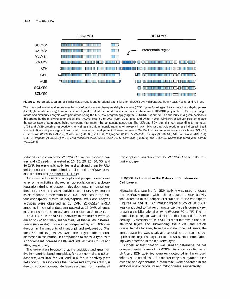
Figure 2 shows a schematic representation of similaritiesbetween the predicted maize polypeptide and other knownmonofunctional and bifunctional LKR/SDH enzymes. A SDHC-terminal domain sharing 42% similarity with the yeastLYS9 protein and an N-terminal domain sharing 27% simi-larity with the yeast LYS1 protein constitute the bifunctionalmaize enzyme. Similarities to the related mouse (F. Papes,E.L. Kemper, G. Cord Neto, F. Langone, and P. Arruda, sub-mitted manuscript),
Caenorhabditis elegans
, and Arabidop-sis proteins are significantly higher (44, 46, and 72%,respectively). The interdomain region (Figure 1A), which is
z
106 residues long in maize, is 57% identical to the corre-sponding Arabidopsis sequence. Interestingly, this regionseems to be absent in
C. elegans
and mouse LKR/SDH pro-teins (Figure 2).
Comparisons of primary protein structures also revealseveral conserved motifs (Figure 2). The initiation ATG andstop codons, as well as some motifs with a high degree ofsimilarity, appear at comparable sites in the open readingframe of all compared proteins.
To confirm the structure of the LKR/SDH bifunctionalpolypeptide, we separated the LKR and SDH domains bylimited proteolysis (Figure 3). A partially purified preparationof LKR/SDH protein was chromatographed on an ion ex-change column (Figure 3A) or digested with elastase beforechromatography (Figure 3B). The most active fractions ofLKR and SDH peaks from Figure 3B were immunoblottedwith anti–LKR/SDH polyclonal antibodies (Figure 3C). Func-tional isolated LKR and SDH domains were recovered, con-firming the bifunctional enzyme structure (Gonçalves-Butruile et al., 1996; Kemper et al., 1998).

LKR/SDH and the High-Lysine
o2
Mutant 1983
ZLKRSDH
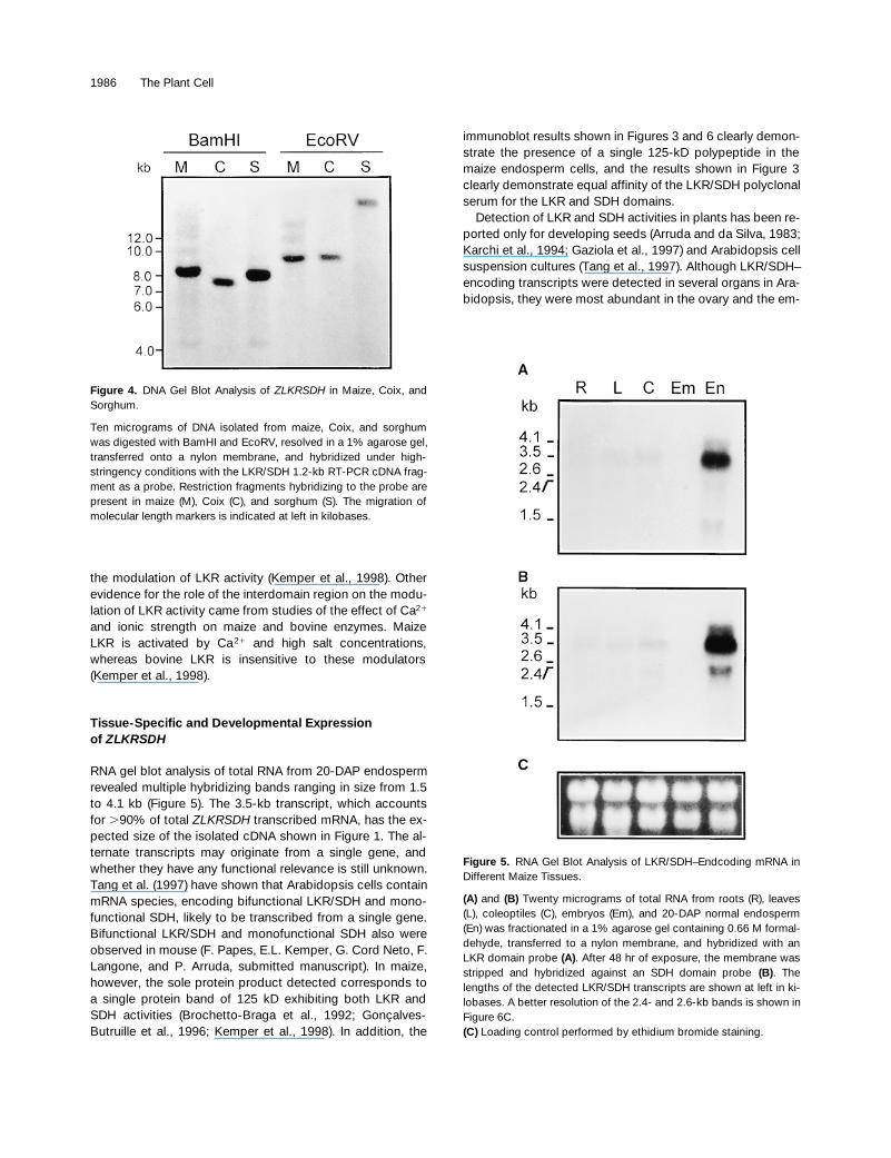
Is Present in Maize and Related Species
We investigated the presence of the
ZLKRSDH
gene inmaize and related cereals. The 1.2-kb RT-PCR DNA frag-ment, comprising part of the LKR domain, the interdomainregion, and part of the SDH domain (residues 444 through849; Figure 1), was hybridized to genomic DNA from maize,Coix, and sorghum. As shown in Figure 4, both BamHI andEcoRV digests gave single, strong hybridizing bands, sug-gesting that
ZLKRSDH
is likely to be present as a single-copy gene in
Andropogoneae
sp.
Analysis of
ZLKRSDH
mRNA Levels in DifferentMaize Tissues
Total RNA extracted from roots, leaves, coleoptiles, em-bryos, and endosperm was hybridized with either LKR- orSDH-specific probes. As shown in Figure 5,
ZLKRSDH
mRNA was detected mainly in the endosperm. No signalwas detected in embryos, and only marginal amounts of ex-pression were detected in roots, leaves, and coleoptiles.Five different transcripts were detected in 20-DAP develop-ing endosperm with both probes. Along with the major 3.5-kb band, which corresponds to
.
90% of the total hybridiz-ing mRNA, alternate transcripts of 4.1, 2.6, 2.4, and 1.5 kbwere detected. The 4.1- and 3.5-kb transcripts were de-tected equally by both probes. The 2.4- and 2.6-kb speciesseemed to contain mainly SDH sequences (Figure 5B), be-cause they hybridized only weakly with the LKR-specificprobe (Figure 5A), whereas the 1.5-kb transcript seemed tocontain only LKR sequences (Figure 5A). The nature andfunction of these alternate transcripts are not known, butthey certainly do not encode monofunctional LKR or SDHenzymes because in the results shown in Figure 3, only onepolypeptide product was detected in the endosperm by theanti–LKR/SDH polyclonal antibodies. This polypeptide isable to recognize with similar efficacy both the LKR andSDH domains.
Developmental Expression of
ZLKRSDH
in Normal and
o2
Mutant Endosperm
We showed previously that LKR activity is reduced two- tothreefold in the
o2
endosperm (Brochetto-Braga et al.,1992). To test whether this lower enzyme activity was due to
Figure 1.
Predicted Sequence of the
ZLKRSDH
cDNA–EncodedProtein.
(A)
The protein sequence encoded by the 3.5-kb
ZLKRSDH
cDNA isshown. Underlined residues highlight the interdomain region. Con-served residues shown in
(B)
are in boldface. The GenBank acces-sion number is AF003551.
(B)
Alignment of the LKR subdomain that is homologous to the puta-tive substrate binding site previously assigned to the yeast LYS1protein. GenBank accession numbers are listed in the legend to Fig-
ure 2. The essential conserved residues H-110, K-113, and R-146 ofthe maize protein are shown in boldface characters. Identical resi-dues are marked by asterisks. ZEA,
Z. mays
; ATH,
Arabidopsisthaliana
; CEL,
C. elegans
; CAL,
Candida albicans
; YLI,
Yarrowia li-polytica
; SCE,
Saccharomyces cerevisiae.
1984 The Plant Cell
reduced expression of the
ZLKRSDH
gene, we assayed nor-mal and
o2
seeds, harvested at 10, 15, 20, 25, 30, 35, and40 DAP, for enzymatic activities and analyzed them by RNAgel blotting and immunoblotting using anti–LKR/SDH poly-clonal antibodies (Kemper et al., 1998).
As shown in Figure 6, transcripts and polypeptides as wellas enzyme activities showed an upregulation and a down-regulation during endosperm development. In normal en-dosperm, LKR and SDH activities and LKR/SDH proteinlevels reached a maximum at 20 DAP, whereas in the mu-tant endosperm, maximum polypeptide levels and enzymeactivities were observed at 25 DAP.
ZLKRSDH
mRNAamounts in normal endosperm peaked at 15 DAP, whereasin
o2
endosperm, the mRNA amount peaked at 20 to 25 DAP.At 20 DAP, LKR and SDH activities in the mutant were re-
duced to
z
2 and 18%, respectively, of the values in normalseeds (Figure 6A). This was accompanied by an
z
90% re-duction in the amounts of transcript and polypeptide (Fig-ures 6B and 6C). At 25 DAP, the polypeptide amountincreased in the mutant in comparison to the wild type, witha concomitant increase in LKR and SDH activities to
z
9 and50%, respectively.
The correlation between enzyme activities and quantita-tive immunoblot band intensities, for both normal and
o2
en-dosperm, was 94% for SDH and 81% for LKR activity (datanot shown). This indicates that decreased enzyme activity isdue to reduced polypeptide levels resulting from a reduced
transcript accumulation from the
ZLKRSDH
gene in the mu-tant endosperm.
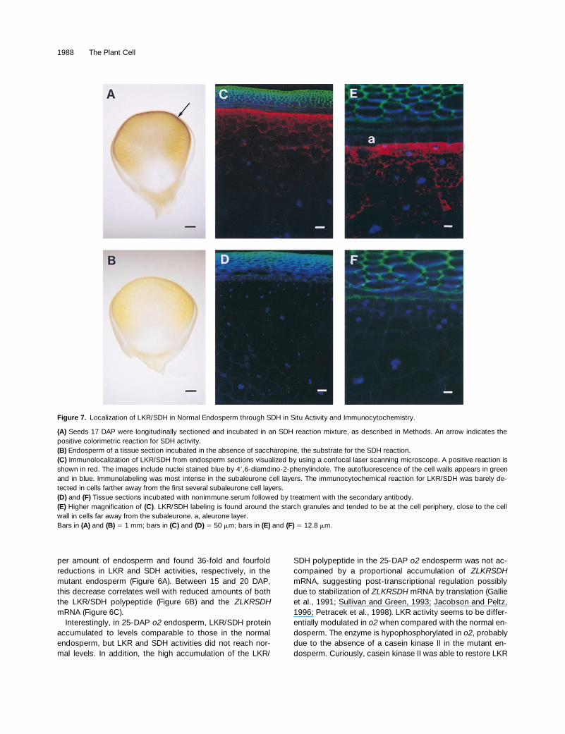
LKR/SDH Is Located in the Cytosol of SubaleuroneCell Layers
Histochemical staining for SDH activity was used to locatethe LKR/SDH protein within the endosperm. SDH activitywas detected in the peripheral distal part of the endosperm(Figures 7A and 7B). An immunological study of LKR/SDHwas conducted to further characterize the cells currently ex-pressing the bifunctional enzyme (Figures 7C to 7F). The im-munolabeled region was similar to that stained for SDHactivity. Expression of LKR/SDH is most intense in the sub-aleurone layers and surrounding the nuclei and starchgrains. In cells far away from the subaleurone cell layers, theimmunostaining was weak and tended to be near the pe-ripheral cell regions, adjacent to cell walls. No immunolabel-ing was detected in the aleurone layer.
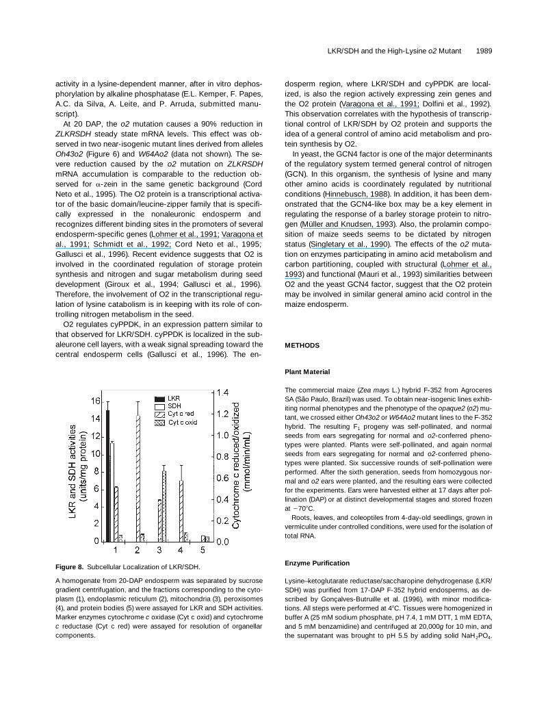
Subcellular fractionation was used to determine the cellcompartmentalization of LKR/SDH. As shown in Figure 8,LKR and SDH activities were only detected in the cytosol,whereas the activities of the marker enzymes, cytochrome
c
oxidase and cytochrome
c
reductase, were observed in theendoplasmatic reticulum and mitochondria, respectively.
Figure 2. Schematic Diagram of Similarities among Monofunctional and Bifunctional LKR/SDH Polypeptides from Yeast, Plants, and Animals.
The predicted amino acid sequences for monofunctional saccharopine dehydrogenase (LYS1, lysine forming) and saccharopine dehydrogenase(LYS9, glutamate forming) from yeast were aligned to plant, nematode, and mammalian bifunctional LKR/SDH polypeptides. Sequence align-ments and similarity analysis were performed using the MACAW program applying the BLOSUM 62 matrix. The similarity at a given position isdesignated by the following color codes: red, .90%; blue, 50 to 90%; cyan, 10 to 49%; and white, ,10%. Similarity at a given position meansthe percentage of sequences being compared that match the consensus sequence. The LKR and SDH domains, corresponding to the yeastLYS1 and LYS9 proteins, respectively, as well as the unique interdomain region present in plant bifunctional polypeptides, are indicated. Blankspaces indicate sequence gaps introduced to maximize the alignment. Nomenclature and GenBank accession numbers are as follows: SCLYS1,S. cerevisiae (P38998); CALYS1, C. albicans (P43065); YLLYS1, Y. lipolytica (P38997); ZMAYS, Z. mays (AF003551); ATH, A. thaliana (U95759);CEL, C. elegans (AF038615); MUS, Mus musculus (AJ224761); SCLYS9, S. cerevisiae (P38999); and SZLYS9, Schizosaccharomyces pombe(AL022244).

LKR/SDH and the High-Lysine
o2
Mutant 1985
DISCUSSION
The biochemical and molecular mechanisms by which the
o2
mutation increases lysine content in the endosperm havebeen investigated during the past 30 years. Both lysine syn-thesis and degradation as well as its incorporation intolysine-rich proteins should contribute to the final content ofthis amino acid in the seed. In this report, we describe theisolation of a maize cDNA clone encoding the lysine-degrad-ing enzyme LKR/SDH and present evidence on how lysinelevels may be controlled in the endosperm. A conspicuouscorrelation between lysine-degrading activities and tran-scriptional regulation by the Opaque2 transcription factorwas observed. LKR/SDH seems to be located within cells inwhich zein protein is being actively synthesized.
The Bifunctional Maize LKR/SDH Enzyme
A cDNA encoding the bifunctional enzyme LKR/SDH wasisolated from the immature maize endosperm mRNA pool.The cDNA predicts a 117-kD protein (Figure 1) bearing dis-tinct N- and C-terminal domains identified, respectively, asLKR and SDH due to similarities to yeast monofunctionalenzymes (SDH lysine– and glutamate-forming enzymes en-coded by the
Lys1
and
Lys9
genes, respectively). Furtherconfirmation of the identity of the maize cDNA was obtainedby comparing sequences from
similar genes recently iso-lated from Arabidopsis (Epelbaum et al., 1997; Tang et al.,1997) and mouse (F. Papes, E.L. Kemper, G. Cord Neto, F.Langone, and P. Arruda, submitted manuscript).
In plants and mammals, the saccharopine pathway isused for lysine catabolism, whereas in yeast and other fungi,this pathway is used in the reverse order for lysine biosyn-thesis. Either bifunctional (maize, soybean, Arabidopsis, bo-vine, murine, and human) or monofunctional (yeast, rat, andArabidopsis) polypeptides operate in this pathway. As illus-trated in Figure 2, the amino acid sequences from these en-zymes share a high degree of similarity. Nevertheless, thereare marked differences between bifunctional LKR/SDHpolypeptides from plants and animals.
First, the enzyme seems to be expressed in different cellcompartments. The mammalian LKR and SDH enzymeshave been located in the mitochondrial fraction (Markovitz etal., 1984; Ameen et al., 1987; Blemings et al., 1994),whereas the maize enzyme seems to be located in the cyto-sol (Figure 8). It is interesting that neither the predicted maizeLKR/SDH (Figure 1) nor the Arabidopsis protein (Epelbaumet al., 1997) contains putative mitochondrial or chloroplasttargeting sequences.
Second, the interdomain region is present in plants butseems to be absent from the animal enzymes (Figure 2). Thisregion is 57% identical in maize and Arabidopsis, but itsfunctional role remains unknown. Limited proteolysis studieswith the maize LKR/SDH suggest that it might be involved in
Figure 3. Delimitation of Maize Bifunctional LKR and SDH Domains.
(A) and (B) Elution profiles for LKR and SDH activities before (A) andafter (B) digestion with elastase. A partially purified DEAE-Sepharose preparation of LKR/SDH was digested with elastase, ap-plied to a Protein-Pak Q 8HR column, and eluted with a linear gradi-ent of 0 to 5 M NaCl in buffer B at a flow rate of 1.5 mL/min (seeMethods for further details). Fractions of 0.75 mL were collected andassayed for LKR and SDH activities. Absorbance at 280 nm is indi-cated at right.(C) Immunoblot of native and elastase-digested LKR/SDH. Equiva-lent aliquots of the most active fractions of LKR and SDH peakswere applied to a 6 to 18% gradient SDS–polyacrylamide gel. Theseparated proteins were blotted onto nylon membranes and incu-bated with anti–LKR/SDH polyclonal antibodies. Numbers at leftcorrespond to the molecular mass markers of the 10-kD protein lad-der (Gibco BRL). The lane labeled LKR/SDH contains native enzyme;digest, elastase digestion products; LKR, LKR peak; SDH, SDHpeak.
1986 The Plant Cell
the modulation of LKR activity (Kemper et al., 1998). Otherevidence for the role of the interdomain region on the modu-lation of LKR activity came from studies of the effect of Ca
2
1
and ionic strength on maize and bovine enzymes. MaizeLKR is activated by Ca
2
1
and high salt concentrations,whereas bovine LKR is insensitive to these modulators(Kemper et al., 1998).
Tissue-Specific and Developmental Expressionof
ZLKRSDH
RNA gel blot analysis of total RNA from 20-DAP endospermrevealed multiple hybridizing bands ranging in size from 1.5to 4.1 kb (Figure 5). The 3.5-kb transcript, which accountsfor
.
90% of total
ZLKRSDH
transcribed mRNA, has the ex-pected size of the isolated cDNA shown in Figure 1. The al-ternate transcripts may originate from a single gene, andwhether they have any functional relevance is still unknown.Tang et al. (1997) have shown that Arabidopsis cells containmRNA species, encoding bifunctional LKR/SDH and mono-functional SDH, likely to be transcribed from a single gene.Bifunctional LKR/SDH and monofunctional SDH also wereobserved in mouse (F. Papes, E.L. Kemper, G. Cord Neto, F.Langone, and P. Arruda, submitted manuscript). In maize,however, the sole protein product detected corresponds toa single protein band of 125 kD exhibiting both LKR andSDH activities (Brochetto-Braga et al., 1992; Gonçalves-Butruille et al., 1996; Kemper et al., 1998). In addition, the
immunoblot results shown in Figures 3 and 6 clearly demon-strate the presence of a single 125-kD polypeptide in themaize endosperm cells, and the results shown in Figure 3clearly demonstrate equal affinity of the LKR/SDH polyclonalserum for the LKR and SDH domains.
Detection of LKR and SDH activities in plants has been re-ported only for developing seeds (Arruda and da Silva, 1983;Karchi et al., 1994; Gaziola et al., 1997) and Arabidopsis cellsuspension cultures (Tang et al., 1997). Although LKR/SDH–encoding transcripts were detected in several organs in Ara-bidopsis, they were most abundant in the ovary and the em-
Figure 4. DNA Gel Blot Analysis of ZLKRSDH in Maize, Coix, andSorghum.
Ten micrograms of DNA isolated from maize, Coix, and sorghumwas digested with BamHI and EcoRV, resolved in a 1% agarose gel,transferred onto a nylon membrane, and hybridized under high-stringency conditions with the LKR/SDH 1.2-kb RT-PCR cDNA frag-ment as a probe. Restriction fragments hybridizing to the probe arepresent in maize (M), Coix (C), and sorghum (S). The migration ofmolecular length markers is indicated at left in kilobases.
Figure 5. RNA Gel Blot Analysis of LKR/SDH–Endcoding mRNA inDifferent Maize Tissues.
(A) and (B) Twenty micrograms of total RNA from roots (R), leaves(L), coleoptiles (C), embryos (Em), and 20-DAP normal endosperm(En) was fractionated in a 1% agarose gel containing 0.66 M formal-dehyde, transferred to a nylon membrane, and hybridized with anLKR domain probe (A). After 48 hr of exposure, the membrane wasstripped and hybridized against an SDH domain probe (B). Thelengths of the detected LKR/SDH transcripts are shown at left in ki-lobases. A better resolution of the 2.4- and 2.6-kb bands is shown inFigure 6C.(C) Loading control performed by ethidium bromide staining.

LKR/SDH and the High-Lysine
o2
Mutant 1987
bryo (Tang et al., 1997). In maize,
ZLKRSDH
mRNA isabundant in the endosperm but is completely absent in theembryo and scarcely detectable in roots, leaves, and co-leoptiles (Figure 5). Enzymatic activity, indeed, was not de-tected in those organs. Furthermore, endosperm specificactivity is
z
30-fold higher in maize than in soybean andcommon bean (data not shown), suggesting that LKR/SDHmay play an important physiological role in maize en-dosperm.
Histochemical and immunological assays revealed thatLKR/SDH is highly expressed in the subaleurone cell layers(Figure 7). This region comprises cells that are actively ex-pressing zein genes at a high rate (Dolfini et al., 1992). Thesecells probably have a low lysine requirement because zeinproteins, which represent .70% of total endosperm protein,are devoid of lysine residues. Thus, we suggest that LKR/SDH activity is important to regulate lysine levels in this nar-row area of the endosperm.
In addition, it could be possible that some product arisingfrom lysine degradation could regulate cellular and develop-mental processes operating in subaleurone cells. Interest-ingly, it has been suggested that lysine degradation mayhave an influence on the growth of the mammalian brain,because LKR is highly active during embryonic rat brain de-velopment (Rao et al., 1992).
Effects of the o2 Mutation on ZLKRSDH mRNA Amounts
Altered lysine catabolism (Sodek and Wilson, 1970; Arrudaand da Silva, 1979) may be one of the mechanisms by whichthe o2 mutation creates a high-lysine maize phenotype.Most of the lysine in the endosperm seems to originate fromlysine-rich proteins, which are more abundant in o2 than innormal endosperm (Habben et al., 1993). Synthesis oflysine-rich proteins presumes the existence of increasedavailability of free lysine to be incorporated. We previouslyobserved increased contents of lysine-rich proteins in thedouble homozygous mutant o2 Ask1 (Azevedo et al., 1990).This was attributed to increased lysine availability due to thefeedback-insensitive AK encoded by Ask1 and to reducedlysine degradation determined by o2.
The amount of LKR activity in the o2 endosperm is re-duced in comparison to the wild type (Brochetto-Braga et al.,1992). In this work, we calculated LKR and SDH activities
Figure 6. Developmental Expression of LKR/SDH in Wild-Type ando2 Endosperm.
(A) Temporal patterns of LKR and SDH activities in normal and o2endosperm. Seeds of wild type (open circles) and o2 (closed circles)were harvested from 10 to 40 DAP at 5-day intervals. Equal amountsof endosperm were homogenized in buffer A and fractionated withammonium sulfate. The fractions collected between 35 and 60%saturation were assayed for LKR and SDH activities. Each point isthe average of duplicate assays, and the bar indicates the standarddeviation.(B) Immunoblot showing LKR/SDH protein temporal profile in nor-mal and o2 endosperms. Aliquots of wild-type (WT) and o2 ammo-nium sulfate–fractionated extracts from (A) were dialyzed againstbuffer A, separated in a 7% SDS–polyacrylamide gel, blotted ontonylon membranes, and incubated with anti–LKR/SDH polyclonal an-tibodies at a 1:10,000 (v/v) dilution. At top are the immunoblots, andat bottom are the protein loading controls stained with Coomassieblue. Numbers at right correspond to the molecular mass markers ofthe 10-kD protein ladder (Gibco BRL).(C) ZLKRSDH gene expression during development of normal ando2 endosperm. Fifteen-microgram samples of total RNA extracted
from the same lots of wild-type (WT) and o2 endosperm shown in(A) were fractionated in a formaldehyde-containing 1.0% agarosegel, transferred to a nylon membrane, and hybridized with the 1.2-kbRT-PCR cDNA fragment as a probe. The lengths of the detectedLKR/SDH–encoding transcripts are shown at right. Loading con-trols, representing an ethidium bromide–stained gel, are shown atbottom.
1988 The Plant Cell
per amount of endosperm and found 36-fold and fourfoldreductions in LKR and SDH activities, respectively, in themutant endosperm (Figure 6A). Between 15 and 20 DAP,this decrease correlates well with reduced amounts of boththe LKR/SDH polypeptide (Figure 6B) and the ZLKRSDHmRNA (Figure 6C).
Interestingly, in 25-DAP o2 endosperm, LKR/SDH proteinaccumulated to levels comparable to those in the normalendosperm, but LKR and SDH activities did not reach nor-mal levels. In addition, the high accumulation of the LKR/
SDH polypeptide in the 25-DAP o2 endosperm was not ac-compained by a proportional accumulation of ZLKRSDHmRNA, suggesting post-transcriptional regulation possiblydue to stabilization of ZLKRSDH mRNA by translation (Gallieet al., 1991; Sullivan and Green, 1993; Jacobson and Peltz,1996; Petracek et al., 1998). LKR activity seems to be differ-entially modulated in o2 when compared with the normal en-dosperm. The enzyme is hypophosphorylated in o2, probablydue to the absence of a casein kinase II in the mutant en-dosperm. Curiously, casein kinase II was able to restore LKR
Figure 7. Localization of LKR/SDH in Normal Endosperm through SDH in Situ Activity and Immunocytochemistry.
(A) Seeds 17 DAP were longitudinally sectioned and incubated in an SDH reaction mixture, as described in Methods. An arrow indicates thepositive colorimetric reaction for SDH activity.(B) Endosperm of a tissue section incubated in the absence of saccharopine, the substrate for the SDH reaction.(C) Immunolocalization of LKR/SDH from endosperm sections visualized by using a confocal laser scanning microscope. A positive reaction isshown in red. The images include nuclei stained blue by 49,6-diamdino-2-phenylindole. The autofluorescence of the cell walls appears in greenand in blue. Immunolabeling was most intense in the subaleurone cell layers. The immunocytochemical reaction for LKR/SDH was barely de-tected in cells farther away from the first several subaleurone cell layers.(D) and (F) Tissue sections incubated with nonimmune serum followed by treatment with the secondary antibody.(E) Higher magnification of (C). LKR/SDH labeling is found around the starch granules and tended to be at the cell periphery, close to the cellwall in cells far away from the subaleurone. a, aleurone layer.Bars in (A) and (B) 5 1 mm; bars in (C) and (D) 5 50 mm; bars in (E) and (F) 5 12.8 mm.
LKR/SDH and the High-Lysine o2 Mutant 1989
activity in a lysine-dependent manner, after in vitro dephos-phorylation by alkaline phosphatase (E.L. Kemper, F. Papes,A.C. da Silva, A. Leite, and P. Arruda, submitted manu-script).
At 20 DAP, the o2 mutation causes a 90% reduction inZLKRSDH steady state mRNA levels. This effect was ob-served in two near-isogenic mutant lines derived from allelesOh43o2 (Figure 6) and W64Ao2 (data not shown). The se-vere reduction caused by the o2 mutation on ZLKRSDHmRNA accumulation is comparable to the reduction ob-served for a-zein in the same genetic background (CordNeto et al., 1995). The O2 protein is a transcriptional activa-tor of the basic domain/leucine-zipper family that is specifi-cally expressed in the nonaleuronic endosperm andrecognizes different binding sites in the promoters of severalendosperm-specific genes (Lohmer et al., 1991; Varagona etal., 1991; Schmidt et al., 1992; Cord Neto et al., 1995;Gallusci et al., 1996). Recent evidence suggests that O2 isinvolved in the coordinated regulation of storage proteinsynthesis and nitrogen and sugar metabolism during seeddevelopment (Giroux et al., 1994; Gallusci et al., 1996).Therefore, the involvement of O2 in the transcriptional regu-lation of lysine catabolism is in keeping with its role of con-trolling nitrogen metabolism in the seed.
O2 regulates cyPPDK, in an expression pattern similar tothat observed for LKR/SDH. cyPPDK is localized in the sub-aleurone cell layers, with a weak signal spreading toward thecentral endosperm cells (Gallusci et al., 1996). The en-
dosperm region, where LKR/SDH and cyPPDK are local-ized, is also the region actively expressing zein genes andthe O2 protein (Varagona et al., 1991; Dolfini et al., 1992).This observation correlates with the hypothesis of transcrip-tional control of LKR/SDH by O2 protein and supports theidea of a general control of amino acid metabolism and pro-tein synthesis by O2.
In yeast, the GCN4 factor is one of the major determinantsof the regulatory system termed general control of nitrogen(GCN). In this organism, the synthesis of lysine and manyother amino acids is coordinately regulated by nutritionalconditions (Hinnebusch, 1988). In addition, it has been dem-onstrated that the GCN4-like box may be a key element inregulating the response of a barley storage protein to nitro-gen (Müller and Knudsen, 1993). Also, the prolamin compo-sition of maize seeds seems to be dictated by nitrogenstatus (Singletary et al., 1990). The effects of the o2 muta-tion on enzymes participating in amino acid metabolism andcarbon partitioning, coupled with structural (Lohmer et al.,1993) and functional (Mauri et al., 1993) similarities betweenO2 and the yeast GCN4 factor, suggest that the O2 proteinmay be involved in similar general amino acid control in themaize endosperm.
METHODS
Plant Material
The commercial maize (Zea mays L.) hybrid F-352 from AgroceresSA (São Paulo, Brazil) was used. To obtain near-isogenic lines exhib-iting normal phenotypes and the phenotype of the opaque2 (o2) mu-tant, we crossed either Oh43o2 or W64Ao2 mutant lines to the F-352hybrid. The resulting F1 progeny was self-pollinated, and normalseeds from ears segregating for normal and o2-conferred pheno-types were planted. Plants were self-pollinated, and again normalseeds from ears segregating for normal and o2-conferred pheno-types were planted. Six successive rounds of self-pollination wereperformed. After the sixth generation, seeds from homozygous nor-mal and o2 ears were planted, and the resulting ears were collectedfor the experiments. Ears were harvested either at 17 days after pol-lination (DAP) or at distinct developmental stages and stored frozenat 2708C.
Roots, leaves, and coleoptiles from 4-day-old seedlings, grown invermiculite under controlled conditions, were used for the isolation oftotal RNA.
Enzyme Purification
Lysine–ketoglutarate reductase/saccharopine dehydrogenase (LKR/SDH) was purified from 17-DAP F-352 hybrid endosperms, as de-scribed by Gonçalves-Butruille et al. (1996), with minor modifica-tions. All steps were performed at 48C. Tissues were homogenized inbuffer A (25 mM sodium phosphate, pH 7.4, 1 mM DTT, 1 mM EDTA,and 5 mM benzamidine) and centrifuged at 20,000g for 10 min, andthe supernatant was brought to pH 5.5 by adding solid NaH2PO4.
Figure 8. Subcellular Localization of LKR/SDH.
A homogenate from 20-DAP endosperm was separated by sucrosegradient centrifugation, and the fractions corresponding to the cyto-plasm (1), endoplasmic reticulum (2), mitochondria (3), peroxisomes(4), and protein bodies (5) were assayed for LKR and SDH activities.Marker enzymes cytochrome c oxidase (Cyt c oxid) and cytochromec reductase (Cyt c red) were assayed for resolution of organellarcomponents.

1990 The Plant Cell
Polyethylene glycol 8000 at 50% (w/v) was added to the homoge-nate to obtain a final concentration of 7.5%. The mixture was centri-fuged at 20,000g for 10 min, and the supernatant was brought to a15% (w/v) polyethylene glycol concentration and centrifuged againat 20,000g for 10 min. The pellet was resuspended in buffer B (50mM Tris-HCl, pH 8.5, 1 mM DTT, and 1 mM EDTA) and dialyzedovernight against the same buffer. The dialyzed sample was appliedto a DEAE-Sepharose column (2.5 3 40 cm) previously equilibratedwith buffer B. The enzyme was eluted from the column with a lineargradient from 0 to 0.5 M NaCl in buffer B. Fractions containing en-zyme activity were combined, brought to 70% saturation with solidammonium sulfate, and centrifuged at 20,000g for 10 min. The pelletwas resuspended in buffer B, dialyzed against the same buffer, andapplied to a Protein-Pak Q 8HR (Waters, Milford, MA) column. Theenzyme was eluted from the column with a linear gradient from 0 to0.5 M NaCl in buffer B. Fractions containing enzyme activity werecombined, brought to 70% saturation with solid ammonium sulfate,and centrifuged at 20,000g for 10 min. The pellet was resuspended inbuffer B and applied to a Superdex 200 HR (Pharmacia) column pre-viously equilibrated with buffer C (buffer B containing 0.3 M NaCl).The enzyme was eluted from the Superdex column with buffer C andstored at 2708C.
N-Terminal Protein Sequencing
The purified LKR/SDH was separated in by SDS-PAGE in a 7% gel.After electrophoresis, the gel was stained with Coomassie Brilliant BlueR 250 and sent to the protein sequencing facility of the Weizmann Insti-tute of Science (Rehovot, Israel). The protein band was eluted from thegel and digested with trypsin, and the major peaks were sequenced.Four internal peptide sequences were obtained: (1) GLIDFLHGL, (2)RYEGFSEIMVTLS, (3) RLTPLYEYI, and (4) RELPAFALEHLPNR.
cDNA Cloning
Cloning of a full-length maize LKR/SDH–encoding cDNA was com-pleted by using a combination of three procedures: (1) reverse tran-scription–polymerase chain reaction (RT-PCR), (2) cDNA libraryscreening, and (3) 59 rapid amplification of cDNA ends (59 RACE).
After protein sequencing, a set of degenerate oligonucleotideswas synthesized based on the tryptic peptide sequences and used inRT-PCR experiments. One microgram of total RNA extracted from17-DAP endosperm was used in an RT reaction by using an RT-PCRkit (Stratagene, La Jolla, CA) according to the manufacturer’s in-structions. A 1.2-kb cDNA fragment was amplified by subsequentPCR reactions (2 mL of template cDNA, 20 mM Tris-HCl, pH 8.4, 50mM KCl, 1.5 mM MgCl2, 0.2 mM deoxynucleotide triphosphate mix,100 pmol of each primer, and 2.5 units of Taq DNA polymerase in afinal volume of 50 mL) by using primers derived from the above-described peptide sequences 2 (59-ISWIARIGTIACCATDATTTC-39)and 3 (59-CTIACICCICTITAYGARTATAT-39). S (G or C), W (A or T), R(A or G), D (A, G, or T), and Y (C or T) designate IUB codes for variablenucleotide sites, and I denotes inosine.
Amplification was performed on a thermal cycler (model 480; Per-kin-Elmer) as follows: 5 min at 948C; 35 cycles of 1 min at 948C, 1 minat 428C, 2 min at 728C; and 5 min at 728C. After sequencing, the 1.2-kb amplified cDNA fragment was confirmed to be part of the maizeLKR/SDH–encoding gene by sequence comparison to the yeastLys1 and Lys9 genes. This fragment then was used as a probe to
screen a maize endosperm cDNA library constructed from RNA ex-tracted from 25-DAP seeds of the R-802 inbreed line (Aukerman etal., 1991). Twenty clones were isolated and sequenced, but all ofthem were incomplete. The 59 cDNA end was cloned by using theRACE system (Gibco BRL), according to the manufacturer’s instruc-tions, with the primers designed on the basis of the cDNA sequences(59-TATCAAATAGGTGCCCAC-39 and 59-GTGTGGGAAAAGAAG-GCGTA-39).
The full-length maize ZLKRSDH cDNA clone was finally isolatedfrom F-352 total RNA extracted from 17-DAP endosperms. Primersannealing to the 59 untranslated region (59-TTCAACTCTCCACTTTCT-CAACCA-39) and 39 untranslated region (59-CTCGTCCGTCTCCGT-TTCCGTC-39) of the cDNA were used to obtain a 3.5-kb completecDNA in RT-PCR reactions catalyzed by Pfu polymerase (Stratagene)according to the manufacturer’s instructions. This fragment subse-quently was cloned into pBluescript KS1 and sequenced in an auto-matic ABI377 DNA sequencer (Perkin-Elmer).
DNA Gel Blot Analysis
Genomic DNA was extracted from maize, Coix, and sorghum seed-lings as described by Rivin et al. (1982). Ten micrograms of genomicDNA was digested to completion with BamHI or EcoRV. The digestswere ethanol precipitated and loaded onto a 0.7% agarose gel, blot-ted onto a nylon membrane (Hybond-N; Amersham), and hybridizedwith the 32P-labeled (Megaprime DNA labeling system; Amersham)1.2-kb RT-PCR fragment. Hybridization was conducted at 658C inSSPE buffer (5 3 SSPE [1 3 SSPE is 150 mM NaCl, 10 mM sodiumphosphate, and 1 mM EDTA, pH 7.4], 5 3 Denhardt’s solution [1 3Denhardt’s solution is 0.02% Ficoll, 0.02% PVP, and 0.02% BSA],and 0.5% SDS), and washes were performed twice at room temper-ature in 2 3 SSPE 0.1% SDS solution for 20 min and repeated twiceat 658C in 0.1 3 SSPE 0.1% SDS. Autoradiography was performedat 2708C for 72 hr by using intensifying screens.
Total RNA Isolation and RNA Gel Blot Analysis
Total RNA used in both RT reactions or RNA gel blot analysis was ex-tracted from developing seeds and seedling tissues, according to theprocedures described by Prescott and Martin (1987). For RNA gelblot experiments, 20 mg of total RNA was electrophoresed in 1.0 or1.5% agarose–formaldehyde gels (0.66 M formaldehyde, 50 mMMops). After electrophoresis, gels were stained with ethidium bro-mide as a loading control. The RNA then was transferred from the gelonto a nylon membrane (Hybond-N1; Amersham). The probes usedwere either the 1.2-kb RT-PCR fragment or specific LKR or SDH do-main probes. The LKR probe consisted of a 0.55-kb cDNA fragmentcorresponding to the region spanning residues 96 through 284, andthe SDH probe (0.49 kb) encompassed residues 652 to 814 (Figure1). DNA labeling, hybridization, and washing were as describedabove for the DNA gel blots. For quantitative experiments, pre-flashed x-ray films were exposed to the radioactive membrane, ac-cording to the manufacturer’s instructions (Amersham). Relativeamounts of hybridized mRNA were analyzed through scanning with alaser densitometer (LKB UltroScan XL, Bromma, Sweden).
Immunoblotting
Immunoblotting was performed as described by Kemper et al.(1998), with minor modifications. After electrophoresis, the gel was
LKR/SDH and the High-Lysine o2 Mutant 1991
soaked in 25 mM Tris-base, 190 mM glycine, and 20% methanol for10 min. Proteins were transferred to nylon membranes (Hybond-N;Amersham) in a semidry electroblotting apparatus (Pharmacia).Membranes were blocked overnight at 48C in 20 mM Tris-HCl, pH7.4, 137 mM NaCl, 0.1% (v/v) Tween 20, and 5% (w/v) nonfat drymilk and then incubated with anti-LKR polyclonal antibodies. Afterincubation with anti–rabbit IgG alkaline phosphatase conjugate, themembranes were incubated for 20 min in the dark in a developingmixture containing 100 mM Tris-HCl, pH 9.5, 100 mM NaCl, and a1:1000 solution of CSPD (Tropix, Bedford, MA). Bands were de-tected by exposure on a preflashed chemiluminescence-sensitivefilm (Amersham) and quantified by using laser densitometry.
Histochemical Staining
Histochemical staining of SDH activity on maize kernel sections wasbased on the gel staining reaction described by Gonçalves-Butruilleet al. (1996). Seventeen-DAP maize kernels were longitudinally handsectioned every 2 mm with a razor blade. Sections were fixed in 4%formalin, pH 7.0, for 30 min, rinsed 10 times in water over 18 hr at 48Cto remove endogenous substrates, and then incubated for 10 min atroom temperature in a reaction mixture containing 2 mM saccha-ropine, 1 mM NAD, 0.5% nitro blue tetrazolium, 0.1 mM phenazinemethasulfate, and 100 mM Tris-HCl, pH 8.5, with rocking. Controlsections were incubated in the absence of saccharopine. The reac-tion was stopped by rinsing the sections in double distilled water.
Immunohistochemistry
Indirect immunofluorescence and confocal laser scanning micros-copy were used to visualize LKR/SDH in frozen sections of maize en-dosperm. Seeds at 17 DAP were dissected from cobs, immersed inice-cold PBS (140 mM NaCl, 3 mM KCl, 8 mM Na2HPO4, 1.5 mMKH2PO4, pH 7.4, and 0.02% [w/v] of sodium azide) containing 4% (w/v) paraformaldeyde and 0.1 M sucrose, and cut into 2- to 3-mm-thicklongitudinal slices. After overnight fixation at 48C, the slices wererinsed three times in PBS containing 0.5 M sucrose for 20 min eachand frozen in liquid nitrogen. The frozen slices were mounted inspecimen supports with Cryo-embedding compound (Microm La-borgerate, Waldorf, Germany) and sectioned at 16 mm in a Microncryostat (Microm Laborgerate). Sections were collected on albumin-coated slides, postfixed with acetone for 15 min, and air dried.
Sections were blocked with 2% (w/v) BSA and 0.3% (v/v) Tween20 in PBS (PBS-T-B) for 30 min in a 200-mL coupling jar before incu-bation with the primary antibody. Sections then were incubated witha polyclonal antibody raised in rabbit against LKR/SDH (Kemper etal., 1998) diluted at 1:200 (v/v) in PBS-T-B overnight at room temper-ature in a 50-mL coupling jar. The control consisted of incubationwith an unrelated rabbit serum at the same dilution. The slides werewashed three times for 20 min in PBS-T in a 200-mL coupling jar.Sections were covered with a rhodamine-conjugated goat anti–rab-bit immunoglobulin (Calbiochem-Novabiochem, La Jolla, CA) diluted1:30 (v/v) in PBS-T-B and incubated for 1 hr at room temperature. Af-ter washing, the sections were counterstained with 49,6-diamidino-2-phenylindole (0.8 mg/mL VectaShield; Vector Laboratories, Burlin-game, CA). Sections were analyzed by using a confocal laser scan-ning microscope (model MRC 1024UV; Bio-Rad Life Sciences,Richmond, CA). Filter sets for rhodamine (immunolabeling), fluores-
cein (natural cell wall fluorescence), and 49,6-diamidino-2-phenylin-dole (nuclear staining plus natural cell wall fluorescence) were used.Immunolabeled and control sections were analyzed using the samelaser power, iris, and gain settings as their experimental counter-parts. This treatment was performed twice with kernels from differentfield seasons. Photographs were taken using Focus Graphics (FocusGraphics Inc., Plymouth, MN) equipment with Kodak ISO 100 filmand processed commercially.
Subcellular Localization of the LKR/SDH Enzyme
Subcellular fractionation was conducted according to Habben et al.(1993), with the following modifications. Buffer A contained 10 mMHepes, pH 7.5, 1 mM EDTA, 10 mM KCl, 200 mM sucrose, 1 mMDTT, and 5 mM benzamidine. After centrifugation in a discontinuoussucrose gradient, the interface fractions were collected by lateralpuncture through the centrifuge tube wall and assayed for LKR andSDH activities. The activities of the marker enzymes cytochrome coxidase and cytochrome c reductase were assayed for resolution oforganellar components, as described by Tolbert (1974) and Larkinsand Hurkman (1978).
Separation of Maize LKR and SDH Domains byLimited Proteolysis
Aliquots of LKR/SDH partially purified by chromatography on DEAE-Sepharose were incubated with elastase (Sigma). Enzyme/proteaseproportions and proteolysis conditions are indicated in the legend toFigure 3. The reaction was stopped by the addition of phenylmethyl-sulfonyl fluoride to a final concentration of 2 mM. The digest was ap-plied to a Protein-Pak Q 8HR column (Waters) previously equilibratedwith buffer B. The column was washed with buffer B and then elutedwith a linear gradient from 0 to 0.5 M NaCl in buffer B. The fractionscontaining separated LKR and SDH domains were used for enzymeassays and immunobloting analysis.
Enzyme Assays
LKR and SDH activities were measured spectrophotometrically byfollowing the oxidation of NADPH and reduction of NAD1, respec-tively, at 308C. LKR assays contained 20 mM L-lysine, 10 mMa-ketoglutaric acid (neutralized to pH 7.0 with potassium hydroxide),0.1 mM NADPH, 150 mM Tris-HCl buffer, pH 7.4, and 0.04 to 0.1 mgof protein in a final volume of 0.3 mL. SDH assays contained 2 mML-saccharopine, 2 mM NAD1, and 100 mM Tris-HCl buffer, pH 8.5, ina final volume of 0.3 mL. Oxidation of NADPH and reduction of NAD1
were monitored at 340 nm in a spectrophotometer (model DU-65;Beckman). One unit of enzyme activity is defined as 1 nmol ofNADPH oxidized or NAD1 reduced per min at 308C. The protein con-centration in the enzyme extracts was determined by the method ofBradford (1976), using the Bio-Rad protein assay dye reagent.
Computer Analysis
Amino acid sequence alignments and similarity analysis were per-formed using the MACAW (Schuler et al., 1991) and BOXSHADE (FTPdownloaded from Vax0.biomed.uni-koeln.de) programs.

1992 The Plant Cell
ACKNOWLEDGMENTS
We are very grateful to Dr. Hernandes F. Carvalho and Sérgio L.Felisbino (Departamento de Biologia Celular, IB, Unicamp, Brazil) foruse of the Bio-Rad MRC 1024UV confocal laser scanning micro-scope (Fundação de Amparo à Pesquisa do Estado de São Paulo[FAPESP], Grant No. 95/6110-2) and for helping us to obtain the con-focal images presented in this study. We are also very grateful to Dr.Robert J. Schmidt (University of California at San Diego) for providingthe lgt11 maize cDNA library, and to Dr. Gad Galili (Weizmann Insti-tute, Rehovot, Israel) for helping with peptide sequencing. We alsothank Sílvia R. Turcinelli for technical help, and Drs. Celso Benedetti,José A. Yunes, and Michel Vincentz for critical reading of the manu-script. This work was supported by grants to P.A. from ConselhoNacional de Desenvolvimento Científico e Tecnológico (CNPq) andFAPESP (Grant No. PTE-90/3808-5). P.A. and A.L. received researchfellowships from CNPq. Postgraduate fellowships were granted byCoodenadoria de Aperfeiçoamento de Pessoal de Nível Superior toG.C.N., and by FAPESP to E.L.K., F.P., and K.C.M.M.
Received June 16, 1999; accepted August 13, 1999.
REFERENCES
Ameen, M., Palmer, T., and Oberholzer, V.G. (1987). Inhibition ofbovine liver lysine–ketoglutarate reductase by urea cycle metabo-lites and saccharopine. Biochem. Int. 14, 589–595.
Arruda, P., and da Silva, W.J. (1979). Amino acid composition ofvascular sap of maize ear peduncle. Phytochemistry 18, 409–410.
Arruda, P., and da Silva, W.J. (1983). Lysine–ketoglutarate reduc-tase activity in maize. Its possible role in lysine metabolism ofdeveloping endosperm. Phytochemistry 22, 206–208.
Arruda, P., Sodek, L., and da Silva, W.J. (1982). Lysine–ketoglu-tarate reductase activity in developing maize endosperm. PlantPhysiol. 69, 988–989.
Aukerman, M.J., Schmidt, R.J., Burr, B., and Burr, F.A. (1991). Anarginine to lysine substitution in the bZIP domain of an opaque2mutant in maize abolishes specific DNA binding. Genes Dev. 5,310–320.
Azevedo, R.A., Arana, J.L., and Arruda, P. (1990). Biochemicalgenetics of the interaction of the lysine plus threonine resistantmutant Ltr*1 with opaque-2 maize mutant. Plant Sci. 70, 81–90.
Azevedo, R.A., Arruda, P., Turner, W.L., and Lea, P.J. (1997). Thebiosynthesis and metabolism of the aspartate derived aminoacids in higher plants. Phytochemistry 46, 395–419.
Bass, H.W., Webster, C., O’Brian, G.R., Roberts, J.K.M., andBoston, R.S. (1992). A maize ribosome-inactivating protein iscontrolled by the transcriptional activator Opaque2. Plant Cell 4,225–234.
Blemings, K.P., Crenshaw, T.D., Swick, R.W., and Benevenga,N.J. (1994). Lysine-a-ketoglutarate reductase and saccharopinedehydrogenase are located only in the mitochondrial matrix in ratliver. J. Nutr. 124, 1215–1221.
Bradford, M.M. (1976). A rapid and sensitive method for the quanti-fication of microgram quantities of protein utilizing the principle ofprotein-dye binding. Anal. Biochem. 72, 248–254.
Bright, S.W.J., Miflin, B.J., and Rognes, S.E. (1982). Threonineaccumulation in the seeds of a barley mutant with an alteredaspartate kinase. Biochem. Genet. 20, 229–243.
Brochetto-Braga, M.R., Leite, A., and Arruda, P. (1992). Partialpurification and characterization of lysine–ketoglutarate reductaseactivity in normal and opaque-2 maize endosperms. Plant Physiol.98, 1139–1147.
Cord Neto, G., Yunes, J.A., Vettore, A.L., da Silva, W.J., Arruda,P., and Leite, A. (1995). The involvement of Opaque2 in b-prola-min gene regulation in maize and Coix suggests a more generalrole for this transcriptional activator. Plant Mol. Biol. 27, 1015–1029.
da Silva, W.J., and Arruda, P. (1979). Evidence for the genetic con-trol of lysine catabolism in maize endosperm. Phytochemistry 18,1803–1805.
Diedrick, T.J., Frisch, D.A., and Gegenbach, B.G. (1990). Tissueculture isolation of a second mutant locus for increased threonineaccumulation in maize. Theor. Appl. Genet. 79, 209–215.
Dolfini, S.F., Landoni, M., Tonelli, C., Bernard, L., and Viotti, A.(1992). Spatial regulation in the expression of structural and regu-latory storage-protein genes in Zea mays endosperm. Dev. Genet.13, 264–276.
Epelbaum, S., McDevitt, R., and Falco, S.C. (1997). Lysine–keto-glutarate reductase and saccharopine dehydrogenase from Arabi-dopsis thaliana: Nucleotide sequence and characterization. PlantMol. Biol. 35, 735–748.
Falco, S.C., Guida, T., Locke, M., Mauvais, J., Sanders, C., Ward,R.T., and Webb, P. (1995). Transgenic canola and soybean seedswith increased lysine. Bio/Technology 13, 577–582.
Frankard, V., Ghislain, M., and Jacobs, M. (1991). High threonineproducer mutant in Nicotiana sylvestris (Spegg. and Comes).Theor. Appl. Genet. 82, 273–282.
Frankard, V., Ghislain, M., and Jacobs, M. (1992). Two feedback-insensitive enzymes of the aspartate pathway in Nicotiana sylves-tris. Plant Physiol. 99, 1285–1293.
Fujioka, M., and Takata, Y. (1981). Role of arginine residue in sac-charopine dehydrogenase (L-lysine forming) from baker’s yeast.Biochemistry 20, 468–472.
Fujioka, M., Takata, Y., Ogawa, H., and Okamoto, M. (1980). Theinactivation of saccharopine dehydrogenase (L-lysine forming) bydiethyl pyrocarbonate. J. Biol. Chem. 255, 937–942.
Gallie, D.R., Feder, J.N., Schimke, R.T., and Walbot, V. (1991).Post-transcriptional regulation in higher eukaryotes: The role ofthe reporter gene in controlling expression. Mol. Gen. Genet. 228,258–264.
Gallusci, P., Varott, S., Matsuoko, M., Maddaloni, M., andThompson R.D. (1996). Regulation of cytosolic pyruvate, ortho-phosphate dikinase expression in developing maize endosperm.Plant Mol. Biol. 31, 45–55.
Gaziola, S.A., Teixeira, C.M., Lugli, J., Sodek, L., and Azevedo,R.A. (1997). The enzymology of lysine catabolism in rice seeds—Isolation, characterization, and regulatory properties of a lysine2-oxoglutarate reductase/saccharopine dehydrogenase bifunc-tional polypeptide. Eur. J. Biochem. 247, 364–371.
Giroux, M.J., Boyer, C., Feix, G., and Hannah, C. (1994). Coordi-nated transcriptional regulation of storage product genes in themaize endosperm. Plant Physiol. 106, 713–722.
Gonçalves-Butruille, M., Szajner, P., Torigoi, E., Leite, A., andArruda, P. (1996). Purification and characterization of the bifunc-
LKR/SDH and the High-Lysine o2 Mutant 1993
tional enzyme lysine–ketoglutarate reductase–saccharopine dehy-drogenase from maize. Plant Physiol. 110, 765–771.
Habben, J.E., Kirleis, A.W., and Larkins, B.A. (1993). The origin oflysine-containing proteins in the opaque-2 maize endosperm.Plant Mol. Biol. 23, 825–838.
Hinnebusch, A.G. (1988). Mechanisms of gene regulation in thegeneral control of amino acid biosynthesis in Saccharomyces cer-evisiae. Microbiol. Rev. 52, 248–273.
Jacobson, A., and Peltz, S.W., (1996). Interrelationships of thepathways of mRNA decay and translation in eukaryotic cells.Annu. Rev. Biochem. 65, 693–739.
Karchi, H., Shaul, O., and Galili, G. (1993). Seed specific expres-sion of a bacterial desensitized aspartate kinase increases theproduction of seed threonine and methionine in transgenictobacco. Plant J. 3, 721–727.
Karchi, H., Shaul, O., and Galili, G. (1994). Lysine synthesis andcatabolism are coordinately regulated during tobacco seed devel-opment. Proc. Natl. Acad. Sci. USA 91, 2577–2581.
Kemper, E.L., Cord Neto, G., Capella, A.N., Gonçalves-Butruille, M.,Azevedo, R.A., and Arruda, P. (1998). Structure and regulation ofthe bifunctional enzyme lysine–oxoglutarate reductase–sac-charopine dehydrogenase in maize. Eur. J. Biochem. 253,720–729.
Larkins, B.A., and Hurkman, W.J. (1978). Synthesis and depositionof zein in protein bodies of maize endosperm. Plant Physiol. 62,256–263.
Lohmer, S., Maddaloni, M., Motto, M., Di Fonzo, N., Hartings, H.,Salamini, F., and Thompson, R.D. (1991). The maize regulatorylocus Opaque-2 encodes a DNA-binding protein which activatesthe transcription of the b-32 gene. EMBO J. 10, 617–624.
Lohmer, S., Maddaloni, M., Motto, M., Salamini, F., and Thompson,R.D. (1993). Translation of the mRNA of the maize transcriptionalactivator Opaque-2 is inhibited by upstream open reading framespresent in the leader sequence. Plant Cell 5, 65–73.
Luehrsen, K.R., and Walbot, V. (1994). The impact of AUG startcodon context on maize gene expression in vivo. Plant Cell Rep.13, 454–458.
Markovitz, D.J., and Chuang, D.T. (1987). The bifunctional amino-adipic semialdehyde synthetase in lysine degradation. J. Biol.Chem. 262, 9353–9358.
Markovitz, D.J., Chuang, D.T., and Cox, R.P. (1984). Familialhyperlysinemias: Purification and characterization of the bifunc-tional aminoadipic semialdehyde synthase with LKR and SDHactivities. J. Biol. Chem. 259, 11643–11646.
Mauri, I., Maddaloni, M., Lohmer, S., Motto, M., Salamini, F.,Thompson, R.D., and Martegani, E. (1993). Functional expres-sion of the transcriptional activator Opaque-2 in yeast. Mol. Gen.Genet. 341, 319–326.
Mertz, E.T., Bates, L.S., and Nelson, O.E. (1964). Mutant gene thatchanges protein composition and increases lysine content of maizeendosperm. Science 145, 279–280.
Müller, M., and Knudsen, S. (1993). The nitrogen response of abarley C-hordein promoter is controlled by positive and negativeregulation of the GCN4 and endosperm box. Plant J. 4, 343–355.
Negrutiu, I., Cattoir-Reynaerts, A., Verbruggen, I., and Jacobs,M. (1984). Lysine overproducer mutants with an altered dihydro-dipicolinate synthase from protoplast culture of Nicotiana sylves-tris (Spegazzini and Comes). Theor. Appl. Genet. 68, 11–20.
Ogawa, H., and Fujioka, M. (1980). The reaction of pyridoxal 59-phosphate with an essential lysine residue of saccharopine dehy-drogenase (L-lysine forming). J. Biol. Chem. 255, 7420–7425.
Ogawa, H., Okamoto, M., and Fujioka, M. (1979). Chemical modifi-cation of the active site sulfhydryl group of saccharopine dehy-drogenase (L-lysine-forming). J. Biol. Chem. 254, 7030–7035.
Pederson, H.B., Galili, G., Knudsen, G., and Holm, P.B. (1996).Engineering of the aspartate family biosynthetic pathway in barley(Hordeum vulgare L.) by transformation with heterologous genesencoding feedback-insensitive aspartate kinase and dihydrodipi-colinate synthase. Plant Mol. Biol. 32, 611–620.
Petracek, M.E., Dickey, L.F., Hansen, E.R., Sowinski, D.A.,Nguyen, T.-T., Allen, G.C., and Thompson, W.F. (1998). Depen-dence of Fed-1 light regulation on translation. In A Look beyondTranscription: Mechanisms Determining mRNA Stability and Trans-lation in Plants, J. Bailey-Serres and D.R. Gallie, eds (Rockville,MD: American Association of Plant Physiologists), pp. 96–101.
Prescott, A., and Martin, C. (1987). Rapid method for quantitativeassessment of levels of specific mRNAs. Plant Mol. Biol. Rep. 4,219–224.
Rao, V.V., Pan, X., and Chang, Y.-F. (1992). Developmentalchanges of L-lysine–ketoglutarate reductase in rat brain and liver.Comp. Biochem. Physiol. 103B, 221–224.
Rivin, C.J., Zimmer, E.A., and Walbot, V. (1982). Isolation of DNAand DNA recombinants from maize. In Maize for BiologicalResearch, W.F. Sheridan, ed (Grand Forks, ND: University Press),pp. 161–164.
Schmidt, R.J., Ketudat, M., Aukerman, M.J., and Hoschek, G.(1992). Opaque-2 is a transcriptional activator that recognizes aspecific target site in 22-kD zein genes. Plant Cell 4, 689–700.
Schuler, G.D., Altschul, S.F., and Lipman, D.J. (1991). A work-bench for multiple alignment construction and analysis. ProteinsStruct. Funct. Genet. 9, 180–190.
Shaul, O., and Galili, G. (1993). Concerted regulation of lysine andthreonine synthesis in tobacco plants expressing bacterial feed-back-insensitive aspartate kinase and dihydrodipicolinate syn-thase. Plant Mol. Biol. 23, 759–768.
Singletary, G.W., Doehlert, D.C., Wilson, C.M., Muhitch, M.J.,and Below, F.E. (1990). Response of enzymes and storage pro-teins of maize endosperm to nitrogen supply. Plant Physiol. 94,858–864.
Sodek, L., and Wilson, C.M. (1970). Incorporation of leucine-C14
and lysine-C14 into protein in the developing endosperm of normaland opaque-2 corn. Arch. Biochem. Biophys. 140, 29–38.
Sullivan, M.L., and Green, P.J. (1993). Post-transcriptional regula-tion of nuclear-encoded genes in higher plants—The roles ofmessenger RNA stability and translation. Plant Mol. Biol. 23,1091–1104.
Tang, G., Miron, D., Zhu-Shimoni, J.X., and Galili, G. (1997). Reg-ulation of lysine catabolism through lysine–ketoglutarate reduc-tase and saccharopine dehydrogenase in Arabidopsis. Plant Cell9, 1305–1316.
Tolbert, N.E. (1974). Isolation of subcellular organelles of metabolismon isopynic sucrose gradients. Methods Enzymol. 31, 734–746.
Varagona, M.J., Schmidt, R.J., and Raikhel, N.V. (1991). Monocotregulatory protein Opaque-2 is localized in the nucleus of maizeendosperm and transformed tobacco plants. Plant Cell 3, 105–113.


![Regulation of Lysine Catabolism through Lysine[mdash]Ketoglutarate Reductase and Saccharopine Dehydrogenase in Arabidopsis](https://static.fdokumen.com/doc/165x107/631cc83693f371de19019c93/regulation-of-lysine-catabolism-through-lysinemdashketoglutarate-reductase-and.jpg)