Lysine glutarylation is a protein posttranslational modification regulated by SIRT5
Upload
khangminh22Category
view
5download
0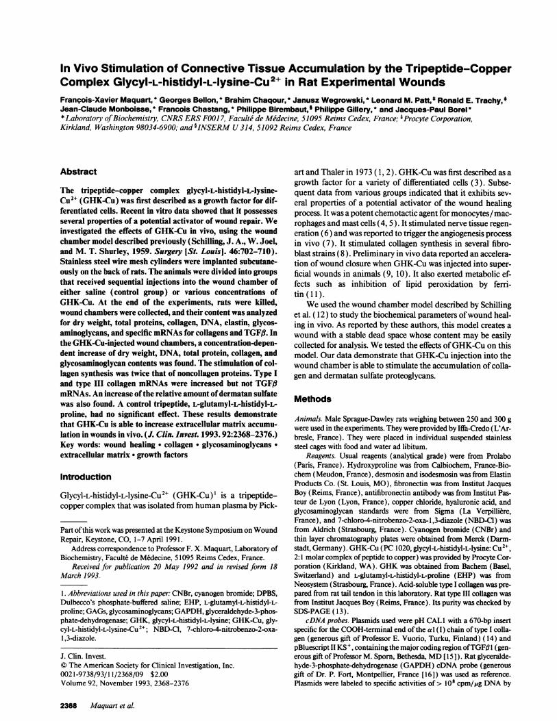
In Vivo Stimulation of Connective Tissue Accumulation by the Tripeptide-CopperComplex Glycyl-L-histidyl-L-lysine-Cu2+ in Rat Experimental WoundsFranpois-Xavier Maquart, * Georges Bellon, * Brahim Chaqour, * Janusz Wegrowski, * Leonard M. Patt,t Ronald E. Trachy,Jean-Claude Monboisse, * Francois Chastang, * Philippe Birembaut, Philippe Gillery, * and Jacques-Paul Borel *
* Laboratory of Biochemistry, CNRSERSFOO17, Faculte de Medecine, 51095 Reims Cedex, France; $Procyte Corporation,Kirkland, Washington 98034-6900; and OINSERMU314, 51092 Reims Cedex, France
Abstract
The tripeptide-copper complex glycyl-L-histidyl-L-lysine-Cu2+ (GHK-Cu) was first described as a growth factor for dif-ferentiated cells. Recent in vitro data showed that it possessesseveral properties of a potential activator of wound repair. Weinvestigated the effects of GHK-Cu in vivo, using the woundchamber model described previously (Schilling, J. A., W. Joel,and M. T. Shurley, 1959. Surgery [St. Louis]. 46:702-710).Stainless steel wire mesh cylinders were implanted subcutane-ously on the back of rats. The animals were divided into groupsthat received sequential injections into the wound chamber ofeither saline (control group) or various concentrations ofGHK-Cu. At the end of the experiments, rats were killed,wound chambers were collected, and their content was analyzedfor dry weight, total proteins, collagen, DNA, elastin, glycos-aminoglycans, and specific mRNAsfor collagens and TGFfI. Inthe GHK-Cu-injected wound chambers, a concentration-depen-dent increase of dry weight, DNA, total protein, collagen, andglycosaminoglycan contents was found. The stimulation of col-lagen synthesis was twice that of noncollagen proteins. Type Iand type III collagen mRNAswere increased but not TGFftmRNAs.An increase of the relative amount of dermatan sulfatewas also found. A control tripeptide, L-glutamyl-L-histidyl-L-proline, had no significant effect. These results demonstratethat GHK-Cu is able to increase extracellular matrix accumu-lation in wounds in vivo. (J. Clin. Invest. 1993.92:2368-2376.)Key words: wound healing * collagen * glycosaminoglycans.extracellular matrix * growth factors
Introduction
Glycyl-L-histidyl-L-lysine-Cu2+ (GHK-Cu)' is a tripeptide-copper complex that was isolated from human plasma by Pick-
Part of this work was presented at the Keystone Symposium on WoundRepair, Keystone, CO, 1-7 April 1991.
Address correspondence to Professor F. X. Maquart, Laboratory ofBiochemistry, Faculte de M&Iecine, 51095 Reims Cedex, France.
Received for publication 20 May 1992 and in revised form 18March 1993.
1. Abbreviations used in this paper: CNBr, cyanogen bromide; DPBS,Dulbecco's phosphate-buffered saline; EHP, L-glutamyl-L-histidyl-L-proline; GAGs, glycosaminoglycans; GAPDH,glyceraldehyde-3-phos-phate-dehydrogenase; GHK, glycyl-L-histidyl-L-lysine; GHK-Cu, gly-cyl-L-histidyl-L-lysine-Cu2"; NBD-C1, 7-chloro-4-nitrobenzo-2-oxa-1,3-diazole.
art and Thaler in 1973 (1, 2). GHK-Cuwas first described as agrowth factor for a variety of differentiated cells (3). Subse-quent data from various groups indicated that it exhibits sev-eral properties of a potential activator of the wound healingprocess. It was a potent chemotactic agent for monocytes/mac-rophages and mast cells (4, 5). It stimulated nerve tissue regen-eration (6) and was reported to trigger the angiogenesis processin vivo (7). It stimulated collagen synthesis in several fibro-blast strains (8). Preliminary in vivo data reported an accelera-tion of wound closure when GHK-Cuwas injected into super-ficial wounds in animals (9, 10). It also exerted metabolic ef-fects such as inhibition of lipid peroxidation by ferri-tin ( 11).
Weused the wound chamber model described by Schillinget al. ( 12) to study the biochemical parameters of wound heal-ing in vivo. As reported by these authors, this model creates awound with a stable dead space whose content may be easilycollected for analysis. Wetested the effects of GHK-Cuon thismodel. Our data demonstrate that GHK-Cuinjection into thewound chamber is able to stimulate the accumulation of colla-gen and dermatan sulfate proteoglycans.
Methods
Animals. Male Sprague-Dawley rats weighing between 250 and 300 gwere used in the experiments. They were provided by Iffa-Credo (L'Ar-bresle, France). They were placed in individual suspended stainlesssteel cages with food and water ad libitum.
Reagents. Usual reagents (analytical grade) were from Prolabo(Paris, France). Hydroxyproline was from Calbiochem, France-Bio-chem (Meudon, France), desmosin and isodesmosin was from ElastinProducts Co. (St. Louis, MO), fibronectin was from Institut JacquesBoy (Reims, France), antifibronectin antibody was from Institut Pas-teur de Lyon (Lyon, France), copper chloride, hyaluronic acid, andglycosaminoglycan standards were from Sigma (La Verpilliere,France), and 7-chloro-4-nitrobenzo-2-oxa-1,3-diazole (NBD-Cl) wasfrom Aldrich (Strasbourg, France). Cyanogen bromide (CNBr) andthin layer chromatography plates were obtained from Merck (Darm-stadt, Germany). GHK-Cu(PC 1020, glycyl-L-histidyl-L-lysine: Cu2",2:1 molar complex of peptide to copper) was provided by Procyte Cor-poration (Kirkland, WA). GHKwas obtained from Bachem (Basel,Switzerland) and L-glutamyl-L-histidyl-L-proline (EHP) was fromNeosystem (Strasbourg, France). Acid-soluble type I collagen was pre-pared from rat tail tendon in this laboratory. Rat type III collagen wasfrom Institut Jacques Boy (Reims, France). Its purity was checked bySDS-PAGE( 13 ).
cDNAprobes. Plasmids used were pH CALl with a 670-bp insertspecific for the COOH-terminal end of the a l (I) chain of type I colla-gen (generous gift of Professor E. Vuorio, Turku, Finland) ( 14) andpBluescript II KS', containingthe major coding region ofTGF#3I (gen-erous gift of Professor M. Sporn, Bethesda, MD[ 15 ]). Rat glyceralde-hyde-3-phosphate-dehydrogenase (GAPDH) cDNA probe (generousgift of Dr. P. Fort, Montpellier, France [16]) was used as reference.Plasmids were labeled to specific activities of > 101 cpm/zg DNAby
2368 Maquart et al.
J. Clin. Invest.© The American Society for Clinical Investigation, Inc.0021-9738/93/1 1/2368/09 $2.00Volume 92, November 1993, 2368-2376
random priming using Klenow fragment (Gibco BRL, Gaithersburg,MD) and [32P]dCTP (Amersham Corp., Arlington Heights, IL). Thelabeled cDNA-probe was separated from dCTP by chromatographythrough a G-50 spin column.
Wound chambers. Wound chambers were made of stainless steelwire mesh (C-CX 20; EDMECInc., Bellevue, WA) as 1-cm-diameterby 2.5-cm-long cylinders. They were closed at both ends by Teflon capsand sterilized by autoclaving.
Surgical procedures. Rats were anesthesized by intraperitoneal in-jection of sodium pentobarbital (40 mg/kg; Clin-Midy, Paris, France).Dorsal hair was clipped widely from scapula to pelvis and the nude areawas sterilized with polyvidone iodine (Betadine*; Laboratories Sarget,Merignac, France). Bilateral incisions were made perpendicular to thespine to the skin's full-thickness through the panniculus carnosus to thefascial plane). A space approximately the size of the chamber wasopened under the dermis and sterile wound chambers with caps wereslipped beneath the skin. The incisions were closed through individual4.0 nylon sutures. Animals were then returned to their cages until thefirst injection.
Experimental design. A first set of experiments was performed forstudying the effects of repeating a single dose of GHK-Cu. Twowoundchambers were inserted on the back of each rat. The first one receivedserial injections of 2.0 mg GHK-Cu dissolved in 0.2 ml Dulbecco'sphosphate-buffered saline (DPBS). The first injection was 7 d afterimplantation, then every 3 d for an additional 2 wk. The other chamberreceived on the same days injection of the same volume of DPBS.Chambers were collected on day 29, immediately frozen at -20'C, andlyophilized. This day (4 wk after insertion) was initially chosen becauseof the previous works of Schilling et al. (12), who designed thechamber model and showed that, at this time, the chamber is nearlyfilled with new connective tissue. The dried chamber content wasweighted. It was then dissolved in 0.5 MNaOHand an aliquot wastaken for DNAmeasurement according to Fiszer-Szafarz et al. ( 17).The remaining solution was neutralized with HCG, and ethanol wasadded to the final concentration of 80% (wt/vol). After 18 h at 40Cand a centrifugation at 5,000 g for 30 min, supernatant was collected,the pellet resuspended in saline, and the ethanol precipitation repeatedonce. Both supernatants were pooled, evaporated under nitrogen, andsubmitted to acid hydrolysis in 6 MHOat 1 10C for 18 h. This frac-tion was used for fluorometric measurement of hydroxyproline con-tained in the small peptides formed by collagen degradation, using thefluorophore 7-chloro-4-nitrobenzo-2-oxa-1,3-diazole (NBD-Cl), anda thin layer chromatography as described previously ( 18). Ethanolprecipitation also eliminated some unidentified pigments that inter-fered in the protein measurement. The ethanol precipitate was redis-solved in 0.5 MNaOHand an aliquot was taken for measurement oftotal proteins by the method of Lowry et al. ( 19). The remaining frac-tion was collected and used for measurement ofcollagen and glycosami-noglycans.
Collagen was measured by its hydroxyproline content. An aliquotof the NaOH-solubilized material was neutralized, and hydrolyzed in 6MHCGat 1 10C for 18 h. Hydroxyproline was quantified in the hydro-lyzate by fluorometry ( 18). For glycosaminoglycan analysis, anotheraliquot was neutralized with acetic acid and digested with pronase in0.05 MTris HCI, pH 8.0,0.02 MCaCG2, for 48 h at 480C. Trichloroace-tic acid was added to the hydrolysate to 10% (wt/vol) final concentra-tion. The samples were centrifuged at 5,000 g for 15 min and the super-natant was dialyzed exhaustively against distilled water at 4VC. Analiquot of the nondialyzable material was used for the measurement ofuronic acid (20) and the remaining part was lyophilized and used forelectrophoresis.
The separation of glycosaminoglycans (GAGs) was carried out bycellulose acetate electrophoresis in 0.1 Mzinc acetate, pH 5.0, for 80min at 80 V followed by electrophoresis in 0.1 MHGfor 20 min at 40V. The GAGswere stained with 0.2% Alcian blue according to Bartoldet al. (21 ). The strips were scanned at 560 nmwith a Sebia gel scanningsystem. The distribution of GAGswas estimated after correction forstaining of GAGstandards. Specific GAGswere identified by hydroly-
sis with specific enzymes: hyaluronidase (EC 4.2.2.1) for hyaluronicacid, chondroitinase ABC (EC 4.2.2.4) and chondroitinase AC (EC4.2.2.5) for chondroitin sulfates, and dermatan sulfate, according toSaito et al. (22). Heparan sulfate was identified as the remaining frac-tion susceptible to HNO2degradation (23).
A second set of experiments was used for studying the effects ofincreasing amounts of GHK-Cu injected into the wound chambers.For that purpose, 18 rats were divided into 6 groups and 2 woundchambers were implanted on the back of each rat. The first group(controls) received the injection of 0.2 ml of DPBS into the woundchambers every 3 d from days 7 to 23 after the chamber implantation.The second group received 0.2 mgGHK-Cudissolved in 0.2 ml DPBS,the third 0.5 mg, the fourth 1.0 mg, the fifth 2.0 mg, and the sixth 4.0mg, on the same days as the control group. The chambers were col-lected on day 29, their contents collected and lyophilized, and theirweights measured. An aliquot of the lyophilized material was taken,dissolved in 0.1% SDS, and used for fibronectin measurement by im-muno-enzyme assay (24). Another aliquot was used for measuringdesmosin and isodesmosin (an estimate of the elastin content) by ionexchange chromatography on a Multichrom B amino-acid analyzer(Beckman Instrs. Inc., Fullerton, CA) after acid hydrolysis (6 MHCl,18 h at 1 10C). The remaining material was dissolved in 0.5 MNaOHand processed as described above for measurement of DNA, total pro-teins, collagen, and uronic acid.
A third set of experiments was designed for investigating the effectsof GHK-Cuon the expression of the genes of type I and type III colla-gens and TGFBinside the chambers. Twowound chambers per animalwere inserted on day 0 on the back of 10 rats. A first series of five rats(controls) received in both wound chambers the injection of 0.2 mlDPBSon day 2, then every 3 d until day 17. The other series receivedon the same days 2.0 mgof GHK-Cudissolved in the same volume ofDPBS. A control and a GHK-Cu-treated rat were killed on days 3, 7,10, 14, and 21. Both chambers were collected. The first one was imme-diately immersed in a formol (10% [vol/vol]) solution in DPBSforhistological examination. The tissues inside were formalin fixed, paraf-fin included, and 4-Mm-thick sections were stained with hematoxylin-eosin and safran. The second chamber was immediately frozen in liq-uid nitrogen before mRNAextraction, according to Chomczinski andSacchi (25). Once deep frozen, the wound chamber content was care-fully collected using a scalpel and homogenized in 4 Mguanidine-isothiocyanate containing 25 mMsodium citrate, pH 7.5, 0.5% sarco-syl, and 0.1 M2-mercaptoethanol in order to inactivate ribonucleases.The homogenate was then extracted with phenol, chloroform, isoamyl-alcohol mixture. After centrifugation at 10,000 g for 20 min at 4°C, theaqueous phase was mixed with an equal volume of isopropanol andplaced at -20°C for at least 2 h to precipitate RNAs. Centrifugation at10,000 g for 20 min was again performed, the resulting RNApelletresuspended in the guanidine-isothiocyanate solution, and precipitatedonce more with isopropanol. After centrifugation, the RNApellet wasresuspended in 75% ethanol, sedimented, vacuum dried, and finallydissolved in diethylpyrocarbonate-treated water. RNA content wasquantitated by its absorbance at 260 nm. Northern blot and slot blotanalyses were then carried out to quantify collagen gene expression atthe mRNAlevel.
For Northern blot analysis, 6 Ag of total RNAwas dissolved in 50%formamide, 2.2 M formaldehyde, 0.1 M3-[N-morpholino]-propanesulfonic acid, pH 7.0,40 mMsodium acetate, 5 mMEDTA, pH 8, anddenatured by heating at 65°C for 10 min. RNAwas then fractionatedby electrophoresis in 1% agarose/formaldehyde gel for 4 h at 70 V(26). Duplicate RNAsamples were either stained with ethidium bro-mide or transferred overnight by capillary blotting in 20X standardsaline citrate solution ( lx SSC = 0.15 MNaCl, 0.015 Msodium ci-trate, pH 7) to nylon filter (Biodyne; Pall Ultrafine Filtration Corp.,Glen-Cowe, NY). The air-dried nylon filter was UV irradiated for 5min to bind transferred RNAs.
The nylon filter was prehybridized for 24 h at 42°C in 10%SDS, 5XSSC, 5x Denhardt's solution, 250 ,g/ml of denatured salmon spermDNA, and 50% formamide. Specific hybridization was carried out
Stimulation of WoundRepair In Vivo 2369
overnight at 420C in freshly prepared prehybridization solution withthe addition of 106 cpm/ml of 32P-labeled cDNAprobe. The filter waswashed twice in 2X SSC, 0.1% SDSat room temperature, twice in 0.1 xSSC, 0.1% SDSat 680C, air dried, and exposed to autoradiography at-80'C using hyperfilm MP(Amersham Corp.) and Kodak X-Omatcassette C2 with intensifying screens. Autoradiographic signals werequantitated using a scanning densitometer Desaga (Desaga GmbH,Heidelberg, Germany).
For slot blot analysis, 8 gg of total RNAwas diluted in 6x SSC, 2.2Mformaldehyde, and 50% formamide, denatured at 650C for 10 min,and applied to a nylon filter in a slot blot apparatus (Bio-Rad Laborato-ries, Richmond, CA) using gentle aspiration. The subsequent stepswere identical to the Northern blot protocol.
Measurement of the ratio of type III to type I collagen. A woundchamber was inserted on day 0 on the back of 18 rats. They were thendivided into two groups. The first one (controls) received in thechamber the injection of 0.2 ml DPBSon day 2, then every 3 d untilday 17. The other series received on the same days 2.0 mgof GHK-Cudissolved in the same volume of DPBS. Three control and three GHK-Cu-treated rats were then killed on days 7, 14, and 21. The chamberswere collected, immediately frozen in liquid nitrogen, and lyophilized.The chamber content was then collected and submitted to CNBr diges-tion according to Epstein (27). After dialysis and lyophilization, CNBrpeptides were separated by SDS-PAGE( 13) using a 5% stacking geland a 10.0% separating gel. The percentage of type III collagen was thendeduced from the ratio of the a I (III)-CB 5 to a I (I)-CB 8 peptides, asdescribed by Chan and Cole (28).
Specificity of GHK-Cueffects. For checking the specificity ofGHK-Cu effects, wound chambers were inserted on day 0 on the back of 40
A ...i.*
rats. 10 rats were used as controls and received serial injections of 0.2ml DPBSwith the usual schedule (every 3 d from days 3 to 17). Asecond group of 10 rats received serial injections of 2.0 mgGHK-Cuinthe same volume of saline. The third group of 10 rats received serialinjections of 1.8 mgof GHKalone, which is not complexed with cop-per. The last group (n = 10) received serial injections of 0.4 mgcopperchloride. The doses of GHKand of copper chloride were chosen forproviding an equimolar amount of product, compared to the chambersinjected with the GHK-Cu complex. The chambers were collected onday 21 for measuring collagen content, as described previously.
An additional experiment was performed for comparing the effectsof GHK-Cu to those of the tripeptide glu-his-pro (EHP), which is theinactive metabolite of thyroid releasing hormone. The same scheduleof injections as above was adopted (every 3 d from days 3 to 17). Thefirst group (controls) received serial injections of 0.2 ml DPBS, thesecond group 2.0 mgGHK-Cudissolved in 0.2 ml DPBS, and the thirdgroup 2.0 mgEHPin 0.2 ml DPBS. Chambers were collected on day 21for analysis.
Statistical analysis. Results were expressed as mean± 1 SEM. Sta-tistical analysis was done by the Student's t test.
Results
Effects of serial injections of a unique (2.0-mg) dose of GHK-Cu into the wound chamber. A series of wound chambers seri-ally injected with 2.0 mg GHK-Cu or with DPBSwere col-lected at various days after insertion and sorted for morphologi-cal analysis. A simple macroscopical observation of the
B
D
Figure 1. Histological examination of the chamber content at days 3 (A and B) and 14 (C and D) after insertion. At day 3, more inflammatorycells (polymorphonuclear cells and macrophages) were present in the treated chambers (B) than in the controls (A) and some neovascularization(v) was already visible. At day 14, large areas of well-organized fibrosis with elongated fibroblasts were seen in the GHK-Cu-injected chambers(D), whereas the control ones (C) were characterized by a cellular accumulation, mainly fibroblastic, with oedematous areas and few extracel-lular matrix deposition (bar = 50 Am).
2370 Maquart et al.
Cs,
chambers clearly showed that more connective tissue accumu-lated in the treated than in the controls (data not shown).Histological examination at day 3 (Fig. 1, A and B) showed amore intense inflammatory cellular infiltrate with some neo-vessels clearly visible in the GHK-Cu-injected chambers. Laterexamination showed a more rapid appearance of collagen withlarge, dense, and well-organized fibrosis areas clearly visible inthe treated chambers as soon as day 14 (Fig. 1, Cand D).
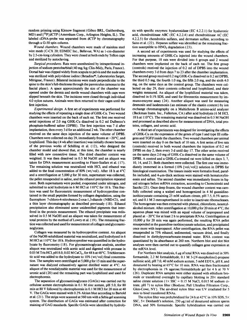
Biochemical analysis of the chamber content at day 29(Fig. 2) demonstrated a significant increase of dry weight(223% of the controls, P< 0.01; Fig. 2 A), total proteins (230%of the controls, P< 0.01; Fig. 2 B), glycosaminoglycans (208%of the controls, P < 0.01; Fig. 2 C), and collagen (344% of thecontrols, P < 0.01; Fig. 2 D) in wound chambers injected with2.0 mg GHK-Cu. Small collagenous peptide hydroxyproline(Fig. 2 E) was also increased in wound chambers ( 188% of thecontrols, P < 0.01 ), showing that collagen degradation wasenhanced as well as its synthesis but to a lesser extent. DNAcontent was not significantly altered (Fig. 2 F).
The increase of collagen in the treated chambers was ap-proximately twice that oftotal proteins, demonstrating a prefer-ential effect of GHK-Cu on collagen synthesis. By contrast,
mg
120-
100 -
.-
*= 80 -
*, 60
40
20 -
0-
Amg
300II
*- 0° 20a
a 'a0
10 -+C G-Cu
B
U
-V-u
1L.
0-OC G-Cu
D
19
150
-l
1001
01.
50- a
0-
E F
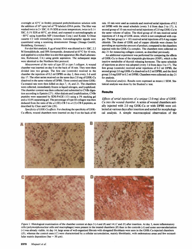
glycosaminoglycans were increased approximately to the sameextent as total proteins. Their distribution, however, was mark-edly altered (Fig. 3). In control wound chambers, the derma-tan sulfate/heparan sulfate/hyaluronic acid ratio was 53:18:29(the total content being considered as 100), whereas inchambers serially injected with 2.0 mg GHK-Cu, it was70:13:17.
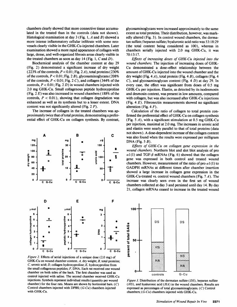
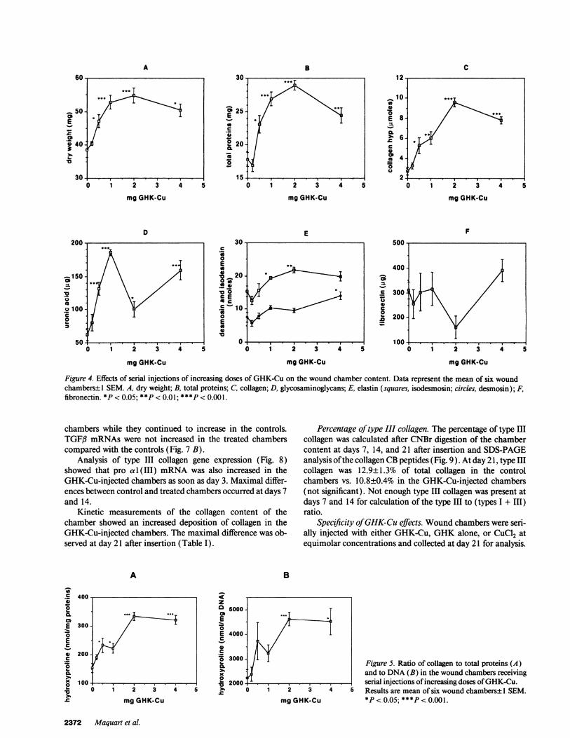
Effects of increasing doses of GHK-Cu injected into thewound chambers. The injection of increasing doses of GHK-Cu demonstrated a dose-effect relationship between theamount of GHK-Cu injected into the wound chamber and thedry weight (Fig. 4 A), total protein (Fig. 4 B), collagen (Fig. 4C), and glycosaminoglycan content (Fig. 4 D) at day 29. Inevery case, the effect was significant from doses of 0.5 mgGHK-Cu per injection. Elastin, as detected by its isodesmosinand desmosin content, was present in low amounts, comparedwith collagen, but was also increased after GHK-Cu injections(Fig. 4 E). Fibronectin measurements showed no significantalteration (Fig. 4 F).
Calculation of the ratio of collagen to total protein con-firmed the preferential effect of GHK-Cuon collagen synthesis(Fig. 5 A), with a significant stimulation at 0.5 mgGHK-Cuper injection, maximal at 2.0 mg. The increases in uronic acidand elastin were nearly parallel to that of total proteins (datanot shown). A dose-dependent increase of the collagen contentwas also found when the results were expressed per milligramDNA(Fig. 5 B).
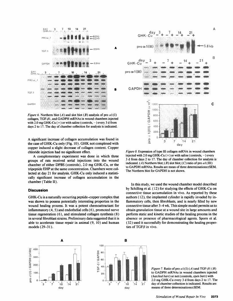
Effects of GHK-Cu on collagen gene expression in thewound chambers. Northern blot and slot blot analysis of proa I(I) and TGF-13 mRNAs(Fig. 6) showed that the collagengene was expressed in both control and treated woundchambers. However, measurement of the ratio of pro a 1(I ) toGADPHmRNAsat different times after chamber insertionshowed a large increase in collagen gene expression in theGHK-Cu-treated vs. control wound chambers (Fig. 7 A). Theincrease was clearly seen even in the first set of woundchambers collected at day 3 and persisted until day 14. By day21, collagen mRNAsceased to increase in the treated wound
pmol
6-
0
= 40
>. 3-
* 2.S .
20
U
-C-
I
o0
o s
0o
ob
G-Cu
* p mol
6-
5.
3-
2]
:1
mg
3 -
2-
z -
1 -
U
-0-
a
1-
C G-Cu
100-
+0 1
a
-0 -45
0o-W
0
G-Cu 0IS
Figure 2. Effects of serial injections of a unique dose (2.0 mg) ofGHK-Cuon wound chamber content. A, dry weight; B, total proteins;C, uronic acid; D, collagen hydroxyproline; E, hydroxyproline fromthe small collagenous peptides; F, DNA. Each rat received one woundchamber on both sides of the back. The first chamber was used ascontrol injected with saline. The second chamber received GHK-Cuinjections. Symbols represent individual results (quantity per woundchamber) for the four rats. Means are shown by horizontal bars. (C)Control chambers injected with DPBS; (G-Cu) chambers injectedwith GHK-Cu.
50-_-
0.
0- K
DS
c .....s::
controls
DS
HA......C......,...........
G-Cu
Figure 3. Distribution of the dermatan sulfate (DS), heparan sulfate(HS), and hyaluronic acid (HA) in the wound chambers. Results areexpressed as percentages of total glycosaminoglycans. (C) Controlchambers; (G-Cu) chambers injected with GHK-Cu.
Stimulation of WoundRepair In Vivo 2371
.
L
B
E 25-E0c
-W
2 20-
-
0
c
0Eto0
._
E0 I
0
0
)- I I I
1 2 3 4
mgGHK-Cu
E
mgGHK-Cu
F
a-
00c06.
mgGHK-Cu mgGHK-Cu
Figure 4. Effects of serial injections of increasing doses of GHK-Cu on the wound chamber content. Data represent the mean of six woundchambers± 1 SEM. A, dry weight; B, total proteins; C, collagen; D, glycosaminoglycans; E, elastin (squares, isodesmosin; circles, desmosin); F,fibronectin. * P < 0.05; **P < 0.01; ***P < 0.001.
chambers while they continued to increase in the controls.TGFI3 mRNAswere not increased in the treated chamberscompared with the controls (Fig. 7 B).
Analysis of type III collagen gene expression (Fig. 8)showed that pro a 1(III) mRNAwas also increased in theGHK-Cu-injected chambers as soon as day 3. Maximal differ-ences between control and treated chambers occurred at days 7and 14.
Kinetic measurements of the collagen content of thechamber showed an increased deposition of collagen in theGHK-Cu-injected chambers. The maximal difference was ob-served at day 21 after insertion (Table I).
Percentage of type III collagen. The percentage of type IIIcollagen was calculated after CNBr digestion of the chambercontent at days 7, 14, and 21 after insertion and SDS-PAGEanalysis of the collagen CBpeptides (Fig. 9). At day 21, type IIIcollagen was 12.9±1.3% of total collagen in the controlchambers vs. 10.8±0.4% in the GHK-Cu-injected chambers(not significant). Not enough type III collagen was present atdays 7 and 14 for calculation of the type III to (types I + III)ratio.
Specificity of GHK-Cu effects. Woundchambers were seri-ally injected with either GHK-Cu, GHKalone, or CuCl2 atequimolar concentrations and collected at day 21 for analysis.
B
1 2 3 4 1
mgGHK-Cu
zo 5000-a.E0E 4000-C
0C
0 3000-0.
0o22 3
mgGHK-Cu
Figure S. Ratio of collagen to total proteins (A)and to DNA(B) in the wound chambers receivingserial injections of increasing doses of GHK-Cu.Results are mean of six wound chambers± 1 SEM.*P < 0.05; ***P < 0.001.
_ 50coE
a* 40
20
mgGHK-Cu
D
_150
.0
0 100'E
mgGHK-Cu
A
00r 400
0.
E 300-02C
* 200-
0._I-
o 100-02.la
2372 Maquart et al.
A C
DAY 3 7 10 14 21GHK-cu- - - - + --
PROo<, gol 6SSW2kb
Aday 3 7 14 21
GHK-Cu - + - + - + - +
ATGF3 t it2.5kb
GAPDH det||w _2-2kb
8RKCAY3 + 7 10 414 21
PROx - - e a e- e e
-.- _ a S_ _- _ - a
TGF _ _ _GAPDH - _l rn-rn -_
GAPOR _- - - - - _
Figure 6. Northern blot (A) and slot blot (B) analysis of pro al (I)collagen, TGF-l1, and GADPHmRNAsin wound chambers injectedwith 2.0 mgGHK-Cu (+) or with saline (controls, -) every 3 d fromdays 2 to 17. The day of chamber collection for analysis is indicated.
A significant increase of collagen accumulation was found inthe case of GHK-Cuonly (Fig. 10). GHKnot complexed withcopper induced a slight decrease of collagen content. Copperchloride injection had no significant effect.
A complementary experiment was done in which threegroups of rats received serial injections into the woundchamber of either DPBS(controls), 2.0 mgGHK-Cu, or thetripeptide EHPat the same concentration. Chambers were col-lected at day 21 for analysis. GHK-Cuonly induced a statisti-cally significant increase of collagen accumulation in thechamber (Table II).
Discussion
GHK-Cu is a naturally occurring peptide-copper complex thatwas shown to possess potentially interesting properties in thewound healing process. It was a potent chemoattractant forinflammatory (4, 5) and endothelial cells (6), promoted nervetissue regeneration (6), and stimulated collagen synthesis (8)in several fibroblast strains. Preliminary data suggested that it isable to accelerate tissue repair in animal (9, 10) and humanmodels (29-31 ).
A
- 400-0
xI300-
aCL4 200-
7,100.-0-_0L. °'-
pro cx 1(111)
day 3GHK-Cu - +
proocl(III) ._-- - a -
* g o -,| - 5.8kb
B_ 7 me -14 + 21 +
a e aa e e
-- - a a a - a
S
o e - - a a e es GAPDH a a-_ a a a e
o e a - - a a a
C
Figure 8. Expression of type III collagen mRNAin wound chambersinjected with 2.0 mgGHK-Cu (+) or with saline (controls, -) every3 d from days 2 to 17. The day of chamber collection for analysis isindicated. (A) Northern blot; (B) slot blot; (C) ratio of pro al (III)to GAPDHmRNAs. Results are mean of three determinations±SEM.The Northern blot for GAPDHis not shown.
In this study, we used the wound chamber model describedby Schilling et al. ( 12) for studying the effects of GHK-Cu onconnective tissue accumulation in vivo. As reported by theseauthors ( 12), the implanted cylinder is rapidly invaded by in-flammatory cells, then fibroblasts, and is nearly filled by newconnective tissue after 3-4 wk. This simple model permits us toobtain granulation tissue at a wound site in large amounts andperform static and kinetic studies of the healing process in theabsence or presence of pharmacological agents. Sporn et al.(32) used it successfully for demonstrating the healing proper-ties of TGFfl in vivo.
B
Figure 7. Ratio of pro al (I) (A) and TGF-fll (B)to GADPHmRNAsin wound chambers injected(hatched bars) or not (controls, open bars) with2.0 mgGHK-Cu every 3 d from days 2 to 17. Theday of chamber collection is indicated. Results aremeans of three determinations±SEM.
Stimulation of WoundRepair In Vivo 2373
c 1, 2 0
m i GOG - *
CL
o 400
-2 00
m - 3 7 14 21aL day
day3 7 10 14 21
day
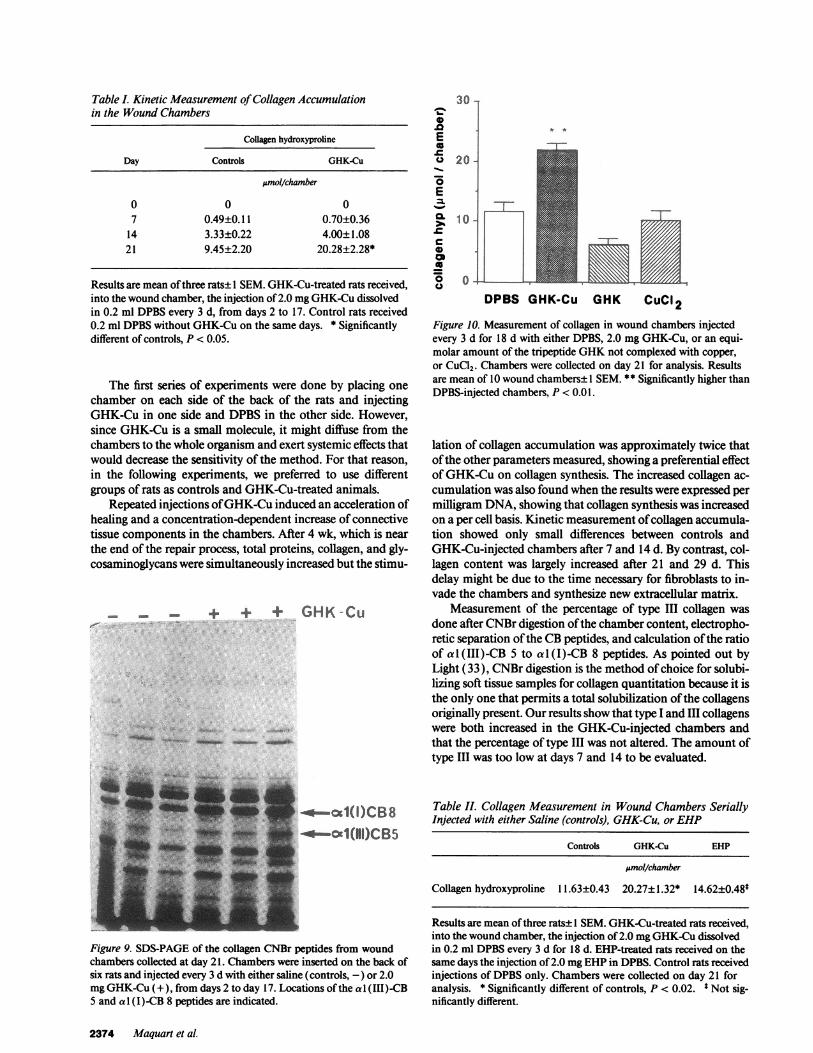
Table I. Kinetic Measurement of Collagen Accumulationin the WoundChambers
Collagen hydroxyproline
Day Controls GHK-Cu
pmol/chamber
0 0 07 0.49±0.11 0.70±0.36
14 3.33±0.22 4.00±1.0821 9.45±2.20 20.28±2.28*
Results are mean of three rats± I SEM. GHK-Cu-treated rats received,into the wound chamber, the injection of 2.0 mgGHK-Cudissolvedin 0.2 ml DPBSevery 3 d, from days 2 to 17. Control rats received0.2 ml DPBSwithout GHK-Cu on the same days. * Significantlydifferent of controls, P < 0.05.
The first series of experiments were done by placing onechamber on each side of the back of the rats and injectingGHK-Cu in one side and DPBS in the other side. However,since GHK-Cu is a small molecule, it might diffuse from thechambers to the whole organism and exert systemic effects thatwould decrease the sensitivity of the method. For that reason,in the following experiments, we preferred to use differentgroups of rats as controls and GHK-Cu-treated animals.
Repeated injections of GHK-Cuinduced an acceleration ofhealing and a concentration-dependent increase of connectivetissue components in the chambers. After 4 wk, which is nearthe end of the repair process, total proteins, collagen, and gly-cosaminoglycans were simultaneously increased but the stimu-
_ - + + + GHK-Cu
-*-cK1(I)CB8-* Cc1(N11)CB5
30 -
0.0E6
0 20-S
Cf 10-
0
*5 0 -~U
DPBS GHK-Cu GHK CuCI 2
Figure 10. Measurement of collagen in wound chambers injectedevery 3 d for 18 d with either DPBS, 2.0 mgGHK-Cu, or an equi-molar amount of the tripeptide GHKnot complexed with copper,or CuC12. Chambers were collected on day 21 for analysis. Resultsare mean of 10 wound chambers±l SEM. ** Significantly higher thanDPBS-injected chambers, P < 0.01.
lation of collagen accumulation was approximately twice thatof the other parameters measured, showing a preferential effectof GHK-Cu on collagen synthesis. The increased collagen ac-cumulation was also found when the results were expressed permilligram DNA, showing that collagen synthesis was increasedon a per cell basis. Kinetic measurement of collagen accumula-tion showed only small differences between controls andGHK-Cu-injected chambers after 7 and 14 d. By contrast, col-lagen content was largely increased after 21 and 29 d. Thisdelay might be due to the time necessary for fibroblasts to in-vade the chambers and synthesize new extracellular matrix.
Measurement of the percentage of type III collagen wasdone after CNBr digestion of the chamber content, electropho-retic separation of the CBpeptides, and calculation of the ratioof al (III)-CB 5 to al (I)-CB 8 peptides. As pointed out byLight (33), CNBr digestion is the method of choice for solubi-lizing soft tissue samples for collagen quantitation because it isthe only one that permits a total solubilization of the collagensoriginally present. Our results show that type I and III collagenswere both increased in the GHK-Cu-injected chambers andthat the percentage of type III was not altered. The amount oftype III was too low at days 7 and 14 to be evaluated.
Table I. Collagen Measurement in Wound Chambers SeriallyInjected with either Saline (controls), GHK-Cu, or EHP
Controls GHK-Cu EHP
Amol/chamber
Collagen hydroxyproline 11.63±0.43 20.27±1.32* 14.62±0.48*
Figure 9. SDS-PAGEof the collagen CNBr peptides from woundchambers collected at day 21. Chambers were inserted on the back ofsix rats and injected every 3 d with either saline (controls, -) or 2.0mgGHK-Cu (+), from days 2 to day 17. Locations of the al (III)-CB5 and al (I)-CB 8 peptides are indicated.
Results are mean of three rats± I SEM. GHK-Cu-treated rats received,into the wound chamber, the injection of 2.0 mgGHK-Cudissolvedin 0.2 ml DPBSevery 3 d for 18 d. EHP-treated rats received on thesame days the injection of 2.0 mgEHPin DPBS. Control rats receivedinjections of DPBSonly. Chambers were collected on day 21 foranalysis. * Significantly different of controls, P < 0.02. * Not sig-nificantly different.
2374 Maquart et al.
* *
-T-
A
Peptidic hydroxyproline was increased, in the treatedchambers, which demonstrated that collagen turnover was ac-celerated, compared with the control ones. Since collagen con-tent was always higher in the treated chambers, the activationof collagen synthesis was globally more efficient than catabo-lism.
Extraction of mRNAsfollowed by Northern blot and slotblot analyses demonstrated an increase of type I procollagenmRNAsin the treated chambers, compared with the controls,from days 3 to 14. This difference did not exist at day 21. TypeIII collagen mRNAfollowed a similar kinetics. These data in-dicated that the effects of GHK-Cu occurred very early andaccelerated the repair process. It was not possible, in such an invivo model, to specify if the effects of GHK-Cu on collagenmRNAswere related to an increased transcription of collagengenes or to an increased stability of the corresponding tran-scripts. Further in vitro studies will be necessary to solve thisquestion. Northern blot hybridization with the rat TGF#lIcDNAprobe revealed the presence of two species of transcripts,the first one 2.5 kb and the second - 1.9 kb. These results areconsistent with previous findings of Qian et al. ( 15 ), who re-ported the appearance of a 1.9-kb TGF-f31 mRNAin infarctedrat heart. As discussed by these authors, this mRNAhas beenobserved also in a variety of other mouse and rat tissues andwas most often elevated after tissue injury. The injection ofGHK-Cu in the treated chambers did not increase TGF-(ilmRNAlevel, showing that GHK-Cu effects are not mediatedthrough an increase of TGF-f 1 gene expression.
The distribution of glycosaminoglycans was markedly al-tered in GHK-Cu-injected chambers, with a large increase ofthe relative amount of dermatan sulfate. Dermatan sulfate-containing proteoglycan is known to interact with collagen(34) and to contribute to the organization and strength of thefibrillar network (35). Its specific accumulation with GHK-Cutreatment might improve the formation of a fully resistant scar.Wereported previously a similar increase of dermatan sulfate-containing proteoglycan in human dermis fibroblast culturesincubated with GHK-Cu (36).
The mechanism of action of GHK-Cuis still under discus-sion. Since GHK-Cuwas shown in vitro to possess activatingproperties on a variety of cells implicated in the wound repairprocess, it is likely that, in vivo, it is also able to trigger a largenumber of events such as increased angiogenesis, increased re-cruitment of inflammatory cells, and fibroblast activation. Aspointed out by Raju et al. (7) and by Odedra and Weiss (37),the presence of the copper ion in the GHK-Cumolecule mightbe of paramount importance for explaining its effects. Severalcopper-containing low molecular mass substances have beenshown to possess angiogenic properties (37). It is the same forthe copper-containing serum protein, ceruloplasmin (7). Cop-per alone is highly cytotoxic in cell cultures and was devoid ofeffect in our in vivo study. On the other hand, an increasedcopper uptake into cells incubated with GHK-Cuwas reportedpreviously (38). It seems likely that copper delivery to the cellsis involved in the effects of the tripeptide. In this regard, it wasof great interest to note that GHKalone, not previously com-plexed with copper, was devoid of any stimulating effects oncollagen synthesis in our model.
Our results demonstrate that GHK-Cu may promotewound repair in vivo. The origin of naturally occurring GHK-Cu and its exact role in physiological tissue repair remain to bedetermined.
Acknowledgments
Mrs. Etienne and Deschamps are greatly acknowledged for typing themanuscript, Professor H. Choisy (University of Reims, France) for hishelp in taking care of the animals, and Dr. Su Wen Qian (NationalHeart, Lung and Blood Institute, Bethesda, MD) for helpful discussionof the results obtained with the TGF(3 probe.
This work was supported by grants from CNRS, the Universit6 deReims-Champagne-Ardenne, and Procyte Corporation.
References
1. Pickart, L., and M. Thaler. 1973. Tripeptide in human serum which pro-longs survival of normal liver cells and stimulates the growth of hepatoma cells.Nature New Biol. 243:85-87.
2. Lau, S. J., and B. Sarkar. 1981. The interaction of copper (II) and glycyl-L-histidyl-L-lysine, a growth modulating tripeptide from plasma. Biochem. J.199:649-656.
3. Pickart, L. 1981. The use of glycyl-histidyl-lysine in culture systems. InVitro (Athens). 17:459-466.
4. Zetter, B. R., N. Rasmussen, and L. Brown. 1985. Methods in laboratoryinvestigation. An in vivo assay for chemoattractant activity. Lab. Invest. 53:362-368.
5. Poole, T. J., and B. R. Zetter. 1983. Stimulation of rat peritoneal mast cellmigration by tumor-derived peptides. Cancer Res. 43:5857-5861.
6. Grosse, G., and G. Lindner. 1980. Experimental influence ofthe pharmaco-logical agents on the regeneration of nervous tissue in vitro. Folia Morphol. ( War-saw). 28:345-347.
7. Raju, K. S., J. Alessandri, M. Ziche, and P. M. Gullino. 1982. Ceruloplas-min, copper ions and angiogenesis. J. Natl. Cancer Inst. 69:1183-1188.
8. Maquart, F. X., L. Pickart, M. Laurent, P. Gillery, J. C. Monboisse, andJ. P. Borel. 1988. Stimulation of collagen synthesis in fibroblast cultures by thetripeptide-copper complex glycyl-L-histidyl-L-lysine-Cu++. FEBS (Fed. Eur.Biochem. Soc.) Lett. 238:343-346.
9. Downey, D., W. F. Larrabee, V. Voci, and L. Pickart. 1985. Acceleration ofwound healing using glycyl-histidyl-lysine-Cu("). Surg. Forum. 48:573-575.
10. Pickart, L. 1987. lamin: a human growth factor with multiple woundhealing properties. In Biology of Copper Complexes. J.R.C. Sorenson, editor.Humana Press, Inc. Totowa, NJ. 273-282.
1 1. Miller, D. M., D. De Silva, L. Pickart, and S. D. Aust. 1990. Effects ofglycyl-histidyl-lysyl chelated CuOO) on ferritin dependent lipid peroxidation. InAntioxidants in Therapy and Preventive Medicine. I. Emerit, editor. PlenumPublishing Corporation, NewYork. 79-84.
12. Schilling, J. A., W. Joel, and M. T. Shurley. 1959. Wound healing: acomparative study of the histochemical changes in granulation tissue containedin stainless steel wire mesh and polyvinyl sponge cylinders. Surgery (St. Louis).46:702-710.
13. Laemmli, U. K. 1970. Cleavage of structural proteins during the assemblyof the head of the bacteriophage T4. Nature (Lond.). 227:680-685.
14. Vuorio, T., J. K. Makela, V. M. Kahari, and E. Vuorio. 1987. Coordinatedregulation of type I and type III collagen production and mRNAlevels of proa I (I) and pro a2(I) collagen in cultured morphea fibroblasts. Arch. Dermatol.Res. 279:154-160.
15. Qian, S. W., P. Kondaiah, W. Casscells, A. M. Roberts, and M. B. Sporn.1991. A second messenger RNA species of transforming growth factor 031 ininfacted rat heart. Cell Regul. 2:241-249.
16. Fort, P., L. Marty, K. Piechaczyk, E. El Sabrouty, C. Dani, P. Jeanteur,and J. M. Blanchard. 1985. Various rat adult tissues express only one majormRNAspecies from the glyceraldehyde-3-phosphate-dehydrogenase multigenefamily. Nucleic Acids Res. 13:1431-1442.
17. Fiszer-Szafarz, B., D. Szafarz, and A. Guevara de Murillo. 1981. A gen-eral, fast and sensitive method for DNAdetermination: application to rat andmouse liver hepatoma, human leukocytes, chicken fibroblasts and yeast cells.Anal. Biochem. 110:165-170.
18. Bellon, G., A. Malgras, A. Randoux, and J. P. Borel. 1978. Further im-provement of the fluorometric assay for hydroxyproline. J. Chromatogr.178:167-172.
19. Lowry, 0. H., N. J. Rosebrough, A. L. Farr, and R. J. Randall. 1951.Protein measurement with Folin-phenol reagent. J. Biol. Chem. 257:5333-5336.
20. Bitter, T., and H. M. Muir. 1962. A modified uronic acid carbazole reac-tion. Anal. Biochem. 4:330-334.
21. Bartold, P. M., 0. W. Wiebkin, and J. C. Thonard. 1982. Molecularweight estimation of sulfated glycosaminoglycans in human gingivae. Connect.Tissue Res. 9:165-172.
22. Saito, H., T. Yamagata, and S. Suzuki. 1968. Enzymatic methods for thedetermination of small quantities of isomeric chondroitin sulfates. J. Biol. Chem.243:1536-1542.
Stimulation of WoundRepair In Vivo 2375
23. Lagunof, D., and G. Warren. 1962. Deamination of 2-deoxy-2-sulfoami-nohexose content of mucopolysaccharides. Arch. Biochem. Biophys. 99:396-400.
24. Vuento, M., E. Salonen, M. Pasanen, and U. H. Stenman. 1981. Compara-tive enzyme immunoassay for human plasma fibronectin. J. Immunol. Methods.40:101-108.
25. Chomczinski, P., and N. Sacchi. 1987. Single step method of RNAisola-tion by acid guanidinium-thiocyanate-phenol-chloroform extraction. Anal. Bio-chem. 162:156-159.
26. Sambrook, J., E. F. Fritsch, and T. Maniatis. 1989. Molecular Cloning: ALaboratory Manual, 2nd ed. Cold Spring Harbor Laboratory, Cold Spring Har-bor, NY.
27. Epstein, E. H. 1974. [a I (III)]3 human skin collagen. Release by pepsindigestion and preponderance in fetal life. J. Biol. Chem. 249:3225-3231.
28. Chan, D., and W. G. Cole. 1984. Quantitation of type I and III collagensusing electrophoresis of alpha chains and cyanogen bromide peptides. Anal. Bio-chem. 139:322-328.
29. Aupaix, F., F. X. Maquart, V. Salagnac, L. Pickart, P. Gillery, J. P. Borel,and B. Kalis. 1990. Effects of the tripeptide glycyl-histidyl-lysine-Cu++ on heal-ing. Clinical and biochemical correlations. J. Invest. Dermatol. 94:390 (Abstr.)
30. Fish, F. S., I. Katz, N. T. Hien, M. E. Briden, J. A. Johnson, and L. M.Patt. 1991. Evaluation of glycyl-L-histidyl-L-lysine copper complex in acutewound healing. Effects in post Mohs' surgery wounds. Wounds. 3:171-177.
31. Massey, P., L. M. Patn, and J. C. D'Aoust. 1992. The effect of glycyl-L-his-tidyl-L-lysine copper chelate on the healing of diabetic ulcers: a pilot study.Wounds. 4:21-28.
32. Sporn, B., A. B. Roberts, J. H. Shull, J. M. Smith, J. M. Ward, and J.Sodek. 1983. Polypeptide transforming growth factors isolated from bovinesources and used for wound healing in vivo. Science (Wounds). 219:1329-1331.
33. Light, N. D. 1982. Estimation of types I and III collagens in whole tissueby quantitation of the CNBr peptides on SDS-polyacrylamide gels. Biochim.Biophys. Acta. 702:30-36.
34. Fleischmajer, R., L. W. Fischer, E. D. McDonald, L. Jacobs, J. S. Perlish,and J. Termine. 1991. Decorin interacts with fibrillar collagen of embryonic andadult human skin. J. Struct. Biol. 106:82-90.
35. Scott, J. E. 1988. Proteoglycan fibrillar collagen interactions. Biochem. J.252:313-323.
36. Wegrowski, Y., F. X. Maquart, and J. P. Borel. 1992. Stimulation ofsulfated glycosaminoglycan synthesis by the tripeptide-copper complex glycyl-L-histidyl-L-lysine-Cu2+. Life Sci. 51:1049-1056.
37. Odedra, R., and J. B. Weiss. 1991. Low molecular weight angiogenesisfactors. Pharmacol. & Ther. 49:111-124.
38. Pickart, L., J. H. Freedman, W. J. Loker, J. Peisach, C. M. Perkins, R. E.Stenkamp, and B. Weinstein. 1980. Growth-modulating plasma tripeptide mayfunction by facilating copper uptake into cells. Nature (Lond.). 288:715-717.
2376 Maquart et al.
Copyright © 2022 FDOKUMEN


![Interfacial interactions between poly[L-lysine]-based branched polypeptides and phospholipid model membranes](https://static.fdokumen.com/doc/165x107/633df5f7df741406dc0b4c83/interfacial-interactions-between-polyl-lysine-based-branched-polypeptides-and.jpg)