Formation and Mechanical Characterization of Aminoplast Core/Shell Microcapsules
Upload
independentCategory
view
0download
0
ARTICLE IN PRESS
0142-9612/$ - se
doi:10.1016/j.bi
�Correspondfax: +1514 340
E-mail addr1These autho2These autho
Biomaterials 26 (2005) 6950–6961
www.elsevier.com/locate/biomaterials
Physicochemical model of alginate–poly-L-lysine microcapsulesdefined at the micrometric/nanometric scaleusing ATR-FTIR, XPS, and ToF-SIMS
Susan K. Tama,1, Julie Dusseaultb,c,1, Stefania Polizua, Martin Menardb,Jean-Pierre Halleb,2, L’Hocine Yahiaa,2,�
aGroupe de Recherche en Biomateriaux/Biomecanique, Ecole Polytechnique de Montreal, C.P. 6079, succ. Centre-ville,
Montreal, Que., Canada H3C 3A7bCentre de Recherche Guy-Bernier, Hopital Maisonneuve-Rosemont, 5415 boul. de l’Assomption, Montreal, Que., Canada H1T 2M4
cFaculte de Medecine, Universite de Montreal, C.P. 6128, succ. Centre-ville, Montreal, Que., Canada H3C 3J7
Received 14 January 2005; accepted 6 May 2005
Available online 22 June 2005
Abstract
Alginate–poly-L-lysine–alginate (APA) microcapsules are currently being investigated as a means to immuno-isolate transplanted
cells, but their biocompatibility is limited. In this study, we verified the hypothesis that poly-L-lysine (PLL), which is immunogenic
when unbound, is exposed at the APA microcapsule surface. To do so, we analysed the microcapsule membrane at the micrometric/
nanometric scale using attenuated total reflectance Fourier transform infrared spectroscopy, X-ray photoelectron spectroscopy, and
time-of-flight secondary ion mass spectrometry. The results indicate that PLL and alginate molecules interact within the membrane.
PLL exists in considerable amounts near the surface, contributing to the majority of the carbon within the outermost 100 A of the
membrane. PLL was also detected at the true surface (the outermost monolayer) of the microcapsules. The exposure of PLL does
not appear to result from defects in the outer alginate coating. This physicochemical model of APA microcapsules could explain
their immunogenicity and will play an important role in the optimization of the microcapsule design.
r 2005 Elsevier Ltd. All rights reserved.
Keywords: Microencapsulation; Biocompatibility; Alginate; Poly-L-lysine; Time-of-flight secondary ion mass spectrometry (ToF-SIMS); Islet
1. Introduction
The immuno-isolation of secretory cells, such as theislets of Langerhans, within semi-permeable microcap-sules is currently being investigated as a means toprotect the cells from the host’s immune system, thusallowing a successful transplantation without the use ofpotentially toxic immunosuppressive drugs [1,2].
e front matter r 2005 Elsevier Ltd. All rights reserved.
omaterials.2005.05.007
ing author. Tel.: +1514 340 4711x4378;
4611.
ess: [email protected] (L. Yahia).
rs contributed equally to this work.
rs contributed equally to this work.
The alginate–poly-L-lysine–alginate (APA) microcap-sule is the most widely studied device for the immuno-isolation and transplantation of living cells [3–6]. Toencapsulate cells within APA microcapsules, they arefirst immobilized in calcium-alginate gel beads, whichare sequentially incubated in a poly-L-lysine (PLL)solution and a dilute alginate solution. Alginate is thepreferred polymer for cell immobilization because of itsability to gel quickly under physiological conditions [7],allowing the transformation from alginate droplets toperfectly spherical gel beads without the use of toxicsolvents or the production of harmful by-products. Theaddition of PLL is necessary to form the semi-permeablemembrane of a controlled porosity, and to providestrength and stability to the microcapsule [8,9]. The final
ARTICLE IN PRESSS.K. Tam et al. / Biomaterials 26 (2005) 6950–6961 6951
incubation of the microcapsules in alginate serves toimprove the biocompatibility of the microcapsules byneutralizing and masking the positively charged PLL,which is known to be immunogenic [10,11].In the case of islet transplantation, the APA micro-
capsule has been successfully employed to induceprolonged normoglycemia in small diabetic animalmodels for several days to months [4,6,12]. While thesereported cases clearly demonstrate that, in concept,APA microcapsules can be used effectively as animmune barrier for transplanted islets, similar successesin larger animal models and clinical trials are rare andhave not been reproduced [13,14]. The main hindrancefor the success in large animals is the need to transplantunreasonably large numbers of islets in order tocompensate for the death or malfunction of a significantportion of the encapsulated cells [15–17]. A number ofparameters that are suspected to influence the survivalrate of encapsulated islets have been investigated,including the choice of implantation site [18,19], theavailability of growth factors such as insulin-like growthfactor-II [15], and immune cell chemotaxis [16]. A morefundamental issue that continues to contribute to isletcell death, however, is the inadequate biocompatibilityof the APA microcapsules and/or of their biomaterialcomponents. Despite encouraging improvements of themicrocapsule biocompatibility by purifying the alginate[20,21], carefully selecting the alginate composition (M/G ratio) [22,23], and optimizing the microcapsule size[18,24], even the most successful published studies reportthat up to 10% of APA microcapsules that are retrievedfrom implantation sites in rats are surrounded bymacrophages and fibroblasts, even in the absence ofgrafted islets [17]. While this percentage may seemminimal, it has been shown that an immune responseagainst one microcapsule can have a deleterious effecton neighbouring encapsulated islets [25]. Moreover,once macrophages at the implant site are activated, thereleased cytokines and chemokines will attract addi-tional immune cells and fibroblasts. As a consequence,even a low-grade inflammatory reaction against a smallportion of implanted APA microcapsules can lead to asevere immune reaction and significantly lower thesurvival rate of the encapsulated islets.In order to ensure a controlled and reproducible
biocompatibility of the implanted APA microcapsulesand achieve near-zero pericapsular overgrowth, webelieve that a rational, stepwise approach must betaken. That is, the physicochemical properties of theempty microcapsules and its biomaterial componentsmust be fully understood before being capable ofproperly evaluating the bioreactivity of cell-loadedmicrocapsules in vitro and in vivo. It is especially crucialto characterize and control the surface properties of themicrocapsules, since the chemical composition andstructure of biomaterial surfaces are known to directly
influence the biological reactions that occur at theinterface between the implant and the host tissue [26].Furthermore, it is particularly important to assess theadequacy in which the alginate binds and masks theimmunogenic PLL. We recently found that during an invitro co-incubation with macrophages, APA microcap-sules induce more macrophage activation than non-PLL-coated alginate microcapsules, as evaluated by themRNA production of pro-inflammatory cytokines [27].The results of this study and others [28,29] have led tothe hypothesis that the PLL layer is exposed at themicrocapsule surface; if the PLL is exposed and notproperly neutralized, then the positively charged aminegroups of the polypeptide may attract negativelycharged cells and proteins from the host tissue andinduce the immune response against the microcapsules[30]. No published studies, however, have directlymeasured the chemical composition of the surface ofAPA microcapsules to confirm or reject this hypothesisof PLL exposure.The purpose of the present study was to develop a
physicochemical model of the APA microcapsulemembrane, with the intention of explaining certainmechanisms of the immune reaction to the microcapsulethat occurs following implantation. Particularly, thisstudy focused on the chemical composition of themicrocapsule surface (the outermost atomic layer) inorder to determine whether unbound PLL, or anotherpotential immunogen, is exposed at the surface and iscapable of generating an interaction with the host tissueat the molecular level. Equally important, the interac-tions between the alginate and PLL molecules within themicrocapsule membrane (up to 3 mm deep) wereinvestigated because of their influence on the surfacechemical composition as well as on the stability of themembrane.To characterize the surface and membrane of the
APA microcapsules, three surface sensitive techniques,each having a different analytical depth and providingcomplementary physicochemical information, were ap-plied: attenuated total reflectance Fourier transforminfrared spectroscopy (ATR-FTIR), X-ray photoelec-tron spectroscopy (XPS), and time-of-flight secondaryion mass spectrometry (ToF-SIMS). Recently, de Vos etal. [31,32] first introduced ATR-FTIR and XPS astechniques for the physicochemical analysis of alginate-based microcapsules and concluded from their resultsthat the outer alginate coating does not exist, i.e. thatthe membrane surrounding the gel core consists of asingle layer of alginate–PLL complexes. The appliedtechniques, however, have depths of analysis that are inthe micrometre range for ATR-FTIR and up to 100 Afor XPS. Alone, these techniques are not sensitiveenough to adequately define the content of an outercoating that is less than 10 nm thick. In order to directlyexamine, for the first time, the chemical composition of

ARTICLE IN PRESSS.K. Tam et al. / Biomaterials 26 (2005) 6950–69616952
the outermost 1–2 nm of APA microcapsules, thepresent study included the use of ToF-SIMS, anextremely sensitive technique that not only has ananalytical depth of a few atomic layers, but also allows avisualization of the lateral distribution of the moleculesat the surface with a spatial resolution in the nanometricrange, and detects concentrations as low as parts perbillion (ppb) [33].
2. Materials and methods
2.1. Study design
The APA microcapsules that were used for this study were
previously determined, in our own laboratory, to have optimal
immuno-isolation properties [9] and mechanical stability [8].
The studied microcapsules were empty, i.e. did not contain
islet cells, and were fabricated using only a high-G alginate
that we previously determined to be of the best biocompat-
ibility and highest purity (publication in progress). ATR-FTIR,
XPS, and ToF-SIMS were used for the physicochemical
characterization of whole APA microcapsules. As controls,
the three biomaterial components of the microcapsules were
examined separately: sodium alginate, PLL, and calcium
alginate. Due to experimental constraints of the applied
techniques, all samples were dehydrated before analysis.
2.2. Materials
PronovaTM UPLVG sodium alginate powder (X60%
guluronic content, as specified by the manufacturer) was
purchased from FMC Biopolymer, Philadelphia, PA. PLL
hydrobromide (Mw ¼ 30; 200Da MALLS, as specified from
the manufacturer) was purchased from Sigma, St-Louis, MO.
All other chemicals (sodium chloride, calcium lactate) were
purchased from Fisher Scientific Ltd., Nepean, Ont., Canada.
2.3. Calcium-alginate gel bead fabrication
The sodium alginate powder was dissolved in a 0.9% NaCl
solution buffered to a pH of 7.4. The concentration of the
alginate solution was adjusted to obtain a viscosity of
200720 cps (1.3% w/v). The solution was sterilized by
filtration (0.2 mm). For the production of alginate droplets,
the solution was extruded through a 25-gauge needle using an
electrostatic drop generator. The droplets were transformed
into calcium-alginate gel beads by immersing them for 30min
in a solution of 100mM calcium lactate.
2.4. APA microcapsule fabrication
For the formation of the microcapsule membrane, the
calcium-alginate gel beads were successively incubated in a
0.05% PLL solution for 5min, rinsed with a saline solution,
incubated in a diluted (1:10) alginate solution for 5min, then
rinsed twice more with a saline solution. The final diameter of
the microcapsules was approximately 280 mm, as measured by
light microscopy.
2.5. Sample preparation for analyses
The microcapsules and gel beads were quickly rinsed three
times with sterile water (in order to wash off excess saline that
could interfere with the results) before placing them onto
pieces of silicon wafer. Drops of aqueous solutions of alginate
(2% w/v) and of PLL (4% w/v) were also placed onto silicon
wafers. For the XPS analyses, the alginate solutions were first
agitated ultrasonically for 30min immediately before casting
them onto the silicon in order to homogenize the solutions. All
samples were fully dehydrated by leaving them to slowly air-
dry for a minimum of 24 h, then storing them in a vacuum
desiccator for at least 24 h before analyses. The alginate and
PLL solutions formed clear films upon dehydration.
2.6. ATR-FTIR measurements
A Biorad UMA 250 Microscope (Digilab Inc.) equipped
with micro-ATR (germanium ATR crystal) was used to
acquire FTIR spectra. Single reflection micro-ATR was used
because the small contact area allowed an easy analysis of the
tiny microcapsules and beads, and because the microscope
accessory permitted a visual examination of the analysed
microcapsule surface. Spectra of the dehydrated APA micro-
capsules, dehydrated calcium-alginate beads, sodium alginate
films, and PLL films were recorded for the range
400–4000 cm�1 at a resolution of 8 cm�1. Each spectrum
represents an average of 256 scans. Background spectra
consisted of the bare Ge crystal under the same experimental
conditions. The experiments were repeated twice to verify that
the spectra were consistent between individual samples.
2.7. XPS measurements
XPS spectra of the dehydrated microcapsules, dehydrated
beads, and the alginate and PLL films were obtained using an
Escalab MKII surface analysis system (VG Scientific). An
unmonochromated Mg Ka anode operated at 216W (18mA,
12 kV) was used for X-ray generation. Survey spectra were
recorded for 0–1200 eV binding energy range, at a pass energy
of 50 eV. High-resolution spectra of C1s, O1s, N1s, and Na1speaks were recorded at 20 eV pass energy. To avoid sample
degradation during analyses, exposure to X-ray radiation was
limited by omitting high-resolution scans of low intensity
peaks and recording scans only once. Spectral analysis was
performed using the software supplied by the company
(Avantage, VG Scientific). Charge shift corrections were made
by setting the C1s peak of saturated hydrocarbons to 285.0 eV.
Peaks were fitted by fixing the full-width at half-maximum of
the C1s, O1s, N1s, and Na1s peaks at 1.6, 1.8, 1.7, and 1.7 eV,
respectively, and setting the Gaussian/Lorentzian ratio to
50%. Three trials were completed for each sample.
2.8. ToF-SIMS measurements
ToF-SIMS data were acquired using a ToF-SIMS IV
instrument (ION-TOF GmbH). High-resolution ion mass
spectra were obtained using a 15-keV Ga+ ion source with a
1.35 pA beam current. Samples were analysed at a pressure of
approximately 1.6� 10�9 Torr. Total area spectra of the intact

ARTICLE IN PRESSS.K. Tam et al. / Biomaterials 26 (2005) 6950–6961 6953
microcapsule surface, the alginate film, and the PLL film were
recorded. A spectral analysis using the provided software was
performed to identify the characteristic ions associated with
peaks in the mass spectrum. Peak intensities were measured as
the Poisson-corrected area of the peak, normalized by the total
counts of the spectrum. Characteristic ion masses were used to
reconstruct an image of the analysed area of the microcapsule
surface.
2.9. Statistical analyses
For the XPS analyses, results were compared using an
unpaired Student’s t-test (two-tailed and unequal variance). A
difference for which po0:05 was considered statistically
significant.
Abs
orba
nce
Abs
orba
nce
1604
1598
1409
1412
1125
1124 10
8710
87 1033
1027
OH−O
Amide A
Wavenumbe
Wavenumber (cm-1)
3500 3000 2500
APA
COO- asym COO- symNa-Alg
1700 1600 1500 1400 1300 1200 1100 1000
(a)
(b)
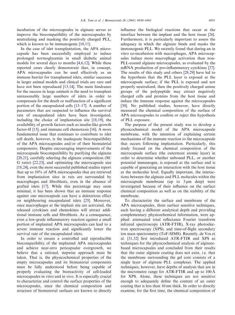
Fig. 1. ATR-FTIR absorbance spectra of the outer 3mm of dehydrated APA
alg), poly-L-lysine film (PLL), dehydrated beads of calcium-alginate gel (C
components. The characteristic peaks for alginate and PLL are identified. (
alginate spectrum in the region 1800–900 cm�1. This region contains the pea
vibrations. The peak wavenumbers are indicated on the figure. (c) A compari
in the region containing the Amide I and Amide II peaks. The peak wavenum
peak positions that correspond to the random coil, a-helix, and unparallel b
3. Results
3.1. ATR-FTIR analyses
To examine the interactions between the moleculeswithin the microcapsule membrane, the outer portion ofAPA microcapsules and their components were exam-ined using ATR-FTIR. Given the experimental condi-tions, it was estimated that the depth of analysis forthese studies was 0.2–3 mm.Fig. 1 compares the ATR-FTIR absorbance spectrum
of the outer portion of dehydrated APA microcapsuleswith the spectra of a sodium alginate film, a PLL film,and dehydrated beads of calcium-alginate gel.
1648
1643
1624
1555
1555
1598
1537
1522
R
α β
R
αβ
r (cm-1)
Abs
orba
nce
2000 1500 1000
Amide I Amide II
APA
APA
COO-
Amide I Amide II
COO-
CO, CC,COH
Na-Alg
PLL
PLL
Ca-Alg
Wavenumber (cm-1)
1700 1650 1600 1550 1500 1450
(c)
microcapsules (APA) and their components: sodium alginate film (Na-
a-alg). (a) Full spectrum for the APA microcapsules and its three
b) A comparison of the APA microcapsule spectrum with the sodium
ks corresponding to the COO� symmetric and asymmetric stretching
son of the APA microcapsule spectrum with the Poly-L-lysine spectrum
bers are indicated on the figure. The symbols R, a, and b indicate the
-sheet conformations of the PLL molecule, respectively.

ARTICLE IN PRESSS.K. Tam et al. / Biomaterials 26 (2005) 6950–69616954
The FTIR spectrum of the APA microcapsules clearlydisplayed absorbance bands that were characteristic ofalginate, which are labelled in Fig. 1a. These include thebroad peak near 3370 cm�1 that indicates stretchingvibrations of hydrogen-bonded OH groups, two peaksnear 1410 and 1600 cm�1 that represent symmetric andasymmetric stretching vibrations of the COO�, respec-tively, and three peaks in the 1200–950 cm�1 region thatcorrespond to various vibrations of the carbohydrate ring.The FTIR spectrum of the microcapsules also
displayed absorbance bands that were characteristic ofPLL, which are labelled in Fig. 1a. In particular, themicrocapsule spectrum contained a small bump in thebroad peak near 3265 cm�1 that coincided with theplacement of the Amide A band, as well as twoshoulders on either side of the peak near 1600 cm�1
that corresponded to the strong Amide I (�1645 cm�1)and Amide II (�1540 cm�1) absorbance bands of thePLL. These amide bands arise from the stretching andbending vibrations of the N–H bonds, and the stretchingvibrations of the CQO and C–N bonds of the amidegroup in the PLL.The precise positions of the absorbance bands for the
APA microcapsules and the sodium alginate in the900–1800 cm�1 region are compared in Fig. 1b. Thepeaks for the COO� stretching vibrations were atslightly lower wavenumbers (1598 and 1409 cm�1) forthe microcapsules than for the sodium alginate (1604and 1412 cm�1). This shift likely indicates that, withinthe microcapsule membrane, the COO� group ofthe alginate interacted with counterions other thansodium [34].A magnified view of FTIR spectra of the micro-
capsules and the PLL for the region containing theAmide I and II bands is shown in Fig. 1c. The bands inthe PLL spectrum are actually composed of several
Table 1
Surface elemental compositions of dehydrated APA microcapsules, sodium
beads as measured from XPS spectra recorded in survey mode
APA microcapsules Sodium alginat
% C 55.970.3 51.270.1
% O 32.170.7 38.370.6
% Na 1.870.3 10.170.8
% N 4.5470.07 0
% S 0 0.3770.13
% P 0 0.0370.02
% Cl 0.0570.05 0
% Br 0 0
% Ca 0 0
% Si 5.771.2 0
C/O 1.770.0 1.370.0**
C/N 12.370.3 —
C/Na 32.975.3 5.270.4*
Note: Values are expressed as the mean atomic percentage7standard error of
of the APA microcapsule surface.
peaks that were assigned, by their positions, ascorresponding to the three possible conformations ofthe molecule: a-helix (1648 and 1537 cm�1), unparallelb-sheet (1624 and 1522 cm�1), and random coil(1555 cm�1). In the microcapsule spectrum, peakshoulders were observed at 1643 and 1555 cm�1,indicating that the PLL molecule, as a component ofthe microcapsule membrane, existed primarily in the a-helix and random coil conformations.
3.2. XPS analyses
To quantitatively determine the elemental and chemi-cal composition of the outermost portion of the APAmicrocapsules, XPS was applied. This technique issurface sensitive in that only the outermost 20–100 Aof a sample is analysed.Table 1 compares the surface elemental composition
of dehydrated APA microcapsules to the compositionsof sodium alginate films, PLL films, and dehydratedcalcium-alginate beads. The compositions were calcu-lated from XPS spectra recorded in survey mode and areexpressed in relative atomic percentages.The surface of the APA microcapsule was composed
primarily of carbon (55.9%), oxygen (32.1%), nitrogen(4.5%), and sodium (1.8%). These elements were alsothe primary components of sodium alginate (51.2% C,38.3% O, 10.1% Na) and PLL (60.1% C, 27.9% O,7.6% N), suggesting that each substance was presentwithin the outer 100 A of the microcapsules. Addition-ally, traces of chlorine (0.05%), presumably originatingfrom NaCl that was used for microcapsule fabrication,were detected. Finally, exposed portions of the siliconsubstrate were detected (5.7%).To verify the origin of the elements that were
detected, the C1s, O1s, N1s, and Na1s peaks of the XPS
alginate films, poly-L-lysine films, and dehydrated calcium-alginate gel
e Poly-L-lysine Calcium-alginate gel
60.173.2 35.070.9
27.972.5 34.770.9
0 0
7.5773.41 0.1770.09
0 0
0 0
0 0
3.771.5 0
0 0.670.1
0.870.8 29.570.2
2.270.1 1.070.1**
10.275.0 —
— —
the mean. *po0:05 and **po0:01 when compared to the composition

ARTICLE IN PRESS
C-O-C
C-CC-O
SiO2
SiO2
C-C
C-NC-O
CO2
C-NH2C-NH3+
C-NH2C-NH3+
C-H
C-CC-H
C-C/Si
APA
APA
O-C-O
HN-C=O
HN-C=ONaO-C=O
NaO-C=O
Na-?
Na-?
HN-C=O
HN-C=O
HN-C=O
C-OHN-C=O
292 290 288 286 284 282
HO-C=O
HO-C=O
-O-C=O-O-C=O
-O-C=O
O-C-O
O-C-O
Cou
nts/
s
Cou
nts/
s
Cou
nts/
s
-O-C=O
C-OH
C-OH
O-C-OC-OH
C-O-C
adsorbed
H2Oadsorbed
Binding Energy [eV]
538 536 534 532 530 528
Binding Energy [eV]
406 404 402 400 398
Binding Energy [eV]
Cou
nts/
s
1076 1074 1072 1070 1068
Binding Energy [eV]
adsorbed
C-OH
Na-Alg
APA
Na-Alg
Na-Alg
PLL PLL
PLL
APA
C-N
(a) (b)
(c) (d)
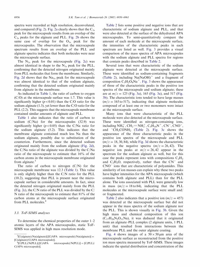
Fig. 2. A comparison of the XPS spectrum for dehydrated APA microcapsules (APA) with the XPS spectra of sodium alginate films (Na-Alg) and
Poly-L-lysine (PLL) films at high resolution. Deconvoluted peaks are assigned to a chemical group based on the binding energy of the peak. (a) C1s
peaks of the APA microcapsules, sodium alginate, and PLL. (b) O1s peaks of the APA microcapsules, sodium alginate, and PLL. (c) N1s peaks of the
APA microcapsules and PLL. (d) Na1s peaks of the APA microcapsules and sodium alginate.
S.K. Tam et al. / Biomaterials 26 (2005) 6950–6961 6955
ARTICLE IN PRESSS.K. Tam et al. / Biomaterials 26 (2005) 6950–69616956
spectra were recorded at high resolution, deconvoluted,and compared (Fig. 2). Fig. 2a clearly shows that the C1s
peak for the microcapsule results from an overlap of theC1s peaks for the alginate and PLL. Fig. 2b shows thesame case of overlap for the O1s peak for themicrocapsules. The observation that the microcapsulespectrum results from an overlap of the PLL andalginate spectra indicates that both molecules were nearthe microcapsule surface.The N1s peak for the microcapsule (Fig. 2c) was
almost identical in shape to the N1s peak for the PLL,confirming that the detected nitrogen originated mainlyfrom PLL molecules that form the membrane. Similarly,Fig. 2d shows that the Na1s peak for the microcapsulewas almost identical to that of the sodium alginate,confirming that the detected sodium originated mainlyfrom alginate in the membrane.As indicated in Table 1, the ratio of carbon to oxygen
(C/O) at the microcapsule surface was 1.7. This value issignificantly higher (po0:01) than the C/O ratio for thesodium alginate (1.3), yet lower than the C/O ratio for thePLL (2.2). This suggests that the microcapsule membranecontained similar amounts of alginate and PLL.Table 1 also indicates that the ratio of carbon to
sodium (C/Na) for the microcapsules (32.9) wassignificantly higher (po0:05) than the C/Na ratio forthe sodium alginate (5.2). This indicates that themembrane alginate contained much less Na than thesodium alginate, possibly due to substitution of thecounterion. Furthermore, since the detected sodiumoriginated mainly from the sodium alginate (Fig. 2d),the C/Na ratio of the alginate was divided by the C/Naratio of the microcapsule to estimate that 16% of thecarbon atoms in the microcapsule membrane originatedfrom alginate.3
The ratio of carbon to nitrogen (C/N) for themicrocapsule membrane was 12.3 (Table 1). This valueis only slightly higher than the C/N ratio for the PLL(10.2), suggesting that PLL is present near the micro-capsule surface in considerable amounts. In fact, sincethe detected nitrogen originated mainly from the PLL(Fig. 2c), the C/N ratio of the PLL was divided by the C/N ratio of the microcapsule to estimate that 81% of thecarbon atoms at the microcapsule surface originatedfrom PLL molecules.4
3.3. ToF-SIMS analyses
To determine the chemical properties of the outer 1–2atomic layers of the APA microcapsules, static ToF-SIMS was applied in high mass resolution mode.
3[C(alginate)/Na(alginate)]/[C(APA microcapsule)/Na(alginate)] ¼
[C(alginate)/C(APA microcapsule)].4[C(PLL)/N(PLL)]/[C(APA microcapsule)/N(PLL)] ¼ [C(PLL)/
C(APA microcapsule)].
Table 2 lists some positive and negative ions that arecharacteristic of sodium alginate and PLL, and thatwere also detected at the surface of the dehydrated APAmicrocapsules. To semi-quantitatively compare theamount of each molecule at the microcapsule surface,the intensities of the characteristic peaks in eachspectrum are listed as well. Fig. 3 provides a visualcomparison of the mass spectra of APA microcapsuleswith the sodium alginate and PLL spectra for regionsthat contain peaks described in Table 2.Several ions that were characteristic of the sodium
alginate were detected at the microcapsule surface.These were identified as sodium-containing fragments(Table 2), including Na(NaOH)+ and a fragment ofcomposition C3H2O4Na
+. Fig. 3 shows the appearanceof three of the characteristic peaks in the positive ionspectra of the microcapsule and sodium alginate; theseare at m=z ¼ 125 (Fig. 3a), 165 (Fig. 3a), and 517 (Fig.3b). The characteristic ions tended to have large masses(m=z ¼ 165 to 517), indicating that alginate moleculescomposed of at least one or two monomers were intactat the microcapsule surface.Many ions that were characteristic of the PLL
molecule were also detected at the microcapsule surface.These were identified as nitrogen-containing ions,including NH4
+, CH2QNH2+, C5H10N
+, CN�, CNO�,and �CH2CONH2 (Table 2). Fig. 3c shows theappearance of the three characteristic peaks in thepositive ion spectra of the microcapsule and PLL(m=z ¼ 18; 30; 84), while Fig. 3d shows the characteristicpeaks in the negative spectra (m=z ¼ 26; 42). Thenegative ion peaks at m=z ¼ 26; 42 appear in thespectrum for the sodium alginate as well, but for thiscase the peaks represent ions with compositions C2H2
and C2H2O, respectively, rather than the CN� andCNO� ions that are characteristic of polyamides. Thissimilarity of ion masses can explain why these two peakshave higher intensities for the APA microcapsule (whichcontains both alginate and PLL) than for the PLLalone. The ions associated with PLL were generally lowin mass (m=z ¼ 18 to 84), indicating that the PLLmolecules at the microcapsule surface were small and/or fragmented.Table 2 also indicates that a positive ion (m=z ¼ 457)
was detected at the microcapsule surface but did notappear in the mass spectra of the sodium alginate northe PLL. This is shown visually in Fig. 3b. Given thehigh mass and chemical composition of this ion(C17H26N2O11Na), it was deduced that it originatedfrom an alginate–PLL complex (2 alginate units, 1 PLLunit) that resulted from interactions between themembrane PLL and the outer alginate coating.Fig. 4 shows images of a 50� 50 mm area of the
microcapsule surface as reconstructed from the positiveion mass spectra measured by ToF-SIMS. These imagesindicate the spatial distribution and concentration of the

ARTICLE IN PRESS
Table 2
Characteristic peaks of sodium alginate and poly-L-lysine that were present the ion mass spectrum of dehydrated APA microcapsules, as measured by
static ToF-SIMS
Molecule Ion mass Ion composition Ion description Intensity (counts� 103)
APA caps PLL Na-alg
Sodium alginate C6H7O6Na (Mw ¼ 108:01)
63+ HONa2 Na(NaOH)+ 1.9 — 40.8
125+ C3H2O4Na Alginate fragment 1.9 — 11.6
165+ C7H10O3Na 1 ring+COONa+H 1.6 — 1.2
517+ C16H23O16Na2 2 intact FU+C4H9O4 0.3 — 6.3
399� C12H17O12Na2 2 intact FU+2H 2.2 — 0.9
Poly-L-lysine C6H13N2O (Mw ¼ 129:10)
18+ H4N NH4+ 4.5 7.7 —
30+ CH4N CH2QNH2+ 16.3 18.6 0.5
84+ C5H10N C5H10N+ 12.2 6.7 0.3
26� CN CN� 86.4 81.1 —
42� CNO CNO� 13.3 11.8 —
58� C2H4NO�CH2CONH2 1.0 5.8 —
Alginate–PLL complex 457+ C17H26N2O11Na 2 alginate FU+1 PLL FU 0.5 — —
Note: The ion masses listed (m/z) correspond to the peak positions in the APA microcapsule spectra. The peak intensities correspond to the area of
the (Poisson-corrected) peaks in the spectra for a dehydrated APA microcapsule, sodium alginate film, and poly-L-lysine film, normalized by the total
counts of each spectrum. A (–) indicates that the peak intensity was negligible (o150 counts). FU ¼ functional unit.
S.K. Tam et al. / Biomaterials 26 (2005) 6950–6961 6957
detected ions. Fig. 4a shows that the sum of all ions (i.e.all chemical species detectable by ToF-SIMS) wasdistributed fairly homogeneously at the microcapsulesurface. Fig. 4b maps the sum of the ions characteristicof alginate, and thus represents the distribution of thealginate molecules at the microcapsule surface. Simi-larly, Fig. 4c represents the distribution of PLL at themicrocapsule surface. These images clearly show thateach the alginate and PLL were distributed evenly at themicrocapsule surface. Fig. 4d shows that the distributionof the ion representing the alginate–PLL complex wasalso distributed evenly at the surface. These imagestogether indicate that there were no localizations of themolecules at the microcapsule surface.
4. Discussion
The combined evidence from the physicochemicalanalyses clearly indicates the presence of both alginateand PLL in the membrane of the APA microcapsules.From the ATR-FTIR analyses, it was confirmed thatthe outermost 3 mm of the microcapsules contained bothmolecules. This result was expected since we measuredthe thickness of the alginate–PLL membrane to be2–3 mm by light microscopy. The elemental compositionof the microcapsules that was measured by XPSsuggested that, in terms of carbon content, relativelyhigh amounts of PLL are very close to the surface,within the outermost 100 A. De Vos et al. [31,35]
obtained similar surface compositions using XPS;therefore, this result was foreseeable. What was lessexpected is that, as suggested from the ToF-SIMSanalyses, the PLL is exposed at the surface of themicrocapsules, i.e. it was detected at the outermost 1–2monolayers of the membrane. This result is supportedby Strand et al.’s [36] morphological observation, usingconfocal laser scanning microscopy, that the outeralginate layer appears to overlap with the PLL layerrather than form an additional outer membrane.The detected presence of PLL in the outermost atomic
layers of the microcapsule implies that a distinct outercoating of alginate does not exist. The results of theToF-SIMS analyses, however, showed that sodiumalginate was also present at the surface, indicating thatat least some of the polysaccharide did in fact bind tothe membrane PLL during the final incubation of thecapsule in dilute alginate (since no calcium was detectedby XPS, we excluded the possibility that the alginate atthe surface originated from the gel core). Usingradiolabelling techniques, Thu et al. [37] have alsodemonstrated that the coating alginate clearly binds tothe PLL layer of the microcapsule. To further supportthis view, this study also provided strong evidence ofinteraction between the alginate and PLL moleculeswithin the membrane. That is, peaks shifts in the ATR-FTIR spectra indicated that the sodium ions that werebound to the COO� groups of the alginate werereplaced by other ions during membrane formation[34,38]. Since nitrogen was detected in the membrane by

ARTICLE IN PRESS
103
5
125
125
165
165
4321
103
54321
103
54321
104
4
3
2
1
104
4
3
2
1
104
4
3
2
1
20 40 60 80
120 140m/z
m/z20 30 40 50 60
m/z
m/z160
Cou
nts
Cou
nts
103
Cou
nts
APA
Na-Alg
PLL
APA
Na-Alg
PLL
104
7
5
3
1
104
7
5
3
1
104
7
5
3
1
Cou
nts
APA
Na-Alg
PLL
APA
Na-Alg
PLL
1.0
0.5
103
1.0
0.5
103
1.0
0.5
450 500 550
457517
517
18
30
18
30 84
84
26
26
42
42
(a) (b)
(c) (d)
Fig. 3. A comparison of the ion mass spectra of a dehydrated APA microcapsule (APA), sodium alginate film (Na-Alg), and poly-L-lysine (PLL)
film, as measured by static ToF-SIMS, for regions of interest. The characteristic ions (labelled) are described in Table 2. (a) Positive ions that are
characteristic of sodium alginate (m=z ¼ 125; 165) are present in the APA microcapsule and alginate spectra, but negligible in the PLL spectrum. (b)
A positive ion that is characteristic of sodium alginate (m=z ¼ 517) is present in the APA microcapsule and alginate spectra, but absent in the PLL
spectrum. Also, a positive ion (m=z ¼ 457) characteristic of an alginate–PLL complex is present in the APA microcapsule spectrum, but is absent in
the sodium alginate and PLL spectra. (c) Positive ions that are characteristic of PLL (m=z ¼ 18; 30; 84) are present in the APA microcapsule and PLL
spectra, but negligible in the alginate spectrum. (d) Negative ions that are characteristic of PLL (m=z ¼ 26; 42) are present in the APA microcapsule
and PLL spectra.
S.K. Tam et al. / Biomaterials 26 (2005) 6950–69616958
XPS, the replacement ions are assumed to be the NH3+
groups of the PLL, confirming the ionic bondingbetween the alginate and PLL molecules. The detectionof a unique fragment at the microcapsule surface, usingToF-SIMS, that was associated with the presence of analginate–PLL complex, also demonstrates that the twomolecules interact closely within the membrane and atthe surface.Despite the evidence that the coating alginate binds to
the PLL of the microcapsule, the degree of interactionbetween the two molecules may vary within themembrane. This view is suggested by the results of theATR-FTIR analyses, which indicated that the mem-brane PLL exists in both the a-helix and random coilconformations. In a solution of neutral pH, PLLnormally exists in a random, or ‘‘charged’’, coilconformation. However, it has been shown that upon
interaction with alginate and similar polyelectrolytes,the PLL will undergo a transition to an a-helixconformation [39,40]. Since for this study, the micro-capsules are fabricated within a solution, it is reasonableto assume that the detected presence of PLL in the a-helix conformation is an indication of sufficient bindingwith the alginate within the membrane, and this view isshared by others [31]. On the other hand, our observa-tion that the membrane PLL is also in random coilconformation does not necessarily indicate insufficientbinding with alginate because the samples were studiedin a dehydrated state; it has been shown that when thePLL interacts with certain sugars, it will conserve itsrandom coil conformation during the dehydrationprocess, as opposed to converting to the b-sheetconformation [41]. Furthermore, the random coilconformation was also observed by Dupuy et al.

ARTICLE IN PRESS
Fig. 4. Images of a 50� 50mm area of a dehydrated APA micro-
capsule surface as reconstructed from the positive ion mass spectrum
that was measured using static ToF-SIMS. The images display the
spatial distribution and concentration of the detected ions (yellow-
higher concentration; red ¼ lower concentration). (a) The distribution
of all of the detected ions from the APA microcapsule surface. (b) The
distribution of the characteristic positive ions of the sodium alginate
molecule. (c) The distribution of the characteristic positive ions of the
poly-L-lysine (PLL) molecule. (d) The distribution of a positive ion
characteristic of an alginate–PLL complex.
Fig. 5. A schematic of the hypothetical conformations of the poly-L-
lysine (black) after dehydration, and when interacting with alginate
(light grey) to variable degrees. (a) PLL converts to a b-sheetconformation in the absence of alginate, (b) PLL retains a random
coil conformation when there is a limited interaction with alginate, (c)
PLL converts to an a-helix conformation, surrounded by a larger helixof the alginate molecule, when the two molecules interact strongly.
S.K. Tam et al. / Biomaterials 26 (2005) 6950–6961 6959
[38], in the case of a calcium-alginate gel immersed inPLL, and by van Hoogmoed et al. [32], in the case ofdehydrated APA microcapsules. Fig. 5 illustrates thehypothetical conformations of PLL, after dehydration,that result from its variable degrees of interaction withalginate.It is noteworthy that the exposure of PLL at the
surface of APA microcapsules does not appear to be dueto defects or inconsistencies in the outer alginatecoating. This was clearly demonstrated by the imagesobtained from the ToF-SIMS analyses, where it wasshown that the alginate molecules are evenly distributedamongst the microcapsule surface. Furthermore, thealginate–PLL complex that was detected by ToF-SIMSwas also homogeneously distributed at the surface.Together, these results confirm the view that, eventhough PLL is exposed at the surface of the APAmicrocapsule, it is consistently interacting with thecoating alginate, not just within the membrane but alsoat the very surface.Despite the evidence of interactions between the
alginate and PLL within the membrane and at themicrocapsule surface, it is clear that PLL is still exposedat the microcapsule surface. Consequently, there
is an increased risk that, during the implantation ofthe microcapsules, a portion of the positively chargedamine groups of the exposed PLL are not sufficientlyneutralized and are coming into contact with thehost tissue, which contains negatively charged cellsand proteins that may play a role in the immuneresponse to the microcapsules [30]. To be able to confirmthis hypothetical role of the exposed PLL in theimmunogenicity of APA microcapsules, further studiesmust be performed in vitro and in vivo, wheremodifications of the surface properties will be closelymonitored.
5. Conclusion
This study provided the first direct evidence thatPLL is exposed at the very surface of APA micro-capsules. It was demonstrated that the exposed PLLinteracts with the outer coating of alginate to form analginate–PLL complex. The degree of interactionbetween the molecules, however, may be variable, whichleaves the possibility that unbound portions of the PLLare exposed at surface of APA microcapsules. If theexposed PLL is not sufficiently neutralized, then thismay explain the observed immune response to APAmicrocapsules. This new model of APA microcapsules,chemically defined at the micrometric/nanometric scale,will be used in future investigations as a tool forinterpreting the interfacial mechanisms that govern thebiocompatibility of the microcapsules, and it will thusplay a pivotal role in the optimization of the APAmicrocapsule design for cell immuno-isolation andtransplantation.

ARTICLE IN PRESSS.K. Tam et al. / Biomaterials 26 (2005) 6950–69616960
Acknowledgements
We thank Dr. K.D. Roberts, Dr. J.X. Zhu, and M.Gauthier for their critical review of the manuscript. Wethank S. Poulin and the Departement de Genie Physiqueat the Ecole Polytechnique for their assistance andtechnical expertise with the physicochemical analyses.We thank the Association Diabete Quebec for providingstudent scholarships to S. Tam and J. Dusseault duringthis project. This work was financially supported by theFonds de Recherche en Sante du Quebec and theNatural Sciences and Engineering Research Council ofCanada.
References
[1] Shapiro AM, Lakey JR, Ryan EA, Korbutt GS, Toth E,
Warnock GL, Kneteman NM, Rajotte RV. Islet transplantation
in seven patients with type 1 diabetes mellitus using a
glucocorticoid-free immunosuppressive regimen. N Engl J Med
2000;343(4):230–8.
[2] Ryan EA, Lakey JR, Rajotte RV, Korbutt GS, Kin T, Imes S,
Rabinovitch A, Elliott JF, Bigam D, Kneteman NM, Warnock
GL, Larsen I, Shapiro AM. Clinical outcomes and insulin
secretion after islet transplantation with the Edmonton protocol.
Diabetes 2001;50(4):710–9.
[3] Orive G, Hernandez RM, Rodriguez Gascon A, Calafiore R,
Chang TM, de Vos P, Hortelano G, Hunkeler D, Lacik I, Pedraz
JL. History, challenges and perspectives of cell microencapsula-
tion. Trends Biotechnol 2004;22(2):87–92.
[4] Lim F, Sun AM. Microencapsulated islets as bioartificial
endocrine pancreas. Science 1980;210(4472):908–10.
[5] Fan MY, Lum ZP, Fu XW, Levesque L, Tai IT, Sun AM.
Reversal of diabetes in BB rats by transplantation of encapsulated
pancreatic islets. Diabetes 1990;39(4):519–22.
[6] Lum ZP, Tai IT, Krestow M, Norton J, Vacek I, Sun AM.
Prolonged reversal of diabetic state in NOD mice by xenografts of
microencapsulated rat islets. Diabetes 1991;40(11):1511–6.
[7] Smidsrod O, Skjak-Braek G. Alginate as immobilization matrix
for cells. Trends Biotechnol 1990;8(3):71–8.
[8] Leblond FA, Tessier J, Halle JP. Quantitative method for the
evaluation of biomicrocapsule resistance to mechanical stress.
Biomaterials 1996;17(21):2097–102.
[9] Robitaille R, Leblond FA, Bourgeois Y, Henley N, Loignon M,
Halle JP. Studies on small (o350 microm) alginate–poly-L-lysine
microcapsules. V. Determination of carbohydrate and protein
permeation through microcapsules by reverse-size exclusion
chromatography. J Biomed Mater Res 2000;50(3):420–7.
[10] Strand BL, Ryan TL, In’t Veld P, Kulseng B, Rokstad AM,
Skjak-Brek G, Espevik T. Poly-L-lysine induces fibrosis on
alginate microcapsules via the induction of cytokines. Cell
Transplant 2001;10(3):263–75.
[11] Duvivier-Kali VF, Omer A, Parent RJ, O’Neil JJ, Weir GC.
Complete protection of islets against allorejection and auto-
immunity by a simple barium-alginate membrane. Diabetes
2001;50(8):1698–705.
[12] O’Shea GM, Sun AM. Encapsulation of rat islets of Langerhans
prolongs xenograft survival in diabetic mice. Diabetes
1986;35(8):943–6.
[13] Soon-Shiong P, Heintz RE, Merideth N, Yao QX, Yao Z, Zheng
T, Murphy M, Moloney MK, Schmehl M, Harris M, et al. Insulin
independence in a type 1 diabetic patient after encapsulated islet
transplantation. Lancet 1994;343(8903):950–1.
[14] Sun Y, Ma X, Zhou D, Vacek I, Sun AM. Normalization of
diabetes in spontaneously diabetic cynomologus monkeys by
xenografts of microencapsulated porcine islets without immuno-
suppression. J Clin Invest 1996;98(6):1417–22.
[15] Robitaille R, Dusseault J, Henley N, Rosenberg L, Halle JP.
Insulin-like growth factor II allows prolonged blood glucose
normalization with a reduced islet cell mass transplantation.
Endocrinology 2003;144(7):3037–45.
[16] de Groot M, Schuurs TA, van Schilfgaarde R. Causes of limited
survival of microencapsulated pancreatic islet grafts. J Surg Res
2004;121(1):141–50.
[17] De Vos P, Van Straaten JF, Nieuwenhuizen AG, de Groot M,
Ploeg RJ, De Haan BJ, Van Schilfgaarde R. Why do micro-
encapsulated islet grafts fail in the absence of fibrotic overgrowth?
Diabetes 1999;48(7):1381–8.
[18] Leblond FA, Simard G, Henley N, Rocheleau B, Huet PM, Halle
JP. Studies on smaller (approximately 315 microM) microcap-
sules: IV. Feasibility and safety of intrahepatic implantations of
small alginate poly-L-lysine microcapsules. Cell Transplant
1999;8(3):327–37.
[19] De Vos P, Hillebrands JL, De Haan BJ, Strubbe JH, Van
Schilfgaarde R. Efficacy of a prevascularized expanded polytetra-
fluoroethylene solid support system as a transplantation site for
pancreatic islets. Transplantation 1997;63(6):824–30.
[20] Klock G, Frank H, Houben R, Zekorn T, Horcher A, Siebers U,
Wohrle M, Federlin K, Zimmermann U. Production of purified
alginates suitable for use in immunoisolated transplantation. Appl
Microbiol Biotechnol 1994;40(5):638–43.
[21] De Vos P, De Haan BJ, Wolters GH, Strubbe JH, Van
Schilfgaarde R. Improved biocompatibility but limited graft
survival after purification of alginate for microencapsulation of
pancreatic islets. Diabetologia 1997;40(3):262–70.
[22] De Vos P, De Haan B, Van Schilfgaarde R. Effect of the alginate
composition on the biocompatibility of alginate–polylysine
microcapsules. Biomaterials 1997;18(3):273–8.
[23] Clayton HA, London NJ, Colloby PS, Bell PR, James RF. The
effect of capsule composition on the biocompatibility of
alginate–poly-L-lysine capsules. J Microencapsul 1991;8(2):
221–33.
[24] Robitaille R, Pariseau JF, Leblond FA, Lamoureux M, Lepage Y,
Halle JP. Studies on small (o350 microm) alginate–poly-L-lysine
microcapsules. III. Biocompatibility of smaller versus standard
microcapsules. J Biomed Mater Res 1999;44(1):116–20.
[25] de Groot M, Schuurs TA, Leuvenink HG, van Schilfgaarde R.
Macrophage overgrowth affects neighboring nonovergrown en-
capsulated islets. J Surg Res 2003;115(2):235–41.
[26] Castner DG, Ratner BD. Biomedical surface science: foundations
to frontiers. Surf Sci 2002;500:28–60.
[27] Juste S, Lessard M, Henley N, Menard M, Halle JP. Effect of
poly-L-lysine coating on macrophage activation by alginate-based
microcapsules: assessment using a new in vitro method. J Biomed
Mater Res A 2005;72A(4):389–98.
[28] Vandenbossche GM, Bracke ME, Cuvelier CA, Bortier HE,
Mareel MM, Remon JP. Host reaction against empty alginate–
polylysine microcapsules. Influence of preparation procedure.
J Pharm Pharmacol 1993;45(2):115–20.
[29] King A, Sandler S, Andersson A. The effect of host factors and
capsule composition on the cellular overgrowth on implanted
alginate capsules. J Biomed Mater Res 2001;57(3):374–83.
[30] Richert L, Lavalle P, Vautier D, Senger B, Stoltz JF, Schaaf P,
Voegel JC, Picart C. Cell interactions with polyelectrolyte
multilayer films. Biomacromolecules 2002;3(6):1170–8.
[31] de Vos P, van Hoogmoed CG, van Zanten J, Netter S, Strubbe
JH, Busscher HJ. Long-term biocompatibility, chemistry, and
function of microencapsulated pancreatic islets. Biomaterials
2003;24(2):305–12.

ARTICLE IN PRESSS.K. Tam et al. / Biomaterials 26 (2005) 6950–6961 6961
[32] van Hoogmoed CG, Busscher HJ, de Vos P. Fourier transform
infrared spectroscopy studies of alginate–PLL capsules with
varying compositions. J Biomed Mater Res 2003;67A(1):172–8.
[33] Belu AM, Graham DJ, Castner DG. Time-of-flight secondary ion
mass spectrometry: techniques and applications for the character-
ization of biomaterial surfaces. Biomaterials 2003;24(21):3635–53.
[34] Sartori C, Dudley SF, Ralph B, Gilding K. Determination of the
cation content of alginate thin films by FTi.r. spectroscopy.
Polymer 1997;38(1):43–51.
[35] de Vos P, Hoogmoed CG, Busscher HJ. Chemistry and
biocompatibility of alginate–PLL capsules for immunoprotection
of mammalian cells. J Biomed Mater Res 2002;60(2):252–9.
[36] Strand BL, Morch YA, Espevik T, Skjak-Braek G. Visualization
of alginate–poly-L-lysine-alginate microcapsules by confocal laser
scanning microscopy. Biotechnol Bioeng 2003;82(4):386–94.
[37] Thu B, Bruheim P, Espevik T, Smidsrod O, Soon-Shiong P,
Skjak-Braek G. Alginate polycation microcapsules. I. Interaction
between alginate and polycation. Biomaterials 1996;17(10):
1031–40.
[38] Dupuy B, Arien A, Perrot Minnot A. FT-IR of membranes made
with alginate/polylysine complexes. Variations with the mannuro-
nic or guluronic content of the polysaccharides. Artif Cells Blood
Substit Immobil Biotechnol 1994;22(1):71–82.
[39] Bystricky S, Malovikova A, Sticzay T. Interaction of alginates
and pectins with cationic polypeptides. Carbohydr Polym
1990;13(3):283–94.
[40] Paradossi G, Chiessi E, Malovikova A. Study of the interactions
of D- and L-polylysine enantiomers with pectate in aqueous
solutions. Biopolymers 1999;50(2):201–9.
[41] Wolkers WF, van Kilsdonk MG, Hoekstra FA. Dehydration-
induced conformational changes of poly-L-lysine as influenced by
drying rate and carbohydrates. Biochim Biophys Acta 1998;
1425(1):127–36.
Copyright © 2022 FDOKUMEN