XPS and XRF depth patina profiles of ancient silver coins
7
This article appeared in a journal published by Elsevier. The attached copy is furnished to the author for internal non-commercial research and education use, including for instruction at the authors institution and sharing with colleagues. Other uses, including reproduction and distribution, or selling or licensing copies, or posting to personal, institutional or third party websites are prohibited. In most cases authors are permitted to post their version of the article (e.g. in Word or Tex form) to their personal website or institutional repository. Authors requiring further information regarding Elsevier’s archiving and manuscript policies are encouraged to visit: http://www.elsevier.com/authorsrights
Transcript of XPS and XRF depth patina profiles of ancient silver coins

This article appeared in a journal published by Elsevier. The attachedcopy is furnished to the author for internal non-commercial researchand education use, including for instruction at the authors institution
and sharing with colleagues.
Other uses, including reproduction and distribution, or selling orlicensing copies, or posting to personal, institutional or third party
websites are prohibited.
In most cases authors are permitted to post their version of thearticle (e.g. in Word or Tex form) to their personal website orinstitutional repository. Authors requiring further information
regarding Elsevier’s archiving and manuscript policies areencouraged to visit:
http://www.elsevier.com/authorsrights

Author's personal copy
Applied Surface Science 272 (2013) 82– 87
Contents lists available at SciVerse ScienceDirect
Applied Surface Science
jou rn al h om epa g e: www.elsev ier .com/ locate /apsusc
XPS and XRF depth patina profiles of ancient silver coins
F. Caridia,b,∗, L. Torrisi c,d, M. Cutroneoc, F. Barrecae, C. Gentilec, T. Serafinof, D. Castriziog
a Facoltà di Scienze MM. FF. NN., Università di Messina, V.le F. Stagno D’Alcontres 31, Messina, Italyb INFN-Sez. CT, Gr. coll. di Messina, V.le F. Stagno D’Alcontres 31, Messina, Italyc Dip.to di Fisica, Università di Messina, V.le F. Stagno D’Alcontres 31, Messina, Italyd INFN-Laboratori Nazionali del Sud, V. S. Sofia 62, 95123 Catania, Italye Advanced and Nanomaterials Research S.r.l., V.le F. Stagno D’Alcontres 31, Messina, Italyf Dip.to di Fisica della materia e ingegneria elettronica, V.le F. Stagno D’Alcontres 31, Messina, Italyg Dip.to di Scienze dell’Antichità, Università di Messina, Italy
a r t i c l e i n f o
Article history:Available online 28 February 2012
Keywords:X-ray photoelectron spectroscopyX-ray fluorescenceSilver coins
a b s t r a c t
Ancient silver coins of different historical periods going from IV cent. B.C. up to recent XIX century, comingfrom different Mediterranean countries have been investigated with different surface physical analyses.X-ray photoelectron spectroscopy (XPS) analysis has been performed by using electron emission inducedby 1.4 keV X-rays. X-ray fluorescence (XRF) analysis has been devoted by using 30 keV electron beam.Scanning electron microscopy (SEM) has been employed to analyze the surface morphology and the X-raymap distribution by using a 30 keV microbeam.
Techniques were used to investigate about the patina composition and trace elements as a function ofthe sample depth obtained coupling XPS to 3 keV argon ion sputtering technique.
© 2012 Elsevier B.V. All rights reserved.
1. Introduction
The compositional analysis of ancient coins, based on bulk andpatina impurity patterns, represents an important aspect of thearcheological research. In particular, the patina layer, representingan important aspect of the coin surface, endures strong changes,as a consequence of the coin conservation environment, impactswith other materials, absorbent contaminants promoted by ther-mal and chemical processes, etc. Its composition may be changed asa result of the interaction between its constituting metal alloy andthe environmental agents or contaminants. On ancient coins, whichhave been usually buried in the ground for thousands of years, thepatina, from an archeometrist’s point of view, is essential for iden-tifying the nature of the environment in which the coins remainedso long. Compositional data constitute a fundamental tool in thestudy of ancient coins and in the knowledge of extractive sites andfabrication technologies [1].
Many physical techniques can be used to investigate the patinaelemental composition.
X-ray photoelectron spectroscopy (XPS), energy dispersed X-rayfluorescence (EDXRF), electron microscopy (SEM), surface pro-filometry (SP), mass spectrometric techniques (MST), inductively
∗ Corresponding author at: Facoltà di Scienze MM. FF. NN., Università di Messina,V.le F. Stagno D’Alcontres 31, Messina, Italy. Tel.: +39 0906765120.
E-mail address: [email protected] (F. Caridi).
coupled plasma-mass spectrometry (ICP-MS), and laser ablation-mass quadrupole spectrometry (LAMQS), represent only someof the many analysis techniques useful for these investigations[2–5].
The main techniques adopted for the present study wereX-ray photoelectron spectroscopy (XPS) and X-ray fluorescence(XRF) coupled to SEM analysis, both possessing easy sample prepa-ration together with a good attainable degree of experimentalinformation. Furthermore, they can be considered non-invasive andcan be safely used according to the integrity requirements of theanalyzed pieces.
In the present investigation, in order to analyze the compositionand the depth profile of peculiar elements, these two techniquesare employed to study a set of ancient silver coins coming from theMediterranean.
2. Materials and methods
The silver coins submitted to XPS and XRF analyses were a 500Lire coin, 1965, Italian Republic, a Denario Romano, II B.C. and aDracma, Alessandro Magno, Macedonia, IV B.C. Each coin, after beencleaned in an ultrasounds bath, was directly analyzed without anyother special preparation of its surface.
A photo of the three coins is reported in the inset of Figs. 1–3.The patina chemical composition was investigated by means
of non-invasive X-ray photoelectron spectroscopy (XPS) acquiringspectra by means of a K� system from Thermo Scientific equipped
0169-4332/$ – see front matter © 2012 Elsevier B.V. All rights reserved.doi:10.1016/j.apsusc.2012.02.071

Author's personal copy
F. Caridi et al. / Applied Surface Science 272 (2013) 82– 87 83
Fig. 1. X-ray photoelectron spectroscopy (XPS) results for the 500 Lire coin, 1965,Italian Republic, for an etching time of 0 s (a) and XPS results for the 500 Lire coin,1965, Italian Republic, for an etching time of 351 s (b).
with a monochromatic Al K� source (1486.6 eV) and operating withan analyzer in constant analyzer energy (CAE) mode with a passenergy from 50 to 200 eV for survey and high resolution spec-tra, respectively. Inside the analyzer are maintained electrostaticfields to allow electrons of a specific kinetic energy (pass energy)by setting voltages, to arrive at the detector slit and onto the detec-tor itself. A spot size diameter on the sample of about 400 �mwas adopted. Surface charging effects occurring in an isolant sam-ples were avoided using an electron flood gun. Ar+ ions with anenergy of 3 keV were used for the sputtering procedure in orderto perform the depth profile measurements. XPS analysis permit-ted to investigate on about 5 nm thickness; the coupling with theion sputtering gave depth profiles of peculiar elements. The sput-tering rate was of the order of 0.3 nm/s and sputtering depths ofthe order of 100 nm were investigated using a time sputtering ofabout 350 s.
The non-invasive XRF spectroscopy permitted to evaluate theelemental composition by using 30 keV electron beam of a SEMmicroprobe (scanning electron microscopy). Elemental composi-tion was monitored for thicknesses of the order of few microns. ASi(Li) detector with a thin Be windows was employed to investi-gated on the elements with atomic number higher than 6.
Fig. 2. XPS results for the Denario Romano, II B.C., for an etching time of 0 s (a) and250 s (b), respectively.
3. Results
Results of the X-ray photoelectron spectroscopy (XPS) arereported in Fig. 1 for the 500 Lire coin, 1965, Italian Republic, for anetching time of 0 s (a) and 351 s (b), respectively. In the first casea great amount of carbon, silver and oxygen and trace elements asCu, Cl and Na was detected on the coin surface, while only a highquantity of silver (carbon, oxygen, chlorine, copper and sodium astrace elements) was detected in the patina at a depth of the orderof 100 nm, after a sputtering procedure with Ar+ ions at an energyof 3 keV, for an etching time of 351 s.
Results obtained for an older silver coin, Denario Romano, II B.C.,are reported in Fig. 2 for an etching time of 0 s (a) and 250 s (b),respectively. In particular a greater amount of trace elements (Si,Au, Ca, Cu, Cl and Na) was detected without the sputtering proce-dure (etching time = 0 s) while, also in this case, a high quantity ofsilver was detected in the patina at a depth of the order of 75 nm(etching time = 250 s).
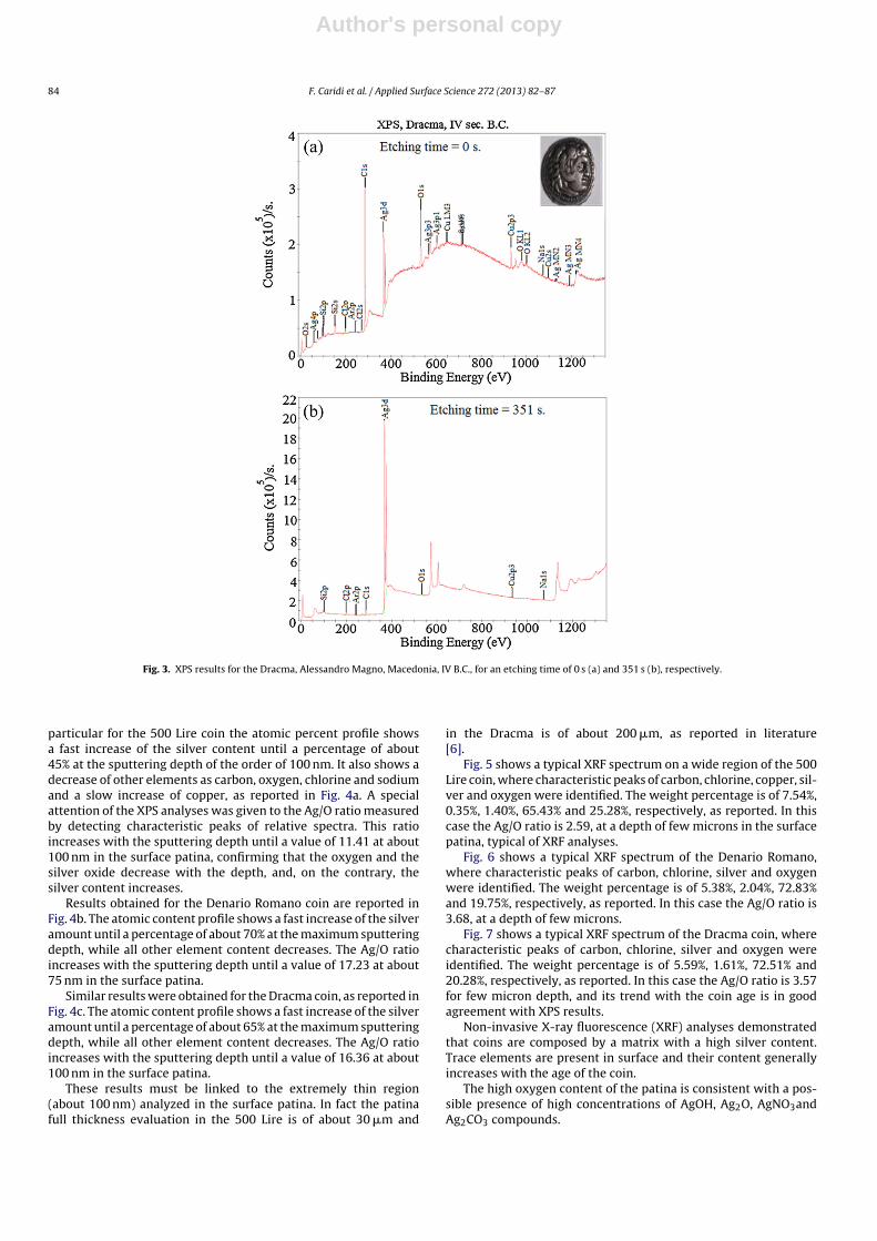
Similar results were obtained for the oldest coin, Dracma,Alessandro Magno, Macedonia, IV B.C., as reported in Fig. 3 for anetching time of 0 s (a) and 351 s (b), respectively. A great amount oftrace elements (Cu, Cl, Na, Si) was detected without etching timeand, also in this case, a high peak of silver was detected in the patinaat a depth of the order of 100 nm (etching time = 351 s).
The XPS permitted to evaluate the elemental composition ofcoins as a function of the etching time, as reported in Fig. 4. In
Author's personal copy
84 F. Caridi et al. / Applied Surface Science 272 (2013) 82– 87
Fig. 3. XPS results for the Dracma, Alessandro Magno, Macedonia, IV B.C., for an etching time of 0 s (a) and 351 s (b), respectively.
particular for the 500 Lire coin the atomic percent profile showsa fast increase of the silver content until a percentage of about45% at the sputtering depth of the order of 100 nm. It also shows adecrease of other elements as carbon, oxygen, chlorine and sodiumand a slow increase of copper, as reported in Fig. 4a. A specialattention of the XPS analyses was given to the Ag/O ratio measuredby detecting characteristic peaks of relative spectra. This ratioincreases with the sputtering depth until a value of 11.41 at about100 nm in the surface patina, confirming that the oxygen and thesilver oxide decrease with the depth, and, on the contrary, thesilver content increases.
Results obtained for the Denario Romano coin are reported inFig. 4b. The atomic content profile shows a fast increase of the silveramount until a percentage of about 70% at the maximum sputteringdepth, while all other element content decreases. The Ag/O ratioincreases with the sputtering depth until a value of 17.23 at about75 nm in the surface patina.
Similar results were obtained for the Dracma coin, as reported inFig. 4c. The atomic content profile shows a fast increase of the silveramount until a percentage of about 65% at the maximum sputteringdepth, while all other element content decreases. The Ag/O ratioincreases with the sputtering depth until a value of 16.36 at about100 nm in the surface patina.
These results must be linked to the extremely thin region(about 100 nm) analyzed in the surface patina. In fact the patinafull thickness evaluation in the 500 Lire is of about 30 �m and
in the Dracma is of about 200 �m, as reported in literature[6].
Fig. 5 shows a typical XRF spectrum on a wide region of the 500Lire coin, where characteristic peaks of carbon, chlorine, copper, sil-ver and oxygen were identified. The weight percentage is of 7.54%,0.35%, 1.40%, 65.43% and 25.28%, respectively, as reported. In thiscase the Ag/O ratio is 2.59, at a depth of few microns in the surfacepatina, typical of XRF analyses.
Fig. 6 shows a typical XRF spectrum of the Denario Romano,where characteristic peaks of carbon, chlorine, silver and oxygenwere identified. The weight percentage is of 5.38%, 2.04%, 72.83%and 19.75%, respectively, as reported. In this case the Ag/O ratio is3.68, at a depth of few microns.
Fig. 7 shows a typical XRF spectrum of the Dracma coin, wherecharacteristic peaks of carbon, chlorine, silver and oxygen wereidentified. The weight percentage is of 5.59%, 1.61%, 72.51% and20.28%, respectively, as reported. In this case the Ag/O ratio is 3.57for few micron depth, and its trend with the coin age is in goodagreement with XPS results.
Non-invasive X-ray fluorescence (XRF) analyses demonstratedthat coins are composed by a matrix with a high silver content.Trace elements are present in surface and their content generallyincreases with the age of the coin.
The high oxygen content of the patina is consistent with a pos-sible presence of high concentrations of AgOH, Ag2O, AgNO3andAg2CO3 compounds.

Author's personal copy
F. Caridi et al. / Applied Surface Science 272 (2013) 82– 87 85
Fig. 4. XPS elemental composition of coins as a function of the etching time.

Author's personal copy
86 F. Caridi et al. / Applied Surface Science 272 (2013) 82– 87
Fig. 5. A typical XRF spectrum of the 500 Lire coin.
Fig. 6. A typical XRF spectrum of the Denario Romano coin.
Fig. 7. A typical XRF spectrum of the Dracma coin.
Author's personal copy
F. Caridi et al. / Applied Surface Science 272 (2013) 82– 87 87
The content of other elements is consistent with the presenceof chemical compounds such as Ag2S, AgCl, Ag2CrO4, AgBrO3 andAgIO3.
4. Discussion and conclusions
XPS and XRF represent useful non-invasive techniques to ana-lyze the surface layers of metallic samples such as coins.
Obtained results for the characterization of the silver oxidepatina thickness of recent and old coins are very interesting fornumismatic classification of coins, for historical and archeologi-cal investigations, for museum coin conservation, for a comparisonbetween coin composition and mineral composition and to distin-guish true coins from false ones [7–10].
In this work investigations concerning the presence of traceimpurity in the patina and the study of the Ag/O ratio were per-formed.
Results reported in this article can be compared with the evalua-tion of the silver oxide patina thickness for the same coins obtainedwith laser ablation-mass quadrupole spectrometry (LAMQS) andsurface profile analysis [6]. LAMQS is a micro-invasive techniqueand it was employed to evaluate the Ag/O ratio as a function of theirradiation time at 1 Hz repetition rate, i.e. by measuring the ratio vs.depth with a resolution given by the ablation yield of 1 �m/pulse.
This technique has demonstrated that analyzing old silver coins andmeasuring the patina thickness, the Ag/O ratio is lower in old coinsand it increases in recent ones, demonstrating that the coin patinais generally rich in silver oxide proportionally to the age of the coin.The bulk is rich in silver, while the surface is rich in silver oxide.
References
[1] D. Castrizio, in: I. Antinoupolis (Ed.), Preliminary Relation, vol. I, Istituto Papiro-logico G. Vitelli Scavi e Materiali, Firenze, 2008, pp. 217–227.
[2] M. Resano, E. Garcia-Ruiz, F. Vanhaecke, Mass Spectrom. Rev. 29 (2010) 55–78.[3] S. Kyuseok, C. Hyungki, K. Dukhyeon, M. Kihyung, Bull. Korean Chem. Soc. 25
(1) (2004) 101–105.[4] B. Giussani, D. Monticelli, L. Rampazzi, Anal. Chim. Acta 635 (2009) 6–21.[5] L. Torrisi, F. Caridi, L. Giuffrida, A. Torrisi, G. Mondio, T. Serafino, M. Caltabiano,
E.D. Castrizio, E. Paniz, A. Salici, NIM B 268 (2010) 1657–1664.[6] L. Torrisi, G. Mondio, A.M. Mezzasalma, D. Margarone, F. Caridi, T. Serafino, A.
Torrisi, Eur. Phys. J. D 54 (2009) 225–232.[7] L. Torrisi, F. Caridi, A. Borrielli, L. Giuffrida, A. Torrisi, G. Mondio, A. Mezzasalma,
T. Serafino, M. Caltabiano, E.D. Castrizio, E. Paniz, M. Romeo, A. Salici, Radiat.Eff. Defects Solids 165 (6–10) (2010) 626–636.
[8] F. Caridi, L. Torrisi, A. Borrielli, G. Mondio, Radiat. Eff. Defects Solids 165 (6)(2010) 668–680.
[9] D. Aiello, A. Buccolieri, G. Buccolieri, A. Castellano, M. Di Giulio, L. Sandra Leo,A. Lorusso, G. Nassisi, V. Nassisi, L. Torrisi, in: XVI International Symp. on GasFlow and Chemical Laser & High Power Laser Conference, Gmunden, Proc. SPIE6346 (2006).
[10] A. Picciotto, L. Torrisi, D. Margarone, P. Bellutti, Radiat. Eff. Defects Solids 165(6–10) (2010) 706–712.