Detection of genomic and intermediate replicative strands of ...

at SciVerse ScienceDirect
Biomaterials 34 (2013) 1967e1979
Contents lists available
Biomaterials
journal homepage: www.elsevier .com/locate/biomater ia ls
Genomic analysis of the role of transcription factor C/EBPd in the regulation of cellbehaviour on nanometric grooves
Jolanta Wiejak 1, Penelope M. Tsimbouri 1, Pawel Herzyk, Matthew J. Dalby, Graham Hamilton,Stephen J. Yarwood*
Institute of Molecular, Cell and Systems Biology, College of Medical, Veterinary and Life Sciences, University of Glasgow, Glasgow, United Kingdom
a r t i c l e i n f o
Article history:Received 1 November 2012Accepted 20 November 2012Available online 13 December 2012
Keywords:NanotopographyCell signallingCell spreadingCell adhesionGene expressionSignal transduction mediator
* Corresponding author. Institute for Molecular, Cellof Medical, Veterinary and Life Sciences, UniversDavidson Building, Glasgow, Scotland G12 8QQ, Unite330 3908; fax: þ44 (0)141 330 4620.
E-mail addresses: [email protected], step(S.J. Yarwood).
1 These authors contributed equally to research.
0142-9612/$ e see front matter � 2012 Elsevier Ltd.http://dx.doi.org/10.1016/j.biomaterials.2012.11.036
a b s t r a c t
C/EBPd is a tumour suppressor transcription factor that induces gene expression involved in suppressingcell migration. Here we investigate whether C/EBPd-dependent gene expression also affects cellresponses to nanometric topology. We found that ablation of the C/EBPd gene in mouse embryonalfibroblasts (MEFs) decreased cell size, adhesion and cytoskeleton spreading on 240 nm and 540 nmnanometric grooves. ChIP-SEQ and cDNA microarray analyses demonstrated that many binding sites forC/EBPd, and the closely related C/EBPb, exist throughout the mouse genome and control the upregulationor downregulation of many adjacent genes. We also identified a group of C/EBPd-dependent, trans-regulated genes, whose promoters contained no C/EBPd binding sites and yet their activity was regulatedin a C/EBPd-dependent manner. These genes include signalling molecules (e.g. SOCS3), cytoskeletalcomponents (Tubb2, Krt16 and Krt20) and cytoskeletal regulators (ArhGEF33 and Rnd3) and are possiblyregulated by cis-regulated diffusible mediators, such as IL6. Of particular note, SOCS3 was shown to beabsolutely required for efficient cell spreading and contact guidance on 240 nm and 540 nm nanometricgrooves. C/EBPd is therefore involved in the complex regulation of multiple genes, including cytoskeletalcomponents and signalling mediators, which influence the nature of cell interactions with nanometrictopology.
� 2012 Elsevier Ltd. All rights reserved.
1. Introduction
C/EBP proteins form a highly conserved family of leucine zipper(bZIP) transcriptional factors that serve as master regulators ofcellular processes such as the cell cycle, differentiation, andinflammatory responses [1]. So far, six C/EBP genes have been iso-lated (a, b, g, d, ε, and z), although protein numbers may well behigher due to variation in polypeptide size through alternatesplicing and protein processing [2]. C/EBP isoforms are structurallysimilar, displaying a characteristic basic leucine zipper domain atthe C terminus (90% homology between isoforms), which facilitatesdimerization and DNA binding [2]. However, C/EBP isoforms arefunctionally and genetically distinct, with their transcriptional
and Systems Biology, Collegeity of Glasgow, Room 239,d Kingdom. Tel.: þ44 (0)141
All rights reserved.
activation domains less well conserved (<20% sequence identitybetween isoforms). This divergence gives rise to the wide range ofcellular responses inwhichC/EBP isoformshavebeen implicated [2].
Attention has recently turned to the functional role of the C/EBPdisoform, which represents a ubiquitously expressed transcriptionalactivator that is robustly induced in a variety of G0 growth arrestedcells [3]. Importantly, “loss of function” alterations in C/EBPd havebeen reported in breast cancer [4e6] and acute myeloid leukaemia(AML) [7] and are generally associated with impaired contactinhibition, increased genomic instability and increased cell migra-tion [8]. The use of knockout mice demonstrated that C/EBPd hasa vital role in mammary duct and epithelial cell proliferation [9] aswell as an obligate role in the differentiation of preadipocytes[10,11]. In addition, recent work suggests that C/EBPd is requiredfor the maintenance of pluripotency in human limbic stem cells [2].C/EBPd activity appears to be regulated at a number of levels,including transcriptional (gene induction by STAT3, Sp1, CREB andNcoA/SRC-1 [12,13]), post-transcriptional (mRNA stability [14]) andpost-translational (ubiquitinylation [15] and SUMOylation [16])mechanisms. Certain C/EBP isoforms have also been shown to besubstrates for various protein kinases, including the MAP kinases,

MEF WT MEF C/EBPδ -/-
FLAT
240 nm
540 nm
a
b
05
101520253035404550
C/EBPδWT Flat
C/EBPδWT 240nm
C/EBPδWT 540nm
C/EBPδ -/-Flat
C/EBPδ -/-240nm
C/EBPδ -/-540nm
Cell A
rea
(sq
uare m
icro
ns/100)
0
500
1000
1500
2000
2500
3000
3500
4000
4500
C/EBPδ WTFlat
C/EBPδ WT240nm
C/EBPδ WT540nm
C/EBPδ -/-Flat
C/EBPδ -/-240nm
Mean
V
in
cu
lin
In
ten
sity
(arb
itrary u
nits)
*** *** **
** *** *
C/EBPδ -/-540nm
Fig. 1. Morphology of wild type (WT) and C/EBPd �/� mouse embryonic fibroblasts (MEFs) cultured on flat or nanogrooved substrates. a) MEF WT and MEF C/EBPd �/� cells werecultured on flat or nanogrooved substrates as indicated and stained for actin cytoskeleton (red) and vinculin (green). MEF WT cells had a well developed actin cytoskeleton anda largely polygonal morphology and became bipolar when cultured on grooves. In contrast C/EBPd �/� MEFs had a less well developed actin cytoskeleton, were generally smallerand became very elongated when grown on the grooved substrate. b) MEF WT and MEF C/EBPd �/� cell area and vinculin intensity were quantified as described in Materials andMethods. Results demonstrated that MEF C/EBPd �/� cells were smaller and had lower levels of vinculin, on both flat and grooved surfaces, than MEF WT cells. (For interpretation ofthe references to colour in this figure legend, the reader is referred to the web version of this article.)

J. Wiejak et al. / Biomaterials 34 (2013) 1967e1979 1969
ERK 1 and 2, and protein kinase C (PKC) [1], and are targets of secondmessenger signalling pathways. For example, elevations in theintracellular levels of cyclic AMP and activation of protein kinase A,has a direct impact on the induction of the constitutively active C/EBPd isoform, which, in turn, regulates the acute-phase plasmaprotein gene haptoglobin, which is involved in the intestinalepithelial cell response to inflammation [12], whereas cyclic AMP-activation of exchange protein activated by cyclic AMP 1 (EPAC1),leads to the C/EBPd-dependent induction of the anti-inflammatorysuppressor of cytokine signalling 3 (SOCS3) gene in vascularendothelial cells [17].
Despite being a transcriptional activator, very few C/EBPdtarget genes have been identified. As a result the mechanisms bywhich C/EBPd controls cell adhesion, migration, differentiationand cell-cycle progression remain poorly understood. Recentreports have demonstrated that sumoylation of C/EBPd promotessequestration to the nuclear periphery, thereby suppressingexpression of C/EBPd-dependent genes associated with cell adhe-sion, including glycoprotein V, protocadherin 9 and integrin b8[8,16]. Given this potential link between transcriptional control andcell adhesion we have used genomic analysis (including gene arrayand high resolution DNA sequencing) to investigate the role of C/EBPd in controlling cell adhesion with biomaterials, comparingplanar and nanometric grooved growth surfaces.
2. Materials and methods
2.1. Materials
Wild type (WT) immortalized mouse embryonic fibroblasts (129SV:C57CI/6,MEFs) and MEFs containing homozygous deletions of the C/EBPb, or C/EBPd geneswere generous gifts from Prof. Peter Johnson (C/EBPb) and Esta Sterneck (C/EBPd)from NCI, National Institutes of Health, Frederick, MD. SOCS3 and FAK knockoutMEFs, together with matched WT cells, were gifts from Prof. Margaret Frame,Edinburgh Cancer Research UK Centre, UK and Dr Timothy Palmer, Institute ofCardiovascular and Medical Sciences, University of Glasgow, UK, respectively. For-skolin, and rolipram were purchased from Merck Biosciences (Nottingham, UK).Anti-SOCS3 and ChIP-grade, anti-C/EBP polyclonal antibodies were from Santa Cruz.
2.2. Generation of grooved and planar growth surfaces
Microstructured quartz substrates were fabricated by acid-cleaning(7:1H2SO4:H2O2 for 5 min) quartz slides which were then spin-coated with AZprimer (4000 rpm for 30sec). A layer of Shipley S1818 photoresist (Shipley) was spunonto to the spin-coated slides and then soft-baked for 30 min in a 90�C oven. Thesubstrates were UV treated on an MA6 mask aligner with exposure energy of ca.7.1 mJ/cm2 per second in hard contact, through an electron-beam fabricated chromemask with 12.5 mm wide lines. The resist was developed (1:1 AZ developer (Micro-chemicals):water) for 65sec. The substrates were rinsed, dried and then etched ina trichloromethane environment at a rate of 25 nm/min in a reactive ion etching unit(RIE80, Plasma Technology) using the polymer pattern as an etch resist to generate240 or 540 nm deep grooves. The residual resist was removed with acetone and theslide was blanket etched for a further 1 min to produce a homogenous surfacechemistry. Planar slides were blanket etched to ensure that the chemistry wascomparable with the structured substrates. Quartz substrates were then cleaned inCaro’s acid solution (2:1 H2SO4:H2O2) for 20 min, rinsed six times with double-distilled water and air-dried under a Class I or II sterile flow hood. Imprints of thequartz substrates into polycaprolactone (PCL) were achieved by hot-embossing. Theresulting imprints, 240 nm and 540 nm grooves, were trimmed for use and planarPCL (Ra of 1.17 nm over 10 mm)was used as a control substrate. The PCL samples weregiven a 30 s treatment in oxygen plasma to allow cell attachment (Harrick Plasma,USA). All PCL substrates were sterilised in ethanol for 30 min, then transferredthrough three rinses in sterile 1� PBS and two rinses in complete medium.
2.3. Cell culture and growth of cells on biomaterials
Matched wild type (WT) or homozygous knockout (�/�) mouse embryonicfibroblasts (MEFs), for C/EBPb, C/EBPd, SOCS3 or FAK, were maintained in 71% (v/v)DMEM (Sigma), supplemented with 17.7% (v/v) medium 199, 9% (v/v) FBS, 1% (v/v)200 mM L-glutamine (Gibco), 0.9% (v/v) 100 mM sodium pyruvate and antibiotics(6.74 U/ml Penicillin-Streptomycin, 0.2 mg/ml Fungizone) at 37 �C in a 5% CO2
environment. Cells were passaged at 70e80% confluence, and the medium wasreplaced regularly. Unless otherwise indicated, MEFs were seeded at a density of1 �104 cells/ml on the PCL thumb-embossed substrates with a 24 h culture period.
2.4. Immunofluorescence
MEFs were fixed in a 10% (v/v) formaldehyde solution (15min at 37 �C), per-meabilised (5min at 4 �C) and blocked in 1% (w/v) BSA/PBS (15min at 37 �C). Thesamples were then stained at 37oC for 1 h with 1:200 (v/v) anti-vinculin (clonehVin-1, Sigma) in 1% (w/v) BSA/PBS and 1:500 (v/v) phalloidin-rhodamine (Molec-ular Probes). In each experiment, two replicas each of planar and nanogroovetopography were stained. Cells were washed 3 � 5min in 1xPBS containing 0.5% (v/v) Tween-20, and appropriate biotinylated secondary antibody (Vector Laboratories)was added at 1:50 in 1% (w/v) BSA/PBS and incubated for a further hour at 37 �C.After washing, 1:50 (v/v) FITC-conjugated streptavidin (Vector Laboratories) wasadded to the samples and incubated for 30 min at 4 �C followed by washing andmounting using Vectashield mountant with DAPI nuclear stain (Vector Laborato-ries). Cell images were captured on a confocal microscope and analysed using ImageJ software (NIH). Significant changes were determined by one-way ANOVA withTukey post-test.
2.5. Microarray analysis
C/EBPbWT, C/EBPb�/�, C/EBPdWTand C/EBPd �/� cells were grown on planargrowth surfaces and then incubated in the presence or absence of a combination of10 mM forskolin plus 10 mM rolipram (F/R) for 5 h at 37 �C in 5% (v/v) CO2. Cells werethen washed with 1 � 1 ml PBS and RNA was isolated using the Qiagen “RNeasy”Mini Kit according to the manufacturers protocol. RNA samples were additionallytreated with a “DNA-free Kit” (Applied Biosystems) to remove any remaining DNA.RNA samples were then prepared for Affymetrix whole transcriptome microarrayanalysis, using the WT Expression Kit (Ambion) according to the manufacturer’sinstructions. Briefly, reverse transcription was used to prime poly(A) and non-poly(A), but not ribosomal, mRNA and generate sense strand cDNA for fragmenta-tion and labelling, using the Affymetrix GeneChip� WT Terminal Labelling Kit (PN900671). Amplified and biotinylated sense-strand DNA targets were hybridised,using a Fluidic Station 400,to Affymetrix GeneChip� Mouse Gene 2.0 ST Array anddata captured using an Affymetrix GeneChip� Scanner 3000 7G. Gene expressionchanges were selected where at least one fold change was greater than 2. Groupingof gene expression data into similarly responsive patterns was done using Treeviewand CLUSTER software [18], freely available from the Eisen lab (http://www.eisenlab.org/eisen/).
2.6. Chromatin immunoprecipitation and sequencing (ChIP-SEQ) analysis
C/EBPb WT and C/EBPd WT cells were stimulated in the presence or absence ofa combination of 10 mM forskolin plus 10 mM rolipram (F/R) for 5 h at 37 �C in 5% (v/v) CO2. Cells were then fixed and chromatin extracted and sheared using theenzymatic “CHIP-IT Express Kit” (Active Motif) according to the manufacturer’sinstructions. Sheared chromatin from F/R-treated and non-treated cells was thenimmunoprecipitated at 4 �C, overnight with 4 mg of either C/EBPb WT or C/EBPdChIP-grade antibodies (Santa Cruz). DNA fragments were eluted from immunopre-cipitated chromatin and used to prepare a ChIP-SEQ DNA library for sequencingusing the “ChIP-SEQ Sample Prep Kit” from Illumina, according to the manufac-turer’s protocols. Briefly, the first step in library preparation was to convert anyoverhangs in the ChIP’d DNA into phosphorylated blunt ends. The 30 ends were thenadenylated and adaptors ligated onto the ends of the fragments. The librarywas thensize selected on an agarose gel and eventually enriched by PCR. The enriched librarysamples were then loaded onto a flow cell at a concentration of 12pM and clusterformationwas done on an Illumina Cluster station. Samples were then sequenced onan Illumina GA IIX giving 76bp reads.
2.7. ChIP-SEQ data analysis
The ChIP DNA was sequenced on an Illumina GA IIx, one lane of the flowcell per sample. The quality of the reads was assessed using Fastqc (www.bioinformatics.babraham.ac.uk/projects/fastqc/). The sequence reads werealigned to the mouse genome (release version mm9) using the bowtie aligner(version 0.12.7) (bowtie-bio.sourceforge.net/index.shtml) [19], which was set up toreport only uniquely aligning reads. Duplicate reads were removed using Samtools(version 0.1.18) [20]. The ChIP analysis was performed using the Homer (version3.9) suite of tools (biowhat.ucsd.edu/homer/ngs/index.html) [21]. The pipeline forthe analysis is shown in Supplementary Fig. 1. Custom scripts were created tocompare the list of known genes closest to ChIP peaks with gene lists generatedfrom RNA microarray experiments. These corroborated gene lists were visualisedon chromosomes using Circos plots (circos.ca; Supplementary Fig. 2) and Venndiagram (Fig. 2b).
2.8. Reverse transcription PCR (RT-PCR)
Total RNA isolation was extracted from MEF WT and MEF �/� cells using anRNeasy Mini Kit (Qiagen) according to the manufacturer’s instructions. RT-PCRreactions were carried out using the OneStep RT-PCR Kit from Qiagen. Briefly, 5e10 ng of RNA per sample was used in 25 ul reaction mixture containing 0.4 mM

Rank Motif P-value
log P-pvalue
% ofTargets
% ofBackground
STD(BgSTD) Best Match/Details
P-value
log P-pvalue
% ofTargets
% ofBackground
STD(BgSTD) Best Match/Details
1 1e-128
-2.955e+02 15.12% 3.09% 47.1bp
(58.8bp)CEBP(bZIP)/CEBPb-ChIP-Seq/Homer
2 1e-69
-1.604e+02 9.61% 2.26% 52.6bp
(59.4bp)CEBP:AP1/ThioMac-CEBPb-ChIP-Seq/Homer
Validation of C/EBPββ ChIP-Sequencing
a
b
Rank Motif
1 1e-869
-2.002e+03 58.50% 6.49% 39.9bp
(61.4bp)MA0102.2_CEBPA
2
Motif
1e-49
-1.134e+02 6.22% 1.23%
57.1bp(62.1bp)
CEBP:AP1/ThioMac-CEBPb-ChIP-Seq/Homer
Validation of C/EBPδ ChIP-Sequencing
638 505 568
173546 512
6647
Microarray
β δ
Fig. 2. Results of C/EBPb and C/EBPd ChIP-SEQ and microarray. a) MEF WT cells were incubated in the presence or absence of a combination of the cyclic AMP-elevating agents,forskolin and rolipram (F/R). Following stimulation cells were fixed, chromatin was isolated and then immunoprecipitated (ChIP’d) with anti-C/EBPb or C/EBPd antibodies, asdescribed in Materials and methods. ChIP’d DNA samples were then sequenced (ChIP-SEQ) on an Illumina GA IIx DNA sequencer. ChIP analysis of the resulting DNA sequenceswas then performed using the Homer (version 3.9) suite of tools (biowhat.ucsd.edu/homer/ngs/index.html). The figure shows part of the Homer analysis indicating thataligned sequences from each ChIP experiments contained bone fide C/EBP consensus binding motifs, hence validating the experimental technique. b) MEF WT, MEF C/EBPb �/� andMEF C/EBPd �/� cells were stimulated for 5 h in the presence or absence of F/R. Cells were then harvested, RNA isolated, biotinylated sense-strand DNA synthesised and hybridisedto mouse whole genome GeneChip� ST Arrays (Affymatrix). A Venn diagram was then generated to illustrate the degree of overlap between genes identified by ChIP-SEQ ascontaining consensus, F/R-dependent C/EBP-binding sites and F/R-induced changes in MEF gene expression.
J. Wiejak et al. / Biomaterials 34 (2013) 1967e19791970
dNTPs and 0.6 mM of each primer. The primer sequences used were mTubb2a(Fwd 50-TGTGTACTACAATGAAGCTG, Rev 50-GGTACTCCTCTCTGATCTTG), mRnd3(Fwd 50-ATATGGCCAAGCAGATCGGA, Rev 50-TCACAGTACAGCTCTTCGCT), mKrt16(Fwd 50-GGAAATGCAGATTGAAAACC; Rev 50-CATACAGTATCTGCCTTTGG), mKrt20(Fwd 50-ACTACGCACAGATTAAAGAG, Rev 50-TCCAAGTCTGTCTTTTGAAG), mNR4a3(Fwd 50-ATGAACCCCGACTACACCAA, Rev 50-GTAGAAGGCGGAGACTGCTT), mIRS(Fwd 50-ACTATATGCCCATGAGCCCC, Rev 50-AGTAAGAGAGGACCGGCTTG), mIL6 (Fwd50-AAATGATGGATGCTACCAAA, Rev 50-TGACTCCAGCTTATCTGTTA) and mActin (Fwd50- GGTCATCACTATTGGCAACG, Rev 50-ACGGATGTCAACGTCACACT). Reactions werecarried out in a thermocycler with the following settings; 30 min at 50�C (RT-PCR),15min at 95�C (hot-start) and then 30 cycles of 30 s at 94 �C, 30 s at 50�C followed by1 min at 72�C followed by 10 min at 72�C. PCR products were visualised followingethidium bromide staining on agarose gels.
3. Results and discussion
3.1. The role of C/EBPd in controlling cell adhesion and spreading onnanometric grooves
In order to determine the role of C/EBPd transcription factor inregulating cell interaction with nanometric topology, embryonalfibroblasts isolated fromwild type (MEFWT), or transgenic mice inwhich both alleles encoding C/EBPd had been deleted (MEF C/EBPd�/�), were grown on planar or nanogroove (240 nm or 540 nmdepth, fixed 12.5 mm groove/ridge width (25 mm pitch)) surfaces.

Fig. 3. Gene expression changes from microarray that are also identified by C/EBPb ChIP-SEQ microarray analysis was carried out on RNA samples isolated from MEF WT, MEFC/EBPb �/� and MEF C/EBPd �/� cells that had been stimulated in the presence or absence (CNTRL) of F/R for 5 h. Gene expression ratios from all cell types were then matched withgenes identified from ChIP-SEQ as containing C/EBPb binding sites. CLUSTER analysis was then used to generate a dendrogram (on the left) to group together gene changes withsimilar expression profiles. The enlarged sections on the right represent groups of genes that are either induced (blue) or repressed (yellow) following F/R treatment and displaya dependency for either C/EBPb, C/EBPd or both, as represented by a directional change in gene expression in samples isolated from either MEF C/EBPb �/� or MEF C/EBPd �/� cells.(For interpretation of the references to colour in this figure legend, the reader is referred to the web version of this article.)
J. Wiejak et al. / Biomaterials 34 (2013) 1967e1979 1971

Fig. 4. Gene expression changes from microarray that are also identified by C/EBPd ChIP-SEQ CLUSTER analysis was carried out on gene expression ratio changes that were alsoidentified by C/EBPd ChIP-SEQ, as described in the legend to Fig. 3. Enlarged elements of the CLUSTER dendogram on the right represent groups of genes whose responsiveness toF/R treatment is dependent on the expression of either C/EBPb, C/EBPd, or both.
J. Wiejak et al. / Biomaterials 34 (2013) 1967e19791972

Fig. 5. Gene expression changes from microarray that are also identified by both C/EBPb and C/EBPd ChIP-SEQ CLUSTER analysis was carried out on microarray gene expressionchanges that were also identified by ChIP-SEQ as genes with consensus binding sites for both C/EBPb and C/EBPd ChIP.
J. Wiejak et al. / Biomaterials 34 (2013) 1967e1979 1973
Cells were then fixed and immunostained with anti-actin and anti-vinculin antibodies (Fig. 1a). From these experiments it wasapparent that MEF WT cells spread normally on planar growthsurfaces, possessing a well developed actin cytoskeleton and
adhesion complexes, as indicated by phalloidin and vinculin stain-ing respectively. In addition, MEF WT cells grown on 240 nm or540 nm grooves became more elongated and aligned themselvesalong the direction of the nanogrooves (Fig. 1a). In contrast, MEF C/

Fig. 6. Gene expression changes from microarray that were not identified by either C/EBPb or C/EBPd ChIP-SEQ CLUSTER analysis was carried out on microarray gene expressionchanges on genes that were not identified by either C/EBPb or C/EBPd ChIP-SEQ. These gene identities probably represent genes that are either regulated by F/R, independently ofC/EBPb or C/EBPd, or genes that are trans-activated by C/EBPb or C/EBPd.
J. Wiejak et al. / Biomaterials 34 (2013) 1967e19791974

J. Wiejak et al. / Biomaterials 34 (2013) 1967e1979 1975
EBPd �/� cells grown on a flat substrate were more rounded, witha poorly developed actin cytoskeleton and reduced focal adhesions,as demonstrated by a reduction in vinculin staining (Fig.1b). MEF C/EBPd �/� cells also appeared more stellate with extensive arbor-isation and, although they underwent contact guidance on nano-metric grooves, became much more elongated and were much lessspread than MEF WT cells, particularly on 540 nm grooves. Theseresults demonstrate that the transcription factor C/EBPd plays anessential role in efficient adhesion and spreading to both planar andnanometric grooved surfaces. These results are consistent witha recent report demonstrating that MEF C/EBPd �/� cells displayincreased migration when compared with MEF WT cells in woundhealing assays [22] and suggests that C/EBPd may play a role inregulating the expression of genes involved in cell adhesion,movement and spreading.
3.2. Identification of C/EBPd consensus binding sites within themouse genome
In order to test whether C/EBPd does in fact regulate geneexpression associated with interactions with cell growth surfaceswe carried out genomic analysis to identify the full range of genesand gene promoters regulated by C/EBPd in MEFs. We stimulatedMEFWTandMEF C/EBPd�/� cells for 5 hwith a combination of thepharmacological agents (F/R), forskolin, which promotes cyclic AMP
Primers
mIL6
mNR4a3
mIRS1
RT-PCR
F/R - + - + - + - +
Primers RT-PCR
F/R - + - + - + - +
C/EBPβWT
C/EBPβWT
C/EBPβ-/-
C/EBPβ-/-
C/EBPδWT
C/EBPδWT
C/EBPδ-/-
C/EBPδ-/-
mTubb2
mRnd3
mKrt16
mKrt20
a
b
actin
Fig. 7. RT-PCR analysis demonstrates that C/EBPd is required for the regulation ofgenes encoding cytoskeletal components and IL6 MEF WT or MEF �/� cells wereincubated for 5 h with a combination of forskolin and rolipram (F/R), following whichRNAwas extracted, reverse transcribed and PCR amplified using the indicated primers.Results demonstrate that F/R regulates the expression of mRNAs encoding cytoskeletalcomponents (a) and IL6 (b) in a C/EBPd-dependent manner.
synthesis, and rolipram, which inhibits cyclic AMP degradation, toincrease the intracellular levels of C/EBPd protein through theactivation of the C/EBPd gene [17] (Fig. 8). As a comparison, and toensure thatwe could identify genes specifically regulated byC/EBPd,C/EBPbWTand�/� cells were treated in a similar fashion. This wasdone becausemany of the cellular actions of C/EBPd are shared by C/EBPb [17]. Following stimulationMEFWTcells werefixed, lysed andcellular chromatin isolated, fragmented and immunoprecipitated(ChIP) with anti-C/EBPd or anti-C/EBPb antibodies. The genomicDNA associated with ChIP’d samples was then sequenced usinga genome analyser (ChIP-SEQ) to identify the C/EBPd are C/EBPbbinding sites throughout the genome. The sequence reads werealigned to the mouse genome using the Bowtie aligner (version0.12.7), whichwas set up to report only uniquely aligning reads, andduplicate reads were removed using Samtools (version 0.1.18). TheHomer (version 3.9) suite of tools was used to verify that themajority of reads contained bone fide C/EBP-binding sites (Fig. 2a).
To complement the ChIP-SEQ analysis, mRNAwas also extractedfrom F/R-treated MEF C/EBPd WT, MEF C/EBPd �/�, MEF C/EBPbWT and MEF C/EBPb �/� cells. The extracted mRNA was thenconverted by reverse transcription to cDNA probes, which werethen hybridised to mouse whole genome microarrays (Affymetrix).This approach was used so as to determine whether the genes thatcontain C/EBP-binding sites, identified by ChIP-SEQ, are also regu-lated at the level of transcription in a C/EBP-dependent manner.
Antibody
SOCS-3
C/EBPδ
C/EBPδ WT
Tubulin
Forskolin/Rolipram
- + - +
Forskolin/Rolipram
- + - +
Primers
SOCS-3
Actin
200bp
C/EBPδ -/-
C/EBPδ WT C/EBPδ -/-
Fig. 8. Deletion of the C/EBPd gene in MEFs blocks the induction of SOCS3 mRNA andprotein MEF WT and MEF C/EBPd �/� cells were stimulated for 5 h with F/R to elevateintracellular cyclic AMP levels. This resulted in the induction of C/EBPd and SOCS3protein in MEF WT, but not MEF C/EBPd �/�, cells as detected by Western blotting inthe upper panel. Deletion of the C/EBPd gene also blocked the ability of F/R to induceSOCS3 mRNA expression, as detected by RT-PCR (lower panel).

a
b
0
20
40
60
80
100
120
SOCS3 WTFlat
SOCS3 WT240nm
SOCS3 WT540nm
SOCS3 -/-Flat
SOCS3 -/-240nm
SOCS3 -/-540nm
Percen
tag
e C
han
ge in
C
ell A
rea
####
240 nm
540 nm
FLAT
MEF WT MEF SOCS3 -/-
Fig. 9. Morphology of Wild Type (WT) and SOCS3 e/e MEFs Cultured on Flat or Nanogrooved Substrates MEF WT and MEF SOCS3 �/� cells were cultured on flat or nanogroovedsubstrates and stained for actin cytoskeleton (red) and vinculin (green). It can be seen in (a) that C/EBP e/e MEFs were smaller and had a poorly developed actin cytoskeleton andwere unable to contact align to nanogrooved substrates. MEF WT and MEF SOCS3 �/� cell area (b) and vinculin intensity (c) were quantified as described in Materials and Methods.Results demonstrated that MEF SOCS3 e/e cells were smaller and had lower levels of vinculin (c) on nanogrooved surfaces, than MEF WT cells. Western blotting with anti-SOCS3antibodies (d) demonstrated that MEF WT cells also expressed significantly lower levels of SOCS3 protein on 540 nm nanometric grooves. (For interpretation of the references tocolour in this figure legend, the reader is referred to the web version of this article.)
J. Wiejak et al. / Biomaterials 34 (2013) 1967e19791976
Genes identified as being regulated by F/R, or by deletion of eitherC/EBPd or C/EBPb, are listed in Supplementary Data Set 2. Customscripts were then created to compare the list of known genesclosest to ChIP peaks with gene lists generated from RNA micro-array experiments (Supplementary Data Set 1).
These corroborated gene lists were visualised on chromosomesusing Circos plots (circos.ca; Supplementary Fig. 2) and a Venndiagram (Fig. 2b). From these analyses we identified over 1100genes that interact with C/EBPb alone and a similar number that
interactwith C/EBPd alone (Fig. 2b; Supplementary Data Set 1). Over650 genes contain binding sites for both C/EBPb and C/EBPd (Fig. 2b;Supplementary Data Set 1). Generally, C/EBPb and C/EBPd bindingsites were distributed evenly throughout the cellular chromosomes(Supplementary Fig. 2). Of the approximate 7800 genes identifiedby cDNAmicroarray as being regulated by the F/R treatment regime(Supplementary Data Set 2), over 500 were identified as containingC/EBPb binding sites, over 500 contained C/EBPd binding sites andover 170 contained binding sites for both C/EBPb and C/EBPd

0
20
40
60
80
100
120
140
160
180
200
SOCS3 WTFlat
SOCS3 WT240nm
SOCS3 WT540nm
SOCS3 -/-Flat
SOCS3 -/-240nm
SOCS3 -/-540nm
Percen
tag
e C
han
ge in
Vin
cu
lin
In
ten
sit
y
** ** *
0
20
40
60
80
100
120
140
160
Flat MEF WT Flat MEF WT+F/R
540nM MEF WT 540nM MEF WT+F/R
Relative S
OC
S3 E
xp
ressio
n
(A
rb
itrary D
en
sito
metric U
nits) ***
***
##
###
c
d
Fig. 9. (continued).
J. Wiejak et al. / Biomaterials 34 (2013) 1967e1979 1977
(Fig. 2b; Supplementary Data Set 1). These figures far exceed theapproximate 100 C/EBPd target genes previously identified throughChIPechip assays in a previous study [8] and demonstrates thesensitivity of our combined ChIP-SEQ and cDNA microarrayapproach. Taken together our data demonstrates that C/EBPb andC/EBPd isoforms interact promiscuously with the mouse genomethrough direct interaction multiple C/EBP-binding sites. Moreover,the fact that C/EBPd specifically interacts with over 1000 genes,independently of C/EBPb, suggests that C/EBPd may play a uniqueand specific role in the control of cell behaviour through the inde-pendent regulation of multiple gene regulatory networks.
3.3. Identification of genes whose expression is specificallyregulated by C/EBPd
Results this far indicate that there are many regulatory C/EBPdbinding sitesdistributed throughout themousegenome.Accordingly,the identities of genes that contain C/EBPd binding sites are diverseand don’t immediately describe shared functions (SupplementaryData Set 1). The same can be said for C/EBPb-interacting genes
(Supplementary Data Set 1). Therefore, in order to further delineatethe function of C/EBPd in MEFs we carried out CLUSTER analysis [18]on the F/R-induced gene expression changes that were identified byboth cDNAmicroarray and SEQ analysis (Supplementary Data Set 2).CLUSTERs were generated for gene expression changes associatedwith C/EBPb (Fig. 3), C/EBPd (Fig. 4) and shared (C/EBPb plus C/EBPd;Fig. 5) ChIP-SEQ and therefore represent genes that are potentiallycis-regulated by C/EBPs b and/or d. From these results in can be seenthat F/R stimulation effects the induction or suppression of a largenumber of genes, where the direction of change is similar betweenMEF WT and MEF �/� cells (Figs. 3e5). This indicates that althoughC/EBPs b and/or d physically interact with these genes, they do notappear tobe required for transcriptional regulationbyF/R. Incontrast,CLUSTER analysis did reveal a number of gene groups (the enlargedportions of Figs. 3e5) that share a common expression patternscharacterised by a directional change in gene expression relative toMEFWT cells, in one or both of MEF C/EBPb �/� and/or MEF C/EBPd�/� cells, indicating a dependency on C/EBPb and/or C/EBPd for theexpression change. However, examining the majority of gene iden-tities from C/EBPd-dependent CLUSTERs, it is not immediately clear
J. Wiejak et al. / Biomaterials 34 (2013) 1967e19791978
which genesmight bedirectly responsible for the effects of C/EBPd oncell adhesion, migration and spreading that we observed on planarand nanometric growth surfaces (Fig. 1). We did note, however, thatthe RHO guanine nucleotide exchange factor 33 (ARHGEF33)appeared to be up-regulated in F/R-stimulated C/EBPd �/� cells(Fig. 4), indicating a possible link between C/EBPd and the regulationof the tubulin cytoskeleton.
Surprisingly we could not find any other C/EBPd-regulated,cytoskeleton linked gene identity in either of the C/EBPb or d ChIP-SEQ lists, apart from RHO-family GTPase 3 (RND3), which wasassociated with C/EBPb ChIP, and hence is likely to be trans-, ratherthan cis-, activated byC/EBPd. Consistentwith a trans-activation rolefor C/EBPd, we did find a number of genes encoding cytoskeletoncomponents in Fig. 6 (tubulin b2 (TUBB2), Keratin 16 (KRT16) andKeratin 20 (KRT20)), which details a CLUSTER of gene expressionchanges that do not match with ChIP-SEQ and therefore representgenes that are possibly trans-regulated by C/EBPs b and/or d. Trans-regulation by C/EBPd could occur through either the induction ofexpressionof additional, “tertiary”, transcription factor(s) or throughthe activation of autocrine/paracrine signalling mechanisms. Inter-estingly, in this respect, F/R treatment was found to up-regulate theexpression of the gene encoding the autocrine/paracrine cytokineinterleukin 6 (IL6), which is a C/EBPd-interacting gene whoseexpression was further enhanced in MEF C/EBPd �/� cells (Fig. 4).We confirmed this observation by RT-PCR analysis of IL6 mRNAlevels in C/EBPWTandC/EBP�/� cells (Fig. 7b) and contrasted theseeffects with the expression levels of the transcription factor nuclearreceptor (NR) 4a3 and the signal transduction intermediate, insulinreceptor substrate (IRS) 1, whose mRNA levels were either inducedor repressed, respectively, following F/R treatment, but were unaf-fected by knockout of the C/EBPd gene (Fig. 7b). Moreover, RT-PCRanalysis confirmed that the genes encoding TUBB2, RND3, KRT16and KRT20 were also induced by F/R treatment, consistent witha potential intermediary role for IL6, however only Krt20 was theonly cytoskeletal component to be further induced following C/EBPddeletion, whereas TUBB2 and KRT16 expression was suppressed(Fig. 7a). Basal levels of RND3 levels were enhanced in MEF C/EBP�/� cells, whereas actin levels were unchanged, demonstrating thatC/EBPd is a general repressor of RND3 gene activity (Fig. 7a).
Together these results demonstrate that C/EBPd plays a centralrole in the regulated expression of cytoskeleton components(TUBB2, KRT16 and KRT20) and cytoskeleton regulators (ARHGEF33and RND3) in MEFs. Since no direct interaction was detectedbetween C/EBPd and the genes encoding these cytoskeletalcomponents, it is likely that an intermediary, trans-activatingelement, such as C/EBPd-regulated IL-6 (Fig. 7b), may be requiredfor their regulation. These observationsmaygo someway to explainthe requirement for C/EBPd for proper adhesion and spreading onplanar and nanometric growth surfaces described in Fig. 1.
3.4. The role of SOCS3 in spreading and contact guidance onnanometric grooves
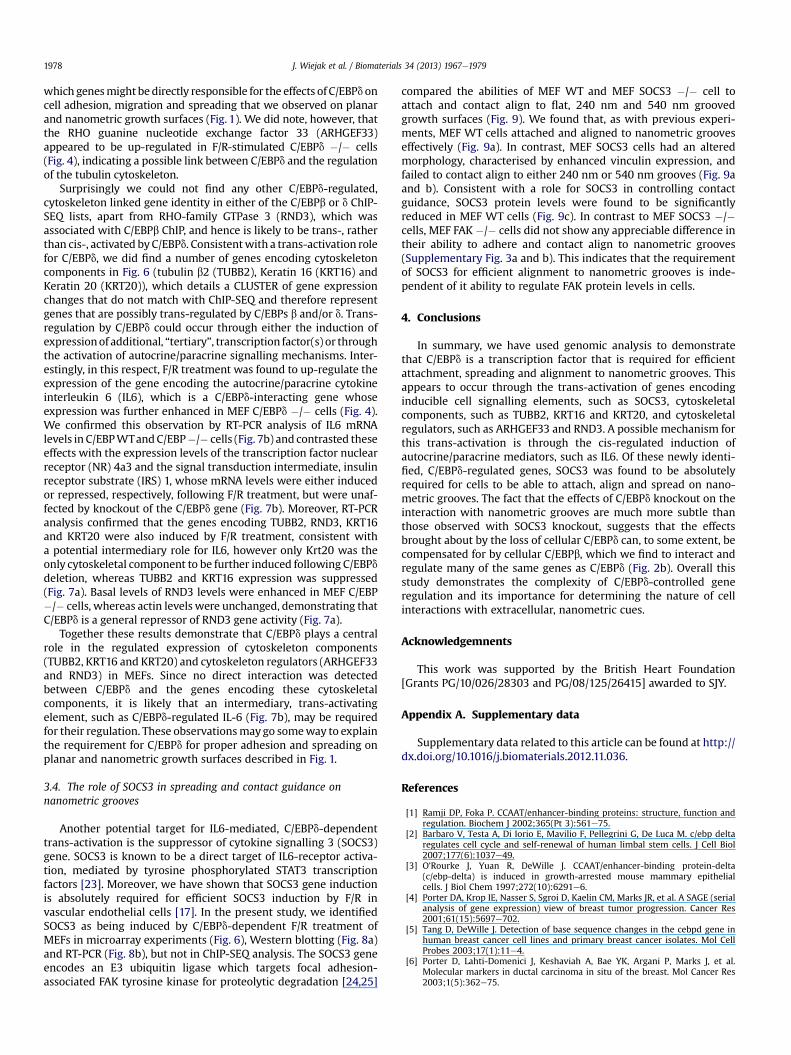
Another potential target for IL6-mediated, C/EBPd-dependenttrans-activation is the suppressor of cytokine signalling 3 (SOCS3)gene. SOCS3 is known to be a direct target of IL6-receptor activa-tion, mediated by tyrosine phosphorylated STAT3 transcriptionfactors [23]. Moreover, we have shown that SOCS3 gene inductionis absolutely required for efficient SOCS3 induction by F/R invascular endothelial cells [17]. In the present study, we identifiedSOCS3 as being induced by C/EBPd-dependent F/R treatment ofMEFs in microarray experiments (Fig. 6), Western blotting (Fig. 8a)and RT-PCR (Fig. 8b), but not in ChIP-SEQ analysis. The SOCS3 geneencodes an E3 ubiquitin ligase which targets focal adhesion-associated FAK tyrosine kinase for proteolytic degradation [24,25]
compared the abilities of MEF WT and MEF SOCS3 �/� cell toattach and contact align to flat, 240 nm and 540 nm groovedgrowth surfaces (Fig. 9). We found that, as with previous experi-ments, MEF WT cells attached and aligned to nanometric grooveseffectively (Fig. 9a). In contrast, MEF SOCS3 cells had an alteredmorphology, characterised by enhanced vinculin expression, andfailed to contact align to either 240 nm or 540 nm grooves (Fig. 9aand b). Consistent with a role for SOCS3 in controlling contactguidance, SOCS3 protein levels were found to be significantlyreduced in MEF WT cells (Fig. 9c). In contrast to MEF SOCS3 �/�cells, MEF FAK �/� cells did not show any appreciable difference intheir ability to adhere and contact align to nanometric grooves(Supplementary Fig. 3a and b). This indicates that the requirementof SOCS3 for efficient alignment to nanometric grooves is inde-pendent of it ability to regulate FAK protein levels in cells.
4. Conclusions
In summary, we have used genomic analysis to demonstratethat C/EBPd is a transcription factor that is required for efficientattachment, spreading and alignment to nanometric grooves. Thisappears to occur through the trans-activation of genes encodinginducible cell signalling elements, such as SOCS3, cytoskeletalcomponents, such as TUBB2, KRT16 and KRT20, and cytoskeletalregulators, such as ARHGEF33 and RND3. A possible mechanism forthis trans-activation is through the cis-regulated induction ofautocrine/paracrine mediators, such as IL6. Of these newly identi-fied, C/EBPd-regulated genes, SOCS3 was found to be absolutelyrequired for cells to be able to attach, align and spread on nano-metric grooves. The fact that the effects of C/EBPd knockout on theinteraction with nanometric grooves are much more subtle thanthose observed with SOCS3 knockout, suggests that the effectsbrought about by the loss of cellular C/EBPd can, to some extent, becompensated for by cellular C/EBPb, which we find to interact andregulate many of the same genes as C/EBPd (Fig. 2b). Overall thisstudy demonstrates the complexity of C/EBPd-controlled generegulation and its importance for determining the nature of cellinteractions with extracellular, nanometric cues.
Acknowledgemnents
This work was supported by the British Heart Foundation[Grants PG/10/026/28303 and PG/08/125/26415] awarded to SJY.
Appendix A. Supplementary data
Supplementary data related to this article can be found at http://dx.doi.org/10.1016/j.biomaterials.2012.11.036.
References
[1] Ramji DP, Foka P. CCAAT/enhancer-binding proteins: structure, function andregulation. Biochem J 2002;365(Pt 3):561e75.
[2] Barbaro V, Testa A, Di Iorio E, Mavilio F, Pellegrini G, De Luca M. c/ebp deltaregulates cell cycle and self-renewal of human limbal stem cells. J Cell Biol2007;177(6):1037e49.
[3] O’Rourke J, Yuan R, DeWille J. CCAAT/enhancer-binding protein-delta(c/ebp-delta) is induced in growth-arrested mouse mammary epithelialcells. J Biol Chem 1997;272(10):6291e6.
[4] Porter DA, Krop IE, Nasser S, Sgroi D, Kaelin CM, Marks JR, et al. A SAGE (serialanalysis of gene expression) view of breast tumor progression. Cancer Res2001;61(15):5697e702.
[5] Tang D, DeWille J. Detection of base sequence changes in the cebpd gene inhuman breast cancer cell lines and primary breast cancer isolates. Mol CellProbes 2003;17(1):11e4.
[6] Porter D, Lahti-Domenici J, Keshaviah A, Bae YK, Argani P, Marks J, et al.Molecular markers in ductal carcinoma in situ of the breast. Mol Cancer Res2003;1(5):362e75.

J. Wiejak et al. / Biomaterials 34 (2013) 1967e1979 1979
[7] Agrawal S, Hofmann WK, Tidow N, Ehrich M, van den Boom D, Koschmieder S,et al. The c/ebp delta tumor suppressor is silenced by hypermethylation inacute myeloid leukemia. Blood 2007;109(9):3895e905.
[8] Zhang Y, Liu T, Yan P, Huang T, Dewille J. Identification and characterization ofCCAAT/enhancer binding proteindelta (c/ebp delta) target genes in G0 growtharrested mammary epithelial cells. BMC Mol Biol 2008;9:83.
[9] Gigliotti AP, Johnson PF, Sterneck E, DeWille JW. Nulliparous CCAAT/enhancerbinding proteindelta (c/ebp delta) knockout mice exhibit mammary glandductal hyperlasia. Exp Biol Med (Maywood) 2003;228(3):278e85.
[10] CaoZ,UmekRM,McKnightSL.Regulatedexpressionof threec/ebp isoformsduringadipose conversion of 3T3-L1 cells. Genes Dev 1991;5(9):1538e52.
[11] Farmer SR. Transcriptional control of adipocyte formation. Cell Metab 2006;4(4):263e73.
[12] Zhang Y, Sif S, DeWille J. The mouse c/ebpdelta gene promoter is regulated bystat3 and sp1 transcriptional activators, chromatin remodeling and c-mycrepression. J Cell Biochem 2007;102(5):1256e70.
[13] Hutt JA, O’Rourke JP, DeWille J. Signal transducer and activator oftranscription 3 activates CCAAT enhancer-binding protein delta genetranscription in G0 growth-arrested mouse mammary epithelial cells and ininvoluting mouse mammary gland. J Biol Chem 2000;275(37):29123e31.
[14] Li B, Si J, DeWille JW. Ultraviolet radiation (uvr) activates p38 map kinaseandinducespost-transcriptional stabilizationof thec/ebpdeltamRNAinG0growtharrestedmammary epithelial cells. J Cell Biochem 2008;103(5):1657e69.
[15] Zhou S, Dewille JW. Proteasome-mediated CCAAT/enhancer-binding proteindelta (C/EBPdelta) degradation is ubiquitin-independent. Biochem J 2007;405(2):341e9.
[16] Zhou S, Si J, Liu T, DeWille JW. PIASy represses CCAAT/enhancer-bindingprotein delta (c/ebp delta) transcriptional activity by sequestering c/ebpdelta to the nuclear periphery. J Biol Chem 2008;283(29):20137e48.
[17] Yarwood SJ, Borland G, Sands WA, Palmer TM. Identification of CCAAT/enhancer-binding proteins as exchange protein activated by camp-activatedtranscription factors that mediate the induction of the socs-3 gene. J BiolChem 2008;283(11):6843e53.
[18] Eisen MB, Spellman PT, Brown PO, Botstein D. Cluster analysis and display ofgenome-wide expression patterns. Proc Natl Acad Sci U S A 1998;95(25):14863e8.
[19] Langmead B, Trapnell C, Pop M, Salzberg SL. Ultrafast and memory-efficientalignment of short DNA sequences to the human genome. Genome Biol2009;10(3):R25.
[20] Li H, Handsaker B, Wysoker A, Fennell T, Ruan J, Homer N, et al. The sequencealignment/map format and SAMtools. Bioinformatics 2009;25(16):2078e9.
[21] Heinz S, Benner C, Spann N, Bertolino E, Lin YC, Laslo P, et al. Simplecombinations of lineage-determining transcription factors prime cis-regulatory elements required for macrophage and B cell identities. Mol Cell2010;38(4):576e89.
[22] Yu X, Si J, Zhang Y, Dewille JW. CCAAT/enhancer binding protein-delta(c/ebp-delta) regulates cell growth, migration and differentiation. Cancer2010;10:48.
[23] Lesina M, Kurkowski MU, Ludes K, Rose-John S, Treiber M, Kloppel G, et al.Stat3/socs3 activation by IL-6 transsignaling promotes progression ofpancreatic intraepithelial neoplasia and development of pancreatic cancer.Cancer 2011;19(4):456e69.
[24] Liu E, Cote JF, Vuori K. Negative regulation of fak signaling by socs proteins.Embo J 2003;22(19):5036e46.
[25] Niwa Y, Kanda H, Shikauchi Y, Saiura A, Matsubara K, Kitagawa T, et al.Methylation silencing of socs-3 promotes cell growth and migration byenhancing jak/stat and fak signaling in human hepatocellular carcinoma.Oncogene 2005;24(42):6406e17.
Copyright © 2022 FDOKUMEN


![4* • Pharingeal grooves/cleft : 4 • [Pharyngeal membrane]](https://static.fdokumen.com/doc/165x107/6334ea00b9085e0bf5093ec7/4-pharingeal-groovescleft-4-pharyngeal-membrane.jpg)