Detection of genomic and intermediate replicative strands of ...
10
Detection of genomic and intermediate replicative strands of hepatitis C virus in liver tissue by in situ hybridization. K T Nouri Aria, … , A L Eddleston, R Williams J Clin Invest. 1993;91(5):2226-2234. https://doi.org/10.1172/JCI116449. Nonisotopic in situ hybridization using a digoxigenin-labeled cDNA probe to the 3' nonstructural region (NS5) of hepatitis C virus (HCV) was performed on liver tissue from 33 patients. The results were compared with PCR detection of HCV RNA performed on 24 of the biopsies. Nonisotopic in situ hybridization correlated well with PCR findings. Hybridization signals were detected, within the cytoplasm and nuclei/nucleoli of hepatocytes, mononuclear, and biliary epithelial cells. In patients with clinically and histologically defined chronic active hepatitis related to active HCV infection, HCV genome was frequently detected in biliary epithelium and correlated well with biliary damage, an otherwise uncommon finding in chronic active hepatitis due to other hepatotropic viruses. Further studies using sense and antisense probes synthesized from the 5' non-coding region of the HCV genome confirmed the localization of positive strand of HCV in the above cell populations. The replicative intermediate strand was also present in all cells, although less frequently observed, apart from biliary epithelium, where negative strand of HCV was undetectable. The findings of HCV genome in liver biopsies of two patients with no significant histological abnormalities may suggest that the damage seen in chronic HCV infection is immune mediated, although the cytopathic effect of the virus may also be important. Research Article Find the latest version: https://jci.me/116449/pdf
-
Upload
khangminh22 -
Category
Documents
-
view
2 -
download
0
Transcript of Detection of genomic and intermediate replicative strands of ...

Detection of genomic and intermediate replicative strands ofhepatitis C virus in liver tissue by in situ hybridization.
K T Nouri Aria, … , A L Eddleston, R Williams
J Clin Invest. 1993;91(5):2226-2234. https://doi.org/10.1172/JCI116449.
Nonisotopic in situ hybridization using a digoxigenin-labeled cDNA probe to the 3' nonstructural region (NS5) of hepatitisC virus (HCV) was performed on liver tissue from 33 patients. The results were compared with PCR detection of HCVRNA performed on 24 of the biopsies. Nonisotopic in situ hybridization correlated well with PCR findings. Hybridizationsignals were detected, within the cytoplasm and nuclei/nucleoli of hepatocytes, mononuclear, and biliary epithelial cells. Inpatients with clinically and histologically defined chronic active hepatitis related to active HCV infection, HCV genome wasfrequently detected in biliary epithelium and correlated well with biliary damage, an otherwise uncommon finding inchronic active hepatitis due to other hepatotropic viruses. Further studies using sense and antisense probes synthesizedfrom the 5' non-coding region of the HCV genome confirmed the localization of positive strand of HCV in the above cellpopulations. The replicative intermediate strand was also present in all cells, although less frequently observed, apartfrom biliary epithelium, where negative strand of HCV was undetectable. The findings of HCV genome in liver biopsies oftwo patients with no significant histological abnormalities may suggest that the damage seen in chronic HCV infection isimmune mediated, although the cytopathic effect of the virus may also be important.
Research Article
Find the latest version:
https://jci.me/116449/pdf
Detection of Genomic and Intermediate Replicative Strandsof Hepatitis C Virus in Liver Tissue by In Situ HybridizationKayhan T. Nouri Aria, Richard Sallie, David Sangar, Graeme J. M. Alexander, Heather Smith,Janice Byrne, * Bernard Portmann, Adrian L. W. F. Eddleston, and Roger WilliamsInstitute of Liver Studies, King's College School of Medicine and Dentistry, LondonSE5 9PJ; and *The Wellcome Foundation, Beckenham, United Kingdom
Abstract
Nonisotopic in situ hybridization using a digoxigenin-labeledcDNAprobe to the 3' nonstructural region (NS5) of hepatitis Cvirus (HCV) was performed on liver tissue from 33 patients.The results were compared with PCRdetection of HCVRNAperformed on 24 of the biopsies. Nonisotopic in situ hybridiza-tion correlated well with PCRfindings. Hybridization signalswere detected, within the cytoplasm and nuclei/ nucleoli of he-patocytes, mononuclear, and biliary epithelial cells. In patientswith clinically and histologically defined chronic active hepati-tis related to active HCV infection, HCVgenome was fre-quently detected in biliary epithelium and correlated well withbiliary damage, an otherwise uncommon finding in chronic ac-tive hepatitis due to other hepatotropic viruses. Further studiesusing sense and antisense probes synthesized from the 5' non-coding region of the HCVgenome confirmed the localization ofpositive strand of HCVin the above cell populations. The repli-cative intermediate strand was also present in all cells, al-though less frequently observed, apart from biliary epithelium,where negative strand of HCVwas undetectable. The findingsof HCVgenome in liver biopsies of two patients with no signifi-cant histological abnormalities may suggest that the damageseen in chronic HCVinfection is immune mediated, althoughthe cytopathic effect of the virus may also be important. (J.Clin. Invest. 1993. 91:2226-2234.) Key words: In situ hybrid-ization * polymerase chain reaction * hepatitis C virus * forma-lin-fixed tissue * liver disease
Introduction
Hepatitis C virus (HCV),' the causative agent of the majorityof cases of posttransfusion hepatitis has recently been isolated,cloned, and sequenced ( 1, 2) and is a single stranded RNAvirus of 10 kb in length. Comparative sequencing studies of
Portions of this work were presented at the European Association forthe Study of the Liver meeting, 11 - 14 September 1991, Palma de Mal-lorca, Spain.
Address correspondence to Dr. Roger Williams, The Institute ofLiver Studies, King's College School of Medicine and Dentistry, Bes-semer Road, London SE5 9PJ, United Kingdom.
Receivedfor publication 4 August 1992 and in revisedform 23 De-cember 1992.
1. Abbreviations used in this paper: CAH, chronic active hepatitis;HCV, hepatitis C virus; NANBH, non-A, non-B hepatitis; NISH, non-isotopic in situ hybridization.
the HCVgenome suggest that this virus is distantly related toflaviviruses and pestiviruses (3, 4). Although the availability oftests for antibody (anti-HCV) to nonstructural and structuralantigens of HCVhas greatly advanced knowledge of the epide-miology of non-A, non-B hepatitis (NANBH), a significantproportion of patients with autoimmune liver disease havefalse positive reactivity to anti-HCV (C 100) ( 5 ) and the signifi-cance of a positive anti-HCV test in patients with liver diseaseis presently unknown. Furthermore, antibody testing cannotdetermine whether a patient with anti-HCV has recoveredfrom the infection or is still viremic. During the early phase ofprimary HCVinfection there is a period of several months ofseronegativity in the presence of ongoing viremia during whichtime HCVRNAdetected by PCRis the only evidence of infec-tion (6, 7). Similarly, in the chronic state, the presence of anti-body does not always correlate with the persistence of infectionas defined by detection of HCVRNAin the serum (8). Re-cently there have been a number of reports using PCRand"nested" PCRto detect the viral genome in serum and livertissue (9, 10). However, PCRof HCVRNAextracted fromeither serum or liver tissue cannot provide information regard-ing the site of infection and/or replication of HCV. In thepresent study we have applied nonisotopic in situ hybridization(NISH) to detect the HCVgenome within liver tissue frompatients with a variety of liver disorders and compared the re-sults obtained with nested PCRperformed on different sectionsobtained from the same tissue.
Methods
Patients. 33 formalin-fixed paraffin-embedded liver biopsy specimensfrom patients with both acute and chronic liver disease were studied(Table I); 20 of the 33 patients had chronic liver disease (mean age40±11, 12 male: 8 female), 8 had acute but unresolved hepatitis (meanage 39+10, 4 male: 4 female), while biopsies from 5 other patientsdemonstrated no histological abnormality (mean age 41±25, 2 male: 3female). 16 of the 20 patients with chronic disease had been diagnosedas NANBHafter serological exclusion of hepatitis A, B, Epstein-Barrvirus, and cytomegalovirus in patients with an antecedent history ofblood transfusion (n = 10), drug abuse (n = 3), or origin from a highrisk area (n = 3). Of the 4 remaining cases, 1 had primary biliarycirrhosis, 1 autoimmune chronic active hepatitis (CAH), and 2 hadhepatitis B virus-related CAH. Both patients with autoimmune CAHand primary biliary cirrhosis had received multiple blood transfusions.17 of patients with chronic liver disease were tested for anti-HCV(United Biomedical Inc., Lake Success, NY), and 12 were positive. Ofthe 8 cases with acute unresolved hepatitis, 7 were thought to haveNANBHaccording to the above criteria and one had acute hepatitis B.All 7 with NANBHwere tested for anti-HCV early in the course of theirillness and 2 were positive. 3 of the 5 biopsies showing no significanthistological abnormalities were obtained from patients biopsied duringinvestigation for chronically abnormal liver function tests and the onlyone routinely tested for anti-HCV was negative. 2 samples were takenfrom normal, "cut-down" donor liver obtained at the time of orthoto-
2226 Nouri Aria et al.
J. Clin. Invest.© The American Society for Clinical Investigation, Inc.0021-9738/93/05/2226/09 $2.00Volume 91, May 1993, 2226-2234
Table I. Clinical, Serological, and Histological Data (Mean±SD)of Patients Studied
Chronic liver Acute Normaldisease hepatitis histology
(n = 20) (n = 8) (n = 5)
Age 40±11 39±10 41±25Male/Female 12/8 4/4 2/3Aspartate aminotransferase 87±22 409±643 36±7Alkaline phosphatase 176± 111 201± 108 126±49Bilirubin 52±59 193±215 15±12
pic liver transplantation to act as negative control tissues. Nested HCVPCRperformed on fresh samples of this tissue was negative. 2 addi-tional biopsy specimens, 1 from skin and the second from duodenumwere obtained from HCV negative patients and used as a negativecontrol for NISH.
The cDNAprobe (designated pDM415) used for NISH was 1,700nucleotides in length and homologous to nucleotides 6,997-8,764 ofthe 3' end of the HCVgenome NS5 (JG2). (Byrne, J., B. Rodgers, andD. Parker, manuscript in preparation). This probe was constructed byinsertion of a PCR fragment into pUC13 plasmid and cloning beforeexcision of the insert and gel purification by electroelution. The probewas digoxigenin labeled by random priming (multi-prime kit; Boeh-ringer Mannheim GmbH, Mannheim, Germany). Briefly, 500 ng ofthe probe was denatured and mixed with hexanucleotide, dNTP digox-igenin labeling mixture, and Klenow enzyme, and incubated for 20 h at370C before purification of the probe by ethanol precipitation.
Sense and antisense RNAprobes were used to detect the replicativeintermediary and positive strands of HCVgenome respectively. TwoRNAprobes were constructed in the following way: a PCR fragmentcorresponding to the 5' noncoding region of the HCVgenome wasinserted into Bluescript plasmid (Stratagene, Cambridge, UK). TheDNAwas linearized using the restriction enzymes BamHI (to preparenegative strand HCVRNA nucleotides 1-390) or NheI (to preparepositive strand HCVRNAnucleotides 1-249). After extraction withphenol/chloroform and ethanol precipitation, the probes were labeledwith digoxigenin (DIG-UTP; Boehringer Mannheim) according to themanufacturer's instruction. Briefly, 1 Mig of linearized DNAwas addedto NTP labeling mixture, DTT, transcription buffer, and either SP6 orT7 RNApolymerase and RNase inhibitor. After incubation for 2 h at37°C the template DNAwas digested with RNase-free DNase andprobes were purified by ethanol precipitation.
The sensitivity of detection using this digoxigenin-labeled probeand the digoxigenin detection system was determined by serial dilu-tions of target sequence fixed to nylon membranes via a dot-blot mani-fold; the target sequence was detectable in the range of 1-5 pg, corre-sponding to 50-250 copies of the cloned HCVcDNA.
Expression ofHCVgenome into insect cells. The NS5 or 5' noncod-ing regions of HCVgenome were inserted into a baculovirus vector andinsect cells (Spodoptera frugipedra, BCH7and BCH10, respectively)were infected with recombinant viruses ( 11 ) and used as positive con-trols. Uninfected cells and insect cells infected with wild-type baculo-virus were used as negative controls. These cells were cultured in Tc100 fluid (Flow Laboratories, Irvine, Scotland) for 72 h at room tem-perature, then fixed in formalin and processed in a similar fashion toliver biopsies.
In situ hybridization. In situ hybridization was performed as previ-ously described ( 12). Briefly, after dewaxing and rehydration, the sec-tions were treated with 0.2 MHCl, then digested with 15 Mg/ml pro-teinase K (Sigma, Poole, UK) in 100 mMTris/50 mMEDTApH 8.0for 30 min at 37°C, rinsed with 2 mg/ml glycine/PBS and then refixedin 4%para-formaldehyde/PBS and acetylated with 0.25% acetic anhy-dride in 0.1 Mtriethanolamine (pH 8). The tissue was then prehybri-dized with a mixture containing 50% formamide (Sigma), 10%dextran
sulfate (Sigma), lx Denhardt's solution (Sigma), 150 Mg/ml shearedsalmon sperm DNA(Sigma), 300 mMDTT (Sigma), Tris (0.05 M),EDTA (5 mM), 600 mMNaCI, and 0.1% sodium pyrophosphate.After incubation for 1 h at 370C, 20 Ad solution containing denaturedlabeled cDNA probe (6-10 ng) and 500 Mg/ml transfer RNA(Sigma)in the above hybridization mixture was applied to each section whichwere then covered with siliconized coverslips and incubated for 16-20h at 420C. In situ hybridization for the detection of positive and inter-mediary replicative strands of HCVRNAwas identical to the one de-scribed above with one exception; tissues were incubated at 950C in anoven for 10 min before hybridization with RNAprobes and the hybrid-ization was carried out at 520C. Control slides were treated with ribonu-cleases (RNases) A and T. and deoxyribonucleases (DNase) (Sigma)at 100 g/ml before hybridization. A cDNA probe specific for IFN-'y,
- 900 nucleotides in length, was labeled in a similar fashion and usedas a control. After incubation the slides were rinsed in 4X SSCand thenwashed twice in 50% formamide/4x SSC for 45 min at 370C. Thesections were washed twice for 30 min in 2x SSC, once in 0.2x SSCat650C and once in 0.1 x SSC for 15 min at room temperature. Afterposthybridization washing, digoxigenin-labeled probes were detectedaccording to the manufacturer's instruction (digoxigenin detection kit;Boehringer Mannheim GmbH). Briefly after preincubation of sectionsfor 30 min with 5% normal sheep serum, 0.3% Triton in Tris-bufferedsaline, the sections were incubated with 1:500 dilution of alkaline phos-phatase conjugated antidigoxigenin mouse monoclonal antibody in theabove buffer for 2 h at room temperature. The slides were then washedfor 30 min and color reaction was developed by the addition of sub-strate containing 2.4 mg/ml levamisole (Sigma) and incubated for 16h in the dark.
Nested PCR. RNAand DNAwas extracted from four I O-Mmthickformalin fixed paraffin embedded sections cut from pathologicalblocks made from either wedge or needle liver biopsy using minormodifications of the methods described by Jackson ( 13) as previouslydescribed ( 14). Briefly, the specimen was dewaxed in xylene at 90°Cfor 5-10 min, centrifuged at 12,000 g for 5 min, and xylene carefullyaspirated from the resulting pellet. The remaining xylene was removedfrom the pellet with alcohol. The pellet was then dried before resuspen-sion in 200 Ml digestion buffer containing proteinase K at 100 U/ml,0.5% SDS, 5 mMEDTA, and 10 mMTris, pH 8, and vanadyl ribonu-cleoside complexes at 40 mM, overlaid with three drops of mineral oil(Sigma), and digested at 42°C for between 2 and 3 d, before phenol/chloroform extraction and overnight ethanol precipitation of nucleicacid at -20°C. Purified glycogen (20 ,g; Boehringer Mannheim) wasadded to facilitate precipitation. Nucleic acid/glycogen was pelleted at15,000 g for 15 min, washed in 70% alcohol, and resuspended in 20 Mlwater. 2 Ml of the resulting RNA/DNAsolution was then added to areverse transcription mixture containing 5 U AMVreverse transcrip-tase (Promega, Southampton, UK), 1 mMDTT, 2.5 mMdNTPs(Pharmacia, Milton Keynes, UK), 2 Ml of 10 x reverse transcriptionbuffer containing 500 mMKCl, 200 mMTris, pH 8.0, and 50 ngrandom hexamers in a total vol of 20 Ml which was incubated at 42°Cfor 1 h. 2 Ml of the resulting DNA/cDNA solution was then added tothe first round PCRmix containing 60 ng of each the "outer" primers,0.2 mMdNTPs, 2.5 Ml of 10 x Taq buffer, 1.0 units of Taq polymerase(Biotech, Perth, Western Australia) in a total reaction vol of 50 ,l. Thereaction mixture was overlaid with three drops of oil before being sub-ject to 30 rounds of amplification on a thermal cycler (Perkin-ElmerCetus, Norwalk, CT) using the following cycling parameters (file 4):94°C for 1 min, 55°C for 1 min, and 72°C for 1 min before a finalextension step of 72°C for 7 min. 2 Ml of the resulting product was thentaken into a second round mixture which was identical to the firstexcept that 120 ng of the inner primers were used. 10 Ml of the resultingproduct was then electrophoresed on agarose gels and visualized usingethidium bromide staining under ultraviolet light.
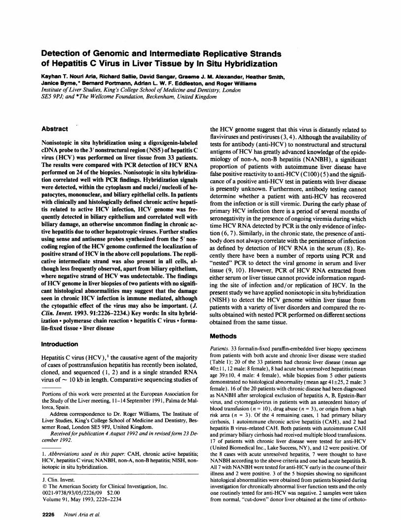
The specificity of the PCRproducts was confirmed by Southernblotting using a probe internal to and not inclusive of the sequences ofthe internal primers (Fig. 1). Normal human liver taken from a cut-down donor organ was used as a negative control for liver tissue, while
Hepatitis C Virus Genomic and Antigenomic RNA in Liver Tissue 2227
1 2 3 4 5 6 7 8 9 10 11
Figure 1. Agarose gel electrophoresis (3% NuSieve GTG[ FMCCorp.,Rockland, ME] 1% agarose) and accompanying Southern blot ofnested PCRproducts amplified as part of these experiments. (LanesI and 11) 123-bp molecular weight marker; (lane 2) negative (re-agent) control; (lane 3) positive control (HCV, tissue); (lane 4) pos-itive control (albumin); (lanes 5 and 6) sample positive for albumin(lane 5) but negative for HCV(lane 6); (lanes 7-10) positive tissuesamples. (Lanes 7 and 9) Positive for albumin; (lanes 8 and 10) pos-itive for HCV. Arrow at level of HCVamplicon ('- 126 bp).
tissue known to be positive for HCVRNAby nested amplificationfrom fresh tissue acted as a positive control. Using this protocol, HCVgenome could reliably be detected in formalin-fixed tissues, but wasabout five times less sensitive than when applied to fresh tissue ( 14). Asan internal control for tissue digestion, extraction, reverse transcrip-tion, and amplification primers directed at albumin mRNAwere de-signed based on the nucleotide sequence described by Minghetti et al.( 15 ). These primers flank a large exon and produce a DNAband of 822bp and an RNAband of 273 bp, and initially were a generous gift of Dr.Kenneth Hillan, Royal Western Infirmary, Glasgow. The cycling pa-rameters of the albumin primers were 940C 10 s, 550C 10 s, 720C 1min. For HCVamplification, primers described by Ulrich (16) were
Table II. Sequence and Position of Primers Usedfor PCRin This Work
ProductPrimer Position* Sequences size
HCVJR12 Sense 5'-GGCGACACTCCACCATAGAT-3' 216 bpnt 1-20
HCVJR 19 Antisense 5'-CGCCCAAATC TCCAGGCATT-3'nt 197-216
HCVJR13 Sense 5'GAACTACTGT CTTCACGCA-3' 126 bpnt 35-53
HCVJR14 Antisense 5'-GGCAATTCCG GTGTACTCACC-3'nt 140-161
Probe 115-135 5'-GAGAGCCATAGTGGTCTGCG-3'
Albumin Sense 5'-TGAAATGGCT GACTGCTGTG-3'Albumin Antisense 5'GCAGCTTTAT CAGCAGCTTG-3'
* HCVprimers numbered according to Okamoto et al. (3). nt, nu-cleotide.
used. The sequences of all primers and the HCVprobe are given inTable II. As well as negative and positive tissue controls, negative "con-tamination" controls were run with each experiment and to minimizeproblems of contamination the general measures suggested by Kwokand Higuchi ( 17) were adhered to. To minimize nonspecific amplifica-tions, all PCRreactions were started "hot" by preliminary incubationof the reaction at 85°C before addition of Taq polymerase (18). Inaddition, to avoid cross contamination from the microtome blade, sec-tions were cut using a fresh disposable blade for each sample and sepa-rate duplicate samples were cut on different days. Both cDNAprepara-tion and HCVamplification were carried out in duplicate and duringdifferent days. PCRassays were carried out "blind" with respect to thein situ hybridization and serology results.
Results
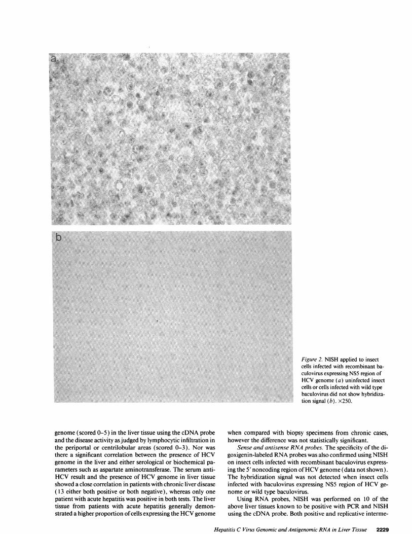
In situ hybridizationcDNAprobes. The specificity of the digoxigenin-labeled probe(pDM 415) was confirmed using insect cells infected with re-combinant baculovirus containing NS5 region of HCVge-nome. The hybridization signal was only detected in insectcells infected with recombinant baculovirus. Uninfected insectcells or cells infected with the wild type of baculovirus did notshow hybridization. (Fig. 2, a and b).
In our original experiments, the nuclear HCVstaining wasmore intense than the cytoplasmic signal ( 19), which was oftenmasked by counterstaining. In subsequent work, performedwithout counterstaining, the cytoplasmic signal became moreprominent, and the cytoplasmic signal was seen in a greaterproportion of cells. When NISH was performed on humantissue using pDM415 as probe, the HCVgenome, when pres-ent was found in both the cytoplasm and nuclei (in some casesalso in nucleoli) of hepatocytes and mononuclear cells, but alsoin bile duct epithelial and sinusoidal cells (Fig. 3, a-d). Hepati-tis C virus genome was detected by NISH in 14 of 20 patientswith chronic liver disease, 7 of 8 patients with acute hepatitis,and in both patients with chronically abnormal liver functiondespite normal histology (Tables III and IV). The HCVge-nome was not detected in either of the two cut-down donorlivers or the two other nonliver tissues (skin and duodenalbiopsies). The proportion of cells positive for HCVgenome inliver tissues generally varied from 1 to 5%. However, in oneexceptional patient, with autoimmune chronic active hepatitison long-term treatment with corticosteroids, a positive NISHsignal was seen in - 20%of hepatocytes. Two patients who didnot have NANBHas the primary diagnosis had HCVgenomedemonstrated in their liver by NISH; one patient had autoim-mune CAHand one primary biliary cirrhosis; both were HCVRNApositive by reverse transcription PCRand had been multi-ply transfused as a consequence of their disease. HCVgenomewas not detected in the two HBV-related liver biopsies testedusing either PCRor in situ hybridization.
RNase treatment of liver biopsies before hybridization re-duced the HCVgenome signals in the tissues by - 75-80%.Although DNase treatment also reduced the proportion of cellsexpressing HCVgenome in the nuclei, the reduction was negli-gible and was probably the result of minor RNase contamina-tion. The cDNA IFN-,y probe was used as a control probe for23 of the liver biopsies and a positive signal was found in only 4of 23 liver biopsies tested and was confined to mononuclearcells infiltrated into the portal areas.
When the results were analyzed as a whole, there was nocorrelation between the intensity of expression of the HCV
2228 Nouri Aria et al.
.2s~~~~~~~~~~~~~~~~~~~~~~~~..1il
O i,.}e~~~~~~~~-.,4. ..
..̂ j
'I
4
a
INN
'.44.
A*,
* r4-'
b
Figure 2. NISH applied to insectcells infected with recombinant ba-culovirus expressing NS5 region ofHCVgenome (a) uninfected insectcells or cells infected with wild typebaculovirus did not show hybridiza-tion signal (b). x250.
genome (scored 0-5) in the liver tissue using the cDNAprobeand the disease activity as judged by lymphocytic infiltration inthe periportal or centrilobular areas (scored 0-3). Nor wasthere a significant correlation between the presence of HCVgenome in the liver and either serological or biochemical pa-rameters such as aspartate aminotransferase. The serum anti-HCVresult and the presence of HCVgenome in liver tissueshowed a close correlation in patients with chronic liver disease( 13 either both positive or both negative), whereas only onepatient with acute hepatitis was positive in both tests. The livertissue from patients with acute hepatitis generally demon-strated a higher proportion of cells expressing the HCVgenome
when compared with biopsy specimens from chronic cases,however the difference was not statistically significant.
Sense and antisense RNAprobes. The specificity of the di-goxigenin-labeled RNAprobes was also confirmed using NISHon insect cells infected with recombinant baculovirus express-ing the 5' noncoding region of HCVgenome (data not shown).The hybridization signal was not detected when insect cellsinfected with baculovirus expressing NS5 region of HCVge-nome or wild type baculovirus.
Using RNA probes, NISH was performed on 10 of theabove liver tissues known to be positive with PCRand NISHusing the cDNAprobe. Both positive and replicative interme-
Hepatitis C Virus Genomic and Antigenomic RNA in Liver Tissue 2229
./ 3.
'O
.i II.
w JWX ,.

w> iE ; . -i
4> *i. 4, Ab1 .Xr4,~~~ ~ ~ ~ ~ ~ ~~C
I;T
:4:,
_ ; >A'A
IAL~~~~~~~~~~~~~~~~.~ ~ ~ .
44 ~ ~ ~ "
.4 S~ ~ ~~~~~~~.3
.4 4.~~~~~~~~~~~~~~~~~~~~~~4
*.r *. r y
4454 A ~ ~ ~ ~ ~ ~ ~ ~ 'R
I
t.
:- 4
.k
4 .0I.M .
.. I0
t:
.4 4*44,
frt
.4~~~~~~~~~~~~~~~~.
V4 A4
.;=r ....
..r.4
it_i .t ,0,#r > X
-̂ ,ro s -
I- 4&.
,44.
~ r .*A
Io
St..
. 9
o ia
W
4* . - 444
A*
s,:+ , . 4*:8 : t '. ': + i
A
.'..
A40
'U
.5'
4)
t
4)
0,4)
C.0
C-0
4)
C)0s0
U?4)
_C_00
0.
*'S0
**W0C-.N
S-
* G0
4)
Co
0_
'0
.:. n
:
* '0;-4)0t
2230 Nouri Aria et al
.0
401.
a.
V
ll.IF 71,
w
p
I
i
V., O4
.1- 4
4L
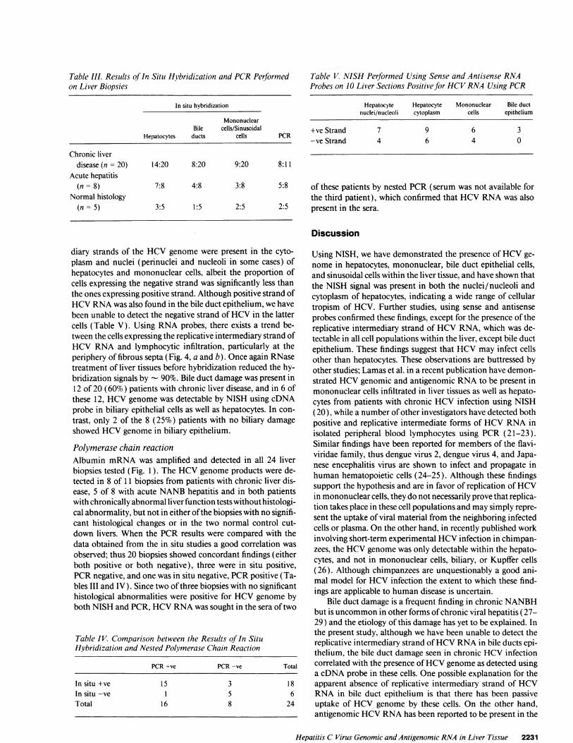
Table III. Results of In Situ Hybridization and PCRPerformedon Liver Biopsies
In situ hybridization
MononuclearBile cells/Sinusoidal
Hepatocytes ducts cells PCR
Chronic liverdisease(n= 20) 14:20 8:20 9:20 8:11
Acute hepatitis(n = 8) 7:8 4:8 3:8 5:8
Normal histology(n = 5) 3:5 1:5 2:5 2:5
Table V. NISH Performed Using Sense and Antisense RNAProbes on 10 Liver Sections Positive for HCVRNAUsing PCR
Hepatocyte Hepatocyte Mononuclear Bile ductnuclei/nucleoli cytoplasm cells epithelium
+ve Strand 7 9 6 3-ve Strand 4 6 4 0
of these patients by nested PCR(serum was not available forthe third patient), which confirmed that HCVRNAwas alsopresent in the sera.
Discussion
diary strands of the HCVgenome were present in the cyto-plasm and nuclei (perinuclei and nucleoli in some cases) ofhepatocytes and mononuclear cells, albeit the proportion ofcells expressing the negative strand was significantly less thanthe ones expressing positive strand. Although positive strand ofHCVRNAwas also found in the bile duct epithelium, we havebeen unable to detect the negative strand of HCVin the lattercells (Table V). Using RNAprobes, there exists a trend be-tween the cells expressing the replicative intermediary strand ofHCVRNAand lymphocytic infiltration, particularly at theperiphery of fibrous septa (Fig. 4, a and b). Once again RNasetreatment of liver tissues before hybridization reduced the hy-bridization signals by - 90%. Bile duct damage was present in12 of 20 (60%) patients with chronic liver disease, and in 6 ofthese 12, HCVgenome was detectable by NISH using cDNAprobe in biliary epithelial cells as well as hepatocytes. In con-trast, only 2 of the 8 (25%) patients with no biliary damageshowed HCVgenome in biliary epithelium.
Polymerase chain reactionAlbumin mRNAwas amplified and detected in all 24 liverbiopsies tested (Fig. 1). The HCVgenome products were de-tected in 8 of 11 biopsies from patients with chronic liver dis-ease, 5 of 8 with acute NANBhepatitis and in both patientswith chronically abnormal liver function tests without histologi-cal abnormality, but not in either of the biopsies with no signifi-cant histological changes or in the two normal control cut-down livers. When the PCRresults were compared with thedata obtained from the in situ studies a good correlation wasobserved; thus 20 biopsies showed concordant findings (eitherboth positive or both negative), three were in situ positive,PCRnegative, and one was in situ negative, PCRpositive (Ta-bles III and IV). Since two of three biopsies with no significanthistological abnormalities were positive for HCVgenome byboth NISH and PCR, HCVRNAwas sought in the sera of two
Table IV. Comparison between the Results of In SituHybridization and Nested Polymerase Chain Reaction
PCR+ve PCR-ve Total
In situ +ve 15 3 18In situ -ve 1 5 6Total 16 8 24
Using NISH, we have demonstrated the presence of HCVge-nome in hepatocytes, mononuclear, bile duct epithelial cells,and sinusoidal cells within the liver tissue, and have shown thatthe NISH signal was present in both the nuclei/nucleoli andcytoplasm of hepatocytes, indicating a wide range of cellulartropism of HCV. Further studies, using sense and antisenseprobes confirmed these findings, except for the presence of thereplicative intermediary strand of HCVRNA, which was de-tectable in all cell populations within the liver, except bile ductepithelium. These findings suggest that HCVmay infect cellsother than hepatocytes. These observations are buttressed byother studies; Lamas et al. in a recent publication have demon-strated HCVgenomic and antigenomic RNAto be present inmononuclear cells infiltrated in liver tissues as well as hepato-cytes from patients with chronic HCV infection using NISH(20), while a number of other investigators have detected bothpositive and replicative intermediate forms of HCVRNAinisolated peripheral blood lymphocytes using PCR (21-23).Similar findings have been reported for members of the flavi-viridae family, thus dengue virus 2, dengue virus 4, and Japa-nese encephalitis virus are shown to infect and propagate inhuman hematopoietic cells (24-25). Although these findingssupport the hypothesis and are in favor of replication of HCVin mononuclear cells, they do not necessarily prove that replica-tion takes place in these cell populations and may simply repre-sent the uptake of viral material from the neighboring infectedcells or plasma. On the other hand, in recently published workinvolving short-term experimental HCVinfection in chimpan-zees, the HCVgenome was only detectable within the hepato-cytes, and not in mononuclear cells, biliary, or Kupffer cells(26). Although chimpanzees are unquestionably a good ani-mal model for HCVinfection the extent to which these find-ings are applicable to human disease is uncertain.
Bile duct damage is a frequent finding in chronic NANBHbut is uncommon in other forms of chronic viral hepatitis (27-29) and the etiology of this damage has yet to be explained. Inthe present study, although we have been unable to detect thereplicative intermediary strand of HCVRNAin bile ducts epi-thelium, the bile duct damage seen in chronic HCVinfectioncorrelated with the presence of HCVgenome as detected usinga cDNAprobe in these cells. One possible explanation for theapparent absence of replicative intermediary strand of HCVRNA in bile duct epithelium is that there has been passiveuptake of HCVgenome by these cells. On the other hand,antigenomic HCVRNAhas been reported to be present in the
Hepatitis C Virus Genomic and Antigenomic RNA in Liver Tissue 2231
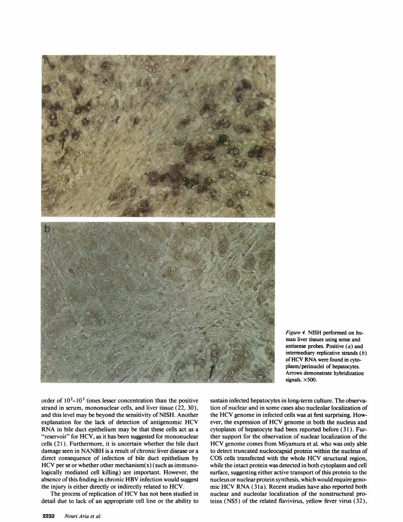
Figure 4. NISH performed on hu-man liver tissues using sense andantisense probes. Positive (a) andintermediary replicative strands (b)of HCVRNAwere found in cyto-plasm/perinuclei of hepatocytes.Arrows demonstrate hybridizationsignals. X500.
order of 102-10i times lesser concentration than the positivestrand in serum, mononuclear cells, and liver tissue (22, 30),and this level may be beyond the sensitivity of NISH. Anotherexplanation for the lack of detection of antigenomic HCVRNAin bile duct epithelium may be that these cells act as a"reservoir" for HCV, as it has been suggested for mononuclearcells (21 ). Furthermore, it is uncertain whether the bile ductdamage seen in NANBHis a result of chronic liver disease or adirect consequence of infection of bile duct epithelium byHCVper se or whether other mechanism(s) (such as immuno-logically mediated cell killing) are important. However, theabsence of this finding in chronic HBVinfection would suggestthe injury is either directly or indirectly related to HCV.
The process of replication of HCVhas not been studied indetail due to lack of an appropriate cell line or the ability to
sustain infected hepatocytes in long-term culture. The observa-tion of nuclear and in some cases also nucleolar localization ofthe HCVgenome in infected cells was at first surprising. How-ever, the expression of HCVgenome in both the nucleus andcytoplasm of hepatocyte had been reported before (31 ). Fur-ther support for the observation of nuclear localization of theHCVgenome comes from Miyamura et al. who was only ableto detect truncated nucleocapsid protein within the nucleus ofCOScells transfected with the whole HCVstructural region,while the intact protein was detected in both cytoplasm and cellsurface, suggesting either active transport of this protein to thenucleus or nuclear protein synthesis, which would require geno-mic HCVRNA(3 la). Recent studies have also reported bothnuclear and nucleolar localization of the nonstructural pro-teins (NS5) of the related flavivirus, yellow fever virus (32),
2232 Nouri Aria et al.
which is also consistent with previous studies showing identifi-cation of other flaviviruses (west Nile virus, yellow fever virus,and Japanese encephalitis virus) antigens (envelope, core, andNSI) within nuclei of infected cells (33-35). Nuclear involve-ment in the replicative stages of these viruses has been postu-lated in these studies. Our observation of both positive and inparticular negative strands of HCVRNA located in nuclei/perinuclei of hepatocytes, in the present study, may furthersupport this hypothesis and suggest that HCVmay use the hostgenome or nuclear apparatus for replication or that parts of theviral particles are stored in the nuclei of infected cells in asimilar fashion to hepatitis B virus. In a recently publishedwork, using short-term experimental HCVinfection in chim-panzees, Negro et al. found exclusively cytoplasmic expressionof HCVin a high proportion of hepatocytes within 22 wk ofinfection, whereas NISH performed subsequently was negativedespite confirmation of viraemia by PCR(26). In contrast, inour chronically infected patients, we found a low proportion ofhepatocytes expressing HCVin both cytoplasm and nucleus.While differences in species may contribute to some of thesediscrepancies, an alternative explanation may be that after anacute phase during which time widespread expression of thegenome within hepatocytes is seen, HCVenters into a latencywhich corresponds to a reduction in serum transaminase levelsand reduced cellular replicative activity, manifested by reducedlevels of detection within hepatocytes.
The discrepancies between NISH and PCRfound in thisstudy might relate to the differences in sensitivity of the twotechniques, as well as to the effect sequence variation has oneach; cases positive by NISH but negative by PCRmight arisethrough minor sequence variation (especially critical at the 3'end of each PCRprimer) which would be insufficient to pre-vent the NISH probe hybridizing. Alternatively, the extensivetissue processing required to permit RNAPCRfrom formalin-fixed tissue may have resulted in some loss of viral genome andfalse negativity; conversely, the PCR-positive, in situ-negativecase may be attributable to low concentration of the viruswithin the tissue which could effect NISH more than PCR.
While it is conceivable that the pDM415 probe used inNISH may have nonspecifically hybridized to some cells result-ing in false positive signals, the absence of hybridization signalafter application of this probe to either normal or HCVnega-tive nonliver tissue (skin and duodenum), and also the lowproportion of positive cells per specimen argues against thispossibility. Furthermore, the specificity of sense or antisenseRNAprobes used for the detection of HCVgenome in thepresent study was confirmed when NISH was performed onvero cells (African green monkey kidney) infected with a flavi-virus, yellow fever virus vaccine (strain 17D) (a generous giftfrom Dr. A. Barrett, Surrey University, UK) and no hybridiza-tion signal was detected (unpublished observations). Similarly,using IFN-y probe, the low proportion of mononuclear cellsexpressing IFN-'y mRNAfurther support the specificity ofNISH signals in our study.
The lack of correlation between the degree of hepatocellu-lar damage as assessed histologically or serologically (e.g., aspar-tate aminotransferase) and the prevalence of HCVgenome inliver tissue as detected by the cDNA probe might suggest thatHCVis not directly cytopathic. This observation is supportedby a recent report (36), where chronic hepatitis was diagnosedhistologically in 9 of 16 individuals with normal levels of ala-nine aminotransferase, who were anti-HCV and HCVRNA
positive using PCR(36). On the other hand, the topographicalrelationship between cells expressing the replicative interme-diary strand of HCVRNAand the severity of liver damage asjudged by lymphocytic infiltration using NISH and RNAprobes may suggest a possible cytopathic effects associated withviral replication, although an additional immune-mediatedliver damage in this condition cannot be excluded. The involve-ment of immune mediated damage in chronic HCVinfectionis supported by studies demonstrating that in patients withNANBHa significant proportion of the lymphocyte destroyingautologous hepatocytes in in vitro cell-mediated cytolysis are Tlymphocytes (37). Furthermore, T cell clones obtained fromthis group of patients showed high cytotoxic activity againsthepatocytes (38). In a recent study Fong et al. (30) reportedthat the severity of liver damage, as assessed histologically orbiochemically, was unrelated to the presence or concentrationof either genomic or antigenomic (replicative intermediary)HCVRNAin either serum or liver tissue. These findings andthat of HCVgenome in healthy carriage of HCV(39) and thefindings of HCVgenome in liver biopsies of two patients withno significant histological abnormalities in the present study,using both in situ hybridization and PCR, all suggest thatchronic liver damage due to HCVinfection is immune me-diated. However, our own findings using RNAprobes showthat direct cytopathic effects of the virus may also be impor-tant.
Acknowledgments
The assistance of Dr. Berywin Clarke and Tony Carroll of The Well-come Research Laboratories, Beckenham, United Kingdom, for tech-nical help, practical advice, and generous access to their laboratory isgratefully acknowledged.
Wethank the HumaneResearch Trust (UK) for generous supportthat has allowed the purchase of some of the equipment and consum-ables used in this research.
References
1. Choo, Q. L., G. Kuo, A. J. Weiner, L. R. Overby, D. W. Bradley, and M.Houghton. 1989. Isolation of a cDNAclone derived from a blood-borne non-A,non-B viral hepatitis genome. Science (Wash. DC). 244:359-362.
2. Kato, N., M. Hijikata, Y. (otsuyama, M. Nakagawa, S. Ohkoshi, T. Sugi-mura, and K. Shimotohno. 1990. Molecular cloning of the human hepatitis Cvirus genome from Japanese patients with non-A, non-B hepatitis. Proc. NatL.Acad. Sci. USA. 87:9524-9528.
3. Okamoto, H., S. Okada, Y. Sugiyama, S. Yotsumoto, T. Tanaka, H. Yoshi-zawa, F. Tsuda, Y. Miyakawa, and M. Mayumi. 1990. The 5'-terminal sequenceof the hepatitis C virus genome. Jpn. J. Exp. Med. 60:167-177.
4. Miller, R. H., and R. H. Purcell. 1990. Hepatitis C virus shares amino acidsequence similarity with pestiviruses and flaviviruses as well as members of twoplant virus supergroups. Proc. NatL. Acad. Sci. USA. 87:2057-2061.
5. McFarlane, I. G., H. M. Smith, P. J. Johnson, G. P. Bray, D. Vergani, andR. Williams. 1990. Hepatitis C virus antibodies in chronic active hepatitis: patho-genetic factor or false-positive result? Lancet. 335:754-757.
6. Farci, P., H. J. Alter, D. Wong, R. H. Miller, J. W. Shih, B. Jett, and R. H.Purcell. 1991. A long-term study of hepatitis C virus replication in non-A, non-Bhepatitis. N. Engl. J. Med. 325:98-104.
7. Shimizu, Y. K., and R. H. Purcell. 1989. Cytoplasmic antigen in hepato-cytes of chimpanzees infected with non-A, non-B, hepatitis virus or hepatitis deltavirus: relationship to interferon. Hepatology. 10:764-768.
8. Shimizu, Y. K., A. J. Weiner, J. Rosenblatt, D. C. Wong, M. Shapiro, T.Popkin, M. Houghton, H. J. Alter, and R. H. Purcell. 1990. Early events inhepatitis C virus infection of chimpanzees. Proc. Nati. Acad. Sci. USA. 1990.87:6441-6444.
9. Weiner, A. J., G. Kuo, D. W. Bradley, F. Bonino, G. Saracco, C. Lee, J.Rosenblatt, Q.-L. Choo, and M. Houghton. 1990. Detection of hepatitis C viralsequences in non-A, non-B hepatitis. Lancet. 335:1-3.
10. Garson, J. A., R. S. Tedder, M. Briggs, P. Tuke, J. A. Glazerbrook, A.Trute, D. Parker, J. A. J. Barbara, M. Contreras, and S. Aloysius. 1990. Detection
Hepatitis C Virus Genomic and Antigenomic RNAin Liver Tissue 2233
of hepatitis C viral sequences in blood donations by "nested" polymerase chainreaction and prediction of infectivity. Lancet. 335:1419-1422.
1 1. Chiba, J., H. Ohba, Y. Matsuura, Y. Watanaba, T. Katayama, S. Kikuchi,1. Saito, and T. Miyamura. 1991. Serodiagnosis of hepatitis Cvirus (HCV) infec-tion with an HCVcore protein molecularly expressed by a recombinant baculo-virus. Proc. Natl. Acad. Sci. USA. 88:4641-4645.
12. Nouri-Aria, K. T., J. Arnold, F. Davison, G. J. M. Alexander, B. Port-mann, A. L. W. F. Eddleston, and R. Williams. 1991. Hepatic interferon alphagene transcripts and the products in acute and chronic HBVrelated liver disease.Hepatology. 13:1029-1034.
13. Jackson, D. P., F. A. Lewis, G. R. Taylor, A. W. Boylson, and P. Quirke.1990. Tissue extraction of DNAand RNAand analysis by the polymerase chainreaction. J. Clin. Pathol. (Lond.). 43:499-504.
14. Sallie, R., A. Rayner, A. L. W. F. Eddleston, B. Portmann, and R. Wil-liams. 1992. Detection of HCVRNAin formalin fixed, paraffin embedded livertissue by nested polymerase chain reaction. J. Med. Virol. 27:310-314.
15. Minghetti, P. P., D. E. Ruffner, W. Kuang, 0. E. Dennison, J. W. Haw-kins, W. G. Beatties, and A. Dugaiczyke. 1986. Molecular structure of the humanalbumin gene is revealed by nucleotide sequence within q 1 1-22 of chromosome4. J. Biol. Chem. 261:6747-6757.
16. Ulrich, P. P., J. M. Romeo, P. K. Lane, I. Kelly, L. J. Daniel, and G. N.Vyas. 1990. Detection, semiquantitation, and genetic variation in hepatitis Cvirus sequences amplified from the plasma of blood donors with elevated alanineaminotransferase. J. Clin. Invest. 86:1609-1614.
17. Kwok, S., and R. Higuchi. 1989. Avoiding false positive with PCR. Nature(Lond.). 339:237-238.
18. Erlich, H. A., D. Gelfand, and J. J. Sninsky. 1991. Recent advances in thepolymerase chain reaction. Science (Wash. DC). 252:1643-1651.
19. Nouri-Aria, K. T., R. Sallie, J. Byrne, G. J. M. Alexander, B. Portmann,A. L. W. F. Eddleston, and R. Williams. 1991. Detection of HCVgenome in theliver tissue using an in situ hybridisation technique. J. Hepatol. 13:S56.
20. Lamas, E., P. Baccarini, C. Housset, D. Kremsdorf, and C. Brechot. 1992.Detection of hepatitis C virus (HCV) RNAsequences in liver tissue by in situhybridisation. J. Hepatol. 16:219-223.
21. Zignego, A. L., D. Macchia, M. Monti, V. Thiers, M. Mazzetti, M. Foschi,E. Maggi, S. Romagnani, P. Gentilini, and C. Brechot. 1992. Infection of periph-eral mononuclear blood cells by hepatitis C virus. J. Hepatol. 15:382-386.
22. Wang, J.-T., J.-C. Sheu, J.-T. Lin, T.-H. Wang, and D. S. Chen. 1992.Detection of replicative form of hepatitis Cvirus RNAin peripheral blood mono-nuclear cells. J. Infect. Dis. 166:1167-1169.
23. Hsieh, T.-T., D.-S. Yao, I.-S. Sheen, Y.-F. Liaw, and C. C. Pao. 1992.Hepatitis C virus in peripheral blood mononuclear cells. Am. J. Clin. Pathol.98:392-396.
24. Hase, T., P. L. Summers, and K. H. Eckels. 1989. Flavivirus entry intocultured mosquito cells and human peripheral blood monocytes. Arch. Virol.104:129-143.
25. Nakao, S., C.-J. Lai, and N. S. Young. 1989. Dengue virus, a flavivirus,propagates in human bone marrow progenitors and hematopoietic cell lines.Blood. 74:1235-1240.
26. Negro, F., D. Pacchioni, Y. Shimizu, R. H. Miller, G. Bussolati, R. H.
Purcell, and F. Bonino. 1992. Detection of intrahepatic replication of hepatitis Cvirus RNAby in situ hybridisation and comparison with histopathology. Proc.Natl. Acad. Sci. USA. 89:2247-2251.
27. Poulson, H., and P. Christoffersen. 1992. Abnormal bile duct epitheliumin chronic aggressive hepatitis and cirrhosis. Hum. Pathol. 3:217-225.
28. Scheuer, P. J., P. Ashrafzadeh, S. Sherlock, D. Brown, and G. M. Du-sheiko. 1992. The pathology of hepatitis C. Hepatology. 15:567-571.
29. Bach, N., S. N. Thung, and F. Schaffner. 1992. The histological features ofchronic hepatitis C and autoimmune chronic hepatitis: a comparative analysis.Hepatology. 15:572-577.
30. Fong, T. L., M. Shindo, S. M. Feinstone, J. H. Hoofnagle, and A. M.DiBiscegli. 1991. Detection of replicative intermediates of hepatitis C viral RNAin liver and serum of patients with chronic hepatitis C. J. Clin. Invest. 88:1058-1060.
31. Negro, F., D. Pacchioni, R. Miller, Y. Shimizu, G. Bussolati, R. H. Pur-cell, and F. Bonino. 1991. Intrahepatic replication of hepatitis C virus in thechimpanzee as detected by in-situ hybridisation. Hepatology. 14:116A.
3 1a.Suzuki, R., A. Ando, S. Harada, Y. Matsura, I. Saito, and T. Miyamura.1991. Analysis of the HCVgenome coding for core protein. The Third Interna-tional Symposium on Viral Hepatitis and Hepatocellular Carcinoma. 5-8 De-cember 1991, Taiwan.
32. Buckely, A., S. Gaidamovitch, A. Turchinskaya, and E. A. Gould. 1992.Monoclonal antibodies identify the NS5 yellow fever virus non-structural proteinin the nuclei of infected cells. J. Gen. Virol. 73:1125-1130.
33. Buckley, A., and E. A. Gould. 1988. Detection of virus specific antigen inthe nuclei or nucleoli of cells infected with Zika or Langat virus. J. Gen. Virol.69:1913-1920.
34. Tadano, M., Y. Makino, F. Fukunaga, Y. Okuno, and K. Fukai. 1989.Detection of dengue 4 virus core protein in the nucleus. I. A monoclonal antibodyto dengue 4 reacts with the antigen in the nucleus and cytoplasm. J. Gen. Virol.70:1409-1415.
35. Gupta, A. K., M. M. Gore, V. J. Lad, and S. N. Ghosh. 1991. Nuclearimmunofluorescence in porcine kidney cells infected with Japanese encephalitisvirus. Acta Virol. (Prague). 35:282-286.
36. Alberti, A., G. Morsica, L. Chemello, D. Cavalletto, F. Noventa, P. Pon-tisso, and A. Ruol. 1992. Hepatitis C viraemia and liver disease in symptom-freeindividuals with anti-HCV. Lancet. 340:697-698.
37. Mondelli, M., A. Alberti, G. Realdi, and E. G. Rondanelli. 1986. In vitroeffect of lymphoblastoid alpha-interferon on subpopulations of effector cells me-diating cytotoxicity for autologous hepatocytes in hepatitis B and non-A, non-B.Int. J. Immunopharmacol. 8:887-891.
38. Imawari, M., M. Nomura, T. Kaieda, T. Moriyama, K. Oshimi, I. Naka-mura, T. Guniji, S. Ohnishi, T. Ishikawa, and H. Nakagama. 1989. Establish-ment of a human T-cell clone cytotoxic for both autologous and allogenic hepato-cytes from chronic hepatitis patients with type non-A, non-B virus. Proc. Nati.Acad. Sci. USA. 86:2883-2887.
39. Takeuchi, K., S. Boonmar, Y. Kubo, T. Katayama, H. Harada, A.Ohbayashi, Q. L. Choo, G. Kuo, M. Houghton, and I. Saito. 1990. Hepatitis Cviral cDNAclones isolated from a healthy carrier donor implicated in post-trans-fusion non-A, non-B hepatitis. Gene (Amst.). 91:287-291.
2234 Nouri Aria et al.


![[INTERMEDIATE 3D MODELING IN TINKERCAD]](https://static.fdokumen.com/doc/165x107/63336b2d4cd921f2410cdab7/intermediate-3d-modeling-in-tinkercad.jpg)