h1-2021-rs-presentation.pdf - Coloplast's Investor Relations site
Weiss et al. Epigenetics & Chromatin 2010, 3:7http://www.epigeneticsandchromatin.com/content/3/1/7
Open AccessR E S E A R C H
ResearchHistone H1 variant-specific lysine methylation by G9a/KMT1C and Glp1/KMT1DThomas Weiss1, Sonja Hergeth1, Ulrike Zeissler1, Annalisa Izzo1, Philipp Tropberger1, Barry M Zee2, Miroslav Dundr3, Benjamin A Garcia2, Sylvain Daujat*1 and Robert Schneider*1
AbstractBackground: The linker histone H1 has a key role in establishing and maintaining higher order chromatin structure and in regulating gene expression. Mammals express up to 11 different H1 variants, with H1.2 and H1.4 being the predominant ones in most somatic cells. Like core histones, H1 has high levels of covalent modifications; however, the full set of modifications and their biological role are largely unknown.
Results: In this study, we used a candidate screen to identify enzymes that methylate H1 and to map their corresponding methylation sites. We found that the histone lysine methyltransferases G9a/KMT1C and Glp1/KMT1D methylate H1.2 in vitro and in vivo, and we mapped this novel site to lysine 187 (H1.2K187) in the C-terminus of H1. This H1.2K187 methylation is variant-specific. The main target for methylation by G9a in H1.2, H1.3, H1.5 and H1.0 is in the C-terminus, whereas H1.4 is preferentially methylated at K26 (H1.4K26me) in the N-terminus. We found that the readout of these marks is different; H1.4K26me can recruit HP1, but H1.2K187me cannot. Likewise, JMJD2D/KDM4 only reverses H1.4K26 methylation, clearly distinguishing these two methylation sites. Further, in contrast to C-terminal H1 phosphorylation, H1.2K187 methylation level is steady throughout the cell cycle.
Conclusions: We have characterised a novel methylation site in the C-terminus of H1 that is the target of G9a/Glp1 both in vitro and in vivo. To our knowledge, this is the first demonstration of variant-specific histone methylation by the same methyltransferases, but with differing downstream readers, thereby supporting the hypothesis of H1 variants having specific functions.
BackgroundIn eukaryotic cells, DNA is packaged into chromatin. Thebuilding block of chromatin is the nucleosomal core par-ticle containing a histone octamer (formed by the his-tones H2A, H2B, H3 and H4) around which 147 bp ofDNA (147 bp) are wrapped [1]. The linker histone H1binds to the DNA between the nucleosomal core parti-cles, and is essential to stabilise higher order chromatinstructures [2].
Human cells possess up to 11 H1 variants, all consistingof a short N-terminal tail, a globular core domain and aC-terminal tail, making up approximately 50% of thewhole protein [3,4]. H1.0 is mainly expressed in termi-nally differentiated cells. H1.1 expression has to date onlybeen reported for a subset of tissues. H1.2 to H1.5 are
expressed in almost all cells, with H1.2 and H1.4 beingthe predominant variants in most somatic cells [5]. Thevariants H1t, H1T2, H1oo and HILS1 are only found ingerm cells [3,4]. Expression of H1x has only been investi-gated in a limited number of cell types [6]. H1 variantswere shown to have a distinct nuclear localisation; forexample, H1.2 seems to localize preferentially to euchro-matic regions, whereas H1.4 is enriched in heterochro-matic regions [7,8]. Whether somatic H1 variants havespecific functions is subject to ongoing research [4]. Sin-gle knockout H1 variants in mice show upregulation ofother H1 variants and relatively mild phenotypes [9].However, knockout of three H1 variants leads to a 50%reduction of overall H1 amount and is embryonicallylethal [10]. This highlights the potential importance ofhistone H1 in maintaining chromatin integrity, and sug-gests two possible functions of H1 variants: a general oneredundant among H1 variants and related to the forma-
* Correspondence: [email protected], [email protected] for Immunobiology, Stübeweg 51, 79108 Freiburg, GermanyFull list of author information is available at the end of the article
BioMed Central© 2010 Weiss et al; licensee BioMed Central Ltd. This is an Open Access article distributed under the terms of the Creative CommonsAttribution License (http://creativecommons.org/licenses/by/2.0), which permits unrestricted use, distribution, and reproduction inany medium, provided the original work is properly cited.
Weiss et al. Epigenetics & Chromatin 2010, 3:7http://www.epigeneticsandchromatin.com/content/3/1/7
Page 2 of 13
tion of higher order chromatin, and an individual, gene-specific one.
Covalent modifications of histones such as lysine meth-ylation are involved in the regulation of all DNA-basedprocesses. Methylation marks are catalysed by histonelysine methyl transferases (HKMTs) using S-adenosylmethionine (SAM) as the methyl group donor. The func-tion of histone lysine methylation depends on the specificsite and the methylation state (mono-, di- or trimethy-lated) [11]. However, not all HKMTs can catalyse thetrimethylation state. G9a/KMT1C and the G9a-like pro-tein 1 (Glp1/KMT1D) were initially described as enzymesthat could mono- and dimethylate H3 on Lys9 (H3K9) ineuchromatic regions, leading to repression of specificgenes [12,13]. Knockout of either of these two enzymes islethal at embryonic day (E9.5). Both knockouts result indrastic reduction of H3K9 mono- and dimethylation,leading to induction of specific genes. Furthermore, het-erochromatin protein 1 (HP1), which binds to H3K9me2/me3, is relocalized in G9a/Glp1-deficient ES cells.Although G9a and Glp1 can independently methylateH3K9 in vitro, they form a heteromeric complex in vivo,explaining their similar phenotypes and the lack ofredundancy [14]. Interestingly, some HKMTs have morethan one target; for example, G9a was reported to methy-late both H3 and H1.4 [15,16].
Most of the methylation marks characterized to date,are located in the N-terminal tails of the core histones H3and H4. Posttranslational modifications of the linker his-tone H1 are less well studied. The best characterised H1modification is phosphorylation [17,18]. This phosphory-lation is cell cycle-dependent, reaching a maximum in Mphase [17], and is mainly catalysed by cdk type kinases[17]. H1.4K26 methylation was the first methylation sitediscovered in H1. This methylation has been reported tobe catalysed by Ezh2 as part of the PRC2 complex andG9a [16,19]. H1.4K26me2 is bound by HP1, leading totranscriptional repression, whereas phosphorylation ofH1.4S27 impairs HP1 binding [20]. Technical improve-ments in mass spectrometry (MS) have lately led to theidentification of new modification sites including methy-lation on various human H1 variants [18], but as yet theirfunctions are unknown.
However, getting complete sequence coverage of H1 inMS analysis is very difficult, and several potential modi-fied sites might be missed during the analysis. We soughtto overcome this problem by using a candidate approachto identify HKMTs with activity on H1 and their targetsites. We report for the first time that G9a and Glp1 areenzymes with activity towards the C-terminal tail of H1both in vitro and in vivo, and we have mapped the methy-lation site on H1.2 to K187. We further show that methy-lation by G9a/Glp1 is H1 variant-specific, as G9a andGlp1 methylate preferentially H1.4K26 and H1.2K187 but
not the corresponding lysines on H1.2 and H1.4, respec-tively. Additionally, these two methylation marks differ intheir readout, as HP1 binds to methylated H1.4K26 butnot to H1.2K187. Similarly, JMJD2D demethylatesH1.4K26me but not H1.2K187me. Interestingly, theH1.2K187 methylation is constant over the cell cycle, incontrast to C-terminal H1 phosphorylation. Our datasuggest that H1 variant-specific functions can beachieved through differential methylation of specific resi-dues in vivo.
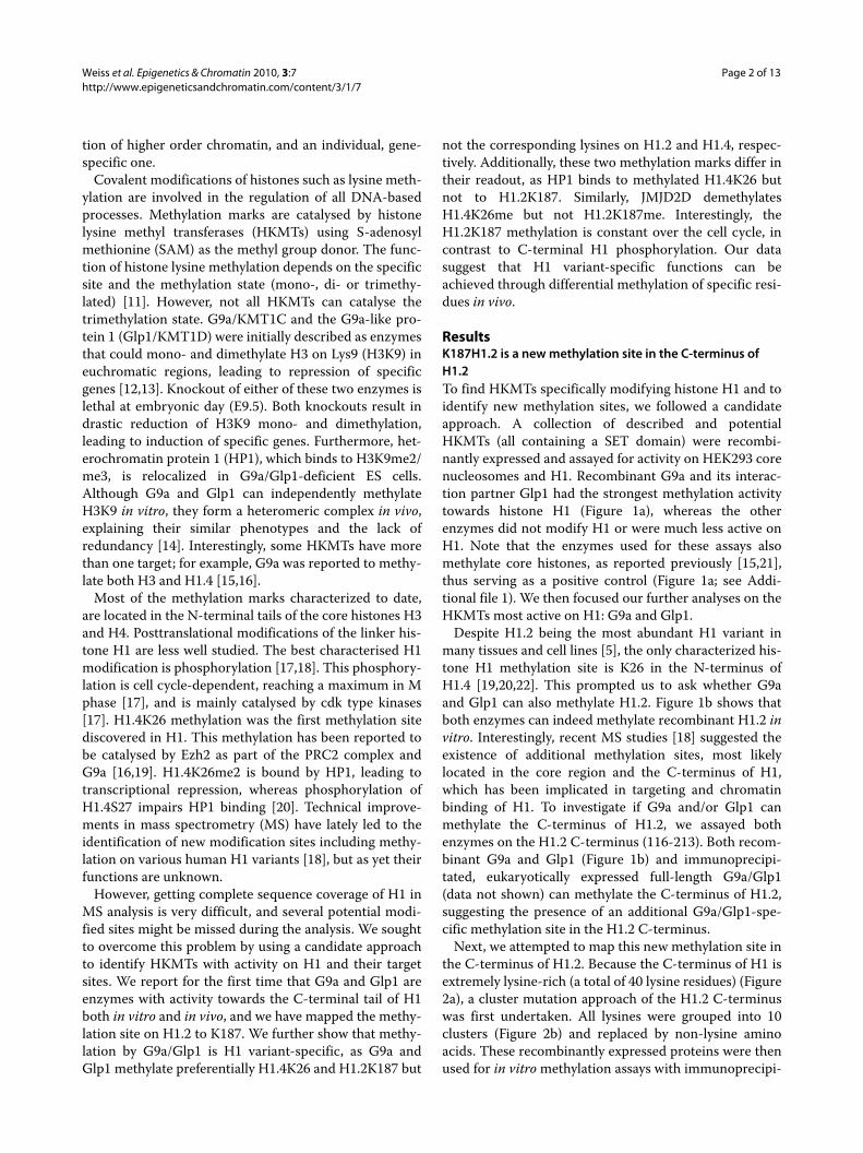
ResultsK187H1.2 is a new methylation site in the C-terminus of H1.2To find HKMTs specifically modifying histone H1 and toidentify new methylation sites, we followed a candidateapproach. A collection of described and potentialHKMTs (all containing a SET domain) were recombi-nantly expressed and assayed for activity on HEK293 corenucleosomes and H1. Recombinant G9a and its interac-tion partner Glp1 had the strongest methylation activitytowards histone H1 (Figure 1a), whereas the otherenzymes did not modify H1 or were much less active onH1. Note that the enzymes used for these assays alsomethylate core histones, as reported previously [15,21],thus serving as a positive control (Figure 1a; see Addi-tional file 1). We then focused our further analyses on theHKMTs most active on H1: G9a and Glp1.
Despite H1.2 being the most abundant H1 variant inmany tissues and cell lines [5], the only characterized his-tone H1 methylation site is K26 in the N-terminus ofH1.4 [19,20,22]. This prompted us to ask whether G9aand Glp1 can also methylate H1.2. Figure 1b shows thatboth enzymes can indeed methylate recombinant H1.2 invitro. Interestingly, recent MS studies [18] suggested theexistence of additional methylation sites, most likelylocated in the core region and the C-terminus of H1,which has been implicated in targeting and chromatinbinding of H1. To investigate if G9a and/or Glp1 canmethylate the C-terminus of H1.2, we assayed bothenzymes on the H1.2 C-terminus (116-213). Both recom-binant G9a and Glp1 (Figure 1b) and immunoprecipi-tated, eukaryotically expressed full-length G9a/Glp1(data not shown) can methylate the C-terminus of H1.2,suggesting the presence of an additional G9a/Glp1-spe-cific methylation site in the H1.2 C-terminus.
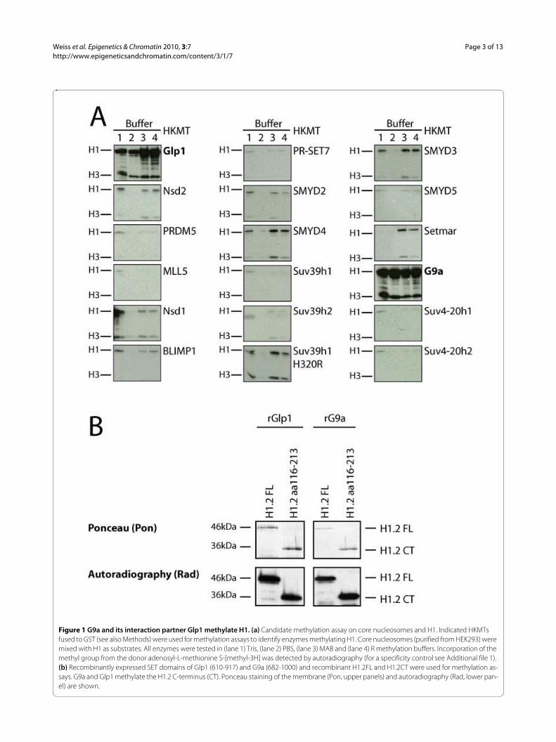
Next, we attempted to map this new methylation site inthe C-terminus of H1.2. Because the C-terminus of H1 isextremely lysine-rich (a total of 40 lysine residues) (Figure2a), a cluster mutation approach of the H1.2 C-terminuswas first undertaken. All lysines were grouped into 10clusters (Figure 2b) and replaced by non-lysine aminoacids. These recombinantly expressed proteins were thenused for in vitro methylation assays with immunoprecipi-
Weiss et al. Epigenetics & Chromatin 2010, 3:7http://www.epigeneticsandchromatin.com/content/3/1/7
Page 3 of 13
Figure 1 G9a and its interaction partner Glp1 methylate H1. (a) Candidate methylation assay on core nucleosomes and H1. Indicated HKMTs fused to GST (see also Methods) were used for methylation assays to identify enzymes methylating H1. Core nucleosomes (purified from HEK293) were mixed with H1 as substrates. All enzymes were tested in (lane 1) Tris, (lane 2) PBS, (lane 3) MAB and (lane 4) R methylation buffers. Incorporation of the methyl group from the donor adenosyl-L-methionine S-[methyl-3H] was detected by autoradiography (for a specificity control see Additional file 1). (b) Recombinantly expressed SET domains of Glp1 (610-917) and G9a (682-1000) and recombinant H1.2FL and H1.2CT were used for methylation as-says. G9a and Glp1 methylate the H1.2 C-terminus (CT). Ponceau staining of the membrane (Pon, upper panels) and autoradiography (Rad, lower pan-el) are shown.
Weiss et al. Epigenetics & Chromatin 2010, 3:7http://www.epigeneticsandchromatin.com/content/3/1/7
Page 4 of 13
Figure 2 G9a and Glp1 methylate H1.2K187. (a) Amino acid sequence of the C-terminal tail of H1.2. Lysines as potential methylation sites are high-lighted in grey. (b) Cluster mutation approach to map methylation sites in the C-terminal part with its 40 potential sites. Cluster definitions and their substituting amino acid sequence are depicted. All lysines in the C-terminal tail of H1.2 were classified into 10 clusters (grey rectangles). Cluster 12 is a fusion of clusters 1 and 2. (Upper) Wild-type H1.2 sequence; (lower) the substituting amino acids. (c) The methylated site is between amino acids 187 and 196. Methylation assay of the cluster mutation constructs with immunoprecipitated Glp1. Used constructs are schematically indicated on top. Grey rectangles, mutated clusters; white rectangles, wild-type clusters. In addition, all constructs carry a lysine to arginine mutation introducing an arginase C cutting site. (Upper panel) Ponceau (Pon) staining as a loading control. The different migration levels of the constructs are due to the sub-stitutions and replacements of different numbers of uncharged and positively charged lysines by uncharged amino acids. Note a strong reduction in methylation in lane 16, corresponding to a cluster 8 mutation (mutated cluster containing K187 is indicated by asterix). (d) Methylation assay with wild-type H1.2CT, H1.2CT K172R, H1.2CT K187R, H1.2CT K172R and K187R and wild-type H1.2CT (2× z-tag) as substrates. Mutation of K187 results in a strong reduction of methylation by immunoprecipitated Glp1. (Upper panel) Pon; (lower panel) autoradiography (Rad). Wild-type H1.2CT (2× z-tag) was added to the methylation assays as an internal control of methyltransferase activity (lanes 5 to 8). (e) Methylation assays on peptides. Peptides containing unmodified K187, monomethylated K187 and dimethylated K187 and immunoprecipitated G9a were used for methylation assay. Rad is shown. Note that unmodified and monomethylated peptide were methylated to similar extents, whereas the dimethylated peptide was not methy-lated.
Weiss et al. Epigenetics & Chromatin 2010, 3:7http://www.epigeneticsandchromatin.com/content/3/1/7
Page 5 of 13
tated, eukaryotically expressed Glp1 and G9a. As shownin Figure 2c, the H1 in which the cluster of amino acids187 to 196 is mutated (lane 16) was markedly less methy-lated by Glp1 than the wild type C-terminus (lane 4) andall other constructs. This suggests that the main methyla-tion site for Glp1 in the C-terminus resides in this aminoacid stretch. Identical results were obtained for G9a (datanot shown).
To identify the lysine residues methylated by G9a andGlp1, individual lysines in this cluster were mutated,starting with Lys187. Mutation of Lys187 to Arg resultedin almost complete loss of methylation activity of the C-terminus of H1.2 (Figure 2d, lanes 1 and 3), but not ofother lysines in the cluster (data not shown). As a speci-ficity control, Lys172, which is within a similar sequencemotif (AKS) was also mutated; however, this mutationdid not reduce methylation activity by Glp1 (Figure 2d,lane 2). To exclude the possibility that the lower methyla-tion of the K187R mutant was due to inhibiting activitiesin the reaction, wild-type H1.2 CT (2× z-tag) was addedas an internal control. Note that wild-type H1.2 CT (2× z-tag) becomes equally methylated in all the reactions (Fig-ure 2d, lanes 5 to 8). Specificity was confirmed by tandemMS (MS/MS) analysis of the in vitro methylated H1.2(data not shown). In parallel, peptides spanning aminoacids 182 to 193 were used as substrates in methylationassays. Peptides unmethylated on K187 could be methy-lated by G9a (Figure 2e). For all these assays, identicalresults were obtained for Glp1 and G9a (data not shown).G9a and Glp1 had been previously reported to catalysemono- and dimethylation [12,23]. Indeed, G9a and Glp1can dimethylate a monomethylated K187 peptide butcannot trimethylate a dimethylated K187 peptide, againindicating that both enzymes can catalyse the mono- anddimethylation (Figure 2e, data not shown) as shown forH3K9 in vitro and in vivo [12,14,24]. Together, these dataidentify K187 as a new methylation site within the C-ter-minus of H1.2 and show that the HKMTs G9a and Glp1can methylate this site in vitro.
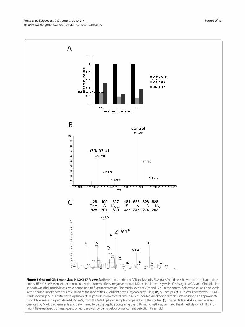
K187H1.2 is methylated by G9a and Glp1 in vivoTo confirm that G9a and Glp1 can also methylate K187 ofH1.2 in vivo, RNA interference (RNAi) analysis was per-formed. HEK293 cells were used with either non-targetnegative control small interfering siRNA or simultane-ously with two siRNAs against G9a and Glp1 to knockdown both enzymes. At 42 hours and 72 hours aftertransfection of the siRNAs, a reduction in G9a RNA toapproximately 20% and Glp1 to 30% was detected by RT-PCR (Figure 3a), and this knockdown was confirmed bywestern blotting (data not shown).
Histones were then acid-extracted from these cells andprepared by derivatization with D0- and isotopic D5-pro-pionic anhydride for quantitative proteomic analysis.
After derivatization, control and knockdown sampleswere mixed equally; they could be distinguished becauseof a mass difference corresponding to a 5 Da shift. Twosets of peaks corresponding to a 2.5 Da shift were seen,which is consistent with D0- and D5-propionyl labellingfor a 2+ charged peptide. The D0 labelled peptide fromthe G9a/Glp1 knockdown sample was decreased byapproximately two fold compared with the control (Fig-ure 3b), and MS/MS analysis of this D0 peak (Figure 3c)confirmed that this peptide contained the sequence pr-AAKprmeSAAKpr (pr = propionyl amide, +56 Da). To con-trol for enzyme specificity, the effect of the knockdownon other histone modifications was analysed, and as pre-viously described [16], a reduction in H1.4K26me andH3K9me was seen, whereas H3K27me and H4K20me didnot change (Additional file 2). In summary, this demon-strates that K187 of H1.2 is methylated in vivo and thatG9a and Glp1 can mediate this methylation.
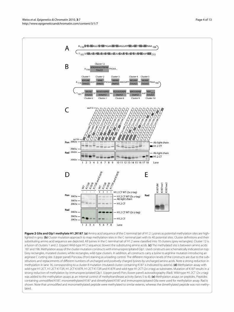
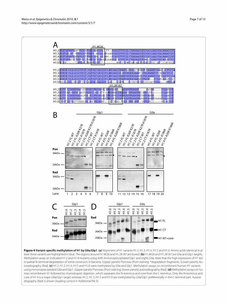
K187 methylation by G9a/Glp1 is H1 variant-specificUp to this point, our studies had been focused on methy-lation of the H1.2 C-terminus. Because it had recentlybeen described that G9a can methylate K26 on H1.4 [16],we then investigated whether H1.2 can also be methy-lated by G9a or Glp1 at K26 or at the neighbouring K27(Figure 4a). Both lysines in full-length H1.2 weremutated, and methylation assays performed in vitro.Whereas mutation of K187 resulted in a strong decreasein methylation (Figure 4b; lanes 1 and 11 versus lanes 3and 13), mutation of K26 and K27 did not impair H1.2methylation by Glp1 or G9a (Figure 4b, lanes 2 and 12).By contrast, mutation of H1.4K26 resulted in an almostcomplete loss of methylation (Figure 4b, lanes 8 and 18)whereas mutation of K186 (corresponding to K187 inH1.2) did not affect the methylation of H1.4 by Glp1 andG9a (Figure 4b, lanes 9 and 19). Thus, G9a/Glp1 methy-lated H1 in a variant-specific manner, preferentially tar-geting the N-terminus of H1.4, but the C-terminus ofH1.2.
These results prompted us to investigate whether theother major H1 variants are methylated by G9a/Glp1 onthe N- or C-terminus. First, in vitro methylation assayswere performed, using recombinant H1.3, H1.5, and themore divergent variant H1.0; the latter is mainlyexpressed in terminally differentiated cells. All of the H1variants analysed could be methylated by G9a/Glp1 (Fig-ure 4c). To distinguish between N- and C-terminal meth-ylation, H1 was digested after the methylation reactionwith chymotrypsin, which cleaves the H1 C-terminus atposition F105 [17]. G9a/Glp1 methylated preferentiallythe N-terminus of H1.4, whereas they methylated prefer-entially the C-termini of other variants (Figure 4d).
Together, these data demonstrate that G9a and Glp1methylate residues within H1 in a variant-specific fash-
Weiss et al. Epigenetics & Chromatin 2010, 3:7http://www.epigeneticsandchromatin.com/content/3/1/7
Page 6 of 13
Figure 3 G9a and Glp1 methylate H1.2K187 in vivo. (a) Reverse transcription PCR analysis of siRNA-transfected cells harvested at indicated time points. HEK293 cells were either transfected with a control siRNA (negative control; NK) or simultaneously with siRNAs against G9a and Glp1 (double knockdown; dkn). mRNA levels were normalised to β-actin expression. The mRNA levels of G9a and Glp1 in the control cells were set as 1 and levels in the double knockdown cells calculated as the ratio of this level (light grey, G9a; dark grey, Glp1). (b) MS analysis of H1.2 after knockdown. Full MS result showing the quantitative comparison of H1 peptides from control and G9a/Glp1 double knockdown samples. We observed an approximate twofold decrease in a peptide (414.750 m/z) from the G9a/Glp1 dkn sample compared with the control. (c) This peptide at 414.750 m/z was se-quenced by MS/MS experiments and determined to be the peptide containing the K187 monomethylation mark. The dimethylation of H1.2K187 might have escaped our mass-spectrometric analysis by being below of our current detection threshold.
C
B
�
� � �
� � �
� � �
� � �
�
� � �
� � � � �
� � � � � � � � � � � � � � � � � � � �
� � � � � � � � � � � � � � � �
� � � � � � � ! "# $ % ! &� � � $ ' ( ) '
� � � $ ' ( ) '
A
Weiss et al. Epigenetics & Chromatin 2010, 3:7http://www.epigeneticsandchromatin.com/content/3/1/7
Page 7 of 13
Figure 4 Variant-specific methylation of H1 by G9a/Glp1. (a) Alignment of H1 variants H1.2, H1.3, H1.4, H1.5 and H1.0. Amino acids identical in at least three variants are highlighted in blue. The regions around H1.4K26 and H1.2K187 are boxed. (b) H1.4K26 and H1.2K187 are G9a and Glp1 targets. Methylation assay on indicated H1.2 and H1.4 mutants using (left) immunoprecipitated Glp1 and (right) G9a. Note that the high expression of H1 led to partial N-terminal degradation of some constructs in bacteria. (Upper panels) Ponceau (Pon) staining: *degradation fragments. (Lower panels) Au-toradiography (Rad). (c) H1.2, H1.3, H1.4, H1.5 and H1.0 were methylated by G9a and Glp1. Methylation assays on recombinant human H1 variants using immunoprecipitated G9a and Glp1. (Upper panels) Ponceau (Pon) staining; (lower panels) autoradiography (Rad). (d) Methylation assays on hu-man recombinant H1 followed by chymotrypsin digestion, which separates the N-terminus and core from the C-terminus. Only the N-terminus and core of H1.4 is a major G9a/Glp1 target whereas H1.2, H1.3, H1.5 and H1.0 are methylated by G9a/Glp1 preferentially in the C-terminal part. Autora-diography (Rad) is shown (loading control in Additional file 3).
Pon
Rad
Glp1 G9a
H1.
2 FL
WT
H1.
2 FL
K26
R K2
7R
H1.
2 FL
K18
7R
H1.
2 FL
K26
R K2
7R K
187R
H1.
2 CT
WT
H1.
2 CT
K18
7R
H1.
4 FL
WT
H1.
4 FL
K26
R
H1.
4 FL
K18
6R
H1.
4 FL
K26
R K1
86R
36kDa
26kDa
36kDa
26kDa
A
B
H1.2 H1.3 H1.4 H1.5 H1.0
H1.2 H1.3 H1.4 H1.5 H1.0
H1.2 H1.3 H1.4 H1.5 H1.0
Rad
G9a Glp1
H1.
0
H1.
2
H1.
3
H1.
4
H1.
5
H1.
0
H1.
2
H1.
3
H1.
4
H1.
5
H1 FL
H1 CT
H1 NT+core
D C
H1.
0
H1.
2
H1.
3
H1.
4
H1.
5
Pon
Rad
H1.4K26
H1.2K187
H1.
2 FL
WT
H1.
2 FL
K26
R K2
7R
H1.
2 FL
K18
7R
H1.
2 FL
K26
R K2
7R K
187R
H1.
2 CT
WT
H1.
2 CT
K18
7R
H1.
4 FL
WT
H1.
4 FL
K26
R
H1.
4 FL
K18
6R
H1.
4 FL
K26
R K1
86R
1 2 3 4 5 6 7 8 9 10 11 12 13 14 15 16 17 18 Lane 19 20
36kDa
36kDa
36kDa
26kDa
20kDa
Glp1
Weiss et al. Epigenetics & Chromatin 2010, 3:7http://www.epigeneticsandchromatin.com/content/3/1/7
Page 8 of 13
ion. H1.4 is specifically methylated at the N-terminus atK26 and H1.2 at the C-terminus at K187, but not the cor-responding lysines on H1.2 and H1.4, respectively. Fur-thermore, the C-terminus of H1 is the main target forG9a/Glp1-mediated methylation in most somatic H1variants.
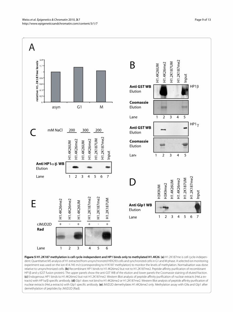
H1.2K187 methylation is cell cycle-independentHistone H1 phosphorylation is also mainly localised tothe C-terminus. This C-terminal H1 phosphorylation ishighly cell cycle-regulated [17], reaching a maximum inG2/M. Based on this, we investigated if H1.2K187 is alsocell cycle-regulated. Therefore, HEK293 cells were syn-chronised and H1 isolated at different time points forquantitative MS analysis. H1.2K187 methylation levelsstayed relatively constant during the cell cycle (Figure 5a),indicating that H1.2 C-terminal phosphorylation andH1.2K187me may act independently of each other.
H1.2K187 and H1.4K26 have different downstream readersTo identify novel, methylation-dependent H1 bindingproteins, we followed an unbiased approach initially,using peptide affinity purifications. We did not detect anynovel H1 methylation-specific binding proteins (data notshown), thus we switched to a candidate approach. Weshowed previously [20] that HP1 binds specifically tomethylated K26 of H1.4. Because H1.2K187 and H1.4K26are methylated by the same enzymes, we investigated ifHP1 could also bind to methylated H1.2K187. Recombi-nant HP1β and γ were expressed and assayed for bindingto K187me2 in peptide affinity purifications. The recom-binant HP1β and γ bound to methylated H1.4K26me2(Figure 5b, lane2) but not to H1.2K187me2 (lane 4). Toconfirm this with endogenous proteins, the peptide affin-ity purifications were repeated using HeLa nuclearextract as HP1 source. There was specific binding ofendogenous HP1 to H1.4K26me2 (Figure 5c, lane 2), butnot to H1.2K187me2 (Figure 5c, lane 6). These data indi-cate selective recognition by HP1: H1.4K26me2 is specifi-cally bound by HP1 whereas H1.2K187me2 is not.
The ankyrin repeats of G9a and Glp1 have been shownto bind specifically to H3K9me1/2 and it has been sug-gested that this binding could be involved in the recruit-ment of the enzymes. Therefore, we investigated if theycould also bind to methylated H1. We did not observe anyspecific binding of Glp1 (Figure 5d) (or G9a, data notshown) to either H1.4K26me2 or H1.2K187me2, arguingagainst a binding of G9a/Glp1 to its H1 substrate sites.
To gain further insight into regulation of K187 methyla-tion we investigated whether the jumonji-C type histonedemethylase JMJD2D/KDM4 (which can reverse dimeth-ylation to monomethylation) could demethylateH1.2K187me2, as shown for H1.4K26me2 [16].H1.2K187me2 and H1.4K26me2 peptides were first dem-
ethylated and then assayed for remethylation with G9aand Glp1. This assay is indirect but very sensitive, as lowlevels of demethylation are sufficient to allow remethyla-tion. Incubation of the dimethylated H1 peptides withrecombinant JMJD2D allowed remethylation by G9a/Glp1 of the H1.4K26 peptide (Figure 5e, lane 1) but not ofthe H1.2K187 peptide (lane 4). Thus H1.4K26me2 butnot H1.2K187me2 can be demethylated by JMJD2D.
Together, these results show that despite being methy-lated by the same HKMTs, H1.4K26 and H1.2K187 meth-ylation are read and removed by different proteins,suggesting novel variant-specific functions.
DiscussionOur data show that K187 in histone H1.2 is a new targetof HKMTs G9a and its interaction partner Glp1 in vitroand in vivo, as confirmed by mutation analysis, knock-down experiments and MS analysis. To our knowledge,this is the first characterisation of a methylation site andits corresponding methylating enzymes in the C-termi-nus of histone H1. G9a and Glp1 were found to be in het-eromeric complexes and to share high sequencehomology, and in the case of histone H3, both contributeto global H3K9 mono- and dimethylation [14]. We showhere that a similar situation occurs for H1, where bothenzymes target identical lysines.
Interestingly, both G9a and Glp1 were able to methylateH1 in a variant-specific way. In H1.4, the main methyla-tion site is K26 in the N-terminus, whereas in H1.2 (andthe other main somatic H1 variants H1.0, H1.3 and H1.5)it is the C-terminus that is preferentially methylated byG9a and Glp1. Methylation of H1.2 at K26 had previouslybeen identified only at low levels by MS in certain mousetissues, but according to our data this seems not to be aG9a target. Indeed, H1.2K26 can be methylated by otherenzymes such as Ezh2 ([18] and our unpublished data).Furthermore, H1.4K26me2 is specifically bound by allHP1 isoforms, but K187me2 is not, and onlyH1.4K26me2 is demethylated by JMJD2D. Both of thesefeatures thus distinguish these two methylation marks.
In our MS analysis we detected monomethylation ofH1.2K187 in vivo, but dimethylation of H1.2K187 mighthave escaped our MS analysis if it was below our currentdetection threshold. Both enzymes have been shown todimethylate H3K9 and H1.4K26 both in vitro and in vivo[16,24]. In line with this, our assays showed that G9a andGlp1 methylate unmodified and monomethylated K187peptides, demonstrating dimethylation activity.
To date, the only characterised modification of the H1C-terminus is phosphorylation. The C-terminal tail of H1can be highly phosphorylated [25] on several serine andthreonine residues. This phosphorylation is mainly catal-ysed by cdk type kinases [26,27], and is highly cell cycle-dependent, reaching maximum levels in G2/M phase.
Weiss et al. Epigenetics & Chromatin 2010, 3:7http://www.epigeneticsandchromatin.com/content/3/1/7
Page 9 of 13
Figure 5 H1.2K187 methylation is cell cycle-independent and HP1 binds only to methylated H1.4K26. (a) H1.2K187me is cell cycle-indepen-dent. Quantitative MS analysis of H1 extracted from unsynchronized HEK293 cells and synchronized cells in G1 and M phase. A selected ion monitoring experiment was used on the ion 414.745 m/z (corresponding to H1K187 methylation) to monitor the levels of methylation. Normalisation was done relative to unsynchronized cells. (b) Recombinant HP1 binds to H1.4K26me2 but not to H1.2K187me2. Peptide affinity purification of recombinant HP1β and γ (GST fusion protein). The upper panels show the anti GST WB of the elution and lower panels the Coomassie staining of eluted fraction. (c) Endogenous HP1 binds to H1.4K26me2 but not H1.2K187me2. Western Blot analysis of peptide affinity purification of nuclear extracts (HeLa ex-tracts) with HP1α/β specific antibody. (d) Glp1 does not bind to H1.4K26me2 or H1.2K187me2. Western Blot analysis of peptide affinity purification of nuclear extracts (HeLa extracts) with Glp1 specific antibody. (e) JMJD2D demethylates H1.4K26me2 only. Methylation assay with G9a and Glp1 after demethylation of peptides by JMJD2D (Rad).
Anti GST WBElution
H1.
4K26
UM
H1.
4K26
me2
H1.
2K18
7UM
H1.
2K18
7me2
Inp
ut
CoomassieElution
B
C
D
Rad
H1.
2K18
7me2
H1.
2K18
7me2
H1.
2K18
7UM
H1.
4K26
me2
H1.
4K26
me2
H1.
4K26
UM
H1.
4K26
UM
H1.
4K26
me2
H1.
4K26
UM
H1.
4K26
me2
H1.
2K18
7UM
H1.
2K18
7me2
Inp
ut
mM NaCl 200 300 200
Anti HP1 WBElution
rJMJD2D + - + + - +
�
Lane 1 2 3 4 5
Lane 1 2 3 4 5 6 7
Lane 1 2 3 4 5 6
Anti GST WBElution
CoomassieElution
H3K
9UM
H3K
9me2
H1.
4K26
UM
H1.
4K26
me2
H1.
2K18
7UM
H1.
2K18
7me2
Inp
ut
E Anti Glp1 WBElution
Lane 1 2 3 4 5 6 7
Lane 1 2 3 4 5
HP1
HP1γ
β
�
� � �
� � �
� � �
� � �
�
� � �
� � �
�
asyn G1 M
A
Weiss et al. Epigenetics & Chromatin 2010, 3:7http://www.epigeneticsandchromatin.com/content/3/1/7
Page 10 of 13
Interestingly, in contrast to phosphorylation, H1.2K187methylation levels remain steady throughout the cellcycle, suggesting that only H1 C-terminal phosphoryla-tion, but not H1.2K187 methylation, has a global role inmitotic chromosome condensation [28]. In a recent MSanalysis [3,4,18,29] a phosphorylation site, S188, wasmapped next to K187 in H1.4 but not H1.2. Although it istempting to speculate that S188 phosphorylation mightblock K187 methylation in H1.4, there are strong argu-ments against this. First, the C-terminus of recombinant,unphosphorylated H1.4 is not (or only very weakly)methylated by G9a/Glp1 and second, only the neighbour-ing phosphorylation is cell cycle-dependent.
Whether H1 variants have specific functions is a matterof an ongoing debate [3,4,29]; however, there are severallines of evidence pointing towards specific functions. Flu-orescence recovery after photobleaching experimentsshowed that different H1 variants span a broad range ofrecovery times, owing to significant differences in theirbinding affinity to chromatin [3,4,29]. The recovery timedepends on the length of the C-terminal tail, on the den-sity of positively charged amino acids, and on the numberand distribution of phosphorylation sites within it. H1.2has one of the shortest tails and the fastest recovery time,whereas H1.4 shows higher affinity for chromatin withlonger residence times [8]. Recently, differences in thebinding affinities of mammalian H1 variants have alsobeen shown both in vitro [30] and in vivo [31]. Addition-ally, a preferential location of H1 variants to differentgenomic regions was described [8]. Both the differencesin the dynamics and the reported variations in globalnuclear distribution patterns suggest that H1 variantssuch as H1.2 and H1.4 (the major H1 variants in manysomatic cells) could have specific functions. This is sup-ported by our findings that different methylation sitestargeted by the same enzymes on distinct H1 variantshave specific readouts.
In addition to G9a/Glp1, H1.4K26 can be methylated byEzh2 as part of the polycomb repressive complex 2(PRC2) [22]; however, the relative in vivo contribution ofthese enzymes is currently unclear. Inhibition of G9a andGlp1 results in a strong decrease in H1.4K26 methylation[16], suggesting that G9a/Glp1 might be the mainenzymes catalysing this modification. This is in line withour knockdown experiments showing that reduced levelsof G9a and Glp1 drastically affect H1.4K26me (see Addi-tional file 2).
Pinpointing a cellular phenotype to histone modifica-tions in mammals is not straightforward, because aminoacid substitutions cannot be applied by geneticapproaches and also because the enzymes catalyzingmodifications often modify multiple proteins. The phe-notype of the G9a/Glp1 knockout is embryonically lethal[24]; however, it is unclear whether this is due to changes
in the histone modifications or in other G9a/Glp1 targets[32]. In an attempt to investigate the cellular function ofH1.2K187, we made stable cell lines expressing taggedwild-type H1.2K187 and a H1.2K187R mutant that can-not be methylated. Because depletion of H1.2 has beenshown to result in cell-cycle arrest in G1 phase [31], wechecked particularly for possible effects on the cell cycle;however, we did not detect any major effect on the cellsor on cell-cycle progression, which is in line withH1.2K187 methylation levels remaining rather steadythroughout the cell cycle.
Interestingly, H1.4K26 methylation by G9a has beenshown to increase the residence time of H1.4 on chroma-tin [16], whereas the more 'dynamic' H1.2 is not methy-lated by G9a at this site. Because both G9a and Glp1 areimportant for H3K9 methylation and silencing of euchro-matic genes [14,24], and because H1.2 has been found tobe enriched in euchromatic regions [7,8], we speculatethat H1.2K187 methylation might have a role in genesilencing in these regions. By contrast, H1.4 has beenfound to be enriched in heterochromatic regions [7], andmethylated H1.4K26 but not H1.2K187 can be specifi-cally bound by HP1 [20], a main component of hetero-chromatin. This might argue for a role of H1.4K26methylation in transcriptional repression in heterochro-matin. It will be interesting in the future to determine thebiological functions and the localisation patterns of theH1 variants and their corresponding modifications at agenome-wide level. To date, in spite of many attemptsand different approaches, we have been unable to raise asatisfactory antibody with unique specificity for methy-lated H1.2K187 that could be used in in vivo experiments.
It has been reported that the ankyrin repeats of G9aand Glp1 recognise H3K9me1/2 [33], and suggested thatthey also bind to H1.4K26me1/2 [16], contributing totheir recruitment to chromatin. We do not observe bind-ing of G9a or Glp1 to methylated H1.4K26 or H1.2K187peptides, suggesting a different recruiting mechanismfrom that for H3K9me1/2. Moreover, recruitment ofG9a/Glp1 to H3K9me1/2 could result in the spreading ofH3K9 methylation mark, whereas our data suggest thatsuch a mechanism is less likely for H1. This is in line withthe globally low levels of H1 methylation, compared withH3K9me1/2; however, binding of the ankyrin repeats toH3K9me1/2 could tether G9a/Glp1 and then direct H1methylation.
ConclusionsIn conclusion, we have identified and characterised anovel methylation site in the C-terminus of histone H1(H1.2K187). We have shown that the HKMTs G9a and itsinteracting partner Glp1 are the enzymes performing thismethylation in vitro and in vivo, resulting in the identifi-cation of the first enzymes that methylate the H1 C-ter-
Weiss et al. Epigenetics & Chromatin 2010, 3:7http://www.epigeneticsandchromatin.com/content/3/1/7
Page 11 of 13
minus and of a new G9a/Glp1 target. Furthermore, wehave demonstrated that the HKMTs G9a and Glp1 canmethylate histone H1 in a variant-specific manner in theN- or C-terminus. To our knowledge, this is the firstexample of variant-specific histone modifications by thesame enzymes. Additionally H1.4K26me andH1.2K187me also differ in their readout, clearly distin-guishing them functionally.
MethodsCluster mutation PCRFor the exchange of lysine clusters, two-step mutagenesisPCR was used. Mutation primers consisted of the 18replacing bases and approximately 20 bases complemen-tary to the template. In the first PCR, DNA fragmentscovering the 5' end to the mutation site and the mutationsite to the 3' end were amplified separately in two reac-tions. In the second PCR, the two products were mixedand primers flanking the 5' and 3' ends were used toamplify the complete mutated DNA fragment.
Protein expression and purificationRecombinant human HKMTs ((numbers are amino acidresidues) Glp1 610-917; Nsd2 926-1210; PRDM5 3-429;MLL5 294-481; Nsd1 1700-1987; BLIMP1 105-323; PR-SET7 FL; SMYD2 FL; SMYD4 FL; Suv39h1 82-412;Suv39h2 157-477; Suv39h1 82-412 H320R; SMYD3 FL;SMYD5 FL; G9a 682-1000; Suv4-20h1 1-384; Suv4-20h21-280; plasmids obtained from T. Jenuwein) wereexpressed as glutathione S-transferase (GST) fusion pro-teins in bacteria using standard techniques and purified(glutathione Sepharose® 4B; GE Healthcare, Piscataway,NJ, USA).
Affinity purification of bacterially expressed 2× z-tagged and histidine (His)-tagged H1 was performed(HIS-Select Nickel Affinity Gel; Sigma, St Louis, MO,USA). C-terminally His-tagged recombinant histone H1was extracted from bacteria with 0.83 M perchloric acid(PCA) and then neutralized by addition of Tris/HCl pH9.5. Further purification was achieved using nickel affin-ity gel (Sigma).
For eukaryotic expression of human G9a and Glp1,HEK293 cells were transiently transfected with plasmidsencoding full-length FLAG-tagged G9a or Glp1 using thecalcium-phosphate method and harvested 72 hours aftertransfection. Purification was performed using Anti-FLAG M2 agarose (Sigma).
His-tagged recombinant human JMJD2D (1-350; plas-mid obtained from Y. Shi) was expressed in bacteria usingstandard techniques and purified with nickel affinity gel.Human FL HP1β and human FL HP1γ (plasmidsobtained from T. Kouzarides) were expressed as GSTfusion proteins in bacteria using standard techniques and
affinity purified (glutathione Sepharose® 4B; GE Health-care). Full-length recombinant H1 variants were obtainedfrom Calbiochem -Novabiochem Corp. (San Diego, CA,USA).
Purification of endogenous nucleosomes and H1Approximately 3 × 10⁷ HEK293 cells were swollen inhypotonic buffer (20 mM HEPES pH 7.0, 20 mM NaCl, 5mM MgCl2), and plasma membranes were destroyed byhomogenizing in a dounce homogenizer. After centrifu-gation, the pellet was resuspended in hypotonic bufferwith a final concentration of 0.5% NP-40 (Nonidet P-40(Fluka AG, Buchs, Switzerland); to lyse nuclei. Chromatinwas pelleted by centrifugation, resuspended in isolationbuffer (10 mM Tris/HCl pH 7.4, 1.5 mM MgCl2, 1 mMCaCl2, 0.25 M sucrose) and optical density at 260 nm(OD260) was measured. Micrococcal nuclease (10 U per50 μg DNA) was added, and the samples were incubatedat 37°C for 1 hour. Mono- and dinucleosomes wereextracted twice with lysis buffer (10 mM Tris/HCl pH6.85, 5 mM EDTA) and separated by centrifugation for 14hours at 280 000 g on a 5% to 40% sucrose gradient (20mM HEPES pH 7.6, 0.5 mM MgCl2, 5% to 40% sucrose).
For endogenous H1 extraction, HEK293 cells werelysed in Triton extraction buffer (1 × phosphate-bufferedsaline (PBS) with 0.5% triton ×100) to remove the cytosol.After pelleting of nuclei, H1 was extracted with 0.83 MPCA.
In vitro methylation assayHKMT assays were carried out as previously described[34]. Briefly, in a total volume of 25 μl, 2-10 μl of purifiedfull-length G9a or Glp1 or recombinant GST fusionenzymes were mixed with 2 μg of recombinant histoneH1 or 10 μg of peptides as substrate (H1.2K187 peptidesPKKAAK187SAAKAVGC with K187 either unmodified(UM), monomethylated (me1) or dimethylated (me2),H1.4K26 peptides VKKKARK26SAGAAKGC with K26either unmodified or dimethylated) (peptides obtainedfrom CloneStar Peptide Services (Brno, Czech Republic)and GeneCust Europe (Dudelang, Luxembourg)). Reac-tions were carried out in Tris (20 mM Tris/HCl pH 9.0),phosphate (10 mM Na2HPO4, 2 mM KH2PO4, 2.7 mMKCl, 137 mM NaCl, pH 7.2), R (50 mM Tris/HCl pH 8.5,5 mM MgCl2) or Methylation Assay Buffer (MAB) (50mM Tris/HCl pH 8.5, 20 mM KCl, 10 mM MgCl2, 0.25mM sucrose) buffers at 30°C for 1 hour. H1 was then sep-arated using SDS-PAGE with 15% to 18.7% gels, and pep-tides were resolved in 16% Schägger Jagow gels. Proteinsand peptides were transferred to nitrocellulose mem-branes and exposed to film (BioMax MS; Kodak).
Weiss et al. Epigenetics & Chromatin 2010, 3:7http://www.epigeneticsandchromatin.com/content/3/1/7
Page 12 of 13
In vitro demethylation assay and subsequent remethylation assayFor the demethylation assay, 20 μl of purified recombi-nantly expressed JMJD2D and 25 μg of peptide weremixed in reaction buffer (50 mM Tris/HCl pH 7.4, 1 mMα-ketoglutarate, 2 mM ascorbate and 70 μMFe2+(NH4)2(SO4)2), and demethylation was performedat 37°C for 6 hours. For subsequent remethylation, 15 μlof the demethylation assay mixture were added to 1 μl of1 M Tris/HCl pH 9.5, 9.4 μl of mixed purified G9a andGlp1, 0.6 μl 0.5 M EDTA (final concentration 10 mM) and4 μl 5× R methylation buffer, and then incubated at 32°Cfor 1 hour. Peptides were separated in 16% SchäggerJagow gels, transferred to nitrocellulose membrane andexposed to film (BioMax MS).
Knockdown of G9a and Glp1 and reverse transcriptase PCRBriefly, 4 × 10⁶ HEK293 cells were plated in a 145 mmdish. The following day, the cells were simultaneouslytransfected according to the manufacturer's instructions,using a mix containing 100 μl of 10 mM anti-Glp1 smallinterfering (si)RNA (AACGAAGAATGGGAACCTATA)(Qiagen, Valencia, CA, USA), 100 μl anti-G9a siRNA(CACCATGAACATCGATCGCAA) (Qiagen) and 100 μlof lipofectamine for double knockdown. Control cellswere transfected with 200 μl 10 mM negative siRNA (All-Stars; Qiagen). The mRNA levels of cells harvested after19, 42 and 72 hours were analysed by reverse tran-scriptase PCR using a commercial kit (RevertAid MinusFirst Strand cDNA Synthesis Kit; Fermentas Inc., GlenBurnie, MD, USA) and the following primers: CTGACA-CAGAGGACAGGAAGC and TCTCGAACTTCTCTG-GGATCTT for Glp1; TCCGACAGCAAGTCTGAAGTTand TGACTGATTCCCTGACTCCTC for G9a; CGGT-TGGCCTTGGGGTTCAGGGGG and ATCGTG-GGGGCGCCCCAGGCACCA for β-actin (used ascontrol). For MS analysis, cells were harvested after 72hours.
Peptide affinity purificationPeptides were coupled to a gel (SulfoLink Coupling Gel;Pierce, Rockford, IL, USA) via their C-terminal cysteineaccording to the manufacturer's instructions. Recombi-nant HP1β and γ or HeLa nuclear extracts were added to20 μl of coupled beads and incubated for 90 minutes at4°C. Beads were washed three times with IPH-X buffer(20 mM Tris/HCl pH 8.0, 0.5% NP-40, NaCl 250 mM oras indicated) before elution and separation on 15% SDS-PAGE gels. As indicated, binding was detected by eitherCoomassie staining or immunostaining using HP1 -spe-cific antibody (10478; Abcam, Cambridge, MA, USA) orGlp1-specific antibody (D220-3; MBL International,Woburn, MA, USA).
Mass spectrometryAcid-extracted histones were diluted in 100 mM ammo-nium bicarbonate buffer (pH 8.0) and digested withtrypsin at a protein:substrate ratio of 15:1 for 8 hours at37°C. The reaction was quenched by acidification to pH3.0 with glacial acetic acid and freezing. The digestionwas then reacted with either D0- or D5-propionic anhy-dride [35] for chemical derivatization labelling and incor-poration of a stable isotope label for quantitativecomparison as previously described (Plazas-Mayorca etal., submitted for publication). Samples were separatedby online reverse-phase liquid chromatography followedby analysis in an orbitrap mass spectrometer operated inthe data-dependent mode as previously described [36].All spectra were manually verified.
Cell-cycle synchronizationExponentially growing HEK293 cells were synchronisedin M phase by treatment with 300 ng/ml nocodazole for18 h. Cells were enriched in G1/G0 phase by serum star-vation in DMEM containing 0,5% FCS for 48 h. Cell-cycledistribution was checked by propidium iodide stainingand fluorescence activated cell sorting.
Chymotrypsin digestionFor chymotrypsin digestion, 4 μl of 3 M Na acetate pH 4.8and 60 ng chymotrypsin (A-Chymotrypsin Type I-S;Sigma) were added to 25 μl of methylation assay, anddigestion was performed at 25°C for 20 minutes. Frag-ments were separated in 18.7% SDS-PAGE gels, trans-ferred to nitrocellulose membrane and exposed to film(BioMax MS).
Additional material
Competing interestsThe authors declare that they have no competing interests.
Authors' contributionsTW carried out most of the experimental work with the help of SH, UZ, AI, MDand PT. BMZ and BAG performed the mass-spectrometrical analysis. TW, SD
Additional file 1 Figure S1 - Recombinant PRSET7 and Suv4-20h1 methylate H4. (a) Coomassie staining of membrane with histones used for Figure 1a. (b) Autoradiography of HKMT assay with PRSET7 and Suv4-20h1 is shown as specificity control of our assays.Additional file 2 Figure S2 - Quantitative mass spectrometry analysis of G9a/Glp1 knockdown versus control samples. Full mass spectra showing peptides from (a) H4K20me2, (b) H3K27me2, (c) H3K9me2 and (d) H1.4K26me1. No changes in H4K20me2 or H3K27me2 were detected, but decreases in H3K9me2 and H1.4K26me1 were seen in the G9a/Glp1 knock-down samples.Additional file 3 Figure S3 - Loading control of digested H1 variants. Coomassie loading control for Fig 4D. H1 variants were methylated by G9a/Glp1 and afterwards digested by Chymotrypsin. One part of the samples was loaded on an SDS gel and afterwards transferred to a nitrocellulose membrane for autoradiography (Fig 4d), the other part of the samples was loaded on an SDS gel and stained with Coomassie to serve as a loading control.
Weiss et al. Epigenetics & Chromatin 2010, 3:7http://www.epigeneticsandchromatin.com/content/3/1/7
Page 13 of 13
and RS participated in the design of the study and prepared the manuscript. Allauthors read and approved the final manuscript.
AcknowledgementsWork in the RS laboratory is supported by the Max Planck Society, the DFG (through SFB 746), the EU (the Epigenome) and an ERC starting grant. We thank T. Jenuwein for providing expression constructs.
Author Details1MPI for Immunobiology, Stübeweg 51, 79108 Freiburg, Germany, 2Department of Molecular Biology, Princeton University, 415 Shultz Laboratory, Princeton, NJ 08540, USA and 3Department of Cell Biology, Rosalind Franklin University, 3333 Green Bay Road, Chicago, IL 60064, USA
References1. Luger K, Mader AW, Richmond RK, Sargent DF, Richmond TJ: Crystal
structure of the nucleosome core particle at 2.8 A resolution. Nature 1997, 389:251-260.
2. van Holde KE: Chromatin Berlin, Heidelberg; 1988. 3. Happel N, Doenecke D: Histone H1 and its isoforms: contribution to
chromatin structure and function. Gene 2009, 431:1-12.4. Izzo A, Kamieniarz K, Schneider R: The histone H1 family: specific
members, specific functions? Biol Chem 2008, 389:333-343.5. Meergans T, Albig W, Doenecke D: Varied expression patterns of human
H1 histone genes in different cell lines. DNA Cell Biol 1997, 16:1041-1049.
6. Happel N, Schulze E, Doenecke D: Characterisation of human histone H1x. Biol Chem 2005, 386:541-551.
7. Parseghian MH, Newcomb RL, Winokur ST, Hamkalo BA: The distribution of somatic H1 subtypes is non-random on active vs. inactive chromatin: distribution in human fetal fibroblasts. Chromosome Res 2000, 8:405-424.
8. Th'ng JP, Sung R, Ye M, Hendzel MJ: H1 family histones in the nucleus. Control of binding and localization by the C-terminal domain. J Biol Chem 2005, 280:27809-27814.
9. Fan Y, Sirotkin A, Russell RG, Ayala J, Skoultchi AI: Individual somatic H1 subtypes are dispensable for mouse development even in mice lacking the H1(0) replacement subtype. Mol Cell Biol 2001, 21:7933-7943.
10. Fan Y, Nikitina T, Zhao J, Fleury TJ, Bhattacharyya R, Bouhassira EE, Stein A, Woodcock CL, Skoultchi AI: Histone h1 depletion in mammals alters global chromatin structure but causes specific changes in gene regulation. Cell 2005, 123:1199-1212.
11. Kouzarides T: Chromatin modifications and their function. Cell 2007, 128:693-705.
12. Patnaik D, Chin HG, Esteve PO, Benner J, Jacobsen SE, Pradhan S: Substrate specificity and kinetic mechanism of mammalian G9a histone H3 methyltransferase. J Biol Chem 2004, 279:53248-53258.
13. Peters AH, Kubicek S, Mechtler K, O'Sullivan RJ, Derijck AA, Perez-Burgos L, Kohlmaier A, Opravil S, Tachibana M, Shinkai Y, Martens JH, Jenuwein T: Partitioning and plasticity of repressive histone methylation states in mammalian chromatin. Mol Cell 2003, 12:1577-1589.
14. Tachibana M, Ueda J, Fukuda M, Takeda N, Ohta T, Iwanari H, Sakihama T, Kodama T, Hamakubo T, Shinkai Y: Histone methyltransferases G9a and GLP form heteromeric complexes and are both crucial for methylation of euchromatin at H3-K9. Genes Dev 2005, 19:815-826.
15. Tachibana M, Sugimoto K, Fukushima T, Shinkai Y: Set domain-containing protein, G9a, is a novel lysine-preferring mammalian histone methyltransferase with hyperactivity and specific selectivity to lysines 9 and 27 of histone H3. J Biol Chem 2001, 276:25309-25317.
16. Trojer P, Zhang J, Yonezawa M, Schmidt A, Zheng H, Jenuwein T, Reinberg D: Dynamic histone H1 isotype 4 methylation and demethylation by histone lysine methyltransferase G9A/KMT1C and the jumonji domain containing JMJD2/KDM4 proteins. J Biol Chem 2009, 284:8395-8405.
17. Sarg B, Helliger W, Talasz H, Forg B, Lindner HH: Histone H1 phosphorylation occurs site-specifically during interphase and mitosis: identification of a novel phosphorylation site on histone H1. J Biol Chem 2006, 281:6573-6580.
18. Wisniewski JR, Zougman A, Kruger S, Mann M: Mass spectrometric mapping of linker histone H1 variants reveals multiple acetylations, methylations, and phosphorylation as well as differences between cell culture and tissue. Mol Cell Proteomics 2007, 6:72-87.
19. Kuzmichev A, Margueron R, Vaquero A, Preissner TS, Scher M, Kirmizis A, Ouyang X, Brockdorff N, Abate-Shen C, Farnham P, Reinberg D: Composition and histone substrates of polycomb repressive group complexes change during cellular differentiation. Proc Natl Acad Sci USA 2005, 102:1859-1864.
20. Daujat S, Zeissler U, Waldmann T, Happel N, Schneider R: HP1 binds specifically to Lys26-methylated histone H1.4, whereas simultaneous Ser27 phosphorylation blocks HP1 binding. J Biol Chem 2005, 280:38090-38095.
21. Ogawa H, Ishiguro K, Gaubatz S, Livingston DM, Nakatani Y: A complex with chromatin modifiers that occupies E2F- and Myc-responsive genes in G0 cells. Science 2002, 296:1132-1136.
22. Kuzmichev A, Jenuwein T, Tempst P, Reinberg D: Different EZH2-containing complexes target methylation of histone H1 or nucleosomal histone H3. Mol Cell 2004, 14:183-193.
23. Collins RE, Tachibana M, Tamaru H, Smith KM, Jia D, Zhang X, Selker EU, Shinkai Y, Cheng X: In vitro and in vivo analyses of a Phe/Tyr switch controlling product specificity of histone lysine methyltransferases. J Biol Chem 2005, 280:5563-5570.
24. Tachibana M, Sugimoto K, Nozaki M, Ueda J, Ohta T, Ohki M, Fukuda M, Takeda N, Niida H, Kato H, Shinkai Y: G9a histone methyltransferase plays a dominant role in euchromatic histone H3 lysine 9 methylation and is essential for early embryogenesis. Genes Dev 2002, 16:1779-1791.
25. Garcia BA, Busby SA, Barber CM, Shabanowitz J, Allis CD, Hunt DF: Characterization of phosphorylation sites on histone H1 isoforms by tandem mass spectrometry. J Proteome Res 2004, 3:1219-1227.
26. Langan TA, Gautier J, Lohka M, Hollingsworth R, Moreno S, Nurse P, Maller J, Sclafani RA: Mammalian growth-associated H1 histone kinase: a homolog of cdc2+/CDC28 protein kinases controlling mitotic entry in yeast and frog cells. Mol Cell Biol 1989, 9:3860-3868.
27. Brown DT: Histone H1 and the dynamic regulation of chromatin function. Biochem Cell Biol 2003, 81:221-227.
28. Roth SY, Allis CD: Chromatin condensation: does histone H1 dephosphorylation play a role? Trends Biochem Sci 1992, 17:93-98.
29. Godde JS, Ura K: Cracking the enigmatic linker histone code. J Biochem 2008, 143:287-293.
30. Orrego M, Ponte I, Roque A, Buschati N, Mora X, Suau P: Differential affinity of mammalian histone H1 somatic subtypes for DNA and chromatin. BMC Biol 2007, 5:22.
31. Sancho M, Diani E, Beato M, Jordan A: Depletion of human histone H1 variants uncovers specific roles in gene expression and cell growth. PLoS Genet 2008, 4:e1000227.
32. Rathert P, Dhayalan A, Murakami M, Zhang X, Tamas R, Jurkowska R, Komatsu Y, Shinkai Y, Cheng X, Jeltsch A: Protein lysine methyltransferase G9a acts on non-histone targets. Nat Chem Biol 2008, 4:344-346.
33. Collins RE, Northrop JP, Horton JR, Lee DY, Zhang X, Stallcup MR, Cheng X: The ankyrin repeats of G9a and GLP histone methyltransferases are mono- and dimethyllysine binding modules. Nat Struct Mol Biol 2008, 15:245-250.
34. Nishioka K, Reinberg D: Methods and tips for the purification of human histone methyltransferases. Methods 2003, 31:49-58.
35. Garcia BA, Hake SB, Diaz RL, Kauer M, Morris SA, Recht J, Shabanowitz J, Mishra N, Strahl BD, Allis CD, Hunt DF: Organismal differences in post-translational modifications in histones H3 and H4. J Biol Chem 2007, 282:7641-7655.
36. Xie W, Song C, Young NL, Sperling AS, Xu F, Sridharan R, Conway AE, Garcia BA, Plath K, Clark AT, Grunstein M: Histone h3 lysine 56 acetylation is linked to the core transcriptional network in human embryonic stem cells. Mol Cell 2009, 33:417-427.
doi: 10.1186/1756-8935-3-7Cite this article as: Weiss et al., Histone H1 variant-specific lysine methyla-tion by G9a/KMT1C and Glp1/KMT1D Epigenetics & Chromatin 2010, 3:7
Received: 1 February 2010 Accepted: 24 March 2010 Published: 24 March 2010This article is available from: http://www.epigeneticsandchromatin.com/content/3/1/7© 2010 Weiss et al; licensee BioMed Central Ltd. This is an Open Access article distributed under the terms of the Creative Commons Attribution License (http://creativecommons.org/licenses/by/2.0), which permits unrestricted use, distribution, and reproduction in any medium, provided the original work is properly cited.Epigenetics & Chromatin 2010, 3:7
Copyright © 2022 FDOKUMEN