2014 Malnoë A Plant Cell Fts H1 Chlamydomonas
30
Thylakoid FtsH Protease Contributes to Photosystem II and Cytochrome b 6 f Remodeling in Chlamydomonas reinhardtii under Stress Conditions W Alizée Malnoë, 1 Fei Wang, Jacqueline Girard-Bascou, Francis-André Wollman, and Catherine de Vitry 2 Institut de Biologie Physico-Chimique, Unité Mixte de Recherche 7141, Centre National de la Recherche Scientifique/Université Pierre et Marie Curie, Paris 6, 75005 Paris, France FtsH is the major thylakoid membrane protease found in organisms performing oxygenic photosynthesis. Here, we show that FtsH from Chlamydomonas reinhardtii forms heterooligomers comprising two subunits, FtsH1 and FtsH2. We characterized this protease using FtsH mutants that we identified through a genetic suppressor approach that restored phototrophic growth of mutants originally defective for cytochrome b 6 f accumulation. We thus extended the spectrum of FtsH substrates in the thylakoid membranes beyond photosystem II, showing the susceptibility of cytochrome b 6 f complexes (and proteins involved in the c i heme binding pathway to cytochrome b 6 ) to FtsH. We then show how FtsH is involved in the response of C. reinhardtii to macronutrient stress. Upon phosphorus starvation, photosynthesis inactivation results from an FtsH-sensitive photoinhibition process. In contrast, we identified an FtsH-dependent loss of photosystem II and cytochrome b 6 f complexes in darkness upon sulfur deprivation. The D1 fragmentation pattern observed in the latter condition was similar to that observed in photoinhibitory conditions, which points to a similar degradation pathway in these two widely different environmental conditions. Our experiments thus provide extensive evidence that FtsH plays a major role in the quality control of thylakoid membrane proteins and in the response of C. reinhardtii to light and macronutrient stress. INTRODUCTION Photosynthesis allows the conversion of light energy, captured by chlorophyll–protein complexes, into reducing power (NADPH) and chemical energy (ATP). In oxygenic photosynthesis, the photoin- duced reduction of NADP + is performed by a photosynthetic electron transfer chain, which joins three major oligomeric proteins embedded in the thylakoid membranes: photosystem II (PSII), cytochrome b 6 f complex, and photosystem I (PSI). The assembly, degradation and repair of these protein complexes require some coordination in the expression of numerous subunits encoded either in the chloroplast or in the nucleus, to which numerous cofactors such as chlorophylls, carotenoids, hemes, and iron-sulfur clusters must be added. The traffic of these proteins and of their molecular cofactors to their proper destination requires the action of a diverse set of chap- erones, assembly factors, and proteases that ensure the proper biogenesis and recycling of these heterooligomeric complexes. In recent years, the field of chloroplast protease studies has drawn increasing interest because of their role in the response to oxidative stress that results from the reaction of molecular oxygen with radical species, both of which are produced during illumination of the photosynthetic apparatus. Genomic and proteomic studies provided extensive identification of a well-defined set of chloroplast proteases of bacterial origin (reviewed in Sokolenko et al., 2002; Adam et al., 2006; Sakamoto, 2006; Huesgen et al., 2009), most of which are encoded by nuclear genes, with the exception of the catalytic subunit ClpP1 of the Clp protease, which is chloroplast encoded. Together with Clp, the Deg and FtsH proteases are the major proteolytic enzymes whose activities have been implicated in the regulation of biogenesis and the repair of photosynthetic pro- teins. A variety of studies proposed a role of these proteases in PSII repair upon photoinhibition (Lindahl et al., 2000; Majeran et al., 2001; Bailey et al., 2002; Silva et al., 2003; Kapri-Pardes et al., 2007; Kato and Sakamoto, 2009; Kato et al., 2012). However, we still have limited knowledge of the diversity of their substrates and regulatory functions. The thylakoid membrane–anchored ATP-dependent protease FtsH is involved in the processive degradation of stroma-exposed thylakoid proteins, both in Arabidopsis thaliana and the cya- nobacterium Synechocystis (reviewed in Lindahl et al., 1996; Narberhaus et al., 2009; Rodrigues et al., 2011). FtsH is a member of the large and diverse AAA+ (for ATPase associated with diverse cellular activities) protein family (Neuwald et al., 1999). It contains an ATPase domain with Walker A and B motifs and a second re- gion of homology (SRH) with a protease domain displaying a zinc binding motif. FtsH is the only membrane-anchored and essential ATP-dependent protease in Escherichia coli, and it participates in protein quality control for only a few known membrane-bound and cytoplasmic substrates (Suzuki et al., 1997; Herman et al., 2003; Ito and Akiyama, 2005; Narberhaus et al., 2009). It has been sug- gested based on study in E. coli that ATPase activity is required for pulling out substrate proteins from the membrane and pushing them into the internal pore of the FtsH ring structure for proteolysis (Ito and Akiyama, 2005). While FtsH is encoded by a unique gene in most prokaryotes, multiple isoforms are found in plants, algae, and cyanobacteria (see the phylogenetic tree of FtsH homologs in 1 Current address: Department of Plant and Microbial Biology, University of California, Berkeley, CA 94720-3102. 2 Address correspondence to [email protected]. The author responsible for distribution of materials integral to the findings presented in this article in accordance with the policy described in the Instructions for Authors (www.plantcell.org) is: Catherine de Vitry ([email protected]). W Online version contains Web-only data. www.plantcell.org/cgi/doi/10.1105/tpc.113.120113 This article is a Plant Cell Advance Online Publication. The date of its first appearance online is the official date of publication. The article has been edited and the authors have corrected proofs, but minor changes could be made before the final version is published. Posting this version online reduces the time to publication by several weeks. The Plant Cell Preview, www.aspb.org ã 2014 American Society of Plant Biologists. All rights reserved. 1 of 18
-
Upload
ucberkeley -
Category
Documents
-
view
1 -
download
0
Transcript of 2014 Malnoë A Plant Cell Fts H1 Chlamydomonas
Thylakoid FtsH Protease Contributes to Photosystem II andCytochrome b6f Remodeling in Chlamydomonas reinhardtiiunder Stress ConditionsW
Alizée Malnoë,1 Fei Wang, Jacqueline Girard-Bascou, Francis-André Wollman, and Catherine de Vitry2
Institut de Biologie Physico-Chimique, Unité Mixte de Recherche 7141, Centre National de la Recherche Scientifique/Université Pierreet Marie Curie, Paris 6, 75005 Paris, France
FtsH is the major thylakoid membrane protease found in organisms performing oxygenic photosynthesis. Here, we show thatFtsH from Chlamydomonas reinhardtii forms heterooligomers comprising two subunits, FtsH1 and FtsH2. We characterized thisprotease using FtsH mutants that we identified through a genetic suppressor approach that restored phototrophic growth ofmutants originally defective for cytochrome b6f accumulation. We thus extended the spectrum of FtsH substrates in the thylakoidmembranes beyond photosystem II, showing the susceptibility of cytochrome b6f complexes (and proteins involved in the ci hemebinding pathway to cytochrome b6) to FtsH. We then show how FtsH is involved in the response of C. reinhardtii to macronutrientstress. Upon phosphorus starvation, photosynthesis inactivation results from an FtsH-sensitive photoinhibition process. In contrast,we identified an FtsH-dependent loss of photosystem II and cytochrome b6f complexes in darkness upon sulfur deprivation. The D1fragmentation pattern observed in the latter condition was similar to that observed in photoinhibitory conditions, which points toa similar degradation pathway in these two widely different environmental conditions. Our experiments thus provide extensiveevidence that FtsH plays a major role in the quality control of thylakoid membrane proteins and in the response of C. reinhardtii tolight and macronutrient stress.
INTRODUCTION
Photosynthesis allows the conversion of light energy, captured bychlorophyll–protein complexes, into reducing power (NADPH) andchemical energy (ATP). In oxygenic photosynthesis, the photoin-duced reduction of NADP+ is performed by a photosynthetic electrontransfer chain, which joins three major oligomeric proteins embeddedin the thylakoid membranes: photosystem II (PSII), cytochrome b6fcomplex, and photosystem I (PSI). The assembly, degradation andrepair of these protein complexes require some coordination in theexpression of numerous subunits encoded either in the chloroplastor in the nucleus, to which numerous cofactors such as chlorophylls,carotenoids, hemes, and iron-sulfur clusters must be added. Thetraffic of these proteins and of their molecular cofactors to theirproper destination requires the action of a diverse set of chap-erones, assembly factors, and proteases that ensure the properbiogenesis and recycling of these heterooligomeric complexes.
In recent years, the field of chloroplast protease studies hasdrawn increasing interest because of their role in the response tooxidative stress that results from the reaction of molecular oxygenwith radical species, both of which are produced during illuminationof the photosynthetic apparatus. Genomic and proteomic studiesprovided extensive identification of a well-defined set of chloroplastproteases of bacterial origin (reviewed in Sokolenko et al., 2002;
Adam et al., 2006; Sakamoto, 2006; Huesgen et al., 2009), most ofwhich are encoded by nuclear genes, with the exception of thecatalytic subunit ClpP1 of the Clp protease, which is chloroplastencoded. Together with Clp, the Deg and FtsH proteases are themajor proteolytic enzymes whose activities have been implicated inthe regulation of biogenesis and the repair of photosynthetic pro-teins. A variety of studies proposed a role of these proteases in PSIIrepair upon photoinhibition (Lindahl et al., 2000; Majeran et al.,2001; Bailey et al., 2002; Silva et al., 2003; Kapri-Pardes et al.,2007; Kato and Sakamoto, 2009; Kato et al., 2012). However, westill have limited knowledge of the diversity of their substrates andregulatory functions.The thylakoid membrane–anchored ATP-dependent protease
FtsH is involved in the processive degradation of stroma-exposedthylakoid proteins, both in Arabidopsis thaliana and the cya-nobacterium Synechocystis (reviewed in Lindahl et al., 1996;Narberhaus et al., 2009; Rodrigues et al., 2011). FtsH is a memberof the large and diverse AAA+ (for ATPase associated with diversecellular activities) protein family (Neuwald et al., 1999). It containsan ATPase domain with Walker A and B motifs and a second re-gion of homology (SRH) with a protease domain displaying a zincbinding motif. FtsH is the only membrane-anchored and essentialATP-dependent protease in Escherichia coli, and it participates inprotein quality control for only a few known membrane-bound andcytoplasmic substrates (Suzuki et al., 1997; Herman et al., 2003; Itoand Akiyama, 2005; Narberhaus et al., 2009). It has been sug-gested based on study in E. coli that ATPase activity is required forpulling out substrate proteins from the membrane and pushingthem into the internal pore of the FtsH ring structure for proteolysis(Ito and Akiyama, 2005). While FtsH is encoded by a unique genein most prokaryotes, multiple isoforms are found in plants, algae,and cyanobacteria (see the phylogenetic tree of FtsH homologs in
1Current address: Department of Plant and Microbial Biology, Universityof California, Berkeley, CA 94720-3102.2 Address correspondence to [email protected] author responsible for distribution of materials integral to the findingspresented in this article in accordance with the policy described in theInstructions for Authors (www.plantcell.org) is: Catherine de Vitry([email protected]).W Online version contains Web-only data.www.plantcell.org/cgi/doi/10.1105/tpc.113.120113
This article is a Plant Cell Advance Online Publication. The date of its first appearance online is the official date of publication. The article has been
edited and the authors have corrected proofs, but minor changes could be made before the final version is published. Posting this version online
reduces the time to publication by several weeks.
The Plant Cell Preview, www.aspb.org ã 2014 American Society of Plant Biologists. All rights reserved. 1 of 18

Supplemental Figure 1). Characterization of the functional contri-bution of these isoforms has only started and combines in vivo andin vitro approaches. Four FtsH homologs (FtsH1 to FtsH4) arefound in Synechocystis sp PCC 6803: FtsH3 copurifies with FtsH2or FtsH1 in heterohexamers (Boehm et al., 2012). In Arabidopsis,the 12 nucleus-encoded FtsH family members (FtsH1 to FtsH12)are distributed between mitochondria and chloroplasts; in addition,five Arabidopsis FtsH-like proteins (FtsHi1 to FtsHi5) targeted tothe chloroplast envelope lack the conserved zinc binding motifHisGluXxxXxxHis and are presumably inactive as protease (re-viewed in Wagner et al., 2012; see also Arabidopsis proteomedatabases [Sun et al., 2009; Ferro et al., 2010]). Four isoforms havebeen identified in Arabidopsis thylakoid membranes: two of type A(FtsH1 and FtsH5) and two of type B (FtsH2 and FtsH8; Friso et al.,2004). The thylakoid enzyme is made of an active heterohexamericring structure containing type A and B subunits whose accumula-tion is concerted, with a proposed stoichiometry of two type A andfour type B subunits (Zaltsman et al., 2005; Moldavski et al., 2012)or three type A and three type B subunits (Yu et al., 2004; Boehmet al., 2012). Arabidopsis type A and type B subunits use differentmembrane insertion pathways (Rodrigues et al., 2011).
Here, we isolated twomutants in the gene encoding the thylakoid-embedded FtsH1 protein in the unicellular green alga Chlamydo-monas reinhardtii. Thesemutants were recovered in a genetic screento identify proteases that would target cytochrome b6f complex todegradation. The cytochrome b6f complex differs from bc1 by anadditional ci heme located at the quinone reduction site (Kurisu et al.,2003; Stroebel et al., 2003). In recent years, we characterized co-factor assembly to complex C subunit B (CCB) factors involved inthe covalent binding of ci heme to Cys-35 of cytochrome b6 inC. reinhardtii (Kuras et al., 1997, 2007) and Arabidopsis (Lezhnevaet al., 2008). Mutants lacking ci heme cannot grow photoautotro-phically because they show too little accumulation of a dysfunctionalb6f complex (Saint-Marcoux et al., 2009). This observation providedthe basis of a screen for suppressor mutations that affected FtsHand restored higher accumulation of dysfunctional b6f complexes toa level sufficient to sustain phototrophic growth.
Here, we show that the thylakoid-bound FtsH protease fromC. reinhardtii is mainly found in heterooligomers, larger than 1 MD,comprising two subunits: FtsH1 and FtsH2. Mutation of an Argresidue required for ATP hydrolysis in FtsH1 hampers the in-tegration of the whole FtsH protease in these large supercomplexesand impairs the processive degradation of photosynthetic mem-brane proteins, especially when they are difficult to unfold. Inparticular, we investigate the role of FtsH in PSII repair uponC. reinhardtii photoinhibition, described by others in cyanobacteriaand plant chloroplasts, but we extend its function to the regulationof PSII accumulation upon macronutrient stress as well as to thequality control of neosynthesized cytochrome b6f complexes.
RESULTS
A Genetic Suppressor Approach to Obtaining ProteaseMutants in C. reinhardtii
In a previous study (de Vitry et al., 2004), we had shown that ccbmutants of C. reinhardtii, which are nonphototrophic as a result
of their inability to bind ci heme to apocytochrome b6, accu-mulate very low amounts of b6f complex subunits. However,Blue Native (BN)-PAGE showed that these remaining subunitsstill assembled into oligomeric complexes corresponding tocytochrome b6f dimers, similar to those found in the wild-typestrain. In addition, we gathered preliminary evidence that theseb6f complexes retained some electron transfer properties, asdetected by their fluorescence induction kinetics, which pointedto some reoxidation of the plastoquinol pool under continuousillumination (Saint-Marcoux et al., 2009).In a search for phototrophic suppressors, we performed a UV
light mutagenesis of nuclear ccb mutants and selected revertantsfor their ability to grow on minimal medium (Figure 1A). Some ofthese, as illustrated by strain Rccb2-303 (Figure 1B), showed wild-type levels of b6f complexes together with a restoration of hemeci binding, as viewed by the recovery of a heme-stained cyto-chrome b6 upon electrophoresis, whereas others, as exemplifiedby Rccb2-306 and Rccb4-301 in Figure 1B, still lacked ci heme,despite a wild-type accumulation of cytochrome b6f subunits. Thelatter strains were promising candidates for being protease mu-tants unable to degrade b6f complexes lacking heme ci. In furthersupport of this hypothesis, Figure 1B shows that Rccb2-306 alsorecovered wild-type levels of CCB4, which was hardly detect-able in the original ccb2 mutant, owing to its concerted accu-mulation with CCB2 (Saint-Marcoux et al., 2009), and reciprocally,Rccb4-301 recovered some CCB2. We note that CCB2 is detectedas a doublet in the absence of CCB4, as we observed previously(Saint-Marcoux et al., 2009).In a backcross between Rccb2-306 and the original ccb2
mutant, we observed only parental ditype tetrads with a Men-delian segregation of cytochrome b6f accumulation and photo-trophy, which indicated that the revertant phenotype was due to asingle suppressor mutation in the nuclear genome (SupplementalFigure 2). The cross between revertant Rccb2-306 and the wild-type strain yielded 2 parental ditype and 10 tetratype tetrads,indicative of an extragenic suppressor mutation, unlinked to theCCB2 gene. We isolated solely the suppressor named Su1-ccb2from a tetratype tetrad and characterized its phenotype (Figure 2).The suppressor effect of Su1-ccb2 was confirmed by crossing itback to ccb2 (Supplemental Figure 3). Figure 2A shows that thecontents of cytochrome b6f subunits and of CCB2/4 proteins inthe suppressor Su1-ccb2 were undistinguishable from those in thewild-type strain. The photosynthetic electron transfer propertieswere examined in vivo on dark-adapted cells by fluorescenceinduction kinetics under continuous illumination, giving initialand steady state fluorescence in the light, followed by a satu-rating pulse to determine maximal fluorescence (Fm). Thesevalues allow calculation of the maximum quantum efficiency ofPSII primary photochemistry, (Fm 2 initial fluorescence in thelight)/Fm, denoted Fv/Fm, and the quantum yield of PSII photo-chemistry, (Fm 2 steady state fluorescence in the light)/Fm, de-noted FPSII (Genty et al., 1989; Baker, 2008). The ccb2 mutantcould be identified by its fluorescence yield, which rose con-tinuously to a stationary fluorescence level close to Fm. Thedouble mutant Rccb2 displayed fluorescence kinetics and FPSIIintermediate between those in ccb2 and wild-type strains. WhenSu1-ccb2 was grown under low light, Fv/Fm and FPSII were de-creased yet comparable to the wild-type strain, indicative of an
2 of 18 The Plant Cell

efficient photosynthetic electron transfer (Figure 2B). However,when grown at higher light intensity, the suppressor mutantSu1-ccb2 underwent a considerable loss in Fv/Fm and FPSII,indicative of a nearly complete loss in PSII activity (Figure 2C). Itsgrowth under high light was heavily compromised even undermixotrophic conditions (Figure 2D), an observation that led us toconclude that the suppressor mutation conferred a highly photo-sensitive phenotype to the Su1-ccb2 strain, a feature that we usedfor map-based cloning and molecular identification of the sup-pressor mutation.
Map-Based Cloning of the Point Mutation ftsh1-1
The above Su1-ccb2 phenotype did not offer a selection schemefor identification of the mutation by genetic complementation.Since it was obtained by UV light mutagenesis, it did not allowfor the possibility to identify DNA-flanking regions as after in-sertional mutagenesis. As an alternative method, we used map-based cloning that relies on crosses of C. reinhardtii with theinterfertile species Chlamydomonas grossii (also known as S1-D2or CC-2290), which has a suitable profusion of genomic poly-morphisms to permit the identification of the region bearinga mutation (Rymarquis et al., 2005). In each of 127 tetrads, wetook one progeny carrying the Su1-ccb2 mutation for linkageanalysis to various markers. We localized the suppressor mutationon chromosome 12 in the vicinity of the PSR1 and BDF1
markers (Supplemental Figure 4). Next, we amplified and se-quenced several candidate genes in this region and found amutation in the gene encoding the protease FtsH1. The Su1-ccb2
mutation is a CC-to-TT modification leading to a substitution ofGly419GGCArg420CGC into Gly419GGTCys420TGC. In the following,the Su1-ccb2 strain, which bears this ftsH1-R420C substitution,will be referred to as ftsh1-1. Similarly, the revertant Rccb4-301harbored an ftsH1-T382Imutation (ftsh1-2) with a Thr382ACC intoIle382ATT substitution. Both Su1-ccb2 and Rccb4-301 displayed awild-type FTSH2 sequence that encodes the other thylakoid-targeted subunit of the FtsH protease (see below). The mutatedArg and Thr residues are well conserved among FtsH sequencesfrom various organisms and are located, respectively, in or be-fore the SRH domain of the FtsH protein (Figures 3A and 3B).Several mutations of the conserved Arg-420 in E. coli wereshown to compromise ATP hydrolysis and thereby the proteaseactivity of FtsH homohexamers (Karata et al., 1999, 2001).
Figure 1. Revertants Recovered the Wild-Type Level of b6f Complexes.
(A) Phototrophic growth on minimal medium and mixotrophic growthcontrol on acetate medium at 10 µE m22 s21.(B) Total cell proteins of the wild type, ccb2 and ccb4 mutants, andRccb2-303, Rccb2-306, and Rccb4-301 revertants grown in acetatemedium at 6 µE m22 s21 were separated by SDS/urea-PAGE and ana-lyzed by immunodetection with antibodies against cytochrome b6f sub-units (cytochrome f and cytochrome b6) and CCB factors involved in thecovalent binding of ci heme to cytochrome b6 (CCB2 and CCB4) and byheme peroxidase activity detection using tetramethylbenzamidine.
Figure 2. The Suppressor Su1-ccb2 Is Photosensitive.
(A) Total proteins of cells grown at 6 mE m22 s21 separated by SDS/urea-PAGE and analyzed by immunodetection with antibodies against cyto-chrome b6f subunits (cytochrome f, cytochrome b6, and subunit IV) andCCB factors (CCB2 and CCB4), with the ATP synthase b-subunit (b F1)as a loading control.(B) Fluorescence induction kinetics in relative units (r.u.) at low actiniclight of dark-adapted cells followed by a saturating pulse to determineFm. Strains were grown at 6 mE m22 s21.(C) Fluorescence kinetics. Strains were grown at 6 mE m22 s21 followedby 16 h at 200 mE m22 s21.(D) Mixotrophic growth of the strains at 200 mE m22 s21.
Cytochrome b6f and PSII Regulation by FtsH Protease 3 of 18

The ftsh1-1 mutation is recessive, as demonstrated by the lowaccumulation of b6f complex subunits observed in vegetativediploids from the cross ftsh1-1 ccb2 arg7 3 FTSH1 ccb2 arg2(Supplemental Figure 5). Transformation of the ftsh1-1 and ccb2ftsh1-1 strains with the vector pSL18 containing FTSH1 genomicDNA (pSL18-FTSH1) under the control of the PSAD promoter (seeMethods) showed that FTSH1 can complement the loss of PSIIactivity upon high-light treatment (Figures 4A and 4B) and restorea ccb2 fluorescence phenotype (Figure 4C), respectively. Trans-formation of these strains with an empty vector (pSL18) did notchange their fluorescence kinetics, demonstrating that the FTSH1gene alone was sufficient for complementation.
ftsh1-1 Still Allows Accumulation of the FtsH1-R420CMutated Protein but Largely Impairs Its Oligomerizationin High Molecular Mass Complexes
Thylakoid FtsH proteases form various heterohexamers in cya-nobacteria (Boehm et al., 2012) and Arabidopsis (Sakamoto et al.,2003; Yu et al., 2004; Zaltsman et al., 2005; Moldavski et al.,2012). Whereas four isoforms of thylakoid FtsH were identified inArabidopsis (Zaltsman et al., 2005), only two thylakoid isoforms,FtsH1 and FtsH2, have been identified by proteomic studies inC. reinhardtii (Allmer et al., 2006). We raised specific antibodiesagainst peptides from each of these isoforms (see Methods).The immunoblots of Figure 3C show that neither the accumula-tion of FtsH1 nor that of FtsH2 was compromised in the ftsh1-1mutant. However, the mutated FtsH1-R420C protein migratesslightly higher than the wild-type FtsH1. We used these electro-phoresis conditions, which separate the wild-type FtsH1 proteinfrom the mutated one, to immunodetect their coexpression asa doublet in strains comprising the ftsh1-1mutation complementedwith pSL18-FTSH1 (Figure 4D).
To examine whether the ftsh1-1 mutation altered the state ofoligomerization of the thylakoid FtsH protease, we combineda mild digitonin solubilization of the thylakoid membranes withtwo-dimensional BN/SDS-PAGE to identify oligomeric forms ofFtsH1 and FtsH2 in C. reinhardtii wild-type and ftsh1-1 strains(Figure 3D). The two isoforms, FtsH1 and FtsH2, showed thesame migration pattern, being mainly found in either of twopopulations, the smaller FtsH(S) and the larger FtsH(L) proteins.FtsH(L) is localized in the 1-MD region, as evidenced by itscomigration with D1-containing PSII supercomplexes (Heinemeyeret al., 2004).
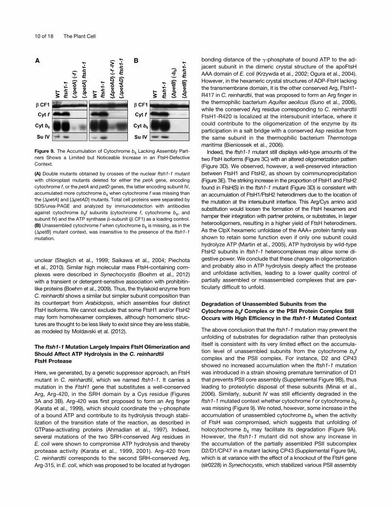
The presence of FtsH in PSII complexes was previously re-ported in cyanobacteria and Arabidopsis and proposed to allowimmediate access to damaged D1 and thereby efficient D1 deg-radation (Kashino et al., 2002; Yoshioka et al., 2010). However, wecould not coimmunoprecipitate PSII subunits with our antibody toFtsH1 (Supplemental Figure 6), which suggests either that thesehigh molecular mass complexes merely comigrate in BN-PAGE orthat they offer limited accessibility of the FtsH1 antigenic peptide.
FtsH(S) was found in the 150- to 200-kD region and is likely tocorrespond to an FtsH1/FtsH2 heterodimer. In stark contrast tothe wild type, which displayed most of the FtsH1 and FtsH2isoforms in the FtsH(L) population, up to approximately two-thirds of FtsH1 and FtsH2 were found in the FtsH(S) populationin the ftsh1-1 mutant. Notably, the PSII supercomplex level was
decreased in the ftsh1-1 mutant, in line with data on the Arabi-dopsis FtsH2 (var2) mutant (Kato et al., 2009). The similardistribution patterns of FtsH1 and FtsH2 suggested an in-teraction between the two within heterooligomers, which wasconfirmed by coimmunoprecipitation with purified antibodiesagainst a peptide of FtsH1 that does not cross-react with FtsH2(Figure 3E). In both the wild-type strain and the ftsh1-1 mutant,FtsH2 was pulled down together with FtsH1. Therefore, thephenotype of the ftsh1-1 mutant results from the expressionof a defective enzyme that is still made of FtsH1/FtsH2 het-erooligomers. However, the ftsh1-1 mutation prevents the in-tegration of most of the FtsH1/FtsH2 heterodimers in largersupercomplexes.
The Accumulation and Lifetime of Cytochrome b6fComplexes Lacking ci Heme Are Highly Increasedin the ftsh1-1 Context
The suppressor strategy that led us to the identification of theftsh1-1 mutant suggested that the assembled b6f complex isa substrate for the FtsH protease. We combined, by geneticcrosses, the ftsh1-1 mutation with the {petB-C35V} mutation,which prevents covalent binding of heme ci to cytochrome b6
and leads to a low accumulation level of cytochrome b6fcomplexes (de Vitry et al., 2004). As was the case for the ccb2ftsh1-1 mutant, the double mutant {petB-C35V} ftsh1-1 nowaccumulated wild-type levels of b6f complex subunits (com-pared with lanes 0 of the single and double mutants in Figure 5,right side). The accumulation of cytochrome b6f subunits inmutants defective for ci binding in the ftsh1-1mutated context isdue to a decrease in their rate of degradation, as also shown inFigure 5. After blocking protein synthesis in the chloroplast withchloramphenicol at time 0, we observed (over the 8-h time spanof the immunochase experiment) that the lifetime of cytochromeb6 and subunit IV showed a considerable increase when theftsh1-1 mutation was introduced into strains containing a mu-tation that impedes the binding of ci heme, as exemplified by theccb2 and {petB-C35V} mutations (Figures 5A and 5B). This re-sult demonstrates that the higher accumulation of b6f com-plexes in the double mutants is due to their lower rate ofdegradation. We attribute the limited degradation of b6f com-plexes still occurring in the double mutants to some residualFtsH activity and/or to other proteases (see Discussion).
The ftsh1-1 Mutation Increases PSII Sensitivity toPhotoinhibition, Prevents PSII Repair, and Leads to theAccumulation of D1 Fragments
Photoinhibition, a common feature of light stress in plants, al-gae, and cyanobacteria, occurs when the rate of light-induceddamage to PSII exceeds the repair capacity. The repair of PSIIinvolves partial disassembly of the damaged complex, selectiveproteolytic degradation and replacement of the damaged sub-unit (predominantly the D1 reaction center subunit) by a de novosynthesized copy, and reassembly. In plants and cyanobacteria,FtsH regulates PSII repair, through its ability to digest photo-oxidatively damaged D1 proteins, either alone in cyanobacteriaor in combination with Deg proteases in plants (reviewed in Kato
4 of 18 The Plant Cell

and Sakamoto, 2009; Nixon et al., 2010). The role of FtsH duringalgal photoinhibition is unclear, and the role of FtsH and thepossible contribution of chloroplast Deg proteases to this pro-cess has not been confirmed experimentally. The ftsh1-1mutantenabled us to address these issues.To explore further the strong decrease in Fv/Fm upon higher
light exposure and the photosensitive growth phenotype of theftsh1-1 mutant observed in Figures 2C and 2D, we monitoredPSII activity by measuring the Fv/Fm during a mild photo-inhibition treatment (250 mE m22 s21) and a subsequent re-covery under low light (6 mE m22 s21). The addition of inhibitorsof chloroplast translation, lincomycin and chloramphenicol (LC),prevents D1 synthesis and permits the discrimination of photo-damage from repair. As shown in Figure 6A, the ftsh1-1 mutationdramatically increased PSII sensitivity to a mild photoinhibition:over a 1-h illumination period with photoinhibitory light, the loss invariable fluorescence was more extensive and developed faster(Figure 6A, inset) in the mutant than in the wild type, and most ofthe functional recovery was prevented in a subsequent low-lightillumination period, irrespective of inhibitor addition. The meansand SD of PSII activity recovery after 1.5 h were 63% 6 12% forthe wild type and 19% 6 8% for the ftsh1-1 mutant (three in-dependent experiments). This lack of recovery is similar to whatwas observed in the wild-type strain when D1 synthesis was in-hibited and thus delineates the important role of FtsH in the PSIIrepair cycle during algal photoinhibition.Using antibodies against the DE loop or the C terminus of D1
(Figure 6B), we probed the fate of full-length D1 protein and theaccumulation of D1 fragments in the same experiment, usinglonger exposure times of the immunoblots for proper detectionof fragments (Figure 6C). Full-length D1 did not decrease duringphotoinhibition and recovery in the wild type or ftsh1-1 unlesstranslation inhibitors were added during the experiment (LCcondition). In the latter condition, the loss of D1 was more ex-tensive in the wild type than in the mutant, as viewed whenprobed with either of the two antibodies. However, D1 fragmentsaccumulated mainly in the ftsh1-1 mutant. They were detectedaround 23 kD with the anti-D1 DE loop (Figures 7B and 8B) andin the 16- to 20-kD and 6- to 10-kD regions with the anti-D1C terminus. We note that the 6- to 10-kD D1 fragments wereparticularly abundant in ftsh1-1 and even could be well detectedbefore photoinhibitory treatment (Figure 6C). We quantified thecontent in D1 upon photoinhibition by chemiluminescence usingthe D1 C-terminal antibody and OEE2 as a loading control whenrepair was prevented by the addition of translation inhibitors (LCcondition). The fraction of full-length D1 degraded over 4 h was53% in the wild type and 35% in ftsh1-1. When using the D1 DEloop antibody, we observed a similar increase in degradation offull-length D1 in the wild type as compared with the ftsh1-1mutant, with an additional 17% of full-length D1 being de-graded in the wild type only. C-terminal epitopes recovered in
Figure 3. Mutant ftsH1-R420C Accumulates Wild-Type Levels of FtsH1and FtsH2 but with Impaired Oligomerization.
(A) Schematic representation of ATP-dependent zinc metalloproteaseFtsH1 with the transit peptide, one thylakoid transmembrane helix (TMH),ATPase Walker A and B motifs, the SRH containing conserved Argresidues required for ATP hydrolysis (one of which is substituted to a Cysresidue in the ftsH1-R420C [ftsh1-1] mutant of C. reinhardtii ), and thezinc binding domain (Zn-BD).(B) Alignment of the SRH (in italics) from T. maritima FtsH (Tm-FtsH),E. coli FtsH (Ec-FtsH), and C. reinhardtii FtsH1 (Cr-FtsH1), indicating theconservation of the Arg and Thr residues (in black) substituted in theftsh1-1 and ftsh1-2 mutants.(C) Mutant ftsH1-R420C accumulates wild-type levels of FtsH1 andFtsH2. Total cell proteins were separated on an 8% polyacrylamide gel inthe presence of 8 M urea and analyzed by immunodetection with specificantibodies against FtsH1, FtsH2, and the ATP synthase b-subunit (bCF1) as a loading control.(D) FtsH complexes after BN-PAGE of digitonin-solubilized membranesfrom cells grown at 6 mE m22 s21. Digitonin-solubilized proteins wereseparated in the first dimension by BN-PAGE (3 to 12%) and in thesecond dimension as in (C) and blotted onto a polyvinylidene difluoridemembrane. The blot was sequentially probed with antibodies specific forFtsH2, FtsH1, and D1. FtsH1 and FtsH2 migrated mainly and similarly intwo populations, the smaller FtsH(S) and the larger FtsH(L) proteins. FtsH(L) is abundant in the wild type and FtsH(S) in ftsh1-1 (dashed boxes).The positions of unassembled proteins (U.P.), monomers [PSII(1)], dimers[PSII(2)], and supercomplexes [PSII(SC)] of PSII are indicated and usedas molecular mass markers.(E) Coimmunoprecipitation analysis of FtsH1 and FtsH2 interactions.Digitonin-solubilized membranes (prepared as for BN-PAGE) from thewild-type and ftsh1-1 strains were incubated with protein A–Sepharosebeads coupled to antibodies against a peptide of FtsH1 (a-FtsH1) or
against preimmune serum (pi). Aliquots of digitonin-solubilized mem-branes (sol) and immunoprecipitates (IP) were separated as in (C) andanalyzed by immunodetection with antibodies against FtsH1 (not cross-reacting with FtsH2, as indicated with gray letters) and FtsH2.
Cytochrome b6f and PSII Regulation by FtsH Protease 5 of 18

D1 fragments amounted to 20% of total initial D1 (full-lengthplus fragments) in the ftsh1-1 mutant, which indicates that D1fragmentation was actively occurring in these experimentalconditions. Thus, the defective activity of the FtsH protease im-pairs the degradation of damaged D1 and leads to some accu-mulation of D1 fragments. We note that the total amount ofantibody-recognized C-terminal epitopes, borne by D1 full-lengthprotein and fragments, decreased by 20% in the mutant in theLC condition, suggesting either that some degradation of the D1C-terminal fragments still occurs or that there is some pro-cessive degradation of D1 that does not result in the productionof D1 C-terminal fragments. A residual FtsH activity in the mu-tant may account for either of these two processes.
PSII Inactivation upon Phosphorus Starvation Is the Resultof Enhanced Photoinhibition
Interestingly, impaired PSII activity also has been reported whenC. reinhardtii is subjected to macronutrient stress, namely upon
phosphorus and sulfur starvation (Wykoff et al., 1998; Zhanget al., 2002). In both cases, a loss of PSII proteins upon star-vation was found responsible for PSII inactivation. The fact thatthe light intensity used in these experiments was ;80 mE m22 s21
suggested to us that a photoinhibitory effect might have developed,since macronutrient deprivation would lower the photosyntheticability to fix carbon. If such was the case, we predicted that theftsh1-1 mutant, being unable to repair damaged D1, would bemore sensitive than the wild type to these nutrient stresses andthat PSII inactivation would only occur in the light.We first subjected the ftsh1-1 mutant to phosphorus starva-
tion for 96 h either under illumination of 80 mE m22 s21 or inthe dark. The cell response marker to phosphorus deprivation,
Figure 4. Complementation of ftsh1-1 and ccb2 ftsh1-1 Strains withFTSH1 Genomic DNA Restores Wild-Type FtsH1 Phenotypes.
(A) Fluorescence induction kinetics in relative units (r.u.) at low actiniclight of dark-adapted cells followed by a saturating pulse to determineFm. Strains were grown at 6 mE m22 s21.(B) Fluorescence kinetics. Strains were grown at 6 mE m22 s21 followedby 19 h at 200 mE m22 s21.(C) Fluorescence kinetics. Strains were grown at 6 mE m22 s21.(D) Total cell proteins were separated on an 8% polyacrylamide gel in thepresence of 8 M urea and analyzed by immunodetection with specificantibodies against FtsH1. Strains ftsh1-1 and ccb2 ftsh1-1 were com-plemented with FTSH1 genomic DNA (pSL18-FTSH1) or as a controlwith empty pSL18 vector (pSL18).
Figure 5. Accumulation and Lifetime of b6f Complexes Lacking ci HemeAre Highly Increased in the ftsh1-1 Context.
(A) Immunochase of ftsh1-1, ccb2, and ccb2 ftsh1-1 mutants. Im-munochase was performed in the presence of 100 mg mL21 chloram-phenicol, an inhibitor of the translation of chloroplast genes. Total cellproteins were loaded at the chlorophyll constant, separated by SDS/urea-PAGE, and blotted onto a polyvinylidene difluoride membrane. Theblot was stained with Coomassie blue (CB) and analyzed by enhancedchemiluminescence immunodetection with antibodies against cytochromeb6f subunits and the nucleotide exchange factor GrpE.(B) Immunochase of the wild type and the {petB-C35V} and {petB-C35V}ftsh1-1 mutants in the same conditions as in (A).Note that for better comparison of the content in cytochrome b6f sub-units between mutants, shorter times of exposure of the immunoblotsare shown at the bottom part of each panel.
6 of 18 The Plant Cell

alkaline phosphatase (PHOX; Hallmann, 1999; Moseley et al.,2006), was induced in the light as well as in the dark in both thewild-type and ftsh1-1 strains (top of Figures 7B and 7D). At80 mEm22 s21, the ftsh1-1mutant showed a more rapid and muchlarger loss in PSII activity than the wild type, as determined bytheir respective changes in Fv/Fm (Figure 7A) and a net de-crease in the content of full-length D1 in the mutant at the latestage of starvation, probed with the D1 N-terminal antibody(Figure 7B, top). We then overexposed immunoblots reactedwith D1 antibodies to the DE loop and C terminus to examinethe pattern of accumulation of D1 fragments upon phospho-rus starvation under 80 mE m22 s21 in the ftsh1-1 mutatedcontext. This pattern was similar to the one observed uponphotoinhibition, showing an increased accumulation of D1 frag-ments, around 23 kD and in the 16- to 20-kD and 6- to 10-kD
regions (Figure 7B). When phosphorus starvation was per-formed in the dark, both the ftsh1-1 and wild-type strainspreserved PSII activity over a period of 96 h (Figure 7C), andneither of them showed any accumulation of D1 fragments(Figure 7D). These results clearly demonstrate that compromisedphotosynthetic activity upon phosphorus starvation merely re-sults from enhanced photoinhibition.
Figure 6. ftsh1-1 Increases PSII Sensitivity to Photoinhibition, PreventsPSII Repair, and Leads to the Accumulation of D1 Fragments.
(A) Time course of photoinhibition at 250 mE m22 s21 and recovery at6 mE m22 s21 in wild-type and ftsh1-1 strains in the absence or presenceof inhibitors of the translation of chloroplast genes (500 mg mL21 linco-mycin and 100 mg mL21 chloramphenicol [LC]) as monitored by PSIIactivity (Fv/Fm). The inset shows kinetics of the PSII activity loss uponphotoinhibition in an independent experiment.(B) D1 model with the localization of the D1 DE loop and C-terminalpeptides recognized by antibody (adapted from Kapri-Pardes et al.,2007).(C) Time course of photoinhibition and recovery in wild-type and ftsh1-1strains as monitored by levels of D1 and D1 fragments immunodetectedwith D1 DE loop and C-terminal antibodies using the PSII OEE2 extrinsicsubunit as a loading control. Total cell proteins were separated by SDS/urea-PAGE.
Figure 7. PSII upon Phosphorus Starvation in Wild-Type and ftsh1-1Strains.
(A) Time course of phosphorus starvation under 80 mE m22 s21 asmonitored by PSII activity detection.(B) Time course of phosphorus starvation under 80 mE m22 s21 asmonitored by immunodetection of inducible PHOX (a marker of the cellresponse to phosphorus deprivation), full D1 and D1 fragments with D1DE loop and C-terminal antibodies, cytochrome b6f subunit IV, and light-harvesting LhcbM5. Total cell proteins were separated on a 7.5 to 15%polyacrylamide gel in the presence of 8 M urea.(C) Time course of phosphorus starvation in the dark as monitored byPSII activity detection.(D) Time course of phosphorus starvation in the dark as monitored byimmunodetection.Means and SD of two independent experiments for each starvationcondition are given.
Cytochrome b6f and PSII Regulation by FtsH Protease 7 of 18

Compromised Photosynthetic Activity upon SulfurStarvation under Illumination Is Due to BothPhotoinhibition and Degradation of b6f Complexes
When the ftsh1-1 mutant was starved of sulfur under illuminationof 80 mE m22 s21 over 96 h, it showed a larger PSII inactivationthan the wild type (Figure 8A). This observation suggests, hereagain, an efficient FtsH-dependent repair process upon sulfurstarvation of C. reinhardtii wild-type cells, similar to the repairupon photoinhibition. Indeed, the decrease in D1 content duringsulfur starvation was far more pronounced in the mutant than inthe wild type (Figure 8B).
However, there were marked differences with regard to phos-phorus starvation or a regular photoinhibition process: the cyto-chrome b6f complex, as probed with an antibody against theRieske protein subunit, was now targeted to degradation inthe two strains, and the drastic degradation of full-length D1 in theftsh1-1mutant was accompanied by a transient overaccumulationof D1 fragments at 21 h of starvation, which were subsequentlydisposed of. Whereas at a late stage of sulfur starvation there wasa general decrease in protein content in the mutant, as shown bythe decreased content in the cell response marker to sulfur dep-rivation, SLT2 (Pootakham et al., 2010), and the b-subunit of CF1(Figure 8B), we noted that the other subunits of the PSII core, D2and CP47, better resisted degradation than D1. This result raisedthe question of whether D2 and CP47 were in a disassembledstate or perhaps still assembled with fragmented D1 in the ftsh1-1mutant. We assayed this hypothesis by two-dimensional BN/SDS-PAGE of dodecyl-maltoside–solubilized membranes isolated after39 h of sulfur starvation (Figure 8E, bottom). We found that frag-mented D1 remained assembled within PSII monomers and re-action center cores lacking CP43 (RC47) in the ftsh1-1 mutant.This finding is in line with what has been reported for the Arabi-dopsis FtsH2 (var2) mutant after 1 h of extreme high-light exposureat 2500 mE m22 s21 (Kato et al., 2012). We note that, beforestarvation, PSII supercomplexes were far more abundant in thewild type than in the ftsh1-1 strain (Figure 8E, top), as also wassuggested by the analysis of digitonin-solubilized membranes(Figure 3D).
We also examined the behavior of the wild type and the ftsh1-1mutant when sulfur starvation was performed in photoautotrophicconditions, under an illumination of 80 mE m22 s21 in the absenceof the carbon source acetate. Both strains showed a delayed lossin PSII activity and downstream electron transfer (SupplementalFigures 7A to 7D). They also showed a retarded induction ofthe sulfate transporter SLT2, probably due to their slower growthand lower sulfur consumption in minimal medium (SupplementalFigures 7E and 7F).
The ftsh1-1 Mutant Conserves Most PSII and b6fComplexes upon Sulfur Starvation in the Dark,in Contrast to the Wild-Type Strain
To further understand to what extent photoinhibitory processescontributed to the loss of PSII upon sulfur starvation, we per-formed the same starvation experiments in complete darkness. Toour surprise, wild-type cells showed a more dramatic inactivationof PSII (Figure 8C), which was now accompanied by a net loss of
D1 and D2 proteins. Thus, the repair process that was efficient inthe light (Figures 8A and 8B) was either heavily compromised inthe dark or unable to make up for higher rates of PSII degradation(Figures 8C and 8D). That the process of PSII degradation in thedark was widely different from the one operating under illuminationwas demonstrated by the behavior of the ftsh1-1 mutant, whichpreserved fully active PSII when starved of sulfur in the dark(Figures 8C and 8D). SLT2 was induced upon sulfur depletion inthe dark in the two strains, indicating that the differences in PSIIdegradation should not be attributed to an absence of signaling ofsulfur deficiency in the ftsh1-1 mutant. The cytochrome b6f com-plexes were degraded in the wild type starved of sulfur in the dark,as they were under 80 mE m22 s21, but this degradation in the darkwas now largely FtsH dependent, since the ftsh1-1 mutantshowed very limited cytochrome b6f degradation in contrast toits behavior in the light (Figures 8B and 8D).The accumulation pattern of D1 fragments upon sulfur star-
vation of the ftsh1-1 mutant in the dark also differed from theone observed upon photoinhibition, with an absence of 23-kDfragments (Figure 8D). This is consistent with the previouslycharacterized light-induced production of a 23-kD D1 fragmentas a substrate of the thylakoid FtsH protease (Lindahl et al.,2000). Importantly, the 6- to 10-kD and 16- to 20-kD fragmentsstill accumulated in the dark. We also note that sulfur starvationinduced extensive accumulation of D1/D2 oligomers of highmolecular weight in the dark but not in the light in the ftsh1-1mutated context only (Supplemental Figure 8).Finally, we tested whether increased expression in some other
major chloroplast proteases would have occurred upon FtsHinactivation, which would explain the more pronounced PSIIdegradation in the ftsh1-1 mutant upon sulfur deprivation inmixotrophic conditions. We used antibodies against FtsH1/FtsH2, stromal ClpP1, lumenal Deg5, and stromal membrane–associated SppA1 to probe the content of major chloroplastproteases in wild-type and ftsh1-1 cells starved of sulfur in thepresence or absence of acetate (Supplemental Figures 7E and7F). The patterns of accumulation were rather similar for the twostrains, indicating that there was no upregulation of the probedproteases in the FtsH-defective mutant. Sulfur starvation causeda rapid loss in Deg5 and ClpP1 in the two strains and someprocessing of SppA1 in mixotrophic conditions only. FtsH andSppA1 were better preserved than Deg5 and ClpP1 when sulfurstarvation was performed in the absence of acetate, especiallyin the ftsh1-1 mutant (Supplemental Figure 7F).
The ftsh1-1 Mutation Has a Limited, if Any, Ability to Preventthe Degradation of Unassembled Subunits from theCytochrome b6f Complex or the PSII Protein Complex
A concerted accumulation of PSII subunits that make up anactive PSII protein has been demonstrated to result from aposttranslational regulation leading to the proteolytic disposal ofunassembled subunit D2 and CP43 and a downregulation of thetranslation of subunit D1 or CP47, which are control by epistasyof synthesis (CES) subunits (de Vitry et al., 1989; Minai et al.,2006). We crossed the ftsh1-1 nuclear mutant to chloroplastmutants with a deleted PsbC (CP43) gene or harboring a pre-mature stop codon in PsbA that leads to the expression of
8 of 18 The Plant Cell

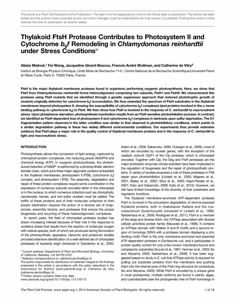
a truncated and labile D1. We found no evidence that the mutationftsh1-1 rescued the accumulation of the other PSII subunits whenPSII assembly was compromised in these mutants (SupplementalFigure 9). To assess whether this protease also determines thelifetime of unassembled subunits of the cytochrome b6f com-plex, most of which are subjected to rapid degradation, withthe exception of cytochrome f, which is a CES subunit (Kurasand Wollman, 1994; Choquet et al., 1998), we tested whetherthe mutation ftsh1-1 rescues the accumulation of cytochromeb6 in the absence of cytochrome f and vice versa. To this end,we crossed the nuclear ftsh1-1 mutant with chloroplast mu-tants lacking either the petA gene, encoding cytochrome f,the petA and petD genes, the latter encoding subunit IV, orthe petB gene, encoding cytochrome b6. The accumulation ofunassembled cytochrome b6 showed a limited but noticeableincrease in an FtsH-defective context (Figure 9A), whereas un-assembled cytochrome f, whose decreased accumulation is dueto a decreased translation initiation according to the CES pro-cess, proved, as expected, insensitive to the status of the FtsHprotease (Figure 9B). In these experiments, the level of accu-mulation of subunit IV in the absence of its assembly partnersremained below detectable levels.
DISCUSSION
The Thylakoid FtsH Protease of C. reinhardtii FormsHeterooligomers Comprising FtsH1 and FtsH2
The C. reinhardtii nuclear genome encodes six members of theFtsH family distributed between the mitochondria (Atteia et al.,2009) and the chloroplast and three FtsH-like proteins (FHL1 toFHL3) that retain a complete AAA+ domain but lack the zincbinding motif (reviewed in Schroda and Vallon, 2009; see phy-logenetic tree of FtsH proteins in Supplemental Figure 1). Onlytwo isoforms have been identified in C. reinhardtii thylakoidmembranes: FtsH1 (type A) and FtsH2 (type B; Allmer et al.,2006). Our two-dimensional BN/SDS-PAGE and coimmuno-precipitation experiments demonstrate that FtsH1 also formsheterocomplexes with FtsH2 in C. reinhardtii (Figures 3D and3E). FtsH(L), however, has a much larger apparent molecularmass than expected for a heterohexamer (;410 kD, to whichshould be added a digitonin contribution that has a micellarmass of ;70 kD). Such large FtsH-containing complexes,around or above 1 MD, were previously observed in E. coli,yeast mitochondria, and Arabidopsis mitochondria, where theyalso include prohibitin-like proteins whose function remains
Figure 8. PSII and Cytochrome b6f upon Sulfur Starvation in Wild-Typeand ftsh1-1 Strains.
(A) Time course of sulfur starvation under 80 mE m22 s21 as monitoredby PSII activity detection.(B) Time course of sulfur starvation under 80 mE m22 s21 as monitoredby immunodetection of a cell response marker to sulfur deprivation(sulfate transporter SLT2) and of PSII subunits (D1, D2, and CP47), cy-tochrome b6f subunits (IV and Rieske), and light-harvesting LhcbM5.Total cell proteins were separated as in Figure 7.(C) Time course of sulfur starvation in the dark as monitored by PSIIactivity detection.(D) Time course of sulfur starvation in the dark as monitored byimmunodetection.(E) PSII complexes after BN-PAGE of dodecyl-maltoside–solubilizedmembranes at 0 and 39 h of sulfur deprivation under 80 mE m22 s21 were
separated by two-dimensional BN (3 to 12%)/SDS-PAGE (12% in thepresence of 8 M urea), blotted onto a polyvinylidene difluoride mem-brane, and immunodetected. The positions of unassembled proteins(U.P.), a reaction center core lacking CP43 (RC47), and monomers [PSII(1)],dimers [PSII(2)], and supercomplexes [PSII(SC)] of PSII are indicated. At0 h, supercomplexes are more abundant in the wild-type than in ftsh1-1;at 39 h, fragmented D1 was found assembled in monomeric PSII andRC47 complexes in the ftsh1-1 mutant (dashed boxes).Means and SD of two independent experiments for each starvationcondition are given.
Cytochrome b6f and PSII Regulation by FtsH Protease 9 of 18
unclear (Steglich et al., 1999; Saikawa et al., 2004; Piechotaet al., 2010). Similar high molecular mass FtsH-containing com-plexes were described in Synechocystis (Boehm et al., 2012)with a transient or detergent-sensitive association with prohibitin-like proteins (Boehm et al., 2009). Thus, the thylakoid enzyme fromC. reinhardtii shows a similar but simpler subunit composition thanits counterpart from Arabidopsis, which assembles four distinctFtsH isoforms. We cannot exclude that some FtsH1 and/or FtsH2may form homohexamer complexes, although homomeric struc-tures are thought to be less likely to exist since they are less stable,as modeled by Moldavski et al. (2012).
The ftsh1-1 Mutation Largely Impairs FtsH Olimerization andShould Affect ATP Hydrolysis in the C. reinhardtiiFtsH Protease
Here, we generated, by a genetic suppressor approach, an FtsHmutant in C. reinhardtii, which we named ftsh1-1. It carries amutation in the FtsH1 gene that substitutes a well-conservedArg, Arg-420, in the SRH domain by a Cys residue (Figures3A and 3B). Arg-420 was first proposed to form an Arg finger(Karata et al., 1999), which should coordinate the g-phosphateof a bound ATP and contribute to its hydrolysis through stabi-lization of the transition state of the reaction, as described inGTPase-activating proteins (Ahmadian et al., 1997). Indeed,several mutations of the two SRH-conserved Arg residues inE. coli were shown to compromise ATP hydrolysis and therebyprotease activity (Karata et al., 1999, 2001). Arg-420 fromC. reinhardtii corresponds to the second SRH-conserved Arg,Arg-315, in E. coli, which was proposed to be located at hydrogen
bonding distance of the g-phosphate of bound ATP to the ad-jacent subunit in the dimeric crystal structure of the apoFtsHAAA domain of E. coli (Krzywda et al., 2002; Ogura et al., 2004).However, in the hexameric crystal structures of ADP-FtsH lackingthe transmembrane domain, it is the other conserved Arg, FtsH1-R417 in C. reinhardtii, that was proposed to form an Arg finger inthe thermophilic bacterium Aquifex aeolicus (Suno et al., 2006),while the conserved Arg residue corresponding to C. reinhardtiiFtsH1-R420 is localized at the intersubunit interface, where itcould contribute to the oligomerization of the enzyme by itsparticipation in a salt bridge with a conserved Asp residue fromthe same subunit in the thermophilic bacterium Thermotogamaritima (Bieniossek et al., 2006).Indeed, the ftsh1-1 mutant still displays wild-type amounts of the
two FtsH isoforms (Figure 3C) with an altered oligomerization pattern(Figure 3D). We observed, however, a well-preserved interactionbetween FtsH1 and FtsH2, as shown by coimmunoprecipitation(Figure 3E). The striking increase in the proportion of FtsH1 and FtsH2found in FtsH(S) in the ftsh1-1 mutant (Figure 3D) is consistent withan accumulation of FtsH1/FtsH2 heterodimers due to the location ofthe mutation at the intersubunit interface. This Arg/Cys amino acidsubstitution would loosen the formation of the FtsH hexamers andhamper their integration with partner proteins, or substrates, in largerheterooligomers, resulting in a higher yield of FtsH heterodimers.As the ClpX hexameric unfoldase of the AAA+ protein family wasshown to retain some function even if only one subunit couldhydrolyze ATP (Martin et al., 2005), ATP hydrolysis by wild-typeFtsH2 subunits in ftsh1-1 heterocomplexes may allow some di-gestive power. We conclude that these changes in oligomerizationand probably also in ATP hydrolysis deeply affect the proteaseand unfoldase activities, leading to a lower quality control ofpartially assembled or misassembled complexes that are par-ticularly difficult to unfold.
Degradation of Unassembled Subunits from theCytochrome b6f Complex or the PSII Protein Complex StillOccurs with High Efficiency in the ftsh1-1 Mutated Context
The above conclusion that the ftsh1-1 mutation may prevent theunfolding of substrates for degradation rather than proteolysisitself is consistent with its very limited effect on the accumula-tion level of unassembled subunits from the cytochrome b6fcomplex and the PSII complex. For instance, D2 and CP43showed no increased accumulation when the ftsh1-1 mutationwas introduced in a strain showing premature termination of D1that prevents PSII core assembly (Supplemental Figure 9B), thusleading to proteolytic disposal of these subunits (Minai et al.,2006). Similarly, subunit IV was still efficiently degraded in theftsh1-1mutated context whether cytochrome f or cytochrome b6
was missing (Figure 9). We noted, however, some increase in theaccumulation of unassembled cytochrome b6 when the activityof FtsH was compromised, which suggests that unfolding ofholocytochrome b6 may facilitate its degradation (Figure 9A).However, the ftsh1-1 mutant did not show any increase inthe accumulation of the partially assembled PSII subcomplexD2/D1/CP47 in a mutant lacking CP43 (Supplemental Figure 9A),which is at variance with the effect of a knockout of the FtsH gene(slr0228) in Synechocystis, which stabilized various PSII assembly
Figure 9. The Accumulation of Cytochrome b6 Lacking Assembly Part-ners Shows a Limited but Noticeable Increase in an FtsH-DefectiveContext.
(A) Double mutants obtained by crosses of the nuclear ftsh1-1 mutantwith chloroplast mutants deleted for either the petA gene, encodingcytochrome f, or the petA and petD genes, the latter encoding subunit IV,accumulated more cytochrome b6 when cytochrome f was missing thanthe {DpetA} and {DpetAD} mutants. Total cell proteins were separated bySDS/urea-PAGE and analyzed by immunodetection with antibodiesagainst cytochrome b6f subunits (cytochrome f, cytochrome b6, andsubunit IV) and the ATP synthase b-subunit (b CF1) as a loading control.(B) Unassembled cytochrome f when cytochrome b6 is missing, as in the{DpetB} mutant context, was insensitive to the presence of the ftsh1-1mutation.
10 of 18 The Plant Cell

intermediates in PSII mutants lacking CP43 or impaired in theassembly or stability of the manganese cluster (Komenda et al.,2006, 2010). Although we cannot fully exclude a residual FtsHactivity that would degrade these unassembled, or partly as-sembled, subunits in the ftsh1-1 mutated context, we tenta-tively attribute these differences to the lower protein qualitycontrol in cyanobacterial thylakoid membranes when comparedwith C. reinhardtii chloroplasts (Wollman et al., 1999). In partic-ular, crippled PSII complexes are less efficiently degraded inSynechocystis (de Vitry et al., 1989; Vermaas, 1998) than in pho-tosynthetic eukaryotes. These observations suggest the presenceof an as yet unidentified additional housekeeping protease thatdegrades these subunits in the thylakoid membranes ofC. reinhardtii.
FtsH Is Involved in Quality Control and the Regulation ofAccumulation of Cytochrome b6f Complexes andCCB Proteins
The first suggestion of a cytochrome b6f subunit degradedby FtsH was the unassembled Rieske iron-sulfur protein im-ported into isolated chloroplasts in vitro (Ostersetzer and Adam,1997). The involvement of FtsH in cytochrome b6f degrada-tion in vivo is demonstrated here with the identification of theftsh1-1 mutant by the present genetic suppressor strategy. Inmarked contrast to the continued degradation of the cyto-chrome b6f subunits when a subunit is absent (Figure 9), a di-versity of labile ci heme–deficient cytochrome b6f complexesproved resistant to degradation in the ftsh1-1 mutated context.They ranged from site-directed substitution of the Cys involvedin the binding of the ci heme (Figure 5) to site-directed sub-stitution of the His ligand of the bh heme (Malnoë et al., 2011)and to mutants in the CCB heme binding pathway (Figure 2).These observations demonstrate that FtsH operates as a majorquality control protease that recognizes altered conformationsin cytochrome b6f assemblies to trigger their degradation throughthe combined action of its unfoldase and proteolytic activities.A similar situation may prevail in Saccharomyces cerevisiaemitochondria, where FtsH proteases with the catalytic sitefacing the matrix (m-FtsH) Yta10/Yta12 have been describedas involved in maturase-dependent intron splicing of cytochromeb transcripts. However, when using an intronless mitochon-drial genome where cytochrome b synthesis was not affected,m-FtsH was shown to contribute to posttranslational bc1 com-plex assembly and degradation (Guzélin et al., 1996; Arlt et al.,1998; Van Dyck and Langer, 1999). As we report here for sulfurstarvation (Figures 8B and 8D) and elsewhere for nitrogenstarvation (Bulté and Wollman, 1992; Wei et al., 2013), FtsH hasa prominent role in regulating the accumulation level of cyto-chrome b6f complexes upon these macronutrient stresses in lowlight or in the dark.
We also found that degradation of unassembled CCB4 in theabsence of CCB2 (and vice versa), both of which are trans-membrane proteins involved in the ci heme binding pathway tocytochrome b6 (Saint-Marcoux et al., 2009), proved to be reg-ulated by FtsH (Figure 1B), in line with our report elsewhere (Weiet al., 2013) of a global role for FtsH in the degradation ofphotosynthetic complexes and their assembly factors.
The ClpP protease also has been demonstrated to play a rolein cytochrome b6f degradation inC. reinhardtii (Majeran et al., 2000),as does Deg1 on cytochrome b6 in Arabidopsis (Zienkiewicz et al.,2012). However, Clp and FtsH may have distinct actions on thedegradation of cytochrome b6f complexes, since a Clp-deficientmutant was unable to protect b6f complexes from degradation inmutants lacking ci heme (Majeran et al., 2000), while the ftsh1-1mutation did so (Figure 5).Here, we observed a cytochrome b6f degradation process that
was selectively activated in sulfur-deficient conditions. Cyto-chrome b6f was degraded in the light but not in darkness in theftsh1-1 mutant, suggesting the action of some additional pro-tease upon illumination. Steady state levels of the chloroplastproteases that we probed, including ClpP1 and Deg5, wererather reduced upon sulfur starvation in the light, but we cannotrule out that some proteases might have better access to cy-tochrome b6f or be activated by oligomerization in the light inthese starvation conditions.
FtsH1 Regulates the Degradation of PSII uponPhotoinhibition and Phosphorus and Sulfur Starvation
In cyanobacteria, FtsH has mainly been studied for its role in D1degradation (reviewed in Nixon et al., 2010; Komenda et al.,2012) and the removal of other damaged or unassembled PSIIproteins (Komenda et al., 2006, 2010; Boehm et al., 2011). Inone study, FtsH was also shown to contribute to the regulationof PSI amounts (Mann et al., 2000). The mechanism for D1protein degradation is not fully elucidated, but it is known toinvolve the reaction of damaging reactive oxygen species, par-tial disassembly of the PSII complex to form the RC47 complex,and the action of several proteases, notably FtsH, that selec-tively remove damaged D1 (reviewed in Edelman and Mattoo,2008; Komenda et al., 2012). The best-documented functionof FtsH in thylakoid membranes is in this PSII repair cycle(reviewed in Kato and Sakamoto, 2009). A model for the pho-toinhibition of cyanobacteria has been proposed in whichdamaged D1 is removed by FtsH heterohexamers, with degrada-tion proceeding from the N terminus of D1 in a highly processivemanner without the requirement of any other proteases (re-viewed in Nixon et al., 2010). In particular, photoinhibitionstudies with the DhtrA/hhoA/hhoB triple mutant, defective forthe three genes encoding Deg proteases in Synechocystis spPCC6803, showed that Deg proteases are not required for theremoval of damaged D1 protein during the PSII repair cyclewhen exposed to high light intensity (Barker et al., 2006). Theseresults are in contrast to several photoinhibition studies per-formed with Arabidopsis showing that PSII protein turnover re-quired both the involvement of FtsH and Deg/Htr proteases(Itzhaki et al., 1998; Kapri-Pardes et al., 2007; Sun et al., 2007).High-light treatment was reported to increase the expression ofDeg/Htr and FtsH types of proteases in Arabidopsis (Sinvany-Villalobo et al., 2004), to raise the level of FtsH hexamerizationand unstacking of thylakoids (reviewed in Yoshioka and Yamamoto,2011), and to activate the lumenal Deg protease, based onin vitro experiments showing its hexamerization upon acidi-fication (Kley et al., 2011). Thus, a model for D1 degradationin the chloroplast was proposed in which Deg proteases act as
Cytochrome b6f and PSII Regulation by FtsH Protease 11 of 18

endoproteases fragmenting D1 and FtsH proteases act as exo-proteases processively degrading D1 and its fragments (reviewedin Kato and Sakamoto, 2009). The reason why cyanobacterialFtsH would be capable of processive degradation of the whole D1protein from the stromal side, while the ability of its homolog inthe chloroplast is far reduced, may rest in the difference in subunitcomposition of FtsH from the two types of organisms as well asfrom the embedment of chloroplast D1 in grana membranes,where it has increased interaction with transmembrane antennaproteins and lumenal oxygen-evolving enhancer subunits (forfurther discussion, see Edelman and Mattoo, 2008).
Here, in agreement with previous photoinhibition studies fromvascular plant chloroplasts and cyanobacteria, we showed thatthe ftsh1-1 mutation increased PSII sensitivity to photoinhibitionand impaired proteolytic degradation and replacement of D1 bya de novo synthesized copy (Figure 6). There is a striking similarityin the fate of D1 in the ftsh1-1 mutant when subjected to lightstress or phosphorus starvation under moderate illumination, bothof which lead to PSII inactivation in C. reinhardtii (Wykoff et al.,1998; Zhang et al., 2002). Here, we have demonstrated that,indeed, compromised photosynthetic activity upon phosphorusstarvation was mainly due to enhanced photoinhibition (Figure 7).In contrast, PSII inactivation upon sulfur starvation cannot beattributed solely to photoinhibition, since it develops even morestrongly when sulfur starvation is performed in complete darkness.Since PSII inactivation and D1 degradation in the ftsh1-1 mutantare markedly increased upon sulfur starvation in the light but fullyprevented when performed in darkness (Figure 8), one couldconsider a light-induced upregulation of ClpP when FtsH activityis defective, as recently demonstrated (Kato et al., 2012). Althoughwe did not detect more ClpP in ftsh1-1 than in the wild-type strainin these conditions (Supplemental Figure 7E), we cannot excludean upregulation of ClpP activity through changes in its state ofoligomerization, substrate accessibility, or ATP availability.
FtsH1 Regulates the Degradation of PSII by RemovingD1 Fragments
A clear consequence of the inactivation of FtsH in the ftsh1-1mutant was the detection of D1 fragments upon photoinhibition,which suggests that primary endoproteolytic cuts occurred in themutant but that their processive degradation by FtsH was inhibited(Figure 6C). According to what has been proposed for plantchloroplasts, there could be a joint action of Deg and FtsH pro-teases in the degradation and repair of D1 upon photoinhibition ofC. reinhardtii. Indeed, the set of chloroplast Deg proteases is similarin Arabidopsis (lumenal Deg1, Deg5, and Deg8 and stromal Deg2,Deg6, Deg7, Deg9, and Deg16) and in C. reinhardtii (lumenalDeg1A, Deg1B, Deg1C, Deg5, and Deg8 and stromal Deg2, Deg7,and Deg9; Huesgen et al., 2009; Schroda and Vallon, 2009;Sun et al., 2010). The model of Kato and Sakamoto (2009), derivedfrom studies with Arabidopsis, predicts the production of Deg-dependent fragments whose apparent molecular mass and epitopecontent match nicely to those we detected with the two distinctD1 antibodies we used (Supplemental Figure 10).
The physiological significance of the Deg-dependent D1degradation pathway has been under debate (for discussion,see Edelman and Mattoo, 2008; Komenda et al., 2012). Is it
synergistic with FtsH, or is it an escape pathway that supple-ments the FtsH-mediated one to accelerate D1 degradationunder photoinhibitory conditions? We conclude that in our ex-perimental conditions, when the function of FtsH is impaired, theDeg pathway would be rather active, with 20% of the C-terminalepitopes of D1 being found in its fragments (Figure 6C).We note, however, that most of these fragments accumulated
to a limited extent. This could be due to a negative feedbackinhibition of the Deg proteases by the accumulation of theseearly D1 degradation products whose stroma-exposed domainsare not processed by FtsH. This is consistent with the obser-vation that the only degradation product that accumulates ex-tensively in the ftsh1-1 mutant is in the ;6-kD region, where theC-terminal fragment of D1, which is lumen localized, should befound. This fragment differs from all the others in that it shouldnever have access to the stroma-located proteolytic chamber ofFtsH in a wild-type context and, therefore, should not be de-graded by FtsH. Its increased production in the ftsh1-1 mutant,as compared with the wild type, probably results from the slowerdigestion of D1 products by FtsH, thus releasing more 6-kDfragments in the lumen.A conspicuous feature of the various PSII degradation pro-
cesses upon macronutrient starvation, whether it be phospho-rus or sulfur starvation under moderate illumination or sulfurstarvation in the dark, is the production of D1 fragments in theftsh1-1 mutated context. With the exception of the 23-kD frag-ment, which is detected only upon illumination and serves asa diagnostic of photoinhibition, as suggested previously (Lindahlet al., 2000), the D1 fragmentation patterns upon macronutrientstress (Figures 7B, 8B, and 8D) are very similar to one resultingfrom photoinhibition (Figure 6C), with a group of fragments ac-cumulating in the 16- to 20-kD and 6- to 10-kD regions. Theseobservations strongly suggest a contribution of the Deg pro-tease family to the degradation of D1 upon nutrient stress asthey do upon photoinhibition. Indeed, Degs were also proposedto contribute to D1 degradation under stress conditions bycleaving cross-links to neighboring proteins (Komenda et al.,2012). A modest expression increase in lumenal Deg1C inC. reinhardtii has been found upon phosphorus and sulfur star-vation (Zhang et al., 2004; Moseley et al., 2006), and a 3-foldincrease of the transcript level of stromal Deg2 upon sulfurstarvation in the light in the wild-type strain was also observed(González-Ballester et al., 2010). Here, however, we detecteda net decrease of the amount of chloroplast proteases uponsulfur depletion in the presence of acetate (Supplemental Figure7E). In the latter experimental conditions, D1 fragments tran-siently overaccumulate in the ftsh1-1 mutant (Figure 8B), whichsupports a degradation pathway in which Deg proteases pro-duce a first set of early endoproteolytic cuts that can be con-tinued only once the resulting D1 fragments are themselvesdegraded by processive proteases such as FtsH.It is of note that light-induced oxidative damage to D1 should
not be considered as a prerequisite for the activation of itsdegradation by the combined effects of FtsH and Deg pro-teases, since D1 degradation yields the same 16- to 20-kD and6- to 10-kD fragments when operating in the dark in sulfur stressconditions (Figure 8D). However, the activation of the Deg pro-teases in the lumen has been shown to be triggered by the high
12 of 18 The Plant Cell

DpH formed in the light (Kley et al., 2011). How they are acti-vated in the dark remains unclear unless a high proton gradientis present during sulfur starvation in the dark, an issue that needsto be addressed in the future.
Enhanced Degradation of PSII upon Sulfur Starvation in theDark Is Fully FtsH Dependent
That PSII degradation was dramatically enhanced when sulfurstarvation was performed in the dark was unexpected (Figure 8D).In this case, PSII degradation became a fully FtsH-dependentprocess. The rationale for this targeted degradation of PSII indarkness upon sulfur starvation remains unclear. It is not aconsequence of functional damage to PSII complexes, since theftsh1-1 mutant, which does not degrade PSII, displays a fluo-rescence pattern with a high Fv/Fm typical of active PSII centers(Figure 8C). It should not be aimed either at scavenging sulfur,since most of the sulfur-containing cofactors for photosynthesisare found within PSI and not in association with PSII. Interestingly,we noted a spectacular increase in the accumulation of D1/D2oligomers in the ftsh1-1 mutant relative to the wild type whendeprived of sulfur in the dark (Supplemental Figure 8). Similarcross-linking products or adducts had been reported previouslyeither during a light stress or in the dark when a heat or oxidativestress was applied (reviewed in Yamamoto et al., 2008). The ac-cumulation of these oligomers in the dark during a nutrient stressis highly unusual and raises the question of how they are formed.These oligomers may be the privileged substrate for PSII degra-dation by FtsH upon sulfur stress in the dark. Being resistant toother proteases, they might be a form of active PSII, which isworthy of further investigation.
The mechanism of activation of this degradation pathway isunknown and might be related to sulfur stress–induced ex-pression of chaperones. Indeed, it has been shown in E. coli thatchaperones can facilitate degradation by FtsH (Rodriguez et al.,2008). The chloroplast DnaJ chaperone Atg8, induced in dark-ness and stabilized by FtsH (Chen et al., 2011), is thus a goodcandidate for being involved in the dark-activated and FtsH-dependent degradation of PSII upon sulfur deprivation.
METHODS
Phylogenetic Analysis
Multiple sequence alignment was performed using ClustalW2.1 (Larkinet al., 2007) with default parameters followed by manual editing. After re-moving ambiguously aligned N- and C-terminal regions, 639 sites wereretained for the phylogenetic analysis (Supplemental Data Set 1). Thebest-fit amino acid substitution model was determined using ProtTest2.4(Abascal et al., 2005). The best-fit modelwas the Le-Gascuelmodel (Le andGascuel, 2008) supplementedwith a g distribution for rate variations amongsites (LG+G). The tree was reconstructed by the maximum likelihoodmethod based on the LG+G model using RaxML7.2.6 (Stamatakis, 2006).Statistical support for nodes was assessed with 1000 bootstrap replicates.
Chlamydomonas reinhardtii Strains, Growth Conditions, andGenetic Methods
C. reinhardtii mutants have been described previously and were as follows:ccb, {petB-C35V}, {petB-H202Q}, {petB-H187G}, {DpetB}, {DpetA}, {Fud34},
{Fud7}, {D1TR}, arg7, and arg2 (Eversole, 1956; Matagne, 1978; de Vitryet al., 1989, 2004; Kuras andWollman, 1994; Kuras et al., 1997, 2007; Minaiet al., 2006). Cells were grown at 25°C in Tris-acetate-phosphate (TAP)medium or, when indicated, in minimal medium lacking acetate, pH 7.2,under dim light at 6 mE m22 s21 and collected during the exponential phaseat 2 3 106 cells mL21. Plasmid pDpetA-D was constructed by ligating theScaI-PstI fragment of 4296 bp of plasmid pDpetA (Kuras andWollman, 1994)bearing theDpetA deletionwith theScaI-PstI fragment of 2134 bp of plasmidpKfDpetD (Choquet et al., 2003) bearing the DpetD deletion and containingthe aadA cassette conferring spectinomycin resistance. Plasmid pDpetA-Dwas transformed by the biolistic method into C. reinhardtii wild-type strain(Boynton et al., 1988; Kuras andWollman, 1994). Nonphotosynthetic {DpetAD}transformants were screened in the dark on TAP-agar-spectinomycin me-dium, and homoplasmy was checked by RNA gel blotting for the absence ofPETA andPETDmRNAasdescribed (Drapier et al. 2002)with probes derivedfrom coding sequences (Eberhard et al., 2002). UV light mutagenesis onccb strains to obtain phototrophic revertants was performed as described(Li et al., 1996). Plasmid pSL18-FTSH1 was constructed by ligating theNdeI/XbaI-digested fragment of 3.5 kb (partially digested to avoid cuttingthe NdeI site in FtsH1g) of the amplified PCR-coding phase of FtsH1genomic DNA, adding the NdeI site before and the XbaI site after thecoding sequence, with the NdeI/XbaI-digested fragment of 6069 bp ofvector pSL18 (Depège et al., 2003) carrying an expression cassette com-posed of the PSAD promoter and polyadenylation site and a paromomycinresistance cassette. Plasmids pSL18-FTSH1 and pSL18 were transformedby the electroporation method (Shimogawara et al., 1998) into C. reinhardtiiftsh1-1 and ftsh1-1 ccb2 mutants. Transformants were screened on TAP-agar-paromomycin medium in the presence of 10 mg mL21 paromomycinandanalyzedbyfluorescence; the expressionofwild-typeFtsH1wascheckedby immunodetection. Crosses were performed and vegetative diploids wereisolated according to published protocols (Harris, 1989).
Map-Based Cloning of Su1-ccb2
The C. reinhardtii Su1-ccb2 mutant was crossed with the interfertile speciesChlamydomonas grossii, which shows suitable profusion of genomicpolymorphism (Rymarquis et al., 2005). In each of 127 tetrads, we tookone progeny carrying the Su1-ccb2 mutation, as identified by its photo-sensitivity, for linkage analysis to various markers by a PCR method. Theprimers were used in a single reaction and generated PCR products thatdiffer in size for the C. reinhardtii or the C. grossii allele and were assayeddirectly on an agarose gel to distinguish alleles.
PCR Detection of the Wild-Type FTSH1 Allele
DNA templates were purified from a pin point of C. reinhardtii cellsresuspended in 10 mL of water to which was added 10 mL of ethanol and80 mL of 5% Chelex 100 molecular biology–grade resin (Bio-Rad) andincubated for 8 min at 95°C. One-microliter samples were used in 10-mLreactions performed in a thermocycler (PTC-200; MJ Research) in aPCR buffer containing 1 M betaine, high-fidelity buffer, and polymerase(PCR extender system; 5PRIME). The FtsH1-WTS allele-specific primer59-CTGCTGCGCCCCGGCC-39 was used for the amplification of wild-typeFTSH1 genomic DNA with reverse FtsH1-A3 primer 59-CGCCCCTTATG-CAGTGCG-39. At the stringent annealing temperature of 65°C, these primersallowed the detection of the wild-type FTSH1 allele by specific amplifi-cation on genomic DNA of an FtsH1-WTS/FtsH1-A3 product of 277 bp(Supplemental Figure 11).
Chlorophyll Fluorescence Analysis
Fluorescence kinetics were performed with a home-built fluorometer andfluorescence camera (Johnson et al., 2009) by using exponential phase
Cytochrome b6f and PSII Regulation by FtsH Protease 13 of 18

cultures maintained in aerobic conditions by vigorous agitation in the darkbefore the experiments. Fluorescence was triggered by continuous ac-tinic light at 590 nm. Fm values were determined in the presence of 10 mMDCMU, an inhibitor of the PSII acceptor side, or by a 250-ms saturatingflash delivered between two 150-ms probe pulses, delivered 300ms apart.Means and SD of two independent experiments for each starvation con-dition are given.
Protein Isolation and Immunoblot Analysis
For polypeptide analysis, cells were resuspended in 200 mM 1,4-DTT and200 mM Na2CO3 and solubilized in the presence of 2% SDS at 100°C for50 s. Polypeptides were separated by SDS/urea-PAGE on 12 to 18%SDS-polyacrylamide gels in the presence of 8M urea (de Vitry et al., 1989),except when indicated differently. Heme peroxidase activity was detectedby using 3,39,5,59-tetramethylbenzidine (Thomas et al., 1976) or on blots byusing chemiluminescence (de Vitry et al., 2004). Proteins were electro-transferred onto Immobilon NC membranes (Westran S membranes; GEHealthcare) in a semidry blotting apparatus at 0.8 mA cm22 for 45 min.Rabbit-specific antibodies against peptides of FtsH1 (DFGRSKSKFQEV-PET) or FtsH2 (QVSVDLPDQKGRLEI) were produced and purified bypeptide affinity by Eurogentec. For immunodetection by the chem-iluminescence method (GE Healthcare), we used antisera against cy-tochrome b6, cytochrome f, cytochrome b6f subunit IV, cytochrome b6firon-sulfur Rieske protein, PSII extrinsic subunits OEE2 and OEE3, SLT2(Agrisera AS07 244), and GrpE at a 1:10,000 dilution; ATP synthase CF1and F1 b-subunit at a 1:100,000 dilution; LhcbM5 (LHCII) at a 1:20,000dilution; PSII subunits CP43, CP47, and D2 (Agrisera AS06 146) andClpP1 at a 1:5000 dilution; D1 C terminus (Agrisera AS05 084) ata 1:100,000 dilution; PHOX and D1 DE loop (Agrisera AS10 704) ata 1:8000 dilution; D1 N terminus (Agrisera AS11 1786) at a 1:5000 dilution;FtsH1 of Arabidopsis thaliana at a 1:80,000 dilution; FtsH1 and FtsH2 ofC. reinhardtii at a 1:5000 dilution; Deg5 (Agrisera AS10 703) at a 1:2500dilution; and CCB2 and CCB4 at a 1:300 dilution.
BN-PAGE and Immunoprecipitations
Membranes were fractionated from sonicated cell lysates (Saint-Marcouxet al., 2009). All steps up to the second dimension analysis were performedat 4°C. Prepared membranes were resuspended in 50 mM Bis-Tris, pH 7.0,750 mM aminocaproic acid, 0.5 mM EDTA, supplemented with 13 com-plete EDTA-free protease inhibitor mixture (Roche) at a final chlorophyllconcentration of 0.75 mg mL21. For PSII complex fractionation (Figure 8E),membranes were solubilized by 3% (w/v) freshly prepared n-dodecyl-b-D-maltoside for 10 min on ice. Supernatant was collected after centrifugationin a TLA-100 rotor (Beckmann Coulter) at 279,000g for 10 min. For FtsHcomplex fractionation (Figure 3D), membranes were solubilized by 4% (w/v)freshly prepared digitonin for 30 min at 4°C, and supernatant was collectedafter centrifugation at 16,110g for 30 min. For each sample, 10 mL of su-pernatant (corresponding to 5 mg of chlorophyll) complemented to 0.5%Coomassie Brilliant Blue G 250 was loaded on a precast 3 to 12%NativePAGE Bis-Tris gel (Invitrogen). Electrophoresis was performed withcathode buffer (50 mM Tricine, 15 mM Bis-Tris, pH 7.0, and 0.04%Coomassie Brilliant Blue G 250) and anode buffer (50 mM Bis-Tris, pH 7.0)for 20 min at 50 V and then 2 h at 150 V. For the denaturing second di-mension analysis, strips cut from the first dimension gel were incubated atroom temperature for 15 min in 2% SDS, 66 mM Na2CO3, and 0.67%b-mercaptoethanol and applied on 12% (Figure 8E) or 8% (Figure 3D) SDS-polyacrylamide gels containing 8 M urea for separating PSII subunits andFtsH subunits, respectively. After electrophoresis, protein content wasdetected by immunoblotting. For immunoprecipitation (Figure 3E), anti-bodies and preimmune serumwere coupled to protein A–Sepharose beads(Schroda et al., 2001). The coupled beads were washed twice in 50 mM
Bis-Tris, pH 7.0, 750mMaminocaproic acid, and 0.5mMEDTAwith 0.25%digitonin and incubated with digitonin-solubilized membranes (prepared asfor BN-PAGE) for 1 h at 4°C. Afterward, the coupled beads were washedthree times in the previous solution followed by two washes in 10 mM Tris-HCl, pH 7.5. Immunoprecipitated proteins were recovered by boiling thebeads in 2% SDS and 12% Suc for 50 s. After centrifugation, the super-natantwas complemented to 0.1MDTT and 0.1MNa2CO3 and analyzed bySDS-PAGE. Protein gel blots of immunoprecipitated sampleswere detectedusing horseradish peroxidase–conjugated protein A instead of horseradishperoxidase–conjugated secondary antibodies to decrease the background(Lal et al., 2005).
Photoinhibition
Strains to be compared were diluted from precultures to equal celldensities and cultured for an additional 15 h before photoinhibition(Schroda et al., 1999). Cells at 2 3 106 cells mL21 were transferred to500-mL Erlenmeyer flasks, and these were placed onto equally illumi-nated spots of a rotary shaker below LEDs. Inhibitors of the translationof chloroplast genes (500 mg mL21 lincomycin and 100 mg mL21 chlor-amphenicol) were added or not just before photoinhibition. Cells werephotoinhibited at 23°C for 1 h at 250mEm22 s21. During the light treatment,the temperature of the cultures remained constant. Recovery of photo-synthesis was allowed to take place at 6 mE m22 s21.
Nutrient Starvation
Precultures were grown at 25°C in TAP medium, pH 7.2, at 6 mE m22 s21
or in the dark for nutrient starvations, respectively, in the light or in thedark. For phosphorus starvation, cells of precultures in the exponentialphase were washed twice in TAP phosphorus-deprived medium, re-suspended at 5 3 105 cells mL21, illuminated at 80 mE m22 s21 ormaintained in the dark, washed again, and resuspended to 106 cells mL21
after 2 d (Wykoff et al., 1998). For sulfur starvation, cells of precultures inthe exponential phase were washed twice in TAP sulfur-deprivedmedium,resuspended at 5 3 105 cells mL21, and illuminated at 80 mE m22 s21 ormaintained in the dark (Wykoff et al., 1998; Zhang et al., 2002). Forcomparison of sulfur starvation at 80 mE m22 s21 in the presence andabsence of acetate, precultures were grown at 25°C in TAPmedium at 6mEm22 s21 followed by 15 h at 80 mE m22 s21 before sulfur starvation. Cellsof precultures in the exponential phase were collected, washed twice inTAP sulfur-deprived medium or in minimum sulfur-deprived medium, re-suspended at 5 3 105 cells mL21, and maintained at 80 mE m22 s21.
Accession Numbers
Sequence data from this article can be found in GenBank/Swiss-Protdatabase under the following accession numbers: Cr-FtsH1, EDP05335.1;Ec-FtsH, AAA23813.1; and Tm-FtsH, Q9WZ49.1.
Supplemental Data
The following materials are available in the online version of this article.
Supplemental Figure 1. Unrooted Phylogenetic Tree of FtsH Proteins.
Supplemental Figure 2. Backcross of Rccb2-306 mt2 with ccb2 mt+Yields Only Parental Ditype Tetrads, Showing That the SuppressorMutation Is Monogenic and Nuclear.
Supplemental Figure 3. Backcross of Su1-ccb2 with ccb2 Confirms theSuppressor Effect.
Supplemental Figure 4. Map-Based Cloning of the Suppressor.
Supplemental Figure 5. Mutation ftsh1-1 Is Recessive in VegetativeDiploids.
14 of 18 The Plant Cell

Supplemental Figure 6. PSII Subunit D1 Did Not Coimmunoprecipi-tate with the Antibody against the FtsH1 Peptide.
Supplemental Figure 7. Cell Response Is Delayed and ThylakoidProteases Are Preserved upon Sulfur Starvation When Cell ViabilitySolely Depends on Photosynthesis.
Supplemental Figure 8. D1/D2 Oligomers Accumulate Only in theftsh1-1 Mutant upon Sulfur Starvation in the Dark.
Supplemental Figure 9. ftsh1-1 Does Not Increase the Accumulationof PSII Integral Subunits Lacking Assembly Partners.
Supplemental Figure 10. D1 Fragments.
Supplemental Figure 11. PCR Test Used to Discriminate betweenftsh1-1 and Wild-Type FTSH1 Alleles.
Supplemental Data Set 1. FtsH Sequence Alignment of the SitesRetained for the Phylogenetic Analysis.
ACKNOWLEDGMENTS
We thank Dominique Drapier and Alix Boulouis for sharing their ex-pertise in map-based cloning, Yves Choquet for assistance with {DpetAD}mutant construction, Stéphane Lemaire for the pSL18 plasmid, KarimMouzannar for help in pSL18-FTSH1 construction, Adrien Quiles, PalakGarg, and Sarah Pousset for assistance with mutant characterization,Olivier Vallon and Nicolas Tourasse for help in FtsH phylogeny, andArthur Grossman, Kevin Redding, Michael Schroda, Yuichiro Takahashi,and Francesca Zito for PHOX, PsaA, GrpE, LhcbM5, and subunit IVantibodies, respectively. This work was funded by the Agence Nationalede la Recherche (projects ANR-07-BLAN-0114, ANR-12-BSV8-0011-01,and ANR-11-LABX-0011), the European Union (project 245070 FP7-KBBE-2009-3 SUNBIOPATH), and the Unité Mixte de Recherche 7141/Centre National de la Recherche Scientifique/Université Paris 6.
AUTHOR CONTRIBUTIONS
A.M., J.G.-B., and C.d.V. isolated C. reinhardtii ccb2 revertant strains anddid the genetic and molecular analysis. A.M., F.W., F.-A.W., and C.d.V.performed biochemical and physiological analyses of mutant ftsh1-1 andof strains combining the ftsh1-1 mutation with assembly mutants ofphotosynthetic complexes. All authors contributed to designing the study,analyzing the data, and writing the article. C.d.V. coordinated the project.
Received October 29, 2013; revised November 28, 2013; acceptedDecember 18, 2013; published January 21, 2014.
REFERENCES
Abascal, F., Zardoya, R., and Posada, D. (2005). ProtTest: Selection ofbest-fit models of protein evolution. Bioinformatics 21: 2104–2105.
Adam, Z., Rudella, A., and van Wijk, K.J. (2006). Recent advances inthe study of Clp, FtsH and other proteases located in chloroplasts.Curr. Opin. Plant Biol. 9: 234–240.
Ahmadian, M.R., Stege, P., Scheffzek, K., and Wittinghofer, A.(1997). Confirmation of the arginine-finger hypothesis for theGAP-stimulated GTP-hydrolysis reaction of Ras. Nat. Struct. Biol. 4:686–689.
Allmer, J., Naumann, B., Markert, C., Zhang, M., and Hippler, M.(2006). Mass spectrometric genomic data mining: Novel insights
into bioenergetic pathways in Chlamydomonas reinhardtii. Proteomics6: 6207–6220.
Arlt, H., Steglich, G., Perryman, R., Guiard, B., Neupert, W., andLanger, T. (1998). The formation of respiratory chain complexesin mitochondria is under the proteolytic control of the m-AAAprotease. EMBO J. 17: 4837–4847.
Atteia, A., et al. (2009). A proteomic survey of Chlamydomonasreinhardtii mitochondria sheds new light on the metabolic plasticityof the organelle and on the nature of the alpha-proteobacterialmitochondrial ancestor. Mol. Biol. Evol. 26: 1533–1548.
Bailey, S., Thompson, E., Nixon, P.J., Horton, P., Mullineaux, C.W.,Robinson, C., and Mann, N.H. (2002). A critical role for the Var2FtsH homologue of Arabidopsis thaliana in the photosystem II repaircycle in vivo. J. Biol. Chem. 277: 2006–2011.
Baker, N.R. (2008). Chlorophyll fluorescence: A probe of photosynthesisin vivo. Annu. Rev. Plant Biol. 59: 89–113.
Barker, M., de Vries, R., Nield, J., Komenda, J., and Nixon, P.J.(2006). The deg proteases protect Synechocystis sp. PCC 6803during heat and light stresses but are not essential for removal ofdamaged D1 protein during the photosystem two repair cycle. J.Biol. Chem. 281: 30347–30355.
Bieniossek, C., Schalch, T., Bumann, M., Meister, M., Meier, R.,and Baumann, U. (2006). The molecular architecture of themetalloprotease FtsH. Proc. Natl. Acad. Sci. USA 103: 3066–3071.
Boehm, M., Nield, J., Zhang, P., Aro, E.M., Komenda, J., and Nixon,P.J. (2009). Structural and mutational analysis of band 7 proteinsin the cyanobacterium Synechocystis sp. strain PCC 6803. J.Bacteriol. 191: 6425–6435.
Boehm, M., Romero, E., Reisinger, V., Yu, J., Komenda, J.,Eichacker, L.A., Dekker, J.P., and Nixon, P.J. (2011). Investigating theearly stages of photosystem II assembly in Synechocystis sp. PCC6803: Isolation of CP47 and CP43 complexes. J. Biol. Chem. 286:14812–14819.
Boehm, M., Yu, J., Krynicka, V., Barker, M., Tichy, M., Komenda,J., Nixon, P.J., and Nield, J. (2012). Subunit organization ofa Synechocystis hetero-oligomeric thylakoid FtsH complex involvedin photosystem II repair. Plant Cell 24: 3669–3683.
Boynton, J.E., Gillham, N.W., Harris, E.H., Hosler, J.P., Johnson, A.M.,Jones, A.R., Randolph-Anderson, B.L., Robertson, D., Klein, T.M.,Shark, K.B., and Sanford, J.C. (1988). Chloroplast transformation inChlamydomonas with high velocity microprojectiles. Science 240: 1534–1538.
Bulté, L., and Wollman, F.A. (1992). Evidence for a selectivedestabilization of an integral membrane protein, the cytochrome b6/fcomplex, during gametogenesis in Chlamydomonas reinhardtii. Eur.J. Biochem. 204: 327–336.
Chen, K.M., Piippo, M., Holmström, M., Nurmi, M., Pakula, E.,Suorsa, M., and Aro, E.M. (2011). A chloroplast-targeted DnaJprotein AtJ8 is negatively regulated by light and has rapid turnoverin darkness. J. Plant Physiol. 168: 1780–1783.
Choquet, Y., Zito, F., Wostrikoff, K., and Wollman, F.A. (2003).Cytochrome f translation in Chlamydomonas chloroplast is autoregulatedby its carboxyl-terminal domain. Plant Cell 15: 1443–1454.
Choquet, Y., Stern, D.B., Wostrikoff, K., Kuras, R., Girard-Bascou, J.,and Wollman, F.A. (1998). Translation of cytochrome f is autoregulatedthrough the 59 untranslated region of petA mRNA in Chlamydomonaschloroplasts. Proc. Natl. Acad. Sci. USA 95: 4380–4385.
Depège, N., Bellafiore, S., and Rochaix, J.D. (2003). Role ofchloroplast protein kinase Stt7 in LHCII phosphorylation and statetransition in Chlamydomonas. Science 299: 1572–1575.
de Vitry, C., Desbois, A., Redeker, V., Zito, F., and Wollman, F.A.(2004). Biochemical and spectroscopic characterization of the
Cytochrome b6f and PSII Regulation by FtsH Protease 15 of 18

covalent binding of heme to cytochrome b6. Biochemistry 43:3956–3968.
de Vitry, C., Olive, J., Drapier, D., Recouvreur, M., and Wollman,F.A. (1989). Posttranslational events leading to the assembly ofphotosystem II protein complex: A study using photosynthesismutants from Chlamydomonas reinhardtii. J. Cell Biol. 109: 991–1006.
Drapier, D., Girard-Bascou, J., Stern, D.B., and Wollman, F.A.(2002). A dominant nuclear mutation in Chlamydomonas identifiesa factor controlling chloroplast mRNA stability by acting on thecoding region of the atpA transcript. Plant J. 31: 687–697.
Eberhard, S., Drapier, D., and Wollman, F.A. (2002). Searchinglimiting steps in the expression of chloroplast-encoded proteins:relations between gene copy number, transcription, transcript abundanceand translation rate in the chloroplast of Chlamydomonas reinhardtii.Plant J. 31: 149–160.
Edelman, M., and Mattoo, A.K. (2008). D1-protein dynamics inphotosystem II: The lingering enigma. Photosynth. Res. 98: 609–620.
Eversole, R.A. (1956). Biochemical mutants of Chlamydomonasreinhardtii. Am. J. Bot. 43: 404–407.
Ferro, M., et al. (2010). AT_CHLORO, a comprehensive chloroplastproteome database with subplastidial localization and curatedinformation on envelope proteins. Mol. Cell. Proteomics 9: 1063–1084.
Friso, G., Giacomelli, L., Ytterberg, A.J., Peltier, J.B., Rudella, A.,Sun, Q., and Wijk, K.J. (2004). In-depth analysis of the thylakoidmembrane proteome of Arabidopsis thaliana chloroplasts: Newproteins, new functions, and a plastid proteome database. PlantCell 16: 478–499.
Genty, B., Briantais, J.M., and Baker, N.R. (1989). The relationshipbetween the quantum yield of photosynthetic electron transport andquenching of chlorophyll fluorescence. Biochim. Biophys. Acta 990:87–92.
González-Ballester, D., Casero, D., Cokus, S., Pellegrini, M.,Merchant, S.S., and Grossman, A.R. (2010). RNA-seq analysis ofsulfur-deprived Chlamydomonas cells reveals aspects of acclimationcritical for cell survival. Plant Cell 22: 2058–2084.
Guzélin, E., Rep, M., and Grivell, L.A. (1996). Afg3p, a mitochondrial ATP-dependent metalloprotease, is involved in degradation of mitochondrially-encoded Cox1, Cox3, Cob, Su6, Su8 and Su9 subunits of the innermembrane complexes III, IV and V. FEBS Lett. 381: 42–46.
Hallmann, A. (1999). Enzymes in the extracellular matrix of Volvox: Aninducible, calcium-dependent phosphatase with a modular composition.J. Biol. Chem. 274: 1691–1697.
Harris, E.H. (1989). The Chlamydomonas Source Book. (San Diego,CA: Academic Press).
Heinemeyer, J., Eubel, H., Wehmhöner, D., Jänsch, L., and Braun,H.P. (2004). Proteomic approach to characterize the supramolecularorganization of photosystems in higher plants. Phytochemistry 65:1683–1692.
Herman, C., Prakash, S., Lu, C.Z., Matouschek, A., and Gross, C.A.(2003). Lack of a robust unfoldase activity confers a unique level ofsubstrate specificity to the universal AAA protease FtsH. Mol. Cell11: 659–669.
Huesgen, P.F., Schuhmann, H., and Adamska, I. (2009). Deg/HtrAproteases as components of a network for photosystem II quality controlin chloroplasts and cyanobacteria. Res. Microbiol. 160: 726–732.
Ito, K., and Akiyama, Y. (2005). Cellular functions, mechanism of action,and regulation of FtsH protease. Annu. Rev. Microbiol. 59: 211–231.
Itzhaki, H., Naveh, L., Lindahl, M., Cook, M., and Adam, Z. (1998).Identification and characterization of DegP, a serine proteaseassociated with the luminal side of the thylakoid membrane. J. Biol.Chem. 273: 7094–7098.
Johnson, X., Vandystadt, G., Bujaldon, S., Wollman, F.A., Dubois,R., Roussel, P., Alric, J., and Béal, D. (2009). A new setup for invivo fluorescence imaging of photosynthetic activity. Photosynth.Res. 102: 85–93.
Kapri-Pardes, E., Naveh, L., and Adam, Z. (2007). The thylakoidlumen protease Deg1 is involved in the repair of photosystem II fromphotoinhibition in Arabidopsis. Plant Cell 19: 1039–1047.
Karata, K., Verma, C.S., Wilkinson, A.J., and Ogura, T. (2001).Probing the mechanism of ATP hydrolysis and substrate translocationin the AAA protease FtsH by modelling and mutagenesis. Mol. Microbiol.39: 890–903.
Karata, K., Inagawa, T., Wilkinson, A.J., Tatsuta, T., and Ogura, T.(1999). Dissecting the role of a conserved motif (the second regionof homology) in the AAA family of ATPases. Site-directed mutagenesisof the ATP-dependent protease FtsH. J. Biol. Chem. 274: 26225–26232.
Kashino, Y., Lauber, W.M., Carroll, J.A., Wang, Q., Whitmarsh, J.,Satoh, K., and Pakrasi, H.B. (2002). Proteomic analysis of a highlyactive photosystem II preparation from the cyanobacterium Synechocystissp. PCC 6803 reveals the presence of novel polypeptides. Biochemistry41: 8004–8012.
Kato, Y., and Sakamoto, W. (2009). Protein quality control inchloroplasts: A current model of D1 protein degradation in thephotosystem II repair cycle. J. Biochem. 146: 463–469.
Kato, Y., Sun, X., Zhang, L., and Sakamoto, W. (2012). CooperativeD1 degradation in the photosystem II repair mediated by chloroplasticproteases in Arabidopsis. Plant Physiol. 159: 1428–1439.
Kato, Y., Miura, E., Ido, K., Ifuku, K., and Sakamoto, W. (2009). Thevariegated mutants lacking chloroplastic FtsHs are defective inD1 degradation and accumulate reactive oxygen species. PlantPhysiol. 151: 1790–1801.
Kley, J., Schmidt, B., Boyanov, B., Stolt-Bergner, P.C., Kirk, R.,Ehrmann, M., Knopf, R.R., Naveh, L., Adam, Z., and Clausen, T.(2011). Structural adaptation of the plant protease Deg1 to repairphotosystem II during light exposure. Nat. Struct. Mol. Biol. 18:728–731.
Komenda, J., Sobotka, R., and Nixon, P.J. (2012). Assembling andmaintaining the photosystem II complex in chloroplasts and cyanobacteria.Curr. Opin. Plant Biol. 15: 245–251.
Komenda, J., Knoppová, J., Krynická, V., Nixon, P.J., and Tichý, M.(2010). Role of FtsH2 in the repair of photosystem II in mutantsof the cyanobacterium Synechocystis PCC 6803 with impairedassembly or stability of the CaMn(4) cluster. Biochim. Biophys. Acta1797: 566–575.
Komenda, J., Barker, M., Kuviková, S., de Vries, R., Mullineaux,C.W., Tichy, M., and Nixon, P.J. (2006). The FtsH protease slr0228is important for quality control of photosystem II in the thylakoidmembrane of Synechocystis sp. PCC 6803. J. Biol. Chem. 281:1145–1151.
Krzywda, S., Brzozowski, A.M., Verma, C., Karata, K., Ogura, T.,and Wilkinson, A.J. (2002). The crystal structure of the AAA domainof the ATP-dependent protease FtsH of Escherichia coli at 1.5 Aresolution. Structure 10: 1073–1083.
Kuras, R., and Wollman, F.A. (1994). The assembly of cytochromeb6/f complexes: An approach using genetic transformation of thegreen alga Chlamydomonas reinhardtii. EMBO J. 13: 1019–1027.
Kuras, R., Saint-Marcoux, D., Wollman, F.A., and de Vitry, C. (2007). Aspecific c-type cytochrome maturation system is required for oxygenicphotosynthesis. Proc. Natl. Acad. Sci. USA 104: 9906–9910.
Kuras, R., de Vitry, C., Choquet, Y., Girard-Bascou, J., Culler, D.,Büschlen, S., Merchant, S., and Wollman, F.A. (1997). Moleculargenetic identification of a pathway for heme binding to cytochromeb6. J. Biol. Chem. 272: 32427–32435.
16 of 18 The Plant Cell

Kurisu, G., Zhang, H., Smith, J.L., and Cramer, W.A. (2003).Structure of the cytochrome b6f complex of oxygenic photosynthesis:Tuning the cavity. Science 302: 1009–1014.
Lal, A., Haynes, S.R., and Gorospe, M. (2005). Clean western blot signalsfrom immunoprecipitated samples. Mol. Cell. Probes 19: 385–388.
Larkin, M.A., et al. (2007). Clustal W and Clustal X version 2.0.Bioinformatics 23: 2947–2948.
Le, S.Q., and Gascuel, O. (2008). An improved general amino acidreplacement matrix. Mol. Biol. Evol. 25: 1307–1320.
Lezhneva, L., Kuras, R., Ephritikhine, G., and de Vitry, C. (2008).A novel pathway of cytochrome c biogenesis is involved in theassembly of the cytochrome b6f complex in Arabidopsis chloroplasts.J. Biol. Chem. 283: 24608–24616.
Li, H.H., Quinn, J., Culler, D., Girard-Bascou, J., and Merchant, S.(1996). Molecular genetic analysis of plastocyanin biosynthesis inChlamydomonas reinhardtii. J. Biol. Chem. 271: 31283–31289.
Lindahl, M., Spetea, C., Hundal, T., Oppenheim, A.B., Adam, Z.,and Andersson, B. (2000). The thylakoid FtsH protease plays a rolein the light-induced turnover of the photosystem II D1 protein. PlantCell 12: 419–431.
Lindahl, M., Tabak, S., Cseke, L., Pichersky, E., Andersson, B., andAdam, Z. (1996). Identification, characterization, and molecularcloning of a homologue of the bacterial FtsH protease in chloroplasts ofhigher plants. J. Biol. Chem. 271: 29329–29334.
Majeran, W., Wollman, F.A., and Vallon, O. (2000). Evidence fora role of ClpP in the degradation of the chloroplast cytochrome b6fcomplex. Plant Cell 12: 137–150.
Majeran, W., Olive, J., Drapier, D., Vallon, O., and Wollman, F.A.(2001). The light sensitivity of ATP synthase mutants of Chlamydomonasreinhardtii. Plant Physiol. 126: 421–433.
Malnoë, A., Wollman, F.A., de Vitry, C., and Rappaport, F. (2011).Photosynthetic growth despite a broken Q-cycle. Nat Commun 2: 301.
Mann, N.H., Novac, N., Mullineaux, C.W., Newman, J., Bailey, S.,and Robinson, C. (2000). Involvement of an FtsH homologue inthe assembly of functional photosystem I in the cyanobacteriumSynechocystis sp. PCC 6803. FEBS Lett. 479: 72–77.
Martin, A., Baker, T.A., and Sauer, R.T. (2005). Rebuilt AAA+ motorsreveal operating principles for ATP-fuelled machines. Nature 437:1115–1120.
Matagne, R.F. (1978). Fine structure of the arg-7 cistron in Chlamydomonasreinhardtii. Complementation between arg-7 mutants defective inargininosuccinate lyase. Mol. Gen. Genet. 160: 95–99.
Minai, L., Wostrikoff, K., Wollman, F.A., and Choquet, Y. (2006).Chloroplast biogenesis of photosystem II cores involves a series ofassembly-controlled steps that regulate translation. Plant Cell 18:159–175.
Moldavski, O., Levin-Kravets, O., Ziv, T., Adam, Z., and Prag, G.(2012). The hetero-hexameric nature of a chloroplast AAA+ FtsHprotease contributes to its thermodynamic stability. PLoS ONE 7:e36008.
Moseley, J.L., Chang, C.W., and Grossman, A.R. (2006). Genome-based approaches to understanding phosphorus deprivation responsesand PSR1 control in Chlamydomonas reinhardtii. Eukaryot. Cell 5: 26–44.
Narberhaus, F., Obrist, M., Führer, F., and Langklotz, S. (2009).Degradation of cytoplasmic substrates by FtsH, a membrane-anchored protease with many talents. Res. Microbiol. 160: 652–659.
Neuwald, A.F., Aravind, L., Spouge, J.L., and Koonin, E.V. (1999).AAA+: A class of chaperone-like ATPases associated with theassembly, operation, and disassembly of protein complexes. GenomeRes. 9: 27–43.
Nixon, P.J., Michoux, F., Yu, J., Boehm, M., and Komenda, J.(2010). Recent advances in understanding the assembly and repairof photosystem II. Ann. Bot. (Lond.) 106: 1–16.
Ogura, T., Whiteheart, S.W., and Wilkinson, A.J. (2004). Conservedarginine residues implicated in ATP hydrolysis, nucleotide-sensing,and inter-subunit interactions in AAA and AAA+ ATPases. J. Struct.Biol. 146: 106–112.
Ostersetzer, O., and Adam, Z. (1997). Light-stimulated degradationof an unassembled Rieske FeS protein by a thylakoid-bound protease:The possible role of the FtsH protease. Plant Cell 9: 957–965.
Piechota, J., Kolodziejczak, M., Juszczak, I., Sakamoto, W., andJanska, H. (2010). Identification and characterization of highmolecular weight complexes formed by matrix AAA proteases andprohibitins in mitochondria of Arabidopsis thaliana. J. Biol. Chem.285: 12512–12521.
Pootakham, W., Gonzalez-Ballester, D., and Grossman, A.R.(2010). Identification and regulation of plasma membrane sulfatetransporters in Chlamydomonas. Plant Physiol. 153: 1653–1668.
Rodrigues, R.A., Silva-Filho, M.C., and Cline, K. (2011). FtsH2 andFtsH5: Two homologous subunits use different integration mechanismsleading to the same thylakoid multimeric complex. Plant J. 65: 600–609.
Rodriguez, F., Arsène-Ploetze, F., Rist, W., Rüdiger, S., Schneider-Mergener, J., Mayer, M.P., and Bukau, B. (2008). Molecular basisfor regulation of the heat shock transcription factor sigma32 by theDnaK and DnaJ chaperones. Mol. Cell 32: 347–358.
Rymarquis, L.A., Handley, J.M., Thomas, M., and Stern, D.B. (2005).Beyond complementation. Map-based cloning in Chlamydomonasreinhardtii. Plant Physiol. 137: 557–566.
Saikawa, N., Akiyama, Y., and Ito, K. (2004). FtsH exists as anexceptionally large complex containing HflKC in the plasma membraneof Escherichia coli. J. Struct. Biol. 146: 123–129.
Saint-Marcoux, D., Wollman, F.A., and de Vitry, C. (2009).Biogenesis of cytochrome b6 in photosynthetic membranes. J. CellBiol. 185: 1195–1207.
Sakamoto, W. (2006). Protein degradation machineries in plastids.Annu. Rev. Plant Biol. 57: 599–621.
Sakamoto, W., Zaltsman, A., Adam, Z., and Takahashi, Y. (2003).Coordinated regulation and complex formation of YELLOWVARIEGATED1 and YELLOW VARIEGATED2, chloroplastic FtsHmetalloproteases involved in the repair cycle of photosystem II inArabidopsis thylakoid membranes. Plant Cell 15: 2843–2855.
Schroda, M., and Vallon, O. (2009). Chaperones and proteases. InThe Chlamydomonas Sourcebook: Organellar and Metabolic Pro-cesses, D. Stern, ed (Ithaca, NY: Academic Press), pp. 671–729.
Schroda, M., Vallon, O., Wollman, F.A., and Beck, C.F. (1999). Achloroplast-targeted heat shock protein 70 (HSP70) contributes tothe photoprotection and repair of photosystem II during and afterphotoinhibition. Plant Cell 11: 1165–1178.
Schroda, M., Vallon, O., Whitelegge, J.P., Beck, C.F., and Wollman,F.A. (2001). The chloroplastic GrpE homolog of Chlamydomonas:Two isoforms generated by differential splicing. Plant Cell 13: 2823–2839.
Shimogawara, K., Fujiwara, S., Grossman, A., and Usuda, H. (1998).High-efficiency transformation of Chlamydomonas reinhardtii byelectroporation. Genetics 148: 1821–1828.
Silva, P., Thompson, E., Bailey, S., Kruse, O., Mullineaux, C.W.,Robinson, C., Mann, N.H., and Nixon, P.J. (2003). FtsH is involvedin the early stages of repair of photosystem II in Synechocystis spPCC 6803. Plant Cell 15: 2152–2164.
Sinvany-Villalobo, G., Davydov, O., Ben-Ari, G., Zaltsman, A.,Raskind, A., and Adam, Z. (2004). Expression in multigene families.Analysis of chloroplast and mitochondrial proteases. Plant Physiol.135: 1336–1345.
Sokolenko, A., Pojidaeva, E., Zinchenko, V., Panichkin, V., Glaser,V.M., Herrmann, R.G., and Shestakov, S.V. (2002). The genecomplement for proteolysis in the cyanobacterium Synechocystis
Cytochrome b6f and PSII Regulation by FtsH Protease 17 of 18

sp. PCC 6803 and Arabidopsis thaliana chloroplasts. Curr. Genet.41: 291–310.
Stamatakis, A. (2006). RAxML-VI-HPC: Maximum likelihood-basedphylogenetic analyses with thousands of taxa and mixed models.Bioinformatics 22: 2688–2690.
Steglich, G., Neupert, W., and Langer, T. (1999). Prohibitins regulatemembrane protein degradation by the m-AAA protease in mitochondria.Mol. Cell. Biol. 19: 3435–3442.
Stroebel, D., Choquet, Y., Popot, J.L., and Picot, D. (2003). Anatypical haem in the cytochrome b6f complex. Nature 426: 413–418.
Sun, Q., Zybailov, B., Majeran, W., Friso, G., Olinares, P.D., and vanWijk, K.J. (2009). PPDB, the Plant Proteomics Database at Cornell.Nucleic Acids Res. 37: D969–D974.
Sun, X., Peng, L., Guo, J., Chi, W., Ma, J., Lu, C., and Zhang, L.(2007). Formation of DEG5 and DEG8 complexes and their involvementin the degradation of photodamaged photosystem II reaction center D1protein in Arabidopsis. Plant Cell 19: 1347–1361.
Sun, X., Fu, T., Chen, N., Guo, J., Ma, J., Zou, M., Lu, C., and Zhang,L. (2010). The stromal chloroplast Deg7 protease participates in therepair of photosystem II after photoinhibition in Arabidopsis. PlantPhysiol. 152: 1263–1273.
Suno, R., Niwa, H., Tsuchiya, D., Zhang, X., Yoshida, M., andMorikawa, K. (2006). Structure of the whole cytosolic region ofATP-dependent protease FtsH. Mol. Cell 22: 575–585.
Suzuki, C.K., Rep, M., van Dijl, J.M., Suda, K., Grivell, L.A., andSchatz, G. (1997). ATP-dependent proteases that also chaperoneprotein biogenesis. Trends Biochem. Sci. 22: 118–123.
Thomas, P.E., Ryan, D., and Levin, W. (1976). An improved stainingprocedure for the detection of the peroxidase activity of cytochromeP-450 on sodium dodecyl sulfate polyacrylamide gels. Anal.Biochem. 75: 168–176.
Van Dyck, L., and Langer, T. (1999). ATP-dependent proteasescontrolling mitochondrial function in the yeast Saccharomycescerevisiae. Cell. Mol. Life Sci. 56: 825–842.
Vermaas, W.F. (1998). Gene modifications and mutation mapping tostudy the function of photosystem II. Methods Enzymol. 297: 293–310.
Wagner, R., Aigner, H., and Funk, C. (2012). FtsH proteases locatedin the plant chloroplast. Physiol. Plant. 145: 203–214.
Wei, L., Derrien, B., Gautier, A., Houille-Vernes, L., Boulouis, A.,Saint-Marcoux, D., Malnoë, A., Rappaport, F., de Vitry, C.,Vallon, O., Choquet, Y., and Wollman, F.-A. (2014). Nitric oxide–
triggered remodelling of chloroplast bioenergetics and thylakoidproteins upon nitrogen starvation in Chlamydomonas reinhardtii.Plant Cell http://dx.doi.org/10.1105/tpc.113.120121.
Wollman, F.A., Minai, L., and Nechushtai, R. (1999). The biogenesisand assembly of photosynthetic proteins in thylakoid membranes1.Biochim. Biophys. Acta 1411: 21–85.
Wykoff, D.D., Davies, J.P., Melis, A., and Grossman, A.R. (1998).The regulation of photosynthetic electron transport during nutrientdeprivation in Chlamydomonas reinhardtii. Plant Physiol. 117: 129–139.
Yamamoto, Y., Aminaka, R., Yoshioka, M., Khatoon, M.,Komayama, K., Takenaka, D., Yamashita, A., Nijo, N., Inagawa,K., Morita, N., Sasaki, T., and Yamamoto, Y. (2008). Qualitycontrol of photosystem II: Impact of light and heat stresses.Photosynth. Res. 98: 589–608.
Yoshioka, M., and Yamamoto, Y. (2011). Quality control ofphotosystem II: Where and how does the degradation of the D1protein by FtsH proteases start under light stress? Facts andhypotheses. J. Photochem. Photobiol. B 104: 229–235.
Yoshioka, M., Nakayama, Y., Yoshida, M., Ohashi, K., Morita, N.,Kobayashi, H., and Yamamoto, Y. (2010). Quality control ofphotosystem II: FtsH hexamers are localized near photosystem II atgrana for the swift repair of damage. J. Biol. Chem. 285: 41972–41981.
Yu, F., Park, S., and Rodermel, S.R. (2004). The Arabidopsis FtsHmetalloprotease gene family: Interchangeability of subunits inchloroplast oligomeric complexes. Plant J. 37: 864–876.
Zaltsman, A., Ori, N., and Adam, Z. (2005). Two types of FtsHprotease subunits are required for chloroplast biogenesis andphotosystem II repair in Arabidopsis. Plant Cell 17: 2782–2790.
Zhang, L., Happe, T., and Melis, A. (2002). Biochemical andmorphological characterization of sulfur-deprived and H2-producingChlamydomonas reinhardtii (green alga). Planta 214: 552–561.
Zhang, Z., Shrager, J., Jain, M., Chang, C.W., Vallon, O., andGrossman, A.R. (2004). Insights into the survival of Chlamydomonasreinhardtii during sulfur starvation based on microarray analysis of geneexpression. Eukaryot. Cell 3: 1331–1348.
Zienkiewicz, M., Ferenc, A., Wasilewska, W., and Romanowska, E.(2012). High light stimulates Deg1-dependent cleavage of the minorLHCII antenna proteins CP26 and CP29 and the PsbS protein inArabidopsis thaliana. Planta 235: 279–288.
18 of 18 The Plant Cell

Supplemental Figure 1. Unrooted Phylogenetic Tree of FtsH Proteins.
Nuclear encoded FtsH sequences of Arabidopsis thaliana (At) and Chlamydomonas reinhardtii (Cr) were obtained from Phytosome v9.1 (http://www.phytozome.net/), FtsH sequences of Synechocystis sp. PCC 6803 (Sy) from cyanobase (http://genome.microbedb.jp/cyanobase/), and FtsH sequences of Escherichia coli (Ec) and Saccharomyces cerevisiae (Sc) from NCBI (http://www.ncbi.nlm.nih.gov/protein): At-FtsH1-12, At-FtsHi1 (AT4G23940), At-FtsHi2 (AT3G16290), At-FtsHi3 (AT3G02450), At-FtsHi4 (AT5G64580), At-FtsHi5 (AT3G04340), Cr-FtsH1 (Cre12.g485800), Cr-FtsH2 (Cre17.g720050), Cr-FtsH3 (Cre01.g019850), Cr-FtsH4 (Cre13.g568400), Cr-FtsH7 (g1437), Cr-FtsH11 (g15070), Cr-FHL1 (Cre03.g201100), Cr-FHL2 (Cre09.g393950), Cr-FHL3 (Cre07.g352350), Sy-FtsH1 (slr1390), Sy-FtsH2 (slr0228), Sy-FtsH3 (slr1604), Sy-FtsH4 (sll1463), Ec-FtsH and Sc-YTA10-12. FtsH-like proteins lacking the Zn-binding motif are annotated At-FtsHi and Cr-FHL. 639 sites were retained for the phylogenetic analysis (see Methods and Supplemental Data Set 1). Cr-FHL2 and At-FtsHi3 sequences are shorter of about 200 amino-acids at their C-terminus and miss part of the protease domain. Bootstrap values above 50% are indicated. The scale bar represents the average number of amino-acids changes per site. The major subcellular localizations of eukaryotic FtsHs, in chloroplasts (in green) or mitochondria (in red), are indicated as determined by proteomic and green fluorescent protein studies (in bold) or as predicted (in italics): chloroplast (C), thylakoid membrane (T) with subunit type (A or B), envelope (E), and mitochondrion (M).
C/TC/T
C/TC
C/TC/TC/T
Type A
Type B
C/EC/EC
MMMMM
MMC/ECM
C
C/E
C/E
C/E
C/E
C/E
C
C
C/E
Supplemental Data. Malnoë et al. (2013). Plant Cell 10.1105/tpc.113.120113

Supplemental Figure 2. Back-Cross of Rccb2-306 mt- with ccb2 mt+ Yields only Parental Ditype Tetrads Showing that the Suppressor Mutation is Monogenic and Nuclear.
(A) Mendelian segregation of b6f complex accumulation as monitored by immunodetection of subunit IV shown here for three tetrads (T).(B) Phototrophic growth on minimum media segregates as b6f accumulation.
A
1
2
4
3
T1 T2 T3
1
2
4
3
T1 T2 T3
BTetrads Tetrads
Supplemental Data. Malnoë et al. (2013). Plant Cell 10.1105/tpc.113.120113

Supplemental Figure 3. Back-Cross of Su1-ccb2 with ccb2 confirms the suppressor effect.
Total cell proteins of two tetratype tetrads (T) grown on acetate medium at 6 µE m-2 s-1
were separated by SDS/urea-PAGE and the segregation of cytochrome b6f complexaccumulation is monitored by immunodetection of cytochrome f (Cyt f) and cytochrome b6(Cyt b6) using ATP synthase subunit ( F1) as loading control. Su1-ccb2 ccb2 progeny (in green) has a wild-type accumulation of b6f subunits with cytochrome b6 lacking covalent heme ci while ccb2 progeny (in orange) accumulates very low amounts of b6f subunits. When cytochrome b6 contains covalent heme ci, it shows up as a diffuse band; when it lacks covalent heme ci, it shows up as a doublet band under these SDS/urea-PAGE conditions. This correlation is demonstrated in Figure 1B by showing immunoblot of cytochrome b6 together with the heme peroxidasic activity assay using tetramethylbenzamidine.
Cyt f
Cyt b6
F1
T1 T2
1 2 3 4 1 2 3 4
Supplemental Data. Malnoë et al. (2013). Plant Cell 10.1105/tpc.113.120113

Su1-ccb2
S1
D2
Example : SAC3 marker
Progeny 1 to 35B
DF
1
Tetrads from Su1-ccb2 X S1-D2
A B
C
Supplemental Figure 4. Map-Based Cloning of the Suppressor.
(A) Tetrads from the cross of Chlamydomonas reinhardtii Su1-ccb2 with the interfertileChlamydomonas grossii specie S1-D2.(B) Linkage analysis of Su1-ccb2 mutation to SAC3 marker by PCR; the primers used in a single PCR reaction generate products that differ in size for the reinhardtii or grossii allele.(C) Chlamydomonas reinhardtii linkage group XII-XIII depicted as a long horizontal rod with known genetic distances between markers (cM) shown by two-headed arrows as published in (Merchant, S. et al. (2007) Science 318: 245-250). The suppressor mutation Su1-ccb2 was localized in the vicinity of the PSR1 and BDF1 markers.
Supplemental Data. Malnoë et al. (2013). Plant Cell 10.1105/tpc.113.120113

Supplemental Figure 5. Mutation ftsh1-1 is Recessive in Vegetative Diploids.
(A) PCR mating type test confirms the presence of both mating types in vegetative diploids growing in absence of arginine from cross of ftsh1-1 ccb2 arg7 with FTSH1 ccb2 arg2.(B) Total cell proteins of vegetative diploids from cross ftsh1-1 ccb2 arg7 X FTSH1 ccb2 arg2 separated by SDS/urea-PAGE and analyzed by immunodetection with antibodies against cytochrome b6f subunits show a low b6f complex accumulation as in the ccb2mutant (Figure 2A).(C) Vegetative diploids from ftsh1-1 ccb2 arg7 X FTSH1 ccb2 arg2 show a b6f deficiency in fluorescence induction kinetics as the ccb2 mutant (Figure 2B).
Diploids
WT
+
WT
-A
C1 2 3 4 WT
β CF1
Cyt f
Cyt b6
DiploidsB
1 2 3 4
Diploids
Flu
ore
scen
ce (
r. u
.)
Time (200 ms/division)
Supplemental Data. Malnoë et al. (2013). Plant Cell 10.1105/tpc.113.120113
Supplemental Figure 6. PSII subunit D1 did not co-immunoprecipitate with the antibody against FtsH1 peptide.
Digitonin-solubilized membranes from wild-type and ftsh1-1 strains were incubated with protein A-sepharose beads coupled to antibodies against a peptide of FtsH1 (-FtsH1) or preimmune serum (pi). Aliquots of digitonin-solubilized membranes (sol) and immunoprecipitates (IP) were separated by SDS-PAGE on a 8% polyacrylamide gel in the presence of 8 M urea and analyzed by immunodetection with antibodies against FtsH1, FtsH2 and D1.
ftsh1-1WT
FtsH2
FtsH1
sol
IP
pi-FtsH1
D1
sol
IP
pi-FtsH1
Supplemental Data. Malnoë et al. (2013). Plant Cell 10.1105/tpc.113.120113

Supplemental Figure 7. Cell Response is Delayed and Thylakoid Proteases are Preserved upon SulfurStarvation when Cell Viability Solely Depends on Photosynthesis.
(A) Time course of sulfur starvation in the presence of acetate under 80 μE m-2 s-1 in wild-type and ftsh1-1strains as monitored by PSII activity (Fv/Fm).(B) Time course of sulfur starvation in the absence of acetate under 80 μE m-2 s-1 as monitored by PSII activity detection.(C) Time course of sulfur starvation in the presence of acetate under 80 μE m-2 s-1 as monitored by downstream electron transfer (PSII). (D) Time course of sulfur starvation in the absence of acetate under 80 μE m-2 s-1 as monitored by downstream electron transfer detection.(E) Time course upon sulfur starvation in the presence of acetate under 80 μE m-2 s-1 as monitored by immunodetection of chloroplast proteases of the thylakoid (FtsH1, FtsH2, SppA1), stroma (unprocessed ClpP1H and processed ClpP1L) and lumen (Deg5), a marker of the cell response to sulfur deprivation (SLT2) and loading control ATP synthase β subunit (β F1). Total cell proteins were separated by SDS/urea-PAGE.(F) Time course upon sulfur starvation in the absence of acetate under 80 μE m-2 s-1 as monitored by immunodetection.Means and standard deviations of two independent experiments for each starvation condition are given.
A B
C D
E
β F1
FTSH1
ClpP1H
Deg5
ClpP1L
0 21 45 72 92 0 21 45Time (h)ftsh1-1WT
SLT2
72 92F ftsh1-1WT
β F1
FTSH1
ClpP1H
Deg5
ClpP1L
Time (h)
SLT2
0 21 45 72 92 0 21 45 72 92
FTSH2 FTSH2
SppA1 SppA1
-0.1
0.0
0.1
0.2
0.3
0.4
0.5
0.6
0.7
0.8
Fv/
Fm
0 24 48 72 96Time (h)
MIN - sulfur 80 µE
WTftsh1-1
TAP - sulfur 80 µE
WTftsh1-1
0 24 48 72 96-0.1
0.0
0.1
0.2
0.3
0.4
0.5
0.6
0.7
0.8
Fv/
Fm
Time (h)
0 24 48 72 96Time (h)
P
SII
-0.1
0.0
0.1
0.2
0.3
0.4
0.5
0.6
0.7
0.8
MIN - sulfur 80 µE
WTftsh1-1
P
SII
0 24 48 72 96Time (h)
-0.1
0.0
0.1
0.2
0.3
0.4
0.5
0.6
0.7
0.8TAP - sulfur 80 µE
WTftsh1-1
Supplemental Data. Malnoë et al. (2013). Plant Cell 10.1105/tpc.113.120113

WT ftsh1-1
{Fud7} {Fud7}0 21 45 72 92 0 21 45 72 92
WT ftsh1-1
0 21 45 72 92 0 21 45 72 92
A
-D1 C-ter -D2
Full D1 Full D2
Sulfur80 µE
WT ftsh1-1{Fud7} {Fud7}0 21 45 72 92 0 21 45 72 92
WT ftsh1-10 21 45 72 92 0 21 45 72 92
B
-D1 C-ter -D2
D1/D2oligomers
D1/D2oligomers
Full D1 Full D2
Sulfurdark
Supplemental Figure 8. D1/D2 Oligomers Accumulate Only in ftsh1-1 Mutant upon Sulfur Starvation in the Dark.
(A) Time course of sulfur starvation under 80 μE m-2 s-1 in wild-type and ftsh1-1 strains as monitored by immunodetection of PSII subunits D1 and D2 and their oligomers. The {Fud7} mutant containing a deletion in the chloroplast psbA gene which encodes D1 protein is used as a negative D1 control. Total cell proteins were separated on a denaturing 7.5-15% polyacrylamide gel in the presence of 8 M urea.(B) Time course of sulfur starvation in the dark.
Supplemental Data. Malnoë et al. (2013). Plant Cell 10.1105/tpc.113.120113

Supplemental Figure 9. ftsh1-1 Does not Increase Accumulation of PSII Integral Subunits Lacking Assembly Partners.
(A) No overaccumulation of D2 or apoCP47 when apoCP43 is missing {Fud34} in ftsh1-1context. Total cell proteins were separated on a 12-18% polyacrylamide gel and analyzed by immunodetection.(B) Not more of D2, CP47 or CP43 when D1 is truncated {D1TR} in ftsh1-1 context; total cell proteins were separated on a 16 % polyacrylamide gel and analyzed by immunodetection.
A B
D2
apoCP47
OEE3
apoCP43
WT
ftsh1-1
{Fud34
} (CP43
)
ftsh1-1
{Fud34
}
apoCP47
apoCP43
D2
OEE3
WT
ftsh1-1
{D1 TR}
fsh1-1
{D1 TR}
Supplemental Data. Malnoë et al. (2013). Plant Cell 10.1105/tpc.113.120113

-D1 DE-loop
-D1 C-ter
Deg7
Deg2/7
Deg1
Deg5/8
D1 fragments probed with -D1 C-ter
16 kDa Deg7 cleavage in BC loopDeg5/8 cleavage in CD loopDeg1 cleavage in CD loop
6 kDa Deg2/7 cleavage in DE loopDeg1 cleavage after TMH E
A
BD1 fragments probed with -D1 DE-loop
23 kDa Deg2/7 cleavage in DE loop
Supplemental Figure 10. D1 Fragments.
(A) D1 model (adapted from (Kapri-Pardes et al., 2007)) with the localization of Deg cleavage sites from Arabidopsis studies (Kapri-Pardes et al., 2007; Kato and Sakamoto, 2009; Kato et al., 2012; Sun et al., 2010) and localization of the D1 DE-loop and C-ter peptides.(B) Obtained D1 fragments in Chlamydomonas detected with D1 DE-loop and C-ter antibodies and corresponding fragments predicted by the Arabidopsis model.
Supplemental Data. Malnoë et al. (2013). Plant Cell 10.1105/tpc.113.120113

1 2 3 4
WT x ftsh1-1Tetrad
WT
ftsh1-1
277 bp10
0 b
p500
100
Supplemental Figure 11. PCR Test Used to Discriminate Between ftsh1-1 and Wild-Type FTSH1 alleles.
The wild-type genomic DNA FTSH1 allele is specifically amplified with FtsH1-WTS and FtsH1-A3 primers as a 277 bpproduct. PCR products of wild-type, ftsh1-1 and a tetrad progeny from a wild-type and ftsh1-1 cross were separated on a 1% agarose gel together with a 100 bp DNA ladder.
Supplemental Data. Malnoë et al. (2013). Plant Cell 10.1105/tpc.113.120113
DOI 10.1105/tpc.113.120113; originally published online January 21, 2014;Plant Cell
Alizée Malnoë, Fei Wang, Jacqueline Girard-Bascou, Francis-André Wollman and Catherine de Vitry under Stress ConditionsChlamydomonas reinhardtii
Remodeling in f6bThylakoid FtsH Protease Contributes to Photosystem II and Cytochrome
This information is current as of January 30, 2014
Supplemental Data http://www.plantcell.org/content/suppl/2013/12/23/tpc.113.120113.DC1.html
Permissions https://www.copyright.com/ccc/openurl.do?sid=pd_hw1532298X&issn=1532298X&WT.mc_id=pd_hw1532298X
eTOCs http://www.plantcell.org/cgi/alerts/ctmain
Sign up for eTOCs at:
CiteTrack Alerts http://www.plantcell.org/cgi/alerts/ctmain
Sign up for CiteTrack Alerts at:
Subscription Information http://www.aspb.org/publications/subscriptions.cfm
is available at:Plant Physiology and The Plant CellSubscription Information for
ADVANCING THE SCIENCE OF PLANT BIOLOGY © American Society of Plant Biologists