by the plus Mating-Type Gene fusl of Chlamydomonas ... - NCBI
14
Molecular Biology of the Cell Vol. 7,1235-1248, August 1996 A Sex Recognition Glycoprotein Is Encoded by the plus Mating-Type Gene fusl of Chlamydomonas reinhardtii Patrick J. Ferris,*t Jeffrey P. Woessner,* and Ursula W. Goodenough* *Department of Biology, Washington University, St. Louis, Missouri 63130; and *Department of Genetics, Washington University Medical School, St. Louis, Missouri 63110 Submitted March 22, 1996; Accepted May 20, 1996 Monitoring Editor: Thomas D. Fox Sexual fusion between plus and minus gametes of the unicellular green alga Chlamydo- monas reinhardtii entails adhesion between plus-specific and minus-specific "fringe" pro- teins displayed on the plasma membrane of gametic mating structures. We report the identification of the gene (fusl) encoding the plus fringe glycoprotein, which resides in a unique domain of the mating-type plus (mt+) locus, and which was identified by transposon insertions in three fusion-defective mutant strains. Transformation with fusl ± restores fringe and fusion competence to these mutants and to the pseudo-plus mutant impll mt-, defective in minus differentiation. The fusl gene is remarkable in lacking the codon bias found in all other nuclear genes of C. reinhardtii. INTRODUCTION The mating-type (mt) locus of the unicellular green alga Chlamydomonas reinhardtii controls gametic recog- nition and fusion, zygote development and meiosis, and the sexual transmission of organelle genomes (Goodenough et al., 1995). The locus, which resides in the left arm of linkage group VI, has been cloned recently (Ferris and Goodenough, 1994) and shown to be a large (-1 Mb) region under recombinational sup- pression and composed of three domains: a centro- mere-proximal (C) domain and a telomere-proximal (T) domain in which the mating-type plus (mt+) and mating-type minus (mt-) sequences are homologous, and a central rearranged (R) domain in which the mt+ and mt- sequences show inversions, deletions/inser- tions, and nonhomologous regions. An obvious infer- ence from these data is that at least some of the non- homologous regions contain genes coding for structural and regulatory proteins unique to either mt+ or mt- gametes. Sexual fusion between C. reinhardtii gametes entails the interaction of mt+ and mt- mating structures, or- ganelles that assemble during gametic differentiation (Martin and Goodenough, 1975) and associate with tCorresponding author: Department of Biology, Campus Box 1229, Washington University, St. Louis, MO 63130. the apical plasma membrane near the basal bodies. A conspicuous coat of material, termed "fringe," extends from the mating-structure membranes, and the initial fusogenic contact between mating gametes involves fringe-fringe associations (Goodenough et al., 1982). Fringe adhesion fails to occur between plus mating structures or between minus mating structures (Goodenough and Jurivich, 1978), and mutations that abolish the display of fringe also abolish ga- metic cell fusion (Goodenough et al., 1982). We have proposed, therefore (Goodenough et al., 1982), that the fringe molecules are sex-specific proteins that interact in a complementary manner to mediate the fusion process. The impl mutation, which is tightly linked to the mt+ locus (Goodenough et al., 1976), generates mt+- mating structures that lack fringe (Goodenough et al., 1982). The mutant can undergo the preliminary agglu- tination step of the mating reaction, which is mediated by sex-specific agglutinin molecules (Adair et al., 1983), but it cannot undergo cell fusion and, therefore, is said to have an agglutinate-but-not-fuse phenotype. Two other mutant strains, fus and bs37, also show this phenotype (Matsuda et al., 1978; Forest, 1987), and the latter has been shown to lack mating-structure fringe (Forest, 1987). Both mutations are in an mt+ back- ground and have been assumed to be allelic to impl, © 1996 by The American Society for Cell Biology 1235
-
Upload
khangminh22 -
Category
Documents
-
view
2 -
download
0
Transcript of by the plus Mating-Type Gene fusl of Chlamydomonas ... - NCBI

Molecular Biology of the CellVol. 7,1235-1248, August 1996
A Sex Recognition Glycoprotein Is Encodedby the plus Mating-Type Gene fusl ofChlamydomonas reinhardtiiPatrick J. Ferris,*t Jeffrey P. Woessner,* and Ursula W. Goodenough*
*Department of Biology, Washington University, St. Louis, Missouri 63130; and *Department ofGenetics, Washington University Medical School, St. Louis, Missouri 63110
Submitted March 22, 1996; Accepted May 20, 1996Monitoring Editor: Thomas D. Fox
Sexual fusion between plus and minus gametes of the unicellular green alga Chlamydo-monas reinhardtii entails adhesion between plus-specific and minus-specific "fringe" pro-teins displayed on the plasma membrane of gametic mating structures. We report theidentification of the gene (fusl) encoding the plus fringe glycoprotein, which resides in aunique domain of the mating-type plus (mt+) locus, and which was identified bytransposon insertions in three fusion-defective mutant strains. Transformation with fusl ±restores fringe and fusion competence to these mutants and to the pseudo-plus mutantimpll mt-, defective in minus differentiation. The fusl gene is remarkable in lacking thecodon bias found in all other nuclear genes of C. reinhardtii.
INTRODUCTION
The mating-type (mt) locus of the unicellular greenalga Chlamydomonas reinhardtii controls gametic recog-nition and fusion, zygote development and meiosis,and the sexual transmission of organelle genomes(Goodenough et al., 1995). The locus, which resides inthe left arm of linkage group VI, has been clonedrecently (Ferris and Goodenough, 1994) and shown tobe a large (-1 Mb) region under recombinational sup-pression and composed of three domains: a centro-mere-proximal (C) domain and a telomere-proximal(T) domain in which the mating-type plus (mt+) andmating-type minus (mt-) sequences are homologous,and a central rearranged (R) domain in which the mt+and mt- sequences show inversions, deletions/inser-tions, and nonhomologous regions. An obvious infer-ence from these data is that at least some of the non-homologous regions contain genes coding forstructural and regulatory proteins unique to eithermt+ or mt- gametes.Sexual fusion between C. reinhardtii gametes entails
the interaction of mt+ and mt- mating structures, or-ganelles that assemble during gametic differentiation(Martin and Goodenough, 1975) and associate with
tCorresponding author: Department of Biology, Campus Box1229, Washington University, St. Louis, MO 63130.
the apical plasma membrane near the basal bodies. Aconspicuous coat of material, termed "fringe," extendsfrom the mating-structure membranes, and the initialfusogenic contact between mating gametes involvesfringe-fringe associations (Goodenough et al., 1982).Fringe adhesion fails to occur between plus matingstructures or between minus mating structures(Goodenough and Jurivich, 1978), and mutationsthat abolish the display of fringe also abolish ga-metic cell fusion (Goodenough et al., 1982). We haveproposed, therefore (Goodenough et al., 1982), thatthe fringe molecules are sex-specific proteins thatinteract in a complementary manner to mediate thefusion process.The impl mutation, which is tightly linked to the
mt+ locus (Goodenough et al., 1976), generates mt+-mating structures that lack fringe (Goodenough et al.,1982). The mutant can undergo the preliminary agglu-tination step of the mating reaction, which is mediatedby sex-specific agglutinin molecules (Adair et al.,1983), but it cannot undergo cell fusion and, therefore,is said to have an agglutinate-but-not-fuse phenotype.Two other mutant strains, fus and bs37, also show thisphenotype (Matsuda et al., 1978; Forest, 1987), and thelatter has been shown to lack mating-structure fringe(Forest, 1987). Both mutations are in an mt+ back-ground and have been assumed to be allelic to impl,
© 1996 by The American Society for Cell Biology 1235

P.J. Ferris et al.
although corroborative genetic analysis has not beenreported.We report here the cloning and sequencing of the
gene defective in the impl, bs37, and fus mutantstrains. The gene, which we designate fusl, is locatedin a portion of the mt+ R domain (region c) that isunique to the mt+ locus, and carries independenttransposon insertions in the impl, bs37, and fus mutantstrains. A 4.7-kb genomic segment carrying the fuslgene can rescue both the fringe and the fusion defectsof the impl mutant in transformation experiments. Theclone can also rescue the fusion defect in the implimutant strain, which also displays an agglutinate-but-not-fuse plus phenotype but is genetically mt- (Good-enough et al., 1982). It has been proposed (Gallowayand Goodenough, 1985) that the impli mutation inac-tivates the expression of the mt- gametogenesis pro-gram and thereby allows the expression of all facets ofthe mt+ program except those encoded in the absentmt+ locus. The ability of the mt+-linked fusl gene toconfer fusogenicity on the impil mutant supports thisinterpretation.Sequence analysis of fusl cDNAs indicates that the
gene encodes a single-pass membrane glycoproteinwith an N-terminal signal sequence and 14 putativeN-glycosylation sites distributed along its length. Wepropose, therefore, that fusl represents the structuralgene for plus fringe.
Particularly interesting is the finding that the fuslgene fails to display the strong codon bias found in the-100 other nuclear genes of C. reinhardtii sequenced todate (LeDizet and Piperno, 1995). One interpretationof this observation is that the gene has been subjectedto unique mutational pressures that override theforces maintaining codon bias. We propose that thesepressures may play a role in the process of speciation.
MATERIALS AND METHODS
General MethodsChlamydomonas strains were propagated on tris-acetate-phosphate(TAP) medium supplemented with 100 jug/mi arginine or 4 ,ug/mlnicotinamide as needed. Gametes were prepared by resuspendingcells in nitrogen-free high-salt minimal (HSM) medium (Harris,1989). Genetic crosses were performed using standard protocols(Harris, 1989). Chlamydomonas transformations were performed byvortexing enzymatically de-walled cells in the presence of DNA,polyethylene glycol, and glass beads (Kindle, 1990) as modified inFerris (1995).The Fus phenotype is scored by mixing gametes of the mt+ strain
to be tested with wild-type mt- (CC-621) gametes. Strains with aFus+ phenotype produce a thick skin of zygote pellicle when al-lowed to mate ovemight (Goodenough et al., 1976). Strains that failto form pellicle are checked for flagellar agglutination to ensure thatthey were truly gametic. A few Fus+ strains are described as fusingwith reduced efficiency. Such strains make less pellicle than otherstrains, and unfused gametes are still observed to be agglutinatingeven after an overnight mating. Mutant strains that are nonfusingby the pellicle assay may still make rare zygotes (too few to make avisible pellicle) as a result of either leakiness or reversion. To screen
for rare zygotes, a mating mixture is plated on HSM medium (1.5%agar), incubated overnight under illumination, placed in darknessfor 5 d, exposed to chloroform vapors for 30 s to kill unmatedgametes, and then illuminated to allow the chloroform-resistantzygotes to form colonies.
Chlamydomonas genomic DNA was prepared essentially as de-scribed in Weeks et al. (1986), and total RNA purification wasessentially as described in Kirk and Kirk (1985). Vegetative RNAwas isolated from cells grown in liquid TAP medium, at a density of2 x 106 cells/ml. Southern blots were prepared by electrophoresingrestriction-digested genomic DNA on agarose gels containing Tris-borate buffer and transferring the DNA to nitrocellulose (Wahl et al.,1979). Northern blots were prepared by transferring RNA to nitro-cellulose after electrophoresis in formaldehyde-agarose gels (Ma-niatis et al., 1982). Blots were hybridized as described previously(Church and Gilbert, 1984) using probes 32P-labeled by nick trans-lation (Maniatis et al., 1982).
Cells were prepared for electron microscopy as described inGoodenough et al. (1982). Before fixation, gametes were treated withgametic lytic enzyme (GLE) (Kinoshita et al., 1992) to remove theirwalls so that the mating structure surfaces were not obscured bytrapped periplasmic materials. Mating efficiency was quantitated asdescribed in Mesland et al. (1980).
Chlamydomonas StrainsThe three mt+ strains with an agglutinate-but-not-fuse phenotypeare renamed as alleles of the fusl locus in this paper. The followingstrains were used: wild-type mt+ (CC-620); wild-type mt- (CC-124and CC-621); fusl-l mt+ (CC-1158, originally impl); fusl-1 arg2 mt+(CC-1865); fusl-2 mt+ (CC-2062, originally fus); fusl-3 mt+ (CC-2392,originally bs37); impll mt- (CC-1148); mat3-1 mt+ (CC-1933); arg2nr-u-2-1 mt+ (CC-1067); and nic7 ac29a pfl4 act2 msrl mt- (CC-1336).The bs37 strain also has a move-backwards-only (mbo) phenotype,which always cosegregates with mt (our unpublished observations),suggesting that this strain carries a separate mutation in the mbollocus (Segal et al., 1984).The diploid strain was constructed by mating CC-1067 to CC-
1336, plating the cells on nonsupplemented TAP medium, andallowing them to grow under light. Arg+ Nic+ colonies were pickedand tested for mating type. Minus colonies should be diploid, be-cause of the tight linkage of nic7 and mt and the dominance of mt-over mt+. Diploidy was verified further by preparing genomic DNAfrom the diploid strain and testing for the presence of restrictionfragment length polymorphisms (RFLPs) from both mt alleles.
Genomic Cloning and SequencingThe fusl + gene was cloned from a AEMBL3 genomic library as partof a chromosome walk through the mt+ locus (Ferris and Good-enough, 1994). A 4.7-kb EcoRI fragment isolated from the walk wassubcloned into the EcoRI site of pUC13, a construct referred to aspFus4.7. Nested deletions of pFus4.7 were obtained using the Dou-ble Strand Nested Deletion Kit (Pharmacia, Uppsala, Sweden) andsequenced using the Sequenase Kit (United States Biochemical,Cleveland, OH). In addition, various restriction fragments frompFus4.7 and from the phage clones were subcloned into pUC118 orpUC1l9 for single-strand sequencing. DNA sequences were com-piled and analyzed with the Genetics Computer Group sequenceanalysis software package (University of Wisconsin, Madison, WI)for VAX/VMS computers.The mutant alleles were cloned as follows. Genomic DNA from
fusl-l was digested to completion with EcoRI and ligated intoEcoRI-cut AEMBL3. Plaque lifts of the resultant library were hybrid-ized with the 1.6-kb HindIII/XbaI fragment from the genomic fusl +(probe A), and hybridizing phage was purified and restriction-mapped. The cloned 14.1-kb EcoRI fragment so derived contains theentire Tcrl insertion with flanking fusl DNA. The same procedurewas followed with fusl-2 genomic DNA. Because there is an EcoRI
Molecular Biology of the Cell1236

Chlamydomonas Sex Recognition Protein
site within Tcr3, fusl-2 was characterized from two adjoining EcoRIfragments of 4.5 and 14.8 kb, cloned in different phage. (The 4.5-kbfragment was isolated in a chimeric phage containing an additional,unrelated EcoRI fragment.) Two AEMBL3 libraries were made usingfusl-3 genomic DNA, one using a complete EcoRI digest, the othera complete BamHI digest. The EcoRI library yielded a phage con-taining the 14.4-kb EcoRI fragment extending from the right EcoRIsite on the wild-type map to the EcoRI site in Tcr3. From the BamHIlibrary, both junction fragments between wild-type DNA and Tcr3were isolated (in chimeric phage). Their restriction maps confirmedthe presence of only a single EcoRI site within Tcr3.
Isolation of fusl-3 RevertantsGametes of fusl-3 and CC-124 were mated for 1 h and then platedfor rare zygotes. Twenty-five chloroform-resistant colonies werepicked and analyzed for their ability to self-mate. Twenty producedzygote pellicle, indicating that both mt+ and mt- progeny hadsurvived in these zygotes and that at least one (presumably both) ofthe mt+ progeny was now Fus+. In two of the colonies, only mt-progeny survived, and in three of the colonies, only mt+ Fus+progeny survived, presumably because of the lethality of somemeiotic products. These data suggest that rare zygotes form onlywhen the fusl-3 mutation reverts.Four of the zygote colonies were cloned to isolate a single mt+
progeny for further analysis. To determine whether reversion hadcreated an excision footprint, -300 bp spanning the fusl-3 integra-tion site was amplified from genomic DNA of the four revertants bypolymerase chain reaction (PCR) using Vent DNA polymerase(New England BioLabs, Beverly, MA). The PCR reaction used prim-ers 10 and 11 under the following conditions: 94°C for 5 min, then25 cycles of 94°C for 1 min, 58°C for 1 min, and 72°C for 1 min. Theamplified product was eluted from a low-melt agarose gel, ligatedinto HinclI-cut pUC119, and sequenced.
Isolation of fusl-l RevertantsRevertants of fusl-1 were isolated during an experiment designed totransform thefusl-1 mt+ (CC-1158) strain to Fus+ by transformationwith pFus4.7 alone, followed by selection for Fus+ cells (see nextsection). As a control, the strain was also transformed with pNic7.1(Ferris, 1995) alone. After vortexing, the 2 x 10⁷ cells were inocu-lated into 5 ml of liquid TAP and cultured for 24-48 h. They werethen pelleted and resuspended in 5 ml of N-free HSM overnight toinduce gametogenesis. The next morning, the gametes were mixedwith an equivalent number of mt- gametes (CC-621), mated for 3-4h, then plated for rare zygotes as above. Sixty colonies were pickedfrom the control transformation and self-mated to determinewhether any of the mt+ progeny were Fus+. Of these, 53 failed toform pellicle even though there was obvious agglutination. Thisresult is consistent with the previous observation (Goodenough etal., 1976) that the fusl-l mutation is leaky and will form zygotes ata low frequency. Two other colonies failed to display self-aggluti-nation and were assumed to lack surviving progeny of both matingtypes. Five colonies self-mated to produce pellicle, potentially theresult of reversion of the fusl-l mutation. These were cloned toisolate an mt+ progeny for further analysis. Excision footprints wereanalyzed by performing PCR amplification of the 300-bp fragmentbetween primers 12 and 13, using the same protocol as for the fusl-3revertants.
Transformation of fus1+ into fusl-1Transformants were obtained in two ways. In most cases, the fusl-1arg2 double mutant (CC-1865) was cotransformed with the plasmidpArg7.8, which complements arg2, and DNA spanning the regionidentified by the transposon insertion sites. In one experiment, thefusl-l strain (CC-1158) was transformed with pFus4.7 alone, fol-lowed by selection for rare zygotes. Most of the resulting 60 colonies
failed to form pellicle after self-mating and were presumed to begenerated by the leakiness of the fusl-l mutation. Five coloniesformed at least some visible pellicle when self-mated, the result ofeither reversion or transformation. The two best pellicle formerswere cloned, and genomic DNA was prepared from an mt+ Fus+progeny. Genomic Southern blot analysis using probe A indicatedthat these two were transformants, because the probe hybridized toboth 14.1- and 4.7-kb EcoRI fragments that correspond in size,respectively, to the endogenous fusl-l allele and the fusl+ trans-gene.
Transformation of fusl+ into impllThe impll mutant is completely sterile, so it was not possible toconstruct strains with specific markers for cotransformation. How-ever, the CC-1148 strain, like most laboratory strains, contains a nitland a nit2 mutation (Harris, 1989) and is unable to grow on eithernitrate or nitrite as sole nitrogen source. The impl 1 strain, therefore,was first transformed with a clone of the nit2+ gene (pMN68, kindlyprovided by R. Schnell; Schnell and Lefebvre, 1993) after growth inliquid TAP medium. Transformants were selected for their ability togrow on a modified Sager and Granick Medium I (modified tocontain 0.3 g/l K2HPO4 and 1.8 g/l sodium acetate), with 2 mMKNO2 as nitrogen source rather than NH4NO3 (Sager and Granick,1953). One of these transformants was then cotransformed withpFus4.7 and the nitl+ plasmid (pMN24) as the selectable marker(Fernandez et al., 1989). For this experiment, the cells were grown inliquid TAP and then pelleted and resuspended in the selectivemedium for 4 h before GLE treatment and transformation. Trans-formants were selected for their ability to grow on modified Sagerand Granick Medium I, with 5 mM KNO3 as nitrogen source. Thepresence of a functional fusl + gene in a transformant was assayedby determining whether visible pellicle formed after mating over-night with mt- gametes.
Isolation of fusl cDNAsPlaque lifts were made from a AZAPII cDNA library constructedfrom polyA+ RNA isolated from zygotes 60 min after mating (Arm-brust et al., 1993). Approximately 2 x 10⁷ plaques were screenedwith probe A radiolabeled with 32P using the Random Primed DNALabelling Kit (Boehringer Mannheim, Indianapolis, IN). The longestcDNA isolated was cDNA4, which was completely sequenced andstarts at base 1466 in the genomic sequence. In an effort to getcDNAs extending more 5' than cDNA4, a 400-bp fragment at the 5'end of cDNA4 was used to screen an additional 2 x 10⁷ plaques,yielding cDNA6, which starts at base 1152. Finally, the 250-bpXbaI/ApaI genomic fragment near the 5' end of cDNA6 was used toscreen 6 x 10⁷ plaques, yielding cDNA17 (starts at base 926) andcDNA24 (the precise 5' end of which was not determined). All fourcDNAs contained a polyA tail, which commences after base 4405 incDNA4, base 4547 in cDNA6, base 4713 in cDNA24, and base 4782in cDNA17.
After screening the cDNA library exhaustively, we used othermethods to attempt to extend the cDNA sequence to the 5' end ofthe fusl transcript. PolyA+ RNA was purified from total RNA ofCC-620 gametes using the BioMag mRNA Purification Kit (PerSep-tive Diagnostics, Cambridge, MA). Reverse transcription (RT)-PCRwas carried out using the RT-PCR Kit from Stratagene (La Jolla, CA)and Vent DNA polymerase, with primers 2 and 3 and the followingconditions: 910C for 1 min, 540C for 1 min, and 720C for 2 min, for30 cycles. No product was observed with total RNA as template, buta 205-bp product was obtained when polyA+ RNA was used. Thisproduct was purified by elution from a low-melt agarose gel, ligatedinto SmaI-cut pUC118, and sequenced.Attempts at RT-PCR using primers more 5' to primer 3 were
unsuccessful and, therefore, the 5' RACE System (Life Technologies,Gaithersburg, MD) was used to extend further the fusl cDNAsequence. PolyA+ RNA from mt' gametes was used as a template
Vol. 7, August 19961237

P.J. Ferris et al.
for cDNA synthesis with primer 1. The RACE PCR conditions were94'C for 1 min, 57°C for 30 sec, and 72'C for 2 min, for 35 cycles,using primer 2, the kit's Anchor primer, and Taq DNA polymerase.The product from this initial PCR was a faint 520-bp fragment thatwas reamplified with nested primers-the kit's Universal Amplifi-cation Primer and primer 8. The reamplification yielded an abun-dant product that was eluted from a low-melt agarose gel, treatedwith T4 DNA polymerase to generate blunt ends, and ligated intoHincIl-cut pUC1l9 for subsequent sequencing.
Inverse PCR (Zeiner and Gehring, 1994) was also attempted as amethod to locate the 5' end of the fusl cDNA. Using 2 ,ug of polyA+RNA, first-strand cDNA was synthesized using primer 1. Second-strand synthesis was performed using the Stratagene AZAP cDNAkit following the manufacturer's protocol to the linker-additionstep. At that point, the blunt-ended cDNA was circularized byligation in 100 ,ul of reaction volume overnight at 12'C. PCR withprimers 1 and 3 confirmed the presence of fusl cDNA in the ligationmix. PCR with primers 7 and 8, which are directed outward aroundthe ligated circle, was performed using Vent DNA polymerase for35 cycles of 91'C for 1 min, 54'C for 1 min, and 72'C for 1 min.Because of low yield, an aliquot of the first amplification wasreamplified with primers 7 and 8. The product of this reaction wasthen eluted from a low-melt agarose gel and cloned into HincII-digested pUC119, and several independent clones were sequenced.All of the analyzed RACE and inverse PCR clones terminated in theregion of genomic sequence from 264 to 280 (numbering from theEcoRI site). Because we predict the starting methionine is an AUG at267-269, either the 5'-untranslated region is unusually short or wehave not succeeded in generating full-length products.
PrimersPrimer 1: GGACAGAGATATCCGCGPrimer 2: AGTGGATAAGGCTGTGGAAAPrimer 3: CAAAGTGATTCCACAAACCCPrimer 7: GTTGCAAGCTCCAGATTTGCPrimer 8: GGGTTTGTGGAATCACTPrimer 10: GGAATGCCCAGGCATCAPrimer 11: TCTCACCAGCCAAGACGPrimer 12: TGGGTGTCTATGCCGTCATTPrimer 13: GAAACGAAAGCGACTCTCCT
RESULTS
Transposon Insertions Create fusl MutationsThe R domain of the mt+ locus contains several non-repetitive regions (a, b, and c) that are not present inthe mt- R domain (Ferris and Goodenough, 1994).These regions might be expected to contain genes forplus-specific functions. When a hybridization probefrom within region c (Figure 1) was used to probe aSouthern blot containing genomic DNA from wild-type mt+, from the mt+-linked mutant mat3 [which isnot defective in gametogenesis (Armbrust et al., 1995)],and from the impl, fus, and bs37 mutants, distinctRFLPs were detected in the three fusion mutants (Fig-ure 2). Genomic libraries were constructed from thethree fusion mutants, and the DNA in the vicinity ofthe region c probe was identified and restriction-mapped. As shown in Figure 1, each mutant carries alarge insertion within the probe sequence. This sug-gests that all three mutants are alleles of the samelocus, which we designate fusl. Henceforth impl, fus,and bs37 are referred to as fusl-1, -2, and -3, respec-tively.Subsequent analysis indicated that the DNA inserts
are transposons. Restriction mapping documentedthat the same 14.6-kb insert is present in both fusl-2and fusl-3, but in opposite orientations. When a re-striction fragment from within the insert was used asa hybridization probe on genomic Southern blots,multiple fragments were seen (our unpublished obser-vations), as expected for a transposon. Restrictionfragments that contain the junctions between the in-serts and the wild-type gene were subcloned and se-quenced for both mutations. Insertion created a 2-bp
- telomere
Sa SC HNXhSHSCBS Sc H S
fus 1-3 fus 1-2 fus 1-1hlrBc
EXhS B E Xb H Sc B E S Sf B St H SB E B ScSp%II II I I I
unstable
probe A
Xh Xh Sc S
1 kb
fusl-1 insert (Tcrl)
Sa XhBXh BSc St
H Sc B B Xh ESc S Xb Si fusl-2insert (Tcr3)
Figure 1. Restriction map of fusl and its transposon inserts. (Top) A detailed restriction map of the mt+-specific region of the mt locuslocated between segments 4 and 1 (Ferris and Goodenough, 1994). The 1.6-kb HindIII/XbaI fragment labeled "probe A" is the nonrepetitivesequence that defined region c. The locations of the transposon insertions in the three fusl mutant alleles are shown above the map, and thelocation and direction of the fusl transcript are drawn below. The 3.2-kb section marked "unstable" is always deleted in phage clones thatspan the section; consequently, this DNA was never present in the restriction fragments used in Chlamydomonas transformation experiments.(The existence of the unstable section was inferred from genomic Southern blot analysis; the segment to the left of its BamHI site has beencloned and restriction-mapped, but the segment to the right has never been cloned and may contain unmapped sites for the restrictionenzymes shown on this map.) (Bottom) Restriction maps for the transposons present in fusl-1 (Tcrl) and in fusl-2 (Tcr3). The fusl-3 allele alsohas a Tcr3 insertion, with an identical restriction map but in opposite orientation. The transposons were not mapped with NotI, Sfil, or SpeI.The restriction enzymes used are BamHI (B), EcoRI (E), HindIIl (H), NotI (N), SacI (Sc), Sall (Sa), Sfil (Sf), SmaI (S), Spel (Sp), XbaI (Xb), andXhoI (Xh).
Molecular Biology of the Cell
segment 4
S S HI
fusl transcnpt
B
segment 1---% r r lr 4Ev&',qL %r -qL qrqr4L-4j3
1238
Chlamydomonas Sex Recognition Protein
Figure 2. Fusi mutants display DNApolymorphisms. Genomic DNA from awild-type mt+ strain, strains carrying thethree fusl alleles, and a mat3-1 strain wasdigested with SmaI, and a Southern blotwas hybridized with labeled probe A.Because the transposons in each of themutants contain SmaI sites, each mutantdisplays two hybridizing fragments, al-though the signals from the second frag-ments in fusl-I and fusl-2 are barely vis-ible in this exposure because of theirlimited overlap with the probe.
+-
*
wa
V-
C's
V! _CI)
(4 c
target site duplication in both cases, and the elementhas a 58-bp inverted repeat containing nine mis-matches and three insertions/deletions (Figure 3).This same transposon has also been identified by S.-C.Wang and P.A. Lefebvre (personal communication)and has been named Tcr3.Revertants of the fusl-3 mutation were recovered
and analyzed (see MATERIALS AND METHODS).Genomic Southern blots of four fusl-3 revertants sug-gested that the Tcr3 DNA had been completely excised(our unpublished observations), which confirms thatthe transposon insertion is responsible for the nonfus-ing phenotype. The excision footprints were clonedafter PCR amplification, and sequencing indicated thatin two revertants the wild-type sequence had beenrestored, whereas the other two displayed an identical3-bp insertion (Figure 3). (Revertants with the samesequence may represent mitotic clones of a single ex-cision and not separate events.) The revertants con-taining the insertion (which adds an Asp after Pro437)fuse less efficiently than the true revertants.The 9.4-kb insert in fusl -1 is also found in multiple
copies in the genome. When the restriction fragmentscontaining the insert/fusl junctions were subclonedand sequenced, it was found that an 8-bp target siteduplication had occurred (Figure 3) and that the ele-ment possesses a terminal 140-bp perfect inverted re-peat. This same transposon has also been identified byR.A. Schnell and P.A. Lefebvre (personal communica-tion) and has been named Tcrl.Genomic Southern blots of five revertants of fusl-l
(see MATERIALS AND METHODS) indicated that theTcrl DNA had been excised in three cases (they mayall be mitotic clones); the excision footprints for two ofthese excisions were cloned after PCR amplification,and both have the wild-type sequence restored (Figure3). In the other two revertants, -1.5 and 2.5 kb of(presumably Tcrl) DNA was still present. Gametescarrying these apparently incomplete excisions fusesignificantly less efficiently than wild-type, but theyhave not been analyzed further.
The fusl+ Gene Rescues the fusl-l MutantCotransformation was used to identify the bound-aries of the gene disrupted by the transposon inser-tions. A fusl-l arg2 double mutant was cotrans-formed with the plasmid pArg7.8, whichcomplements arg2, and DNA spanning the transpo-son insertion sites. The DNA used was either the16.9-kb NotI/Spel, 10.6-kb NotI/SfiI, or 6.0-kb SmaIrestriction fragment purified from phage clones, orthe pFus4.7 plasmid, which contains the 4.7-kbEcoRI fragment (Figure 1). Transformants prototro-phic for arginine were tested for their ability to formpellicle when mated with mt- gametes overnight.All of the fragments tested yielded Fus+ cotransfor-mants, indicating that the fusl + gene is containedwithin the 4.7-kb EcoRI fragment. The pFus4.7 con-struct has only 266 bp of sequence 5' to the starting
2722fus 1+ taaggtgccaCttacgtacatctatg.. A
fus 1-1 taaggtgCcacttacgtGAGCTATGGGCGGCTCCCCAGGGACCTTTTTGCACCGGGCTGGGGAGGGCAGAAAAGACCGAAAGCTCTAGGGGCTGGGGACCTAGGCATCTCCCCAGGAACAATTTGTAATCAACCGCGATGGCAGCACAGCTCTCCCGACCTGCAGGCATGCAAGCTTGGCG-..Tcrl. -.CATTGGCAGCACGCTTTTGAGGCCCGGGAGAGCTGTGCTGCCATCGCGGTTGATTACAAATTGTTCCTGGGGAGATGCCTAGGTCCCCAGCCCCTAGAGCTTTCGGTCTTTTCTGCCCTCCCCAGCCCGGTGCAAAAAGGTCCCTGGGGAGCCGCCCATAGCTCacttacgtacatctatg..
..taaggtgccacttacgtacatctatg. . revertant
2676 Bfus + ..cccaaacgctaccacacttc.
fus 1-2 cccaaacgct.CACTATAACAACGGGAAACCGCGTCTGGGCGCGTCTGGGCCGCGTCTGACCTGTGAGACGCGGTGTCAGACGCGCCGAGACGCTGCGCGTTAAGGGGGAA. . . Tcr3. .. CACTCGCGTCACGATACGCGGCCATACGCCCCCTGACGCATCCTGACGCGCTCGGACGCGCTGAGACGCGCCGAGACSCGAGTTTCCCGTCTTACAGTGtaccacacttc..
CfLJS 1+ .971fs+..cttcattcctgttgagccgg..
fus 1-3 . cttcattcctgCACTGTAAC . . Tcr3. . GTTATAGTGt'gttgagccgg.
.cttcattcctgttgagccgg..
cttcattcctgAtgttgagccgg..I revertants
Figure 3. Sequence of the transposon insertions in fusl mutations.(A) The fusl-1 mutation is the result of insertion of the transposonTcrl (upper case) into the indicated fusl+ sequence (lower case),creating an 8-bp target site duplication (shaded). One class of re-vertant has restored the wild-type sequence. The inverted repeat inTcrl is underlined. (B) The fusl-2 mutation is the result of insertionof the transposon Tcr3 into fusl +, creating a 2-bp target site dupli-cation. The inverted repeat of Tcr3 is underlined; gaps indicatesequence differences between the repeats. (C) The fusl-3 mutation isthe result of a Tcr3 insertion. The sequences of two classes ofrevertants are shown.
Vol. 7, August 1996 1239

P.J. Ferris et al.
AUG (see below), which leaves open the issue ofwhether these transformants have a complete fuslpromoter and whether fusl expression is properlyregulated. We have not investigated this issue.To verify transformation and rule out reversion, one
of the NotI/Spel-fragment transformants was mated toCC-124 and subjected to tetrad analysis. Figure 4shows Southern analysis of the parent and two sets ofprogeny. The endogenous (E) and transformation-in-troduced (T) copies of fusl are readily distinguished inthe transformed parent. The two copies assort inde-pendently in both tetrads, indicating that the trans-gene has not inserted into the mt+ locus and is pre-sumably located in some other chromosome. The mt+progeny that fail to inherit the transgene display theagglutinate-but-not-fuse phenotype of fusl-l, furtherconfirming that reversion of the original mutation hasnot occurred. The mt- progeny that inherit the trans-gene proceed to carry out an apparently normal mt-gametogenesis.Electron microscopy was used to document that
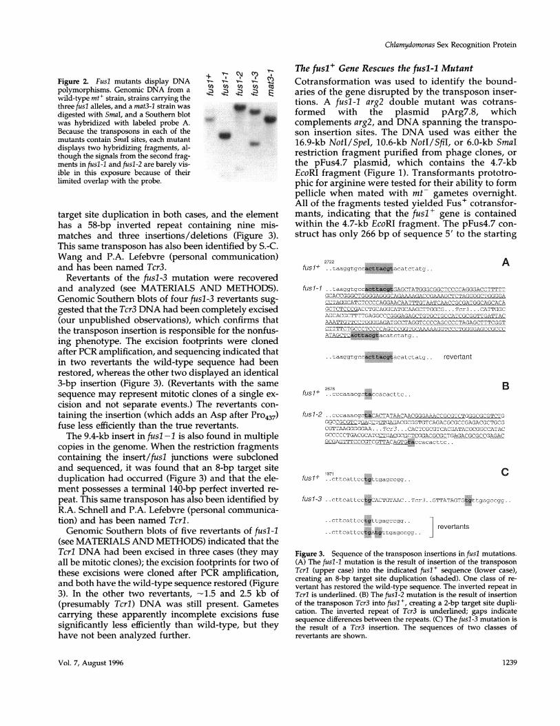
pFus4.7 transformants had acquired the ability to pro-duce mating-structure fringe. Figure 5a shows the nor-mal complement of fringe (arrowheads) associatedwith a wild-type mt+ mating structure; Figure 5b
c
V- 0
+ I-&- tetrad 1
IvI-tetrad 2
11
-E
*...F.h~.. mw w
§'§ § § : § §: fusability+ + + + + + + M
Figure 4. Southern blot analysis of a transformant and meiotictetrads. A Southern blot was prepared with EcoRI-digested genomicDNA from the indicated strains and hybridized with probe A. Thelane marked "transformant" is a Fus+ Arg+ transformant of CC-1865, transformed with the 16.9-kb NotI/SpeI fragment. It displaystwo fragments: the 14.1-kb fragment corresponding to the endoge-nous (E) fusl-l allele and a 4.7-kb fragment corresponding to thefusl + transforming (T) DNA. The meiotic progeny from two tetradsin a cross between the transformant and CC-124 (which has no
hybridizing fragments) are also analyzed. Their mating type (+ or
-) and whether they are (wt) or are not (Fus-) capable of fusingwith gametes of the opposite mating type are indicated. A faintband at 11 kb is visible in some lanes. This band cosegregates withthe transforming DNA and may indicate that another, incompletecopy of the transforming DNA has also been integrated.
shows the fringe-defective appearance of a fusl-1 mat-ing structure. Figure 5c shows a mating structure froma transformed fusl-I strain, in which a robust comple-ment of fringe has been restored. Expression of thetransgene is apparently variable in this strain in thatsome gametes display more fringe than others. Con-sistent with this evaluation, its mating efficiency peaksat 57% cell fusion after 20 min, compared with 96% forwild-type controls and 0% for the original fusl-1 arg2-recipient strain.
The fusl Gene also Rescues the impll MutantThe impll mutation confers gametes of an mt- strainwith a "pseudo-plus" phenotype: they produce plusflagellar agglutinins and mating structures with a plusmorphology, but the mating structures lack fringe andcannot effect cell fusion with wild-type mt- gametes(Goodenough et al., 1982). Because the implI mutationis tightly linked to the mt- locus (Galloway and Good-enough, 1985), it was proposed that the lesion pre-vents expression of an mt--specific gene called mid(minus dominance) that ordinarily acts to switch onthe minus gametogenesis program and switch off theplus program. It follows that the impl 1 mutantshould express plus genes but be defective for anygenes encoded in the absent mt+ locus, an obviousexample being the fusl + gene. We therefore createdan impil1 nitl double mutant (see MATERIALS ANDMETHODS), cotransformed with the pMN24 andpFus4.7 plasmids, selected transformants that couldgrow on nitrate as the sole nitrogen source, andtested these for their ability to form zygotes withmt- gametes. Several fusion-competent clones wereverified by Southern analysis as carrying the fusl+transgene. Genetic analysis demonstrated that thetransgene assorts independently of mt and, conse-quently, pseudo-plus progeny emerge from the cross(our unpublished observations), documenting thatthe acquisition of fusion competency is the result offusl+ transformation.One impll fusl-transformed strain was subjected
to additional analysis. By electron microscopy, its mat-ing structures were found to carry abundant levels offringe (compare Figure 5, d and e). However, its mat-ing behavior was quite different from the pFus4.7-transformed fusl-l strain described previously. After20 min of mating, 44% of the cells were found to beclosely apposed in pairs, but none of them had fused.Instead, apices remained paired for the next few hours(a transient stage in the normal mating reaction), as iffringe adhesion had occurred but fusion was pre-cluded. By the next morning, many of these pairs werefound to have formed zygotes, and genetic analysis ofsuch zygotes demonstrated that they are able to un-dergo a normal meiosis.
Molecular Biology of the Cell1240
Chlamydomonas Sex Recognition Protein
A , *tL
Figure 5. Mating structures of plus strains. (a) Wild type; (b) fusl-1; (c) fusl-l transformed with pFus4.7; (d) impli; (e) impil transformedwith pFus4.7. Arrowheads point to fringe. Bar, 100 nm.
To ascertain whether the pairs, in fact, were adheredvia their mating structures, the mating mixture wassubjected to pH shock (Witman et al., 1972), a proce-dure that causes the loss of flagella in response to an
influx of calcium (Quarmby and Hartzell, 1994). Giventhis stimulus, the paired cells immediately fused.When the procedure was repeated in the absence ofexternal calcium, the cells failed to fuse.
Vol. 7, August 1996 1241

P.J. Ferris et al.
The fus Gene Encodes a Putative GlycopolypeptideAs detailed in MATERIALS AND METHODS, the4813-bp EcoRI/SmaI section of genomic DNA thatrescues the nonfusing phenotype of fusl-l has beensequenced (GenBank accession number U49864).Probes were selected from this section for exhaus-tive screening of a cDNA library. Because thisscreening did not yield a full-length fusl cDNA, weused RT-PCR, 5' RACE, and inverse PCR to extendthe cDNA sequence more 5' to include the putativeinitiating methionine.Figure 6 shows schematically the location and sizes
of the 13 exons and 12 introns of fusl. These intronsare unusual in that all except the first and last are nolarger than 68 bp, the smallest being 57 bp (Table 1)[the smallest previously reported Chiamydomonas in-trons are 67 and 69 bp (LeDizet and Piperno, 1995)].Also indicated in Figure 6 are the sites at which fourindependent fusl cDNAs are polyadenylated. None ofthese sites is preceded by a canonical, TGTAA, poly-adenylation signal sequence. In fact, there is noTGTAA sequence in the entire genomic sequence fromthe fusl stop codon to the SmaI site (782 bp). Possibly,the lack of an exact match forces random selection ofalternate polyadenylation sites.Figure 7 presents the derived amino acid sequence
for fusl. The initiating methionine is followed by ashort 13-amino-acid sequence with the properties of asignal peptide (von Heijne, 1985). A hydropathy plotpredicts a single transmembrane domain near the Cterminus (boxed in Figure 7), indicating that the bulkof the molecule extends out from the membrane (com-pare with Figure 5). The presumed extracellular do-main carries 14 putative N-glycosylation sites distrib-uted along the length of the polypeptide.
Expression of the fusl GeneWhen a Northern blot of RNA isolated from variousstrains and life-cycle stages was hybridized with alabeled restriction fragment carrying only fusl codingsequence, a 3.0-kb message was observed in mt+ ga-metes but not in mt- gametes (which do not contain
the gene) or in mt+ vegetative (nongametic) cells (Fig-ure 8). A faint signal is sometimes seen in RNA fromzygotes, which could represent mRNA still present inthe zygotic cells and/or in residual unmated mt+ ga-metes in some zygote preparations. Gametes from anmt+/mt- heterozygous diploid (phenotypically mi-nus) also express the fusl gene.
The fusl Gene Lacks the ChlamydomonasCodon BiasThe 95 C. reinhardtii genes analyzed by LeDizet andPiperno (1995) all display a strong codon bias (Table2), with the GC content of the coding regions aver-aging 64.4% (M. LeDizet, personal communication)and with a particular bias for C in the third position(LeDizet and Piperno, 1995). The degree of bias foran individual gene can be quantified as a B value(Long and Gillespie, 1991), which indicates hownonrandomly synonymous codons are used (Figure9), with a B value of 0 indicating that all synony-mous codons are used with equal frequency, and 1.0indicating that only one codon is used for eachamino acid. Bias can also be expressed as a graphgenerated by the Codonpreference program of theGCG package, which scores codon usage in a par-ticular sequence against a table of Chlamydomonascodon preferences (i.e., Table 2). Several examplesof the latter are displayed in Figure 10, in whichvalues above the dashed horizontal line indicate theuse of preferred codons and tick marks indicate theuse of rare codons. P,-1 tubulin (Figure 10A) and ida4(a component of an inner-arm dynein; Figure 10B)are displayed to illustrate genes with particularlyhigh (B = 0.74) and low (B = 0.316) bias.The fusl cDNA sequence, in contrast, shows no
codon bias. Its coding region averages 47.7% GC,and when its codon usage is directly tabulated (Ta-ble 2), all possible codon choices are comparablyrepresented, generating a B value (Figure 9) of 0.050,and a dramatically different Codonpreference graph(Figure 10E). Importantly, biased codon usage isfound in two other genes located in the mt locus,
co z, C'C.o Figure 6. Structure of the fusl tran-4 z4 C script. The 4813-bp fusl genomic se-
<: <: < 5 quence is depicted, and the mRNA'u '
a, .K structure is shown diagrammatically.
v vn=. J w Filled boxes represent exons; linesbetween boxes are introns. The open
IXb box represents the longest 3'-un-E V Xb V A H Sc B A E Sm translated region. Triangles point to<8 <2 <1 10> <11 12> <13 the5' and3' endsofindicatedcDNA
3> 7> 200 bp clones (the 5' end of cDNA24 was notdetermined). The positions of the
transposon insertions in the three fusl mutants are indicated, as are restriction sites. Numbered carats mark the approximate locations ofoligonucleotide primers, with the point of the carat being the 3' end. The key for restriction sites is as in Figure 1, with the addition of ApaI(A) and EcoRV (V).
Molecular Biology of the Cell1242

Chlamydomonas Sex Recognition Protein
namely, the ezyl gene involved in the uniparentalinheritance of chloroplast DNA (Armbrust et al.,1993) and ezy2 (contained within the 16-kb repeat inFerris and Goodenough, 1994) (Figures 9 and 10, Cand D). Therefore, the absence of codon bias in thefusl gene is not a general property of genes sub-jected to recombination suppression in the mt locus.The Chlamydomonas genome is 62% GC (Harris,
1989), suggesting that the codon bias may be the con-sequence, at least in part, of an overall genomic pref-erence for G+C. Supporting this idea, an analysis of116 Chlamydomonas introns shows them to average63% GC (S. Fabry and M. Liss, personal communica-tion). The fusl introns, by contrast, are 51.6% GC (Ta-ble 1); if the large intron 12 is excluded, the introns are47% GC. Thus, the fusl introns, and not just the codingsequence, are different from other Chlamydomonasgenes, although the possibility exists that the basecomposition of the small fusl introns is constrained byrequirements of the splicing machinery. The 3'- and5'-untranslated regions of fusl are also low in %GC(Table 1).
DISCUSSION
The fusl GeneThe phenotype of the fusl-1 mutant (Goodenough etal., 1982) originally suggested that a gene necessaryfor fringe display on the plus-mating structure re-sided in the mt+ locus. Until this gene was clonedand sequenced, however, it was not known whetherit represented the structural gene for the fringe pro-tein or whether it played a more indirect role, such
Table 1. % GC in the fusl gene
SequenceSegment length (bp) % GC
5' upstreama 266 42.9coding region 2472 47.7intron 1 154 48.7intron 2 61 44intron 3 64 48intron 4 57 53intron 5 68 50intron 6 58 47intron 7 60 38intron 8 58 50intron 9 61 49intron 10 58 45intron 11 59 42intron 12 525 58.3all introns 1283 51.73' UTRb 761 54.0
a From the EcoRl site to the initiating AUG.b 3' UTR of cDNA17.
as encoding a transcription factor necessary for theexpression of the structural gene. The deduced geneproduct has the properties expected of a surfaceglycoprotein: it carries an N-terminal signal se-quence, a single putative transmembrane domainwith a short, highly charged cytoplasmic domain atthe C terminus, and 14 putative N-glycosylationsites along its putative extracellular domain. Nosignificant homologies to the fusl protein werefound in a search of the databases available usingthe NCBI BLAST network service.As might be expected for a gene whose product
functions in gamete recognition/fusion, the fuslmRNA is present in plus gametic cells but not invegetative cells. Interestingly, the fusl mRNA ispresent in gametes of heterozygous mtI/mt- dip-loids, which are functionally minus because of thedominance of the mt- locus. This suggests that themechanism that represses plus-specific genes in dip-loids (and presumably also in haploid mt- cells)does not recognize the fusl gene, which is not sur-prising given that fusl is not normally present in aminus cell. We have noticed, as have others (Gloeck-ner and Beck, 1995), that diploid gametes fuse withpoor efficiency compared with haploid gametes. Wehave also observed that haploid mt- strains carryinga fusl+ transgene fuse inefficiently, and we specu-
1
51
MPIFLILVLLAAVAKSQDCSVVADFKIDFQTSIFIAGNAYNITLTLLDSY
GDPTCVLYDPLLSVSCPSSSSNGRDFCSLQLSPLYNGVYNIKVIPQTLWG
101 GHTVWPPPYSPVAPLPDVYFAGDTAIRITYNGRDIQGSPFPVTVQAEPHI
151 STALSTVNVAVPAKGVASSRFAIAYFYQISDRFTNWIREKSIATQLRVSA
201 YPDADISVQWWQNWWIVLYANSTSAGMYRFQVYFIDDDGSEVPIRILSPG
251 LGLSYDGSFEVLPLALDEAKITASGLPQEVEAGIPVSLTLQAMDIYSNPT
301 RLVDPESYQPFGQQPDNKTLLQVRLVTVDSGALQPNVVAIPTNTTGACSW
351
401
SITFFTSMDYSVSVTYKESVLHMFSITVRNAQASPSNSTALLPEIGQAGT
TLLYVTPRDLWGNIAPLANNDLSIGLTGSTFFHSFIPVEPVRKGDYSVYS
451 LTLTEAGLYVVSIQLHNSSWLEKNITIEASYPSLQRSYVLGFGAGDPYGF
501 APTPLVAGEQYVLRVFIKDLYGNTIQADKVVDLNIVGPGQVLMNMSMLPS
551 GAFEVIYQPIVWGVYAVIANLTTGLLLHRGAVYVQPGTFNPNATTLQVPD
601 YIVAGEASSFKLAFHDSYGNAAASSGEASVVVFKYGGESLSFPLNLSGKF
651 IEELPFRLMHSGLYAFSISVNSVLVKSNNGYLHVVPGSAYALNITELKMT
701 DRLLEVYAYVVDEYLNSLNAAAMTAFAVAIDPPILFHGNVTLSSTGITLQ
751 LDKDPAIENTLQVQLKISNTIFFNCTWKPTAAHATLRRVREIGPLIAITA801 VCVCILFASIALVWSFRMLRHKP
Figure 7. Fusi protein sequence. The putative signal sequence isdouble underlined, the transmembrane sequence is boxed, and theN-glycosylation sites are underlined, with the asparagine residue inbold. The arrow marks the location of an inserted aspartate in somefusl-3 revertants.
Vol. 7, August 1996 1243
P.J. Ferris et al.
Figure 8. Regulation of fusl ex- + +
pression. RNA was prepared > (+
N
from vegetative mt+ cells (V+),mt- gametes (G-), mt+ gametes
fs
(G+), zygotes 150 min after mat-ing (Z), and gametes of an mt+ /
mt- diploid (G±). Approximately10 gg of total RNA was loaded on 204
each lane of a formaldehyde-con-taining 1% agarose gel, whichwas subsequently blotted to nitrocellulose and probed with a
1.65-kb EcoRV/BamHI fragment (labeled fusl) from cDNA17. Thesame blot was also hybridized with a cDNA called 204 (Ferris andGoodenough, 1987), which is expressed at fairly constant levels inall life-cycle stages, to ensure that all RNA samples were unde-graded and had comparable levels of mRNA.
late that the presence of fusl protein in both of thesecell types may in some way impede minus matingstructure function.Sexual recognition in C. reinhardtii is effected first
by flagellar agglutination and then by fringe-fringeinteractions at the plasma membrane (reviewed inGoodenough et al., 1995). The genes encoding the
flagellar agglutinins have not yet been cloned, butthe proteins have been purified and characterized(Cooper et al., 1983; Collin-Osdoby and Adair, 1985)as hydroxyproline-rich glycoproteins with aminoacid compositions very different from the fusl pro-
tein. Therefore, these two recognition systemsclearly represent independent evolutionary ideas.To our knowledge, this is the first report of a
sex-specific adhesion molecule being encoded in a
mating-type locus. In yeast, the a and a agglutininproteins, and the a and a pheromones and phero-mone receptors, are encoded by genes that are ex-
pressed in only one mating type but are not linkedto MAT (reviewed in Herskowitz, 1989; Lipke andKurjan, 1992). Genes encoding recognition mole-cules have been reported in the mating-type loci oftwo higher fungi: the pheromone/pheromone re-
ceptor genes are located in the al/a2 loci of hetero-basidiomycete Ustilago maydis (Bolker et al., 1992;Spelling et al., 1994), and analogous gene pairs are
located in the B locus of the basidiomycete Schizo-phyllum commune (Wendland et al., 1995).
Table 2. Codon usage in C. reinhardtii and in the fusl gene
Aa Codon Genome fusl Aa Codon Genome fusl Aa Codon Genome fusl
A GCGGCAGCTGCC
C TGTTGC
D GATGAC
E GAGGAA
F TTTTTC
G GGGGGAGGTGGC
H CATCAC
I ATAATTATC
27 115 4415 2553 20
496
1387
983
1288
321580
2575
3565
5644
6139
23291731
9 6791 33
1 1824 5175 31
K AAGAAA
L TTGTTACTGCTACTTCTC
N AATAAC
P CCGCCACCTCCC
Q CAGCAA
R AGGAGACGGCGACGTCGC
99 57 S AGT1 43 AGC
TCGTCA
2 22 TCT0 12 TCC
82 202 12 T ACG4 19 ACA
11 15 ACTACC
5 5695 44 V GTG
GTA24 26 GTT4 42 GTC
11 1762 15 Y TAT
TAC95 555 45 End TGA
TAGTAA
3 81 178 211 8
10 3376 13
Molecular Biology of the Cell
2 1229 3230 62 178 18
29 15
23 223 28
10 3064 20
69 291 246 34
25 13
5 5595 45
5 018 10077 0
The genome column shows the relative frequency of possible codons for each amino acid in all C. reinhardtii nuclear sequences in the GenBankdatabase (95 sequences, 36787 codons, LeDizet and Pipemo, 1995). The fusl column shows the relative frequency of codons in the fusl gene(824 codons).
1244

Chlamydomonas Sex Recognition Protein
(0)G)0C
U)C00
.0o
Ez
Figure 9. Codon usage bias in nuclear genes of C.reinhardtii. The "standardized synonymous codonbias" B (Long and Gillespie, 1991) was evaluated forsequences by LeDizet and Piperno (1995) and M.LeDizet (unpublished observations). A highly biasedcodon usage is reflected by a high value of B, whichvaries from 0 to 1. The fusl gene shows virtually nobias (B = 0.050). Two genes just outside the R domainare biased (ezyl = 0.43; ezy2 = 0.35), as are two geneswhose transcripts are present in low abundance(arg7 = 0.54; nitl = 0.55). A previous report (deHostos et al., 1989) claiming a low bias in the arylsul-fatase (ars) gene may be the consequence of sequenc-ing errors (Hallmann and Sumper, 1994); corrected,ars = 0.52.
Mutations in the fusl GeneThree mutations in the fusi gene generate an agglu-tinate-but-not-fuse phenotype in mt+ cells. Thefusl-1 mutation is shown here to be the consequenceof a Tcrl transposon insertion in the 10th intron ofthe fusl gene. Since the fusl-1 mutant is slightlyleaky (Goodenough et al., 1976), the presence of thetransposon in an intron may render correct splicingunlikely but occasionally feasible. The partial resto-ration of fusability in two revertants that appear tohave incompletely excised Tcrl is consistent withthis hypothesis. The fusl-2 and -3 mutants, whichare both caused by the insertion of Tcr3 into anexon, behave quite differently. When rare zygotesresulting from a fusl-2 or -3 cross are analyzed (thefusl-2 data are not described here), the mt+ progenyinvariably mate normally. Therefore, it appears thatthese two strains may be able to form zygotes onlyif the mutations revert; they either are not leaky orare leaky at a frequency below that of excision of theTcr3 transposon. The fact that all three alleles of fuslare transposon insertions is consistent with the pos-sibility that the fusl gene might be a "hot spot" fortransposon insertions.
Fusl Rescue of the impll Fusion DefectThe ability of the fusl+ gene to confer fringe andfusability to the impil mutant strain supports ouroriginal interpretation of the impil phenotype. Weproposed (Goodenough et al., 1982; Galloway andGoodenough, 1985; Goodenough and Ferris, 1987)that the mt- impil mutation inactivates a gene nec-essary to switch on the minus gametogenesis pro-gram and switch off the plus program, a gene thataccounts for the dominance of mt- over mt+ indiploid Chlamydomonas (Ebersold, 1967). By this rea-soning, most of the genes in the plus gametogenesispathway would reside outside the mt+ locus and,
arg7nitl
ars I
tubulin
IIida4
fusl
0.0 0.1 0.2 0.3 0.4 0.5 0.6 0.7 0.8
B value
thus, would be present in impil1 gametes, explainingtheir ability to express most plus gametic traits. Anygenes resident in the mt+ locus, however, would beabsent in impl 1 gametes. The lack of fringe on impl 1mating structures, and its restoration when impilcells are transformed with the fusl + gene, is concor-dant with this proposal.
In experiments to be reported elsewhere, we haveshown that the zygotes resulting from a cross betweenfusl-transformed impil and mt- are able to undergomeiosis and germination normally but fail to transmittheir chloroplast DNA to the meiotic progeny in auniparental manner. Therefore, a second gene productabsent from impil1 gametes, and therefore presumablyencoded by the mt+ locus, plays a key role in thetransmission of mt+ chloroplast DNA. The fact thatmeiosis proceeds normally when the zygote has nomt+ chromosome indicates that any plus functionsnecessary for zygote development are encoded else-where in the genome and are expressed in the absenceof minus dominance.The phenotype of the fusl -transformed impil
strain is not fully wild-type. This transformant estab-lishes fringe-fringe adhesions with its mt- partners,but fusion is slow and episodic and is aided by ma-nipulations that allow calcium influx at the cell apex.Although it is possible that suboptimal expression ofthe transgene affects the efficiency of the process, theexpression of the same transgene in a fusl-l back-ground permits rapid fusion. Therefore, a third geneproduct may also be encoded in the mt+ locus,namely, a molecule necessary for efficient gameticfusion. Of interest in this regard is the finding thatmammalian sperm display an a/13 dimer on theirmembranes, the 1 subunit involved in recognizingan egg integrin receptor (Almeida et al., 1995) andthe a subunit effecting membrane fusion via a do-
Vol. 7, August 1996 1245

P.J. Ferris et al.
2.0 -
1.5 -
1.0 -
0.5 -
0.0 -
2.0.2
1.5
1. 0
0.5
0.0
2.0 -
1.5
1.0 -
0.5-
0.0
1, 000
2.0
1.5
1.0
0.5
0.0
2, 000 3, 000
Figure 10. Lack of Chlamydomonas codon bias in the fusi gene. The graphical output of the GCG program Codonpreference is shown
for several C. reinhardtii cDNA sequences by using a window of 25 codons and by using the data from LeDizet and Piperno (1995) (Table
2) for the C. reinhardtii codon usage table. The start and stop codons of each open reading frame are marked by downward and upwardarrowheads, respectively. Tick marks at the bottom of each panel represent rare codons (those used no more than 10% of the time in
the Chlamydomonas gene set; compare with Table 2). Values above the dashed line indicate use of preferred codons. Note how noncodingsequences (i.e., the 3'-untranslated region) do not show a codon bias. (A) ,13 tubulin (Youngblom et al., 1984); (B) ida4 (LeDizet and
Piperno, 1995); (C) ezyl (Armbrust et al., 1993); (D) ezy2 (E.V. Armbrust, unpublished observations); (E) fusl.
Molecular Biology of the Cell
CV A
I111I111 IlEIllII 11 I1 I11 III 1111 *uiuuiinmu,'i
D
IgoIIinn mimmniiui.i Il IIlIIi Il IlnIlIiiIII Il 11 lliliiI1111 1 111111 1111111111IIMIIII**iii.m.nm;nIIuuiuumm 1111
EV .A
imnm11U1inmummum *iiunu mmiimfium miiu mmimm 111um
1246
Clilaiiiydomio01nas Sex Recognition Protein
main homologous to viral fusion peptides (Wolfs-berg et a!., 1995).
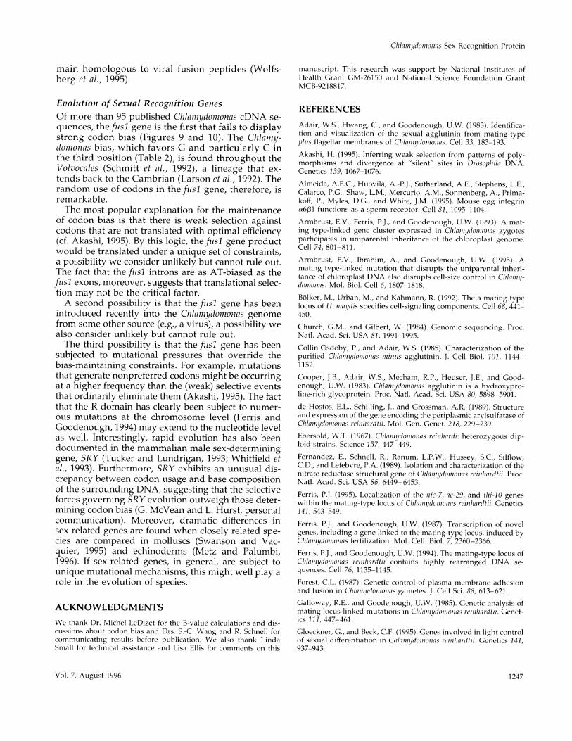
Evolution of Sexual Recognition GenesOf more than 95 published Chlamydomonas cDNA se-quences, the fisl gene is the first that fails to displaystrong codon bias (Figures 9 and 10). The Chlamy-domnonas bias, which favors G and particularly C inthe third position (Table 2), is found throughout theVolvocales (Schmitt et al., 1992), a lineage that ex-tends back to the Cambrian (Larson et al., 1992). Therandom use of codons in thefitsl gene, therefore, isremarkable.The most popular explanation for the maintenance
of codon bias is that there is weak selection againstcodons that are not translated with optimal efficiency(cf. Akashi, 1995). By this logic, the ftisl gene productwould be translated under a unique set of constraints,a possibility we consider unlikely but cannot rule out.The fact that the fisl introns are as AT-biased as thefuisl exons, moreover, suggests that translational selec-tion may not be the critical factor.A second possibility is that the fitsl gene has been
introduced recently into the Chlaiamydomoinas genomefrom some other source (e.g., a virus), a possibility wealso consider unlikely but cannot rule out.The third possibility is that the fiisl gene has been
subjected to mutational pressures that override thebias-maintaining constraints. For example, mutationsthat generate nonpreferred codons might be occurringat a higher frequency than the (weak) selective eventsthat ordinarily eliminate them (Akashi, 1995). The factthat the R domain has clearly been subject to numer-ous mutations at the chromosome level (Ferris andGoodenough, 1994) may extend to the nucleotide levelas well. Interestingly, rapid evolution has also beendocumented in the mammalian male sex-determininggene, SRY (Tucker and Lundrigan, 1993; Whitfield etal., 1993). Furthermore, SRY exhibits an unusual dis-crepancy between codon usage and base compositionof the surrounding DNA, suggesting that the selectiveforces governing SRY evolution outweigh those deter-mining codon bias (G. McVean and L. Hurst, personalcommunication). Moreover, dramatic differences insex-related genes are found when closely related spe-cies are compared in molluscs (Swanson and Vac-quier, 1995) and echinoderms (Metz and Palumbi,1996). If sex-related genes, in general, are subject tounique mutational mechanisms, this might well play arole in the evolution of species.
ACKNOWLEDGMENTSWe thank Dr. Michel LeDizet for the B-value calculations and dis-cussions about codon bias and Drs. S.-C. Wang and R. Schnell forcommunicating results before publication. We also thank LindaSmall for technical assistance and Lisa Ellis for comments on this
manuscript. This research was support by National Institutes ofHealth Grant GM-26150 and National Science Foundation GrantMCB-9218817.
REFERENCES
Adair, W.S., Hwang, C., and Goodenough, U.W. (1983). Identifica-tion and visualization of the sexual agglutinin from mating-typeplhs flagellar membranes of Clilam,ydoioioiias. Cell 33, 183-193.
Akashi, H. (1995). Inferring weak selection from patterns of poly-morphisms and divergence at "silent" sites in Drosoplila DNA.Genetics 139, 1067-1076.
Almeida, A.E.C., Huovila, A.-P.J., Sutherland, A.E., Stephens, L.E.,Calarco, P.G., Shaw, L.M., Mercurio, A.M., Sonnenberg, A., Prima-koff, P., Myles, D.G., and White, J.M. (1995). Mouse egg integrina601 functions as a sperm receptor. Cell 81, 1095-1104.
Armbrust, E.V., Ferris, P.J., and Goodenough, U.W. (1993). A mat-ing type-linked gene cluster expressed in Clia iiiydoiniouas zygotesparticipates in uniparental inheritance of the chloroplast genome.Cell 74, 801-811.
Armbrust, E.V., Ibrahim, A., and Goodenough, U.W. (1995). Amating type-linked mutation that disrupts the uniparental inheri-tance of chloroplast DNA also disrupts cell-size control in Clilampiy-doamioanas. Mol. Biol. Cell 6, 1807-1818.
Bolker, M., Urban, M., and Kahmann, R. (1992). The a mating typelocus of U. miiaydis specifies cell-signaling components. Cell 68, 441-450.
Church, G.M., and Gilbert, W. (1984). Genomic sequencing. Proc.Natl. Acad. Sci. USA 81, 1991-1995.
Collin-Osdoby, P., and Adair, W.S. (1985). Characterization of thepurified Chlaltnydoinoiuas mi111ls agglutinin. J. Cell Biol. 1()1, 1144-1152.
Cooper, J.B., Adair, W.S., Mecham, R.P., Heuser, J.E., and Good-enough, U.W. (1983). Chlamiiydoamaonas agglutinin is a hydroxypro-line-rich glycoprotein. Proc. Natl. Acad. Sci. USA 80, 5898-5901.de Hostos, E.L., Schilling, J., and Grossman, A.R. (1989). Structureand expression of the gene encoding the periplasmic arylsulfatase ofChlalmiydomauaiias rcilihardtii. Mol. Gen. Genet. 218, 229-239.Ebersold, W.T. (1967). Chlilamydoamaoinas reijuhardi: heterozygous dip-loid strains. Science 157, 447-449.
Fernandez, E., Schnell, R., Ranum, L.P.W., Hussey, S.C., Silflow,C.D., and Lefebvre, P.A. (1989). Isolation and characterization of thenitrate reductase structural gene of Chlilamydomio0inas rciulhlardtii. Proc.Natl. Acad. Sci. USA 86, 6449-6453.Ferris, P.J. (1995). Localization of the 11ic-7, ac-29, and thi-10 geneswithin the mating-type locus of Chlamiiydomiiouias rcinlhardtii. Genetics141, 543-549.
Ferris, P.J., and Goodenough, U.W. (1987). Transcription of novelgenes, including a gene linked to the mating-type locus, induced byClhlan1ydom)iionas fertilization. Mol. Cell. Biol. 7, 2360-2366.
Ferris, P.J., and Goodenough, U.W. (1994). The mating-type locus ofChilamiiydomniouias reiuhlardtii contains highly rearranged DNA se-quences. Cell 76, 1135-1145.
Forest, C.L. (1987). Genetic control of plasma membrane adhesionand fusion in Cllauuiytidomtiouias gametes. J. Cell Sci. 88, 613-621.
Galloway, R.E., and Goodenough, U.W. (1985). Genetic analysis ofmating locus-linked mutations in Clilamiiydouiiouias rcinliardtii. Genet-ics 111, 447-461.
Gloeckner, G., and Beck, C.F. (1995). Genes involved in light controlof sexual differentiation in Chilauuydomi1onas rciuhiardtii. Genetics 141,937-943.
Vol. 7, August 1996 1 247

P.J. Ferris et al.
Goodenough, U.W., Armbrust, E.V., Campbell, A.M., and Ferris, P.J.(1995). Molecular genetics of sexuality in Chlamydomonas. Annu.Rev. Plant Physiol. Plant Mol. Biol. 46, 21-44.Goodenough, U.W., Detmers, P.A., and Hwang, C. (1982). Activa-tion for cell fusion in Chlamydomonas: analysis of wild-type gametesand nonfusing mutants. J. Cell Biol. 92, 378-386.Goodenough, U.W., and Ferris, P.J. (1987). Genetic regulation ofdevelopment in Chlamydomonas. In: Genetic Regulation of Develop-ment, ed. W. Loomis, New York: Alan R. Liss.Goodenough, U.W., Hwang, C., and Martin, H. (1976). Isolation andgenetic analysis of mutant strains of Chiamydomonas reinhardi defec-tive in gametic differentiatiop. Genetics 82, 169-186.Goodenough, U.W., and Jurivich, D. (1978). Tipping and mating-structure activation induced in Chlamydomonas gametes by flagellarmembrane antisera. J. Cell Biol. 79, 680-693.Hallmann, A., and Sumper, M. (1994). An inducible arylsulfatase ofVolvox carteri with properties suitable for a reporter-gene system.Eur. J. Biochem. 221, 143-150.Harris, E.H. (1989). The Chlamydomonas Sourcebook, San Diego, CA:Academic Press.Herskowitz, I. (1989). A regulatory hierarchy for cell specializationin yeast. Nature 342, 749-757.Kindle, K.L. (1990). High-frequency nuclear transformation ofChlamydomonas reinhardtii. Proc. Natl. Acad. Sci. USA 87,1228-1232.Kinoshita, T., Fukuzawa, H., Shimada, T., Saito, T., and Matsuda, Y.(1992). Structure-function relationships in a cell wall-degradingmetalloprotease of Chlamydomonas reinhardtii: similarity of func-tional domains to matrix metalloproteases. Proc. Natl. Acad. Sci.USA 89, 4693-4697.Kirk, M.M., and Kirk, D.L. (1985). Translational regulation of pro-tein synthesis, in response to light, at a critical stage of Volvoxdevelopment. Cell 41, 419-428.Larson, A., Kirk, M.M., and Kirk, D.L. (1992). Molecular phylogenyof the Volvocine flagellates. Mol. Biol. Evol. 9, 85-105.LeDizet, M., and Piperno, G. (1995). The light chain p28 associateswith a subset of inner dynein arm heavy chains in Chlamydomonasaxonemes. Mol. Biol. Cell 6, 697-711.Lipke, P.N., and Kurjan, J. (1992). Sexual agglutinins in buddingyeasts: structure, function, and regulation of yeast cell adhesionproteins. Microbiol. Rev. 56, 180-194.Long, M., and Gillespie, J.H. (1991). Codon usage divergence ofhomologous vertebrate genes and codon usage clock. J. Mol. Evol.32, 6-15.
Maniatis, T., Fritsch, E.F., and Sambrook, J. (1982). Molecular Clon-ing: A Laboratory Manual, Cold Spring Harbor, NY: Cold SpringHarbor Laboratory Press.Martin, N.C., and Goodenough, U.W. (1975). Gametic differentia-tion in Chlamydomonas reinhardtii. I. Production of gametes and theirfine structure. J. Cell Biol. 67, 587-605.
Matsuda, Y., Tamaki, S., and Tsubo, Y. (1978). Mating-type-specificinduction of cell-wall lytic factor by agglutination of gametes inChlamydomonas reinhardtii. Plant Cell Physiol. 19, 1253-1261.Mesland, D.A.M., Hoffman, J.L., Caligor, E., and Goodenough, U.W.(1980). Flagellar tip activation stimulated by membrane adhesionsin Chlamydomonas gametes. J. Cell Biol. 84, 599-617.Metz, E.C., and Palumbi, S.R. (1996). Positive selection and sequencerearrangements generate extensive polymorphism in the gameterecognition protein binding. Mol. Biol. Evol. 13, 397-406.
Quarmby, L.M., and Hartzell, H.C. (1994). Two distinct, calcium-mediated, signal transduction pathways can trigger deflagellationin Chlamydomonas reinhardtii. J. Cell Biol. 124, 807-815.
Sager, R., and Granick, S. (1953). Nutritional studies with Chlamy-domonas reinhardi. Ann. NY Acad. Sci. 56, 831-838.
Schmitt, R., Fabry, S., and Kirk, D.L. (1992). In search of the molec-ular origins of cellular differentiation in Volvox and its relatives. Int.Rev. Cytol. 139,189-265.
Schnell, R.A., and Lefebvre, P.A. (1993). Isolation of the Chiamydo-monas regulatory gene NIT2 by transposon tagging. Genetics 134,737-747.
Segal, R.A., Huang, B., Ramanis, Z., and Luck, D.J.L. (1984). Mutantstrains of Chlamydomonas reinhardtii that move backwards only. J.Cell Biol. 98, 2026-2034.
Spelling, T., Bolker, M., Lottspeich, F., Frand, R.W., and Kahmann,R. (1994). Pheromones trigger filamentous growth in Ustilago may-dis. EMBO J. 13, 1620-1627.
Swanson, W.J., and Vacquier, V.D. (1995). Extraordinary divergenceand positive Darwinian selection in a fusogenic protein coating theacrosomal process of abalone spermatozoa. Proc. Natl. Acad. Sci.USA 92, 4957-4961.
Tucker, P.K., and Lundrigan, B.L. (1993). Rapid evolution of thesex-determining locus in Old World mice and rats. Nature 364,715-717.
von Heijne, G. (1985). Signal sequences. The limits of variation. J.Mol. Biol. 184, 99-105.
Wahl, G.M., Stern, M., and Stark, G.R. (1979). Efficient transfer oflarge DNA fragments from agarose gels to diazobenzyloxymethyl-paper and rapid hybridization by using dextran sulfate. Proc. Natl.Acad. Sci. USA 76, 3683-3687.
Weeks, D.P., Beerman, N., and Griffith, O.M. (1986). A small-scalefive-hour procedure for isolating multiple samples of CsCl-purifiedDNA; application to isolations from mammalian, insect, higherplant, algal, yeast, and bacterial sources. Anal. Biochem. 152, 376-385.
Wendland, J., Vaillancourt, L.J., Hegner, J., Lengeler, K.B., Laddison,K.J., Specht, C.A., Raper, C.A., and Kothe, E. (1995). The mating-type locus Bal of Schizophyllum commune contains a pheromonereceptor gene and putative pheromone genes. EMBO J. 14, 5271-5278.
Whitfield, L.S., Lovell-Badge, R., and Goodfellow, P.N. (1993).Rapid sequence evolution of the mammalian sex-determining geneSRY. Nature 364, 713-715.
Witman, G.B., Carlson, K., Berliner, J., and Rosenbaum, J.L. (1972).Chlamydomonas flagella. I. Isolation and electrophoretic analysis ofmicrotubules, matrix, membranes, and mastigonemes. J. Cell Biol.54, 507-539.
Wolfsberg, T.G., Straight, P.D., Gerena, R.L., Huovila, A.-P.J., Pri-makoff, P., Myles, D.G., and White, J.M. (1995). ADAM, a widelydistributed and developmentally regulated gene family encodingmembrane proteins with a distintegrin and metalloprotease do-main. Dev. Biol. 169, 378-383.
Youngblom, J., Schloss, J.A., and Silflow, C.D. (1984). The two13-tubulin genes of Chlamydomonas reinhardtii code for identical pro-teins. Mol. Cell. Biol. 4, 2686-2696.
Zeiner, M., and Gehring, U. (1994). Cloning of 5' cDNA regions byinverse PCR. Biotechniques 17, 1051-1053.
Molecular Biology of the Cell1248