Bifunctional ligands of the bradykinin B2 and B1 receptors
58
1 2 3 4 5 6 7 8 9 10 11 12 13 14 15 16 17 18 19 20 21 22 23 24 25 26 27 28 29 30 31 32 33 34 35 36 37 38 39 40 41 42 43 44 45 46 47 48 49 50 51 52 53 54 55 56 57 58 59 60 61 62 63 64 65 1 Review Bifunctional ligands of the bradykinin B 2 and B 1 receptors: an exercise in peptide hormone plasticity François Marceau*, Marie-Thérèse Bawolak, Jean-Philippe Fortin, Guillaume Morissette, Caroline Roy, Hélène Bachelard, Lajos Gera , Xavier Charest-Morin CHU de Québec Université Laval, Québec QC, Canada G1V 4G2 ; Department of Biochemistry, University of Colorado Denver, Aurora, CO, 80045, U.S.A. *Corresponding author : François Marceau, M.D., Ph.D., Centre de recherche en rhumatologie et immunologie, Room T1-49, Centre Hospitalier Universitaire de Québec, 2705 Laurier Blvd., Québec QC Canada G1V 4G2. Tel.: 418-525-4444, ext. 46155. Fax: 418-654-2765. e-mail: [email protected]
-
Upload
khangminh22 -
Category
Documents
-
view
0 -
download
0
Transcript of Bifunctional ligands of the bradykinin B2 and B1 receptors
1 2 3 4 5 6 7 8 9 1011121314151617181920212223242526272829303132333435363738394041424344454647484950515253545556575859606162636465
1
Review
Bifunctional ligands of the bradykinin B2 and B1 receptors: an exercise in
peptide hormone plasticity
François Marceau*, Marie-Thérèse Bawolak, Jean-Philippe Fortin, Guillaume Morissette,
Caroline Roy, Hélène Bachelard, Lajos Gera , Xavier Charest-Morin
CHU de Québec Université Laval, Québec QC, Canada G1V 4G2 ; Department of
Biochemistry, University of Colorado Denver, Aurora, CO, 80045, U.S.A.
*Corresponding author: François Marceau, M.D., Ph.D., Centre de recherche en rhumatologie et
immunologie, Room T1-49, Centre Hospitalier Universitaire de Québec, 2705 Laurier Blvd.,
Québec QC Canada G1V 4G2. Tel.: 418-525-4444, ext. 46155. Fax: 418-654-2765. e-mail:

1 2 3 4 5 6 7 8 9 1011121314151617181920212223242526272829303132333435363738394041424344454647484950515253545556575859606162636465
2
Abstract
Kinins are the small and fragile hydrophilic peptides related to bradykinin (BK) and derived from
circulating kininogens via the action of kallikreins. Kinins bind to the preformed and widely
distributed B2 receptor (B2R) and to the inducible B1 receptor (B1R). B2Rs and B1Rs are related G
protein coupled receptors that possess natural agonist ligands of nanomolar affinity (BK and Lys
BK for B2Rs, Lys-des-Arg9-BK for B1R). Decades of structure-activity exploration have resulted
in the production of peptide analogs that are antagonists, one of which is clinically used (the B2R
antagonist icatibant), and also non-peptide ligands for both receptor subtypes. The modification
of kinin receptor ligands has made them resistant to extracellular or endosomal peptidases and/or
produced bifunctional ligands, defined as agonist or antagonist peptide ligands conjugated with a
chemical fluorophore (emitting in the whole spectrum, from the infrared to the ultraviolet), a
drug-like moiety, an epitope, an isotope chelator/carrier, a cleavable sequence (thus forming a
pro-drug) and even a fused protein. Dual molecular targets for specific modified peptides may be
a source of side effects or of medically exploitable benefits. Biotechnological protein ligands for
either receptor subtype have been produced: they are enhanced green fluorescent protein or the
engineered peroxidase APEX2 fused to an agonist kinin sequence at their C-terminal terminus.
Antibodies endowed with pharmacological actions (agonist, antagonist) at B2R have been
reported, though not monoclonal antibodies. These findings define classes of alternative ligands
of the kinin receptor of potential therapeutic and diagnostic value.
Keywords: bradykinin, B2 receptors, B1 receptors, synthetic peptides, fluorescent ligands,
biotechnological agents

1 2 3 4 5 6 7 8 9 1011121314151617181920212223242526272829303132333435363738394041424344454647484950515253545556575859606162636465
3
List of major abbreviations
ACE, angiotensin converting enzyme
- -aminocaprolyl
APEX2, engineered soya bean peroxidase
Arg-CP, arginine carboxypeptidase
B1R, B1 receptor
B2R, B2 receptor
B2R-GFP, rabbit B2 receptor fused to green fluorescent protein
BK, bradykinin
CF, 5(6)-carboxyfluorescein
Cy7,cyanine dye 7
EGFP, enhanced green fluorescent protein
FTC, fluorescein-5-thiocarbamoyl
GPCR, G protein coupled receptor
GRK, G protein coupled receptor kinase
HSA, human serum albumin
KLK-1, tissue kallikrein
MK, maximakinin
NG, asparaginyl-glycyl
PTH, parathyroid hormone
TM, transmembrane (domain)
Other abbreviations are defined in the figure legends

1 2 3 4 5 6 7 8 9 1011121314151617181920212223242526272829303132333435363738394041424344454647484950515253545556575859606162636465
4
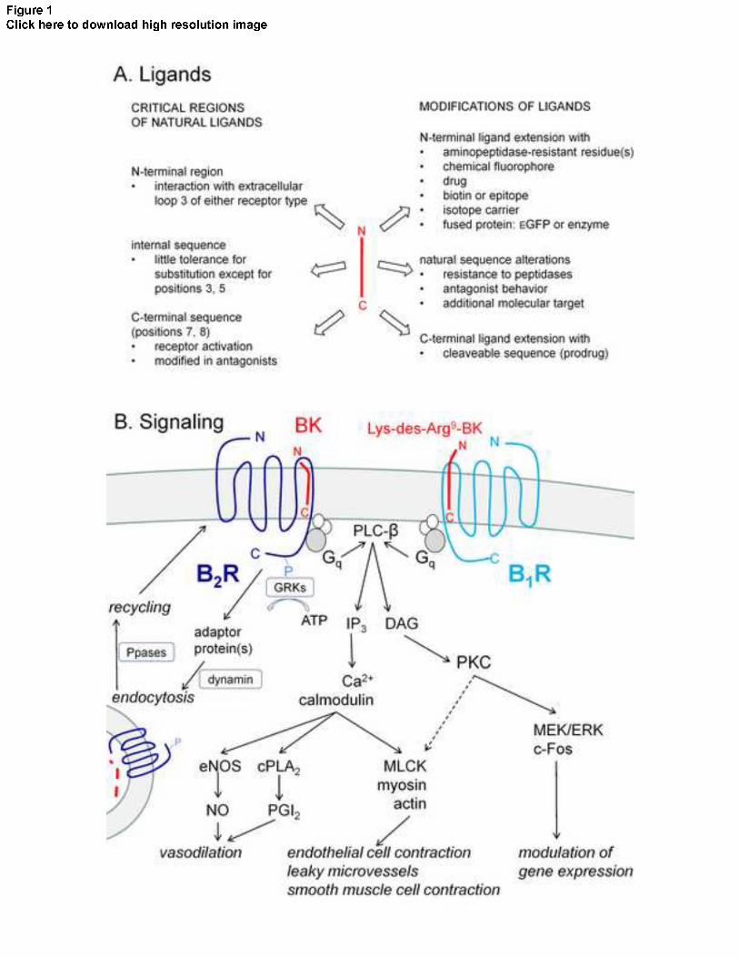
1. Formation, signaling, metabolism and medical importance of kinins
The bradykinin (BK) sequence, H-Arg-Pro-Pro-Gly-Phe-Ser-Pro-Phe-Arg-OH, is imbedded in
domain 4 of 2 circulating proteins, the low and high molecular weight kininogens, produced by
the liver from a single gene subjected to mRNA alternative splicing [1-3]. Kinins, the BK-related
peptides, are generated from these kininogens, mainly by two major types of serine proteases,
plasma and tissue kallikreins, that generate the native kinins BK and Lys-BK (also known as
kallidin). Several constituents of the kallikrein-kinin system are found in the blood plasma, but
also at the surface of cells or in their secretions (e.g., tissue kallikrein = KLK-1) [1, 3].
Two related receptors belong to the kallikrein-kinin system, the B1 and B2 receptors (B1R, B2R).
2R is expressed at a rather
constant level in many tissues and cell types and submitted to a classical
endocytosis/reexpression cycle following stimulation, the B1R is highly regulated by tissue
injury, notably via cytokines or the innate immune system [1, 2]. Both receptor types are mainly
coupled to the Gq protein (also Gi), with ensuing stimulation of a phospholipase C and generation
of second messengers such as calcium and diacylglycerol [2] (schematic representation, Fig. 1B).
The B2R is widely distributed, the vascular endothelial cells being a privileged physiological site
of BK action, but the receptor is also expressed in sensory neurons, smooth muscle cells,
epithelial (intestinal, respiratory) and some types of leukocytes. B1R tend to be present in the
same cell types when expressed.
Kinins are often considered inflammatory mediators, with such effects as edema, pain, diarrheic
states [4] and flu-like airway irritation [5], but are as well compensatory vasodilator autacoids

1 2 3 4 5 6 7 8 9 1011121314151617181920212223242526272829303132333435363738394041424344454647484950515253545556575859606162636465
5
that release nitric oxide (NO) and prostanoids from vascular endothelial cells, with salutary
effects in the circulation of the heart, kidney, brain and the promotion of angiogenesis [1, 6].
Cultured human umbilical vein endothelial cells (HUVECs) are a conventional model where both
NO production and actin reorganization, predictive of microvascular leakage, can be recorded in
response to BK (Supplementary Figures 1 and 2).
Kinins are fragile peptides, being degraded by several metallopeptidases present both in blood
plasma and at the cell surface. Angiotensin converting enzyme (ACE), present at the surface of
endothelial cells and also circulating under a cleaved form, is by far the major kinin-destroying
enzyme in the extracellular compartment both in human blood plasma and in vivo in rats [7, 8].
Second in importance for BK inactivation in both milieus is aminopeptidase P that cleaves Arg1
and inactivates the peptides. Arginine carboxypeptidases (Arg-CPs), such as plasma
carboxypeptidase N and membrane carboxypeptidase M, are minor pathways, but ones that have
a particular importance: they generate des-Arg9-BK and Lys-des-Arg9-BK which have high
affinity for the B1R, the latter peptide being optimal at the human form of this receptor subtype.
Aminopeptidase N is an ectopeptidase that removes the N-terminal Lys residue in Lys-BK and
Lys-des-Arg9-BK; the effect is pharmacologically neutral for the former B2R agonist, but the
latter peptide is partly inactivated by this reaction vs. the B1R [9].
While a large body of literature suggests that interventions on the kallikrein-kinin systems are
therapeutic in animals subjected to various experimental pathologies [2], clinical applications are
presently minimal [10, 11]. The B2R antagonist icatibant is approved to abort attacks of
hereditary angioedema, an autosomal dominant condition where the serpin G1 is deficient in
abundance or function, leading to explosive activation of the contact system and plasma kallikein

1 2 3 4 5 6 7 8 9 1011121314151617181920212223242526272829303132333435363738394041424344454647484950515253545556575859606162636465
6
[12]. For the same rare indication, biotechnological inhibitors of plasma kallikreins are either
approved (ecallantide) or under final clinical development (the neutralizing monoclonal antibody
lanadelumab) [13]. Orally bioavailable, small molecule inhibitors of this serine protease are also
considered for hereditary angioedema and various edematous retinopathies (e.g., KV123833 from
Kalvista). However, these are not kinin receptor ligands. Clinical trials concerning B2R and B1R
antagonists have been so far largely negative, despite positive effects in animal models. An early
B2R peptide antagonist, deltibant, failed to improve the condition of patients with sepsis [14].
Anatibant, a non-peptide B2R antagonist, had no clear effect on post-traumatic brain edema [15].
At least 2 sophisticated non-peptide B1R antagonists have failed to reduce inflammatory pain in
humans [14].
ACE inhibitors, such as enalapril, lisinopril and many others, are used in cardiovascular
conditions where the synthesis of the vasopressor hormone angiotensin II needs to be repressed;
however this metallopeptidase is also the main BK-inactivating enzyme. A fraction of the
antihypertensive effect of ACE inhibitors is attributed to the potentiation of the vasodilator action
of endogenous kinins in humans (evidence based on blunting of their effect by icatibant co-
administration) [16-18]. Recombinant active KLK-1 (DM-199) is being clinically developed as a
parenterally administered drug to take advantage of the salutary effect of kinins on the circulation
and metabolism [19]. The BK analog labradimil was designed to deliberately exploit the B2R-
mediated extravasation and open the blood-brain barrier, thus possibly facilitating the delivery of
chemotherapeutic agents. However, a formal clinical trial failed to show a therapeutically
favorable interaction between labradimil and carboplatin in patients with primary brain tumors
[20].

1 2 3 4 5 6 7 8 9 1011121314151617181920212223242526272829303132333435363738394041424344454647484950515253545556575859606162636465
7
The development of kinin receptor ligands mentioned in this section will be further reviewed
below. The reader is referred to previous reviews for a full discussion of these issues and
historical background [1, 2, 21]. We rather propose an excursion into the medicinal chemistry of
kinins with emphasis on the plasticity of these small peptide hormones (overview, Fig. 1A).
Many synthetic peptides and even biotechnological ligands of the B2R and B1R have now been
produced and are case studies for the concept of bifunctional ligands for receptors of small
peptide hormones. The first author has been an actor in the field for the past 40 years and there
will be a certain autobiographical bias in the selection of the reviewed material.
2. Overview of the molecular pharmacology of kinins receptor ligands
Fig. 1A introduces the plasticity of the ligands by summarizing the critical regions of BK and
Lys-des-Arg9-BK and the steps to produce synthetic analogs. The current docking model of the
agonist ligands to either receptor type involve the interaction of the N-terminal positive charges
(NH2+-Arg- for BK, NH2
+-Lys-Arg- for Lys-des-Arg9-BK) with negatively charged residues in
the extracellular loop 3 of the receptors [2]. The C-terminal region plunges into the rosette
formed by the transmembrane (TM) domains and it is its interaction with TM6 and TM7 in
particular that is believed to activate receptors. This is supported by the fact that specific residue
substitutions in this region cause the transition from the agonist to the antagonist behavior (see
below). Residues in the internal sequence (Pro2 to Pro7) have generally little tolerance for
substitution.

1 2 3 4 5 6 7 8 9 1011121314151617181920212223242526272829303132333435363738394041424344454647484950515253545556575859606162636465
8
Dynorphin A and specific fragments are cationic peptides unrelated to the BK sequence;
however, they can bind both types of kinin receptors, based on radioligand competition assays
applied to recombinant receptors [22]. This interaction of micromolar affinity is nevertheless
proposed to be a basis on neuropathic pain via the stimulation of spinal B2R by dynorphin.
GPCR heterodimerization has also the potential to generate alternative pharmacology, as well as
alterations of receptor stability, amplification or extension of signaling and alteration of cycling
[23]. However, these issues are technically difficult to address, often involving heterologous
systems involving highly expressed recombinant GPCRs and inconsistent application of
approaches that prove close molecular interaction between partners, such as energy transfer
techniques. The BK B2R has been proposed to form heteromers with the B1R, the dopamine D2,
-opioid, the angiotensin AT1 and the catecholami 2 receptors [24-28]. In turn, the B1R-apelin
receptor heterodimer has been proposed [29]. Further, both B1R and B2R may associate with the
non-receptor peptidase ACE in the presence of an ACE inhibitor to promote increased signaling
[30], although this is controversial because protection from kinin inactivation by ACE is a major
confounding factor. The B2R has been also proposed to be one of the components of a multi-
molecular complex that transactivates insulin receptors [31]. These reports have generally not
documented changes in the structure-activity relationship of the kinin receptor ligands, which is
the focus of the present review: this front of research remains open for future investigation.
3. B2R agonists, antagonists and stabilization vs. peptidases
BK is the smallest sequence endowed with a nanomolar affinity for the B2R and this nonapeptide
is a convenient basic structure to discuss the development of peptide/protein ligands for this kinin

1 2 3 4 5 6 7 8 9 1011121314151617181920212223242526272829303132333435363738394041424344454647484950515253545556575859606162636465
9
receptor subtype (Fig. 2). N-terminally extended peptides derived from the human kininogen
sequence, Lys-BK and Met-Lys-BK, are approximately equipotent [2]. One of the well
investigated kinins derived from animal toxins is the 19-mer peptide maximakinin (MK), also
called bombinakinin M, (H-Asp-Leu-Pro-Lys-Ile-Asn-Arg-Lys-Gly-Pro-Arg-Pro-Pro-Gly-Phe-
Ser-Pro-Phe-Arg-OH). MK possesses the C-terminal sequence of BK, is an agonist of the rabbit
and rat B2Rs (though not of the human form) and elicits prolonged signaling in cultured HEK 293
cells [32, 33]. MK has a lower affinity for ACE than BK and is also less susceptible than the
latter nonapeptide to the endosomal inactivation that follows the endocytosis of the agonist-
receptor complex [32], suggesting that this little studied inactivation pathway is mediated by
cathepsin(s) that operate as aminopeptidase(s).
Nonetheless, the main kininase is ACE and efforts to protect BK for this and other
carboxypeptidases included a modified peptide bond between residues 8 and 9 as in [Phe82-
NH)Arg9]BK and its derivative labradimil (Fig. 2, marker 1). Exploiting the vascular leakage that
is mediated by the B2R at the level of endothelial cells, labradimil infusion has been developed as
an adjuvant to open the blood-brain barrier, thus facilitating the delivery of chemotherapeutic
agents to patients with brain tumors. However, as mentioned above, a clinical trial has
disappointed [20].
The first generation of BK B2R antagonists was based on the introduction of a peptide backbone
structural constraint as in [D-Phe7]-BK and further prototypes (Fig. 2, marker 2) [34]. This
structure-activity exploration has been subsequently intensely pursued and one of the very
successful peptide B2R antagonist is icatibant (Hoe 140) (Fig. 2), currently referenced in more
than 1,200 articles in PubMed. Icatibant, possessing the backbone-orienting D-Tic7 substitution,

1 2 3 4 5 6 7 8 9 1011121314151617181920212223242526272829303132333435363738394041424344454647484950515253545556575859606162636465
10
is the first antagonist with a steric limitation of rotation conferred by the neighbouring bulky Oic8
residue; this produced a remarkable gain of affinity, presumably by freezing an optimal docking
conformation to the B2R [35]. The N-terminal of the peptide is protected by its extention with D-
Arg0 from aminopeptidase P cleavage. Icatibant is the only kinin receptor ligand in clinical use,
injected subcutaneously to abort attacks of hereditary angioedema [12]. Icatibant at micromolar
concentration levels has some documented off-target actions: it is an inhibitor of the ubiquitous
ectoenzyme aminopeptidase N [36], an effect of speculative significance, and an agonist of a
peculiar GPCR found in connective tissue mast cells termed MRGPRX2. The latter mediates
degranulation induced by various highly cationic peptides and the universal redness reaction at
the site of subcutaneous icatibant injection was attributed to this [37].
B-9430 is an alternate B2R antagonist of high potency that applies all the innovations found in
icatibant (D-Arg0, the conformationally locked combination D-Igl7-Oic8, Fig. 1). An exact isomer
of this peptide, B-9972, lacks the D-conformation residue (Oic7-Igl8; Fig. 2, marker 3) and is a
highly selective B2R full agonist with remarkable resistance to inactivation, including endosomal
inactivation [38, 39]. Despite a certain loss of potency vs. BK at the B2R level, B-9972 is as apt
as BK to reorganize actin in cultured HUVECs and more potent to release NO from these cells
(Supplementary Figs. 2 and 3). Both the antagonist version B-9430 and its agonist isomer B-9972
were the basis for developing several bifunctional ligands (Fig. 2, markers 4, 5). B-9870 (CU201)
is a dimer of B-9430 held together by a linker and has been developed as an anti-cancer agent
(Fig. 2, marker 6). It has activity against various lung cancer lines, both small cells and non-small
cells, head and neck squamous cell carcinoma and MDA-MB-231 breast cancer cells, in vitro or
in xenotransplantation [41-43]. Although it has measurable nanomolar affinity for and antagonist
action at the B2R, its anti-mitotic effect is not likely to be related to B2R; its polycationic

1 2 3 4 5 6 7 8 9 1011121314151617181920212223242526272829303132333435363738394041424344454647484950515253545556575859606162636465
11
structure may make it a cell-penetrating peptide with undefined intracellular targets at
micromolar concentrations [43].
Efforts to develop non-peptide ligands of the BK B2R are exemplified in the Supplementary Fig.
3, its legend and supplementary references. The antagonists did not reach clinical use. A series of
peculiar compounds derived from these antagonists consists of non-peptide partial B2R agonists
with a large intrinsic activity; compound 47a is one of them and parallels the pharmacological
profile of the protected peptide agonist B-9972 [39].
Biased agonism, or functional selectivity, refers to compounds that preferably recruit one of the
receptor signaling pathways over the others; this is currently a topic of high interest in the
development of GPCR ligands [44]. For the kinin receptor ligands, there are yet no compelling
examples of this, especially with proper quantification of selectivity [44]. In a sense, the
inactivation-resistant agonists B-9972 and 47a induce biased signaling as a function of time in
HEK 293 cells, because their calcium mobilization is as short as that induced by BK, but the
recruitment of arrestins, ERK and c-Fos signaling induced by the former agents is much
prolonged vs. the effects of BK [16]. Activated arrestins in persistent endosomal structures have
been proposed as the nucleus of a signaling scaffold for the angiotensin AT1 receptor [55], a
possible (but undocumented) mechanism for biased signaling of peptidase-resistant agonists of
the related B2R.
4. B1R agonists, antagonists and stabilization vs. peptidases

1 2 3 4 5 6 7 8 9 1011121314151617181920212223242526272829303132333435363738394041424344454647484950515253545556575859606162636465
12
Starting from the optimal agonist of the human B1R, Lys-des-Arg9-BK, an agonist protected from
both aminopeptidases and carboxypeptidases, Sar-[D-Phe8]des-Arg9-BK (Fig. 3, marker 1), has
been exploited in various experimental settings where inactivation is a concern, such as in vivo in
LPS-treated rabbits (prolonged hypotensive effects vs. Lys-des-Arg9-BK) [46, 47] and in long
term cultures of human vascular smooth muscle cells (inhibition of cell migration [48];
slow induction of B1R expression [49]). Obtaining an antagonist from a B1R
agonist was relatively easy: substituting Phe8 by a residue possessing an aliphatic side chain, such
as Leu, was successful [50] (Fig. 3, marker 2). However, the removal of the Arg9 residue in
modern constrained B2R antagonists, such as icatibant, B-9430, etc., also provided B1R
antagonists of high selectivity with better metabolic stability (some examples are shown in Fig. 3,
marker 3). The agonist Lys-des-Arg9-BK and the antagonist B-9958 have both been prolonged at
their N-terminus to confer resistance to aminopeptidase N or to produce bifunctional ligands (Fig.
3, markers 4 and 5). B-10356 is B-9958 N-terminally prolonged with D-Arg: it antagonizes the
B1R at nanomolar levels but inhibits aminopeptidase N as well at micromolar concentrations
[51]. Although not tested in vivo, B-10356 is a potential anti-angiogenic drug with a dual mode of
action, targeting two relevant molecules [51]. Many non-peptide antagonists of the B1R have
been developed since the 1990 [52, 53].
5. Conjugation of peptide ligands with chemical fluorophores
Bifunctional ligands of the kinin receptors include agonist or antagonist peptides conjugated with
a chemical fluorophore. A whole range of chemical fluorophores, emitting from the infrared to
the ultraviolet, have been included in sets of B2R ligands that were pharmacologically

1 2 3 4 5 6 7 8 9 1011121314151617181920212223242526272829303132333435363738394041424344454647484950515253545556575859606162636465
13
characterized (Fig. 2, markers 4, 5 and 7) [54, 55]. All such peptides were prolonged at their N-
terminus, believed to be close to the receptor/extracellular fluid interface in the current docking
model of ligands to B2Rs [2]. Ligands that included 5(6)-carboxyfluorescein (CF) or AlexaFluor-
-aminocaproyl spacer condensed at the N-terminus of peptides,
which was positively shown to increase the affinity in CF- -BK, vs. CF-BK [56]. Other
peptides protected from peptidases, such as the agonist B-9972 or the antagonist B-9430, were
both directly conjugated with the infrared fluorophore Cy7 - [55].
All constructions involving a chemical fluorophore suffered from important losses of affinity vs.
the parent peptide, but less so for antagonists. All were tested using the displacement of [3H]BK
binding from recombinant B2Rs and for their agonist/antagonist behavior in the contractility
assay based on the human isolated umbilical vein [54, 55]. These ligands were essentially used as
visualization aids in epifluorescence or confocal microscopy, with some applications to
cytofluorometry. Colocalization experiments were extensively conducted with fluorophore-
labeled receptors, arrestins, Rab proteins and organelle markers by exploiting different
combinations of emissions. The findings illustrated that the activated B2R transports its agonists
into endosomes in parallel to its well-known internalization behavior [54-56]. The antagonists
label only the resting plasma membrane receptors in intact cells. Further, comparing the ligand
structures opens an interesting window on the endosomal breakdown of the B2R agonists which
is required for receptor recycling at the cell surface: the endosomal fluorescence associated with
CF- -BK is progressively transferred to the cytosol of cells incubated at 37°C, but not in the
presence of bafilomycin A1, an inhibitor of organelle acidification [56], suggesting release of
fluorescein after the peptide breakdown. Further, the fluorescence of an alternate agonist of
similar affinity, FTC-B-9972, remained in endosomes in incubated cells that express B2Rs [54],
suggesting that the intrinsic resistance to peptidases of B-9972 applied also to the endosomal

1 2 3 4 5 6 7 8 9 1011121314151617181920212223242526272829303132333435363738394041424344454647484950515253545556575859606162636465
14
inactivation pathway. A possible drawback of fluorescent peptides based on the BK sequence is
that they have an affinity for ACE, visualized as a plasma membrane protein, at concentrations
that label B2Rs [54, 56]. However, this should not apply to B-9972 derivatives, with their highly
modified C-terminus.
Carboxyfluorescein-conjugated B1R ligands have been developed based on the extension of the
agonist Lys-des-Arg9-BK or the antagonist B-9958 (Fig. 3, markers 5 and 6) [54, 57]. In cells that
express recombinant B1Rs, the antagonist version retains a good affinity and labels the plasma
membrane. The agonist version, B-10378, does not label endosomes in living cells, consistent
with the lack of agonist-induced phosphorylation and internalization of the B1R, but the labeling
is subtly heterogenous at the cell surface, possibly consistent with some redistribution of
activated B1Rs to caveolae.
6. Conjugation of peptide ligands with other small N-terminal extensions (drugs, epitopes,
isotope carriers)
N-terminal extension of the B2R agonists BK or B-9972 with drug-like molecules to confer an
additional pharmacological effect following release from endosomes of the regenerated drug has
been attempted (Fig. 2, markers 5 and 7) and has disappointed. Former realizations have been
described by Gera et al. [54, 58] and literature cited herein. An unpublished attempt was the
fusing of either the agonist B-9972, or the antagonist B-9430 for comparison, with emtansine,
which consists of the highly cytotoxic drug maytansine with a labile coupler that releases the
drug in endosomes (Suppl. Fig. 4; conjugates termed B-10648 and B-10647, respectively). This

1 2 3 4 5 6 7 8 9 1011121314151617181920212223242526272829303132333435363738394041424344454647484950515253545556575859606162636465
15
scheme is clinically applied with transtuzumab emtansine, a drug-conjugated anti-HER2
monoclonal antibody [59]. HER2 is a tyrosine kinase receptor overexpressed at the surface of
cancer cells in a subset of breast cancer cases and endocytosis is believed to follow antibody
engagement. However, several emtansine substitutions are likely to be present in the drug-
conjugated antibody, increasing the toxicity. This is not the case with the emtansine conjugates of
B2R ligands (a 1:1 molar ratio). Very typical of the N-terminally extended kinins, the agonist
version emtansine-B-9972 exhibits an affinity ~100-fold lower than that of the parent peptide for
B2Rs, whereas the loss of affinity is modest in the antagonist version emtansine-B-9430 (Suppl.
Fig. 5). At suitable concentrations, emtansine-B-9972 induced the endocytosis of the stably
expressed B2R-GFP construction in HEK 293 cells, and emtansine-B-9430 rather prevented the
effect of BK (microscopy, Suppl. Fig. 6), confirming their pharmacological identities. However,
in these cells that express a high density of functional B2R-GFP or in other cells, such as the non-
transfected HEK 293 or MBA-MD-231 breast cancer cells, the emtansine-conjugated peptides
were not cytotoxic over 24 hrs (assayed using the cell impermeant DNA stain DRAQ7) and did
not modify the cell cycle in a receptor-dependent manner (data not shown). The general problem
with drug conjugates of the B2R ligands may be the low efficiency of the transport mediated by
agonist-induced receptor endocytosis, a few tens of femtomols per flask of cells densely
expressing recombinant B2Rs.
Other ligands were generated by the extension of the N-terminus of B2R ligands with a moiety
recognized by an antibody, as in myc-tagged B-9972 [60] and digoxigenin- -BK [61], or by
streptavidin, in biotinyl-B-9430 [62]. The digoxigenin conjugate was developed as a tracer to
support an enzyme immunoassay of BK based on anti-BK antibodies, thus not as a receptor
ligand. However, when assembled with a large macromolecule, such peptides do not label B2Rs

1 2 3 4 5 6 7 8 9 1011121314151617181920212223242526272829303132333435363738394041424344454647484950515253545556575859606162636465
16
at the surface of cells [62], or very barely so [60], certainly indicating steric hindrance due to the
small distance between this GPCR and the protein. On the other hand, the receptor binding and
cycling is easily visualized for larger myc-tagged peptides bound to fluorescent anti-myc
antibodies, for instance parathyroid hormone-myc and the chemokine CCL19-myc [63, 64]; these
peptide hormones protrude more than BK in the extracellular fluid when bound to their cognate
receptors. The steric hindrance limitation will also be a concern with proteins fused with kinin
receptor ligands (see below).
The B1R is selectively expressed in inflamed tissues but, in addition, all tumors have a more or
less inflammatory stroma and many transformed cell lines also express B1Rs. The B1R antagonist
B-10324 includes an N-terminal pentafluorocinnamyl extension that logically makes it resistant
to aminopeptidase N, vs. the parent peptide B-9958 that possesses a free Lys-Arg N-terminal
sequence (Fig. 3) [54, 65]. B-10324 has anti-proliferative effects on some human tumor cell lines,
but the mechanism is not clear at this time: the drug-like extension of the B1R antagonist may or
not participate to the cellular effect. An ambitious project is to map B1R expression in mice that
bear a transplanted tumor by virtue of an isotope-labelled B1R ligand (examples of such peptides
are cited in Fig. 3). DOTA and NODA are chelators of positron-emitting metal ions or metal-F
complexes that are condensed with a spacer and with the antagonist B-9958 in the work of Zhang
et al. [66]. Following injection into tumor-bearing animals, positron emission tomography neatly
identified the tumoral mass in a specific manner (i.e., labeling prevented by the co-treatment with
an unlabeled ligand). The antagonist versions were estimated superior to the agonist ones.
However, the urinary tract (kidneys and urinary bladder) is heavily labeled in a non-specific
manner, due to the effective glomerular clearance of the low molecular weight peptides. The

1 2 3 4 5 6 7 8 9 1011121314151617181920212223242526272829303132333435363738394041424344454647484950515253545556575859606162636465
17
project elegantly shows the potential diagnostic value of detecting B1R expression in vivo, but
also the potential therapeutic perspective if a receptor ligand is a vector for a cytotoxic conjugate.
7. Conjugation of peptide ligands with cleavable sequences
A vast body of literature shows the protective effects of kinins, more often mediated by B2Rs, in
ischemic and renal disease [1]. KLK-1 administration, notably under the form of an expression
vector, attenuates hypertension and ischemic renal injury [39]. Prolonged infusion of peptidase-
resistant B2R agonists have been attempted in animals submitted to experimental pathologies
with beneficial results, but side effects were not reported, if any [68-70]. We explored in recent
years the idea that prodrug B2R agonist sequences may be developed by C-terminally extending
BK with a cleavable sequence that regenerates BK following a definite cleavage rule by a
specific peptidase (Fig. 2, marker 8; Fig. 4, top). The concept also included the considerations
that C-terminal extension of the BK sequence is not well tolerated for B2R binding, that the
chosen activating peptidases must be associated with the circulation to limit extravascular side
effects (such as the stimulation of afferent nerve terminals, epithelia, etc.) and that the
regenerated peptide, BK or an homolog, is itself fragile (a soft regenerated drug) and not likely to
escape the circulation. The project was inspired by the characterization of a natural sequence
derived from high molecular weight kininogen following its hydrolysis by purified neutrophil
proteinase 3, Met-Lys-BK-Ser-Ser [71]. Indeed, this peptide was found to possess little affinity
for recombinant B2Rs, but an affinity for ACE comparable to that of BK [72]. The isolated
human umbilical vein is a contractile bioassay for the ligands of the B2R and, as a freshly isolated
vascular system, possesses peptidases activities. In this system, Met-Lys-BK-Ser-Ser is a
1 2 3 4 5 6 7 8 9 1011121314151617181920212223242526272829303132333435363738394041424344454647484950515253545556575859606162636465
18
contractile agonist, but it potency is reduced ~30-fold in the presence of an ACE inhibitor,
supporting that this carboxydipeptidase removes the Ser-Ser extension at once, thus regenerating
the known high-affinity B2R agonist Met-Lys-BK [72].
Extending the idea to synthetic peptides, we developed BK-Arg as a potential kinin activated by
Arg-CPs and other prodrugs prolonged with a dipeptides as potential ACE substrates, e.g., BK-
His-Leu (Fig. 4) [73] Arg-CPs, decreased 15-fold the apparent
potency in the umbilical vein contractility assay, consistent with in situ regeneration of BK, and
also nearly abolished the hypotensive effect of BK-Arg in rats, while leaving that of BK
unaffected (Fig. 4) [73, 74]. While the ACE inhibitor enalaprilat neatly decreased the potency of
BK-His-Leu in the vein [73], the cleavage rule involving ACE in anesthetised rats was much less
clear, with possible alternate competing regenerating pathways [74]. A more recent realization is
D-Arg0-BK-Arg-Arg, a peptide protected from aminopeptidase P that is essentially regenerated in
vivo by 2 cycles of Arg-CP catalysis, although ACE has an affinity for it in vitro [75]. Such
agents might be intravenously infused to patients in intensive care settings.
8. Biotechnological ligands
The definite test of the ligand plasticity for small peptides such as kinins is the development of
fusion proteins that include a kinin sequence at its C-terminus, with a functional protein
protruding in the extracellular fluid (Fig. 5). A spacer sequence has been systematically used in
the reported designs. For B2Rs, an amphibian peptide possessing the intact BK sequence at its C-
terminus and a 10-residue hydrophilic extension, MK -agonist
1 2 3 4 5 6 7 8 9 1011121314151617181920212223242526272829303132333435363738394041424344454647484950515253545556575859606162636465
19
[76, 77] (see section 2). The compact enhanced green fluorescent protein (EGFP)
was successfully fused to MK to produce a nanomolar potency fluorescent B2R probe [76, 77]. A
repeat of the relatively hydrophilic and flexible sequence Asn-Gly (NG) sequence was used as a
spacer fused to the B1R agonist Lys-des-Arg9-BK in the construction enhanced green fluorescent
protein (EGFP)-(NG)15-Lys-des-Arg9-BK [78]. It is not possible to encode a B2R antagonist in
an expression vector because all peptide antagonists include non-natural amino acid residues (see
section 2). However, it is feasible in the B1R antagonist ligand EGFP-(NG)5-Lys-[Leu8]des-Arg9-
BK, but it showed little receptor affinity. Consistent with microscopy results obtained with small
agonist peptides conjugated with chemical fluorophores, EGFP-MK is internalized along with the
B2Rs in endosomes in live cells whereas EGFP-(NG)15-Lys-des-Arg9-BK stays mostly at the
level of the plasma membrane in cells that express B1Rs [76-78]. Fig. 5 and its legend show the
exploitation of EGFP-MK to study the cycling of the agonist-stimulated B2R with microscopy
results that are in part original. Advantages of the EGFP fusion proteins as kinin receptor probes
include a brighter, more stable fluorescence than that of many chemical fluorophores and the
exclusion of the active sites of peptidases such as ACE, leading to a surprising stability of the
agonist sequence both in the cytosol of producer cells and in the extracellular milieu. A
disadvantage of EGFP-MK is its very low affinity for the human form of the B2R, whereas rabbit
and rat B2Rs are readily labeled by this fusion protein. EGFP-(NG)15-Lys-des-Arg9-BK binds
well to human recombinant B1Rs with an estimated Ki of 7 nM [78].
Another construction that includes MK at its C-terminus involves the peroxidase APEX2 (36.7
kDa) with an extended spacer (Fig. 5): APEX2-(NG)15-MK is a bona fide agonist of the rat B2R
and is compatible with various substrate/co-substrate commercial detection systems that allow
receptor visualization (histochemistry) and luminescent or colorimetric quantification under a cell

1 2 3 4 5 6 7 8 9 1011121314151617181920212223242526272829303132333435363738394041424344454647484950515253545556575859606162636465
20
well plate format [77]. As such, competition of APEX2-(NG)15-MK binding to recombinant B2Rs
can be used for drug discovery in a non-isotopic ligand scheme. This is the perfect illustration of
a bifunctional ligand. However, attempts to fuse conformationally correct human serum albumin
(HSA, 66.5 kDa) to various C-terminally positioned spacer-MK or spacer-BK modules uniformly
failed to produce high affinity ligands of the rat B2R [77]. On the other hand, the myc-HSA-MK
construction, synthesised in a denatured form in the cytosol of producer cells, was a B2R agonist,
indicating possible steric hindrance between the correctly folded and secreted protein HSA and
the B2R [77].
9. Antibodies
Anti-GPCR antibodies are perhaps counter-intuitive drug candidates since this class of receptor is
amenable to the development of small ligands, including antagonists. However, there is a recent
interest in this field: therapeutic human or humanized monoclonal antibodies are being actively
developed as long-lived GPCR ligands that can be pharmacological antagonists, according to
various types of molecular interactions with the receptor, or agonists by stabilizing the active
receptor conformation; in addition some antibodies may promote GPCR internalization without
signaling or promote dimerization [79]. As for the kinin receptors, the Müller-Esterl laboratory
has experimented with polyclonal antibodies that exhibited affinity to the native BK B2R, though
not with monoclonal antibody ligands. Firstly, polyclonal antibodies raised against an anti-BK
monoclonal behaved as an agonist anti-idiotype antibody, suggesting that a successive molding
process may have retained some conformational features of BK in the final antibodies [80]. An
ambitious project consisted of raising polyclonal antibodies to each extracellular domain of the
rat B2R, and to some intracellular domains as well [53]. Antibodies to extracellular loops 2 and 3

1 2 3 4 5 6 7 8 9 1011121314151617181920212223242526272829303132333435363738394041424344454647484950515253545556575859606162636465
21
(or extracellular domains 3 and 4, respectively) behaved as pharmacologically active ligands, the
former being agonists and the latter antagonists of the B2R. Interestingly, both antisera displaced
[3H]BK binding from receptors, but only the anti-extracellular domain 4 antibodies displaced the
radiolabeled form of the antagonist icatibant, suggesting differential orientation of the 2 types of
ligands close to the extracellular fluid interface [81]. These experiments support the feasibility of
developing therapeutic monoclonal antibodies directed to kinin receptor. Further, as high
molecular weight proteins, those could theoretically be restricted to the circulation, thus
preventing side effects that could originate from extravascular sites.
Fig. 7 illustrates the sequence of the rabbit B2R with several structural features of functional
significance identified; those were generally discovered in the human B2Rs but are very well
conserved in the rabbit sequence. A more prosaic application of antibodies that bind the kinin
receptor rests on the introduction of an epitope in a recombinant construction, such as the myc tag
at the N-terminus (Fig. 7, marker 3). Excellent commercial anti-myc tag monoclonals, such as
the clones 4A6 or 9E10, bind to rabbit myc-B2Rs but exert no agonist or antagonist activities [33,
82], like the pharmacologically inert anti-N-terminal domain serum in another study [81].
However, agonist-stimulated myc-B2R transports the anti-myc tag antibodies into endosomal
structures in intact cells, as well as very large cargoes constructed around the 4A6 antibody, such
Qdot nanomaterials coated with secondary antibodies [33, 82].
10. Is KLK-1 a B2R ligand?

1 2 3 4 5 6 7 8 9 1011121314151617181920212223242526272829303132333435363738394041424344454647484950515253545556575859606162636465
22
The extracellular domains of any GPCR can certainly include residue sequences susceptible to
concentrated proteases; for instance, there is evidence that a micromolar concentration of
sequencing grade trypsin or endoproteinase Lys-C applied to intact cells can partly degrade the
construction B2R-GFP, itself based on the rabbit receptor sequence (Fig. 7, marker 2) [83, 84].
There are a few lysyl or arginyl residues in the extracellular loops of the rabbit receptor that may
be the basis of these actions (Fig. 7). However, the specificity of KLK-1 is now better known,
and it has a dual tryptic and chymotryptic action with sensitivity to the sequences flanking the
cleavage site [85]. There is an acceptable chymotryptic site of cleavage for KLK-1 in the
extracellular loop 2 of the rabbit B2R ( Fig. 7, marker 5); this site is not conserved in the
human sequence [86]. Thus, KLK-1 can degrade in part the rabbit B2R-GFP construction
expressed at the cell surface, based on the generation of stable GFP-sized fragments recovered in
the cell extract [84, 86].
While the serine protease KLK-1 indirectly stimulates kinin receptors by generating Lys-BK, that
can be metabolized into alternate active ligands such as Lys-des-Arg9-BK and BK, it has been
proposed that the enzyme can bind directly to and activate B2Rs with a nanomolar affinity and in
in a kininogen-independent manner [87-89]. Basically, we did not obseve that a recombinant
form of KLK-1 of high specific activity and purity could displace [3H]BK binding to the human
and the rat recombinant B2Rs, but some minor competition was seen at the rabbit B2R [86],
possibly in line with its unique docking However, various pharmacologic
activities of nanomolar and subnanomolar levels of KLK-1 are essentially dependent on the local
generation of kinins from kininogen(s) often hidden in various experimental systems, including
isolated organs and cultured cells [84, 86]. Other findings supporting indirect effects are that the
catalytic activity of KLK-1 is always needed for a pharmacologic activity, that KLK-1 effects are

1 2 3 4 5 6 7 8 9 1011121314151617181920212223242526272829303132333435363738394041424344454647484950515253545556575859606162636465
23
highly tachyphylactic but can be restored after kininogen replenishment and that the protease
does not desensitize the B2R population to the subsequent effects of the bona fide agonist BK.
11. Perspectives
Kinin receptor ligands presently have a very limited impact in medicine, with the B2R antagonist
icatibant used on a small scale. However, the reviewed material and much preclinical data
suggest that antagonist may have a great potential as anti-inflammatory drugs [2, 21]. The
detection of B1R expression has a diagnostic interest. The cardiovascular and renal benefits of
B2R stimulation are already obtained in patients via ACE inhibition, but we have argued that a
more direct approach based on biotechnological or prodrug agonists could be tried [73-75, 77].
Novel approaches promise to facilitate the design of original kinin receptor ligands in the future,
such as the in silico definition of pharmacophores from the analysis of the docking of several
non-peptide antagonists; this has been done for 3-dimensional models of both the B1R and B2R
[90, 91]. The pro-drug agonist peptides may be further optimized for additional peptidases that
will provide targeted actions on a specific tissue of a selective physiological function. As for the
(1) Kinins are very small peptides without a stable conformation in aqueous solutions and that
they are essentially engaged below the interface of the plasma membrane and extracellular fluid
when bound to receptors (Figs. 1, 7) [2]. This situation is more challenging than that of large
peptide hormones that dock to their cognate GPCRs with a sizeable fraction of the ligand
protruding in the extracellular fluid. For instance, parathyroid hormone (PTH) binds to its

1 2 3 4 5 6 7 8 9 1011121314151617181920212223242526272829303132333435363738394041424344454647484950515253545556575859606162636465
24
receptor1 subtype with its C-terminus free in the extracellular space. Thus, the PTH sequence can
be readily fused without a spacer to that of partner proteins to produce bifunctional ligands such
as PTH1-34-EGFP and PTH-peroxidases [76, 92].
(2) There is a need to produce high-molecular weight kinin receptor ligands that, such as HSA,
resist to both glomerular and endothelial filtrations and remain for a long time in the circulation.
These ligands could activate (as in cardio- and nephro-protection) or block (as in angioedema
states) the vascular kinin receptors with selectivity, or support mapping of B1Rs in tumors or
inflammatory sites without a heavy labeling of the urinary tract. Such high molecular ligands
could be monoclonal antibodies, but also the HSA or Fc immunoglobulin domain sequences
fused to a suitable spacer and a kinin sequence.
(3) A spacer- module applicable to the human B2R has yet to be found [33, 37]; a
combinatorial approach may be needed to achieve this.
(4) Bifunctional ligands endowed with enzymatic activity, as in the prototype APEX2-(NG)15-
MK [77]
-glucuronidase, also supported by a
range of commercial substrates, offer perspectives for increased sensitivity in histochemistry and
non-isotopic receptor binding assays.
These approaches will define classes of alternative ligands of the kinin receptor of potential
therapeutic and diagnostic value.

1 2 3 4 5 6 7 8 9 1011121314151617181920212223242526272829303132333435363738394041424344454647484950515253545556575859606162636465
25
Acknowledgements
Supported by the grant MOP-93773 from the Canadian Institutes of Health Research and by a
research contract from DiaMedica, Inc. (Winnipeg, MB, Canada). We thank Dr. Marc Pouliot for
facilitating the access to microscopic equipment. X. C.-M. is the recipient of a Studentship from
Fonds de recherché du Québec-Santé.

1 2 3 4 5 6 7 8 9 1011121314151617181920212223242526272829303132333435363738394041424344454647484950515253545556575859606162636465
26
References
[1] M.E. Moreau, N. Garbacki, G. Molinaro, N.J. Brown, F. Marceau, A. Adam, The kallikrein-
kinin system: current and future pharmacological targets, J. Pharmacol. Sci. 99 (1) (2005) 6-38.
doi:10.1254/jphs.SRJ05001X.
[2] L.M.F. Leeb-Lundberg, F. Marceau, W. Müller-Esterl, D.J. Pettibone, B.L. Zuraw,
International union of pharmacology. XLV. Classification of the kinin receptor family: from
molecular mechanisms to pathophysiological consequences, Pharmacol. Rev. 57 (1) (2005) 27-
77. doi:10.1124/pr.57.1.2.
[3] A.P. Kaplan, K. Joseph, Y. Shibayama, Y. Nakazawa, B. Ghebrehiwet, S. Reddigari S, et al.,
Bradykinin formation. Plasma and tissue pathways and cellular interactions, Clin. Rev. Allergy
Immunol. 16 (4) (1998) 403-429. doi: 10.1007/BF02737659.
[4] A.W. Cuthbert, C. Huxley, The primary and final effector mechanisms required for kinin-
induced epithelial chloride secretion. Am. J. Physiol. 274 (3 pt 1) (1998): G578-G583.
[5] K.J. Broadley, A.E. Blair, E.J. Kidd, J.J. Bugert, W.R. Ford, Bradykinin-induced lung
inflammation and bronchoconstriction: role in parainfluenze-3 virus-induced inflammation and
airway hyperreactivity. J. Pharmacol. Exp. Ther. 335 (3) (2010) 681-692. doi:
10.1124/jpet.110.171876.

1 2 3 4 5 6 7 8 9 1011121314151617181920212223242526272829303132333435363738394041424344454647484950515253545556575859606162636465
27
[6] C. Emanueli, A. Minasi, A. Zacheo, J. Chao, L. Chao, M.B. Salis, et al., Local delivery of
human tissue kallikrein gene accelerates spontaneous angiogenesis in mouse model of hindlimb
ischemia. Circulation. 103 (1) (2001) 125-132.
[7] M. Cyr, Y. Lepage, C. Blais, C. Jr, N. Gervais, M. Cugno, J.-L. Rouleau, et al., Bradykinin
and des-Arg9-bradykinin metabolic pathways and kinetics of activation of human plasma. Am. J.
Physiol. Heart Circ. Physiol. 281 (1) (2001) H275-H283. doi:10.1152/ajpheart.2001.281.1.H275.
[8] R.M. Fryer, J. Segreti, J., P.N. Banfor, D.L.Widomski, B.J. Backes, C.W. Lin, et al., Effect of
bradykinin metabolism inhibitors on evoked hypotension in rats: rank efficacy of enzymes
associated with bradykinin-mediated angioedema. Br. J. Pharmacol. 153 (5) (2008) 947-955.
doi:10.1038/sj.bjp.0707641.
[9] J.-P. Fortin, L. Gera, J. Bouthillier, J.M. Stewart, A. Adam, F. Marceau, Endogenous
aminopeptidase N decreases the potency of peptide agonists and antagonists of the kinin B1
receptors in the rabbit aorta. J. Pharmacol. Exp. Ther. 314 (3) (2005) 1169-1176.
doi:10.1124/jpet.105.088799.
[10] F. Marceau, F. (2011). Drugs in the kallikrein-kinin system. In: Kinins. M. Bader editor. De
Gruyter, Berlin. (2011) pp. 69-84.
[11] C.I. Fincham, A. Bressan, M. Paris, C. Rossi, D. Fattori, Bradykinin receptor antagonists a
review of the patent literature 2005-2008. Expert Opin. Ther. Pat. 19 (7) (2009) 919-941. doi:
10.1517/13543770902994389.

1 2 3 4 5 6 7 8 9 1011121314151617181920212223242526272829303132333435363738394041424344454647484950515253545556575859606162636465
28
[12] H. Farkas, Icatibant as acute treatment for hereditary angioedema in adults. Expert Rev.
Clin. Pharmacol. 9 (6) (2016) 779-788. doi:10.1080/17512433.2016.1182425.
[13] A. Banerji, P. Busse, M. Shennak, W. Lumry, M. Davis-Lorton, H.J. Wedner, et al.,
Inhibiting plasma kallikrein for hereditary angioedema prophylaxis. N. Engl. J. Med. 376 (8)
(2017) 717-728. doi: 10.1056/NEJMoa1605767.
[14] A.M. Fein, G.R. Bernard, G.J. Criner, E.C. Fletcher, J.T. Good Jr, W.A. Knaus, et al.,
Treatment of severe systemic inflammatory response syndrome and sepsis with a novel
bradykinin antagonist, deltibant (CP-0127). Results of a randomized, double-blind, placebo-
controlled trial. CP-0127 SIRS and Sepsis Study Group. JAMA. 277 (6) (1997) 482-487.
[15] H. Shakur, P. Andrews, T. Asser, L. Balica, C. Boeriu, J.D. Quintero, et al., The BRAIN
TRIAL: a randomised, placebo controlled trial of a bradykinin B2 receptor antagonist (Anatibant) in
patients with traumatic brain injury. Trials 10 (2009) 109. doi: 10.1186/1745-6215-10-109.
[16] J.V. Gainer, J.D. Morrow, A. Loveland, D.J. King, N.J. Brown, Effect of bradykinin-
receptor blockade on the response to angiotensin-converting-enzyme inhibitor in normotensive
and hypertensive subjects. N. Engl. J. Med. 339 (18) (1998) 1285 1292. doi:
10.1056/NEJM199810293391804
[17 , N. Anderson, J.L. Reid, Bradykinin B2 receptor antagonism
attenuates blood pressure response to acute angiotensin-converting enzyme inhibition in normal
men. Hypertension 36 (1) (2000) 132 136.

1 2 3 4 5 6 7 8 9 1011121314151617181920212223242526272829303132333435363738394041424344454647484950515253545556575859606162636465
29
[18] M. Pretorius, D. Rosenbaum, D.E. Vaughan, N.J. Brown, Angiotensin converting enzyme
inhibition increases human vascular-type plasminogen activator release through endogenous
bradykinin. Circulation 107 (4) (2003) 579 585.
[19] T. Kolodka, M.L. Charles, A. Raghavan, I.A. Radichev, C. Amatya, J. Ellefson, et al.,
Preclinical characterization of recombinant human tissue kallikrein-1 as a novel treatment for
type 2 diabetes mellitus. PLoS One 9 (8) (2014) e103981. doi:10.1371/journal.pone.0103981.
[20] K. Warren, R. Jakacki, B. Widemann, A. Aikin, M. Libucha, R. Packer, et al., Phase II trial
of intravenous lobradimil and carboplatin in childhood brain tumors: a report from the Children's
Oncology Group. Cancer Chemother. Pharmacol. 58 (2006) 343-347. doi: 10.1007/s00280-005-
0172-7.
[21] F. Marceau, D. Regoli, Bradykinin receptor ligands: therapeutic perspectives. Nat. Rev.
Drug Discov. 3 (10) (2004) 845-852. doi:10.1038/nrd1522.
[22] J. Lai, M.-C. Luo, Q. Chen, S. Ma, L.R. Gardell, M.H. Ossipov et al., Dynorphin A
activates bradykinin receptors to maintain neuropathic pain. Nat. Neurosci. 9 (12) (2006) 1534-
1540. doi: 10.1038/nn1804.
[23] I. Gomes, M.A. Ayoub, W. Fujita, W.C. Jaeger, K.D. Pfleger, L.A. Devi. G protein-coupled
receptor heteromers. Annu. Rev. Pharmacol. Toxicol. 56 (2016) 403-425. doi: 10.1146/annurev-
pharmtox-011613-135952.

1 2 3 4 5 6 7 8 9 1011121314151617181920212223242526272829303132333435363738394041424344454647484950515253545556575859606162636465
30
[24] X. Zhang, V. Brovkovych, Y. Zhang, F. Tan, R.A. Skidgel, Downregulation of kinin B1
receptor function by B2 receptor heterodimerization and signaling. Cell Signal. 27 (1) (2015) 90-
103. doi: 10.1016/j.cellsig.2014.09.019.
[25] A. Niewiarowska-Sendo, A. Polit, M. Piwowar, M. Tworzydlo, A. Kozik, I. Guevara-Lora,
Bradykinin B2 and dopamine D2 receptors form a functional dimer. Biochim. Biophys. Acta
1864 (10) (2017) 1855-1866. doi: 10.1016/j.bbamcr.2017.07.012.
[26] B -
opioid receptor/bradykinin B2 receptor heterodimers. Cell. Signal. 31 (2017) 66-78. doi:
10.1016/j.cellsig.2017.01.005.
[27] J. L. Hansen, J.T. Hansen, T. Speerschneider, C. Lyngso, N. Errikstrup, E.S. Burstein, et al.,
Lack of evidence for ATY1/B2R heterodimerization in COS-7, HEK293, and NIH3T3 cells: how
common is the AT1R/B2R heterodimer? J. Biol. Chem. 284 (3) (2009) 1831-1839.
[28] P.C. Wilson, M.H. Lee, K.M. Appleton, H.M. El-Shewy, T.A. Morinelli, Y.K. Peterson, et
al., The arrestin-selective angiotensin AT1 receptor agonist [Sar1, Ile4, Ile8]-AngII negatively
regulates bradykinin B2 receptor signaling via AT1-B2 receptor heterodimers. J. Biol. Chem. 288
(26) (2013) 18872-18884. doi: 10.1074/jbc.M113.472381.
1 2 3 4 5 6 7 8 9 1011121314151617181920212223242526272829303132333435363738394041424344454647484950515253545556575859606162636465
31
[29] B. Bai, L. Liu, N. Zhang, C. Wang, Y. Jiang, J. Chen. Heterodimerization of human apelin
and bradykinin 1 receptors: novel signal transduction characteristics. Cell. Signal. 26 (7) (2014)
1549-1559. doi: 10.1016/j.cellsig.2014.03.022.
[30] E.G. Erdös, F. Tan, R.A. Skidgel, Angiotensin I-converting enzyme inhibitors are allosteric
enhancers of kinin B1 and B2 receptor function. Hypertension 55 (2) (2010) 214-220. doi:
10.1161/HYPERTENSIONAHA.109.144600.
[31] F. Haxho, S. Haq, M.R. Szewczuk, Biased G protein-coupled receptor agonism mediates
Neu1 sialidase and matrix metalloproteinase-9 crosstalk to induce transactivation of insulin
receptor signaling. Cell. Signal. 43 (2018) 71-84.
[32] M.-T. Bawolak, C. Roy, L. Gera, F. Marceau, Prolonged signalling and trafficking of the
bradykinin B2 receptor stimulated with the amphibian peptide maximakinin: insight into the
endosomal inactivation of kinins. Pharmacol. Res. 65 (2) (2012) 247-253.
doi:10.1016/j.phrs.2011.11.004.
[33] X. Charest-Morin, H. Bachelard, M. Jean, F. Marceau, F. Species-specific pharmacology of
maximakinin, an amphibian homologue of bradykinin: putative prodrug activity at the human B2
receptor and peptidase resistance in rats. PeerJ 5 (2017) e2911. doi:10.7717/peerj.2911.
[34] R.J. Vavrek, J.M. Stewart, Competitive antagonists of bradykinin. Peptides 6 (2) (1985) 161-
164. doi:10.1016/0196-9781(85)90033-6.

1 2 3 4 5 6 7 8 9 1011121314151617181920212223242526272829303132333435363738394041424344454647484950515253545556575859606162636465
32
[35] F. J. Hock, K. Wirth, U. Albus, W. Linz, H.J. Gerhards, G. Wiemer, G., et al., Hoe 140 a
new potent and long acting bradykinin-antagonist: in vitro studies. Br. J. Pharmacol. 102 (3)
(1991) 769-773. doi:10.1111/j.1476-5381.1991.tb12248.x.
[36] M.-T. Bawolak, J.-P. Fortin, L.K. Vogel, A. Adam, F. Marceau, The bradykinin B2 receptor
antagonist icatibant (Hoe 140) blocks aminopeptidase N at micromolar concentrations: off-target
alterations of signaling mediated by the bradykinin B1 and angiotensin receptors. Eur. J.
Pharmacol. 551 (1-3) (2006) 108-111. doi:10.1016/j.ejphar.2006.08.077.
[37] B.D. McNeil, P. Pundir, S. Meeker, L. Han, B.J. Undem, M. Kulka, et al., Identification of a
mast-cell-specific receptor crucial for pseudo-allergic drug reactions. Nature 519 (7542) (2015)
237-241. doi:10.1038/nature14022.
[38] M.-T. Bawolak, L. Gera, G. Morissette, J.M. Stewart, F. Marceau, B-9972 (D-Arg-
[Hyp3,Igl5,Oic7,Igl8]-bradykinin) is an inactivation-resistant agonist of the bradykinin B2 receptor
derived from the peptide antagonist B-9430 (D-Arg-[Hyp3,Igl5,D-Igl7,Oic8]-bradykinin):
pharmacologic profile and effective induction of receptor degradation. J. Pharmacol. Exp. Ther.
323 (2) (2007) 534-546. doi:10.1124/jpet.107.123422.
[39] M.T. Bawolak, S. Fortin, J. Bouthillier, A. Adam, L. Gera, R. C.-Gaudreault, F. Marceau,
Effects of inactivation-resistant agonists on the signalling, desensitization and down-regulation of
bradykinin B2 receptors. Br. J. Pharmacol. 158 (5) (2009) 1375-1386. doi:10.1111/j.1476-
5381.2009.00409.x.

1 2 3 4 5 6 7 8 9 1011121314151617181920212223242526272829303132333435363738394041424344454647484950515253545556575859606162636465
33
[40] D. Chan, L. Gera, J. Stewart, B. Helfrich, M. Verella-Garcia, G. Johnson, et al., Bradykinin
-4613.
doi:10.1073/pnas.072077299.
[41] D.C. Chan, L. Gera, J.M. Stewart, B. Helfrich, T.L. Zhao, W.Y. Feng, et al., Bradykinin
antagonist dimer, CU201, inhibits the growth of human lung cancer cell lines in vitro and in vivo
and produces synergistic growth inhibition in combination with other antitumor agents. Clin.
Cancer Res. 8 (5) (2002) 1280-1287.
[42] S.M. Thomas, N.E. Bhola, Q. Zhang, S.C. Contrucci, A.L. Wentzel, M.L. Freilino, et al.,
Cross-talk between G protein-coupled receptor and epidermal growth factor receptor signaling
pathways contributes to growth and invasion of head and neck squamous cell carcinoma. Cancer
Res. 66 (24) (2006) 11831-11839.
[43] G. Morissette, S. Houle, L. Gera, J.M. Stewart, F. Marceau, Antagonist, partial agonist and
antiproliferative actions of B-9870 (CU201) as a function of the expression and density of the
bradykinin B1 and B2 receptors. Br. J. Pharmacol. 150 (3) (2007) 369-379.
doi:10.1038/sj.bjp.0706982.
[44] Z. Rankovic, T.F. Brust, L.M. Bohn. Biased agonism: an emerging paradigm in GPCR drug
discovery. Bioorg. Med. Chem. Lett. 26 (2) (2016) 241-250. doi: 10.1016/j.bmcl.2015.12.024.

1 2 3 4 5 6 7 8 9 1011121314151617181920212223242526272829303132333435363738394041424344454647484950515253545556575859606162636465
34
[45] S.M. DeWire, J. Kim, E.J. Whalen, S. Ahn, M. Chen, R.J. Lefkowitz, Beta-arrestin-mediated
signaling regulates protein synthesis. J. Biol. Chem. 283 (16) (2008) 10611-10620. doi:
10.1074/jbc.M710515200.
[46] G. Drapeau, D. deBlois, F. Marceau, Hypotensive effects of Lys-des-Arg9-bradykinin and
metabolically protected agonists of B1 receptors for kinins. J. Pharmacol. Exp. Ther. 259 (3)
(1991) 997 1003.
[47] R. Audet, F. Rioux, G. Drapeau, F. Marceau, Cardiovascular effects of Sar-[D- Phe8]des-Arg9-
bradykinin, a metabolically protected agonist of B1 receptor for kinins, in the anesthetized rabbit
pretreated with a sublethal dose of bacterial lipopolysaccharide. J. Pharmacol. Exp. Ther. 280 (1)
(1997) 6-15.
[48] G. Morissette, T. Sabourin, A. Adam, F. Marceau, Inhibition of human and rabbit arterial
smooth muscle cell migration mediated by the kinin B1 receptor: role of receptor density and
released mediators. Can. J. Physiol. Pharmacol. 84 (11) (2006) 1107-1119. doi:10.1139/y06-031.
[49] C. Roy, E. Marceau, L. Gera, F. Marceau, An in vitro reconstitution system to address the
mechanism of the vascular expression of the bradykinin B receptor in response to angiotensin
converting enzyme inhibition. Vascul. Pharmacol. 57 (1) (2012) 15-23.
doi:10.1016/j.vph.2011.09.003.

1 2 3 4 5 6 7 8 9 1011121314151617181920212223242526272829303132333435363738394041424344454647484950515253545556575859606162636465
35
[50] D. Regoli, J. Barabé, W.K. Park, Receptors for bradykinin in rabbit aortae. Can. J. Physiol.
Pharmacol. 55 (4) (1977) 855-867. doi:10.1139/y77-115.
[51] L. Gera, J.-P. Fortin, A. Adam, J. M. Stewart, F. Marceau, Discovery of a dual-function
peptide that combines aminopeptidase N inhibition and kinin B1 receptor antagonism. J.
Pharmacol. Exp. Ther. 317 (1) (2006) 300-308. doi: 10.1124/jpet.105.095661.
[52] E.T. Whalley, C.D. Figueroa, L. Gera, K.D. Bhoola, Discovery and therapeutic potential of
kinin receptor antagonists. Expert Opin. Drug Discov. 7 (12) (2012) 1129-1148.
doi:10.1517/17460441.2012.729038.
[53] É. Bozó, J. Éles, G.M. -
2012. Expert Opin. Ther. Pat. 22 (12) (2012) 1443-1452. doi: 10.1517/13543776.2012.730521.
[54] L. Gera, C. Roy, M.-T. Bawolak, X. Charest-Morin, F. Marceau, N-terminal extended
conjugates of the agonists and antagonists of both bradykinin receptor subtypes: structure-activity
relationship, cell imaging using ligands conjugated with fluorophores and prospect for
functionally active cargoes. Peptides 34 (2) 433-446. doi:10.1016/j.peptides.2012.02.007.
[55] L. Gera, X. Charest-Morin, M. Jean, H. Bachelard, F. Marceau, Infrared-emitting, peptidase-
resistant fluorescent ligands of the bradykinin B2 receptor: application to cytofluorometry and
imaging. BMC Res. Notes 9 (1) (2016) 452. doi:10.1186/s13104-016-2258-1.

1 2 3 4 5 6 7 8 9 1011121314151617181920212223242526272829303132333435363738394041424344454647484950515253545556575859606162636465
36
[56] L. Gera, M.-T. Bawolak, C. Roy, R. Lodge, F. Marceau, Design of fluorescent bradykinin
analogs: application to imaging of B2 receptor-mediated agonist endocytosis and trafficking and
angiotensin-converting enzyme. J. Pharmacol. Exp. Ther. 337 (1) (2011) 33-41.
doi:10.1124/jpet.110.177147.
[57] M.-T. Bawolak, L. Gera, G. Morissette, J. Bouthillier, J.M. Stewart, L.A. Gobeil, et al.,
Fluorescent ligands of the bradykinin B1 receptors: pharmacologic characterization and
application to the study of agonist-induced receptor translocation and cell surface receptor
expression. J. Pharmacol. Exp. Ther. 329 (1) (2009) 159-168. doi:10.1124/jpet.108.149724.
[58] L. Gera, C. Roy, X. Charest-Morin, F. Marceau, Vasopeptidase-activated latent ligands of
the histamine receptor-1. Int. Immunopharmacol. 17 (3) (2013) 667-683.
doi:10.1016/j.intimp.2013.08.014.
[59] G.D. Lewis Phillips, G. Li, D.L. Dugger, L.M. Crocker, K.L. Parsons, E. Mai, et al.,
Targeting HER2-positive breast cancer with trastuzumab-DM1, an antibody-cytotoxic drug
conjugate. Cancer Res. 68 (22) (2008) 9280-9290. doi:10.1158/0008-5472.CAN-08-1776.
[60] L. Gera, C. Roy, F. Marceau, Bifunctional epitope-agonist ligands of the bradykinin B2
receptor. Biol. Chem. 394 (3) (2013) 379-383. doi:10.1515/hsz-2012-0286.
[61] A. Décarie, G. Drapeau, J. Closset, R. Couture, A. Adam, Development of digoxigenin-
labeled peptide: application to chemiluminoenzyme immunoassay of bradykinin in inflamed
tissues. Peptides 15 (3) (1994) 511-518. doi:10.1016/0196-9781(94)90214-3.

1 2 3 4 5 6 7 8 9 1011121314151617181920212223242526272829303132333435363738394041424344454647484950515253545556575859606162636465
37
[62] M.-T. Bawolak, L. Gera, J. Bouthillier, J.M. Stewart, A. Adam, F. Marceau, A fluorescent
version of the bradykinin B2 receptor antagonist B-9430: pharmacological characterization and
use in live cell imaging. Peptides 29 (9) (2008) 1626-1630. doi:10.1016/j.peptides.2008.05.007
[63] X. Charest-Morin, R. Pépin, A. Gagné-Henley, G. Morissette, R. Lodge, F. Marceau, C-C
chemokine receptor-7 mediated endocytosis of antibody cargoes into intact cells. Front.
Pharmacol. 4 (2013) 122. doi:10.3389/fphar.2013.00122.
[64] X. Charest-Morin, J.-P. Fortin, R. Lodge, I. Allaeys, P.E. Poubelle, F. Marceau, A tagged
parathyroid hormone derivative as a carrier of antibody cargoes transported by the G protein
coupled PTH1 receptor. Peptides 60 (2014) 71-79. doi:10.1016/j.peptides.2014.08.001.
[65] L. Gera, J.M. Stewart, J.-P. Fortin, G. Morissette, F. Marceau, Structural modification of the
highly potent peptide bradykinin B1 receptor antagonist B9958. Int. Immunopharmacol. 8 (2)
(2008) 289-292. doi: 10.1016/j.intimp.2007.06.006.
[66] Z. Zhang, G. Amouroux, J. Pan, S. Jenni, J. Zeisler, C. Zhang, et al., Radiolabeled B9958
derivatives for imaging bradykinin B1 receptor expression with positron emission tomography:
effect of the radiolabel-chelator complex on biodistribution and tumor uptake. Mol. Pharmaceut.
13 (8) (2016) 2823-2832. doi:10.1021/acs.molpharmaceut.6b00428.

1 2 3 4 5 6 7 8 9 1011121314151617181920212223242526272829303132333435363738394041424344454647484950515253545556575859606162636465
38
[67] W.C. Wolf, H. Yoshida, J. Agata, L. Chao, J. Chao, Human tissue kallikrein gene delivery
attenuates hypertension, renal injury, and cardiac remodeling in chronic renal failure. Kidney Int.
58 (2) (2000) 730-739. doi:10.1046/j.1523-1755.2000.00219.x.
[68] L. Taraseviciene-Stewart, R. Scerbavicius, J.M. Stewart, L. Gera, Y. Demura, C. Cool, et al.,
Treatment of severe pulmonary hypertension: A bradykinin receptor 2 agonist B9972 causes
reduction of pulmonary artery pressure and right ventricular hypertrophy. Peptides 26 (8) (2005)
1292-1300. doi: 10.1016/j.peptides.2005.03.050.
[69] M. Marketou, E. Kintsurashvili, K.N. Papanicolaou, H.A. Lucero, I. Gavras, H. Gavras,
Cardioprotective effects of a selective B2 receptor agonist of bradykinin post-acute myocardial
infarct. Am. J. Hypertens. 23 (5) (2010) 562-568. doi:10.1038/ajh.2010.20.
[70] L. Potier, L. Waeckel, M.P. Vincent, C. Chollet, F. Gobeil Jr; M. Marre, et al., Selective
kinin receptor agonists as cardioprotective agents in myocardial ischemia and diabetes. J.
Pharmacol. Exp. Ther. 346 (1) (2013) 23-30. doi:10.1124/jpet.113.203927.
[71] R. Kahn, T. Hellmark, L.M. Leeb-Lundberg, N. Akbari, M. Todiras, T. Olofsson, et al.,
Neutrophil-derived proteinase 3 induces kallikrein-independent release of a novel vasoactive
kinin. J. Immunol. 182 (12) (2009) 7906-7915. doi:10.4049/jimmunol.0803624.
[72] L. Gera, C. Roy, M.-T. Bawolak, J. Bouthillier, A. Adam, F. Marceau, Met-Lys-bradykinin-
Ser-Ser, a peptide produced by the neutrophil from kininogen, is metabolically activated by

1 2 3 4 5 6 7 8 9 1011121314151617181920212223242526272829303132333435363738394041424344454647484950515253545556575859606162636465
39
angiotensin converting enzyme in vascular tissue. Pharmacol. Res. 64 (5) (2011) 528-534. doi:
10.1016/j.phrs.2011.08.001.
[73] X. Charest-Morin, C. Roy, E.-J. Fortin, J. Bouthillier, F. Marceau, Pharmacological
evidence of bradykinin regeneration from extended sequences that behave as peptidase-activated
B2 receptor agonists. Front. Pharmacol. 5 (2014) 32. doi:10.3389/fphar.2014.00032.
[74] M. Jean, L. Gera, X. Charest-Morin, F. Marceau, H. Bachelard, In vivo effects of bradykinin
B2 receptor agonists with varying susceptibility to peptidases. Front. Pharmacol. 6 (2016) 306.
doi: 10.3389/fphar.2015.00306.
[75] H. Bachelard, X. Charest-Moxin, F. Marceau, D-Arg0-bradykinin-Arg-Arg, a latent
vasoactive bradykinin B2 receptor agonist metabolically activated by carboxypeptidases. Front.
Pharmacol. 9 (2018) 273. doi: 10.3389/fphar.2018.00273.
[76] X. Charest-Morin, J.-P. Fortin, M.-T. Bawolak, R. Lodge, F. Marceau, Green fluorescent
protein fused to peptide agonists of two dissimilar G protein-coupled receptors: novel ligands of
the bradykinin B2 (rhodopsin family) receptor and parathyroid hormone PTH1 (secretin family)
receptor. Pharmacol. Res. Perspect. 1 (1) (2013) e00004. doi:10.1002/prp2.4.
[77] X. Charest-Morin, R. Lodge, F. Marceau, Bifunctional fusion proteins containing the
sequence of the bradykinin homologue maximakinin: activities at the rat bradykinin B2 receptor.
Can. J. Physiol. Pharmacol. 96 (5) (2018) 459-470. doi:10.1139/cjpp-2017-0692.

1 2 3 4 5 6 7 8 9 1011121314151617181920212223242526272829303132333435363738394041424344454647484950515253545556575859606162636465
40
[78] X. Charest-Morin, F. Marceau, Biotechnological fluorescent ligands of the bradykinin B1
receptor: protein ligands for a peptide receptor. PLoS One 11 (2) (2016) e0148246.
doi:10.1371/journal.pone.0148246.
[79] C.J. Hutchings, M. Koglin, W.C. Olson, F.H. Marshall, Opportunities for therapeutic
antibodies directed at G-protein-coupled receptors. Nat. Rev. Drug Discov. 16 (9) (2017) 787-
810. doi:10.1038/nrd.2017.91.
[80] M. Haasemann, J. Buschko, A. Faussner, A.A. Roscher, J. Hoebeke, R. Burch, et al., Anti-
idiotypic antibodies against the kinin receptor. J. Cardiovasc. Pharmacol. 20 (suppl. 9) (1992)
S47-S54.
[81] S. Abd Alla, U. Quitterer, S. Grigoriev, A. Maidhof, M. Haasemann, K. Jarnagin, et al.,
Extracellular domains of the bradykinin B2 receptor involved in ligand binding and agonist
sensing defined by anti-peptide antibodies. J. Biol. Chem. 271 (3) (1996) 1748-1755. doi:
10.1074/jbc.271.3.1748.
[82] M.-T. Bawolak, R. Lodge, G. Morissette, F. Marceau, Bradykinin B receptor-mediated
transport into intact cells: anti-receptor antibody-based cargoes. Eur. J. Pharmacol. 668 (1-2)
(2011) 107-114. doi: 10.1016/j.ejphar.2011.06.041.
[83] S. Houle, J.-F. Larrivée, M. Bachvarova, J. Bouthillier, D.R. Bachvarov, F. Marceau,
Antagonist-induced intracellular sequestration of rabbit bradykinin B2 receptor. Hypertension 35
(6) (2000) 1319-1325. doi:10.1161/01.HYP.35.6.1319.

1 2 3 4 5 6 7 8 9 1011121314151617181920212223242526272829303132333435363738394041424344454647484950515253545556575859606162636465
41
[84] S. Houle, G. Molinaro, A. Adam, F. Marceau, Tissue kallikrein actions at the rabbit natural
or recombinant kinin B2 receptors. Hypertension 41 (3) (2003) 611-617.
doi:10.1161/01.HYP.0000054971.03046.9B.
[85] H.X. Li, B.Y. Hwang, G. Laxmikanthan, S.I. Blaber, M. Blaber, P.A. Golubkov, et al.,
Substrate specificity of human kallikreins 1 and 6 determined by phage display. Protein Sci. 17
(4) (2008) 664-672. doi:10.1110/ps.073333208.
[86] X. Charest-Morin, A. Raghavan, M.L. Charles, T. Kolodka, J. Bouthillier, M. Jean, et al.,
Pharmacological effects of recombinant human tissue kallikrein on bradykinin B2 receptors.
Pharmacol. Res. Perspect. 3 (2) (2015) e00119. doi:10.1002/prp2.119.
[87] C. Hecquet, F. Tan, B.M. Marcic, E.G. Erdös, Human bradykinin B2 receptor is activated by
kallikrein and other serine proteases. Mol. Pharmacol. 58 (4) (2000) 828-836.
doi:10.1124/mol.58.4.828.
[88] D. Biyashev, F. Tan, Z. Chen, K. Zhang, P.A. Deddish, E.G. Erdös, et al., Kallikrein
activates bradykinin B2 receptors in absence of kininogen. Am. J. Physiol. Heart Circ. Physiol.
290 (3) (2006) H1244-H1250. doi: 10.1152/ajpheart.00934.2005.
[89] J. Chao, H. Yin, L. Gao, M. Hagiwara, B. Shen, Z.R. Yang, et al. Tissue kallikrein elicits
cardioprotection by direct kinin B2 receptor activation independent of kinin formation.
Hypertension 52 (4) (2008) 715-720. doi:10.1161/HYPERTENSIONAHA.108.114587.

1 2 3 4 5 6 7 8 9 1011121314151617181920212223242526272829303132333435363738394041424344454647484950515253545556575859606162636465
42
[90] C.S. Lupala, P. Gomez-Gutierrez, J.J. Perez, New insights into the stereochemical
requirements of the bradykinin B1 receptor antagonists binding. J. Mol. Graph. Model. 68 (2016)
184-196. doi:10.1016/j.jmgm.2016.06.010.
[91] C.S. Lupala, P. Gomez-Gutierrez, J.J. Perez, New insights into the stereochemical
requirements of the bradykinin B2 receptor antagonist binding. J. Comput. Aided Mol. Des. 30
(1) (2016) 85-101. doi:10.1007/s10822-015-9890-z.
[92] X. Charest-Morin, P.E. Poubelle, F. Marceau, Production and evaluation of parathyroid
hormone receptor1 ligands with intrinsic or assembled peroxidase domains. Sci. Rep. 7 (1)
(2017) 13099. doi:10.1038/s41598-017-13548-0.
[93] G. Drapeau, N.E. Rhaleb, S. Dion, D. Jukic, D. Regoli, [Phe82NH)Arg9]bradykinin, a
B2 receptor selective agonist which is not broken down by either kininase I or kininase II. Eur. J.
Pharmacol. 155 (1-2) (1988) 193 195, doi:10.1016/0014-2999(88)90423-2.
[94] T. Inamura, T. Nomura, R.T. Bartus, K.L. Black, Intracarotid infusion of RMP-7, a
bradykinin analog: a method for selective drug delivery to brain tumors. J. Neurosurg. 81 (5)
(1994) 752-758, doi:10.3171/jns.1994.81.5.0752.
[95] J.M. Stewart, L. Gera, W. Hanson, J.S. Zuzack, M. Burkard, R. McCullough, et al., A new
generation of bradykinin antagonists. Immunopharmacology 33 (1-3) (1996) 51-60.
doi:10.1016/0162-3109(96)00084-7.

1 2 3 4 5 6 7 8 9 1011121314151617181920212223242526272829303132333435363738394041424344454647484950515253545556575859606162636465
43
[96] D. Bironaite, L. Gera, J.M. Stewart, Characterization of the B2 receptor and activity of
bradykinin analogs in SHP-77 cell line by Cytosensor microphysiometer. Chem. Biol. Interact.
150 (3) (2004) 283-293. doi:10.1016/j.cbi.2004.09.021.
[97] X. Charest-Morin, S. Fortin, R. Lodge, C. Roy, L. Gera, R. C.-Gaudreault, et al., Inhibitory
effects of cytoskeleton disrupting drugs and GDP-locked Rab mutants on bradykinin B2 receptor
cycling. Pharmacol. Res. 71 (2013) 44-52. doi:10.1016/j.phrs.2013.02.007.
[98] J.-F. Larrivée, L. Gera, S. Houle, J. Bouthillier, D.R. Bachvarov, J.M. Stewart, et al., Non-
competitive pharmacological antagonism at the rabbit B1 receptor. Br. J. Pharmacol. 131 (5)
(2000) 885-892. doi:10.1038/sj.bjp.0703656.
[99] A. Blaukat, S. Abd Alla, M.J. Lohse, W. Müller-Esterl, Ligand-induced phosphorylation/
dephosphorylation of the endogenous bradykinin B2 receptor from human fibroblasts. J. Biol.
Chem. 271 (50) (1996) 32366-32674. doi:10.1074/jbc.271.50.32366.
[100] A. Blaukat, K. Herzer, C. Schroeder, M. Bachmann, N. Nash, W. Müller-Esterl,
Overexpression and functional characterization of kinin receptors reveal subtype-specific
phosphorylation. Biochemistry 38 (4) (1999) 1300-1309. doi:10.1021/bi981727r.
[101] A. Blaukat, A. Pizard, A. Breit, C. Wernstedt, F. Alhenc-Gelas, W. Muller-Esterl, W. et al.,
Determination of bradykinin B2 receptor in vivo phosphorylation sites and their role in receptor
function. J. Biol. Chem. 276 (44) (2001) 40431-40440. doi: 10.1074/jbc.M107024200.

1 2 3 4 5 6 7 8 9 1011121314151617181920212223242526272829303132333435363738394041424344454647484950515253545556575859606162636465
44
[102] F.F. Hamdan, M.D. Rochdi, B. Breton, D. Fessart, D.E. Michaud, P.G. Charest, et al.,
Unraveling G protein-coupled receptor endocytosis pathways using real-time monitoring of
agonist-promoted interaction between -arrestins and AP-2. J. Biol. Chem. 282 (40) (2007)
29089-29100. doi:10.1074/jbc.M700577200.
[103] A. Pizard, A. Blaukat, W. Müller-Esterl, F. Alhenc-Gelas, R.M. Rajerison, Bradykinin-
induced internalization of the human B2 receptor requires phosphorylation of three serine and two
threonine residues at its carboxyl tail. J. Biol. Chem. 274 (18) (1999) 12738-12747.
doi:10.1074/jbc.274.18.12738.
[104] M. von Zastrow, Mechanisms regulating membrane trafficking of G protein-coupled
receptors in the endocytic pathway. Life Sci. 74 (2-3) (2003) 217-224. doi:
10.1016/j.lfs.2003.09.008.
[105] N. Fernandez, F. Monczor, A. Baldi, C. Davio, C. Shayo, Histamine H2 receptor
trafficking: role of arrestin, dynamin and clathrin in histamine H2 receptor internalization. Mol.
Pharmacol. 74 (4) (2008) 1109-1118. doi:10.1124/mol.108.045336.
[106] M. Simaan, S. Bédard-Goulet, D. Fessart, J.P. Gratton, S.A. Laporte, Dissociation of -
arrestin from internalized bradykinin B2 receptor is necessary for receptor recycling and
resensitization. Cell Signal. 17 (9) (2005) 1074-1083. doi:10.1016/j.cellsig.2004.12.001.

1 2 3 4 5 6 7 8 9 1011121314151617181920212223242526272829303132333435363738394041424344454647484950515253545556575859606162636465
45
[107] S.K. Shenoy, R.J. Lefkowitz, -arrestin-mediated receptor trafficking and signal
transduction. Trends Pharmacol. Sci. 32 (9) (2011) 521-533. doi:10.1016/j.tips.2011.05.002.
[108] M.A. Vidal, A. Astroza, C.E. Matus, P. Ehrenfeld, F. Pavicic, T. Sanchez, et al., Kinin B2
receptor-coupled signal transduction in human cultured keratinocytes. J. Invest. Dermatol. 124
(1) (2005) 178-186. doi:10.1111/j.0022-202X.2004.23518.x.
[109] D.R. Bachvarov, E. Saint-Jacques, J.-F. Larrivée, L. Levesque, F. Rioux, G. Drapeau, et al.,
Cloning and pharmacological characterization of the rabbit bradykinin B2 receptor. J. Pharmacol.
Exp. Ther. 275 (3) (1995) 1623-1630.
[110] A. Pizard, A. Blaukat, S. Michineau, I. Dikic, W. Müller-Esterl, F. Alhenc-Gelas, et al.,
Palmitoylation of the human bradykinin B2 receptor influences ligand efficacy. Biochemistry 40
(51) (2001) 15743-15751. doi:10.1021/bi011600t.
[111] J. Marie, C. Koch, D. Pruneau, J.-L. Paquet, T. Groblewski, R. Larguier, et al.,
Constitutive activation of the human bradykinin B2 receptor induced by mutations in
transmembrane helices III and VI. Mol. Pharmacol. 55 (1) (1999) 92-101.
doi:10.1124/mol.55.1.92.

1 2 3 4 5 6 7 8 9 1011121314151617181920212223242526272829303132333435363738394041424344454647484950515253545556575859606162636465
46
Figure legends
Figure 1. A. Overview of the molecular pharmacology of natural and synthetic ligands of both
B2R and B1R, related GPCRs. B. Overview of the signaling and cycling of those receptors and of
selected ensuing physiological responses (italics). See main text for details. Abbreviations:
cPLA2, cytosolic phospholipase A2; DAG: diacylglycerol; eNOS: endothelial nitric oxide
synthase; ERK: one of the mitogen-activated protein kinases; Gq: protein Gq; GRKs: G protein
coupled receptor kinases; IP3: inositol triphosphate; MEK: one of the mitogen-activated protein
kinases; MLCK, myosin light chain kinase; PGI2, prostacyclin; PKC, protein kinase C; PLC-
phospholipase C-
Figure 2. Some of the key modifications to BK structure to produce novel peptide ligands of the
B2R. Markers: 1, Protection from carboxypeptidases, including ACE, by an isosteric, non-
hydrolysable bond between Phe8 and Arg9 [93, 94]. 2, Production of competitive antagonists by
structural constraint of the peptide backbone in the C-terminal region and secondary gain of
resistance vs. peptidases [35, 35, 95]. 3, Isomerization of the antagonist B-9430 that produced B-
9972, a specific and full agonist highly resistant to inactivation [38, 96]. 4, N-terminal
prolongation of B-9430 [55, 62]. 5, N-terminal extension of B-9972 by a fluorophore [54, 55,
97], emtansine (unpublished) or the myc tag-KPG sequence [60]. 6, Antagonist dimers developed
as anti-neoplasic agents, the prototype, B-9870, being made from two B-9430 sequences joined at
their N-terminus by a chemical linker [40, 43]. 7, N-terminal prolongation of the BK sequence as
produced by alternate cleavage of kininogens [2], kinin homologues found in animal venoms
(such as maximakinin) [32], selective protection against aminopeptidases (D-Arg0-BK) [75],
extension with fluorophore or drug-like moieties [54, 56] (emtansine unpublished) or fused

1 2 3 4 5 6 7 8 9 1011121314151617181920212223242526272829303132333435363738394041424344454647484950515253545556575859606162636465
47
proteins [77] (see also Fig. 5). 8, Prodrug agonists that are activated by vascular peptidases such
as ACE or arginine-carboxypeptidases [72-75]. * Peptide pro-drugs that releases BK-related
agonist upon cleavage; peptide modified for resistance or inherently resistant to peptidases;
bifunctional ligand. Numbers that follow the peptide names are nanomolar IC50 values for
displacing 3 nM [3H]BK from human (hu), rabbit (rb) or rat (rt) B2Rs in our previous studies
when the experimental conditions were comparable. Abbreviations for non-conventional
residues and peptides: AF350: AlexaFluor-350; CF, 5(6)- -
- -aminocaproic acid; EGFP, enhanced green
fluorescent protein; FTC, fluorescein-5-thiocarbamoyl; HSA, human serum albumin; Hyp, trans-
4- -(2-indanyl)glycine; KPG, Lys-Pro-Gly spacer tripeptide; MK,
maximakinin; Oic: (3as,7as)-octahydroindole-2- -2-
thienylalanine; Tic, 1,2,3,4-tetrahydroisoquinoline-3-carboxylic acid.
Figure 3. Some of the key modifications to Lys-des-Arg9-BK structure to produce novel peptide
ligands of the B1R. Markers: 1, Agonist protected from amino- and carboxypeptidases [46]. 2,
First generation antagonists: replacement of Phe8 by a residue possessing an aliphatic side chain
[50]. 3, Some of the second generation peptide antagonists possessing a constrained structure [9,
98]. 4, N-terminal protection of Lys-[Leu8]des-Arg9-BK conferring resistance to aminopeptidase
N [9]. 5, Further modification the antagonist B-9958 with N-terminal extensions to confer
aminopeptidase resistance [51, 65], conjugation with a fluorophore of with an isotope-labeled
moiety (the latter from Zhang et al., [66]). 6, N-terminal prolongation of Lys-des-Arg9-BK to
obtain bifunctional agonist ligands [57, 78]. Abbreviations for non-conventional residues and
moieties: APEX2: soybean-derived peroxidase; CF, 5(6)- -
cyclopentylglycine; DOTA: chelator tetraazacyclododecane-1,4,7,10- - -
1 2 3 4 5 6 7 8 9 1011121314151617181920212223242526272829303132333435363738394041424344454647484950515253545556575859606162636465
48
aminocaproic acid; EGFP, enhanced green fluorescent protein; Hyp, trans-4-hydroxyproline; Igl,
-(2-indanyl)glycine; Mpaa: 4-methylphenylacetic acid; NODA: 1,4,7-triazacyclononane-1,4-
diacetic acid; Oic: (3as,7as)-octahydroindole-2-carboxylic acid; Pip: 4-amino-(1-
carboxymethyl)piperidine; Sar, sarcosine; Tic, 1,2,3,4-tetrahydroisoquinoline-3-carboxylic acid.
peptide modified for resistance or inherently resistant to peptidases; bifunctional ligand.
Numbers that follow the peptide names are nanomolar IC50 values for displacing 1 nM [3H]Lys-
des-Arg9-BK from human (hu) or rabbit (rb) B1Rs in our previous studies when the experimental
conditions were comparable.
Figure 4. The extended BK sequence BK- 2R
activated by Arg-carboxypeptidases. A. Schematic representation of the prodrug concept. B. The
C-terminal sequence extension determines a profound loss of potency at the B2R, determined by
a radioligand competition assay conducted at 0°C [73]. C. The human umbilical vein contractility
assay
of function to BK-Arg, but not to BK. Reproduced in a modified form from Charest-Morin et al.
[73 tor, whereas this drug
inhibits that of BK-Arg (* P<0.05, ** P<0.01). Reproduced in a modified form from Jean et al.
[74].
Figure 5. Some biotechnological ligands of the BK B2R and B1R. Left: the docking of BK to the
B2R is schematically represented [2]; the N-terminus interacts with 2 residues of extracellular
loop 3 (EL3). Maximakinin (MK), an amphibian sequence that contains BK at its C-terminus and
a 10-residue N-terminal extension, is an agonist with a fair potency at the rabbit and rat B2Rs [32]
and the extension should protrude out in the extracellular fluid. EGFP-MK is a fluorescent fusion

1 2 3 4 5 6 7 8 9 1011121314151617181920212223242526272829303132333435363738394041424344454647484950515253545556575859606162636465
49
protein based on MK that is a nanomolar agonist of the rabbit and rat B2R and is translocated to
endosomes with it [33, 76, 77]. APEX2-(Asn- Gly)15-MK is an enzymatic ligand, endowed with
peroxidase activity [77]. Right: the optimal agonist of the human B1R is Lys-des-Arg9-BK and its
N-terminus is also closed to the receptor-extracellular fluid interface [2]. EGFP-(Asn-Gly)15-Lys-
des-Arg9-BK is a nanomolar potency fluorescent ligand of the human B1R that is not subjected to
receptor-mediated endocytosis [78]. * construction inherently resistant to extracellular and
cytosolic peptidases/proteases; bifunctional construction. Reproduced in part, in a modified
manner, from Charest-Morin et al. [77] with the permission of the Editor.
Figure 6. BK B2R cycling illustrated with the fluorescent agonist ligand EGFP-MK, a cytosolic
fusion protein obtained from the lysate of producer cells. To support microphotography, the non-
fluorescent rabbit myc-B2R was transiently expressed in HEK 293a cells, in some cases with a
red fluorescence emitting partner protein (indicated in red). The microphotographs (original
magnification 1000 ×) are rendered with variable enlargement, the w
The duration of incubation period of EGFP-MK with the cells at 37°C are indicated close to each
microphotograph. Markers: 1, G protein-mediated signaling from the cell surface upon agonist
binding. 2, B2R phosphorylation by G protein coupled receptor kinases (GRKs) [99-101], a first
step toward desensitization and internalization. AP2/clathrin is involved, at least in part, in B2R
endocytosis [102], that is largely inhibited by receptor truncation [56, 82, 103] or dominant
negative dynamin (inset a, original microscopy of EGFP-MK; the dynK44n vector given by Dr.
J. Benovic, Thomas Jefferson University, Philadelphia, PA). Dynamin is a GTPase that assists
y applicable to
agonist-stimulated GPCRs [104, 105]. Co-expression of dominant negative dynamin significantly
reduces the endocytotis of [3H]BK mediated by myc-B2R, but does not abate BK-induced

1 2 3 4 5 6 7 8 9 1011121314151617181920212223242526272829303132333435363738394041424344454647484950515253545556575859606162636465
50
signaling in cells (data not shown). 3, Rab5-mediated formation of early ensodomes that travel
toward the (-) end of microtubules and that contain trimolecular complexes agonist-receptor- -
arrestin [97] (inset b, original microscopy). The tubulin turnover inhibitor paclitaxel prevents the
progression of EGFP-MK-containing endosomes but not endocytosis per se (inset c, original
microscopy). At the endosomal stage, the B2 -arrestins and ligand, as
shown by the colocalization of EGFP-MK and mCherry- -arrestin1 (inset d, from [76]). The
cellular expression of a mutated GTP-locked Rab5 protein causes the formation of giant
endosomes in which the EGFP-MK cargo is visualized (inset e, from [76]). Agonist destruction
and the action of phosphatases must precede the extensive receptor recycling [32, 101, 106]. In
the case of EGFP-MK, a loss of BK immunoreactivity and of molecular mass was observed when
this protein ligand progressed into the endosomal tract (immunoblots) [77]. 4, Pathways for the
extensive B2R recycling. While a Rab11-mediated pathway travels along the actin cytoskeleton,
another less defined Rab4-facilitated mechanism has also been identified [97]. It is uncertain
whether internalized B2Rs are the source of signaling, e.g., via scaffolding of kinases on arrestin
as in the related angiotensin AT1 receptor [107], notably because chemical inhibitors of protein
kinase C suppress the downstream ERK signaling [39, 108]. 5, The degraded EGFP-MK, now
reduced to a GFP-sized protein, is sorted to LAMP1-positive lysosomes (inset f, from [77], with
permission of the editor). General outlook reproduced from [97] with the permission of the
editor.
Figure 7. Structure of the rabbit BK B2R [109] and derived constructions and docking of BK.
Transmembrane domains, the 2 sites of cysteinyl palmytoylation (from the conserved motif of the
human B2R) [110] and the 3 putative glycosylation sites are indicated, as well as the position of a
conserved disulfide bond. Aspartic residues interacting with 2 positive charges at the N-terminus

1 2 3 4 5 6 7 8 9 1011121314151617181920212223242526272829303132333435363738394041424344454647484950515253545556575859606162636465
51
of BK (H3N+-Arg1) are indicated in red; those involved in a stabilizing bridge between TM3 and
TM6 in green [111]. Markers: 1, GRK substrate region (boxed; sequence identical in the human
B2R) [103]. 2, sequence prolonged with GFP in B2R-GFP [83]. 3, inserting 9 residues after the
initial M residue in myc-B2R constitutes the 10-residue myc-tag epitope (MEQKLISEEDLN)
supporting the binding and internalization of anti-myc monoclonal antibodies [38, 82, 97]. 4, C-
terminal 28-mer deleted in the endocytosis-resistant myc-B2Rtrunc construction [56]. EL2, EL3:
extracellular loops 2 and 3. 5, Postulated chymotryptic site of hydrolysis of B2R-GFP by tissue
kallikrein (KLK-1) [86]; this site is not well conserved in other mammalian species. 6, Docking
of BK to the B2R with particular reference to the position of the peptide N-terminal sequence [2].