'Isoltion and identification of oil degrading bacteria and amplification of their gene'
86
A DISSERTATION ON Biochemical and Molecular characterization of some diesel degrading bacterial isolates from oil contaminated soil sample SUBMITTED TO THE DEPARTMENT OF BIOTECHNOLOGY INTEGRALUNIVERSITY, LUCKNOW IN PARTIAL FULFILMENT FOR THE DEGREE OF MASTER OF SCIENCE IN MICROBIOLOGY BY Hashim Ali M. Sc. Microbiology (IV semester) Department of Biosciences Integral University, Lucknow
Transcript of 'Isoltion and identification of oil degrading bacteria and amplification of their gene'

A DISSERTATION ON
Biochemical and Molecular
characterization of some diesel
degrading bacterial isolates from oil
contaminated soil sampleSUBMITTED TO THE
DEPARTMENT OF BIOTECHNOLOGY
INTEGRALUNIVERSITY, LUCKNOW
IN PARTIAL FULFILMENT
FOR THE
DEGREE OF MASTER OF SCIENCE
IN MICROBIOLOGY
BY
Hashim Ali
M. Sc. Microbiology (IV semester)
Department of Biosciences
Integral University, Lucknow

UNDER THE SUPERVISION
OF
Dr. Irafn Ahmed
Ansari AssistantProfessor
Department of
Biosciences
Integral
University
LUCKNOW
N INTEGRAL UNIVERSITYEstablished Under U.P. Act No 09 of 2004 by
State Legislation Approved by University Grants
Commission Phone No.: +91 (0552) 2890812,
2890730, 3296117, 6451039, Fax No.: 0522-
2890809
Kursi Road, Lucknow-226026, Uttar Pradesh (INDIA)
TO WHOM IT MAY CONCERN
This is to certify that Mr. Hashim Ali, a student of M.
Sc. Microbiology (IV semester), Integral University has
completed his/her three months dissertation work entitled
“Biochemical and molecular characterization of some

diesel degrading Bacterial isolates from oil contaminated
soil sample ” successfully. He has completed this work
from1st Jan to 31st March Integral University Lucknow under
the guidance of Dr.Irfan Ahmad Ansari. The dissertation
was a compulsory part of his M. Sc. degree.
I wish him good luck and bright future.
Prof. Dr. A.K. Srivastava
Head
Department of Biosciences
E-mail: [email protected] Web: www.integraluniversity.ac.in
E-mail: [email protected] Web: www.integraluniversity.ac.in
CERTIFICATE OF ORIGINAL WORK
This is to certify that the study conducted by Hashim Ali
during the months Jan – March, 2013 reported in the
E-mail: [email protected] Web:

present thesis was under my guidance and supervision. The
results reported by his are genuine and script of the
thesis has been written by the candidate his self. The
thesis entitled “Biochemical and molecular characterizationof some diesel degrading Bacterial isolates from oil
contaminated soil sample)” is therefore, being forwarded
for the acceptance in partial fulfillment of the
requirements for the award of the degree of Master of
Science in Microbiology, Department of Biosciences,
Integral University, Lucknow, (U.P).
Dr. Irfan Ahmad Ansari
Assistant Professor
(Department Of Biosciences)
E-mail: [email protected] Web:
www.integraluniversity.ac.in

List of abbreviations:
NA -
Nutrient agar.
NB -
Nutrient brroth
MSM -
Minral salt media
IMViC -
Indole, methyl red, Voges Proskauer, citrate.
MR-VP -
Methyl red, Voges Proskauer.
NaCl -
Sodium chloride
NaoH -
Sodium hydroxide.
K2HPO4 -
Dipotassium hydrogen phosphate.
SDS -
Sodium dodecyl Sulphate.
EDTA -
Ethylene diamine tetraacetic acid.
E-mail: [email protected] Web:

H2S -
Hydrogen sulfide.
H2O2 -
Hydrogen Peroxide.
CFU -
Colony forming units.
EtBr -
Ethidium Bromide.
µg -
Microgram.
µl -
Microliter.
CONTENTS

S.NO. Particulars
Page No.
1 List of Figures i-
ii
2 List of Tables
iii-vi
3 Introduction 1
4 Aims and Objective
5
5 Review of Literature 10
6 Methodology
18
7 Result 44
8 Discussions
50
9 Summary and Conclusion 57
10 Bibliography 59

Introduction

AIM AND OBJECTIVES


Review ofliterature

Material and
Methods

Result

Discussions


Summary and
Conclusion

References or
Bibliography

ACKNOWLEDGMENT
First of all I bow in reverence to the Almighty for blessing me with strong will
power, patience and confidence, which helped me in completing the present
work.
I would like to express my special thanks to Prof (Dr.) A. K.
Srivastava (Head, Department of Biosciences) for given me an opportunity
to join the department laboratory and providing all the necessary facilities
ever since I started my work.
I would like to express my deep sense of gratitude to Dr. Irfan Ahmad
Ansari (Department of Biosciences) for their invaluable guidance
throughout the course of my dissertation work and academic session. It
would have been impossible to complete this work in so short a time without
his constant guidance. I wish every trainee and research student were
fortunate enough to have such an affectionate guide.
I am elated with delight to avail this wonderful opportunity to express my
sincere thanks to Mr. Mohd Ali and Mr. Anil Pandey (lab instructors), for his
co-operation, affection, encourage me during my academic pursuit. I
gratefully acknowledge to Mr. Firoz Akhtar and Mr. Salman Khan (Ph D
Scholars) who inspired and encouraged me during various steps of my work.
Thanks are due to my friends who have helped me in my work. Other to whom
I am grateful and also like to put on the record Mr. Shadif (Computer- lab

assistant) for providing computer and internet facility during my whole
academic session and project work.
My acknowledgement will be incomplete if I do not mention my parents with
whose blessing I was able to achieve my goal successfully. There are no words
to express my feelings toward them. I silently acknowledge my debt to them.
Date
Name.
IntroductionPetroleum based products are the major source of energy
for industry and daily life. Leaks and accidental spills
occur regularly during the exploration, production,
refining, transport, and storage of petroleum products.
The amount of natural crude oil seepage was estimated to
be 600,000 metric tons per year with a range of
uncertainty of 200,000 metric tons per year (Kvenyolden &
Cooper, 2003). Release of hydrocarbons into the
environment whether accidentally or due to human
activities is a main cause of water and soil pollution
(Holliger et al 1997). Soil contamination with

hydrocarbons causes extensive damage of local system
since accumulation of pollutants in animals and plant
tissue may cause death or mutations (Alverase & Vogel,
1992). The technology commonly used for the soil
remediation includes mechanical, burying, evaporation,
dispersion, and washing. However, these technologies are
expensive and can lead to incomplete decomposition of
contaminants. The process of bioremediation, defined as
the use of microorganisms to detoxify or remove
pollutants owing to their diverse metabolic capabilities
is an evolving method for the removal and degradation of
many environmental pollutants including the products of
petroleum industry (Median et al., 2005). In addition,
bioremediation technology is believed to be noninvasive
and relatively cost-effective (April et al., 2000).
Biodegradation by natural populations of microorganisms
represents one of the primary mechanisms by which
petroleum and other hydrocarbon pollutants can be removed
from the environment (Ulrici et al., 2000) and is cheaper
than other remediation technologies (Leahy & Colwell,
1990).The success of oil spill bioremediation depends on
one’s ability to establish and maintain conditions that
favor enhanced oil biodegradation rates in the
contaminated environment. There are the two main
approaches to oil spill bioremediation: (a)
bioaugmentation, in which known oil degrading bacteria
are added to supplement the existing microbial
population, and (b) biostimulation, in which the growth

of indigenous oil degraders is stimulated by the addition
of nutrients or other growth-limiting co substrates.
Biodegradation of petroleum hydrocarbons is a complex
process that depends on the nature and on the amount of
the hydrocarbons present. Petroleum hydrocarbons can be
divided into four classes: the saturates, the aromatics
the asphaltenes (phenols, fatty acids, ketones, esters,
and porphyrins), and the resins (pyridines, quinolines,
carbazoles, sulfoxides, and amides (Venosa et al 2007).
Different factors influencing hydrocarbon degradation
have been reported by Cooney et al (Cooney et al., 1985).
One of the important factors that limit biodegradation of
oil pollutants in the environment is their limited
availability to micro organism diesel hydrocarbon
compounds bind to soil components, and they are difficult
to be removed or degraded (Barathi & Vasudevan, 2001).
Hydrocarbons differ in their susceptibility to microbial
attack. The susceptibility of hydrocarbons to microbial
degradation can be generally ranked as follows: linear
alkenes> branched alkenes>small aromatics> cyclic alkenes
[6, 22]. Some compounds, such as the high molecular
weight polycyclic aromatic hydrocarbons (PAHs), may not
be degraded at all (Atlas & Bragg, 2009).
Microbial degradation is the major and ultimate natural
mechanism by which one can clean up the diesel
hydrocarbon pollutants from the environment (Atlas et
al., 1992). The recognition of biodegraded diesel-derived

aromatic hydro carbons in marine sediments was reported
by Jones et al. (Jones et al., 1993). They studied the
extensive biodegradation of alkyl aromatics in marine
sediments which occurred priodetectable biodegradation
of n-alkane profile of the crude oil and the
microorganisms, namely, Arthrobacter, Burkholderia, Mycobacterium,
Pseudomonas, Sphingomonas, and Rhodococcus were found to be
involved for alkyl aromatic degradation. Microbial
degradation of diesel hydrocarbons in a polluted tropical
stream in Lagos, Nigeria was reported by Adebusoye et al.
(Adebusoye et al., 2007). Nine bacterial strains, namely,
Pseudomonas fluorescens, P. aeruginosa, Bacillus subtilis, Bacillus sp.,
Alcaligenes sp., Acinetobacter lwoffi, Flavobacterium sp, Micrococcus
roseus, and Corynebacterium sp. were isolated from the
polluted stream which could degrade crude oil.
Hydrocarbons in the environment are biodegraded primarily
by bacteria, yeast, and fungi. The reported efficiency of
biodegradation ranged from 6% (Jones et al 1970) to 82%
[for soil fungi, 0.13% [Jones et al 1970] to 50% [Rinholt
et al 1979] for soil bacteria, and 0.003% [Hollaway et al
1980] to 100% [Mulkins Phillips& Stewart 1974] for marine
bacteria. Many scientists reported that mixed populations
with over all broad enzymatic capacities are required to
degrade complex mixtures of hydrocarbons such as crude
oil in soil [Bartha & Bossert 1984], freshwater [Coorey
et al 1984], and marine environments [Atlas & Floodagate
1984]. Bacteria are the most active agents in petroleum

degradation, and they work as primary degraders of
spilled oil in environment [Rahman et al 2003].
Several bacteria are even known to feed exclusively on
hydrocarbons [Broojmans et al 2009]. Floodgate [Flodgate,
1984] listed 25 genera of hydrocarbon degrading bacteria
and 25 genera of hydrocarbon degrading fungi which were
isolated from marine environment. In earlier days, the
extent to which bacteria, yeast, and filamentous fungi
participate in the biodegradation of diesel hydrocarbons
was the subject of limited study, but appeared to be a
function of the ecosystem and local environmental
conditions. Crude petroleum oil from diesel contaminated
soil from North East India was reported by Das and
Mukherjee . Acinetobacter sp .was found to be capable of
utilizing n-alkanes of chain length C10–C40 as a sole
source of carbon [Das Mukherji, 2007]. Bacterial genera,
namely, Gordonia, Brevibacterium, Aeromicrobium, Dietzia, Burkholderia,
and Mycobacterium isolated from diesel contaminated soil
proved to be the potential organisms for hydrocarbon
degradation [42]. The degradation of polyaromatic
hydrocarbons by Sphingomonas as was reported by Daugulis
and McCracken [43]. Fungal genera, namely, Amorphoteca,
Neosartorya, Talaromyces, and Graphium and yeast genera,
namely, Candida, Yarrowia, and Pichia were isolated from diesel
contaminated soil and proved to be the potential
organisms for hydrocarbon degradation [Chailan et al
2004]. Singh also reported a group of terrestrial fungi,
namely, Aspergillus, Cephalosporium and Pencillium which were also

found to be the potential degrader of crude oil
hydrocarbons. The yeast species, namely, Candida lipolytica,
Rhodoorula mucilaginosa, Geotrichumsp, and Trichosporon mucoides
isolated from contaminated water were noted to degrade
petroleum compounds. Though algae and protozoa are the
important members of the microbial community in both
aquatic and terrestrial ecosystems, reports are scanty
regarding their involvement in hydrocarbon
biodegradation. Walker et al isolated an alga, which was
capable of utilizing crude oil and a mixed hydrocarbon
substrate and exhibited extensive degradation of n-
alkanes and iso-alkane as well as aromatic hydrocarbons.
Review of Literature
Hydrocarbon degrading bacteria can be defined as bacteria
with the capability to degrade (leading to the formation
of less complex intermediate compounds) and/or to
mineralize (leading to the formation of water and carbon
dioxide) hydrocarbons. Heterotrophic bacteria depend on
carbon and electron sources from environment to be used
as their energy resource. Microbial degradation of
organic contaminants therefore occurs as result of
microorganisms using the contaminants as carbon, energy

or nutrient source for their own growth and reproduction.
Besides environmental factors such as oxygen,
temperature, soil physical‐chemical conditions,
bioavailability of the contaminant and available
nutrients (Romantschuk, et al., 2000), the capability to
degrade hydrocarbons in soil is also influenced by other
factors such as a) bacterial species‐dependent
capability; that makes every species differ in their
capability to metabolize hydrocarbons (Siciliano, et al.,
1998), b) the bacterial ability to quickly distribute
genetic information within a population and thereby to
adapt to environmental changes (van Elsas, et al., 2003),
c) the presence of the contaminant as selective pressure
to maintain their degrading capability (van der Lelie, et
al., 2005) and d) catabolic genes encoding degradation
enzymes (Romantschuk, et al., 2000).
Bioremediation makes use of indigenous oil–consuming
microorganisms, called petrophiles, by enhancing and
fertilizing them in their natural habitats. Petrophiles
are very unique organisms that can naturally degrade
large hydrocarbons and utilize then as a food source
(Harder, 2004). Microorganisms degrade these compounds by
using enzymes in the metabolism and can be useful in
cleaning up contaminated sites (Alexander, 1999).
Microbial remediation of a hydrocarbon–contaminated site
is accomplished with the help of a diverse group of
microorganisms, particularly the indigenous bacteria

present in soil. These microorganisms can degrade a wide
range of target constituents present in oily sludge
(Barathi and Vasudevan, 2001; Mishra et al., 2001;
Eriksson et al., 1999). A large number of Pseudomonas
strains capable of degrading polyaromatic hydrocarbons
(PAHs) have been isolated from soil and aquifers (Johnson
et al., 1996; Kiyohara et al., 1992; Fall et al., 1979).
Other petroleum hydrocarbon degraders include Yokenella
spp.,Alcaligenes spp., Roseomonas spp., Stenotrophomonas spp.,
Acinetobacter spp., Flavobacter spp., Corynebacterium spp.,
Streptococcus spp., Providencia spp., Sphingobacterium spp.,
Capnocytophaga spp., Moraxella spp., and Bacillus spp.
(Rusanskyet al., 1987; Antai, 1990; Bhattacharya et al.,
2002).
The most rapid and complete degradation of the majority
of organic pollutants is brought about under aerobic
conditions. The initial intracellular attack of organic
pollutants is an oxidative process and the activation as
well as incorporation of oxygen is the enzymatic key
reaction catalyzed by oxygenases and peroxidases (Fig.
1). Peripheral degradation pathways convert organic
pollutants step by step in to intermediates of the
central intermediary metabolism, for example, the
tricarboxylic acid cycle. Biosynthesis of cell biomass
occurs from the central precursor metabolites, for
example, acetyl-CoA, succinate, pyruvate. Sugars required
for various biosynthesis and growth are synthesized by
gluconeogenesis. The degradation of diesel hydrocarbons

can be mediated by specific enzyme system. Fig. 2 shows
the initial attack on xenobiotics by oxygenases (Fritsche
et al., 2000).The mechanisms involved are (1) attachment
of microbial cells to the substrates and (2) production
of biosurfactants. The uptake mechanism linked to the
attachment of cell to oil droplet is still unknown but
production of biosurfactants has been well studied.
Fig 1: Main principle of aerobic degradation of
hydrocarbons by microorganisms.
Enzymes Participating in Degradation of Hydrocarbons
Cytochrome P450 alkenes hydroxylases constitute a super
family of ubiquitous Heme- thiolate Monooxygenases which
play an important role in the microbial degradation o
foil, chlorinated hydrocarbons, fuel additives, and many

other compounds. Depending on the chain length, enzyme
systems are required to introduce oxygen in the substrate
to initiate biodegradation. Higher eukaryotes generally
contain several different P450 families that consist of
large number of individual P450 forms that may contribute
as an ensemble of is forms to the metabolic conversion of
given substrate. In microorganisms such P450 multiplicity
can only be found in few species Zimmer et al., 1998.
Cytochrome P450 enzyme systems was found to be involved
in biodegradation of petroleum hydrocarbons (Table 1).
The capability of several yeast species to use n-alkanes
and other aliphatic hydrocarbons as a sole source of
carbon and energy is mediated by the existence of
multiple microsomal Cytochrome P450 forms. These
cytochrome P450 enzymes had been isolated from yeast
species such as Candida maltosa, Candida tropicalis, and Candida
apicola . The diversity of alkane oxygenases systems in
prokaryotes and eukaryotes hat are actively participating
in the degradation of alkanes under aerobic conditions
like Cytochrome P450 enzymes, integral membrane di-iron
alkane hydroxylases (e.g., alkB), soluble di-iron
methane Mono oxygenases, and membrane bound copper
containing methane Mono oxygenases have been discussed by
Van Beilen and Funhoff (2005).
Uptake of Hydrocarbons by Biosurfactants
Biosurfactants are heterogeneous group of surface active
chemical compounds produced by a wide variety of

microorganisms. Surfactants enhance solubilization and
removal of contaminants. Biodegradation is also enhanced
by surfactants due to increased bioavailability of
pollutants. Bioremediation of oil sludge using
biosurfactants has been reported by Cameotra and Singh
[87]. Pseudomonas are the best known bacteria capable of
utilizing hydrocarbons as carbon and energy sources
producing biosurfactants.
Fig 2: Enzymatic reactions involved in the processes of
hydrocarbons degradation.
Table 1 : Enzymes involved in biodegradation of petroleum
hydrocarbons.

Among Pseudomonads, P. aeruginosa is widely studied for the
production of glycol lipid type biosurfactants. However,
glycol lipid type biosurfactant are also reported from
some other species like P. putida and P. chlororaphis.
Biosurfactants increase the oil surface area and that
amount of oil is actually available for bacteria to
utilize it. Biosurfactant can act as emulsifying agents
by decreasing the surface tension and forming micelles.
The micro droplets encapsulated in the hydrophobic
microbial cell surface are taken inside and degraded.
Although capability in degrading hydrocarbons spread
across wide range of bacterial species, but in general,
bacterial genera Pseudomonas, Arthrobacter, Alcaligenes,
Corynebacterium, Flavobacterium, Achromobacter,Micrococcus,
Mycobacterium, and Nocardia have reported as the most active
bacteria in the degradation of hydrocarbons in soil

(Frick et al., 1999), whereas Cellulomonas, Clavibacter,
Curtobacterium, Pseudomonas and Microbacteriumhave been
suggested as the most promising endophytic bacteria
(Ryan, et al., 2008). Endophytic bacteria are defined as
those bacteria that colonize the internal tissue of the
plant showing no negative effects on their host (Ryan,
2008).
Bacterial pathways for the degradation of hydrocarbons
contaminants have been the subject of intense study and
suggest several important physiological events as key
factors that lead to the efficient catabolism of these
compounds, which are: bioavailability, or the amount of a
substance that is physiochemically accessible to
microorganisms; chemotaxis, or the directed movement of
motile organisms towards or away from chemicals in the
environment; and transport mechanisms for the
intracellular accumulation of aromatic molecules
(Parales, et al., 2008). Degradation of hydrocarbon by
bacteria takes place through complex sequence of
reduction‐oxidation reactions, which are catalyzed by
enzymes. Aliphatic hydrocarbons are oxidized by several
alkane hydroxylase enzyme systems including cytochrome
P450 enzymes, an integral membrane mono or di‐iron alkane
hydroxylase (i.e. alkane monooxygenase), a soluble di‐
iron methane mono oxygenase (sMMO) and a membrane‐bound
copper‐containing (possibly iron‐containing) methane
monooxygenase (pMMO) (van Beilen and Funhoff, 2005). For
the reaction to take place,the compound must pass through
the bacteria’s cell membrane so the organism’s
electrontransport system can be used for energy storage.
Frequently, the bacteria are attached to alkanedroplets,
which make it more available to bacterial attack,
however, the enhanced bioavailability can cause a
toxicity problem for the bacteria. In addition, soil
moisture lower than 50% and pH above 8.5 appear to
inhibit hydrocarbon degradation (Cookson, 1995).The
degradation of hydrocarbons can be divided into aerobic
and anaerobic metabolic modes.
Aerobic metabolic mode:-
Alkane degradation pathway is performed by oxidation or
incorporating molecular oxygen in the hydrocarbon by a
membrane‐bound alkane mono oxygenase and two soluble
enzymes, rubredoxin and rubredoxireductase, which act as
electrons carriers between NADH and the hydroxylase for
conversion of alkane to alcohol. Commonly, the oxidation
takes place onone or both terminal methyl group or at a
sub‐terminal location .The alcohol can be further
oxidized to an aldehyde and acid prior to proceeding into
the β‐oxidation and tricarboxylic acid cycle (TCA cycle)
to produce energy (van Beilen, et al., 2003).

Figure 3: Metabolic pathway for degradation of alkanes by
terminal and sub‐terminal oxidation.
This pathway is best describing the alkane degradation by
Pseudomonas putidaGPO1, in which the genes coding for alkane
monooxygenase are located on a plasmid (Marín, et al.,
2001). Short chain alkanes except me thane are more
difficult to degrade and may require co‐metabolism‐
defined as the degradation of a compound only in the
presence of other organic material that serves as the
primary energy source. Branched alkanes and cyclic
alkanes are much less susceptible to degradation.
Anaerobic metabolic mode:
Degradation of hydrocarbons is restricted to anaerobic
photo heterotrophic bacteria (i.e. Blastochlorissulfoviridis),
Fe(III)‐reducing bacteria (i.e. Geobacter), denitrifying
bacteria (i.e. Azoarcus, Dechloromonas, Pseudomonas and Thauera),
and sulfate‐reducing bacteria (i.e. Desulfobacterium,
Desulfobacula)or to proton‐reducing and methano genic
bacteria living in syntrophic consortia (phenomenon that
one species lives off the products of another species).
It has been reported that hydrocarbons such as toluene,
alkyl benzenes, benzene, naphthalene, phenanthrene, >C6
n‐alkanes, branched alkanes and hydrocarbon mixtures can
be degraded under anaerobic conditions.
Diversity of Alkane Hydroxylase Systems
As described above, the first step for alkane degradation
under aerobic conditions is oxidation by an alkane
hydroxylase enzyme. Hydroxylases catalyze the addition of
hydroxyl groups (‐OH) by at taching oxygen atoms
(oxidation) during hydroxylation reactions. The terms of
hydroxylase andoxygenase is commonly used
interchangeably. However, oxygenases belong to the class
of oxidoreductases that catalyze the incorporation of
oxygen to the substrate.In prokaryotes and eukaryotes
several enzyme systems to mediate hydroxylation or
oxygenation of aliphatic hydrocarbons. Depending on the
chain‐length of the alkane substrate, different enzyme
systems are required to oxidate the irsubstrate and
thereby initiate the biodegradation process. In summary,
the responsible alkane hydroxylase enzymes, their
encoding genes, substrate range and examples of host
organisms areas follows:

1. Bacterial soluble di‐iron methane monooxygenase (sMMO)
encoded by the mmogenecluster (McDonald, et al., 1997);
substrate range C1‐C10 (Methylisinus trichosporium, Methylococcus
capsulatus), C2‐C8 for butane monooxygenase (Pseudomonas
butanovora) (van Beilen and Funhoff, 2005).
2. Bacterial integral membrane copper or iron‐containing
particulate methane mono oxygenase (pMMO) encoded by
pmoA, pmoB and pmoC genes (Hoffmann, et al.,2002); substrate
range for short alkanes C1–C5 (found in all known
methanothrops) (vanBeilen and Funhoff, 2005).
3. Cytochrome P450 alkane hydroxylase enzyme family; in
bacteria these enzymes are encoded by CYP153 genes
(encoding class I P450s), in eukaryotic yeast and fungi
are encoded by CYP52 genes, in mammals are CYP2E and
CYP4B (encoding class II P450s); substrate range for
bacteria C4 – C16 (e.g. Sphingomonas sp., Mycobacterium sp.,
Acinetobacter sp.), substrate range for eukaryote C10 – C16
(e.g. Candida maltose, Yarrowia lipolytica), substrate range for
mammals C6 – C10 (humans and rabbits) (van Beilen
andFunhoff, 2005).
4. Bacterial integral membrane non‐ heme iron AlkB‐
related alkane hydroxylase or alkane Monooxygenases
(alkB) encoded by a alk gene cluster; substrate range
between medium and long chain alkanes C5 – C16 (e.g.
Acinetobacter, Burkholderia, Mycobacterium, Pseudomonas) and for
RhodococcusC6 – C36 (van Beilen, et al., 2001, 2003, van
Beilen and Funhoff, 2005).

In recent reviews by van Beilen et al. (2003, 2007) it has
been suggested that in most bacterial strains, alkane
hydroxylase genes seem to be distributed over the genome,
they can be located on chromosomes, plasmids, or
transposons. Moreover, very frequent multiple alkane
hydroxylase genes are present in one alkane degrading
bacterium rendering the capactity to utilize a wide range
of alkanes.
Soluble and particulate methane monooxygenase (sMMO and
pMMO) are two types of enzyme systems that are known to
oxidize methane, propane and butane. Besides the
oxidation of methane, sMMO is able to oxidize saturated
and unsaturated alkanes as well as halogenated, aromatic
and heterocyclic compounds, whilst pMMO has a much
narrower substrate range, which appears to be restricted
to alkanes and alkenes up to C5 (van Beilen and Funh off,
2005).
The cytochrome P450 alkane hydroxylase enzyme system
comprises up to date more than 4000 different enzymes, of
which 10‐15% are found in prokaryotes. So far only few
P450s enzymes have been identified and characterized.
Class I P450 enzymes are soluble enzymes located in the
cytoplasm, and consists of 3‐component systems comprising
Cytochrome P450, ferredoxin and ferredoxin reductase
subunits. These enzymes need heme (conjugated protein) as
well as iron‐sulfur as cofactor during catalysis. This
enzyme system is found among bacteria that oxidize C5‐C10
alkanes, salicylic compounds and limonene, encoded by

CYP153 gene family (van Beilen and Funhoff, 2005) and is
commonly found in alkane degrading bacteria that lack the
integral membrane alkane hydroxylase (van Beilenet al.,
2006). Class II P450s enzymes are contained in the
microsome, consist of two‐component systems comprising a
membrane‐bound cytochrome P450 and a reductase, and need
heme as cofactor. The enzymes are encoded by genes
belonging to the CYP52 family and are usually found in
multiple copies in various yeast strains. In mammals, the
CYP2E1 gene is involved in the metabolism of endogenous
compounds and xenobiotics and is the key enzyme in the
microsomal pathway for ethanol oxidation.
AlkB‐related integral membrane alkane hydroxylase or
alkane mono oxygenase (alkB) is the most common enzyme
found in alkane degrading bacteria, first discovered in a
hexane‐degrading Fluorescent Pseudomonas strain now known
as P. putida strain GPo1.This enzyme system is a cytoplasmic
integral membrane protein comprising alkane monooxygenase
(AlkB), one or two rubredoxins and electron providing
rubredoxin reducates, and needs iron as cofactor.

Materials and Methodology:
Sample Collection:-
The study includes three types of samples to isolate the
hydrocarbon degrading bacteria. Soil sample extending
from the ground surface to a depth of 10–20 cm were
collected from diesel-contaminated areas near petrol
station, refining area. Samples were then transported to
laboratory under sterile conditions.
Serial dilution:
The technique involved the removal of a small amount of
an original solution to another container that is then
brought up to a predetermined volume using the working
solution (i.e. ddH2O). To make a 1:100 dilution (10-2),
remove 10μl and place this volume in a tube containing
990μl of ddH2O. This is often represented as 1:100 or 10-
2.

To dilute this by a factor of 1:1000, remove 1μl of the
1:100 dilutions and place it in a tube containing 999μl
of ddH2O or media. The secondary concentration (1:100) has
been diluted by a factor of 1,000 and the original
solution has been diluted by a factor of 100,000 (the
dilution factor). Same process was repeated to obtain 10-
2, 10-3, 10-4, 10-5, 10-6 and 10-7 dilutions.
Media:
Although Nutrient agar was the main medium used for the
isolation of bacteria, some other important selective
media were also involved in the bacterial isolation
process such as-
Media
Function
MSM (Minral salt media)
support oil degrading bacteria
Nutrient Agar
Nutrient Agar was used for the cultivation of less
fastidious microorganisms. Nutrient media are basic
culture media used for maintaining microorganism. This

relatively simple formula is still widely used in the
microbiological examination of variety of materials and
is also recommended by standard methods. It is one of the
several non-selective media useful in routine cultivation
of microorganisms. It can be used for the cultivation and
enumeration of bacteria which are not particularly
fastidious.
Composition of nutrient agar media (NAM) for one liter
(gm. /L)
Peptone :
5gm.
Beef extract :
3gm.
NaCl :
5gm.
Agar :
20gm.
Yeast extract :
10gm.
Isolation of Hydrocarbon Degrading Bacteria :
The bacteria were isolated by inoculating the soil
samples on enrichment medium that contains the autoclaved
mineral salt medium (MSM) supplemented with single
hydrocarbon compound as sole carbon source (1% liquid

diesel). The medium contains K2HPO4 (1.8 g/L); NH4Cl (4
g/L); MgSO4.7H2O (0.2 g/L); NaCl (0.1 g/L); Na2SO4.7H2O
(0.01 g/L); agar (20 g/L); carbon source (1% diesel);
and distilled water (1L) with pH 7.2. The medium without
hydrocarbons was sterilized by autoclaving at 121°C for
15 min. The medium was supplemented with 1% (v/v) filter
sterilized hydrocarbons ( diesel) to serve as the only
source of carbon and energy .the medium was incubated at
370C for 5 to 10 days .after the incubation period the
bacterial colonies that were grown on the medium were
identified by Gram`s staining and biochemical
characterization according to Bergy`s manual.
Isolation of bacterial strain by selective media-
The soil sample was collected from Petrol Pump. A
quantity of 1gm of soil 100 ml of minimal sample was
suspended in salt medium containing Na2HPO4 (6 g), KH2PO4
(3 g), NaCl(0.5 g), NH4Cl (1 g), CaCl2.2H2O (1 M)
andMgSO47.H2O (1 M) in 1000 ml double distilled water.1%
diesel was used as sole source ofCarbon and then
incubated in plate at 370C in incubator for a week.After
growth a single colony was picked and streak on a NA
plate and incubated at 37 0c for 24 hours. After the
incubation pure colony was isolated.

Determination of Bacterial Biodegradative Activity by
Turbidometry: Turbido metry is to determine the bacterial growth by
utilizing the hydrocarbons (1% diesel given as carbon
source in MSM broth. This shows whether the bacterium
possess the degrading activity of hydrocarbons like
phenol, petrol and diesel. The degrading activities of
each isolates were obtained by using Mineral salt broth
(MSB) in which 1% of each hydrocarbon (petrol and diesel)
was added and incubated at room temperature for 15 days.
The growth of the bacterium was measured by taking the
O.D readings at 595nm from 0hrs- 15 days at regular
intervals of 2 days against mineral salt medium as
bMaintenance of culture:
Experimental and stock cultures were grown and maintained
in nutrient agar slants. They were kept in the
refrigerator (40C).
GRAM.STAINING-
Gram staining is a technique which is used for
differentiation of Gram negative and Gram positive
Bacteria
Representation

Pink color – Gram negative
Purple color Gram positive
Procedure-
1: culture was taken on a sterilized slide with help of
inoculation loop.
2: Heat fixed takes the slide.
3: Add one or two drop of Cristal violet for 30 sec -1
min and then wash
water with Distilled.
4: Add one or two drop of iodine on slide for 30 sec – 1
min and then wash with 70% Alcohol.
5: Add one or two drop safranin for 30 sec – 1 min and
then wash with
distilled water.
6: Observation take place under the electron microscope.
BIOCHEMICAL CHARACTERIZATION-Oxidase test, catalase test, TSI test, Indole test,
methyl red test, Voges-Proskauer test, citrate test,
urease test, nitrate test and sugar fermentation tests
(glucose, lactose and mannitol) were carried out
according to standard procedure for the biochemical
characterization of isolated bacteria.
Oxidase Test

This test is used to identify microorganisms containing
the enzyme cytochrome oxidase (important in the electron
transport chain). It is commonly used to distinguish
between oxidase negative Enterobacteriaceae and oxidase
positive Pseudomadaceae.
Cytochrome oxidase transfers electrons from the electron
transport chain to oxygen (the final electron acceptor)
and reduces it to water. In the oxidase test, artificial
electron donors and acceptors are provided. When the
electron donor is oxidized by cytochrome oxidase it turns
a dark purple. This is considered a positive result. In
the picture below the organism on the right (Pseudomonas
aeruginosa) is oxidase positive.
Procedure
1.Placed a few amount of oxidase reagent on to watman
filter paper.
2. Add small amount of culture onto filter paper from
loop.
3. Positive result showed BLUE colour within a
minutes.
4. A negative result showed COLOURLESS colour within a
minutes.
Catalase test-
This test is used to identify organisms that produce the
enzyme, catalase. This enzyme detoxifies hydrogen
peroxide by breaking it down into water and oxygen gas.

2H2o2 --------catalase
--------------->2H2o
and o2
The bubbles resulting from production of oxygen gas
clearly indicate a catalase positive result. The sample
on the right below is catalase positive.
TheStaphylococcus spp. and the Micrococcus spp. are catalase
positive. The Streptococcus and Enterococcus spp. are catalase
negative.
Procedure:-
1. Placed a small amount of growth from culture onto a
clean microscope slide.
2. Added a few drops of H2O2 onto the smear. If needed,
mix with a toothpick. DO NOT use a metal loop or needle
with H2O2; it will give a false positive and degrade the
metal.
3. A positive result showed rapid evolution of O2 as
evidenced by bubbling.
4. A negative result showed no bubbles or only a few
scattered bubbles.

ISOLATION OF BACTERIAL GENOMIC DNA-
Introduction-
Genomic DNA……
The isolation of total genomic DNA from a culture of
bacterial cell comprises of following steps-
1.Growing and harvesting a bacterial culture
2. Purification of DNA from a cell extract
3. Concentration of DNA samples
1. Growing and harvesting a bacterial culture-
The bacteria can be grown in a liquid LB medium (broth
culture) at 370C with constant shaking at 150-250 rpm on a
rotatory platform. The growth of the culture can be
monitored by reading the optical density at 600 nm.

Preparation of cell extract: In order to prepare a cell
extract, the bacterial cells are harvested by spinning
the culture in a centrifuge. The bacterial cell is
enclosed in a cytoplasmic membrane and an outer rigid
cell wall. In some E. coli, the cell wall may be
enveloped by a second, outer membrane. These barriers are
disrupted by the use of lysis solution which contains
Nacl, Tris, EDTA, SDS & Protease k.
Tris – maintain the optimum pH of cell.
EDTA – chelates the divalent cations like Mg+2, which is
an activator of nucleases.
Nacl- maintains the osmotic pressure so that cells do not
burst open and genome does not get sheared.
SDS- help in lysis by removing the lipid molecules and
thereby causing the disruption of the cell membranes.
Lysozyme- degrade the membrane proteins.
For its activity, the cells are incubated for 4-6hrs at
500C.
2. Purification of DNA from a cell extract:
Besides genomic DNA, significance amount of protein and
RNA contamination is observed at the time of isolation.
The cell extract is deproteinized by adding a mixture of
phenol: chloroform: isoamylalcohol (25:24:1). These
organic solvents precipitate proteins but leave the
nucleic acids (DNA & RNA) in aqueous solution.
Centrifugation precipitates the contaminating proteins as
a white coagulated mass at the in the interface between

the aqueous and organic layers. The interphase being
produced as result of isoamylalcohol which acts as a
antifoaming agent. The aqueous layer of nucleic acid can
then be removed with a micropipette.
4 Concentration of DNA sample:
Mainly DNA samples are concentrated by ethanol
precipitation. In the presence of salt (mainly monovalent
cations) and at low temperature, absolute ethanol
precipitates nucleic acids. The acetate group replaces
the hydroxyl group of water and DNA is able to make
hydrogen bonds and becomes heavier. It can be easily
separated by centrifugation or by spooling.
Spooling is the sticking of DNA threads to the glass rod.
A glass rod can be directly put into the DNA containing
absolute ethanol solution and as a result DNA molecules
get adhered to the glass rod in form of fibers. DNA is
then dried in air and with appropriate amount of TE
buffer (pH =8.0) it is dissolved and can be at -200C or
lyophilized ( for long term storage) or can be kept at 40C
(for regular use).
REQUIREMENTS:
Equipment:
Refrigerated centrifuge, Incubator shaker, Laminar air
flow, micropipettes, Refrigerator, Deep freezer, Agarose
gel electrophoresis, UV-Transilluminator, Power pack,
Autoclave, pH meter, Electronic balance,
Magneticstirrer,Vortexer, Hot plate (Microwave oven),
Incubator, UV-spectrophotometer.
Stock Solution-
TE buffer (pH=8.0)
1M Tris –Cl (pH=8.0)
0.5 M EDTA(pH=8.0)
RNA ase A (Bovine pancreatic)
Phenol: Chloroform: Iso-amyl alcohol (25:24:1)
3 M Potassium acetate (pH=4.8)
5 M NaCl
Lysozyme (20mg/ml)
10 % SDS
Lysis Solution:
50 x TAE
0.5 M EDTA
Ethidium Bromide (10mg/ml)
6 x DNA loading dye
OR….Preparations: stock solutions……….
For 40ml SET Buffer:
50mM Sucrose: Dissolved 0.171gm of sucrose in 10ml
distilled water.
25mM EDTA: Dissolved 0.104gm of EDTA in 10ml distilled
water.
50mM Tris HCl: Dissolved 0.031gm of Tris HCl in 10ml
distilled water (pH 7.5).

75mM NaCl: Dissolved 0.0438gm of NaCl in 10ml distilled
water.
(Final pH should be 8.0)
Lysozyme: 1mg/ml.
10% SDS: 55µl.
5M NaCl: Dissolved 2.922gm of NaCl in 10ml distilled
water.
Chloroform: Isoamyl alcohol (24:1): used in 1:1
ratio
Isopropanol: 300µl/0.5ml.
Ethanol: 1ml
For 100µl TE Buffer:s
1M Tris HCl: Dissolved 7.88gm in 25ml distilled water (pH
7.5).
0.5M EDTA (pH 8.0).
Procedure:
1. Bacteria were cultured in Nutrient broth.
2. Cells were harvested from 1-5ml of culture at
12,000rpm for 5 minutes.
1. Pellet was suspended in 0.5ml (500µl) of SET buffer
with 500µl lysozyme.
2. Incubated for 1hr at 370C.

3. Added 1/10th volume of 10% SDS and incubated for 30
minutes at 370C followed by addition of 100µl of 5M
NaCl and then chloroform: Isoamyl alcohol (24:1) in
1:1 ratio.
4. Incubated at room temperature for 30 minutes.
5. Centrifuged at 12,000rpm for 5 minutes.
6. Aqueous phase was transferred to new tube.
7. DNA was precipitated by adding 0.6th volume of
isopropanol and incubated at -200C for overnight.
8. Recovered the precipitate by centrifugation at
15,000rpm for 5-10 minutes.
9. Discard aqueous solution and add 1ml ethanol.
10. Added 40µl of TE buffer.
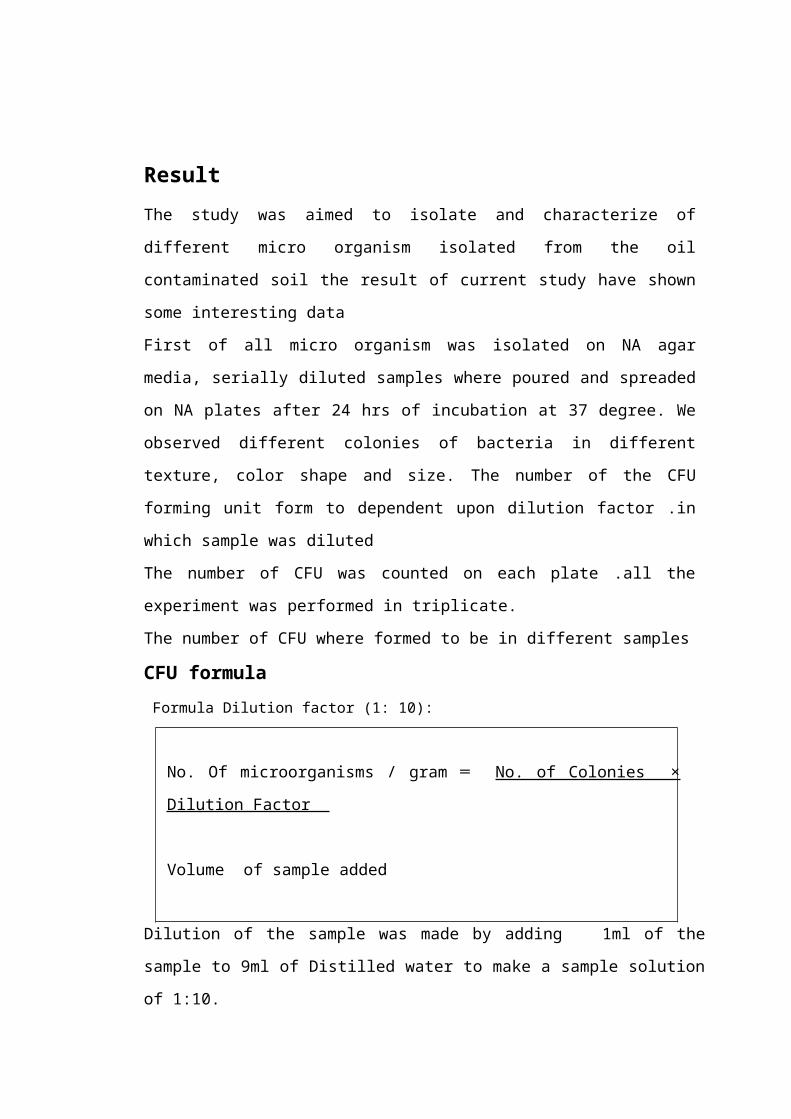
ResultThe study was aimed to isolate and characterize of
different micro organism isolated from the oil
contaminated soil the result of current study have shown
some interesting data
First of all micro organism was isolated on NA agar
media, serially diluted samples where poured and spreaded
on NA plates after 24 hrs of incubation at 37 degree. We
observed different colonies of bacteria in different
texture, color shape and size. The number of the CFU
forming unit form to dependent upon dilution factor .in
which sample was diluted
The number of CFU was counted on each plate .all the
experiment was performed in triplicate.
The number of CFU where formed to be in different samples
CFU formula Formula Dilution factor (1: 10):
No. Of microorganisms / gram ═ No. of Colonies ×
Dilution Factor
Volume of sample added
Dilution of the sample was made by adding 1ml of the
sample to 9ml of Distilled water to make a sample solution
of 1:10.
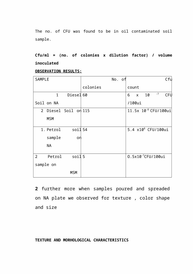
The no. of CFU was found to be in oil contaminated soil
sample.
Cfu/ml = (no. of colonies x dilution factor) / volume
inoculated
OBSERVATION RESULTS:
SAMPLE No. of
colonies
Cfu
count 1 Diesel
Soil on NA
60 6 x 10 -7 CFU
/100ui2 Diesel Soil on
MSM
115 11.5x 10⁻⁸ CFU/100ui
1. Petrol soil
sample on
NA
54 5.4 x10⁸ CFU/100ui
2 Petrol soil
sample on
MSM
5 O.5x10-7CFU/100ui
2 further more when samples poured and spreaded
on NA plate we observed for texture , color shape
and size
TEXTURE AND MORHOLOGICAL CHARACTERISTICS

SAMPLE MEDIUM ELEVATION EDGE SIZE COLONY COLOR Soil NA small yellow
soil NA large yellow
soil
NA large white
3. The different micro organism isolated from the
NA plate where further grown on some specific
media such as MSM, mineral salt media for their
further identification
Serially diluted sample was spread on NA medium
and we observed that

Diesel colony on NA media
Fig A
Three type colony growths take place on NA medium
1 White colony
2 Yellow small colony
3 Yellow large colony

Streaking- All type colony streaked on NA
plate and we observed that result
Fig1
Fig2

Fig3
Fig 1- Showing yellow large colony streaked on NA plate
Fig 2- Showing yellow small colony streaked on NA plate
Fig 2- Showing white colony streaked on NA plate
4.Then thease three different colony isolated from NAplate where streaked on MSM media and we observed that
Yellow small colony
Yellow small colony
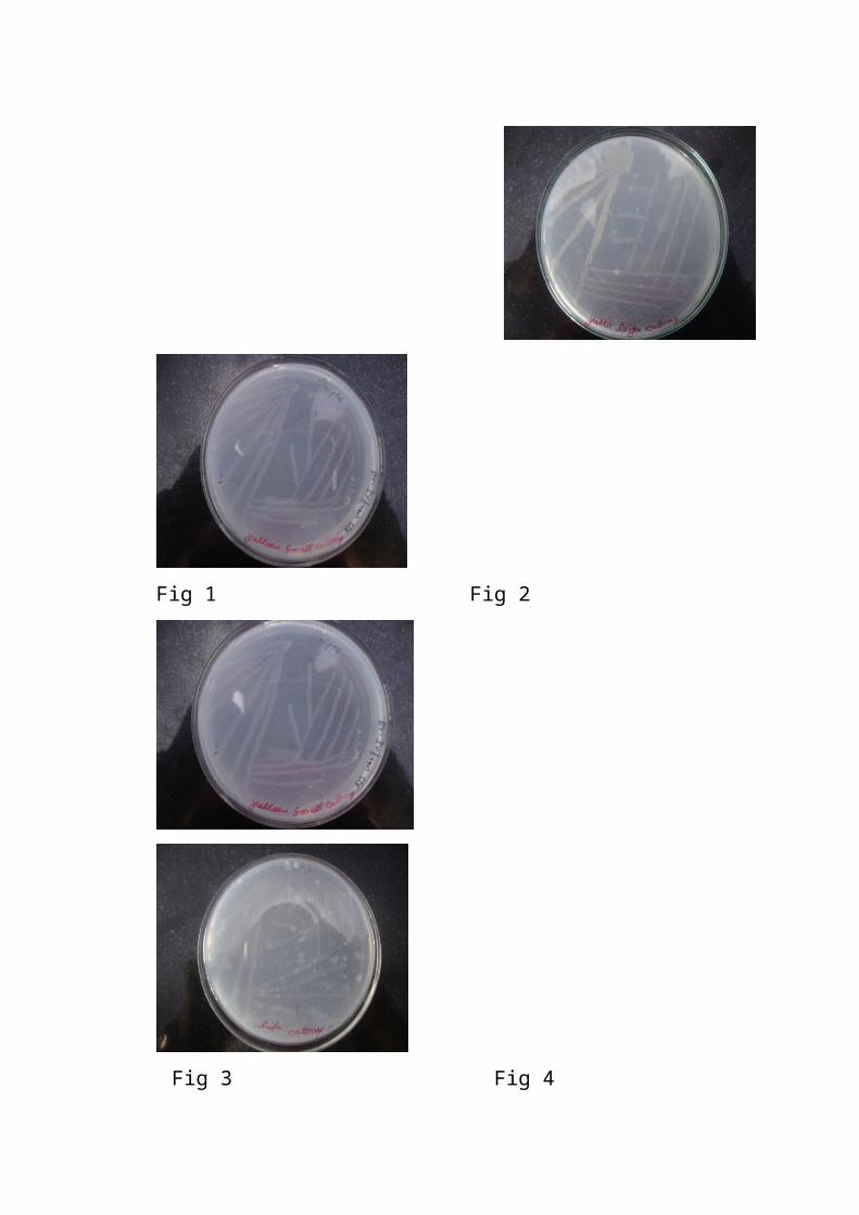
Fig 1 Fig 2
Fig 3 Fig 4

Yellow small colony
white colony
Fig 1- showing yellow large colony streaked on
MSM plate
Fig 2&3- Showing yellow small colony streaked
dill 105 &106 on MSM plate
Fig 3- Showing white colony streaked on MSM plate
5.We further wanted to find out the presence of somedifferent bacterial species in our sample.
We further diluted sample spreaded on MSM media and
incubated at 28 degree centigrade for
seven days and observed that
Diesel colonies on
MSM

Gram staining – After the gram staining we observed that some
intresting result

Diesel white
colony
Fig 1a- Gram negative Coco Fig 2b-
Gram negative Coco Bacilli
Bacilli.

Fig 3c- Gram negative
Coco Bacilli
Fig 4d-Gram
positive Coco
Bacilli suspected Staphylococcus aureus
Diesel yellow colony

Fig 5e- Gram negative Coco Bacilli. Fig 6f-Gramnegative Coco Bacilli
Diesel colony on MSM media

Fig 7g- Gram negative Coco Fig 8h- Gram
negative Coco Bacilli.
Bacilli.
Petrol colony

Gram negative Bacilli
Gram negative Bacilli

Gram negative Bacilli Gram
negative Bacilli

CATALASE TESTSample culture was taken on the sterilized slide
by inoculation loop and then add one or two drop
of H2O2 and we observed bubble formation take
place
Catalase positive

OXIDASE TEST - Oxidase reagent adds 2 or 4 drop on the filter paper and then
the samples are put on this
filter paper. and observe the color
Change.
Oxidase positive

Bacterial typing by 16s r DNA amplification-In order to identify bacterial isolate at species
level .we further aimed to analyse the 16 s Ribosomal DNA
amplification for the Bacterial typing species in this
scenario .we first of all isolated Bacterial Genomic DNA
from different isolates by following a generalized
protocol for the isolation of Genomic DNA
The isolated bacterial DNA was visualized on agarose gel
containing eithedium bromide as shown in following figure

Quantification –The amount of genomic DNA was quantified are to the
formula as earlier discussed in material and methods. and
the purity of DNA was also checked and was found to be
between the range
Isolates absorbence 260 to 280

References

[1] K. A. Kvenvolden and C. K. Cooper, “Natural seepage
ofcrude oil into the marine environment,” Geo-Marine
Letters,vol. 23, no. 3-4, pp. 140–146, 2003.
[2] C. Holliger, S. Gaspard, G. Glod et al.,
“Contaminatedenvironments in the subsurface and
bioremediation: organiccontaminants,” FEMS Microbiology
Reviews, vol. 20, no. 3-4,pp. 517–523, 1997.
[3] P. J. J. Alvarez andT.M. Vogel, “Substrate
interactions of benzene,toluene, and para-xylene during
microbial degradationby pure cultures and mixed culture
aquifer slurries,” Applied
and Environmental Microbiology, vol. 57, no. 10, pp. 2981–2985,
1991.
[4] J. I.Medina-Bellver, P.Mar´ın, A. Delgado et al.,
“Evidence forin situ crude oil biodegradation after the
Prestige oil spill,” Environmental Microbiology, vol. 7, no. 6,
pp. 773–779, 2005.
[5] T. M. April, J. M. Foght, and R. S. Hurrah,
“Hydrocarbondegradingfilamentous fungi isolated from
flare pit soilsin northern and western Canada,” Canadian
Journal of
Microbiology,vol. 46, no. 1, pp. 38–49, 2000.
[6] W. Ulrici, “Contaminant soil areas, different
countries andcontaminant monitoring of contaminants,” in
EnvironmentalProcess II. Soil Decontamination Biotechnology, H. J. Rehm
and G. Reed, Eds., vol. 11, pp. 5–42, 2000.

[7] J. G. Leahy and R. R. Colwell, “Microbial degradation
ofhydrocarbons in the environment,” Microbiological
Reviews,vol. 54, no. 3, pp. 305–315, 1990.
[12] J. M. Foght and D. W. S. Westlake, “Biodegradation
ofhydrocarbons in freshwater,” in Oil in Freshwater:
Chemistry,Biology, Countermeasure Technology, J. H. Vandermeulen
and
S. R. Hrudey, Eds., pp. 217–230, Pergamon Press, New
York,NY, USA, 1987.
[13] R. M. Atlas and R. Bartha, “Fundamentals and
applications,”in Microbial Ecology, pp. 523–530,
Benjamin/Cummings, SanFrancisco, Calif, USA, 4th edition,
1998.
[14] A. J. Mearns, “Cleaning oiled shores: putting
bioremediationto the test,” Spill Science and Technology Bulletin,
vol. 4, no. 4,pp. 209–217, 1997.
[15] R. C. Prince, “Petroleum spill bioremediation in
marineenvironments,” Critical Reviews in Microbiology, vol. 19,
no.4, pp. 217–242, 1993. [18] A. D. Venosa, D.W. King,
and G. A. Sorial, “The baffled flask test for dispersant
effectiveness: a round Robin evaluation of
Reproducibility and repeatability,”Spill Science and Technology
Bulletin, vol. 7, no. 5-6, pp. 299–308, 2002.
[19] R. R. Colwell, J. D. Walker, and J. J. Cooney,
“Ecologicalaspects of microbial degradation of petroleum

in the marineenvironment,” Critical Reviews in Microbiology, vol.
5, no. 4,pp. 423–445, 1977.
[20] J. J. Cooney, S. A. Silver, and E. A. Beck, “Factors
influencinghydrocarbon degradation in three freshwater
lakes,” MicrobialEcology, vol. 11, no. 2, pp. 127–137, 1985.
[21] S. Barathi and N. Vasudevan, “Utilization of
petroleumhydrocarbons by Pseudomonas fluorescensisolated from
apetroleum-contaminated soil,” Environment International,
vol. 26, no. 5-6, pp. 413–416, 2001.
[23] R. Atlas and J. Bragg, “Bioremediation of marine oil
spills:when and when not—the Exxon Valdez experience,”
MicrobialBiotechnology, vol. 2, no. 2, pp. 213–221, 2009.
[24] R. M. Atlas, “Petroleum microbiology,” in Encyclopedia
ofMicrobiology, pp. 363–369, Academic Press, Baltimore,
Md,USA, 1992.
[26] B. Lal and S. Khanna, “Degradation of crude oil
byAcinetobactercalcoaceticusand Alcaligenesodorans,” Journalof Applied
Bacteriology, vol. 81, no. 4, pp. 355–362, 1996.
[27] D. M. Jones, A. G. Douglas, R. J. Parkes, J. Taylor,
W.Giger, and C. Schaffner, “The recognition of
biodegradedpetroleum-derived aromatic hydrocarbons in
recent marine
sediments,” Marine Pollution Bulletin, vol. 14, no. 3, pp. 103–
108, 1983.
[28] S. A. Adebusoye, M. O. Ilori, O. O. Amund, O. D.
Teniola,and S. O. Olatope, “Microbial degradation of
petroleumhydrocarbons in a polluted tropical stream,”
World Journal ofMicrobiology and Biotechnology, vol. 23, no. 8, pp.
1149–1159,2007.
[29] J. Jones, M. Knight, and J. A. Byron, “Effect of
grosspopulation by kerosene hydrocarbons on the
microflora ofa moorland soil,” Nature, vol. 227, p. 1166,
1970.
[30] Y. Pinholt, S. Struwe, and A. Kjoller, “Microbial
changesduring oil decomposition in soil,” Holarctic Ecology,
vol. 2,pp. 195–200, 1979.
[31] S. L. Hollaway, G. M. Faw, and R. K. Sizemore,
“Thebacterial community composition of an active oil
field in theNorthwestern Gulf of Mexico,”Marine Pollution
Bulletin, vol.11, no. 6, pp. 153–156, 1980.
[32] G. J. Mulkins Phillips and J. E. Stewart,
“Distributionof hydrocarbon utilizing bacteria in
Northwestern Atlanticwaters and coastal sediments,”
Canadian Journal of Microbiology,
vol. 20, no. 7, pp. 955–962, 1974.
[33] R. Bartha and I. Bossert, “The treatment and
disposal ofpetroleum wastes,” in Petroleum Microbiology, R.
M. Atlas,Ed., pp. 553–578, Macmillan, New York, NY, USA,
1984.

[34] J. J. Cooney, “The fate of petroleum pollutants
infresh water ecosystems,” in Petroleum Microbiology, R.
M.Atlas, Ed., pp. 399–434, Macmillan, New York, NY,
USA,1984.
[35] R. M. Atlas, “Effects of hydrocarbons on micro-
organismsand biodegradation in Arctic ecosystems,” in
PetroleumEffects in the Arctic Environment, F. R. Engelhardt, Ed.,
pp.63–99, Elsevier, London, UK, 1985.
[36] G. Floodgate, “The fate of petroleum in marine
ecosystems,”in Petroleum Microbiology, R. M. Atlas, Ed., pp.
355–398,Macmillion, New York, NY, USA, 1984.
[37] K. S. M. Rahman, T. J. Rahman, Y. Kourkoutas, I.
Petsas,R. Marchant, and I. M. Banat, “Enhanced
bioremediationof n-alkane in petroleum sludge using
bacterial consortium
amended with rhamnolipid and micronutrients,”
BioresourceTechnology, vol. 90, no. 2, pp. 159–168, 2003.
[38] R. J. W. Brooijmans, M. I. Pastink, and R. J.
Siezen,Hydrocarbon-degrading bacteria: the oil-spill
clean-upcrew,” Microbial Biotechnology, vol. 2, no. 6, pp.
587–594,2009.
[39] M. M. Yakimov, K. N. Timmis, and P. N. Golyshin,
“Obligate
oil-degrading marine bacteria,” Current Opinion in
Biotechnology,vol. 18, no. 3, pp. 257–266, 2007.

[40] K. Das and A. K.Mukherjee, “Crude petroleum-oil
biodegradationefficiency of Bacillus subtilisand Pseudomonas
aeruginosastrains isolated from a petroleum-oil
contaminated soil
from North-East India,” Bioresource Technology, vol. 98, no.7,
pp. 1339–1345, 2007.
[41] M. Throne-Holst, A. Wentzel, T. E. Ellingsen, H.-
K.Kotlar, and S. B. Zotchev, Identification of novel
genes involved inlong-chain n-alkane degradation by
Acinetobactersp. StrainDSM 17874,” Applied and Environmental
Microbiology, vol.73, no. 10, pp. 3327–3332, 2007.
[42] F. Chaillan, A. Le Fl`eche, E. Bury et al.,
“Identification andbiodegradation potential of tropical
aerobic hydrocarbondegradingmicroorganisms,” Research in
Microbiology, vol.155, no. 7, pp. 587–595, 2004.
[44] H. Singh, Mycoremediation: Fungal Bioremediation, Wiley-
Interscience, New York, NY, USA, 2006.
[45] E. Bogusławska-Was and W. Da¸browski, “The
seasonalvariability of yeasts and yeast-like organisms in
water andbottom sediment of the Szczecin Lagoon,”
InternationalJournal of Hygiene and Environmental Health, vol. 203,
no.5-6, pp. 451–458, 2001.
[51] J. D. Walker, R. R. Colwell, Z. Vaituzis, and S. A.
Meyer,Petroleum degrading achlorophyllous alga
Protothecazopfii,”Nature, vol. 254, no. 5499, pp. 423–424,
1975.

[52] C. E. Cerniglia, D. T. Gibson, and C. Van Baalen,
“Oxidation of naphthalene by cyanobacteria and
microalgae,” Journal ofGeneral Microbiology, vol. 116, no. 2,
pp. 495–500,
1980.
[53] M. L. Brusseau, “The impact of physical, chemical
andbiological factors on biodegradation,” in Proceedings of
the International Conference on Biotechnology for Soil Remediation:Scientific
Bases and Practical Applications, R. Serra, Ed.,pp. 81–98,
C.I.P.A. S.R.L., Milan, Italy, 1998.
[54] R. M. Atlas, “Effects of temperature and crude oil
compositionon petroleum biodegradation,” Journal of
AppliedMicrobiology, vol. 30, no. 3, pp. 396–403, 1975.
[62] J. M. Foght, D. W. S. Westlake, W. M. Johnson, and
H.F. Ridgway, “Environmental gasoline-utilizing isolates
andclinical isolates of Pseudomonas aeruginosaare
taxonomically
indistinguishable by chemotaxonomic and molecular
techniques,”Microbiology, vol. 142, no. 9, pp. 2333–2340,
1996.
[63] A. D. Venosa and X. Zhu, “Biodegradation of crude
oilcontaminating marine shorelines and freshwater
wetlands,”Spill Science and Technology Bulletin, vol. 8, no. 2, pp.
163–
178, 2003.

[64] E. Pelletier, D. Delille, and B. Delille, “Crude oil
bioremediationin sub-Antarctic intertidal sediments:
chemistry andtoxicity of oiled residues,” Marine
Environmental Research,
vol. 57, no. 4, pp. 311–327, 2004.
[65] D. Delille, F. Coulon, and E. Pelletier, “Effects of
temperaturewarming during a bioremediation study of
naturaland nutrient-amended hydrocarbon-contaminated sub-
Antarctic soils,” Cold Regions Science and Technology, vol.
40,no. 1-2, pp. 61–70, 2004.
[66] W. J. Mitsch and J. G. Gosselink, Wetlands, John
Wiley &Sons, New York, NY, USA, 2nd edition, 1993.
[67] S.-C. Choi, K. K. Kwon, J.H. Sohn, and S.-J. Kim,
“Evaluationof fertilizer additions to stimulate oil
biodegradation insand seashore mesocosms,” Journal of
Microbiology and
Biotechnology, vol. 12, no. 3, pp. 431–436, 2002.
[68] S.-J. Kim, D. H. Choi, D. S. Sim, and Y.-S. Oh,
“Evaluationof bioremediation effectiveness on crude oil-
contaminatedsand,” Chemosphere, vol. 59, no. 6, pp. 845–
852, 2005.
[69] F. Chaillan, C. H. Chaˆıneau, V. Point, A. Saliot,
and J. Oudot,“Factors inhibiting bioremediation of soil
contaminated withweathered oils and drill cuttings,”
Environmental Pollution,vol. 144, no. 1, pp. 255–265, 2006.

[70] J. Oudot, F. X. Merlin, and P. Pinvidic, “Weathering
rates of oil components in a bioremediation experiment in
estuarinesediments,” Marine Environmental Research, vol. 45,
no. 2,pp. 113–125, 1998.
[71] C. H. Chaˆıneau, G. Rougeux, C. Y´epr´emian, and J.
Oudot,“Effects of nutrient concentration on the
biodegradation ofcrude oil and associated microbial
populations in the soil,”Soil Biology and Biochemistry, vol. 37,
no. 8, pp. 1490–1497,2005.
[72] L. M. Carmichael and F. K. Pfaender, “The effect of
inorganicand organic supplements on the microbial
degradation ofphenanthrene and pyrene in soils,”
Biodegradation, vol. 8, no.1, pp. 1–13, 1997.
[73] J. C. Okolo, E. N. Amadi, and C. T. I. Odu, “Effects
ofsoil treatments containing poultry manure on crude
oildegradation in a sandy loam soil,” Applied Ecology
andEnvironmental Research, vol. 3, no. 1, pp. 47–53, 2005.
[74] H. Maki, T. Sasaki, and S. Haramaya, “Photooxidation
ofbiodegradable crude oil and toxicity of the
photooxidizeproducts,” Chemosphere, vol. 44, pp. 1145–
1151, 2005.
[75] W. Fritsche and M. Hofrichter, “Aerobic
degradationby microorganisms,” in Environmental Processes-
SoilDecontamination, J. Klein, Ed., pp. 146–155, Wiley-VCH,
Weinheim, Germany, 2000.

[76] R. K. Hommel, “Formation and phylogenetic role of
biosurfactants,”Journal of Applied Microbiology, vol. 89, no.
1,pp. 158–119, 1990.
[77] J. B. Van Beilen and E. G. Funhoff, “Alkane
hydroxylasesinvolved in microbial alkane degradation,”
AppliedMicrobiology and Biotechnology, vol. 74, no. 1, pp. 13–
21,2007.
[78] T. Zimmer, M. Ohkuma, A. Ohta, M. Takagi, and W.-H.
Schunck, “The CYP52 multigene family of Candidacytochromes
p450,” Biochemical and Biophysical ResearchCommunications, vol.
224, no. 3, pp. 784–789, 1996.
[79] U. Scheuer, T. Zimmer, D. Becher, F. Schauer, and
W.-H.Schunck, “Oxygenation cascade in conversion of n-
alkanes toα,ω-dioic acids catalyzed by cytochrome P450
52A3,” Journal
of Biological Chemistry, vol. 273, no. 49, pp. 32528–
32534,1998.
[80] J. B. Van Beilen and E. G. Funhoff, “Expanding the
alkaneoxygenase toolbox: new enzymes and applications,”
CurrentOpinion in Biotechnology, vol. 16, no. 3, pp. 308–314,
2005.12 Biotechnology Research International
[81] M. O. Ilori, S. A. Adebusoye, and A. C. Ojo,
“Isolation and characterization of hydrocarbon-degrading
andbiosurfactant-producing yeast strains obtained from a
polluted lagoon water,” World Journal of Microbiology
andBiotechnology, vol. 24, no. 11, pp. 2539–2545, 2008.

[82] G. S. Kiran, T. A. Hema, R. Gandhimathi et
al.,“Optimization and production of a biosurfactant from
thesponge-associated marine fungus
AspergillusustusMSF3,”Colloids and Surfaces B, vol. 73, no. 2, pp.
250–256, 2009.
[83] O. S. Obayori, M. O. Ilori, S. A. Adebusoye, G. O.
Oyetibo,A. E. Omotayo, and O. O. Amund, “Degradation
ofhydrocarbons and biosurfactant production by
Pseudomonassp. strain LP1,” World Journal of Microbiology
andBiotechnology, vol. 25, no. 9, pp. 1615–1623, 2009.
[84] M. L. Brusseau, R. M. Miller, Y. Zhang, X. Wang, and
G. Y.Bai, “Biosurfactant and cosolvent enhanced
remediation ofcontaminated media,” ACS Symposium Series, vol.
594, pp.82–94, 1995.
[85] G. Bai, M. L. Brusseau, and R. M. Miller,
“Biosurfactantenhancedremoval of residual hydrocarbon
from soil,”Journal of Contaminant Hydrology, vol. 25, no. 1-2,
pp.157–170, 1997.
[86] T. Barkay, S. Navon-Venezia, E. Z. Ron, and E.
Rosenberg,“Enhancement of solubilization and
biodegradation ofpolyaromatic hydrocarbons by the
bioemulsifieralasan,”Applied and Environmental Microbiology, vol.
65, no. 6, pp.2697–2702, 1999.
[87] S. S. Cameotra and P. Singh, “Bioremediation of oil
sludgeusing crude biosurfactants,” International

Biodeteriorationand Biodegradation, vol. 62, no. 3, pp. 274–280,
2008.
[88] R. Beal and W. B. Betts, “Role of
rhamnolipidbiosurfactantsin the uptake and mineralization
of hexadecane inPseudomonas aeruginosa,” Journal of Applied
Microbiology,vol. 89, no. 1, pp. 158–168, 2000.
[89] O. Pornsunthorntawee, P. Wongpanit, S. Chavadej,
M.Abe, and R. Rujiravanit, “Structural and
physicochemicalcharacterization of crude biosurfactant
produced byPseudomonas aeruginosaSP4 isolated from petroleum
—contaminated soil,” Bioresource Technology, vol. 99, no. 6,
pp.1589–1595, 2008.
[90] M.NikolopoulouandN. Kalogerakis, “Biostimulation
strategiesfor fresh and chronically polluted marine
environmentswith petroleum hydrocarbons,” Journal of Chemical
Technologyand Biotechnology, vol. 84, no. 6, pp. 802–807,
2009.
[91]:Butler CS, JR Mason (1997). Structure–functionanalysis of the bacterial aromatic ring–hydroxylatingdioxygenase. Advanced Microbial Physiology. 38: 47–84.
[92]:Hag well IS, LM Delfino, JJ Rao (1992). Partitioningof Polycyclic Aromatic Hydrocarbons from oil into water.Environ. Sci. Technol. 26: 2104–2110.
[93]:Mishra S, J Jyot, RC Kuhad, B Lal (2001). Evaluationof inoculum addition to stimulate in situ Bioremediationof oily–sludge–contaminated soil. Appl. Environ.Microbial. 67(4): 1675–1681.

[94]:Propst TL, RL Lochmiller, CW Qualis, Jr. K McBee(1999). In situ (mesocosm) assessment of immunotoxicityrisks to small mammals inhabiting petrochemical wastesites. Chemosphere. 38: 1049–1067.
[95]:Lloyd CA, TA Cackette (2001). Diesel Engines:Environmental Impact and Control. Air and WasteManagement Association. 51: 805–847.
[96]: Van Hamme JD, Singh A, Ward OP (2003). RecentAdvances in Petroleum Microbiology. Microbial. Mol. Biol.Rev. 67(4): 503–549.
[97]:Blodgett WC (2001). Water–soluble mutagen productionduring the bioremediation of oil–contaminated soil.Florida Scientist. 60(1):28–36.
98:Roling WFM, Milner MG, DM Jones, K Lee, F Daniel,Swannell RJP, Head IM (2002). Robust hydrocarbondegradation and dynamics of bacterial communities duringnutrient–enhanced oil spill bioremediation. Appl.Environ. Microbial. 68 (11): 5537-5548.
[99]:Bushnell LD, HF Haas (1941). The utilization ofhydrocarbons by microorganisms. J. Bacterial. 41: 653–673.
[100]:Speight JG (1991). The chemistry and technology ofpetroleum. Marcel Dekker, New York, N.Y.
[101]: Ehrlich HL (1995). Geomicrobiology. Marcel Dekker,Inc., New York, N.Y.
[102]: Harder E (2004). Bioremediation of engine oil.Little Flower Academy. Dallas, Texas.
[103]:Alexander M (1999). Biodegradation andBioremediation (2nd edition) Academic Press, San Diego

[104]:Barathi S, N Vasudevan (2001). Utilization ofpetroleum hydrocarbons by Pseudomonas fluoresces isolatedfrom a petroleum–contaminated soil. Environ. Int. 26: 413– 416.
[105]:Eriksson M, Dalhammer G, AK Borg–Karlson. (1999).Aerobic degradation of a hydrocarbon mixture in naturaluncontaminated potting soil by indigenous microorganismsat 20oC and 6oC. Appl.Microbial. Biotechnology. 51: 532–535
[106]:Johnson K, Anderson S, CS Jacobson (1996).Phenotypic and genotypic characterization ofphenanthrene–degrading fluorescent Pseudomonas biovars.Appl. Environ. microbial. 62: 3818–3825.
[107]:Kiyohara H, Takizawa N, Nagao K (1992). Naturaldistribution of bacteria metabolizing many kinds ofpolyaromatic hydrocarbons. J. Ferment. Bioeng. 74: 49–51.
[108]:Fall RR, JL Brown, TL Schaeffer (1979). Enzymerecruitment allows the biodegradation of recalcitrant–branched hydrocarbons by Pseudomonas citronellals. Appl.Environ. Microbial. 38: 715–722.
[109]:Rusansky S, Avigad R, Michaeli S, Gutnick DL(1987). Involvement of a plasmid in Growth on andDispersion of Crude Oil by Acinetobactersp calcoaceticusRA57. Appl. Environ. Microbial. 53: 1918-1923.
[110]: Prenafeta–Boldu XF, Kuhn A, DMAM L, Anke H, JW VanGroenestijn, JAM De Bont (200)1. Isolation andCharacterization of fungi growing on volatile aromatichydrocarbons as their sole carbon and energy source.Mycological Res. 4: 477–484.

[111]:McLaughlin B (2001). Soil Remediation. Enginr. Sci.Rev. 2:69–77. Mishra S, J Jyot, RC Kuhad, B Lal (2001).Evaluation of inoculum addition to stimulate in situBioremediation of oily–sludge–contaminated soil. Appl.Environ.Microbiol. 67(4): 1675–1681.
[112]:Swannell RPJ, K Lee K, McDonagh M (1996). Fieldevaluation of Marine Oil Spill Bioremediation. Microbial,Rev. 60: 342–365.