Cloning of the Gene for Indoleacetic Acid-Lysine Synthetase ...
Upload
independentCategory
view
3download
0
MOLECULAR AND CELLULAR BIOLOGY,0270-7306/00/$04.0010
Nov. 2000, p. 8458–8467 Vol. 20, No. 22
Copyright © 2000, American Society for Microbiology. All Rights Reserved.
Multiple C-Terminal Lysine Residues Target p53 forUbiquitin-Proteasome-Mediated Degradation
MANUEL S. RODRIGUEZ,1 JOANA M. P. DESTERRO,1 SONIA LAIN,2 DAVID P. LANE,2
AND RONALD T. HAY1*
School of Biology, University of St. Andrews, St. Andrews Fife KY16 9ST,1 and Surgery and Molecular Oncology,University of Dundee, Ninewells Hospital and Medical School, Dundee DD1 9SY,2 Scotland, United Kingdom
Received 13 April 2000/Returned for modification 8 June 2000/Accepted 14 August 2000
In normal cells, p53 is maintained at a low level by ubiquitin-mediated proteolysis, but after genotoxic insultthis process is inhibited and p53 levels rise dramatically. Ubiquitination of p53 requires the ubiquitin-activating enzyme Ubc5 as a ubiquitin conjugation enzyme and Mdm2, which acts as a ubiquitin protein ligase.In addition to the N-terminal region, which is required for interaction with Mdm2, the C-terminal domain ofp53 modulates the susceptibility of p53 to Mdm2-mediated degradation. To analyze the role of the C-terminaldomain in p53 ubiquitination, we have generated p53 molecules containing single and multiple lysine-to-arginine changes between residues 370 and 386. Although wild-type (WT) and mutant molecules show similarsubcellular distributions, the mutants display a higher transcriptional activity than WT p53. Simultaneousmutation of lysine residues 370, 372, 373, 381, 382, and 386 to arginine residues (6KR p53 mutant) generatesa p53 molecule with potent transcriptional activity that is resistant to Mdm2-induced degradation and isrefractory to Mdm2-mediated ubiquitination. In contrast to WT p53, transcriptional activity directed by the6KR p53 mutant fails to be negatively regulated by Mdm2. Those differences are also manifest in HeLa cellswhich express the human papillomavirus E6 protein, suggesting that p53 C-terminal lysine residues are alsoimplicated in E6-AP-mediated ubiquitination. These data suggest that p53 C-terminal lysine residues are themain sites of ubiquitin ligation, which target p53 for proteasome-mediated degradation.
Cell cycle arrest, DNA repair, and apoptosis are the mostcommon responses to DNA damage in normal mammaliancells. Genetic instability and malignant transformation are con-sequences of improper DNA damage responses (9, 26, 32).The p53 protein plays an important role in the initiation of theDNA damage response, cell cycle arrest, and tumor suppres-sion (25, 31). One of the best-characterized functions of p53 isits transcriptional activity (26, 32). p53 increases the transcrip-tion of genes coding for important cell growth inhibitors, suchas the p21Waf1/Cip1 gene (10), and apoptotic genes, such as thatcoding for Bax (42). The identification of the Mdm2 gene as ap53 target gene, the product of which can negatively regulatep53 functions, revealed a feedback loop that regulates both p53activity and expression of Mdm2 (3, 30, 43, 44, 65).
In normal cells p53 levels are maintained at low or unde-tectable levels by continual proteolytic degradation, but whencells are exposed to stress signals, such as hypoxia and DNAdamage, degradation is inhibited and p53 levels rise (25, 35).Sustained degradation of p53 is mediated by the ubiquitin-proteasome pathway (34) and is carried out by the ubiquitin-activating enzyme (E1), a ubiquitin-conjugating enzyme(Ubc5), and a ubiquitin protein ligase (Mdm2) (13, 19). Ubiq-uitin molecules are attached to p53 by an isopeptide bondbetween the C terminus of ubiquitin and the ε-amino group ofa lysine residue. Multiple ubiquitin molecules are required totarget protein substrates for degradation (18). Since degrada-tion is an efficient means to abrogate all p53 functions, p53-induced transcription of Mdm2 generates an efficient mecha-nism for controlling p53 levels (3, 65). Many tumors and tumorcell lines have low levels of Mdm2 because they express tran-
scriptionally inactive p53, and as a consequence, p53 levels arevery high (41). The importance of this regulatory mechanism isillustrated by the fact that deletion of the mdm2 gene in miceinduces an early embryonic lethality which is rescued by dele-tion of the p53 gene (24). A wide variety of mechanisms havebeen proposed which can block the interaction of Mdm2 withp53 and as a consequence increase the levels of p53. Phosphor-ylation of p53 or Mdm2 by a DNA-dependent protein kinasereduces the interaction between the two molecules (37, 58),resulting in reduced ubiquitination of p53 by the ubiquitinligase activity of Mdm2 (20). The tumor suppressor p19 ARFblocks the degradation of p53 by directly inhibiting the ubiq-uitin ligase activity of Mdm2 (20, 39) and sequestering Mdm2in the nucleolus (64, 66).
Subcellular distribution plays an important role in the con-trol of p53 transcriptional activity, and nuclear transport isdependent on three nuclear localization signals (NLSs) locatedin the C-terminal region of the protein (57). Fusion of themajor NLS (NLSI, amino acids [aa] 316 to 322) to a heterol-ogous cytoplasmic protein directs the chimeric protein to thenucleus (7), while NLSII (aa 370 to 377) and NLSIII (aa 380 to386) appear to increase the efficiency of nuclear import (57).Moreover, it has been proposed that the interaction of lysine305 with the cytoplasmic sequestration domain (aa 326 to 355)regulates p53 subcellular distribution (33). The cytoplasmicsequestration domain also contains the p53 oligomerizationdomain and a putative leucine-rich nuclear export signal (LR-NES) (aa 340 to 351) (60). Mdm2-mediated p53 degradationhas been reported to be a cytoplasmic process dependent on anLR-NES present in Mdm2 (54). Consequently, p53 degrada-tion is blocked by leptomycin B (12, 29), which is a competitiveinhibitor of the LR-NES receptor CRM1 (1, 11, 46, 62).
The p53-Mdm2 interaction is mediated by the N-terminalregions of the proteins. Mdm2 can negatively regulate p53transcriptional activity by binding to and occluding the p53
* Corresponding author. Mailing address: School of Biology, BMSBuilding, University of St. Andrews, North Haugh, St. Andrews FifeKY16 9ST, United Kingdom. Phone: 44 1334 463399. Fax: 44 1334462595. E-mail: [email protected].
8458
on October 4, 2015 by guest
http://mcb.asm
.org/D
ownloaded from
transcriptional activation domain (5, 43, 45). The C-terminalregion of p53 modulates the susceptibility of p53 to Mdm2-mediated degradation, and deletion of the last 30 aa of p53inhibits this process (28). Six of the C-terminal 30 aa of p53 arelysine residues which are potential sites of ubiquitination. Toinvestigate the role of the C-terminal lysine residues in thedegradation of p53, we have generated single and multipleK-to-R changes in this region of p53. The altered proteinsdisplay an increased ability to activate a p53-dependent tran-scriptional reporter, and this ability is maximal with the p53mutant in which all six lysine residues have been changed toarginine residues. In vivo ubiquitination and degradation as-says and in vitro ubiquitination assays suggest that p53 C-terminal lysine residues are involved in ubiquitin-mediateddegradation of p53. Our results also support the notion thatMdm2-mediated ubiquitination is an important mechanism forcontrolling p53 activity.
MATERIALS AND METHODS
Proteins and antibodies. Ubiquitin was purchased from Sigma. Human E1ubiquitin-activating enzyme was purified from recombinant baculovirus-infectedcells as described previously (53). Human Mdm2 (residues 6 to 491) was ex-pressed in bacteria and purified as previously described for p53 (40). HumanUbc5 was expressed in bacteria and purified as previously described (49). Glu-tathione S-transferase (GST)–Mdm2 fusion protein was purified as reportedpreviously (4). Monoclonal antibody 4B2 (5) recognizes Mdm2. Monoclonalantibody DO.1 (63) and polyclonal antibody CM1 recognize p53. Green fluores-cent protein (GFP) was detected with a mixture of two mouse monoclonalantibodies (anti-GFP clones 7.1 and 13.1) purchased from Boehringer Mann-heim.
Plasmid construction. The previously described wild-type (WT) p53 BamHI-EcoRI cassette (49) was replaced to generate multiple K-to-R mutants by site-directed mutagenesis using a PCR strategy. DNA sequences were determined bythe University of St. Andrews DNA sequencing facility (with an ABI 377 se-quencer). The pcDNA3-Mdm2 plasmid was described previously (39). ThepEGFP-C2 plasmid was purchased from Clontech.
Cell culture and transfections. HeLa, p53 null (Saos-2 and H1299), and mousep532/2 Mdm22/2 cells were grown in Dulbecco’s modified Eagle’s medium(DMEM) supplemented with 10% fetal bovine serum. Cells were transfected byelectroporation (950 mF, 240 V; Equibio Easyject Plus) as previously described(49) or with Lipofectamine (GIBCO/BRL). pG13-luciferase (pG13-Luc) re-porter assays were performed with 2 mg of reporter plasmid and 5 ng of plasmidencoding WT p53 or one of the K-to-R p53 mutants per 106 Saos-2 cells. Tennanograms of p53-encoding and 30 ng of Mdm2-encoding plasmids were used forcotransfection into p532/2 Mdm22/2 cells. Reporter assays in HeLa cells wereperformed with 5 ng of WT p53 or the 6KR mutant. Empty pcDNA3 vector wasused to maintain a constant level of plasmid DNA. After electroporation, cellswere grown in six-well plates for 10 to 12 h and processed for luciferase activityas previously described (51). For immunofluorescence, 105 cells were cotrans-fected with 375 ng of plasmid encoding WT p53 or a K-to-R mutant and 2.125 mgof Bluescript plasmid DNA. Mdm2-directed degradation in vivo was performedwith 200 ng of WT p53 or K-to-R mutants, 1.2 mg of Mdm2, and 600 ng ofGFP-encoding plasmids per 106 cells. Empty pcDNA3 vector was used to main-tain a constant level of plasmid DNA. Transfected cells were divided into twogroups, and after 16 h in culture one of the groups was harvested. The secondgroup of Mdm2-transfected cells was treated for 4 h prior to harvesting with 10mM MG132. p53 ubiquitination in vivo was detected in 1.5 3 105 p532/2
Mdm22/2 cells transfected with 200 ng of GFP, 200 ng of WT p53 or K-to-R p53mutants, and 600 ng of pcDNA3 or an Mdm2-encoding plasmid. After 16 h inculture, Mdm2-transfected cells were treated with 10 mM MG132 for 4 h prior toharvesting. A total of 106 HeLa cells were cotransfected with 4 mg of an SV5-tagged version of b-galactosidase (27) and 4 mg of WT p53- and 6KR-encodingplasmids. After 16 h, cells were lysed and analyzed by Western blotting.
Immunofluorescence. After transfection, H1299 or Saos-2 cells were culturedon glass or Permanox chamber slides (Nunc). Twenty-four hours after transfec-tion, slides were washed in phosphate-buffered saline (PBS)–1 mM MgCl2, fixedfor 8 min in cold methanol-acetone (1:1), rinsed in PBS–1 mM MgCl2–0.1%Tween 20, and incubated for 1 h at room temperature with DO.1 mouse mono-clonal antibody diluted in DMEM–10% fetal calf serum. Slides were washed twotimes in PBS–1 mM MgCl2, rinsed in PBS–1 mM MgCl2–0.1% Tween 20, andincubated with a mixture of fluorescein isothiocyanate-conjugated donkey anti-mouse immunoglobulin G in DMEM–10% fetal calf serum at the dilution spec-ified by the manufacturer (Jackson ImmunoResearch Laboratories) for 30 min atroom temperature. After two washes in PBS–1 mM MgCl2 and rinsing in PBS–1mM MgCl2–0.1% Tween 20, the slides were mounted with Mowiol-Dabco, dried,and analyzed using a Bio-Rad MRC 600 LSM confocal microscope and customBio-Rad software.
Western blotting. Whole-cell extracts were lysed in sodium dodecyl sulfate(SDS) sample buffer as described previously (8). Lysates were boiled for 10 minprior to fractionation by electrophoresis in 10% polyacrylamide gels containingSDS. Proteins were transferred to a polyvinylidene difluoride membrane (Sigma)by electroblotting. p53 (DO.1), Mdm2 (4B2), and GFP monoclonal antibodieswere used as primary antibodies. Blots were developed with an enhanced chemi-luminescence detection system.
Interaction of Mdm2 and p53 in vitro. [35S]methionine-labeled in vitro-tran-scribed and -translated p53 proteins (10 ml) were incubated for 2 h at 4°C with1 mg of GST or GST-Mdm2 prebound to glutathione-agarose beads in 200 ml ofpreviously described binding buffer (21). For immunoprecipitation, [35S]methi-onine-labeled in vitro-cotranscribed and -cotranslated Mdm2 and p53 proteins(20 ml) were incubated for 2 h at 4°C with an equal mixture of protein A- andprotein G-agarose beads (Sigma) and p53-specific polyclonal antibody CM1 in200 ml of previously described binding buffer (21). Beads were washed as de-scribed previously (21), and bound protein was fractionated in a 10% polyacryl-amide gel containing SDS. The dried gel was analyzed by phosphorimaging.
In vitro ubiquitination assays. [35S]methionine-labeled in vitro-transcribedand -translated p53 proteins were used in ubiquitination assays as previouslyreported (49). Reaction products were fractionated in a 10% polyacrylamide gelcontaining SDS, and dried gels were analyzed by phosphorimaging (Fijix BAS1500, MacBAS software). Ubiquitinated p53 was quantified by determining theproportion of the total radioactive p53 that migrated more slowly than unmod-ified p53.
RESULTS
K-to-R p53 mutants show higher transcriptional activitythan does WT p53. Reporter assays have been used previouslyto identify lysine residues which are targets for ubiquitinationof transcriptionally active proteins or of transcriptional regu-lators (2, 51). The C-terminal 30 aa of p53 are required forMdm2-mediated degradation (28) and contain six lysine resi-dues which are potential targets for ubiquitination. To inves-tigate the role of these residues on ubiquitin-proteasome-de-pendent degradation of p53, single and multiple K-to-Rchanges were introduced into the C-terminal region of p53(Fig. 1). It was previously reported that K386R p53 has aninherently higher transcriptional activity than does WT p53(49). To test if other K-to-R mutations have similar conse-quences on p53 transcriptional activity, Saos-2 cells, which lackendogenous p53, were cotransfected with the p53-responsivereporter plasmid pG13-Luc and expression plasmids for WTp53 and K-to-R p53 mutants. WT p53 activated transcription57-fold, while all K-to-R p53 mutants showed higher transcrip-tional rates (Fig. 2A). The previously reported K386R mutantwas the single-point mutant with the highest transcriptionalactivity (284-fold activation). The triple mutant 3CKR (483-fold activation) showed a higher transcriptional activity thandid 3NKR (268-fold activation), suggesting that lysine residues381, 382, and 386 may be the preferred targets for ubiquitina-tion. However, 6KR p53, in which all six lysine residues arechanged to arginine, displayed the highest transcriptional ac-tivity (613-fold activation). To confirm these results, the samep53 expression plasmids were cotransfected with the Mdm2-Luc reporter plasmid into Saos-2 cells or with the pG13-Lucreporter plasmid into H1299 p53 null cells, with similar resultsin each case (data not shown). This effect is likely to be medi-ated by Mdm-2, as p532/2 Mdm22/2 cells cotransfected withpG13-Luc and plasmids encoding p53 mutants did not showsignificant differences in their abilities to activate the reporter(see Fig. 8). This indicates that WT p53 and various K-to-Rmutants have inherently similar transcriptional activation po-tential and that the observed differences in reporter activity area reflection of the susceptibility of these proteins to ubiquitin-mediated proteolysis.
Subcellular localization of K-to-R p53 mutants is similar tothat of WT p53. p53 lysine residues 370, 372, 373, 381, 382, and386 represent the basic amino acid core of both p53 NLSII andNLSIII (57). Although NLSI (aa 316 to 322) is the predomi-nant nuclear import signal in p53 (7, 57) and conservative
VOL. 20, 2000 UBIQUITINATION OF p53 8459
on October 4, 2015 by guest
http://mcb.asm
.org/D
ownloaded from

K-to-R changes were introduced to create the p53 mutants, itwas important to eliminate the possibility that the differencesin transcriptional activity were a consequence of altered nu-clear import. To establish this point, H1299 cells were trans-fected with WT p53 or K-to-R p53 mutants, and the subcellular
localization of p53 was determined by indirect immunofluores-cence with a p53-specific monoclonal antibody. WT p53 andK-to-R p53 mutants showed similar subcellular distributions.Ninety percent of transfected cells showed exclusive nuclearlocalization of immunoreactive p53 (Fig. 3). In the remaining10% of transfected cells, p53 was found in both the nucleus andthe cytoplasm. Similar results were obtained in Saos-2 cells(data not shown). These results suggest that the differences intranscriptional activity observed between WT p53 and K-to-Rmutants cannot be explained by differences in subcellular lo-calization.
C-terminal lysine residues of p53 control Mdm2-directeddegradation of p53. To assess the role of p53 C-terminal lysineresidues in Mdm2-directed degradation of p53, K-to-R p53mutants were either expressed alone or coexpressed withMdm2 in Saos-2 cells. p53 and Mdm2 expression were ana-lyzed by Western blotting, and p53 molecules containing singleor double K-to-R changes all appeared to be susceptible toMdm2-induced degradation (Fig. 4A). In contrast, p53 mutant6KR was resistant to Mdm2-mediated degradation. Underthese conditions, Mdm2 was detected only when the film wasoverexposed but was at a relatively constant level in each case(data not shown). GFP expressed from a cotransfected plasmidwas used as an internal control and indicated that the observeddifferences were not due to variations in gel loading or trans-fection efficiency. The remaining 50% of transfected cells werepretreated with proteasome inhibitor MG132 for 4 h prior toharvest and Western blot analysis, as described above. As ex-pected, MG132 blocked Mdm2-directed degradation of p53proteins (Fig. 4B). Concomitant accumulation of p53 andMdm2 after MG132 treatment confirmed that degradation ofboth proteins is proteasome mediated. Although 6KR wasresistant to Mdm2-mediated degradation, limited degradationcould be observed when Mdm2 was expressed at high levels,suggesting that alternative lysine residues can be used to me-
FIG. 1. Schematic representation of p53 mutants with C-terminal K-to-R changes.
FIG. 2. p53 mutants containing C-terminal K-to-R substitutions show highertranscriptional activity than does WT p53. Saos-2 cells were cotransfected byelectroporation with expression plasmids for WT p53 or K-to-R mutants and thepG13-Luc reporter plasmid. Each point is the mean of four independent trans-fections, with error bars representing 1 standard deviation.
8460 RODRIGUEZ ET AL. MOL. CELL. BIOL.
on October 4, 2015 by guest
http://mcb.asm
.org/D
ownloaded from

diate p53 degradation (data not shown). Thus, lysine residues370, 372, 373, 381, 382, and 386 have an important role in theMdm2-mediated degradation of p53.
K-to-R mutations in the C terminus of p53 do not affect itsinteraction with Mdm2. In addition to the N-terminal region,which is required for interaction with Mdm2, it has been sug-gested that the conformation of the C-terminal region of p53may be important for targeting Mdm2-dependent p53 ubiquiti-nation (20). To exclude the possibility that the observed resis-tance of 6KR to Mdm2-mediated degradation results from afailure to bind Mdm2, this interaction was analyzed in vitro. Invitro-translated and 35S-labeled WT p53 and K-to-R mutantswere allowed to interact with GST-Mdm2, and the bound pro-teins were analyzed by SDS-polyacrylamide gel electrophoresis(PAGE) and phosphorimaging. No differences were observedbetween the binding abilities of WT and mutant p53 molecules(Fig. 5A). The observed interactions were specific, since GSTalone did not interact with p53 and GST-Mdm2 failed to in-teract with 35S-labeled NF-kB p50 (Fig. 5A). Similar resultswere obtained by coimmunoprecipitation of Mdm2 and p53molecules (which were generated by cotranslation in vitro)using anti-p53 antibody (Fig. 5B). Thus, C-terminal K-to-Rpoint mutations do not affect the ability of p53 to be recog-nized by Mdm2.
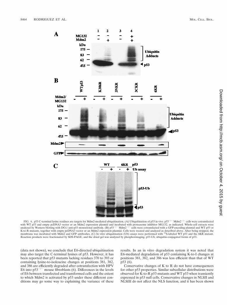
p53 C-terminal lysine residues are targets for Mdm2-depen-dent ubiquitination of p53. Since lysine residues are targets forubiquitination, it is likely that the inability of the 6KR mutantto undergo Mdm2-dependent degradation is due to the factthat it cannot be ubiquitinated. To test this hypothesis, WT p53
and K-to-R mutants were expressed alone or together withMdm2 in p532/2 Mdm22/2 cells. Twelve hours after transfec-tion, Mdm2-transfected cells were exposed to 10 mM MG132for 4 h. Whole-cell extracts were analyzed by Western blottingwith the DO.1 monoclonal antibody. Slowly migrating forms ofp53, consistent with ubiquitination, were observed with WTp53 and the K386R and 3NKR mutants (Fig. 6A and B). In theabsence of proteasome inhibitor, p53 ubiquitinated forms wereobserved only when film was overexposed (Fig. 6A, lanes 1 and2). The appearance of slowly migrating forms was reduced forthe 3CKR mutant and virtually eliminated for the 6KR mutant.GFP expressed from a cotransfected plasmid was also analyzedby Western blotting, and the results indicated that the ob-served differences were not due to variations in gel loading ortransfection efficiency. The relatively small proportion of p53that accumulated as ubiquitinated products was a consequenceof the intracellular removal of ubiquitin by multiple ubiquitinC-terminal hydrolases. Similar results were obtained for theH1299 and Saos-2 cell lines (data not shown). Ubiquitinationof WT p53 and the 6KR mutant was also compared in an invitro system containing 35S-labeled substrate, ubiquitin, andpurified recombinant E1, Ubc5, and Mdm2 (49). Phosphorim-ager quantification of ubiquitin adducts indicated that ubiq-uitination of the 6KR mutant was reduced by 40% comparedto that of WT p53 (Fig. 6C). We conclude that C-terminallysine residues are involved in the Mdm2-mediated ubiquiti-nation of p53.
Role of p53 C-terminal lysine residues in E6- and E6-AP-mediated p53 degradation. To investigate the role of p53 C-
FIG. 3. Subcellular localization of WT p53 and mutants with C-terminal K-to-R changes. H1299 cells were transfected with empty pcDNA3 vector (a) and plasmidsexpressing WT p53 (b), 6KR (c), 3NKR (d), 3CKR (e), K372/373R (f), K381/382R (g), K370R (h), and K386R (i). Indirect immunofluorescence with antibody againstp53 DO.1 is shown.
VOL. 20, 2000 UBIQUITINATION OF p53 8461
on October 4, 2015 by guest
http://mcb.asm
.org/D
ownloaded from

terminal lysine residues in E6-AP-mediated degradation,HeLa cells which express the E6 protein of human papilloma-virus (HPV) were cotransfected with WT p53 or the 6KRmutant, together with SV5-tagged b-galactosidase expressionplasmids. Twenty-four hours after transfection, whole-cell ex-tracts were analyzed by Western blotting with a monoclonalantibody recognizing p53. Higher levels of the 6KR mutantthan of WT p53 were observed (Fig. 7A). Similar results wereobtained with a polyclonal antibody to p53 (data not shown).The increased accumulation of 6KR compared to WT p53 wasnot due to variations in transfection efficiency, because thesedifferences were not reflected in the levels of the cotransfectedSV5-tagged b-galactosidase used as an internal control. Toconfirm these results, HeLa cells were cotransfected with thepG13-Luc reporter plasmid and expression plasmids for WTp53 or the 6KR mutant. The 6KR mutant showed three timesmore transcriptional activity than did WT p53 (Fig. 7B). Thus,it is likely that p53 C-terminal lysine residues are also targetsfor E6-AP-mediated regulation.
The susceptibility of p53 to ubiquitination correlates withthe ability of Mdm2 to negatively regulate p53-dependent tran-scription. To evaluate the contribution of p53 degradation tothe negative regulation of p53 functions by Mdm2, we cotrans-fected p532/2 Mdm22/2 cells with WT p53 and the K386R,3NKR, 3CKR, and 6KR mutants and the p53 reporter plasmidpG13-Luc in the absence or presence of an Mdm2 expressionplasmid. It was reasoned that K-to-R mutants resistant toMdm2-mediated ubiquitination and degradation may showsome resistance to the Mdm2-mediated negative regulation ofp53-dependent transcription. Mdm2 expression reduced WTp53-dependent transcription by 60 to 70% (Fig. 8). Reductionsof 40 to 60% of transcriptional activity were observed when theK386R, 3NKR, and 3CKR mutants were cotransfected withMdm2 (Fig. 8). In contrast, 6KR transcriptional activity wasnot significantly reduced by Mdm2 (Fig. 8). Our results suggestthat ubiquitin-proteasome-mediated degradation of p53 is a
major function of Mdm2 that contributes to the negative reg-ulation of p53-dependent transcriptional activity.
DISCUSSION
Degradation is the only mechanism which abrogates all func-tions of p53, and in normal cells this is accomplished by theubiquitin-proteasome system (34). To identify lysine residueswhich are the sites of modification by ubiquitin, a series of p53molecules were created that contain lysine-to-arginine changesin the C-terminal region of the protein. Using p53-dependentreporter assays, we demonstrated that the p53 molecules con-taining K-to-R changes display a higher transcriptional activity,which is a reflection of their increased stability and resistanceto ubiquitin modification (Fig. 2). In addition to acting as aubiquitin protein ligase (19), it has been reported that Mdm2negatively regulates p53 transcriptional activity by binding tothe transactivation domain of p53 (45, 65). Since p53 mutantswhich accumulate may also induce more efficient synthesis ofMdm2, differences in transcription are more obvious at earlystages after transfection. The observed transcriptional differ-ences between K-to-R p53 mutants cannot be explained as aconsequence of conformational changes induced by the muta-tions, since reporter assays performed in p532/2 Mdm22/2
cells showed no significant differences in the transactivationpotential of WT and mutant versions of p53 in the absence ofMdm2 (Fig. 8). This result also suggests that Mdm2 is the keyregulatory molecule for the control of p53 stability. In HPV-transformed cells p53 is regulated by the viral E6 protein,which, in conjunction with E6-AP, targets p53 for ubiquitin-mediated proteolysis. The 6KR mutant also appears to bemore resistant to degradation than WT p53 when introducedinto HeLa cells which constitutively express HPV E6. Becausecomplex formation between E6 and p53 is not mediatedthrough the C terminus of p53 (6, 36), and since WT p53 and6KR showed similar subcellular distributions in HeLa cells
FIG. 4. Mdm2-mediated degradation is dependent on lysine residues in the C-terminal region of p53. Saos-2 cells were electroporated with plasmids encoding GFP,WT p53, or K-to-R p53 mutants, together with empty pcDNA3 vector or Mdm2 vector as indicated. (A) Twenty-four hours after transfection, whole-cell extracts wereprepared and analyzed by Western blotting with anti-p53, anti-Mdm2, and anti-GFP monoclonal antibodies. (B) Transfected cells were treated with 10 mM MG132for 4 h prior to harvest. Western blotting against p53 and Mdm2 was performed as described above.
8462 RODRIGUEZ ET AL. MOL. CELL. BIOL.
on October 4, 2015 by guest
http://mcb.asm
.org/D
ownloaded from

FIG. 5. K-to-R changes in the p53 C-terminal region do not affect p53-Mdm2 interaction. (A) 35S-labeled NF-kB (p50), WT p53, and K-to-R p53 mutants generatedby in vitro transcription and translation were incubated with GST-Mdm2 or GST as indicated. GST pull-down assays were performed at 4°C. Mdm2-associated materialwas fractionated by SDS-PAGE, and the dried gel was analyzed by phosphorimaging (lower panel). (B) For coimmunoprecipitation, 35S-labeled Mdm2, DN Mdm2,WT p53, and K-to-R p53 mutants generated by in vitro transcription and translation were incubated with protein A- and protein G-agarose beads and p53-specificpolyclonal antibody CM1, as indicated (IPa p53). p53-associated material was fractionated by SDS-PAGE, and the dried gel was analyzed by phosphorimaging (lowerpanel).
VOL. 20, 2000 UBIQUITINATION OF p53 8463
on October 4, 2015 by guest
http://mcb.asm
.org/D
ownloaded from
(data not shown), we conclude that E6-directed ubiquitinationmay also target the C-terminal lysines of p53. However, it hasbeen reported that p53 mutants lacking residues 370 to 393 orcontaining lysine-to-isoleucine changes at positions 381, 382,and 386 are efficiently degraded after cotransfection with HPVE6 into p532/2 mouse fibroblasts (6). Differences in the levelsof E6 between transfected and transformed cells and the extentto which Mdm2 is activated by p53 under these different con-ditions may go some way to explaining the variance of these
results. In an in vitro degradation system it was noted thatE6-mediated degradation of p53 containing K-to-I changes atpositions 381, 382, and 386 was less efficient than that of WTp53 (6).
Conservative changes of K to R do not have consequencesfor other p53 properties. Similar subcellular distributions wereobserved for K-to-R p53 mutants and WT p53 when transientlyexpressed in p53 null cells. Conservative changes in NLSII andNLSIII do not affect the NLS function, and it has been shown
FIG. 6. p53 C-terminal lysine residues are targets for Mdm2-mediated ubiquitination. (A) Ubiquitination of p53 in vivo. p532/2 Mdm22/2 cells were cotransfectedwith WT p53 and empty pcDNA3 vector or an Mdm2 expression plasmid and incubated with proteasome inhibitor MG132, as indicated. Whole-cell extracts wereanalyzed by Western blotting with DO.1 anti-p53 monoclonal antibody. (B) p532/2 Mdm22/2 cells were cotransfected with a GFP-encoding plasmid and WT p53 orK-to-R mutants, together with empty pcDNA3 vector or an Mdm2 expression plasmid. Cells were treated and analyzed as described above. After being stripped, themembrane was incubated with Mdm2 and GFP antibodies. (C) In vitro ubiquitination (Ub) assays were performed with 35S-labeled WT p53 and the 6KR mutant.Reaction products were fractionated by SDS-PAGE, and the dried gel was analyzed by phosphorimaging. p53-Ub, ubiquitin-conjugated forms of p53.
8464 RODRIGUEZ ET AL. MOL. CELL. BIOL.
on October 4, 2015 by guest
http://mcb.asm
.org/D
ownloaded from

that NLSI is the predominant NLS of p53 (7, 57). Likewise,K-to-R p53 mutants show binding to Mdm2 similar to that ofWT p53. Thus, the transcriptional activity of K-to-R p53 mu-tants cannot be explained by differences in nuclear localizationor Mdm2 binding.
Multiple sites for ubiquitin ligation are often required totarget a substrate for degradation (2, 51, 56, 61). Only the 6KRmutant was resistant to Mdm2-mediated ubiquitination anddegradation under the experimental conditions used. Although
a dramatic reduction in the detection of ubiquitinated forms ofthe 6KR mutant was observed in vivo, it should be noted thatwhen the Western blot was exposed for a long time, someubiquitinated forms could still be detected (data not shown),suggesting that neighboring lysine residues, such as K351and/or K357, may also be used as alternative ubiquitinationsites. This seems to be the case in vitro, where only a 40%reduction in the amount of ubiquitinated forms of 6KR wasobserved compared to that of WT p53. The remaining 60% ofubiquitination in the 6KR mutant could also be explained bythe excess of E1, E2, and E3 used in our in vitro assay or theabsence of other cofactors regulating p53 ubiquitination. Forinstance, the interaction of p19 ARF with Mdm2 inhibits theubiquitin ligase activity of Mdm2 (20, 39), and retinoblastomaprotein has been proposed to block Mdm2-mediated degrada-tion of p53 (21). Together, our results suggest that the previ-ously reported resistance to Mdm2-mediated degradation ob-served in a p53 mutant lacking the extreme C-terminal 30 aa(28) can be explained by the failure of this mutant to beefficiently ubiquitinated (20).
Reporter assays performed in p532/2 Mdm22/2 cells showthat exogenous Mdm2 is incapable of inhibiting the transcrip-tional activity of the 6KR mutant (Fig. 8). Therefore, thepredominant function of Mdm2 is to act as a ubiquitin proteinligase rather than as a direct transcriptional inhibitor (13, 19,20, 39). This is consistent with other published work whichindicates that under special circumstances, when p53 andMdm2 are present in the same cellular compartment, no inhi-bition of p53-dependent transcription is observed. Thus, whenp53 and Mdm2 are retained in the nucleolus by treatment withleptomycin B, p53 is transcriptionally active (29). Likewise,c-Abl nonreceptor tyrosine kinase appears to stabilize p53 byinhibiting Mdm2-mediated degradation. In this situation, p53retains its transcriptional activity even though the amount of
FIG. 7. p53 C-terminal lysine residues are targets for E6-AP-mediated deg-radation. (A) HeLa cells which express HPV E6 protein were cotransfected withWT p53 or the 6KR mutant and SV5-tagged b-galactosidase expression plas-mids. Twenty-four hours after transfection, whole-cell extracts were analyzed byWestern blotting with a monoclonal antibody to p53 (DO.1). After beingstripped, the membrane was blotted with SV5 antibody (b-gal SV5). (B) HeLacells were cotransfected with the pG13-Luc reporter plasmid and expressionplasmids for WT p53 or the 6KR mutant. Twelve hours after transfection,reporter activity was determined. Each point is the mean of four independenttransfections, with error bars representing 1 standard deviation.
FIG. 8. Negative regulation of p53 transcriptional activity by Mdm2 requiresubiquitin-mediated degradation. p532/2 Mdm22/2 cells were cotransfected withthe pG13-Luc reporter plasmid and expression plasmids for WT p53 or the 6KRmutant, together with an empty vector or an Mdm2 expression plasmid. Twelvehours after transfection, reporter activity was determined. Each point is the meanof five independent transfections, with error bars representing 1 standard devi-ation.
VOL. 20, 2000 UBIQUITINATION OF p53 8465
on October 4, 2015 by guest
http://mcb.asm
.org/D
ownloaded from

p53 bound to Mdm2 does not change (59). It is now widelyaccepted that several regions in p53 are required for Mdm2-mediated degradation. It has been recently reported that aregion from aa 92 to 112 of p53 functions as a degradationsignal for Mdm2 (15). Interestingly, this region is part of anextensive N-terminal PEST region in p53 (52) that is presum-ably exposed on the surface of the protein, since it is suscep-tible to proteolysis in vitro (47). Gu et al. (15) also recognizethat in addition to the N-terminal region of p53, the C-terminalregion of p53 also contributes to Mdm2-mediated degradationof p53. Thus, it appears that Mdm2 binds to the N terminus ofp53 but directs ubiquitin ligation at the C terminus of p53.
A complex requirement for degradation is not confined top53, and striking similarities can be recognized between thedegradation of the NF-kB inhibitor protein IkBa and that ofp53. Degradation of both molecules seems to require both C-and N-terminal regions, one of which contains a PEST region,while the other contains lysine residues which are the targetsfor ubiquitination (27, 48, 50–52). Moreover, the IkBa and p53lysine residue implicated in SUMO-1 conjugation is also in-volved in ubiquitination (8, 49). In fact, p53 mutant K386R,which cannot conjugate SUMO-1 (49), is also the single-pointmutation with the highest transcriptional rate, suggesting thatlysine residue 386 may be a major ubiquitination site. Sinceubiquitin conjugation of the K386R mutant is possible, we canconclude that SUMO-1 and ubiquitin conjugation of p53 donot compete for the same lysine residue. However, since theC-terminal region of p53 is recognized by both ubiquitin andSUMO-1 enzymes, an interference between the two conjuga-tion systems cannot be excluded. The balance betweenSUMO-1 and ubiquitin conjugation may be critical for control-ling p53 activity, because SUMO-1 conjugation activates p53transcriptional activity (14, 49), while ubiquitination reducesp53 levels and as a consequence abrogates these functions.
Other posttranslational modifications in the C terminus ofp53 have also been reported. Phosphorylation of serine resi-dues 376, 378, and 392 (17, 22, 23, 38) and acetylation of lysineresidues 320 and 382 (16, 55) of p53 enhance sequence-specificDNA binding in vitro. Interestingly, lysine residue 382 be-comes acetylated and lysine residue 386 is conjugated toSUMO-1 after cells are exposed to UV light (49, 55). In addi-tion to their consequences for the transcriptional activity ofp53, those modifications may subtly influence p53 ubiquitina-tion by altering the selection of lysine residues that are avail-able for ubiquitination. Thus, phosphorylation, acetylation,and SUMO-1 conjugation may contribute to the induction ofp53 responses after DNA damage, whereas the role of ubiq-uitination is to maintain a low steady-state level of p53 innormal cells. This fine balance allows the p53 system to quicklyrespond to signals from the extracellular environment by dra-matically increasing the levels and activity of p53.
ACKNOWLEDGMENTS
We thank Alex Houston, University of St. Andrews, for DNA se-quencing and Mark Rolfe, Mitotix, for provision of the E1 baculovirus.
This work was supported by the Medical Research Council, theBiotechnology and Biological Research Council, and the Cancer Re-search Campaign. D.P.L. is a Gibb fellow of the Cancer ResearchCampaign.
REFERENCES
1. Askjaer, P., T. H. Jensen, J. Nilsson, L. Englmeier, and J. Kjems. 1998. Thespecificity of the CRM1-Rev nuclear export signal interaction is mediated byRanGTP. J. Biol. Chem. 273:33414–33422.
2. Baldi, L., K. Brown, G. Franzoso, and U. Siebenlist. 1996. Critical role forlysine-21 and lysine-22 signal-induced, ubiquitin-mediated proteolysis ofIkBa. J. Biol. Chem. 271:376–379.
3. Barak, Y., T. Juven, R. Haffner, and M. Oren. 1993. mdm2 expression isinduced by wild type p53 activity. EMBO J. 12:461–468.
4. Bottger, V., A. Bottger, S. F. Howard, S. M. Picksley, P. Chene, C. Garcia-Echeverria, H. K. Hochkeppel, and D. P. Lane. 1996. Identification of novelmdm2 binding peptides by phage display. Oncogene 13:2141–2147.
5. Chen, J., V. Marechal, and A. J. Levine. 1993. Mapping of the p53 andmdm-2 interaction domains. Mol. Cell. Biol. 13:4107–4114.
6. Crook, T., R. L. Ludwig, N. J. Marston, D. Willkomm, and K. H. Vousden.1996. Sensitivity of p53 lysine mutants to ubiquitin-directed degradationtargeted by human papillomavirus E6. Virology 217:285–292.
7. Dang, C. V., and W. M. Lee. 1989. Nuclear and nucleolar targeting sequencesof c-erb-A, c-myb, N-myc, p53, HSP70, and HIV tat proteins. J. Biol. Chem.264:18019–18023.
8. Desterro, J. M. P., M. S. Rodriguez, and R. T. Hay. 1998. SUMO-1 modi-fication of IkBa inhibits NF-kB activation. Mol. Cell 2:233–239.
9. Eizenberg, O., A. Faber-Elman, E. Gottlieb, M. Oren, V. Rotter, and M.Schwartz. 1995. Direct involvement of p53 in programmed cell death ofoligodendrocytes. EMBO J. 14:1136–1144.
10. el-Deiry, W. S., T. Tokino, V. E. Velculescu, D. B. Levy, R. Parsons, J. M.Trent, D. Lin, W. E. Mercer, K. W. Kinzler, and B. Vogelstein. 1993. WAF1,a potential mediator of p53 tumor suppression. Cell 75:817–825.
11. Fornerod, M., M. Ohno, M. Yoshida, and I. W. Mattaj. 1997. CRM1 is anexport receptor for leucine-rich nuclear export signals. Cell 90:1051–1060.
12. Freedman, D. A., and A. J. Levine. 1998. Nuclear export is required fordegradation of endogenous p53 by MDM2 and human papillomavirus E6.Mol. Cell. Biol. 18:7288–7293.
13. Fuchs, S. Y., V. Adler, T. Buschmann, X. Wu, and Z. Ronai. 1998. Mdm2association with p53 targets its ubiquitination. Oncogene 17:2543–2547.
14. Gostissa, M., A. Hengstermann, V. Fogal, P. Sandy, S. E. Schwarz, M.Scheffner, and G. Del Sal. 1999. Activation of p53 by conjugation to theubiquitin-like protein SUMO-1. EMBO J. 18:6462–6471.
15. Gu, J., D. Chen, J. Rosenblum, R. M. Rubin, and Z.-M. Yuan. 2000. Iden-tification of a sequence element from p53 that signals for Mdm2-targeteddegradation. Mol. Cell. Biol. 20:1243–1253.
16. Gu, W., and R. T. Roeder. 1997. Activation of p53 sequence-specific DNAbinding by acetylation of the p53 C-terminal domain. Cell 90:595–606.
17. Herrmann, C. P., S. Kraiss, and M. Montenarh. 1991. Association of caseinkinase II with immunopurified p53. Oncogene 6:877–884.
18. Hershko, A., and A. Ciechanover. 1992. The ubiquitin system for proteindegradation. Annu. Rev. Biochem. 61:761–807.
19. Honda, R., H. Tanaka, and H. Yasuda. 1997. Oncoprotein MDM2 is aubiquitin ligase E3 for tumor suppressor p53. FEBS Lett. 420:25–27.
20. Honda, R., and H. Yasuda. 1999. Association of p19(ARF) with Mdm2inhibits ubiquitin ligase activity of Mdm2 for tumor suppressor p53. EMBOJ. 18:22–27.
21. Hsieh, J. K., F. S. Chan, D. J. O’Connor, S. Mittnacht, S. Zhong, and X. Lu.1999. RB regulates the stability and the apoptotic function of p53 viaMDM2. Mol. Cell 3:181–193.
22. Hupp, T. R., and D. P. Lane. 1994. Allosteric activation of latent p53 tet-ramers. Curr. Biol. 4:865–875.
23. Hupp, T. R., D. W. Meek, C. A. Midgley, and D. P. Lane. 1992. Regulationof the specific DNA binding function of p53. Cell 71:875–886.
24. Jones, S. N., A. E. Roe, L. A. Donehower, and A. Bradley. 1995. Rescue ofembryonic lethality in Mdm2-deficient mice by absence of p53. Nature 378:206–208.
25. Kastan, M. B., O. Onyekwere, D. Sidransky, B. Vogelstein, and R. W. Craig.1991. Participation of p53 protein in the cellular response to DNA damage.Cancer Res. 51:6304–6311.
26. Ko, L. J., and C. Prives. 1996. p53: puzzle and paradigm. Genes Dev.10:1054–1072.
27. Kroll, M., M. Conconi, M. J. Desterro, A. Marin, D. Thomas, B. Friguet,R. T. Hay, J. L. Virelizier, F. Arenzana-Seisdedos, and M. S. Rodriguez.1997. The carboxy-terminus of IkBa determines susceptibility to degradationby the catalytic core of the proteasome. Oncogene 15:1841–1850.
28. Kubbutat, M. H. G., R. L. Ludwig, M. Ashcroft, and K. H. Vousden. 1998.Regulation of Mdm2-directed degradation by the C terminus of p53. Mol.Cell. Biol. 18:5690–5698.
29. Lain, S., C. Midgley, A. Sparks, E. B. Lane, and D. P. Lane. 1999. Aninhibitor of nuclear export activates the p53 response and induces the local-ization of HDM2 and p53 to U1A-positive nuclear bodies associated with thePODs. Exp. Cell Res. 248:457–472.
30. Lane, D. P. 1998. Awakening angels. Nature 394:616–617.31. Lane, D. P. 1992. Cancer. p53, guardian of the genome. Nature 358:15–16.32. Levine, A. J. 1997. p53, the cellular gatekeeper for growth and division. Cell
88:323–331.33. Liang, S. H., D. Hong, and M. F. Clarke. 1998. Cooperation of a single lysine
mutation and a C-terminal domain in the cytoplasmic sequestration of thep53 protein. J. Biol. Chem. 273:19817–19821.
34. Maki, C. G., J. M. Huibregtse, and P. M. Howley. 1996. In-vivo ubiquitina-tion and proteasome-mediated degradation of p53. Cancer Res. 56:2649–2654.
35. Maltzman, W., and L. Czyzyk. 1984. UV irradiation stimulates levels of p53
8466 RODRIGUEZ ET AL. MOL. CELL. BIOL.
on October 4, 2015 by guest
http://mcb.asm
.org/D
ownloaded from

cellular tumor antigen in nontransformed mouse cells. Mol. Cell. Biol.4:1689–1694.
36. Mansur, C. P., B. Marcus, S. Dalal, and E. J. Androphy. 1995. The domainof p53 required for binding HPV 16 E6 is separable from the degradationdomain. Oncogene 10:457–465.
37. Mayo, L. D., J. J. Turchi, and S. J. Berberich. 1997. Mdm-2 phosphorylationby DNA-dependent protein kinase prevents interaction with p53. CancerRes. 57:5013–5016.
38. Meek, D. W., S. Simon, U. Kikkawa, and W. Eckhart. 1990. The p53 tumoursuppressor protein is phosphorylated at serine 389 by casein kinase II.EMBO J. 9:3253–3260.
39. Midgley, C. A., J. M. Desterro, M. K. Saville, S. Howard, A. Sparks, R. T.Hay, and D. P. Lane. 2000. An N-terminal p14ARF peptide blocks Mdm2-dependent ubiquitination in vitro and can activate p53 in vivo. Oncogene19:2312–2323.
40. Midgley, C. A., C. J. Fisher, J. Bartek, B. Vojtesek, D. Lane, and D. M.Barnes. 1992. Analysis of p53 expression in human tumours: an antibodyraised against human p53 expressed in Escherichia coli. J. Cell Sci. 101:183–189.
41. Midgley, C. A., and D. P. Lane. 1997. p53 protein stability in tumour cells isnot determined by mutation but is dependent on Mdm2 binding. Oncogene15:1179–1189.
42. Miyashita, T., and J. C. Reed. 1995. Tumor suppressor p53 is a directtranscriptional activator of the human bax gene. Cell 80:293–299.
43. Momand, J., G. P. Zambetti, D. C. Olson, D. George, and A. J. Levine. 1992.The mdm-2 oncogene product forms a complex with the p53 protein andinhibits p53-mediated transactivation. Cell 69:1237–1245.
44. Oliner, J. D., K. W. Kinzler, P. S. Meltzer, D. L. George, and B. Vogelstein.1992. Amplification of a gene encoding a p53-associated protein in humansarcomas. Nature 358:80–83.
45. Oliner, J. D., J. A. Pietenpol, S. Thiagalingam, J. Gyuris, K. W. Kinzler, andB. Vogelstein. 1993. Oncoprotein MDM2 conceals the activation domain oftumour suppressor p53. Nature 362:857–860.
46. Ossareh-Nazari, B., F. Bachelerie, and C. Dargemont. 1997. Evidence for arole of CRM1 in signal-mediated nuclear protein export. Science 278:141–144.
47. Pavletich, N. P., K. A. Chambers, and C. O. Pabo. 1993. The DNA-bindingdomain of p53 contains the four conserved regions and the major mutationhot spots. Genes Dev. 7:2556–2564.
48. Rechsteiner, M., and S. W. Rogers. 1996. PEST sequences and regulation byproteolysis. Trends Biochem. Sci. 21:267–271.
49. Rodriguez, M. S., J. M. Desterro, S. Lain, C. A. Midgley, D. P. Lane, andR. T. Hay. 1999. SUMO-1 modification activates the transcriptional responseof p53. EMBO J. 18:6455–6461.
50. Rodriguez, M. S., I. Michalopoulos, F. Arenzana-Seisdedos, and R. T. Hay.1995. Inducible degradation of IkBa in vitro and in vivo requires the acidicC-terminal domain of the protein. Mol. Cell. Biol. 15:2413–2419.
51. Rodriguez, M. S., J. Wright, J. Thompson, D. Thomas, F. Baleux, J. L.
Virelizier, R. T. Hay, and F. Arenzana-Seisdedos. 1996. Identification oflysine residues required for signal-induced ubiquitination and degradation ofIkBa in vivo. Oncogene 12:2425–2435.
52. Rogers, S., R. Wells, and M. Rechsteiner. 1986. Amino acid sequencescommon to rapidly degraded proteins: the PEST hypothesis. Science 234:364–368.
53. Rolfe, M., P. Beerromero, S. Glass, J. Eckstein, I. Berdo, A. Theodoras, M.Pagano, and G. Draetta. 1995. Reconstitution of p53-ubiquitinylation reac-tions from purified components—the role of human ubiquitin-conjugatingenzyme Ubc4 and E6-associated protein (E6AP). Proc. Natl. Acad. Sci. USA92:3264–3268.
54. Roth, J., M. Dobbelstein, D. A. Freedman, T. Shenk, and A. J. Levine. 1998.Nucleo-cytoplasmic shuttling of the hdm2 oncoprotein regulates the levels ofthe p53 protein via a pathway used by the human immunodeficiency virus revprotein. EMBO J. 17:554–564.
55. Sakaguchi, K., J. E. Herrera, S. Saito, T. Miki, M. Bustin, A. Vassilev, C. W.Anderson, and E. Appella. 1998. DNA damage activates p53 through aphosphorylation-acetylation cascade. Genes Dev. 12:2831–2841.
56. Scherer, D. C., J. A. Brockman, Z. Chen, T. Maniatis, and D. W. Ballard.1995. Signal-induced degradation of IkBa requires site-specific ubiquitina-tion. Proc. Natl. Acad. Sci. USA 92:11259–11263.
57. Shaulsky, G., N. Goldfinger, A. Ben-Ze’ev, and V. Rotter. 1990. Nuclearaccumulation of p53 protein is mediated by several nuclear localizationsignals and plays a role in tumorigenesis. Mol. Cell. Biol. 10:6565–6577.
58. Shieh, S. Y., M. Ikeda, Y. Taya, and C. Prives. 1997. DNA damage-inducedphosphorylation of p53 alleviates inhibition by MDM2. Cell 91:325–334.
59. Sionov, R. V., E. Moallem, M. Berger, A. Kazaz, O. Gerlitz, Y. Ben-Neriah,M. Oren, and Y. Haupt. 1999. c-Abl neutralizes the inhibitory effect ofMdm2 on p53. J. Biol. Chem. 274:8371–8374.
60. Stommel, J. M., N. D. Marchenko, G. S. Jimenez, U. M. Moll, T. J. Hope,and G. M. Wahl. 1999. A leucine-rich nuclear export signal in the p53tetramerization domain: regulation of subcellular localization and p53 activ-ity by NES masking. EMBO J. 18:1660–1672.
61. Treier, M., L. M. Staszewski, and D. Bohmann. 1994. Ubiquitin-dependentc-jun degradation in-vivo is mediated by the delta-domain. Cell 78:787–798.
62. Ullman, K. S., M. A. Powers, and D. J. Forbes. 1997. Nuclear export recep-tors: from importin to exportin. Cell 90:967–970.
63. Vojtesek, B., H. Dolezalova, L. Lauerova, M. Svitakova, P. Havlis, J. Ko-varik, C. A. Midgley, and D. P. Lane. 1995. Conformational changes in p53analysed using new antibodies to the core DNA binding domain of theprotein. Oncogene 10:389–393.
64. Weber, J. D., L. J. Taylor, M. F. Roussel, C. J. Sherr, and D. Bar-Sagi. 1999.Nucleolar Arf sequesters Mdm2 and activates p53. Nat. Cell Biol. 1:20–26.
65. Wu, X., J. H. Bayle, D. Olson, and A. J. Levine. 1993. The p53-mdm-2autoregulatory feedback loop. Genes Dev. 7:1126–1132.
66. Zhang, Y., and Y. Xiong. 1999. Mutations in human ARF exon 2 disrupt itsnucleolar localization and impair its ability to block nuclear export of MDM2and p53. Mol. Cell 3:579–591.
VOL. 20, 2000 UBIQUITINATION OF p53 8467
on October 4, 2015 by guest
http://mcb.asm
.org/D
ownloaded from
Copyright © 2022 FDOKUMEN