Biodiversity Impact assessment if the project involves 50 ha of ...
Upload
independentCategory
view
1download
0
Aiqin Cao and Jingwen LiuBin Dong, Hai Li, Amar Bahadur Singh, Pathway
Degradationthrough Ubiquitin-proteasome Hepatic HNF1a Protein ExpressionBerberine Involves Downregulation of Inhibition of PCSK9 Transcription byLipids:
published online December 24, 2014J. Biol. Chem.
10.1074/jbc.M114.597229Access the most updated version of this article at doi:
.JBC Affinity SitesFind articles, minireviews, Reflections and Classics on similar topics on the
Alerts:
When a correction for this article is posted•
When this article is cited•
to choose from all of JBC's e-mail alertsClick here
http://www.jbc.org/content/early/2014/12/24/jbc.M114.597229.full.html#ref-list-1
This article cites 0 references, 0 of which can be accessed free at
by guest on Decem
ber 24, 2014http://w
ww
.jbc.org/D
ownloaded from
by guest on D
ecember 24, 2014
http://ww
w.jbc.org/
Dow
nloaded from

1
Inhibition of PCSK9 Transcription by Berberine Involves Downregulation of Hepatic HNF1
Protein Expression through Ubiquitin-proteasome Degradation Pathway
Bin Dong, Hai Li, Amar Bahadur Singh, Aiqin Cao, and Jingwen Liu*
Department of Veterans Affairs Palo Alto Health Care System, Palo Alto, California 94304
Running title: Regulation of HNF1 protein expression by ubiquitin-proteasome pathway
To whom correspondence should be addressed: Jingwen Liu, Ph.D., VA Palo Alto Health Care
System. 3801 Miranda Avenue, Palo Alto, CA 94304; Tel. 650 493-5000, ext. 64411
Fax. 650 496-2505; Email: [email protected]
Key words: HNF1, PCSK9, LDL receptor, berberine, ubiquitination, proteasome, hyperlipidemia
Background: Berberine inhibits PCSK9
transcription via HNF1 binding site of PCSK9
promoter, but the underlying mechanism is
unclear.
Results: Berberine inhibits PCSK9 expression by
inducing proteasomal degradation of hepatic
HNF1 protein.
Conclusion: Ubiquitin-proteasomal system is
intrinsically connected to the PCSK9-LDLR
pathway through regulation of HNF1 protein
stability.
Significance: We have uncovered a new aspect of
PCSK9 regulation by ubiquitin-induced
proteasomal degradation of HNF1.
ABSTRACT
Our previous in vitro studies have identified
hepatocyte nuclear factor 1 (HNF1) as an
obligated trans-activator for PCSK9 gene
expression and demonstrated its functional
involvement in the suppression of PCSK9
expression by berberine (BBR), a natural
cholesterol lowering compound. In this study,
we investigated the mechanism underlying the
BBR inhibitory effect on HNF1-mediated
PCSK9 transcription. Administration of BBR
to hyperlipidemic mice and hamsters lowered
circulating PCSK9 concentrations and hepatic
PCSK9 mRNA levels without affecting the gene
expression of HNF1. However, hepatic
HNF1 protein levels were markedly reduced
in BBR-treated animals as compared with the
control. Using HepG2 cells as a model system,
we obtained the evidence that BBR treatment
let to accelerated degradation of HNF1
protein. By applying inhibitors to selectively
block ubiquitin proteasome system (UPS) and
autophagy-lysosomal pathway, we show that
HNF1 protein content in HepG2 cells was not
affected by bafilomycin A1 treatment but it was
dose-dependently increased by UPS inhibitors
bortezomib and MG132. Bortezomib treatment
elevated HNF1 and PCSK9 cellular levels
with concomitant reductions of LDLR protein.
Moreover, HNF1 protein displayed a multi-
ubiquitination ladder pattern in cells treated
with BBR or overexpressing ubiquitin. By
expressing GFP-HNF1 fusion protein in cells,
we observed that blocking UPS resulted in
accumulation of GFP-HNF1 in cytoplasm.
Importantly, we show that the BBR reducing
effects on HNF1 protein and PCSK9 gene
transcription can be eradicated by proteasome
inhibitors. Altogether, our studies using BBR as
a probe uncovered a new aspect of PCSK9
regulation by ubiquitin-induced proteasomal
degradation of HNF1.
Mounting evidence has demonstrated that
proprotein convertase subtilisin/kexin type 9
(PCSK9) is a critical player in LDL-cholesterol
(LDL-C) metabolism through its interaction with
hepatic LDL receptor (LDLR) (1-4). PCSK9 is
mainly synthesized in liver and is rapidly secreted
into plasma after its maturation through a self-
engaged autocatalytic cleavage in the endoplasmic
reticulum (3). PCSK9 regulates plasma LDL-C
http://www.jbc.org/cgi/doi/10.1074/jbc.M114.597229The latest version is at JBC Papers in Press. Published on December 24, 2014 as Manuscript M114.597229
Copyright 2014 by The American Society for Biochemistry and Molecular Biology, Inc.
by guest on Decem
ber 24, 2014http://w
ww
.jbc.org/D
ownloaded from

2
levels by diverting cell surface LDLR of
hepatocytes to lysosomes for degradation (5,6). Thus, PCSK9 plasma levels directly influence the
level of circulating LDL-C (7,8). Recent
successful demonstrations of neutralizing anti-
PCSK9 antibodies that lowered serum LDL-C
levels in dyslipidemic and hypercholesterolemic
patients have provided strong validation to support
the notion that lowering circulating PCSK9 levels
to upregulate hepatic LDLR is beneficial for
reducing the risk of cardiovascular disease in
humans (9). In liver tissue, PCSK9 synthesis is largely
controlled at the gene transcriptional level by two
transcription factor families, sterol regulatory
element binding proteins (SREBPs) (10-12) and
hepatocyte nuclear factor 1 (HNF1), a dimeric
transcriptional activator containing homeodomain
(13). PCSK9 gene expression is positively
regulated by SREBP through a SRE motif of the
proximal promoter in response to depletion of
intracellular levels of sterols. Within PCSK9
promoter, a highly conserved HNF1 binding site is
located between SRE and Sp1 site that functions
as a tissue-specific cis-regulatory sequence of
PCSK9 promoter through the binding of the liver-
enriched transcription factor HNF1(14-16). We
have previously reported that the interaction of
HNF1α with HNF1 motif is not only requisite for
the high level transcriptional activity of the
PCSK9 promoter in hepatic cells; it is also a
regulatory site to mediate the suppression of
PCSK9 transcription by berberine (BBR), a
natural cholesterol lowering compound (17). In
HepG2 cells, levels of PCSK9 mRNA and protein
were substantially reduced after BBR treatment
(14,18). Mutation or deletion of HNF1 binding site
on PCSK9 promoter resulted in the loss of BBR-
mediated inhibition of PCSK9 promoter activity in
HepG2 cells. Likewise, siRNA mediated depletion
of intracellular HNF1 protein attenuated the
suppression of PCSK9 expression by BBR
treatment (14).
Our subsequent in vivo study of dyslipidemic
hamsters showed that BBR treatment of 100
mg/kg for one week lowered hepatic PCSK9
mRNA levels by 50% as compared to the PCSK9
mRNA levels in liver samples of control hamsters
(15). However, the involvement of HNF1 in
BBR-mediated reduction of PCSK9 mRNA in
liver tissue was not examined in that hamster
study. Thus, the in vivo evidence for a functional
role of HNF1 in BBR-mediated inhibition of
PCSK9 gene transcription is presently lacking.
Furthermore, the underlying molecular
mechanisms as to how BBR inhibits PCSK9 gene
expression via HNF1 site remain unclear. Since
inhibition of PCSK9 transcription in liver tissue
will directly reduce circulating PCSK9 levels and
hence lower the risk for developing cardiovascular
disease, it is important to conduct further
investigations to elucidate the regulatory pathway
that is elicited by BBR to constrain HNF1-
mediated transactivation of PCSK9 gene
expression.
In this current study, by utilizing a
hyperlipidemic mouse model, we demonstrate that
BBR treatment reduced circulating PCSK9
concentrations and hepatic PCSK9 mRNA levels
without affecting HNF1 gene expression.
However, hepatic HNF1 protein content was
greatly reduced in BBR-treated mice as compared
with the control mice. Examination of liver tissues
of BBR-treated hamsters further confirmed that
BBR lowered HNF1 protein cellular abundance
without inhibiting its gene expression. These in
vivo observations from two different animal
models suggest that BBR regulates HNF1
expression at translational levels. Through
different lines of investigations conducted in
cultured hepatic cells, we provide strong evidence
to demonstrate that the ubiquitin proteasome
system (UPS) is involved in BBR-mediated
reduction of HNF1 protein cellular abundance,
which negatively regulates PCSK9 gene
transcription.
EXPERIMENTAL PROCEDURES
Cells and reagents- The human hepatoma cell
line HepG2 was obtained from American Type
Culture Collection and cultured in Minimum
Essential Medium Eagle (MEM) supplemented
with 10% fetal bovine serum (FBS) and 1%
penicillin/streptomycin solution. HEK293 cells
were maintained in Dulbecco’s modified Eagle’s
medium (DMEM) supplemented with 10% FBS
and 1% penicillin/streptomycin solution. FuGENE
by guest on Decem
ber 24, 2014http://w
ww
.jbc.org/D
ownloaded from

3
6 transfection reagent (Roche Diagnostics,
Indianapolis, IN) was used to transfect plasmids
into HepG2 cells or HEK293 cells according to
manufacturer’s instructions. Anti-HNF1α, anti-
Myc, and anti-HDAC1 antibodies were purchased
from Santa Cruz Biotechnology. Anti-β-actin and
anti-Flag antibodies were purchased from Sigma-
Aldrich. Anti-GAPDH antibody was obtained
from Life Technologies. Anti-LDLR antibody was
obtained from BioVision. Anti-hamster PCSK9
antibody was developed in our laboratory and was
previously reported (19). Anti-human PCSK9
antibody was described previously (14). Anti-
ubiquitin antibody (P4D1) was obtained from
Cell Signaling. Berberine chloride (BBR),
cycloheximide (CHX), bortezomib (BTZ),
MG132, and bafilomycin A1 (BA1) were
purchased from Sigma-Aldrich.
Animal diet and BBR treatment- Two to three
month old FVB mice expressing a luciferase
reporter gene (20) were used in the BBR study.
The expression of the luciferase in these mice is
irrelevant to this study. Mice were housed (4
animals/cage) under controlled temperature (72°F)
and lighting (12 h light/dark cycle). Animals had
free access to autoclaved water and food. Mice
were fed a rodent high-cholesterol diet containing
1.25% cholesterol (Product D12108, Research
Diet, Inc.) for four weeks. Mice were then divided
into two groups (n = 10 per group) and were given
a daily dose of BBR at 200 mg/kg by oral gavage.
The control group received vehicle (0.5% methyl
cellulose). The drug treatment lasted 16 days.
Serum samples were collected after a 4 h fasting
before, during and after the drug treatment. After
the last dosing, all animals were euthanized for
collection of serum and liver tissues. Sera and
liver tissues were stored at –80ºC until analysis.
Male Syrian golden hamsters with body
weights of 100 - 120 g were purchased from
Harlan. Hamsters were fed a high-fructose diet
(60% fructose; Dyets, Inc., Bethlehem, PA) for
three weeks to induce dyslipidemia (15). After 21
days, while continuously on the high-fructose diet,
eighteen hamsters were randomly divided into
vehicle control group or BBR group (n = 9 per
group). Hamsters were given a daily dose of BBR
at 100 mg/kg by oral gavage. The control group
received vehicle (10% 2-hydroxypropyl-β-
cyclodextrin in autoclaved water) by oral gavage.
The BBR treatment lasted 7 days. At the
experimental termination, hamsters were fasted
overnight before euthanization for serum and liver
tissue collections. Sera and liver tissues were
stored at –80ºC until analysis. Animal use and
experimental procedures were approved by the
Institutional Animal Care and Use Committee of
the VA Palo Alto Health Care System.
Serum isolation and cholesterol
determination- Fasting blood samples (0.1 ml)
were collected from the retro-orbital plexus using
heparinized capillary tubes under anesthesia (2–
3% isoflurane and 1–2 L/min oxygen) and serum
was isolated at room temperature and stored at -
80ºC. Standard enzymatic methods were used to
determine TC and LDL-C with commercially
available kits purchased from Stanbio Laboratory
(Texas, USA). Each sample was assayed in
duplicate.
Detection of mouse PCSK9 in serum-
Secreted PCSK9 in mouse serum samples were
measured using mouse PCSK9 ELISA kit obtained
from R&D System according to the instruction.
Detection of hamster PCSK9 in serum-
Hamster serum PCSK9 was directly measured
using the mouse PCSK9 ELISA kit to obtain
relative PCSK9 levels. Additionally, PCSK9 in
hamster serum samples was detected by
immuoprecipitation (IP), followed by Western
blotting using anti-hamster PCSK9 antibody as we
previously described (21, 22).
RNA isolation, cDNA generation and real-
time quantitative PCR (qPCR)- Total RNA
isolation, generation of cDNA and real-time PCR
were conducted as previously reported (23). Each
cDNA sample was run in duplicate for liver
samples and triplicate for HepG2 cells. Target
mRNA expression in each sample was normalized
to the housekeeping gene GAPDH. The 2-ΔΔCt
method was used to calculate relative mRNA
expression levels. Primer sequences of mouse,
hamster and human genes used in real-time PCR
are listed in Table 1.
Western blot detection of HNF1 protein in
liver tissues of mouse and hamster and in HepG2
cells- For tissue samples, approximately 50 mg of
frozen liver tissue was homogenized in RIPA
buffer containing 1 mM PMSF and protease
by guest on Decem
ber 24, 2014http://w
ww
.jbc.org/D
ownloaded from

4
inhibitor cocktail (Roche). For HepG2 cells, total
cell lysates, cytoplasmic and nuclear fractions
were isolated as previously described (24, 25).
Protein concentration was determined using BCA
protein assay reagent (PIERCE). Protein samples
were resolved by SDS-PAGE and transferred onto
a nitrocellulose membrane. Immunoreactive bands
were visualized using Super Signal West Substrate
(Thermo Scientific) and quantified with the Alpha
View Software.
Plasmid constructions- For construction of
Flag-tagged HNF1 expression vector, human
HNF1 coding sequence was amplified with the
following primers containing Sgf I site
(underlined) at the 5’-end and Mlu I site (bold) at
the 3’-end:
5’-gccgcgatcgccatggtttctaaactgagccagc-3’
and 5’-gtacgcgtctgggaggaagaggccatc-3’.
The PCR product was then inserted into pCMV6-
Entry vector (Origen), at the Sgf I and Mlu I sites
of the cut vector to express HNF1 with a Flag tag
at the C-terminal. The final sequence of the vector
pCMV6-HNF1 and the gene product HNF1-
Flag were validated by sequencing and Western
blotting respectively.
For construction of GFP-HNF1 fusion
protein expression vector, human HNF1 coding
sequence was amplified with the following
primers containing Bgl II site (underlined) at the
5’-end and EcoR1 site (bold) at the 3’-end: 5’-
CTCAGATCTATGGTTTCTAAACTGAGCCA-
3’ and 5’-
GCAGAATTCTTACTGGGAGGAAGAGGCCA
-3’. The PCR product was then inserted into
pAcGFP1-C vector (Clontech), at the Bgl II and
EcoRI sites of the cut vector to express GFP-
HNF1. The final sequence of the vector
pAcGFP-HNF1 and the gene product GFP-
HNF1 were validated by sequencing and
Western blotting respectively.
Transient transfection of PCSK9 promoter
reporter construct- PCSK9 promoter reporter
pGL3-PCSK9-D4 (14) was cotransfected with
pCMV-β-galactosidase control vector. One day
post-transfection the medium was changed to
0.5% FBS in MEM overnight and BBR at a dose
of 40 M was added for 24 h in the absence or
presence of increasing concentrations of UPS
inhibitors. Cells were lysed in 100 µl reporter
lysis buffer per well of which 50 µl of cell lysate
were used to measure β-galactosidase activity by
using β-Galactosidase Enzyme Assay System
(Promega). The remaining 50 µl of lysate were
used to measure the firefly luciferase activity by
using Luciferase Assay System (Promega).
Absolute luciferase activity was normalized
against β-galactosidase activity to correct for
transfection efficiency. Triplicate wells were
assayed for each transfection condition.
Detection of ubiquitinated HNF1 by IP-
HEK293 cells were cotransfected with plasmid
pCMV6-HNF1 and plasmid pCW7 expressing an
N-terminal myc-tagged ubiquitin (Myc-Ub). Mock
transfections with empty vectors (pCMV6-entry or
pCMV-myc) were performed in parallel as
control. At 48 h post-transfection, cells were lysed
in lysis buffer containing 250 mM NaCl, 25 mM
Tris/HCl, pH 7.4, 1 mM EDTA, 1% NP-40 and
protease inhibitors. ANTI-FLAG M2-agarose
(Sigma-Aldrich) or Protein A-agarose (Millipore)
and anti-Myc antibody were individually mixed
with 200 μg of cell lysates at 4°C for overnight.
After incubation, the agarose beads were washed
three times with lysis buffer. All proteins were
released from agarose beads by boiling in 20 μl of
1x of Laemmli sample buffer and then subjected to
SDS/PAGE and Western blotting with the
indicated antibodies.
Detection of endogenously ubiquitinated
HNF1 in HepG2 cells- HepG2 cells were treated
for 24 h with BBR or control. During the last 8 h
of BBR treatment, proteasome inhibitor MG132
(20 M) or DMSO was added to cells and cells
were lysed by addition of modified RIPA Buffer
(50 mM Tris pH 7.4, NP-40 1%, Na-deoxycholate
0.25%, NaCl 150mM, EDTA 1mM). Protein A-
agarose (Millipore) beads were mixed with 1 µg
anti-HNF1 antibody or a control antibody (goat
IgG) at 4oC for 1 hour. 0.5 mL cell lysates
containing 500 g protein was then added to the
mixture and incubated at 4oC overnight under
agitation. The beads were collected by
centrifugation and washed three times by modified
RIPA buffer. All proteins were released from
agarose beads by boiling in 20 μl of 1x of
Laemmli sample buffer and then subjected to
SDS/PAGE and Western blotting using anti-
Ubiquitin (Ub) or anti-HNF1 antibodies.
by guest on Decem
ber 24, 2014http://w
ww
.jbc.org/D
ownloaded from

5
Transient transfection of GFP-HNF1
construct and fluorescence microscopy- HEK293
or HepG2 cells seeded in 6-well plates were
transiently transfected with plasmid pAc-GFP-
HNF1 or the control vector pAc-GFP. At 48 h
after transfection, cells were treated with BTZ for
24 h. Three wells were used in each condition. The
cellular localizations of GFP and GFP-HNF1
fusion protein were examined with a fluorescence
microscope. To obtain percent of cells with GFP-
HNF1 localization in cytoplasm, pictures were
taken for 10-15 fields of view in each well, and the
number of GFP-positive cells with nuclear
localization or with cytoplasmic and nuclear
localization were separately counted and recorded.
Proteasome activity assays- HepG2 cells
cultured in MEM containing 0.5% FBS were
treated with BBR (40 M), BTZ (100 nM) or
MG132 (1 M) for 4 h or 24 h. At indicated
treatment time, cells were incubated for 10 min
with the Promega Proteasome-Glo Cell-Based
Assay Reagent (Proteasome-Glo™ 3-Substrate
Cell-Based Assay System). The chymotrypsin-
like, trypsin-like activity or caspase-like
proteasome activities were detected as the relative
light unit (RLU) generated from the cleaved
substrate in the reaction mixture. Luminescence
generated from each reaction condition was
measured by a 96-well plate reader (SpectraMax®
L microplate luminometer, Molecular Devices).
Statistical analysis- Values are presented as
the mean ± SEM. Significant differences between
treatment groups were assessed by two-tailed
Student t-test (nonparametric Mann Whitney test).
Statistical significance is displayed as p < 0.05
(one asterisk), p < 0.01 (two asterisks) or p <
0.001 (three asterisks).
RESULTS
Reduction of circulating PCSK9 and hepatic
HNF1 protein levels in hyperlipidemic mice
and hamsters treated with BBR- To obtain in vivo
evidence for BBR-mediated suppression of
PCSK9 gene expression via HNF1, we employed
a dyslipidemic mouse model. Mice fed a high-
cholesterol diet for four weeks were given BBR at
a daily dose of 200 mg/kg (n=10) or the vehicle
(n=10) for 16 days. BBR treatment led to a time-
dependent reduction of serum TC (Fig. 1A) and
LDL-C (Fig. 1B) whereas TC and LDL-C levels
in control mice remained stable during the dosing
period. At the end of treatment, serum PCSK9
levels were measured that showed a ~50%
reduction in BBR-treated mice as compared with
that in the control mice (Fig. 1C). qRT-PCR
measurement of mRNA levels of PCSK9, HNF1
and four other SREBP target genes including
LDLR in all liver samples revealed that mRNA
levels of PCSK9 were reduced 46% (p < 0.05) by
BBR treatment while mRNA levels of LDLR and
other SREBP2-target genes were unchanged (Fig.
1D). Importantly, HNF1 mRNA levels were not
reduced by BBR treatment.
We analyzed protein levels of HNF1 and
LDLR in individual mouse liver homogenates by
Western blotting and the signals were quantified
(Fig. 1E). In contrast to the steady HNF1
mRNA levels, amounts of HNF1 protein were
42% lower (p < 0.05) and LDLR protein levels
were approximately 67% higher (p < 0.05),
respectively in BBR treated group as compared
with the vehicle group. Taken together, these
results demonstrate that BBR treatment reduced
HNF1 protein levels that attenuated PCSK9 gene
expression. As a consequence, serum PCSK9
levels were decreased in BBR treated mice that led
to an upregulation of hepatic LDLR protein and an
increased removal of circulating LDL-C.
We questioned next whether BBR could
reduce the expression of HNF1 protein in liver
tissue of other animal models. To this end, we
examined the hepatic expression of HNF1
protein and mRNA levels from dyslipidemic
hamsters (15) that were treated with BBR at a
daily dose of 100 mg/kg (n = 9) or the vehicle (n =
9) for 7 days. Similar to the mice study results,
BBR treatment reduced hepatic PCSK9 mRNA
and HNF1 protein levels without any effect on
HNF1 and SREBP2 mRNA expression (Fig.
2A&B). Because an ELISA kit for hamster
PCSK9 is not available, we used the mouse
ELISA kit to estimate serum PCSK9 levels in all
hamster serum samples (Fig. 2C). Serum PCSK9
concentration was 30% (p < 0.01) lower in BBR
group as compared with the control group. To
validate the ELISA results, utilizing a highly
specific anti-hamster PCSK9 antibody, we
by guest on Decem
ber 24, 2014http://w
ww
.jbc.org/D
ownloaded from

6
performed IP of pooled serum samples obtained
from BBR and vehicle groups and the presence of
PCSK9 in immunoprecipitates was detected by
Western blotting. Fig. 2D shows that serum
PCSK9 levels in BBR group were significantly
lower as compared with the vehicle, which agreed
with the ELISA results. Taken together, these in
vivo data showed that BBR inhibited HNF1-
mediated PCSK9 transcription by reducing hepatic
HNF1 protein content without affecting its
mRNA levels.
BBR downregulates hepatic HNF1
expression by accelerating its protein
degradation in cultured hepatic cells- We
examined the time-dependent effects of BBR on
the mRNA and protein expressions of PCSK9 and
HNF1 in HepG2 cells. Reductions of both
PCSK9 and HNF1 protein levels in total cell
lysates were clearly observed after 16 h and 24 h
of BBR treatment (Fig. 3A). In contrast to the
concomitant reductions of both proteins, qRT-PCR
analysis showed that HNF1 mRNA levels
remained constant during the 24 h treatment
course, whereas PCSK9 mRNA levels decreased
with similar kinetics as PCSK9 protein in BBR-
treated cells (Fig. 3B). These results are consistent
with the in vivo results described above and
suggest that HepG2 cells are a suitable in vitro
model system to further decipher the molecular
mechanism by which BBR downregulates HNF1
protein cellular abundance. The effect of BBR on HNF1 protein could be
a result of accelerated protein degradation or the
reduction of new protein synthesis. Since it has not
been previously reported in literature about the
protein half-life of HNF1, initially we decided to
use cycloheximide (CHX) to block new protein
synthesis and assess the HNF1 protein turnover
rate in control and BBR treated cells. HepG2 cells
were treated for 0, 1, 2, 4, and 8 h with CHX in the
absence or the presence of BBR and total lysates
were isolated for analyzing HNF1 protein levels
by Western blotting. Two separate experiments
with identical conditions were performed. Figure
3C is a representative Western blot analysis.
Figure 3D shows the protein decay curves of both
experiments. BBR treatment caused an accelerated
degradation of HNF1 protein with approximate
T1/2 = 8.3 h as compared with T1/2 = 16.2 h
calculated from DMSO treated control cells (left
panel). A similar 1.8-fold reduction of HNF1T1/2
by BBR was obtained from the second assay (T1/2
= 7.3 h in BBR vs. T1/2 = 12.9 h in DMSO
control).
Evaluation of autophagy-lysosomal pathway
and ubiquitin proteasome system in HNF1
protein degradation in hepatic cells- The
reduction in HNF1 protein half-life in BBR-
treated cells suggested that HNF1 is subject to an
enhanced degradation process upon BBR
treatment. The autophagy-lysosomal pathway and
the UPS are the two major cellular proteolytic
systems for intracellular protein degradation in
eukaryotic cells. It was recently reported that HCV
infection of human hepatoma derived Huh-7.5
cells resulted in HNF1 protein degradation in
lysosomes (26). Matsui et.al further demonstrated
that the HCV NS5A protein is responsible for the
HCV-induced degradation of HNF1 protein.
Thus, we first assessed whether lysosomes are
involved in the normal clearance of HNF1 in
HepG2 cells without HCV infection. We treated
HepG2 cells with autophagy/lysosome inhibitor
bafilomycin A1 (BA1) at an effective dose of 50
nM or DMSO as the vehicle control for 24 h and
total cell lysates were isolated for Western
blotting. Fig. 4A shows that cellular levels of
LDLR and PCSK9 were markedly increased by
BA1 treatment, which was in line with literature
reports for lysosome-mediated degradations of
PCSK9 and LDLR (5,6). In contrast, the cellular
abundance of HNF1 was not affected by the
lysosomal inhibitor, suggesting that HNF1 is not
degraded by autophagy-lysosomal pathway in
HepG2 cells.
We next utilized the proteasome inhibitors
bortezomib (BTZ) and MG132 to examine the role
of UPS in HNF1 degradation in HepG2 cells.
Cells were treated with different concentrations of
BTZ or MG132 for 24 h and total cell lysates were
analyzed for HNF1 protein abundance. Fig. 4B
shows that proteasome inhibitors dose-
dependently increased the protein level of HNF1
in HepG2 cells. In parallel experiments, we
examined the cell viability in control and treated
cells and we did not detect significant changes
induced by BBR or by the proteasome inhibitors.
by guest on Decem
ber 24, 2014http://w
ww
.jbc.org/D
ownloaded from

7
To assess the impact of blocking proteasome-
mediated HNF1 degradation on cellular levels of
PCSK9 and LDLR, HepG2 cells seeded in
triplicate dishes were either treated with 100 nM
of BTZ or DMSO for 24 h and cell lysates were
harvested. Fig. 4C shows the results of Western
blotting of HNF1, PCSK9, and LDLR. The
signal intensity of each protein was quantified and
normalized to β-actin. The quantitative data are
presented in Fig. 4D. BTZ treatment increased
HNF1 abundance 60% (p < 0.05) over control,
which was accompanied by a 40% increase in
PCSK9 protein amount (p < 0.05) and a 30%
reduction of LDLR protein (p < 0.01). These
results demonstrate that inhibition of proteasome
inversely affected PCSK9 and LDLR protein
levels through HNF1 protein stabilization.
Blocking UPS abolished reducing effects of
BBR on HNF1 protein and PCSK9 mRNA and
protein expressions- We utilized BTZ to examine
the involvement of UPS in BBR-mediated
downregulation of HNF1 and PCSK9 protein
levels in HepG2 cells. Cells in triplicate wells
were treated with vehicle control DMSO, BBR,
BTZ, and the combination of BBR and BTZ. Fig.
5A shows results of Western blotting of HNF1,
PCSK9, and LDLR. The signal intensity of each
protein was quantified and normalized to β-actin.
The quantitative data are presented in Fig. 5B. In
the absence of proteasome inhibitor BTZ, BBR
treatment significantly lowered HNF1 and
PCSK9 while increasing LDLR protein levels. In
the presence of BTZ, however, these effects of
BBR were largely lost. BTZ treatment led to a
marked elevation of cellular abundances of
HNF1 and PCSK9 and reversals of BBR-induced
decrease in HNF1 and PCSK9 protein contents
(Fig. 5B).
In parallel experiments, we examined the
effects of BTZ on mRNA expressions of HNF1,
PCSK9 and LDLR (Fig. 5C) by qPCR. As we
expected, while HNF1 mRNA levels were barely
changed by any of the treatments, PCSK9 mRNA
expression levels were reduced by BBR and
increased by BTZ without or with BBR, which
were consistent with the results of Western blot
analysis. It is worthy to note that our previous
studies have demonstrated that BBR increases
LDLR mRNA levels by mRNA stabilization
mediated through LDLR mRNA 3’untranslated
region (17,20), and this BBR effect was not
affected by the proteasome inhibitor. BTZ
treatment did not alter LDLR mRNA levels in un-
induced control cells nor in BBR-stimulated cells.
To provide a direct link between proteasome
blockage and BBR-mediated suppression of
PCSK9 transcription, we analyzed PCSK9
promoter activity in HepG2 cells that were treated
with BBR in the absence or the presence of BTZ at
concentrations of 50 nM and 100 nM. Reporter
assay confirmed that the inhibitory effect of BBR
on PCSK9 promoter activity was largely
attenuated by co-treatment with BTZ (Fig. 5D).
The UPS recognizes cellular proteins that are
tagged by ubiqintin (27-29). Thus, it is important
to obtain direct evidence linking the UPS to BBR-
regulated HNF1 degradation. To this end, first,
we overexpressed a C-terminal Flag-tagged
HNF1 and a Myc-tagged ubiqintin (Myc-Ub) in
HEK293 cells. In parallel, cells were transfected
with respective empty vectors as negative controls.
Whole cell lysates were subjected to IP with either
anti-Flag or anti-Myc conjugated agarose beads.
We blotted anti-Myc precipitates with anti-HNF1
antibody. Multi-ubiquitination ladder pattern was
readily observed in cells expressing HNF1 (Fig.
5E, lane 1) but not in cells transfected with the
empty vector (Fig. 5E, lane 2). Likewise, blotting
anti-Flag precipitates with anti-ubiquitin antibody
detected the polyubiquitinated HNF1 in cells that
were cotransfected with Flag-HNF1 and Myc-Ub
(Fig. 5E, lane 3). Without overexpression of Myc-
Ub, a single band possibly representing the mono
ubiquitinated HNF1 was detected by anti-
ubiquitin antibody in cells transfected with Flag-
HNF1 (Fig. 5E, lane 4).
Next, we examined the ubiquitination status of
endogenous HNF1 in HepG2 cells without and
with BBR treatment. HepG2 cells were treated for
24 h with BBR or DMSO as control. During the
last 8 h of BBR treatment, proteasome inhibitor
MG132 (20 M) or DMSO was added to cells.
Equal amounts of whole cell lysates were
subjected to IP with anti-HNF1 antibody or a
control antibody (goat IgG), followed by Western
blotting using anti-HNF1 or anti-ubiquitin
antibody. Detection of HNF1 and ubiquitin in IP
samples showed that the amount of pulled down
by guest on Decem
ber 24, 2014http://w
ww
.jbc.org/D
ownloaded from
8
unubiquitinated HNF1, as shown by anti-
HNF1 Western blot, was lower in the
precipitates of BBR treated sample than that of the
control sample (Fig. 5F, compare lane 8 with lane
7), however, the BBR treated sample clearly had
higher amount of polyubiqutinated HNF1 than
untreated control. In contrast with anti-HNF1 IP,
Western blotting with anti-HNF1 or anti-
ubiquitin antibodies did not detect specific bands
in control IgG immunoprecipitates (Fig. 5F, lanes
1-4). These data provide direct evidence that BBR
exposure led to enhanced HNF1ubiquitination
and its proteasomal degradation.
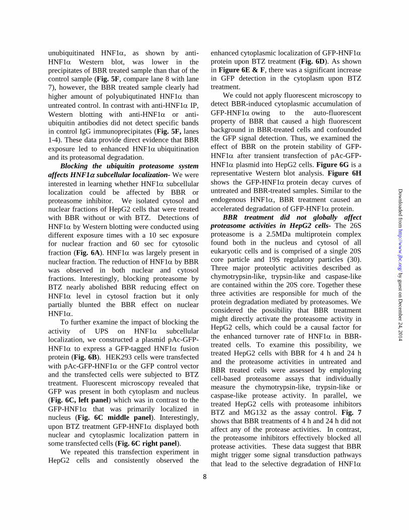
Blocking the ubiquitin proteasome system
affects HNF1 subcellular localization- We were
interested in learning whether HNF1 subcellular
localization could be affected by BBR or
proteasome inhibitor. We isolated cytosol and
nuclear fractions of HepG2 cells that were treated
with BBR without or with BTZ. Detections of
HNF1 by Western blotting were conducted using
different exposure times with a 10 sec exposure
for nuclear fraction and 60 sec for cytosolic
fraction (Fig. 6A). HNF1 was largely present in
nuclear fraction. The reduction of HNF1 by BBR
was observed in both nuclear and cytosol
fractions. Interestingly, blocking proteasome by
BTZ nearly abolished BBR reducing effect on
HNF1 level in cytosol fraction but it only
partially blunted the BBR effect on nuclear
HNF1.
To further examine the impact of blocking the
activity of UPS on HNF1 subcellular
localization, we constructed a plasmid pAc-GFP-
HNF1 to express a GFP-tagged HNF1 fusion
protein (Fig. 6B). HEK293 cells were transfected
with pAc-GFP-HNF1 or the GFP control vector
and the transfected cells were subjected to BTZ
treatment. Fluorescent microscopy revealed that
GFP was present in both cytoplasm and nucleus
(Fig. 6C, left panel) which was in contrast to the
GFP-HNF1 that was primarily localized in
nucleus (Fig. 6C middle panel). Interestingly,
upon BTZ treatment GFP-HNF1 displayed both
nuclear and cytoplasmic localization pattern in
some transfected cells (Fig. 6C right panel).
We repeated this transfection experiment in
HepG2 cells and consistently observed the
enhanced cytoplasmic localization of GFP-HNF1
protein upon BTZ treatment (Fig. 6D). As shown
in Figure 6E & F, there was a significant increase
in GFP detection in the cytoplasm upon BTZ
treatment.
We could not apply fluorescent microscopy to
detect BBR-induced cytoplasmic accumulation of
GFP-HNF1owing to the auto-fluorescent
property of BBR that caused a high fluorescent
background in BBR-treated cells and confounded
the GFP signal detection. Thus, we examined the
effect of BBR on the protein stability of GFP-
HNF1 after transient transfection of pAc-GFP-
HNF1 plasmid into HepG2 cells. Figure 6G is a
representative Western blot analysis. Figure 6H
shows the GFP-HNF1protein decay curves of
untreated and BBR-treated samples. Similar to the
endogenous HNF1, BBR treatment caused an
accelerated degradation of GFP-HNF1 protein.
BBR treatment did not globally affect
proteasome activities in HepG2 cells- The 26S
proteasome is a 2.5MDa multiprotein complex
found both in the nucleus and cytosol of all
eukaryotic cells and is comprised of a single 20S
core particle and 19S regulatory particles (30).
Three major proteolytic activities described as
chymotrypsin-like, trypsin-like and caspase-like
are contained within the 20S core. Together these
three activities are responsible for much of the
protein degradation mediated by proteasomes. We
considered the possibility that BBR treatment
might directly activate the proteasome activity in
HepG2 cells, which could be a causal factor for
the enhanced turnover rate of HNF1 in BBR-
treated cells. To examine this possibility, we
treated HepG2 cells with BBR for 4 h and 24 h
and the proteasome activities in untreated and
BBR treated cells were assessed by employing
cell-based proteasome assays that individually
measure the chymotrypsin-like, trypsin-like or
caspase-like protease activity. In parallel, we
treated HepG2 cells with proteasome inhibitors
BTZ and MG132 as the assay control. Fig. 7
shows that BBR treatments of 4 h and 24 h did not
affect any of the protease activities. In contrast,
the proteasome inhibitors effectively blocked all
protease activities. These data suggest that BBR
might trigger some signal transduction pathways
that lead to the selective degradation of HNF1
by guest on Decem
ber 24, 2014http://w
ww
.jbc.org/D
ownloaded from

9
protein by proteasome. This possibility requires
further investigation.
DISCUSSION
One important aspect of the PCSK9-LDLR
pathway in mediating LDL clearance is that their
transcription is coordinately regulated by sterols
through a common SRE motif embedded in their
gene promoters and is co-induced by current
cholesterol lowering drugs such as statins through
activation of SREBPs (10-12). Statin treatment
increases the transcription of both LDLR and
PCSK9. The undesirable inducing effect of statins
on PCSK9 transcription is increasingly recognized
as a major limitation to statin therapeutic efficacy
in further lowering plasma LDL-C (31-33). Since
HNF1 binding site is unique to PCSK9 promoter
and is not present in the LDLR promoter,
modulations of PCSK9 transcriptions through
HNF1 sequence will not affect LDLR gene
expression. Thus, HNF1 binding site represents a
divergent point to disconnect the coregulation of
PCSK9 with LDLR and other SREBP-target
genes. Indeed, in this study, we have shown that
the natural cholesterol lowering compound BBR
suppressed hepatic PCSK9 gene expression and
reduced serum PCSK9 concentrations in mice
without affecting mRNA levels of LDLR and
other SREBP-target genes. Importantly, in the
absence of an increase in gene expression, liver
LDLR protein levels in BBR-treated mice were
significantly higher than that of control mice,
which was convoyed by a 30% reduction of serum
LDL-C levels. Analysis of HNF1 mRNA and
protein levels in all liver samples clearly
demonstrated that BBR attenuated PCSK9
transcription by reducing the liver content of
HNF1 protein without lowering its mRNA
expression. These results provided the first in vivo
example of downregulation of HNF1 alone by a
lipid lowering compound and the beneficial impact
on plasma LDL-C metabolism through the
PCSK9-LDLR pathway. By analyzing liver
samples of control and BBR-treated hamsters, we
observed essentially the same phenomena that
BBR lowered PCSK9 expression by reducing
HNF1 protein levels. We further confirmed these
in vivo findings by experiments conducted in
HepG2 cells, a model human hepatic cell line.
Hence, it is likely that a similar cellular
mechanism is utilized by BBR to suppress PCSK9
gene expression in liver tissue in vivo and in
cultured hepatic cells.
HNF1 is a homeodomain-containing
transcription factor (13,34) that is important for
diverse metabolic functions of liver, pancreatic
islets, kidney and intestine (35-37). Although the
regulatory network that controls HNF1 gene
expression has been well studied (38-40), less is
known about the regulation of HNF1 at the
protein level. Searching literatures we found two
reports that described the regulation of HNF1
protein. An early study conducted by Park et al
reported that ceramide treatment of H4IIE, a rat
hepatocyte-derived cell line, repressed HNF1
protein via the ubiquitin proteasome system (41),
while a recent study reported that HCV infection
of human hepatoma derived Huh-7.5 cells resulted
in HNF1 protein degradation in lysosomes (26).
To determine which protein degradation pathway
might be operated by BBR to induce HNF1
protein degradation in HepG2 cells, initially we
applied BA1 and BTZ to separately shutdown the
cellular protein clearance machineries. The
cellular content of HNF1 was not affected by
BA1 but was markedly elevated by proteasome
inhibitors BTZ and MG132. It is noteworthy that
the elevated HNF1 protein levels were
accompanied by increased PCSK9 and reduced
LDLR abundance in BTZ-treated cells. Our results
provided the first evidence that the UPS-mediated
degradation of HNF1 is directly linked to the
PCSK9-LDLR pathway.
BBR treatment consistently lowered HNF1
protein and PCSK9 mRNA and protein levels in
HepG2 cells, however, this effect was not
observed in the presence of UPS inhibitors. Both
MG132 and BTZ showed antagonism to BBR-
mediated repression of HNF1 protein. Utilizing
BTZ we further showed that BBR-mediated
suppression of PCSK9 promoter activity was
largely abolished. By co-transfection of flag-
tagged HNF1 and myc-taged ubiquitin in
HEK293 cells, we detected a multi-ubiquitination
ladder pattern of HNF1, which provided
additional evidence supporting that HNF1 is
targeted by the ubiquitin-induced proteasomal
degradation for its intracellular clearance.
by guest on Decem
ber 24, 2014http://w
ww
.jbc.org/D
ownloaded from

10
Importantly, by detection of ubiquitinated
endogenous HNF1 in HepG2 cells, our study
provides strong evidence showing that BBR
treatment resulted in an enhanced
HNF1ubiquitination and its proteasomal
degradation, which negatively impacted PCSK9
gene transcription and protein abundance in
hepatic cells. Possibly, these BBR effects, at least
in part, account for the LDL-C lowering effect of
BBR in hypercholesterolemic patients.
The precise mechanism by which BBR
induces HNF1 degradation via the UPS is
presently unknown. Assessment of proteasome
activities in HepG2 cells indicated that BBR
treatment did not affect any of the three major
proteolytic activities that are contained within the
proteasome 20S core. By analysis of HNF1
subcellular localization, we observed that HNF1
is accumulated in cytoplasm upon BTZ treatment.
Additionally, BTZ treatment totally reversed the
reducing effect of BBR on HNF1 in cytoplasmic
fraction but it only partially reversed BBR effect
on nuclear HNF1. It is tempting to speculate that
BBR might increase HNF1 nuclear export
activity resulting in enhanced degradation in
cytoplasm. Verification of this point awaits further
experimental examination.
In summary, we have demonstrated that BBR
inhibits HNF1mediated transactivation of
PCSK9 gene expression by downregulation of
hepatic HNF1 protein content in animal models
and in cultured cells where BBR treatment
accelerated HNF1 protein degradation. This
effect can be blocked by proteasome inhibitors
that increase intracellular content of HNF1 in
untreated cells. Importantly, we demonstrate that
blocking ubiquitin-proteasome pathway in HepG2
cells unfavorably affected PCSK9-LDLR pathway
by increasing PCSK9 and lowering LDLR.
Considering that BTZ is currently used in clinic as
an anticancer drug (42,43) and new proteasome
inhibitors are under development for treating
patients with certain types of cancer, our findings
in this study warrant further investigations in
animal models to determine whether proteasome
inhibitors would increase HNF1 protein levels in
liver tissue and elevate circulating PCSK9 and
LDL-C levels.
REFERENCES
1. Peteson, A. S., Fong, L. G. & Young, S. G. (2008) PCSK9 function and physiology. J Lipid Res 49:
1595-1599.
2. Qian, Y.-W., Schmidt, R. J., Zhang, Y., Chu, S., Lin, A., Wang, H., Wang, X., Beyer, T. P.,
Bensch, W. R. et al. (2007) Secreted PCSK9 downregulates low density lipoprotein receptor
through receptor-mediated endocytosis. J Lipid Res 48: 1488-1498.
3. Seidah, N. G., Benjannet, S., Wickham, L., Marcinkiewicz, J., Bélanger, J., Stifani, S., Basak, A.,
Prat, A. & Chrétien, M. (2003) The secretory proprotein convertase neutal apoptosis-regulated
convertase 1 (NARC-1): liver regeneration and neuronal differentiation. Proc. Natl. Acad. Sci. USA
100: 928-933.
4. Seidah, N. G. (2009) PCSK9 as a therapeutic target of dyslipidemia. Expert Opin. Ther. Targets 13:
19-28.
5. Zhang, D. W., Lagace, T. A., Garuti, R., Zhao, Z., McDonald, M., Horton, J. D., Cohen, J. C. &
Hobbs, H. (2007) Binding of proprotein convertase subtilisin/kexin type 9 to epidermal growth
factor-like repeat A of low density lipoprotein receptor decreases receptor recycling and increases
degradation. J Biol Chem 282: 18602-18612.
by guest on Decem
ber 24, 2014http://w
ww
.jbc.org/D
ownloaded from

11
6. McNutt, M. C., Kwon, H. J., Chen, C., Chen, J. R., Horton, J. D. & Lagace, T. A. (2009)
Antagonism of secreted PCSK9 increases low density lipoprotein receptor expression in HepG2
cells. J Biol Chem 284: 10561-10570.
7. Lambert, G., Ancellin, N., Charlton, F., Comas, D., Pilot, J., Keech, A., Patel, S., Sullivan, D. R.,
Cohn, J. S. et al. (2008) Plasma PCSK9 concentration correlate with LDL and total cholesterol in
diabetic patients and are decreased by fenofibrate treatment. Clinical Chemistrry 54: 1038-1045.
8. Grefhorst, A., McNutt, M. C., Lagace, T. A. & Horton, J. D. (2008) Plasma PCSK9 preferentially
reduces liver LDL receptors in Mice. J Lipid Res 49: 1303-1311.
9. Ling, H., Burns, T. L. & Hilleman, D. E. (2014) An update on the clinical development of
proprotein convertase subtilisin kexin 9 inhibitors, novel therapeutic agents for lowering low-
density lipoprotein cholesterol. Cardiovasc. Ther. 32: 82-88.
10. Horton, J. D., Shah, N. A., Warrington, J. A., Anderson, N. N., Park, S. W., Park, S. W., Brown, M.
S. & Goldstein, J. L. (2003) Combined analysis of oligonucleotide microarray data from transgenic
and knockout mice identifies direct SREBP target genes. Proc. Natl. Acad. Sci. USA 100: 12027-
12032.
11. Dubuc, G., Chamberland, A., Wassef, H., Davignon, J., Seidah, N. G., Bernier, L. & Prat, A. (2004)
Statins upregulate PCSK9, the gene encoding the proprotein convertase neural apoptosis-regulated
convertase-1 implicated in familial hypercholesterolemia. Arterioscler Thromb Vasc Biol 24: 1454-
1459.
12. Jeong, H. J., Lee, H.-S., Kim, K.-S., Kim, Y.-K., Yoon, D. & Park, S. W. (2008) Sterol-dependent
regulation of proprotein convertase subtilisin/kexin type 9 expression by sterol-regulatroy element
binding protein-2. J Lipid Res 49: 399-409.
13. Mendel, D. B. & Crabtree, G. R. (1991) HNF-1, a member of a novel class of dimerizing
homeodomain proteins. J. Biol. Chem. 266: 677-680.
14. Li, H., Dong, B., Park, S. W., Lee, H. S., Chen, W. & Liu, J. (2009) Hepatocyte nuclear factor
1alpha plays a critical role in PCSK9 gene transcription and regulation by the natural
hypocholesterolemic compound berberine. J Biol Chem. 284: 28885-28895.
15. Dong, B., Wu, M., Li, H., Kraemer, F. B., Adeli, K., Seidah, N. G., Park, S. W. & Liu, J. (2010)
Strong induction of PCSK9 gene expression through HNF1alpha and SREBP2: mechanism for the
resistance to LDL-cholesterol lowering effect of statins in dyslipidemic hamsters. J Lipid Res. 51:
1486-1495.
16. Li, H. & Liu, J. (2012) The novel function of HINFP as a co-activator in sterol-regulated
transcription of PCSK9 in HepG2 cells. Biochem. J 443: 757-768.
17. Kong, W., Wei, J., Abidi, P., Lin, M., Inaba, S., Li, C., Wang, Y., Wang, Z., Si, S. et al. (2004)
Berberine Is a Promising Novel Cholesterol-lowering Drug Working through a Unique Mechanism
Distinct from Statins. Nature Medicine 10: 1344-1352.
18. Cameron, J., Ranheim, T., Kulseth, M. A., Leren, T. P. & Berge, K. E. (2008) Berberine decreases
PCSK9 expression in HepG2 cells. Atherosclerosis 201: 266-273.
by guest on Decem
ber 24, 2014http://w
ww
.jbc.org/D
ownloaded from

12
19. Cao, A., Wu, M., Li, H. & Liu, J. (2011) Janus kinase activation by cytokine oncostatin M
decreases PCSK9 expression in liver cells. J Lipid Res. 52: 518-530.
20. Singh, A. B., Kan, C. F., Shende, V., Dong, B., Liu, J. (2014). A novel posttranscriptional
mechanism for dietary cholesterol-mediated suppression of liver LDL receptor expression. J Lipid
Res. 55:1397-1407.
21. Cao, A., Wu, M., Li, H. & Liu, J. (2011) Janus kinase activation by cytokine oncostatin M
decreases PCSK9 expression in liver cells. J Lipid Res 52: 518-530.
22. Wu, M., Dong, B., Cao, A., Li, H. & Liu, J. (2012) Delineation of molecular pathways that regulate
hepatic PCSK9 and LDL receptor expression during fasting in normolipidemic hamsters.
Atherosclerosis 224: 401-410.
23. Astudillo, A. M., Perez-Chacon, G., Meana, C., Balgoma, D., Pol, A., Del Pozo, M. A., Balboa, M.
A. & Balsinde, J. (2011) Altered arachidonate distribution in macrophages from caveolin-1 null
mice leading to reduced eicosanoid synthesis. J Biol Chem 286: 35299-35307.
24. Dong, B., Kan, C. F., Singh, A. B. & Liu, J. (2013) High-fructose diet downregulates long-chain
acyl-CoA synthetase 3 expression in liver of hamsters via impairing LXR/RXR signaling pathway.
J Lipid Res 54: 1241-1254.
25. Dong, B., Singh, A. B., Fung, C., Kan, K. & Liu, J. (2014) CETP inhibitors downregulate hepatic
LDL receptor and PCSK9 expression in vitro and in vivo through a SREBP2 dependent
mechanism. Atherosclerosis 235: 449-462.
26. Matsui, C., Shoji, I., Kaneda, S., Sianipar, I. R., Deng, L. & Hotta, H. (2012) Hepatitis C virus
infection suppresses GLUT2 gene expression via downregulation of hepatocyte nuclear factor
1alpha. J Virol. 86: 12903-12911.
27. Hershko, A. (2005) The ubiquitin system for protein degradation and some of its roles in the control
of the cell-division cycle (Nobel lecture). Angew. Chem. Int. Ed Engl. 44: 5932-5943.
28. Hershko, A., Ciechanover, A. & Varshavsky, A. (2000) Basic Medical Research Award. The
ubiquitin system. Nat. Med. 6: 1073-1081.
29. Huang, L. & Chen, C. H. (2009) Proteasome regulators: activators and inhibitors. Curr Med. Chem.
16: 931-939.
30. Careskey, H. E., Davis, R. A., Alborn, W. E., Troutt, J. S., Cao, G., and Konrad, R. J.
(2008) Atovastatin increases human serum levels of proprotein convertase subtilisin/kexin
type 9. J Lipid Res. 49: 394-398.
31. Raal, F., Panz, V., Immelman, A. & Pilcher, G. (2013) Elevated PCSK9 levels in untreated patients
with heterozygous or homozygous familial hypercholesterolemia and the response to high-dose
statin therapy. J Am. Heart Assoc. 2: e000028.
32. Costet, P., Hoffmann, M. M., Cariou, B., Guyomarc'h, D. B., Konrad, T. & Winkler, K. (2010)
Plasma PCSK9 is increased by fenofibrate and atorvastatin in a non-additive fashion in diabetic
patients. Atherosclerosis 212: 246-251.
by guest on Decem
ber 24, 2014http://w
ww
.jbc.org/D
ownloaded from

13
33. Welder, G., Zineh, I., Pacanowski, M. A., Troutt, J. S., Cao, G. & Konrad, R. J. (2010) High-dose
atorvastatin causes a rapid sustained increase in human serum PCSK9 and disrupts its correlation
with LDL cholesterol. J Lipid Res. 51: 2714-2721.
34. Cereghini, S. (1996) Liver-enriched transcription factors and hepatocyte differentiation. FASEB J.
10: 267-282.
35. Shih, D. Q., Bussen, M., Sehayek, E., Ananthanarayanan, M., Shneider, B. L., Suchy, F. J., Shefer,
S., Bollileni, J. S., Gonzalez, F. J. et al. (2001) Hepatocyte nuclear factor-1alpha is an essential
regulator of bile acid and plasma cholesterol metabolism. Nat. Genet. 27: 375-382.
36. Pontoglio, M., Barra, J., Hadchouel, M., Doyen, A., Kress, C., Bach, J. P., Babinet, C. & Yaniv, M.
(1996) Hepatocyte nuclear factor 1 inactivation results in hepatic dysfunction, phenylketonuria, and
renal Fanconi syndrome. Cell 84: 575-585.
37. Shih, D. Q., Screenan, S., Munoz, K. N., Philipson, L., Pontoglio, M., Yaniv, M., Polonsky, K. S. &
Stoffel, M. (2001) Loss of HNF-1alpha function in mice leads to abnormal expression of genes
involved in pancreatic islet development and metabolism. Diabetes 50: 2472-2480.
38. Costa, R. H., Kalinichenko, V. V., Holterman, A. L. & Wang, X. (2003) Transcription factors in
liver development, differentiation, and regeneration. Hepatplogy 38: 1331-1347.
39. Armendariz, A. D. & Krauss, R. M. (2009) Hepatic nuclear factor 1-alpha: inflammation, genetics,
and atherosclerosis. Curr Opin Lipidol 20: 106-111.
40. Pontoglio, M., Pausa, M., Doyen, A., Viollet, B., Yaniv, M. & Tedesco, F. (2001) Hepatocyte
nuclear factor 1alpha controls the expression of terminal complement genes. J Exp. Med. 194:
1683-1689.
41. Park, I. N., Cho, I. J. & Kim, S. G. (2004) Ceramide negatively regulates glutathione S-transferase
gene transactivation via repression of hepatic nuclear factor-1 that is degraded by the ubiquitin
proteasome system. Mol. Pharmacol. 65: 1475-1484.
42. Yoshizawa, K., Mukai, H. Y., Miyazawa, M., Miyao, M., Ogawa, Y., Ohyashiki, K., Katoh, T.,
Kusumoto, M., Gemma, A. et al. (2014) Bortezomib therapy-related lung disease in Japanese
patients with multiple myeloma: incidence, mortality and clinical characterization. Cancer Sci 105:
195-201.
43. Thompson, J. L. (2013) Carfilzomib: a second-generation proteasome inhibitor for the treatment of
relapsed and refractory multiple myeloma. Ann. Pharmacother. 47: 56-62.
FOOTNOTE
* This study was supported by the Department of Veterans Affairs (Office of Research and Development,
Medical Research Service) and by grants (1R01 AT002543-01A1 and 1R01AT006336-01A1) from
National Center of Complementary and Alternative Medicine.
by guest on Decem
ber 24, 2014http://w
ww
.jbc.org/D
ownloaded from

14
The abbreviations used are: BA1, bafilomycin A1; BBR, berberine; BTZ, bortezomib;
HNF1hepatocyte nuclear factor 1 LDLR, LDL receptor; PCSK9, proprotein convertase
subtilisin/kexin type 9; SREBP, sterol regulatory element binding protein; UPS, ubiquitin proteasome
system
FIGURE LEGENDS
Figure 1. BBR treatment lowered serum PCSK9 and cholesterol levels with reduced HNF1
protein content in liver of mice fed a high-cholesterol diet. Male mice fed a high-cholesterol diet for
four weeks were orally dosed 200 mg/kg/day BBR (n=10) or equal volume of vehicle (0.5% methyl
cellulose) as the control group (n=10) for 16 days. Mouse serum TC (A) and LDL-C (B) were measured
before, during and after BBR treatment. (C) Mouse serum PCSK9 levels after BBR treatment were
quantified by ELISA. (D) Real-time PCR analysis of mouse liver mRNA levels of indicated genes after
BBR treatment was shown. (E) Two mouse liver samples of the same treatment group were pooled
together. For each group, 50 µg of homogenate proteins of 5 pooled samples were resolved by SDS-
PAGE. HNF1 LDLR and β-actin were detected by immunoblotting. The protein abundance of HNF1
and LDLR were quantified with the Alpha View Software with normalization by signals of β-actin. The
values are presented as mean ± SEM. *p < 0.05, **p < 0.01 and ***p < 0.001 as compared to the
vehicle group.
Figure 2. Reduction of serum PCSK9 and hepatic HNF1 protein in dyslipidemic hamsters by BBR
treatment. Male hamsters were fed a high-fructose diet for three weeks which elevated serum TC and
LDL-C (15). Hamsters on the high-fructose diet were orally dosed 100 mg/kg/day BBR (n=9) or equal
volume of vehicle (10% 2-hydroxypropyl-β-cyclodextrin) as the control group (n=9) for 7 days.
(A) 50 µg of homogenate proteins of six representative hamster liver samples from each group were
resolved by SDS-PAGE. HNF1 and β-actin were detected by immunoblotting. The protein abundance of
HNF1 was quantified with the Alpha View Software with normalization by signals of β-actin.
(B) qPCR analysis of hamster liver mRNA levels of indicated genes after BBR treatment was shown.
(C) Hamster serum PCSK9 levels after BBR treatment were determined using a mouse PCSK9 ELISA
kit.
(D) Three hamster serum samples of the same treatment group were pooled together. For each group, 20
µL of pooled serum of 3 pooled samples were used for conducting PCSK9 IP and Western blotting.
Hamster liver homogenate protein was loaded in lane 1 as a positive control for anti-PCSK9 antibody
Western blotting. The values are presented as mean ± SEM. *p < 0.05, **p < 0.01 and ***p < 0.001 as
compared to the vehicle group.
Figure 3. BBR downregulates HNF1 expression by accelerating its protein degradation in HepG2
cells.
(A) HepG2 cells were treated with 40 M of BBR for the indicated times. Total cell lysates were isolated
from cells and analyzed for PCSK9 and HNF1 protein levels by Western blotting. The results shown are
representative of three separate experiments with similar results.
(B) HepG2 cells were treated with 40 M of BBR for the indicated times. Total RNA was isolated and
PCSK9 and HNF1 mRNA levels were assessed by qRT-PCR using human-specific PCR primers, and
triplicate measurements were conducted for each cDNA sample. The results shown are representative of
three separate experiments with similar results.
(C) HepG2 cells were treated with CHX at 5 g/ml concentration at indicated times in the absence or the
presence of 40 M of BBR. Total cell lysates were subjected to Western blotting and bands were
visualized with antibody against HNF1 or -actin. The data shown are representative of two separate
experiments with similar results.
by guest on Decem
ber 24, 2014http://w
ww
.jbc.org/D
ownloaded from

15
(D) After normalization to -actin, the HNF1 signal intensity was plotted against the CHX treatment
time to calculate T1/2 of HNF1 protein. Half-life was calculated from each of the two separate
experiments.
Figure 4. Evaluation of lysosome inhibitor BA1 and proteasome inhibitors BTZ and MG132 in
HNF1 protein stability in HepG2 cells.
(A) HepG2 cells were incubated with 50 nM of BA1 or DMDO for 24 hours. Total cell lysates were
harvested and 30 g of total proteins were resolved by SDS-PAGE and HNF1, LDLR and PCSK9 were
detected by Western blotting. The data shown are representative of three separate experiments with
similar results.
(B) HepG2 cells were treated with indicated concentrations of BTZ or MG132 for 24 hours. The data
shown are representative of three separate experiments with similar results.
(C) HepG2 cells in triplicate dishes were treated with BTZ at 80 nM or DMSO for 24 h, and total cell
lysates were analyzed by Western blotting. The data shown are representative of three separate
experiments with similar results.
(D) The protein abundance of HNF1, PCSK9 and LDLR in (C) were quantified with the Alpha View
Software with normalization by signals of β-actin. Values are mean ± SEM of 3 samples per group. * p <
0.05 and ** p < 0.01 compared to DMSO.
Figure 5. Proteasomal pathway participates in BBR-induced degradation of HNF1 protein.
(A) HepG2 cells were treated with 40 M BBR in the absence or the presence of BTZ (1 M) for 16 h
and cell lysates were utilized for immunoblotting using anti-HNF1anti-PCSK9 and anti-LDLR
antibodies. Triplicate wells were used for each treatment. The data shown are representative of three
separate experiments with similar results.
(B) The protein abundances of HNF1, PCSK9 and LDLR in (A) were quantified with the Alpha View
Software with normalization by signals of β-actin. The values are presented as mean ± SEM. * p < 0.05,
** p < 0.01 and *** p < 0.001compared to DMSO.
(C) HepG2 cells were treated with 40 M BBR in the absence or the presence of BTZ for 16 h and total
RNA was isolated for qPCR analysis of mRNA levels of HNF1, PCSK9 and LDLR. Duplicate wells
were used for each treatment and individual cDNA samples were measured in triplicates. The values are
presented as mean ± SEM. The data shown are representative of two separate experiments with similar
results.
(D) The PCSK9 promoter reporter construct pGL3-PCSK9-D4 was transiently cotransfected with pSV--
gal vector into HepG2 cells. One day post transfection, cells were incubated in 0.5% FBS medium
overnight, followed by a 24-treamtent of 40 M BBR in the absence or the presence of BTZ at indicated
concentrations. Cells were harvested and the luciferase and β-galactosidase activities were measured as
described in experimental procedures. Normalized luciferase activity of untreated cells is expressed as
100 percent. The data shown are representative of three separate experiments with similar results.
(E) Plasmids encoding Flag-tagged HNF1 and Myc-tagged ubiquitin were cotransfected into HEK293
cells. The empty vectors of pCMV-Entry and pCMV-Myc were transfected as mock control. Cell lysates
were harvested after two days of transfection and immunoprecipitated with anti-Myc or anti-Flag
antibodies. Ubiquitinated HNF1 was detected by immunoblotting using anti-Myc or anti-Flag
antibodies, respectively. The data shown are representative of three separate experiments with similar
results.
(F) HepG2 cells were treated for 24 h with BBR or DMSO as control. During the last 8 h of BBR
treatment, MG132 (20 M) or DMSO was added to cells. Total cell lysates were prepared and 500 g
proteins from total lysates were subjected to IP with anti-HNF1 antibody or control antibody goat IgG.
by guest on Decem
ber 24, 2014http://w
ww
.jbc.org/D
ownloaded from

16
IP complexes were analyzed for ubiquitinated HNF1 with anti-HNF1 antibody and anti-ubiquitin
antibody. The data shown are representative of two separate experiments with similar results.
Figure 6. Accumulation of HNF1 in cytoplasm by proteasome inhibition
(A) HepG2 cells were treated with BBR without and with 100 nM of BTZ for 24 h. Nuclear fraction and
cytoplasmic fraction of total cell lysates from each sample were prepared and analyzed for HNF1
protein levels by Western blotting. The membranes were reprobed with anti-HDAC1 antibody as a
control of equal nuclear protein loading or GAPDH as a control of equal cytoplasmic protein loading. The
data shown are representative of two separate experiments with similar results.
(B) Diagram of GFP and GFP-HNF1 fusion constructs. Human HNF1 coding sequence was subcloned
into pAcGFP-C1 vector at the Bgl II and EcoR1 sites to produce the N-terminal GFP-tagged fusion
protein.
(C, D) HEK293 cells or HepG2 cells were transfected with pAc-GFP or pAc-GFP-HNF1 Two days
after transfection, cells were either treated with BTZ or DMSO for 24 h.. The subcellular localization of
GFP-HNF1 fusion protein was examined with a fluorescence microscope. The pictures shown are
representative of three separate experiments with similar results.
(E) From each well, approximately 80-160 GFP-HNF1 positive HepG2 cells were examined for their
subcellular localization and graphed.
(F) The percentage of transfected HepG2 cells with GFP-HNF1 in cytoplasm was calculated. The values
are presented as mean ± SEM of triplicate wells per condition.
(G) HepG2 cells transfected with pAc-GFP-HNF1weretreated with CHX at 5 g/ml concentration at
indicated times in the absence or the presence of 40 M of BBR. Total cell lysates were subjected to
Western blotting and bands were visualized with antibody against GFP or -actin. The data shown are
representative of two separate experiments with similar results.
(H) After normalization to -actin, the GFP-HNF1 signal intensity in (G) was plotted against the CHX
treatment time to calculate T1/2 of GFP-HNF1 fusion protein.
Figure 7. BBR treatment is not associated with altered cellular proteasomal activity.
HepG2 cells were treated with BBR (40 M), BTZ (100 nM) and MG132 (1 M) for 4 h or 24 h. At
indicated treatment time, cells were incubated for 10 min with the Promega Proteasome-Glo Cell-Based
Assay Reagent (Proteasome-Glo™ 3-Substrate Cell-Based Assay System). The chymotrypsin-like,
trypsin-like activity or caspase-like proteasome activities were detected as the relative light unit (RLU)
generated from the cleaved substrate in the reaction mixture. The values are presented as mean ± SEM.
The data shown are representative of two separate experiments with similar results.
by guest on Decem
ber 24, 2014http://w
ww
.jbc.org/D
ownloaded from

Forward Reverse
Mouse
GAPDH AACTTTGGCATTGTGGAAGG GGATGCAGGGATGATGTTCT
HMGCR CTTTCAGAAACGAACTGTAGCTCAC CTAGTGGAAGATGAATGGACATGAT
HNF1α GCACCAGAGACCCACGTGCC GGCTTCCCCTCAGCTCCCGA
LDLR ACCTGCCGACCTGATGAATTC GCAGTCATGTTCACGGTCACA
PCSK9 TTGCAGCAGCTGGGAACTT CCGACTGTGATGACCTCTGGA
SREBP1 CAAGGCCATCGACTACATCCG CACCACTTCGGGTTTCATGC
SREBP2 CCAAAGAAGGAGAGAGGCGG CGCCAGACTTGTGCATCTTG
Hamster
GAPDH AACTTTGGCATTGTGGAAGG GGATGCAGGGATGATGTTCT
HNF1α GAGGTGGCTCAGCAATTCAC CACTCCTCCACCAAGGTCTC
PCSK9 TGCTCCAGAGGTCATCACAG GTCCCACTCTGTGACATGAAG
SREBP1 GCACTTTTTGACACGTTTCTTC CTGTACAGGCTCTCCTGTGG
SREBP2 GAGAGCTGTGAATTTTCCAGTG CTACAGATGATATCCGGACCAA
Human
GAPDH ATGGGGAAGGTGAAGGTCG GGGGTCATTGATGGCAACAATA
HNF1α TGGCGCAGCAGTTCACCCAT TGAAACGGTTCCTCCGCCCC
PCSK9 AGGGGAGGACATCATTGGTG CAGGTTGGGGGTCAGTACC
Table 1. Real-time PCR primer sequences.
by guest on Decem
ber 24, 2014http://w
ww
.jbc.org/D
ownloaded from

Vehicle BBR0
10
20
30
40
50
* (-49.5%)
Seru
m P
CSK
9 (n
g/m
L)
0 7 14 160
50
100
150
200
250
300
VehicleBBR, 200 mg/kg
*
BBR treatment, days
Seru
m T
C, m
g/dL
0 7 14 160
50
100
150
200
250
VehicleBBR, 200 mg/kg
**** **
BBR treatment, days
Seru
m L
DL-C
, mg/
dL
PCSK9
HNF1ALDLR
SREBP2
SREBP1
HMGCR0.00
0.25
0.50
0.75
1.00
1.25VehicleBBR
*
Rela
tive
mRN
A ex
pres
sion
(fold
of c
ontr
ol)
A B
HNF1α
β-actin
LDLR
C D
E
Figure 1
Control BBR
HNF1α/β-actin: 1.0 ± 0.13 0.58 ± 0.04 (p < 0.05)LDLR/β-actin: 1.0 ± 0.04 1.67 ± 0.20 (p < 0.05)
by guest on Decem
ber 24, 2014http://w
ww
.jbc.org/D
ownloaded from

con BBR
HNF1α
β-actin
PCSK9 HNF1A SREBP1 SREBP20.00
0.25
0.50
0.75
1.00
1.25
1.50 controlBBR 100 mg/kg
*
Norm
aliz
ed m
RNA
leve
ls
Figure 2
HNF1α/β-actin 1.0 ± 0.05 0.69 ± 0.03 (p < 0.001)
A
B
C
Vehicle BBR0
20
40
60
80
**
Ser
um P
CS
K9
(ng/
mL)
Live
r
PCSK9-PPCSK9-M
Vehicle BBR
PCSK9-MN.S.
DSerum
1 2 3 4 5 6 7
by guest on Decem
ber 24, 2014http://w
ww
.jbc.org/D
ownloaded from

HNF1α
β-actin
BBR (h): 0 1 2 4 8 16 24
PCSK9-P
PCSK9-M
0 4 8 12 16 20 240.00
0.25
0.50
0.75
1.00
1.25
1.50 PCSK9HNF1A
BBR treatment (h)
Norm
aliz
ed m
RNA
leve
ls(fo
ld o
f con
trol)
Figure 3
A B
C
D
HNF1α
β-actin
CHX (h): 0 1 2 4 8 0 1 2 4 8 DMSO BBR
HNF1α/Actin: 100 98.6 85.3 83.2 79.4 100 86.9 77.5 73.0 56.5
0 1 2 3 4 5 6 7 810
100
control T 1/2 = 16.2 hBBR T1/2 = 8.3 h
Cycloheximide (h)
HN
F1α
pro
tein
rem
aini
ng
0 1 2 3 4 5 6 7 810
100
control T1/2 = 12.9 hBBR T1/2 = 7.3 h
Cycloheximide (h)
HN
F1α
pro
tein
rem
aini
ng
by guest on Decem
ber 24, 2014http://w
ww
.jbc.org/D
ownloaded from

A B
LDLR
HNF1α
β-actin
PCSK9
BA
1
DM
SO
+ ++BTZ:
LDLR
HNF1α
β-actin
PCSK9
HNF1α
GAPDH
BTZ
20
nM
BTZ
50
nM
BTZ
100
nM
Con
trol
MG
132
1 µM
MG
132
2 µM
C D
Figure 4
HNF1α PCSK9 LDLR0
50
100
150
200controlBTZ* *
**
Rel
ativ
e p
rote
in le
vels
(% o
f co
ntr
ol)
by guest on Decem
ber 24, 2014http://w
ww
.jbc.org/D
ownloaded from

A BFigure 5
C
0
20
40
60
80
100
120
C BBR BBR BBRBTZ (nM): 0 0 50 100
PC
SK
9 pr
omot
er a
ctiv
ity(%
of c
ontr
ol)
Ub-HNF1α
IP: anti-Myc IP: anti-Flag
WB: HNF1a WB: Ub1 2 3 4
Flag-HNF1α: + - + + Myc-Ub: + + + -
BTZ BTZ+BBR
LDLR
HNF1α
β-actin
PCSK9
DMSO BBR
D DMSOBBR
BTZ
BTZ+BBR
DMSOBBR
BTZ
BTZ+BBR
DMSOBBR
BTZ
BTZ+BBR
0.0
0.5
1.0
1.5
2.0
2.5
LDLRPCSK9HNF1α
* ***
***
****
Rela
tive
prot
ein
leve
ls(fo
ld o
f con
trol
)
DMSOBBR
BTZ
BTZ+BBR
DMSOBBR
BTZ
BTZ+BBR
DMSOBBR
BTZ
BTZ+BBR
0.0
0.5
1.0
1.5
2.0
LDLRPCSK9HNF1α
Rela
tive
mRN
A le
vels
(fold
of c
ontr
ol)
WB: anti-Ubiquitin
WB: anti-HNF1α
IP: goat IgG IP: anti-HNF1α
75kD
100kD
150kD
250kD
80kD
BBR: - + - +MG132: + +- -
- + - ++ +- -
1 2 3 4 5 6 7 8
FE
by guest on Decem
ber 24, 2014http://w
ww
.jbc.org/D
ownloaded from

CMV GFPpAc-GFP
CMV GFP HNF1αpAc-GFP-HNF1α
pAC-GFP pAC-GFP-HNF1α pAc-GFP-HNF1α + BTZ
A
B
C
Figure 6
HNF1α
GAPDH
HNF1α
HDAC1
BBR: BTZ:
Cytosol Nuclear
(shorter exposure)(longer exposure)
E F
- + - + - + - +- +- + - +- +
HEK293
HepG2D
DMSO DMSO DMSO BTZ BTZ BTZ0
20406080
100120140160
Nuclear localizationNuclear and cytoplasmic localization
GFP
-HNF
1α p
ositi
ve c
ells
DMSO BTZ0
20
40
60
80
100
***
Cyt
opla
smic
pos
itive
cel
ls(%
of t
otal
tran
sfec
ted
cells
)
by guest on Decem
ber 24, 2014http://w
ww
.jbc.org/D
ownloaded from

GFP-HNF1α
β-actin
CHX (h): 0 1 2 4 8 0 1 2 4 8 DMSO BBR
GFP-HNF1α/β-actin: 100 93.6 83.6 94.3 89.4 100 84.8 76.5 71.5 61.0
Figure 6
G
H
0 1 2 3 4 5 6 7 810
100
control T1/2 = 29.1 hBBR T1/2 = 8.7 h
Cycloheximide (h)
GFP
-HNF
1α p
rote
in re
mai
ning
by guest on Decem
ber 24, 2014http://w
ww
.jbc.org/D
ownloaded from

Figure 7
DM
SO 4
hD
MD
O 2
4hB
BR
4h
BB
R 2
4hB
orte
zom
ib 4
hB
orte
zom
ib 2
4hM
G-1
32 4
hM
G-1
32 2
4h
DM
SO 4
hD
MD
O 2
4hB
BR
4h
BB
R 2
4hB
orte
zom
ib 4
hB
orte
zom
ib 2
4hM
G-1
32 4
hM
G-1
32 2
4h
DM
SO 4
hD
MD
O 2
4hB
BR
4h
BB
R 2
4hB
orte
zom
ib 4
hB
orte
zom
ib 2
4hM
G-1
32 4
hM
G-1
32 2
4h
0
500000
1000000
1.5x100 6
2.0x100 6
chymotrypsin-like activitytrypsin-like activitycaspase-like activity
RLU
by guest on Decem
ber 24, 2014http://w
ww
.jbc.org/D
ownloaded from
Copyright © 2022 FDOKUMEN