Damage response involves mechanisms conserved across plants, animals and fungi

MOLECULAR AND CELLULAR BIOLOGY,0270-7306/00/$04.0010
Apr. 2000, p. 2915–2925 Vol. 20, No. 8
Copyright © 2000, American Society for Microbiology. All Rights Reserved.
Cellular Response to Oncogenic Ras Involves Induction of theCdk4 and Cdk6 Inhibitor p15INK4b
MARCOS MALUMBRES,† IGNACIO PEREZ DE CASTRO, MARIA I. HERNANDEZ,MARIA JIMENEZ, TERESA CORRAL, AND ANGEL PELLICER*
Department of Pathology and Kaplan Comprehensive Cancer Center, New YorkUniversity Medical Center, New York, New York 10016
Received 26 July 1999/Returned for modification 13 September 1999/Accepted 27 January 2000
The cell cycle inhibitor p15INK4b is frequently inactivated by homozygous deletion together with p16INK4a andp19ARF in some types of tumors. Although the tumor suppressor capability of p15INK4b is still questioned, it hasbeen found to be specifically inactivated by hypermethylation in hematopoietic malignancies in the absence ofp16INK4a alterations. Here we show that, in vitro, p15INK4b is a strong inhibitor of cellular transformation byRas. Surprisingly, p15INK4b is induced in cultured cells by oncogenic Ras to an extent similar to that ofp16INK4a, and their expression is associated with premature G1 arrest and senescence. Ras-dependent induc-tion of these two INK4 genes is mediated mainly by the Raf-Mek-Erk pathway. Studies with activated anddominant negative forms of Ras effectors indicate that the Raf-Mek-Erk pathway is essential for induction ofboth the p15INK4b and p16INK4a promoters, although other Ras effector pathways can collaborate, giving rise toa stronger response. Our results indicate that p15INK4b, by itself, is able to stop cell transformation by Ras andother oncogenes such as Rgr (a new oncogene member of the Ral-GDS family, whose action is mediatedthrough Ras). In fact, embryonic fibroblasts isolated from p15INK4b knockout mice are susceptible to trans-formation by the Ras or Rgr oncogene whereas wild-type embryonic fibroblasts are not. Similarly, p15INK4b-deficient mouse embryo fibroblasts are more sensitive than wild-type cells to transformation by a combinationof the Rgr and E1A oncogenes. The cell cycle inhibitor p15INK4b is therefore involved, at least in some cell types,in the tumor suppressor activity triggered after inappropriate oncogenic Ras activation in the cell.
The transforming activity of oncogenes has been extensivelystudied in the last 2 decades. Early on, transformation of pri-mary cells was observed to require two cooperating oncogenesto convert normal cells to a tumorigenic state (27, 59). Theresistance of primary cells to transformation by single onco-genes can now be explained as the effect of the induction oftumor suppressor genes by inappropriate oncogenic signals(reviewed in reference 73). Thus, prolonged oncogenic Rasactivity produces in primary cells an increase in the levels ofp16INK4a, p21Cip1, and p53, which, in turn, is dependent on thetranscriptional induction of its positive regulator p19ARF (32,48, 64). Induction of the expression of these genes by Ras isaccompanied by growth arrest in the G1 phase of the cell cycleand a phenotype indistinguishable from premature senescence(33, 64). Both the p16INK4a and p19ARF proteins are expressedfrom a complex gene structure, the INK4a locus (for reviews,see references 13 and 67). Each of the two proteins uses adifferent exon 1, and both use the same exon 2, but eachprotein is translated in a different reading frame (53). Al-though their amino acid sequences are completely different,both proteins are cell cycle inhibitors. p16INK4a is a potentinhibitor of cyclin-dependent kinases 4 and 6 (Cdk4/6) (61),whereas p19ARF stabilizes the p53 tumor suppressor gene (forreviews, see references 7, 66, and 67).
In both humans and mice, the INK4a locus is located close toa second gene of the INK4 family, p15INK4b, which also func-
tions as a Cdk4/6 inhibitor and is strongly induced by trans-forming growth factor b (TGF-b) (14, 22, 54). Both loci, INK4aand INK4b, are frequently deleted in a variety of tumors andcell lines (22, 58). In addition, these proteins can also beinactivated by point mutations or methylation (reviewed inreferences 50 and 58). The expression of proteins p16INK4a,p15INK4b, and p19ARF can be decreased by hypermethylation ofthe CpG island upstream of corresponding exon 1 in bothhumans (17, 41, 56) and rodents (36, 69). No clear tumorsuppressor role has been assigned to the other two members ofthe INK4 family, p18INK4c and p19INK4d.
Whereas the evidence for a tumor suppressor role ofp16INK4a is abundant, the role of p15INK4b in tumor suppres-sion is more controversial. In most tumors, homozygous dele-tions affect both the INK4a and INK4b loci or the INK4a locusalone. In only a few cases have specific deletions of p15INK4b
sequences been reported, i.e., leukemias and lymphomas,which are among the tumors with higher involvement ofp15INK4b deletions (58). Point mutations, which are relativelyfrequent in INK4a, only rarely occur in p15INK4b (36, 50). Incontrast, inactivation of p15INK4b by hypermethylation seemsto be selectively frequent in leukemias and lymphomas anddoes occur independently of p16INK4a status (4, 17, 18, 36, 38),suggesting a tissue-specific tumor suppressor role for p15INK4b
in hematopoietic malignancies. In concordance with thesedata, Lois et al. (34) demonstrated an inverse relationshipbetween p15INK4b expression and proliferation of lymphocytesafter mitogenic stimuli, suggesting a specific role for this genein maintaining cell quiescence in lymphocytes.
Early studies on Ras mitogenic potential demonstrated thatRas induces and is required for DNA synthesis in serum-stimulated cells (44). Only recently have the pathways linkingRas activity with cell cycle control begun to be dissected. Rasacts on the cell cycle machinery by inactivating Cdk inhibitors
* Corresponding author. Mailing address: Department of Pathologyand Kaplan Comprehensive Cancer Center, New York UniversityMedical Center, 550 First Ave., New York, NY 10016. Phone: (212)263-5342. Fax: (212) 263-8211. E-mail: [email protected].
† Present address: Centro Nacional de Investigaciones OncologicasCarlos III, Crta. Majadahonda-Pozuelo, 28220 Majadahonda, Madrid,Spain.
2915

such as p27Kip1 and inducing cyclins, giving rise to an increasein Cdk4/6 and Cdk2 kinase activities (for reviews, see refer-ences 11 and 35). Thus, Ras activity is linked directly to theG1/S transition of the cell cycle and, in fact, G1 is the onlyphase in which inhibition of Ras affects cell cycle progression.Ras is required for activation of both Cdk2 and Cdk4/6 com-plexes until 2 h before the G1/S transition, a time correspond-ing to the so-called restriction point. Once cells have entered Sphase, Ras becomes dispensable until the next cell cycle (19,44). Although Ras signals through a growing number of dif-ferent effector pathways, effects on both cyclin D induction andp27Kip1 degradation seem to be dependent on the Raf1-Erkpathway. The specific activation of the Erk pathway, however,is not sufficient to trigger p27Kip1 degradation, and it seems tobe involved in a RhoA-associated pathway that could require aphosphatidylinositol 39-kinase (PI3K)-dependent but proteinkinase B-independent pathway (for a review, see reference 35).
Whereas different experiments have clearly shown thatp16INK4a is able to suppress cellular transformation by Ras andcan contribute to cellular senescence (2, 20, 47, 62), the abilityof p15INK4b to inhibit cellular transformation has not beenstudied. In this article, we show that the cell cycle inhibitorp15INK4b is able to produce cell cycle arrest and stop cellulartransformation by Ras. Interestingly, this Cdk4/6 inhibitor isstrongly induced in cultured cells by oncogenic Ras and, thus,can cooperate in causing the premature cellular senescenceresulting from oncogenic signals. Using luciferase constructscarrying the p15INK4b and p16INK4a promoters, we show thatthe Raf-Mek-Erk pathway is the main effector pathway in theinduction of the INK4 promoters although other Ras effectorscooperate in the transcriptional induction of both p15INK4b andp16INK4a in NIH 3T3 cells. Finally, using mouse embryonicfibroblasts (MEFs) from p15INK4b knockout mice, we show thatthe lack of p15INK4b protein is sufficient to render these fibro-blasts susceptible to transformation by the Ras or Rgr onco-gene.
MATERIALS AND METHODS
Plasmids and DNA manipulation. Mouse genomic N-ras containing a codon61 point mutation (pMZNT-17) or the wild-type sequence (pMZNN-1) wassubcloned into the Zeocin-resistant vector pcDNA3.1/Zeo(1) (Invitrogen).Mouse p15INK4b cDNA was isolated by reverse transcription-PCR and subclonedinto the pCR3.1 (Invitrogen) or pMAMneo (Clontech) vector, giving rise toplasmids pHM414 (cytomegalovirus [CMV] promoter orientation), pHM411(opposite orientation), and pMAMneo-p15. The same fragment was subclonedinto the pBabe-puro vector for retroviral transduction of primary cells. A 6-kbgenomic fragment containing the p15INK4b and 59 upstream sequences was am-plified by long-template PCR (Expand System; Boehringer Mannheim) usingprimers Mp15-P-1F (59-GGC CAA AAC AGG ATC CCT TGG GAT GTGTTA-39) and Mp15-39-1R (59-TAA CCA TGG AGA TCT CTC CAG GCTCCA-39) as described previously (37). This fragment includes about 700 bpupstream of the p15INK4b coding region and was subcloned into the bacterialplasmid pCR2.1 (Invitrogen) to generate pMM134. A different 8-kb genomicfragment containing the mouse p15INK4b gene was obtained from pmp15 (36)and subcloned into pCR3.1 in the orientation opposite to that of the CMVpromoter (pCRpmp15). The integrity and orientation of the inserts were con-firmed by sequencing with an automatic 373 DNA Sequencer (Applied Biosys-tems). p16INK4a, p27Kip1 and retinoblastoma expression plasmids were a gift fromM. Pagano. Plasmids expressing wild-type, activated, or dominant negative formsof Raf (25), Mek1 (8), Erk1 and Erk2 (74), RalA (71, 76), p53 (70), or the PI3Kp110 and p85 subunits (15, 55) were described previously. Ras effector domainmutants containing an activating G12V mutation (57) were used to specificallystudy the activity of the Raf, RalGDS, and PI3K effector pathways. The p16-luc5plasmid contained a 1.1-kb promoter fragment of p16INK4a upstream of theluciferase reporter gene (29). The plasmids pGal4-ElkC and p5xGal-luc were forassaying transactivation of Elk-1 (74). RhoA, Rac1, Cdc42, JNK1, PKCz, andMEKK1 expression plasmids were graciously provided by P. Crespo. The mouseRalGDS gene (1) was subcloned into the pCR3.1 (Invitrogen) vector for expres-sion in mammalian cells (pCR-RalGDS).
For hybridizations, digested DNA or RNA was separated on agarose gels andtransferred to nitrocellulose membranes (Schleicher & Schuell). DNA probeswere labeled with [a-32P]dCTP (3,000 Ci/mmol; Dupont-NEN, Boston, Mass.)
using a random primed labeling kit (Boehringer Mannheim, Indianapolis, Ind.)in accordance with the manufacturer’s protocol. Hybridizations were visualizedand quantified by use of a PhosphorImager (Molecular Dynamics) or by expo-sure of X-ray films (Kodak), digital scanning, and analysis with the NIH Imagesoftware.
Cell culture, transfection, and retroviral infection assays. All cultures weremaintained in Dulbecco’s modified Eagle medium (DMEM; Gibco) supple-mented with 10% calf serum and 1% penicillin G–streptomycin sulfate (Gibco).Stable transfection of mammalian cells was performed using the calcium phos-phate technique, and G418 (Gibco) at 400 mg/ml or Zeocin (Invitrogen) at 500mg/ml was used for selection of clones. Expression of recombinant proteins inpooled transfected cells or in individual clones was analyzed by Western blotting.Inducible expression of p15INK4b in NIH 3T3 clones containing pMAMneo-p15was achieved after addition of 1 mM dexamethasone dissolved in ethanol. Un-induced cells were treated with an equivalent amount of ethanol. For the focusassay, cells were cotransfected with 1 mg of pMZNT-17 (Ras) or pNM11 (apMEXneo-derivative plasmid expressing the rabbit Rgr oncogene [9]) and 3 mgof other expression plasmids or empty vectors. After transfection, cells were split1:5 and maintained in DMEM with 5% calf serum for 2 weeks. Foci were scoredafter staining with cresyl violet.
For soft-agar assays, cells were resuspended in 0.33% agar in DMEM supple-mented with 10% calf serum at a density of 5,000/65-mm-diameter plate andseeded onto solidified 0.5% agar-containing culture medium. Cultures were fedweekly, and photomicrographs of colonies were taken 2 weeks postplating.
MEFs were isolated from C57BL/6J 3 DBA2 mice using E14.5 embryos.Embryonic tissues were disaggregated with trypsin, and primary fibroblasts werecultured at intermediate densities in DMEM with 10% calf serum. p15INK4b-deficient MEFs and those of wild-type littermates were kindly provided by E.Latres and M. Barbacid (unpublished data). For the transformation assays, 106
cells were plated and transfected with 10 mg of a Ras (pMZNT-17), Rgr(pNM11), or E1A (a gift of M. Serrano) expression plasmid. When needed,transfection mixtures were supplemented with up to 20 mg of mouse genomicDNA as carrier DNA. After transfection, MEFs were maintained in DMEM plus10% fetal bovine serum and visible foci (.2 mm in diameter) were scored after2 weeks of culture and cresyl violet staining. Infection of three-passage MEFswith a pZIPneoSV(X)-derivative retrovirus carrying the oncogenic N-ras cDNA(pZip4) was performed essentially as previously described (39) using cloneC2A2. Mouse p15INK4b and N-ras N61 cDNAs were cloned in the pBabe-puroretroviral vector. One day before the infection, MEFs were plated at 8 3105/10-cm dish. Infections were performed by replacing the medium with thevirus-containing supernatant which had previously been filtered (0.45-mm-pore-size filter; Millipore) and supplemented with Polybrene (Sigma) at 4 mg/ml.Infected cell populations were selected for 4 days using puromycin at 2 mg/ml. At1 and 3 days after selection, the cells were incubated for 5 h with 10 mMbromodeoxyuridine (BrdU) (Boehringer Mannheim) and BrdU incorporationwas analyzed by flow cytometry (see below).
The activity of senescence-associated b-galactosidase (SA-bGal) was detectedby following the original protocol described by Dimri et al. (10). About 100,000MEFs were infected with the retrovirus carrying p15INK4b, oncogenic Ras, or theempty vector, and SA-bGal activity was assayed 7 days postinfection.
Protein expression. Western blotting was used to assess the level of p15INK4b
in cultured cells. Samples were homogenized with a Tissumizer in ice-cold,freshly prepared lysis buffer (1% NP-40, 20 mM HEPES [pH 7.5], 5 mM MgCl2,aprotinin at 10 mg/ml, leupeptin at 2 mg/ml, pepstatin A at 1 mg/ml, 0.5 mMphenylmethylsulfonyl fluoride, 1 mM dithiothreitol) and spun at 100,000 3 g for45 min at 4°C. The amount of protein in the supernatants was quantified by theBradford method using bovine serum albumin as the standard. Aliquots contain-ing 50 mg of protein per sample were subjected to sodium dodecyl sulfate–10 to18% polyacrylamide gel electrophoresis and transferred onto nitrocellulosemembranes, which were blocked with 5% nonfat dried milk in TBST buffer(0.1% Tween 20, 132 mM NaCl, 20 mM Tris [pH 7.5]) for 12 h. The polyclonalantibody that recognizes the murine p15INK4b protein was kindly provided by E.Latres and M. Barbacid. A commercial antibody was used to detect the Erkproteins (C-16; Santa Cruz Biotechnologies) as a control for protein loading.Peroxidase signal was detected by the enhanced-chemiluminescence method(Amersham). Protein band intensity after different exposure times was quantifiedusing a Umax PowerLook scanner and the NIH Image software.
Flow cytometry. The cell cycle status of mammalian cells was analyzed bypropidium iodide staining of DNA. For double staining of DNA content andBrdU, cells were pulsed with 10 mM BrdU, washed, and fixed in 70% ethanol.After a 30-min treatment with 2 N HCl and pepsin at 0.2 mg/ml, the pH wasstabilized with 0.1 M sodium tetraborate (pH 8.5) and the cells were preincu-bated in 0.5% Tween 20–2% normal mouse serum in phosphate-buffered saline.Incorporated BrdU was detected with a fluorescein isothiocyanate-conjugatedanti-BrdU antibody (Boehringer Mannheim) and resuspended in a solutioncontaining propidium iodide at 50 mg/ml and DNase-free RNase at 0.5 mg/ml inphosphate-buffered saline. Fluorescence was analyzed with a FACScan cytome-ter, and data were interpreted using the CellQuest and ModFitLT applications(Becton Dickinson).
Luciferase assays. The pLUC1 luciferase reporter plasmid used in this workand its derivatives were described previously (3). Mouse p15INK4b genomic se-quences upstream of the coding region were obtained from plasmid pmp15 (36).
2916 MALUMBRES ET AL. MOL. CELL. BIOL.

For deletion mapping of the murine p15INK4b promoter, different fragmentscarrying p15INK4b upstream sequences were amplified by PCR and subclonedinto pLUC1 digested with XhoI and HindIII. The following oligonucleotidesthat incorporate a HindIII (forward primers) or XhoI (reverse primers) recog-nition site were used for priming: 1F (59-AGG GGA AGC TTG TAA AGACAG GCC-39), 2F (59-TGC GCA AGC TTC TAA GAT CTT CCG AC-39), 3F(59-AAC AAG CTT GGG GGA GGG GTT AG-39), 6F (59-GCT AAG CTTCTG CGG GCT CCC C-39), 1R (59-CAG AAC TCG AGG TTT CCT AGTCTG GAA C-39), and 2R (59-CCC CTC TCG AGA CCC AGT AGC TTCGG-39). DNA fragments were digested with HindIII and XhoI and subclonedinto the pLUC1 vector to generate p15-1F1R-luc, p15-2F1R-luc, and so on. Theidentities and orientations of the new constructs were confirmed by DNA se-quencing. About 500 ng of the pLUC1 constructs, 100 ng of b-galactosidaseplasmid pCH110 (Pharmacia) or pQP-CH110 (a pCH110 derivative in which theCMV promoter has been replaced with the Q fragment of the mouse N-raspromoter [21]), and 3 mg of the expression plasmids described above were usedto cotransfect different cell lines. When several expression plasmids were used inone transfection, the total amount of the mixture was 3 mg. Transient transfec-tions were performed by the calcium phosphate method (NIH 3T3 and HaCaTcells and MEFs) or Lipofectin (A431 cells). Cells were plated onto six-well platesat a density of 100,000 per well, grown for 24 h, and transfected. Forty hours aftertransfection, cells were collected and lysed using the buffer provided in theLuciferase Assay System (Promega). In some cases, TGF-b1 (Boehringer Mann-heim) was added to transfected HaCaT cells to a concentration of 100 pM andluciferase activity was measured 20 h later, following the timing described by Liet al. (28). Luciferase was assayed in accordance with the manufacturer’s rec-ommendations (Luciferase Assay System; Promega Corp.). The same proteinextracts were used then to measure the chemiluminescence produced by b-ga-lactosidase using the Galacto-Light Plus system (Tropix). All of the luciferaseexperiments were performed at least in triplicate.
RESULTS
p15INK4b inhibits cellular transformation by Ras. NIH 3T3fibroblasts, similar to many other immortal cell lines, lack theINK4a and INK4b loci due to homozygous deletion of theirchromosomal region (31, 54). In order to analyze the effect ofthe reintroduction of p15INK4b, NIH 3T3 cells were stablytransfected with pMAMneo-p15, where p15INK4b expression iscontrolled by the dexamethasone-responsive mouse mammarytumor virus promoter. Addition of 1 mM dexamethasone pro-duces enforced expression of p15INK4b compared to cells grownin the presence of the solvent (ethanol). The effect of p15INK4b
induction was investigated by growth curve analysis of trans-fected clones and double staining for BrdU incorporation andDNA content. NIH 3T3 cells expressing p15INK4b grew moreslowly than noninduced cells or nontransfected NIH 3T3 cells(Fig. 1).
The growth-inhibitory effect of p15INK4b expression wasmaintained even in Ras-transformed cells. NIH 3T3 cells weretransfected with pMZNT-17, containing the activated N-rasgene in a Zeocin-resistant vector. Transformed clones thatexpressed the Ras oncogene were selected with Zeocin. Clonesexpressing the N-ras oncogene and showing transformed mor-phology were chosen and transfected with pMAMneo-p15.Zeocin- and G418-resistant pooled cells were treated withdexamethasone or left untreated. Induction of p15INK4b bydexamethasone was able to provoke cell cycle arrest in thepresence of activated Ras, as in untransformed NIH 3T3 cells.Ras-transformed NIH 3T3 cells grew more slowly, and thepercentage of BrdU-incorporating cells decreased from 25 to16% when p15INK4b was induced. However, the morphology ofRas-transformed NIH 3T3 cells remained unchanged in thepresence of p15INK4b. These cells maintained the typical mor-phology of Ras-transformed cells, being small and highly re-fractile and presenting long, thin cellular prolongations (Fig.1).
In the focus formation assay, p15INK4b was also able to in-hibit the formation of foci by Ras to an extent similar to that ofthe p16INK4a or p27Kip1 protein (Fig. 2A and B). Only about30% of the foci produced by oncogenic Ras were scored whenRas was cotransfected with pHM414, a G418-resistance plas-
mid in which p15INK4b is driven by the CMV promoter. Thereduction in the number of foci was similar to that found whenRas was cotransfected with the p16INK4a or p27Kip1 expressionplasmid but not when p15INK4b was placed in the orientationopposite to that of the CMV promoter (pHM411). Individualclones resistant to Zeocin and G418 were selected, and Rasand p15INK4b expression was determined by Western blotting.
FIG. 1. Cell cycle-inhibitory activity of p15INK4b in Ras-transformed or un-transformed NIH 3T3 cells. (A) Western blot of NIH 3T3 cell lysates. ControlNIH 3T3 cells have no p15INK4b protein, whereas fibroblasts containingpMAMneo-p15 express the protein at low levels without dexamethasone treat-ment (2 Dex). Twenty hours after the addition of dexamethasone (1 Dex), thelevels of p15INK4b are strongly increased. Erk proteins were detected as loadingcontrols. (B) Photomicrographs of untransformed or Ras-transformed NIH3T3/pMAMneop-15 fibroblasts. About 100,000 cells were seeded and grown inDMEM–10% calf serum without or with dexamethasone. Pictures were takenafter 7 days of culture. (C) Growth curve of the same cultures. (D) Doublestaining for BrdU incorporation and DNA content using a flow cytometer.Ras-transformed NIH 3T3 cells containing plasmid pMAMneo-p15 were grownin the absence or presence of dexamethasone for 24 h, pulsed with BrdU for 2 h,and fixed in 70% ethanol. Incorporation of BrdU was measured in a FACScancytometer (FL1-Height) and plotted against propidium iodide staining (FL2-A)(left panels). BrdU-positive cells (M1 marker in the right panels) decreased from25 to 16% after p15INK4b induction.
VOL. 20, 2000 SUPPRESSION OF Ras ONCOGENESIS BY p15INK4b 2917

Some of these clones presented a typical Ras-transformedmorphology even in the presence of the p15INK4b protein.However, clones expressing both oncogenic Ras and p15INK4b
grew more slowly than the Ras-positive, p15INK4b-deficientclones (data not shown). In addition, the presence of p15INK4b
decreased the number and size of colonies formed in soft agarby Ras-transformed NIH 3T3 cells (Fig. 2C).
p15INK4b and the other Cdk inhibitors (p16INK4a and p27Kip1)are also able to inhibit transformation by Rgr (Fig. 2B), anoncogene belonging to the Ral family of guanine nucleotideexchange factors (9). This oncogene has recently been shownto activate the Erk kinases and to induce Fos expression andcyclin D1 transcription by using pathways similar to those usedby Ras (M. I. Hernandez and A. Pellicer, unpublished data).
Oncogenic Ras induces p15INK4b expression and growth ar-rest. Since oncogenic Ras produces transformation in NIH 3T3cells but G1 arrest in primary fibroblasts, we analyzed the effectof oncogenic Ras on NIH 3T3 cells containing a p15INK4b
genomic fragment. NIH 3T3 cells were transfected withpCRpmp15 or pMM134, containing an 8- or 6-kb genomicp15INK4b fragment, respectively, and selected with G418. BothNIH 3T3/CRpmp15 and NIH 3T3/MM134 cells are highlyresistant to focus formation when transfected with the onco-genic Ras plasmid pMZNT-17, compared to parental NIH 3T3cells in the focus formation assay (Fig. 2D). The expression ofp15INK4b in NIH 3T3 cells decreased the tumorigenic potentialof Ras in these cells similarly to what happens in primarycultures. Expression of the oncogenic N-ras N61 mutant inMEFs produced the same morphological changes and cellcycle arrest found previously by Serrano et al. (64) using H-rasV12. Indeed, similar results were observed when MEFswere infected with a p15INK4b-expressing retrovirus (Fig. 3A).p15INK4b-transduced cells became flat and enlarged, stainedpositive for SA-bGal, and grew more slowly than cells infectedwith the Ras retrovirus or an empty vector. BrdU incorpora-tion and cell cycle analysis by flow cytometry revealed that thisphenomenon was produced through arrest in the G1 phase ofthe cell cycle and a decrease in the number of cells that pro-gressed to S phase (Fig. 3B). Thus, similar to that of Ras,retroviral expression of p15INK4b in primary fibroblasts pro-duces a senescence morphology accompanied by an arrest incell proliferation. These results indicate that Ras-dependentinduction of p15INK4b is physiologically relevant, since onlyoverexpression of p15INK4b is able to produce G1 arrest and asenescence phenotype in primary cells similar to that describedfor oncogenic Ras.
We next measured the expression of the p15INK4b and Rasproteins in transfected NIH 3T3 cells or infected primary cul-tures. Surprisingly, when analyzing NIH 3T3/MM134 cells, car-rying the genomic p15INK4b region, we observed strong induc-tion of p15INK4b expression in Ras-transfected cells (Fig. 4A).A similar induction was not found when a p15INK4b cDNAdriven by the CMV promoter (pHM414) was used instead ofthe genomic fragment, suggesting the presence of Ras-respon-sive sequences in the p15INK4b genomic region.FIG. 2. Effects of several cell cycle inhibitors on NIH 3T3 cellular transfor-
mation by Ras. (A) Photograph of foci obtained after cotransfection of NIH 3T3cells with pMZNT-17 (expressing oncogenic N-ras N61) and the empty vectorpCR3.1 (left) or pMZNT-17 and pHM414 (expressing p15INK4b) (right). (B)Suppression activity of p15INK4b and other cell cycle inhibitors (p16INK4a andp27Kip1) on focus formation by Ras (left panel) or Rgr (right panel) in NIH 3T3cells. Cells were cotransfected with pMZNT-17 (Ras) or pNM11 (Rgr) and theexpression plasmid for p15INK4b, p16INK4a, or p27Kip1. The empty vector or thevector containing p15INK4b in the opposite orientation (pHM411) was used as acontrol. Results are represented as percentages of the number of foci obtainedwith Ras or Rgr and the empty vector (taken as 100%). The mean and standarderror were calculated from three independent experiments. (C) Effect ofp15INK4b expression on anchorage-independent growth of Ras-transformed NIH3T3 cells. Transformed cells expressing or not expressing p15INK4b were seededin agar-containing medium and grown for 2 weeks. The presence of this cell cycle
inhibitor was accompanied by a decrease in the number and size of colonies intwo separate experiments. A sample of the colonies obtained in cells expressingRas or Ras plus p15INK4b is shown. (D) Focus formation by oncogenic Ras inNIH 3T3 cells containing a p15INK4b genomic fragment. The same number ofNIH 3T3, NIH 3T3/CRpmp15, or NIH 3T3/MM134 cells was transfected withpMZNT-17, and the number of foci was scored after 15 days. Results arepresented as percentages of the number of foci obtained with Ras in NIH 3T3cells (taken as 100%). The mean and standard error were calculated from threeindependent experiments.
2918 MALUMBRES ET AL. MOL. CELL. BIOL.
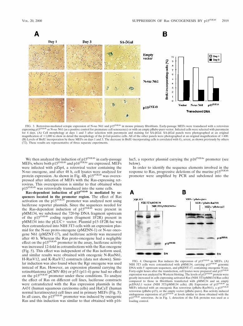
We then analyzed the induction of p15INK4b in early-passageMEFs, where both p15INK4b and p16INK4a are expressed. MEFswere infected with pZip4, a retroviral vector containing theN-ras oncogene, and after 48 h, cell lysates were analyzed forprotein expression. As shown in Fig. 4B, p15INK4b was overex-pressed after infection of MEFs with the Ras-expressing ret-rovirus. This overexpression is similar to that obtained whenp15INK4b was retrovirally transduced into the same cells.
Ras-dependent induction of p15INK4b is mediated by se-quences located in the promoter region. The effect of Rasactivation on the p15INK4b promoter was analyzed next usingluciferase reporter plasmids. Since the sequences needed forthe Ras-dependent induction of p15INK4b were present inpMM134, we subcloned the 720-bp DNA fragment upstreamof the p15INK4b coding region (fragment 1F2R) present inpMM134 into the pLUC1 vector. Plasmid p15-1F2R-luc wasthen cotransfected into NIH 3T3 cells with an expression plas-mid for the N-ras proto-oncogene (pMZNN-1) or N-ras onco-gene N61 (pMZNT-17), and luciferase activity was measuredafter 40 h. Whereas the Ras proto-oncogene had a negligibleeffect on the p15INK4b promoter in the assay, luciferase activitywas increased 12-fold in cotransfections with the Ras oncogene(Fig. 5). This effect was independent of the Ras isoform used,and similar results were obtained with oncogenic N-RasN61,H-RasV12, and K-RasV12 constructs (data not shown). Simi-lar induction was also found when the Rgr oncogene was usedinstead of Ras. However, an expression plasmid carrying theretinoblastoma (pCMV-Rb) or p53 (p11-4) gene had no effecton the p15INK4b promoter under these conditions. To analyzethe effect of Ras on different cell lines, luciferase constructswere cotransfected with the Ras expression plasmids in theA431 (human squamous carcinoma cells) and HaCaT (humannormal keratinocytes) cell lines and in primary MEFs (Fig. 5).In all cases, the p15INK4b promoter was induced by oncogenicRas and this induction was similar to that obtained with p16-
luc5, a reporter plasmid carrying the p16INK4a promoter (seebelow).
In order to identify the sequence elements involved in theresponse to Ras, progressive deletions of the murine p15INK4b
promoter were amplified by PCR and subcloned into the
FIG. 3. Retrovirus-mediated ectopic expression of N-ras N61 and p15INK4b in mouse primary fibroblasts. Early-passage MEFs were transduced with a retrovirusexpressing p15INK4b or N-ras N61 (as a positive control for premature cell senescence) or with an empty pBabe-puro vector. Infected cells were selected with puromycinfor 4 days. (A) Cell morphology at days 1 and 3 after selection with puromycin and staining for SA-bGal. SA-bGal panels were photographed at an originalmagnification of 31,000 to show in detail the morphology of the b-Gal-positive cells. All of the other panels were photographed at an original magnification of 3400.(B) Levels of BrdU incorporation by these MEFs on days 1 and 3. The decrease in BrdU-incorporating cells is correlated with G1 arrest, as shown previously by others(72). These results are representative of three separate experiments.
FIG. 4. Oncogenic Ras induces the expression of p15INK4b in MEFs. (A)NIH 3T3 cells were cotransfected with pMM134, carrying p15INK4b genomicDNA with 59-upstream sequences, and pMZNT-17, containing oncogenic N-ras.Forty-eight hours after the transfection, cell lysates were prepared and p15INK4b
expression was analyzed by Western blotting. The levels of p15INK4b protein weregreatly increased in cells expressing activated Ras (NIH 3T3/pMM134/Ras cells)compared to those in fibroblasts transfected with pMM134 and an emptypcDNA3.1 vector (NIH 3T3/pMM134 cells). (B) Expression of p15INK4b inMEFs infected with an oncogenic Ras retrovirus (pBabe-RasN61), a p15INK4b
retrovirus (pBabe-p15), or the empty vector (pBabe-puro). Ras activity inducedendogenous expression of p15INK4b at levels similar to those obtained with thep15INK4b retrovirus. As in Fig. 1, detection of the Erk proteins was used as aloading control.
VOL. 20, 2000 SUPPRESSION OF Ras ONCOGENESIS BY p15INK4b 2919

pLUC1 reporter plasmid. These constructs were cotrans-fected with pMZNT-17 or the empty vector (pcDNA3.1), andluciferase activity was assayed (Fig. 6). Induction by Ras wasobtained with all constructions carrying at least 120 bp proxi-mal to exon 1. However, no response was found with p15-1F1R-luc, carrying a distal fragment, or p15-6F2R-luc, contain-ing only 55 bp upstream of exon 1. Thus, some Ras responseelements must be located at positions 2122 to 265. Computersearches revealed the presence of three Sp1 consensus sitesbetween these positions (Fig. 6A), in an organization similar tothat of the human sequences (28). The Sp1 and Sp3 sitespresent in the human gene seem to be responsible for itsinduction by TGF-b (28). To determine whether these homol-ogous sequences are involved in the induction by TGF-b in themurine promoter, the deletion constructs described abovewere transfected into HaCaT keratinocytes. Twenty hours af-ter transfection, cells were treated with 100 pM TGF-b1 or leftuntreated and luciferase activity was assayed 20 h later. Onlyconstruct p6F2R-luc had lost inducibility by TGF-b, showingthat the same region responsible for Ras induction seems to beinvolved, at least partially, in the TGF-b response. These re-sults agree with those of Li et al. (28) showing that the pro-moter response to TGF-b resides in Sp1 sites proximal (posi-tions 279 to 255) to the p15INK4b gene. However, when weused a 720-bp fragment upstream of the mouse p15INK4b cod-ing sequence (similar to the human fragment used as describedin reference 28), only a threefold induction of p15INK4b could
be observed after TGF-b treatment. Since a 30-fold inductionof p15INK4b was observed in keratinocytes after TGF-b treat-ment (14), other elements needed for a complete TGF-b re-sponse could be located outside this promoter region (ourresults and A. Iavarone and J. Massague, personal communi-cation).
The Raf-Mek-Erk effector pathway is essential for Ras-de-pendent INK4 induction. Cellular effects of Ras activation arediverse and are mediated by several different effector pathways(5, 24, 35). In order to investigate which of the signaling cas-cades downstream of Ras are involved in the induction of thep15INK4b and p16INK4a cell cycle inhibitors, several expressionplasmids containing activated versions of signaling proteinswere cotransfected with the p15-1F2R-luc (p15INK4b pro-moter) and p16-luc5 (p16INK4a promoter) plasmids. Both on-cogenic Ras and Rgr induce the INK4 promoters to similarextents (about 12-fold). Activated versions of Raf1 (RafBXB)
FIG. 5. Induction of the murine p15INK4b promoter by oncogenic Ras incultured cells. Plasmid p15-1F2R-luc, containing 720 bp of p15INK4b 59 sequencesdriving a luciferase reporter gene, was cotransfected with different expressionplasmids in NIH 3T3, A431, or HaCaT cells or in primary MEFs. Luciferaseactivities from three separate experiments with each cell line were measured.The promoter activity of p15-1F2R-luc cotransfected with an empty vector wasarbitrarily chosen as 1. Cotransfection with an N-ras proto-oncogene, p53, orretinoblastoma expression plasmid had little or no effect on promoter induction.However, there was a 12-fold increase in luciferase activity when an oncogenicN-ras plasmid (pMZNT-17) was used in NIH 3T3 cells and similar results wereobtained with the other cells. wt, wild type.
FIG. 6. Deletion analysis of the mouse p15INK4b promoter. (A) Proximalsequence upstream p15INK4b exon 1. Nucleotide positions are numbered back-ward from the first base of the cDNA sequence (nucleotides in boldface). Dif-ferent fragments upstream of the p15INK4b gene were amplified by PCR andsubcloned into the luciferase reporter vector. The 59-end position of some prim-ers used for amplification (3F and 6F) is shown by arrows. Primer 2R (notshown) is a reverse oligonucleotide inside the cDNA sequence. Boxes indicateputative recognition sites for the Sp1 family of transcription factors. (B) Schemeof the deletion fragments and promoter activity (black bars). Circles indicate thepositions of putative Sp1-binding sites. The data shown are as percentages of theactivity of p15-1F2R-luc (100%) carrying a 720-bp insert. (C) Induction ofpromoter activity by Ras and TGF-b. The constructs were cotransfected with anactivated Ras plasmid (pMZNT-17) or the vector pcDNA3.1. Fold induction byoncogenic Ras (white bars) was compared to the luciferase activity of the samefragment cotransfected with the empty vector. Fold induction by TGF-b (graybars) corresponded to the increase in luciferase activity after TGF-b treatmentof HaCaT cells transfected with the constructs. All of the data shown here werenormalized to b-galactosidase activity as described in Materials and Methods andare representative of three separate experiments.
2920 MALUMBRES ET AL. MOL. CELL. BIOL.

and Mek1 (Mek1 EE) showed partial induction of p15INK4b
and p16INK4a compared with Ras (Fig. 7A). Other activatedproteins, such as RalA (RalA23V), RhoA (RhoA63), and thecatalytic subunit of PI3K (rCD2p110), or overexpression of thewild-type RalGDS, JNK, or PKCz protein had a reduced or noeffect on the p15INK4b or p16INK4a promoter. To ensure the
functional integrity of the Raf1 and Mek1 constructs, theseproteins were used in Elk-1 transactivation assays using lucif-erase reporters (74). Although the three proteins strongly in-duced Elk-1 transactivation (data not shown), only a fivefoldincrease on p15INK4b or p16INK4a promoter activity was ob-served (Fig. 7A).
Using a similar experimental approach, dominant negativevariants of several proteins involved in the signaling cascadesdownstream of Ras were cotransfected with the luciferase re-porter plasmids and the oncogenic Ras construct (pMZNT-17). Only the dominant negative forms of Raf1 (RafC4B),Mek1 (Mek1 MANA), and Erks (KR-Erk1 and K52R-Erk2)were able to suppress the luciferase activity induced by Ras(Fig. 7B). No significant effect was observed with a RalA(RalAN28), Rac1 (Rac1N17), Cdc42 (Cdc42N17), or PKCz(PKCzW281) inhibitory mutant protein (data not shown), andonly a slight decrease in luciferase activity was detected whenthe PI3K (p85D) and RhoA (RhoAN19) dominant negativeproteins were used. We also took advantage of several effectordomain mutant forms of Ras which are impaired in specificdownstream pathways. Thus, S35 Ras mutant proteins signalthrough the Raf1 pathway but not through the RalGDS orPI3K effector. G37 and E38 mutant proteins are specific forthe RalGDS pathway, and C40 interacts with PI3K but notwith the other two effectors (57, 75). We cotransfected acti-vated forms (V12) of these mutant proteins with a p15-1F2R-luc or p16-luc5 reporter using the HrasV12 mutant protein(with full effector response) or the empty vector as a control.As shown in Fig. 7C, HrasV12 produced 8- to 10-fold in-duction of the promoters whereas Raf1-interacting mutantHrasV12S35 mutant showed partial (3-fold) induction. A slightincrease in luciferase activity was found with HrasV12G37 andHrasV12E38 (less than twofold induction in the RalGDS-in-teracting forms). No significant increase in luciferase activitywas found with PI3K-interacting Ras mutant HrasV12C40.Interestingly, higher induction of the INK4 promoters wasobserved when combinations of these mutants were used, es-pecially in the case of Raf1- and RalGDS-interacting proteinsor when a mixture of the three Ras mutant proteins was used,indicating that, at least in NIH 3T3 cells, several Ras effectorpathways can cooperate in p15INK4b and p16INK4a induction.
p15INK4b-deficient MEFs are susceptible to transformationby a single oncogene. Since the in vitro results described abovesuggested a role for p15INK4b in suppressing the oncogenicactivity of Ras in cultured fibroblasts, we decided to analyzethe effect of Ras activation in MEFs obtained from p15INK4b
knockout mice. These MEFs and those from wild-type litter-mates were transfected at early passages with plasmids express-ing oncogenic Ras, Rgr, or the E1A protein. After transfection,cells were cultured for 2 weeks and visible foci were scored.Whereas no foci were observed in wild-type MEFs transfectedwith either the Ras or the Rgr oncogene, a few foci werescored in p15INK4b-deficient MEFs transfected with either ofthese proteins (Fig. 8). The efficiency of transformation ofp15INK4b-deficient MEFs (averages of five foci per assay withRas and seven foci per assay with Rgr) was, however, lowerthan that of INK4aD2,3 knockout MEFs lacking both thep16INK4a and p19ARF proteins (an average of 36 foci per assay;data not shown). These results are similar to those obtained byLatres and Barbacid (unpublished) showing that p15INK4b
knockout MEFs are more susceptible to transformation by Rasor a combination of the Ras and Myc oncogenes. The numbersof foci obtained with Ras and Rgr were very similar, supportingan oncogenic mechanism common to both proteins (Hernan-dez and Pellicer, unpublished). When Rgr was cotransfectedwith the E1A oncoprotein, transformed foci were observed in
FIG. 7. Analysis of the Ras effector pathways involved in p15INK4b andp16INK4a promoter induction. The p15INK4b (p15-1F2R-luc; black columns) andp16INK4a (p16-luc5, white columns) reporter plasmids were cotransfected intoNIH 3T3 cells with empty plasmids (Vector) or with expression plasmids carryingwild-type, activated, or dominant negative forms of molecules involved in thepathways downstream of Ras. Luciferase activity was then compared to the effectof oncogenic Ras (NrasN61 in pMZNT-17 or HrasV12 in pSG5), and the dataare shown as fold induction with respect to the effect of the empty vector.Averages and standard errors from at least three different experiments areshown. Luciferase activity was normalized to b-galactosidase activity as a controlfor transfection efficiency. (A) Effect of overexpression of wild-type or activatedproteins in p15INK4b and p16INK4a promoters. Whereas the Ras or Rgr oncogeneproduced a 12-fold increase in luciferase activity, only partial induction wasfound with single molecules downstream of Ras. (B) NIH 3T3 cells were co-transfected with the p15INK4b and p16INK4a reporter plasmids, oncogenic Ras(pMZNT-17), and the dominant negative forms of Raf1, Mek1, Erk1, PI3K(p85D), and RhoA. All of the Raf1-Mek1-Erk inhibitory forms suppressed Ras-dependent induction, whereas the other proteins had little or no effect. (C)p15INK4b and p16INK4a promoter induction by effector domain mutant forms ofRas. Only partial induction was achieved with HasV12S35 (Raf1-interactingmutant), and a very modest effect was obtained with the other forms. However,the three different forms cooperated to produce a stronger response.
VOL. 20, 2000 SUPPRESSION OF Ras ONCOGENESIS BY p15INK4b 2921
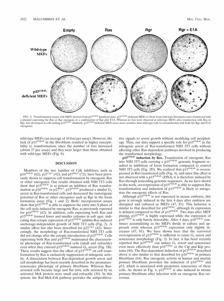
wild-type MEFs (an average of 10 foci per assay). However, thelack of p15INK4b in the fibroblasts resulted in higher suscepti-bility to transformation, since the number of foci increased(about 27 per assay) and they were larger than those obtainedwith wild-type MEFs (Fig. 8).
DISCUSSION
Members of the two families of Cdk inhibitors, such asp16INK4a (62), p21Cip1 (42), and p57Kip2 (72), have been previ-ously shown to suppress cell transformation by oncogenic Rasor other oncogenes. Our results obtained with NIH 3T3 cellsshow that p15INK4b is as potent an inhibitor of Ras transfor-mation as p16INK4a or p27Kip1. p15INK4b produced a similar G1arrest in Ras-transformed cells and decreased the tumorigenicpotential of Ras or other oncogenes such as Rgr in the focusformation assay (Fig. 1 and 2). BrdU incorporation assaysshow that p15INK4b is able to suppress the entry into S phase ofthe cell cycle induced by oncogenic Ras, as previously reportedfor p16INK4a (62). In addition, cells expressing both Ras andp15INK4b formed fewer and smaller colonies in soft agar, indi-cating that ectopic expression of p15INK4b inhibits Ras-inducedanchorage-independent growth of NIH 3T3 cells (Fig. 2). Asimilar effect has also been described for p21Cip1 (42). Inter-estingly, the morphology of Ras-transformed NIH 3T3 cellsdid not change after the ectopic introduction of p15INK4b. Cellsexpressing both Ras and p15INK4b maintained the characteris-tic phenotype of Ras-transformed cells (small and refractile)even when they entered p15INK4b-induced G1 arrest (Fig. 1B).These results suggest that the effect of p15INK4b on cell trans-formation by Ras is exclusively suppression of mitogenic activ-ity. A dissociation between Ras-dependent growth arrest andcell morphology has been recently described in the premature-senescence phenotype of primary fibroblasts. Whereas Ras-arrested cells became large and flat (64), cells arrested by anactivated Mek protein were small and refractile (30). In thissystem, the Raf-Mek-Erk pathway provides the antiprolifera-
tive signals to arrest growth without modifying cell morphol-ogy. Thus, our data support a specific role for p15INK4b in themitogenic arrest of Ras-transformed NIH 3T3 cells withoutaffecting other Ras-dependent pathways involved in producingthe transformed morphology.
p15INK4b induction by Ras. Transfection of oncogenic Rasinto NIH 3T3 cells carrying a p15INK4b genomic fragment re-sulted in inhibition of focus formation compared to controlNIH 3T3 cells (Fig. 2D). We realized that p15INK4b is overex-pressed in Ras-transfected cells (Fig. 4), and since this effect isnot observed with a p15INK4b cDNA, it is therefore induced byRas through noncoding genomic sequences. As we have shownin this work, overexpression of p15INK4b is able to suppress Rastransformation and induction of p15INK4b is likely to antago-nize the oncogenic effects of Ras.
Although p15INK4b is not expressed in mouse embryos, thisgene is strongly induced in the first 4 days after embryos aredisrupted and cultured as MEFs (47, 81). This behavior issimilar to that described for p16INK4a, although its expressionis delayed compared to that of p15INK4b. Two days after MEFplating, p15INK4b is highly expressed while the expression ofp16INK4a is only barely detectable. After 4 days, p16INK4a con-tinues accumulating as the MEFs divide in culture and ap-proach crisis whereas p15INK4b expression only slightly in-creases (47, 81). We have shown here that the retroviraloverexpression of p15INK4b is sufficient to cause G1 arrest andsenescence morphology (Fig. 3). In fact, it has been recentlyreported that p15INK4b can induce G1 arrest and senescenceeven more effectively than p16INK4a or the Cip and Kip pro-teins (40). The Ras-dependent induction of p15INK4b describedabove is also similar to that described for p16INK4a in primaryfibroblasts (64). Ras oncogenic activity in human and murineprimary fibroblasts provokes the induction of p16INK4a andp53, which is associated with premature senescence of thesecells. As shown in Fig. 4, p15INK4b is also induced in mouseprimary fibroblasts after infection with an oncogenic Ras ret-rovirus.
FIG. 8. Transformation assays with MEFs derived from p15INK4b knockout mice. p15INK4b-deficient MEFs or those from wild-type littermates were transfected witha plasmid expressing the Ras or Rgr oncogene or a combination of Rgr plus E1A. Whereas no foci were observed in wild-type MEFs after transfection with Ras orRgr, foci developed in cells lacking p15INK4b. Similarly, p15INK4b-deficient MEFs were more sensitive than wild-type cells to cotransfection with both the Rgr and E1Aoncogenes.
2922 MALUMBRES ET AL. MOL. CELL. BIOL.
This effect on cell cycle arrest and senescence could be evenmore dramatic in lymphoid tissues, where p15INK4b has beenshown to play an important role in lymphocyte activation(34) or tumorigenesis (4, 17, 36). T lymphocytes accumulatep15INK4b protein during successive population doublings anddisplay high levels of this molecule as they enter replicativesenescence (12). Accumulation of INK4 proteins is accompa-nied by their increased binding to Cdk6 and decreased Cdk6and Cdk2 activity. Thus, absence of the p15INK4b protein couldmake it possible for tumoral lymphocytes to proliferate, avoid-ing replicative senescence.
Transfection experiments using pMM134 (p15INK4b genomicDNA containing about 720 bp of the promoter sequence) andluciferase reporter assays show that Ras-dependent inductionof p15INK4b and p16INK4a occurs through sequences located inthe promoter region. In the case of p15INK4b, induction by Rasis blocked when 60 bp located between positions 2122 and255 are deleted from the promoter. Similar sequences alsoseem to be necessary for response to TGF-b in the humanp15INK4b promoter (28). Sp1-binding sites are also importantfor the regulation of the p19ARF promoter (56), and sincep19ARF is also induced by Ras activation (48), we are currentlyanalyzing whether these sequences are also involved in Ras-dependent expression of the p19ARF protein. Ras, therefore,could use the same sequence motifs that TGF-b uses for in-duction of p15INK4b. In fact, a role for Ras in TGF-b transcrip-tional activation has been suggested (16, 45, 78). In epithelialcells, growth inhibition by TGF-b1 and TGF-b2 is associatedwith rapid activation of both Ras and Erk1 (45). Moreover,expression of the Ras dominant negative form (RasN17) par-tially abrogates Erk1 activation and inhibition of DNA synthe-sis by TGF-b (16) and Ras activation is also needed for TGF-bupregulation of p21Cip1 and p27Kip1 (78). Since p27Kip1 upregu-lation by TGF-b has been described as an indirect effect ofp15INK4b induction and displacement of p27Kip1 from cyclinD-Cdk4/6 complexes (49, 51, 60), Ras could be physiologicallyinvolved in p15INK4b induction. If this is the case, the ability ofRas to induce the expression of some Cdk inhibitors could notonly be a protective or stress response of the cell but have afunction in normal physiological stages.
The constant levels of p15INK4b expression during the cellcycle and its induction by TGF-b have suggested a role forp15INK4b as an important effector in TGF-b-induced growtharrest, rather than in regulation of the timing of events in thecell cycle itself (14, 68). Our results suggest that p15INK4b couldalso provoke G1 arrest in response to cellular signals mediatedby not only TGF-b but also other stimuli depending on Rassignaling. When Ras is required to send a mitogenic stimulus,other signals must cooperate to suppress the induction ofp15INK4b and other inhibitors. For instance, in Ras-dependentinduction of p21Cip1, once Ras has been activated, Rho signal-ing is required for suppression of p21Cip1 induction (46). Whensignaling through Rho is inhibited, constitutively active Rasinduces p21Cip1 and entry into the DNA synthesis phase of thecell cycle is blocked.
Ras effector pathways and cell cycle control. Since p15INK4b
is also induced by Ras in NIH 3T3 cells containing the genomicINK4b gene, the downstream effector pathways used by Rasseem to be intact in these cells. Among the Ras effectors, theRaf-Mek-Erk pathway has been shown to function not only inthe stimulation of cellular proliferation but also in growtharrest. There is growing evidence that a sustained increase inErk activity can lead to inhibition of Cdk activity and cell cyclearrest (30, 33, 52, 77). In some systems, these inhibitory effectshave been attributed to induction of the Cdk inhibitor p21Cip1
or a decrease in cyclin A levels (32, 65). This induction has
been proposed to occur in a p53-dependent manner in pri-mary rat Schwann cells (33) but independently of p53 inmouse primary fibroblasts (77). In other cases, the increase inp16INK4a expression seems more relevant and activated Raf orMek protein is sufficient to provoke its induction and prema-ture senescence in primary fibroblasts (30, 79).
In our study, analysis of the pathways involved in the Ras-dependent transcriptional activation of the p15INK4b andp16INK4a promoters showed that in NIH 3T3 cells, differentcooperating pathways can be required to emulate the effect ofoncogenic Ras. Activated forms of the Raf1 and Mek (MEKEE) kinases only partially increased luciferase activity from thep15INK4b and p16INK4a promoter, whereas the effect was almostnull in other pathways (Fig. 7). Interestingly, a recently de-scribed oncogene named Rgr (9) is able to induce the INK4proteins to an extent similar to that of oncogenic Ras. Rgr is astrong oncogene belonging to the RalGDS family of exchangefactors. Rgr is able to activate RalA, similar to the othermembers of the family, but is also able to activate some Rho-mediated pathways and the mitogen-activated protein kinases,and this activation is at least partially dependent on Ras (Her-nandez and Pellicer, unpublished). All of these pathways arestrongly related to transcriptional activation and could there-fore account for the induction of the INK4 inhibitors.
The use of dominant negative proteins shows that the Raf-Mek-Erk pathway is essential for Ras-dependent activation ofp15INK4b and p16INK4a. These results agree with the recentreports by Lin et al. (30) and Zhu et al. (79) showing thatactivation of the mitogen-activated protein kinase cascade isessential for induction of Cdk inhibitors and promotion ofpremature senescence. However, while they described compa-rable data on growth arrest and senescence after Ras, Raf1, orMek activation, we repeatedly found only partial induction ofthe INK4 promoters when using activated Raf or Mek protein.These results can be explained by differences between theexperimental approaches. While we analyzed the transcrip-tional activation of individual promoters, the work of Lin et al.(30) was based mainly on measurement of growth arrest andcellular senescence. Thus, small increases in the individuallevels of p16INK4a, p15INK4b, and perhaps p21Cip1 induced byH-RasV12S35 could still cooperate and produce cell cycle ar-rest. On the other hand, strong mutant proteins used in theirwork, such as MekQ56P, could produce a stronger signal thanthe activated forms used in the present work. In fact, activatedMekQ56P causes cell cycle arrest even more rapidly than on-cogenic Ras (30) and differences in the strength of the signalhave been previously shown to be essential in the stimulationof the Raf-Mek-Erk cascade outcome (26, 77). It has beensuggested that the ultimate cellular response (cell cycle arrestor cellular proliferation) to activation of the Mek-Erk pathwaycan depend on the strength and duration of the Erk signal,which is the transient or cyclical activation responsible for theproliferative output, while sustained high levels of Erk activitycould result in cell cycle arrest (52, 77). Thus, low levels of Rafactivity lead to activation of cyclin D1-Cdk4 and cyclin E-Cdk2complexes and cell cycle progression, whereas higher Raf ac-tivity elicited cell cycle arrest correlating with p21Cip1 inductionand inhibition of cyclin-Cdk activity (77). Finally, the signalingpathways from Ras to p16INK4a and p15INK4b induction couldbe modified in NIH 3T3 cells, so that the Raf and Mek proteinsmight not be able to produce as high levels of INK4 inductionas Ras does. We therefore do not exclude the possibility thatdifferent pathways could cooperate with the Raf-Mek-Erk cas-cade in NIH 3T3 and other cell types to produce growth arrestmediated by the Cdk inhibitors. In fact, in NIH 3T3 cellsexpressing human TrkA, PD98059 (a Mek inhibitor) blocked
VOL. 20, 2000 SUPPRESSION OF Ras ONCOGENESIS BY p15INK4b 2923

the ability of nerve growth factor (NGF) to inhibit Cdk2 andCdk4 activities but only partially prevented the NGF inductionof p21Cip1. Thus, NGF-dependent up-regulation of the p21Cip1
protein appears to be only partially mediated through theMek-Erk pathway (52), suggesting that other pathways canalso be involved.
Susceptibility of p15INK4b-deficient MEFs to cell transfor-mation by single oncogenes. The need for oncogene coopera-tion in the transformation of primary cells was reported almost20 years ago (27, 59). In the last few years, it has been dem-onstrated both in vitro and in vivo that oncogenic activationand loss of inhibitory effects might cooperate in cell trans-formation. In fact, primary cells from INK4aD2,3-, p19ARF-,p21Cip1-, or p53-deficient mice can be transformed by Rasalone (23, 43, 63). On the other hand, Ras transgenics in anINK4aD2,3 background have a significantly higher tumor inci-dence (6). We show here that p15INK4b-deficient MEFs aresusceptible to transformation by the Ras or Rgr oncogene,although with a lower efficiency than INK4aD2,3 MEFs. TheseMEFs lack both the p16INK4a and p19ARF proteins, and there-fore, the contribution of each protein to this phenotype has notbeen assessed yet. Moreover, MEFs lacking only p19ARF arealso susceptible to transformation by Ras (23) and the effect ofp16INK4a inactivation in primary MEFs remains to be deter-mined. The absence of the other INK4 proteins (p18INK4c andp19INK4d) in the corresponding knockout MEFs does not resultin increased sensitivity to transformation by Ras (82; Latresand Barbacid, unpublished). Our results and those obtained byLatres and Barbacid (unpublished) indicate that p15INK4b hasa suppressor effect on MEF transformation by either the Rasor the Rgr oncogene or by the combination of Ras and differ-ent oncogenes such as Myc or E1A.
The role of p15INK4b as a tumor suppressor in lymphocyteshas also been recently highlighted as a result of methylationstudies with both human and mouse cells (4, 17, 18, 36, 38, 80).Thus, p15INK4b is frequently (up to 88%) inactivated by meth-ylation of the promoter region in both humans (4, 17, 18) andexperimental systems (36). In agreement with these observa-tions, p15INK4b knockout mice frequently develop lymphopro-liferative disorders (Latres and Barbacid, unpublished). Thefact that oncogenic Ras can activate p16INK4a and p15INK4b
therefore provides a new insight into their role as tumor sup-pressor genes and suggests that Ras oncogenic signals mightcooperate with p15INK4b inactivation in tumorigenesis in vivo.These INK4 proteins have similar activities in vitro and arelikely to participate in the growth arrest response to oncogenicsignals. Their different role in vivo could be dependent on theirspecific pattern of expression or in their ability to be inducedby different stimuli. Among them, Ras seems to be a strongINK4 inducer and an important one due to its ability to par-ticipate in physiological and tumorigenic processes.
ACKNOWLEDGMENTS
We thank J. Altschmied, M. Barbacid, J. L. Bos, D. A. Brenner, P.Crespo, C. J. Der, L. A. Feig, M. Kasuga, D. Levy, J. Massague, M.Pagano, U. R. Rapp, K. Reif, P. Rodrıguez-Viciana, M. Serrano, R. A.Weinberg, and Y. Xiong for kindly providing some of the plasmids andreagents used in this work. We are specially indebted to E. Latres andM. Barbacid for providing us with the p15INK4b-deficient MEFs.
M.M., M.I.H., and I.P.C. received fellowships from the Ministeriode Educacion (Madrid, Spain). This work was supported by grants CA36327 and CA 50434 from NIH to A.P.
REFERENCES
1. Albright, C. F., B. W. Giddings, J. Liu, M. Vito, and R. A. Weinberg. 1993.Characterization of a guanine nucleotide dissociation stimulator for a ras-related GTPase. EMBO J. 12:339–347.
2. Alcorta, D. A., Y. Xiong, D. Phelps, G. Hannon, D. Beach, and J. C. Barret.1996. Involvement of the cyclin-dependent kinase inhibitor p16 (INK4a) inreplicative senescence of normal human fibroblasts. Proc. Natl. Acad. Sci.USA 93:13742–13747.
3. Altschmied, J., and J. Duschl. 1997. Set of optimized luciferase reportergene plasmids compatible with widely used CAT vectors. BioTechniques 23:436–438.
4. Batova, A., M. B. Diccianni, J. C. Yu, T. Nobori, M. P. Link, J. Pullen, andA. L. Yu. 1997. Frequent and selective methylation of p15 and deletion ofboth p15 and p16 in T-cell acute lymphoblastic leukemia. Cancer Res. 57:832–836.
5. Campbell, S. L., R. Khosravi-Far, K. L. Rossman, G. J. Clark, and C. J. Der.1998. Increasing complexity of Ras signaling. Oncogene 17:1395–1413.
6. Chin, L., J. Pomerantz, D. Polsky, M. Jacobson, C. Cohen, C. Cordon-Cardo,J. W. Horner 2nd, and R. A. DePinho. 1997. Cooperative effects of INK4aand ras in melanoma susceptibility in vivo. Genes Dev. 11:2822–2834.
7. Chin, L., J. Pomerantz, and R. A. DePinho. 1998. The INK4a/ARF tumorsuppressor: one gene—two products—two pathways. Trends Biochem. Sci.23:291–296.
8. Cowley, S., H. Paterson, P. Kemp, and C. J. Marshall. 1994. Activation ofMAP kinase kinase is necessary and sufficient for PC12 differentiation andfor transformation of NIH 3T3 cells. Cell 77:841–852.
9. D’Adamo, D. R., S. Novick, J. M. Kahn, P. Leonardi, and A. Pellicer. 1997.rsc: a novel oncogene with structural and functional homology with the genefamily of exchange factors for Ral. Oncogene 14:1295–1305.
10. Dimri, G. P., X. Lee, G. Basile, M. Acosta, G. Scott, C. Roskelley, E. E.Medrano, M. Linskens, I. Rubelj, O. Pereira-Smith, M. Peacocke, and J.Campisi. 1995. A biomarker that identifies senescent human cells in cultureand in aging skin in vivo. Proc. Natl. Acad. Sci. USA 92:9363–9367.
11. Downward, J. 1997. Routine role for Ras. Curr. Biol. 7:R258–R260.12. Erickson, S., O. Sangfelt, M. Heyman, J. Castro, S. Einhorn, and D.
Grander. 1998. Involvement of the Ink4 proteins p16 and p15 in T-lympho-cyte senescence. Oncogene 17:595–602.
13. Haber, D. A. 1997. Splicing into senescence: the curious case of p16 andp19ARF. Cell 91:555–558.
14. Hannon, G. J., and D. Beach. 1994. p15INK4b is a potential effector of TGF-b-induced cell cycle arrest. Nature 371:257–261.
15. Hara, K., K. Yonezawa, H. Sakaue, A. Ando, K. Kotani, T. Kitamura, Y.Kitamura, H. Ueda, L. Stephens, T. R. Jackson, et al. 1994. 1-Phosphatidyl-inositol 3-kinase activity is required for insulin-stimulated glucose transportbut not for RAS activation in CHO cells. Proc. Natl. Acad. Sci. USA 91:7415–7419.
16. Hartsough, M. T., R. S. Frey, P. A. Zipfel, A. Buard, S. J. Cook, F. McCor-mick, and K. M. Mulder. 1996. Altered transforming growth factor b sig-naling in epithelial cells when Ras activation is blocked. J. Biol. Chem. 271:22368–22375.
17. Herman, J. G., J. Jen, A. Merlo, and S. B. Baylin. 1996. Hypermethylation-associated inactivation indicates a tumor suppressor role for p15INK4B.Cancer Res. 56:722–727.
18. Herman, J. G., C. I. Civin, J. P. Issa, M. I. Collector, S. J. Sharkis, and S. B.Baylin. 1997. Distinct patterns of inactivation of p15INK4B and p16INK4Acharacterize the major types of hematological malignancies. Cancer Res. 57:837–841.
19. Hitomi, M., and D. W. Stacey. 1999. Cellular Ras and cyclin D1 are requiredduring different cell cycle periods in cycling NIH 3T3 cells. Mol. Cell. Biol.19:4623–4632.
20. Huschtscha, L. I., and R. R. Reddel. 1999. p16INK4a and the control ofcellular proliferative life span. Carcinogenesis 20:921–926.
21. Jeffers, M., and A. Pellicer. 1994. Identification of multiple promoters withinthe N-ras proto-oncogene. Biochim. Biophys. Acta 1219:623–635.
22. Kamb, A., N. A. Gruis, J. Weaver-Feldhaus, Q. Liu, K. Harshman, S. V.Tavtigian, E. Stockert, R. S. Day 3rd, B. E. Johnson, and M. H. Skolnick.1994. A cell cycle regulator potentially involved in genesis of many tumortypes. Science 264:436–440.
23. Kamijo, T., F. Zindy, M. F. Roussel, D. E. Quelle, J. R. Downing, R. A.Ashmun, G. Grosveld, and C. J. Sherr. 1997. Tumor suppression at themouse INK4a locus mediated by the alternative reading frame productp19ARF. Cell 91:649–659.
24. Katz, M. E., and F. McCormick. 1997. Signal transduction from multiple Raseffectors. Curr. Opin. Genet. Dev. 7:75–79.
25. Kerkhoff, E., and U. R. Rapp. 1997. Induction of cell proliferation in quies-cent NIH 3T3 cells by oncogenic c-Raf-1. Mol. Cell. Biol. 17:2576–2586.
26. Kerkhoff, E., and U. R. Rapp. 1998. High-intensity Raf signals convertmitotic cell cycling into cellular growth. Cancer Res. 58:1636–1640.
27. Land, J., L. F. Parada, and R. A. Weinberg. 1983. Tumorigenic conversion ofprimary embryo fibroblasts requires at least two cooperating oncogenes.Nature 304:596–602.
28. Li, J. M., M. A. Nichols, S. Chandrasekharan, Y. Xiong, and X. F. Wang.1995. Transforming growth factor activates the promoter of cyclin-depen-dent kinase inhibitor p15INK4B through an Sp1 consensus site. J. Biol. Chem.270:26750–26753.
29. Li, Y., M. A. Nichols, J. W. Shay, and Y. Xiong. 1994. Transcriptional
2924 MALUMBRES ET AL. MOL. CELL. BIOL.

repression of the D-type cyclin-dependent kinase inhibitor p16 by the reti-noblastoma susceptibility gene product pRb. Cancer Res. 54:6078–6082.
30. Lin, A. W., M. Barradas, J. C. Stone, L. van Aelst, M. Serrano, and S. W.Lowe. 1998. Premature senescence involving p53 and p16 is activated inresponse to constitutive MEK/MAPK mitogenic signaling. Genes Dev. 12:3008–3019.
31. Linardopoulos, S., A. J. Street, D. E. Quelle, D. Parry, G. Peters, C. J. Sherr,and A. Balmain. 1995. Deletion and altered regulation of p16INK4a andp15INK4b in undifferentiated mouse skin tumors. Cancer Res. 55:5168–5172.
32. Lloyd, A. C. 1998. Ras versus cyclin-dependent kinase inhibitors. Curr. Opin.Genet. Dev. 8:43–48.
33. Lloyd, A. C., F. Obermuller, S. Staddon, C. F. Barth, M. McMahon, and H.Land. 1997. Cooperating oncogenes converge to regulate cyclin/cdk com-plexes. Genes Dev. 11:663–677.
34. Lois, A. F., L. T. Cooper, Y. Geng, T. Nobori, and D. Carson. 1995. Expres-sion of the p16 and p15 cyclin-dependent kinase inhibitors in lymphocyteactivation and neuronal differentiation. Cancer Res. 55:4010–4013.
35. Malumbres, M., and A. Pellicer. 1998. Ras pathways to cell cycle control andcell transformation. Front. Biosci. 3:887–912.
36. Malumbres, M., I. Perez de Castro, J. Santos, B. Melendez, R. Mangues, M.Serrano, A. Pellicer, and J. Fernandez-Piqueras. 1997. Inactivation of thecyclin-dependent kinase inhibitor p15INK4b by deletion and de novo methyl-ation with independence of p16INK4a alterations in murine primary T-celllymphomas. Oncogene 14:1361–1370.
37. Malumbres, M., R. Mangues, N. Ferrer, S. Lu, and A. Pellicer. 1997. Isola-tion of high molecular weight DNA for reliable genotyping of transgenicmice. BioTechniques 22:1114–1119.
38. Malumbres, M., I. Perez de Castro, J. Santos, J. Fernandez-Piqueras, and A.Pellicer. 1999. Hypermethylation of the cell cycle inhibitor p15INK4b 39-untranslated region interferes with its transcriptional regulation in primarylymphomas. Oncogene 18:385–396.
39. Matesanz, F., and A. Pellicer. 1995. In vivo and in vitro analysis of retroviralvectors carrying the N-ras oncogene. Int. J. Oncol. 7:443–451.
40. McConnell, B. B., M. Starborg, S. Brookes, and G. Peters. 1998. Inhibitorsof cyclin-dependent kinases induce features of replicative senescence in earlypassage human diploid fibroblasts. Curr. Biol. 8:351–354.
41. Merlo, A., J. G. Herman, L. Mao, D. J. Lee, E. Gabrielson, P. C. Burger, S. B.Baylin, and D. Sidransky. 1995. 59 CpG island methylation is associated withtranscriptional silencing of the tumour suppressor p16/CDKN2/MTS1 in hu-man cancers. Nat. Med. 1:686–692.
42. Michieli, P., W. Li, M. V. Lorenzi, T. Miki, R. Zakut, D. Givol, and J. H.Pierce. 1996. Inhibition of oncogene-mediated transformation by ectopicexpression p21Waf1 in NIH3T3 cells. Oncogene 12:775–784.
43. Missero, C., F. Di Cunto, H. Kiyokawa, A. Koff, and G. P. Dotto. 1996. Theabsence of p21Cip1/WAF1 alters keratinocyte growth and differentiationand promotes ras-tumor progression. Genes Dev. 10:3065–3075.
44. Mulcahy, L. S., M. R. Smith, and D. W. Stacey. 1985. Requirements for rasproto-oncogene function during serum-stimulated growth of NIH 3T3 cells.Nature 313:241–243.
45. Mulder, K. M., and S. L. Morris. 1992. Activation of p21ras by transforminggrowth factor beta in epithelial cells. J. Biol. Chem. 267:5029–5031.
46. Olson, M. F., H. F. Paterson, and C. J. Marshall. 1998. Signals from Ras andRho GTPases interact to regulate expression of p21Waf1/Cip1. Nature 394:295–299.
47. Palmero, I., B. McConnell, D. Parry, S. Brookes, E. Hara, S. Bates, P. Jat,and G. Peters. 1997. Accumulation of p16INK4a in mouse fibroblasts as afunction of replicative senescence and not of retinoblastoma gene status.Oncogene 15:495–503.
48. Palmero, I., C. Pantoja, and M. Serrano. 1998. p19ARF links the tumoursuppressor p53 to Ras. Nature 395:125–126.
49. Peters, G. 1994. Stifled by inhibitors. Nature 371:204–205.50. Pollock, P. M., J. V. Pearson, and N. K. Hayward. 1996. Compilation of
somatic mutations of the CDKN2 gene in human cancers: non-randomdistribution of base substitutions. Genes Chromosomes Cancer 15:77–88.
51. Polyak, K., J. Y. Kato, M. J. Solomon, C. J. Sherr, J. Massague, J. M.Roberts, and A. Koff. 1994. p27Kip1, a cyclin-Cdk inhibitor, links transform-ing growth factor-b and contact inhibition to cell cycle arrest. Genes Dev. 8:9–22.
52. Pumiglia, K. M., and S. J. Decker. 1997. Cell cycle arrest mediated by theMEK/mitogen-activated protein kinase pathway. Proc. Natl. Acad. Sci. USA94:448–452.
53. Quelle, D. E., F. Zindy, R. A. Ashmun, and C. J. Sherr. 1995. Alternativereading frames of the INK4a tumor suppressor gene encode two unrelatedproteins capable of inducing cell cycle arrest. Cell 83:993–1000.
54. Quelle, D. E., R. A. Ashmun, G. J. Hannon, P. A. Rehberger, D. Trono, K. H.Richter, C. Walker, D. Beach, C. J. Sherr, and M. Serrano. 1995. Cloningand characterization of murine p16INK4a and p15INK4b genes. Oncogene 11:635–645.
55. Reif, K., C. D. Nobles, G. Thomas, A. Hall, and D. A. Cantrell. 1996.Phosphatidylinositol 3-kinase signals activate a selective subset of Rac/Rho-dependent effector pathways. Curr. Biol. 6:1445–1455.
56. Robertson, K. D., and P. A. Jones. 1998. The human ARF cell cycle regu-
latory gene promoter is a CpG island which can be silenced by DNA meth-ylation and down-regulated by wild-type p53. Mol. Cell. Biol. 18:6457–6473.
57. Rodriguez-Viciana, P., P. H. Warne, A. Khwaja, B. M. Marte, D. Pappin, P.Das, M. D. Waterfield, A. Ridley, and J. Downward. 1997. Role of phospho-inositide 3-OH kinase in cell transformation and control of the actin cy-toskeleton by Ras. Cell 89:457–467.
58. Ruas, M., and G. Peters. 1998. The p16INK4a/CDKN2A tumor suppressorand its relatives. Biochim. Biophys. Acta 1378:F115–F177.
59. Ruley, H. E. 1983. Adenovirus early region 1A enables viral and cellulartransforming genes to transform primary cells in culture. Nature 304:602–606.
60. Sandhu, C., J. Garbe, N. Bhattacharya, J. Daksis, C. H. Pan, P. Yaswen, J.Koh, J. M. Slingerland, and M. R. Stampfer. 1997. Transforming growthfactor b stabilizes p15INK4B protein, increases p15INK4B-cdk4 complexes, andinhibits cyclin D1-cdk4 association in human mammary epithelial cells. Mol.Cell. Biol. 17:2458–2467.
61. Serrano, M., G. J. Hannon, and D. Beach. 1993. A new regulatory motif incell-cycle control causing specific inhibition of cyclin D/CDK4. Nature 366:704–707.
62. Serrano, M., E. Gomez-Lahoz, R. A. DePinho, D. Beach, and D. Bar-Sagi.1995. Inhibition of Ras-induced proliferation and cellular transformation byp16INK4. Science 267:249–252.
63. Serrano, M., H. W. Lee, L. Chin, C. Cordon-Cardo, D. Beach, and R. A.DePinho. 1996. Role of the INK4a locus in tumor suppression and cellmortality. Cell 85:27–37.
64. Serrano, M., A. W. Lin, M. E. McCurrach, D. Beach, and S. W. Lowe. 1997.Oncogenic ras provokes premature cell senescence associated with accumu-lation of p53 and p16INK4a. Cell 88:593–602.
65. Sewing, A., B. Wiseman, A. C. Lloyd, and H. Land. 1997. High-intensity Rafsignal causes cell cycle arrest mediated by p21Cip1. Mol. Cell. Biol. 17:5588–5597.
66. Sharpless, N. E., and R. A. DePinho. 1999. The INK4A/ARF locus and its twogene products. Curr. Opin. Genet. Dev. 9:22–30.
67. Sherr, C. J. 1998. Tumor surveillance via the ARF-p53 pathway. Genes Dev.12:2984–2991.
68. Stone, S., P. Dayananth, P. Jiang, J. M. Weaver-Feldhaus, S. V. Tavtigian, L.Cannon-Albright, and A. Kamb. 1995. Genomic structure, expression andmutational analysis of the P15 (MTS2) gene. Oncogene 11:987–991.
69. Swafford, D. S., S. K. Middleton, W. A. Palmisano, K. J. Nikula, J. Tesfaigzi,S. B. Baylin, J. G. Herman, and S. A. Belinsky. 1997. Frequent aberrantmethylation of p16INK4a in primary rat lung tumors. Mol. Cell. Biol. 17:1366–1374.
70. Tan, T.-H., J. Wallis, and A. J. Levine. 1986. Identification of the p53 proteindomain involved in formation of the simian virus 40 large T-antigen–p53protein complex. J. Virol. 59:574–583.
71. Urano, T., R. Emkey, and L. A. Feig. 1996. Ral-GTPases mediate a distinctdownstream signaling pathway from Ras that facilitates cellular transforma-tion. EMBO J. 15:810–816.
72. Watanabe, H., Z. Q. Pan, N. Schreiber-Agus, R. A. DePinho, J. Hurwitz, andY. Xiong. 1998. Suppression of cell transformation by the cyclin-dependentkinase inhibitor p57KIP2 requires binding to proliferating cell nuclear anti-gen. Proc. Natl. Acad. Sci. USA 95:1392–1397.
73. Weinberg, R. A. 1997. The cat and mouse games that genes, viruses, and cellsplay. Cell 88:573–575.
74. Westwick, J. K., A. D. Cox, C. J. Der, M. H. Cobb, M. Hibi, M. Karin, andD. A. Brenner. 1994. Oncogenic Ras activates c-Jun via a separate pathwayfrom the activation of extracellular signal-regulated kinases. Proc. Natl.Acad. Sci. USA 91:6030–6034.
75. White, M. A., C. Nocolette, A. Minden, A. Polverino, L. Van Aelst, M. Karin,and M. H. Wigler. 1995. Multiple Ras functions can contribute to mamma-lian cell transformation. Cell 80:533–541.
76. Wolthuis, R. M., N. D. de Ruiter, R. H. Cool, and J. L. Bos. 1997. Stimulationof gene induction and cell growth by the Ras effector Rlf. EMBO J. 16:6748–6761.
77. Woods, D., D. Parry, H. Cherwinski, E. Bosch, E. Lees, and M. McMahon.1997. Raf-induced proliferation or cell cycle arrest is determined by the levelof Raf activity with arrest mediated by p21Cip1. Mol. Cell. Biol. 17:5598–5611.
78. Yue, J., A. Buard, and K. M. Mulder. 1998. Blockade of TGFbeta3 up-regulation of p27Kip1 and p21Cip1 by expression of RasN17 in epithelialcells. Oncogene 17:47–55.
79. Zhu, J., D. Woods, M. McMahon, and J. M. Bishop. 1998. Senescence ofhuman fibroblasts induced by oncogenic Raf. Genes Dev. 12:2997–3007.
80. Zhuang, S. M., A. Schippert, A. Haugen-Strano, R. W. Wiseman, and P.Soderkvist. 1998. Inactivations of p16INK4a-a, p16INK4a-b and p15INK4b genesin 29,39-dideoxycytidine- and 1,3-butadiene-induced murine lymphomas. On-cogene 16:803–808.
81. Zindy, F., D. E. Quelle, M. F. Roussel, and C. J. Sherr. 1997. Expression ofthe p16INK4a tumor suppressor versus other INK4 family members duringmouse development and aging. Oncogene 15:203–211.
82. Zindy, F., J. van Deursen, G. Grosveld, C. J. Sherr, and M. Roussel. 2000.INK4d-deficient mice are fertile despite testicular atrophy. Mol. Cell. Biol.20:372–378.
VOL. 20, 2000 SUPPRESSION OF Ras ONCOGENESIS BY p15INK4b 2925
Copyright © 2022 FDOKUMEN