Adenosine receptors and brain diseases: Neuroprotection and neurodegeneration
Upload
independentCategory
view
0download
0
BRAINA JOURNAL OF NEUROLOGY
Delayed post-ischaemic neuroprotection followingsystemic neural stem cell transplantation involvesmultiple mechanismsMarco Bacigaluppi,1,2,3,*,† Stefano Pluchino,2,3,*,† Luca Peruzzotti Jametti,2,3,† Ertugrul Kilic,1
Ulkan Kilic,1 Giuliana Salani,2,† Elena Brambilla,2 Mark J. West,4 Giancarlo Comi,2,3
Gianvito Martino2,3,* and Dirk M. Hermann1,5,*
1 Department of Neurology, University Hospital Zurich, Frauenklinikstrasse 26, CH-8091 Zurich, Switzerland
2 Neuroimmunology Unit-DIBIT2 and Institute of Experimental Neurology, San Raffaele Scientific Institute, Via Olgettina 58, I-20132 Milan, Italy
3 Department of Neurology and Neurophysiology, San Raffaele Scientific Institute, Via Olgettina 58, I-20132 Milan, Italy
4 Institute of Anatomy, University Park, DK-8000 Aarhus, Denmark
5 Department of Neurology, University Hospital, University of Duisburg-Essen, Hufelandstrasse 55, D-45122 Essen, Germany
*These authors contributed equally to this work.
†Present address: CNS Repair Unit-DIBIT2, and Institute of Experimental Neurology,
San Raffaele Scientific Institute,
Via Olgettina 58,
I-20132 Milan,
Italy
Correspondence to: Prof. Dr Dirk M. Hermann,
Chair of Vascular Neurology,
Dementia and Ageing Disorders,
Department of Neurology,
University Hospital Essen,
Hufelandstr. 55,
D-45122 Essen,
Germany
E-mail: [email protected]
Recent evidence suggests that neural stem/precursor cells (NPCs) promote recovery in animal models with delayed neuronal
death via a number of indirect bystander effects. A comprehensive knowledge of how transplanted NPCs exert their therapeutic
effects is still lacking. Here, we investigated the effects of a delayed transplantation of adult syngenic NPCs—injected intra-
venously 72 h after transient middle cerebral artery occlusion—on neurological recovery, histopathology and gene expression.
NPC-transplanted mice showed a significantly improved recovery from 18 days post-transplantation (dpt) onwards, which
persisted throughout the study. A small percentage of injected NPCs accumulated in the brain, integrating mainly in the infarct
boundary zone, where most of the NPCs remained undifferentiated up to 30 dpt. Histopathological analysis revealed a hitherto
unreported very delayed neuroprotective effect of NPCs, becoming evident at 10 and 30 dpt. Tissue survival was associated with
downregulation of markers of inflammation, glial scar formation and neuronal apoptotic death at both mRNA and protein levels.
Our data highlight the relevance of very delayed degenerative processes in the stroke brain that are intimately associated
with inflammatory and glial responses. These processes may efficaciously be antagonized by (stem) cell-based strategies at
time-points far beyond established therapeutic windows for pharmacological neuroprotection.
doi:10.1093/brain/awp174 Brain 2009: 132; 2239–2251 | 2239
Received December 17, 2008. Revised May 15, 2009. Accepted May 21, 2009
� The Author (2009). Published by Oxford University Press on behalf of the Guarantors of Brain. All rights reserved.
For Permissions, please email: [email protected]
by guest on February 9, 2016http://brain.oxfordjournals.org/
Dow
nloaded from

Keywords: stroke; neural stem/precursor cells; transplantation; inflammation; gliosis
Abbreviations: CC = corpus callosum; CNS = central nervous system; dpt = days post-transplantation; i.v. = intravenously;LDF = laser Doppler flow; MCAO = middle cerebral artery occlusion; NPCs = neural stem/precursor cells
IntroductionAccording to current pathophysiological concepts, the structural
histological injury following stroke evolves within several hours
to up to 3–4 days, depending on the duration and severity of
the ischaemic event (Namura et al., 1998; Hata et al., 2000;
Hermann et al., 2001). During that time, secondary energy failure
develops in the ischaemic penumbra via multiple mechanisms
including excitotoxicity, lactacidosis and peri-infarct depolarizations
(Hossmann, 2006). In the ischaemic border zone surrounding the
evolving infarct, inflammatory responses and apoptotic programs
are activated in this phase that further contribute to injury devel-
opment (Dirnagl et al., 1999; Hermann et al., 2001; Hossmann,
2006). Following the acute stroke phase, the tissue undergoes
substantial matrix remodelling that results in (astro)glial scar
formation, which is widely regarded as a major hampering factor
for tissue repair and regeneration (Fawcett and Asher, 1999;
Nedergaard and Dirnagl, 2005; Yiu and He, 2006).
Several lines of evidence suggest that the structural and
functional changes in the ischaemic boundary zone are of key
importance for the final stroke outcome (Witte, 1998; Nudo,
1999). This might imply that therapies favouring endogenous
tissue repair and/or remodelling may have realistic chances of
success also when applied beyond established time-windows of
tissue protection, which in the case of thrombolytics in humans
are in the range of 3–6 h (Hacke et al., 2008). The pathophysiol-
ogy of this second phase is highly heterogeneous in time and
space, and the underlying mechanisms are still poorly understood
(Lee et al., 2008). There is common sense that classical
pharmacological strategies aiming at improving neuronal survival
are no more useful at this latter stage (Zivin, 1998).
Neural stem/precursor cells (NPCs) display remarkable ther-
apeutic plasticity upon transplantation in experimental conditions
mimicking central nervous system (CNS) diseases, by adapting
their fate and function(s) to specific environments under
pathological conditions (Martino and Pluchino, 2006). Key func-
tions such as the replacement of neural cells (Pluchino et al., 2003;
Karimi-Abdolrezaee et al., 2006) and the delivery of therapeutic
gene(s) (Lee et al., 2007) have been recently challenged by
intrinsic bystander capacities, mainly exerted by undifferentiated
NPCs, releasing at the site of tissue damage, a milieu of immune-
regulatory molecules that is temporally and spatially orchestrated
by specific environmental needs (Pluchino et al., 2005).
Furthermore, NPCs synergize with local immune (e.g. T cells and
microglia) (Ziv et al., 2006) and CNS resident (e.g. endothelial
cells, astrocytes) cells (Pluchino et al., 2005), modulating the
focal release of stem cell regulators and—in turn—promoting
functional recovery from CNS injuries. The molecular and cellular
mechanisms mediating such bystander effects still remain to be
characterized.
The brain repair potential of NPCs acutely transplanted in
stroke-like conditions—both ischaemic and haemorrhagic—has
solid pre-clinical evidence (Bliss et al., 2007; Lee et al., 2008).
Thus, the issue as whether (and how) NPCs also modulate
delayed cerebral responses after stroke has important therapeutic
relevance that should be clarified in order to correctly adjust cell-
based therapies to tissue needs. Here, we report the capacity of
the post-acute transplantation of syngenic adult NPCs injected
intravenously (i.v.) at 72 h post-ischaemia to induce functional
neurological recovery in mice with transient middle cerebral
artery occlusion (MCAO). Histological analysis revealed a so far
unreported very delayed neuroprotective effect that began to
evolve at 10 days post-transplantation (dpt) and was even
more pronounced at 30 dpt. Molecular biological and histo-
chemical studies revealed profound anti-inflammatory, glial
scar-inhibitory and anti-apoptotic effects of NPCs, which
were responsible for the neuroprotection and the functional
improvements.
Materials and Methods
Intraluminal MCAODetails on the study design are presented in Supplementary Fig. 1.
Forty-five minutes of MCAO were induced in adult male 8- to
10-week old (weight 20–25 g) C57Bl/6 mice (Harlan Nossan,
Netherlands), as previously described (Hata et al., 2000; Hermann
et al., 2001). Experiments were performed at the University Hospital
Zurich according to the National Institutes of Health guidelines for the
care and use of laboratory animals with approval of local government
authorities (Cantonal Veterinary Office, ZH 169/2005). During the
experiments, up to 15 min after reperfusion, laser Doppler flow
(LDF) was monitored above the core of the middle cerebral artery
territory. Further information is provided in the Supplementary
materials and Methods section.
NPC preparation and transplantationAdult neurospheres were generated from the subventricular zone of
8-month-old C57Bl/6 mice, as described (Pluchino et al., 2005),
and i.v. delivered at 72 h after reperfusion via the tail vein. Further
information is provided in the Supplementary Materials and Methods
section.
Behavioural analysisModified neurological severity score (mNSS) was evaluated at base-
line, on the day of transplantation prior to cell delivery, on a daily
basis between 1 and 11 dpt, and then every second to fourth day up
to 30 dpt. Grip strength test was performed at baseline, at 3 days
after MCAO, immediately before cell transplantation, and at 3, 10, 20
and 30 dpt. Behavioural tests were performed during the light
phase of the circadian cycle beginning 4 h after lights on. Further
2240 | Brain 2009: 132; 2239–2251 M. Bacigaluppi et al.
by guest on February 9, 2016http://brain.oxfordjournals.org/
Dow
nloaded from

information is provided in the Supplementary Materials and
Methods section.
Tissue pathologyAt sacrifice, mice were transcardially perfused with 4% paraformalde-
hyde and brain, liver, spleen, kidneys and lungs were removed
and processed for tissue histopathology as frozen tissue samples
(Pluchino et al., 2005). Further information is provided in the
Supplementary Materials and Methods section.
Stereological analysisIn the striatum, cell numbers for NeuN, Darpp-32 and D2R were
quantified according to the optical fractionator method with the assis-
tance of the Stereo Investigator v 3.0 software (MicroBrightField, Inc.,
Colchester, VT, USA) (West et al., 1991).
Estimates of the length of capillaries (threshold smaller than 8 mm)
were made throughout the striatum (ischaemic and contralateral) on
CD 31 stained sections with the use of virtual isotropic hemispherical
probes (radius = 22 mm) on the Stereo Investigator v 3.0 software
(MicroBrightField) (Mouton et al., 2002). Further information is pro-
vided in the Supplementary Materials and Methods section.
Real-time PCRReal-time quantitative PCR was performed using pre-developed
TaqmanTM Assay Reagents on an ABI PrismTM 7700 Sequence
Detection System (Applied Biosystems, Carlsbad, California, USA)
according to the manufacturer’s protocol. Further information is pro-
vided in the Supplementary Materials and Methods section.
Statistical analysisFor statistical analyses, we used a standard software package
(GraphPad Prism version 4.00). To test the treatment effect on each
of the behaviour scores, behavioural tests were evaluated by means of
repeated measurement analysis of variance (ANOVA). Whenever
a treatment by time interaction or treatment effect was present at
the 0.05 level, a post hoc analysis was done by Bonferroni test.
Histological data were evaluated by unpaired two-tailed t-tests.
Gene expression analyses were evaluated by two-tailed t-tests and
by two-way ANOVA followed by Bonferroni test.
Results
Progressive improvement ofneurological deficits after NPCtransplantationMice subjected to transient MCAO (Hata et al., 2000) were
injected i.v. with syngenic subventricular zone-derived green
fluorescent protein (GFP)+ adult NPCs (1.0� 106 cells per
mouse) at 72 h after reperfusion, following a delayed (post-
acute) cell transplantation scheme (Pluchino et al., 2003, 2005).
Post-randomization analysis of the LDF measurements, weight loss
and neurological deficit scores (mNSS and grip strength test) both
prior to the induction of cerebral ischaemia (�3 dpt) and before
treatment (Day 0) did not show any differences between the
groups (Fig. 1A–D).
Neurological performance was monitored all along the
follow-up until 30 dpt. Indeed, remarkable development of
neurological deficits was observed upon MCAO in both groups
of mice. NPC-transplanted MCAO mice showed a progressively
enhanced recovery of motor skills in the mNSS—starting to be
significant from 18 dpt onwards (P40.05)—as compared with
sham-treated controls. This improvement was also reflected by
stronger grip strength in the right paretic forepaw (Fig. 1 C–D).
The grip strength in the left non-paretic forepaw did not differ
between the groups (data not shown).
Accumulation and long-termpersistence of transplanted NPCsin the ischaemic boundary zoneTo assess the distribution and the identity of i.v.-injected NPCs in
the ischaemic brain, a detailed morphometric analysis was carried
out at 3, 10 and 30 dpt in the brain and in peripheral tissues such
as spleen, liver, kidneys and lungs. Only 0.09% of injected NPCs
were detected in the brain by 3 dpt and the cells accumulated in
both hemispheres, ipsilateral and contralateral to the stroke
(3.47� 0.96 and 4.25� 1.6 cells/mm3, respectively). At later
time-points, NPCs were detected exclusively in the ischaemic
hemisphere. As such, the number of NPCs increased by 5.5-fold
at 10 dpt (0.23% of transplanted NPCs, 19.4� 4.87 cells/mm3,
P40.05 compared with 3 dpt), the majority GFP+ NPCs accumu-
lating in the ischaemic boundary zone. At 30 dpt numbers of
NPCs remained similarly high (0.28% of transplanted NPCs,
20.5� 4.2 cells/mm3, P40.05 compared with 3 dpt). Sham-
treated ischaemic mice did not display any immunoreactivity for
GFP, neither in the brain nor in peripheral tissues (Fig. 2A and B).
To characterize the proliferation and differentiation capacity of
transplanted NPCs, different immunohistochemical stainings were
performed. Twenty-five percent of GFP+ cells at 3 dpt expressed
the Ki67 antigen, a molecular marker that is expressed exclusively
in proliferating cells during the late G1, S, G2 or M phase of the
cell cycle. The great majority of NPCs within the brain at 3 and 10
dpt did not express any of the major antigens of the neural line-
age, such as glial fibrillary acidic protein, doublecortin (Dcx),
microtubule-associated protein (MAP)-2 and the oligodendroglial
transcription factor (Olig 2) (data not shown). The percentage of
Ki67+ NPCs decreased at later time-points (6.9% at 10 dpt, 0.8%
at 30 dpt), whereas the majority of cells still remained undiffer-
entiated. Even at 30 dpt, only 4.4% and 0.8% of
i.v.-injected NPCs at 30 dpt expressed Olig2 and doublecortin,
respectively (Fig. 2C–F).
Similarly to what others and we have shown in experimental
autoimmune encephalomyelitis, NPCs detected in the ischaemic
boundary zone established anatomical interaction(s) with
von Willebrand factor+ endothelial cells, CD45+ blood-derived
leucocytes and f4/80+ phagocytes. Occasionally, f4/80+ phago-
cytes being immunoreactive also for GFP were identified, thus
Delayed neuroprotection by stem cells Brain 2009: 132; 2239–2251 | 2241
by guest on February 9, 2016http://brain.oxfordjournals.org/
Dow
nloaded from

suggesting that only very low numbers of i.v.-injected NPCs might
have been phagocytosed in vivo (Fig. 2G–H).
Few scattered GFP+ NPCs were found in the spleen, liver,
kidneys and lungs up to 30 dpt (Supplementary Fig. 2). No patho-
logical signs suggestive of overt toxic effects (e.g. inflammation,
necrosis, cell degeneration) were found in any of these organs.
Protection from delayed ischaemicinjury and enhancement of neuronalsurvivalTo understand how NPCs delivery improved functional recovery
after stroke, we analysed the treatment effect on histopathological
brain injury. Cresyl violet-based morphometrical analysis of brain
swelling or atrophy showed no difference, when NPC-treated
MCAO mice were compared with sham-treated controls at both
0, 3 and 10 dpt (Fig. 3A and C). In contrast, significant reduction
of the lesion volume by 21.5% was detected at 30 dpt in
NPC-treated mice (P4 0.05) (Fig. 3B–E). The regional analysis
of the surviving tissue indicated that NPC effect on lesion size
was due to a protection of residual striatal brain tissue (total
striatal volume in the ischaemic hemisphere: sham treated,
1.99� 0.20 mm3; NPC treated, 2.59� 0.20 mm3, P = 0.05; 23%
lesion volume reduction). The morphometric analysis of the
surviving cortical tissue showed no significant difference between
the groups.
The total lesion volume was found to be inversely correlated
with grip strength at 30 dpt (r =�0.877, r2 = 0.769; P = 0.0002),
but not to the mNSS.
Modulation of inflammation-,astrogliosis- and neuronalsurvival-related genes at mRNA levelIn order to elucidate the molecular mechanism(s) underlying
the observed NPC-mediated protection from brain ischaemic
injury progression, we next performed a wide TaqMan�-based
semi-quantitative gene expression profiling in the two brain hemi-
spheres (both ipsi- and contralateral to the stroke) from sham-
treated and NPC-treated MCAO mice at 0, 3, 10 and 30 dpt.
Forty-five different mRNA species involved in angiogenesis,
astrogliosis, inflammation and neuronal survival were measured
in the brain. Significant and homogeneous downregulation of
several mRNA species was observed between 3 and 30 dpt in
NPC-treated MCAO mice, as shown by progressive decrease
of the regression curve slope values (m) at 3 (m = 1.15),
10 (m = 0.67) and 30 dpt (m = 0.57), respectively. Further corre-
lation index analysis on scatter plots revealed that differences in
gene expression were mostly seen in the ischaemic hemispheres at
Figure 1 Amelioration of neurological deficits. (A) LDF recordings before (�3 to 0 min), during and after 45 min of intraluminal
MCAO. No differences on LDF were noted before, during and after MCAO between the two groups. (B) Body weight in the 4 weeks
following MCAO. No significant difference in body weight was ever observed between the two groups. (C) mNSS and (D) grip strength
test, evaluated on the paretic right forepaw. Note the functional improvement in NPC-treated mice. Open circles in A–D refer to sham-
treated mice, while filled circles refer to NPC-treated mice. Data are expressed as mean values� SE. *P4 0.05, compared with sham-
treated MCAO mice.
2242 | Brain 2009: 132; 2239–2251 M. Bacigaluppi et al.
by guest on February 9, 2016http://brain.oxfordjournals.org/
Dow
nloaded from

10 dpt (r2 = 0.82), when compared with 3 and 30 dpt (r2 = 0.96
and 0.93, respectively) (Supplementary Fig. 3), which is notably
significant before group differences in brain injury became evident.
Interestingly, 42.2% (19/45) of the genes did not change at any
time-point. Only 2.2% (1/45) of the genes were upregulated in
NPC-treated mice at 3 dpt, among which we identified DARPP-32
(1.7-fold) (Fig. 4A). On the other hand, 55.6% (25/45) of
the genes were downregulated in NPC-treated mice. Among
these, the most prominent were (i) inflammatory regulators
[e.g. interferon (IFN)-g at 3 dpt (0.4-fold, P4 0.05) and at
10 dpt (0.6-fold), tumour necrosis factor (TNF)-� (0.5-fold,
P40.05) at 10 dpt, interleukin (IL)-1b (0.5-fold, P4 0.05), IL-6
(0.5-fold, P40.05) and leptin receptor (0.7-fold, P = 0.07) at
30 dpt]; (ii) regulators of (astro)glial proliferation and reactivity
[e.g. fibroblast growth factor (FGF)-II (0.7-fold, P4 0.01) at
10 dpt, the marker of reactive astrocytes vimentin (0.4-fold,
P40.05) (Galou et al., 1996) at 30 dpt] and (iii) neuronal death
and plasticity [e.g. the executioner caspase-3 (0.7-fold, P40.05)
at 10 dpt, the growth-associated protein (GAP)-43 (0.6-fold,
P40.01) and the chondroitin sulphate proteoglycan versican
Figure 2 Accumulation and persistence of injected NPCs in the brain ischaemic boundary zone. (A) Quantification of the number
of i.v.-injected NPCs accumulating in the ischaemic brain at 3, 10 or 30 dpt. Filled circles correspond to individual mice. I indicate ipsilateral
to ischemia, while C indicates contralateral to ischemia. *P40.05, compared with sham-treated MCAO mice. (B) Low-magnification
view of the ischaemic brain hemisphere showing a cluster of GFP+ NPCs (in brown) within the lesion border zone beneath the hippo-
campus at 10 dpt. The right image is a magnification of the boxed area. Nuclei were counterstained with haematoxylin. (C) Consistent
proportion of i.v.-injected NPCs in the brain ischaemic boundary zone at 3 dpt are Ki67+ (arrowhead, red). (D–F) At 30 dpt, some of
transplanted NPCs express the oligodendrocyte-specific transcription factor Olig2 (red in E) or the neuronal marker doublecortin (red in F).
No co-expression of GFP with the astroglial marker GFAP (red in D) was found at any time-point. (G and H) NPCs (arrowheads) at
the ischaemic boundary zone were often detected in close proximity to von Willebrand factor+ endothelial cells (blue in G), CD45+
blood-derived leukocytes (red in G) and f4/80+ phagocytes (red in H). Occasionally, f4/80+ phagocytes being immunoreactive also for
GFP (arrowhead in H) were identified. The white dotted line in G represents a blood vessel. GFP+ cells are green in C–H. Nuclei (labelled
with DAPI) are in blue in C–F and H. Images in F and G are deconvolved projections optimized by using Delta Vision (Applied Precision)
software. Scale bars: B, 200mm; C, 40 mm; D, 80 mm; E, 60 mm; F, 100 mm; E, G and H, 40 mm.
Delayed neuroprotection by stem cells Brain 2009: 132; 2239–2251 | 2243
by guest on February 9, 2016http://brain.oxfordjournals.org/
Dow
nloaded from

(0.7-fold, P = 0.059) at 30 dpt] (Fig. 4). Genes involved in the angio-
genic response to ischaemia (Hif-1�, KDR, EPO, Flt-1, VEGF-�) in
the perilesional area changed only slightly after ischaemia and only
subtle changes were observed between the two treatment groups.
At 30 dpt, stereological-based estimates of the total density and
length of capillaries in sham- and NPC-treated mice were not
significantly different (total capillary density: 6.751� 10�4� 3.978
�10�5mm/mm3 versus 6.682� 10�4� 2.846� 10�5mm/mm3,
P = 0.890; total length of capillaries: 1.732� 106� 6.82�105 mm
versus 2.20� 106� 4.05� 105mm, P = 0.163, in sham and
NPC-treated mice, respectively). In the contralateral non-ischaemic
hemisphere, the large majority of mRNA species (29/45, 64.4%)
remained unchanged. A comprehensive description of the gene
expression data is shown in the Supplementary Table 1. These
data indicate that the delayed i.v. injection of NPCs potently regu-
lates the expression of several mRNA species in the ischaemic brain.
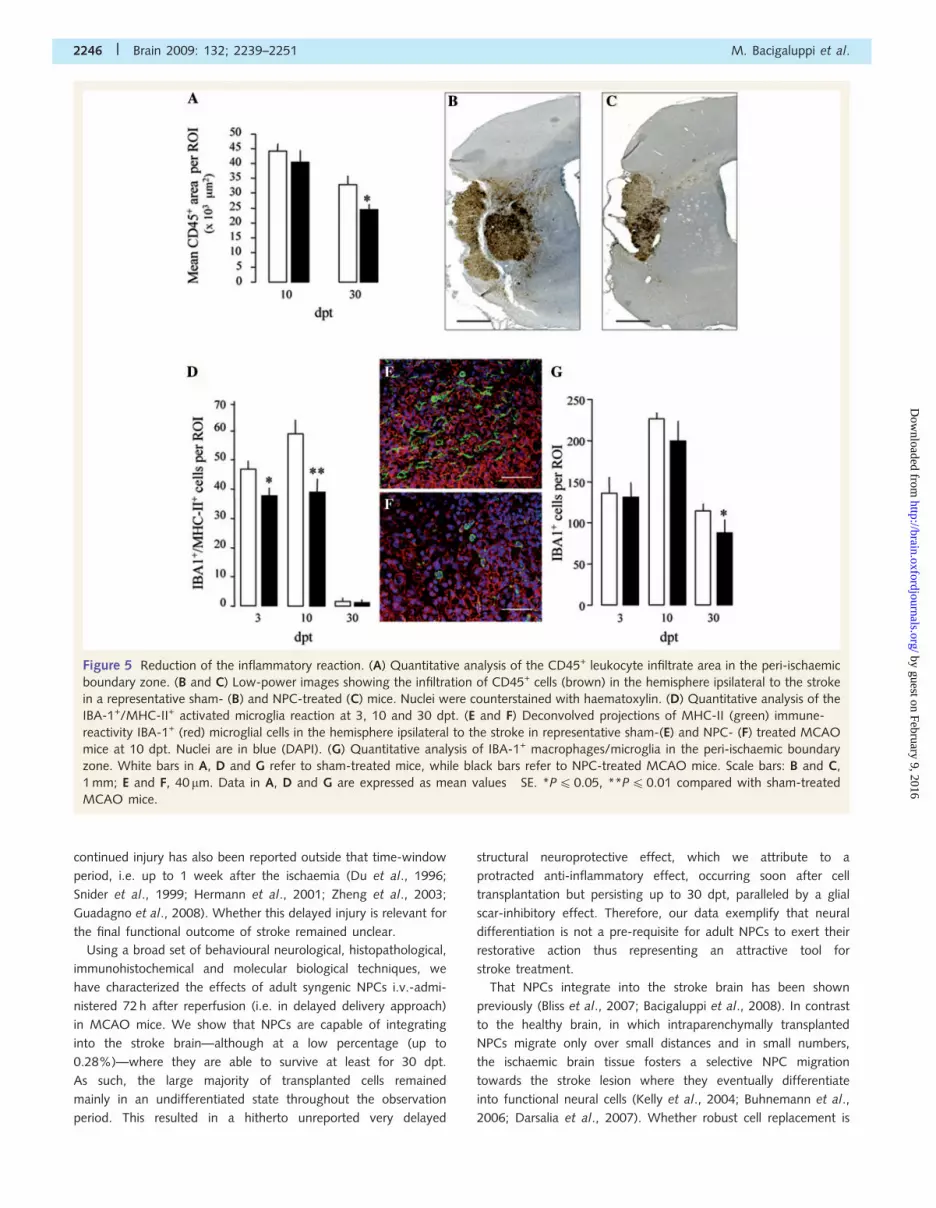
Attenuation of the inflammatoryresponseTo further characterize the effects of NPC transplantation on the
local inflammatory response, we analysed morphometrically the
total brain area stained with the common leucocyte antigen
CD45 as well as the density of microglial cells in the ischaemic
boundary zone both at 3, 10 and 30 dpt.
While NPC-treated mice showed a significant reduction of both
CD45+ and ionized calcium binding adaptor molecule 1 (IBA-1+)
cells at 30 dpt only (both P40.05, when compared with sham-
treated controls) (Fig. 5A–C and G), a significant reduction
of IBA-1+/major histocompatibility complex class II molecule
(MHC-II+) cells was observed at both 3 and 10 dpt (both
P40.05) (Fig. 5D–F).
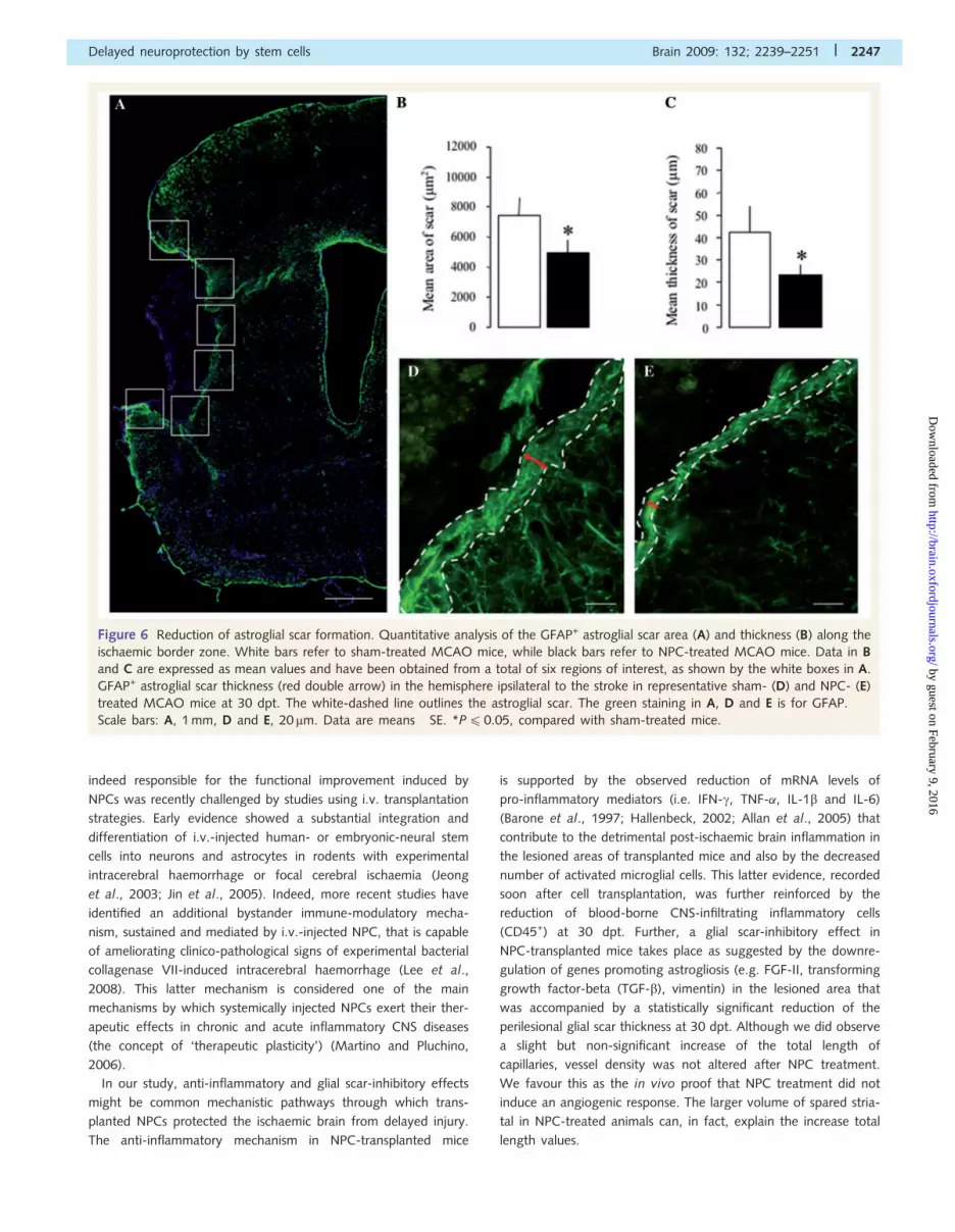
Inhibition of (astro)glial scar formationGliotic scar formation is regarded as a major factor hampering
recovery from stroke (Li et al., 2005; Yiu and He, 2006). Thus,
we next measured the area and thickness of the Glial fibrillary
acidic protein (GFAP+) glial scar wall in the infarct border zone
(Fig. 6A). Again, NPC-treated mice showed a smaller scar area at
30 dpt (P4 0.05). When corrected for the border length profile
irregularities, this also resulted in a significantly thinner scar
(P4 0.01) (Fig. 6B–E), the difference among the two treatment
Figure 3 Protection from the progression of brain ischaemic injury. (A) Brain oedema/atrophy. Brain hemisphere volume ipsilateral to
ischemia is maximal at 0 dpt. At 3 dpt the oedema decreases while a substantial degree of atrophy is observed at 10 dpt and it further
progresses up to 30 dpt. (B) Evolution of the ischaemic lesion volume over time. (C) Spared striatal tissue volume of the ipsilateral
hemisphere at 30 dpt as calculated from the analysis of serial sections for stereology-based counts. (D and E) Representative 3D
reconstructions of the forebrain (grey), striatum (red) and ischaemic lesion (yellow) at 30 dpt of a representative sham-treated (D) and
NPC-treated mice (E) obtained from the sequential sections (n = 18 sections per mouse) analysed for stereology. White bars refer to
sham-treated MCAO mice, while black bars refer to NPC-treated MCAO mice. Data in A, B and C are expressed as means� SE.
*P = 0.05, compared with sham-treated MCAO mice.
2244 | Brain 2009: 132; 2239–2251 M. Bacigaluppi et al.
by guest on February 9, 2016http://brain.oxfordjournals.org/
Dow
nloaded from
groups of mice was particularly evident at the rostrocaudal level of
the bregma.
Promotion of neuronal survivalFurther analysis showed that NPC-treated MCAO mice had higher
densities of surviving neuronal nuclear antigen (NeuN)+ neurons in
the striatum ipsilateral to the stroke starting at 3 and 10 dpt,
getting significant at 30 dpt, compared with sham-treated controls
(Fig. 7A–C and Supplementary Fig. 4). No differences were seen in
other structures, namely in the cortex, hippocampus and thalamus
(data not shown). These findings were also substantiated with
unbiased stereological counts (Fig. 7D–G). Furthermore, by apply-
ing the optical-fractionator methodology at tissues obtained at 30
dpt only, we observed significantly more Darpp-32+ medium spiny
neurons, as well as D2R+ neurons (a subpopulation of medium
spiny neurons) (Kawaguchi, 1997) (Fig. 7F and E, P40.05 and
P4 0.01, respectively) in the striatum of NPC-treated ischaemic
mice, when compared with sham-treated controls. Finally, though
NPC-transplanted mice displayed a slightly higher number of
ChAT+ interneurons (a subpopulation of interneurons particular
resistant to ischaemia) (Andsberg et al., 2001), the difference to
sham-treated animals was not statistically significant (P = 0.33)
(Fig. 7G).
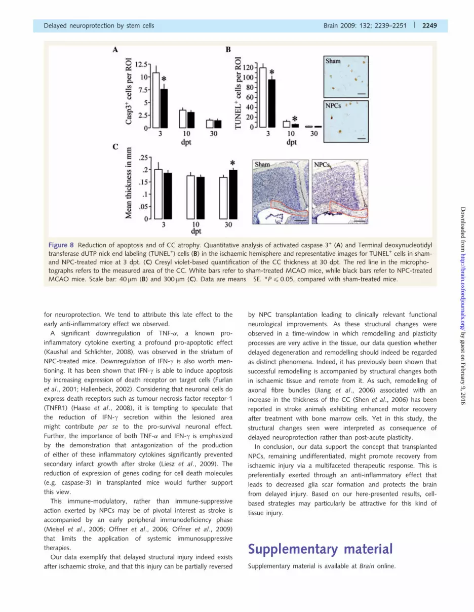
The enhanced neuronal survival was also reflected by a
significant reduction (P40.05) of the total number of apoptotic
cells (in particular neurons) displaying either cleavage of caspase 3
(3 dpt) or DNA fragmentation (3 and 10 dpt) in the ischaemic
hemisphere of NPC-treated MCAO mice, when compared with
sham-treated controls (Fig. 8A and B, Supplementary Fig. 4).
Prevention of corpus callosum atrophyby NPCsThere is substantial evidence that ischaemic lesions may induce
degeneration of long distance, interhemispheric projections
(Napieralski et al., 1996), such as those crossing the corpus callo-
sum (CC). Indeed, the quantification of the medial CC ipsilateral
to the stroke revealed that NPC-treated ischaemic mice had
a significantly thicker CC at 30 dpt, when compared with sham-
treated controls (P40.05) (Fig. 8C). These latter data point
towards a more successful remodelling of interhemispheric
projections in NPC—as compared with sham-treated MCAO mice.
DiscussionIn ischaemic stroke the time-window, in which survival promoting
therapies are efficacious, is extremely limited. As such, recanalizing
drugs (i.e. thrombolytics) (Busch et al., 1998; Kilic et al., 2000;
Hacke et al., 2008) as well as neuroprotective compounds
[e.g. NMDA receptor antagonists (Ma et al., 1998), free radical
scavengers (Yang et al., 2000), growth factors (Cerami, 2001) or
caspase-3 inhibitors (Endres et al., 1998)] protect brain tissue only
when delivered in the first 1–3 and in some conditions up to 6 h
after stroke. That the therapeutic window is so narrow has been
explained by the fact that brain infarct evolves very rapidly, typ-
ically within a few hours up to 3–4 days after stroke, depending
on the duration and severity of ischaemia (Garcia et al., 1993;
Namura et al., 1998; Hata et al., 2000; Li et al., 2000;
Hermann et al., 2001). In case of ischaemias exhibiting a severity
that is close to the threshold at which brain infarcts develop, some
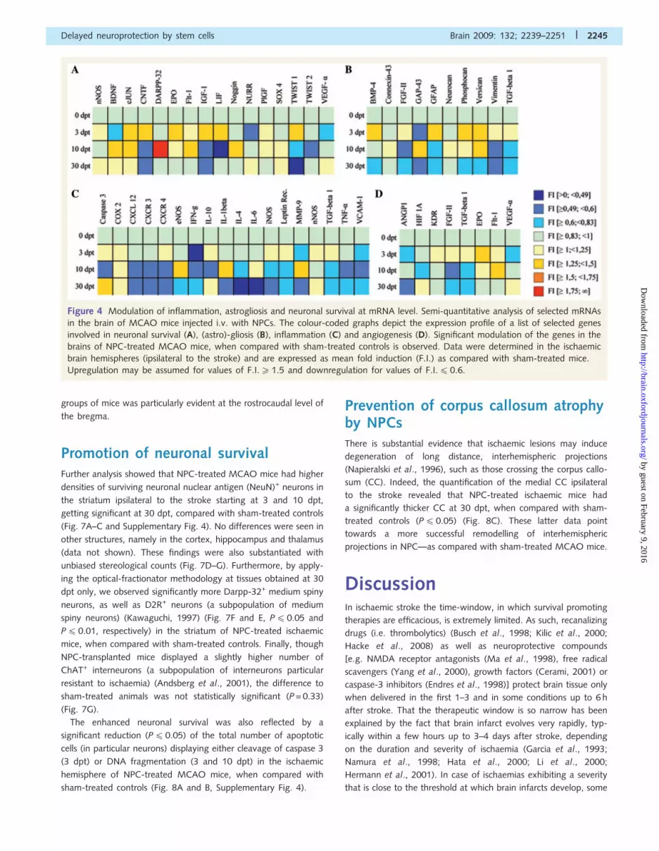
Figure 4 Modulation of inflammation, astrogliosis and neuronal survival at mRNA level. Semi-quantitative analysis of selected mRNAs
in the brain of MCAO mice injected i.v. with NPCs. The colour-coded graphs depict the expression profile of a list of selected genes
involved in neuronal survival (A), (astro)-gliosis (B), inflammation (C) and angiogenesis (D). Significant modulation of the genes in the
brains of NPC-treated MCAO mice, when compared with sham-treated controls is observed. Data were determined in the ischaemic
brain hemispheres (ipsilateral to the stroke) and are expressed as mean fold induction (F.I.) as compared with sham-treated mice.
Upregulation may be assumed for values of F.I.51.5 and downregulation for values of F.I.4 0.6.
Delayed neuroprotection by stem cells Brain 2009: 132; 2239–2251 | 2245
by guest on February 9, 2016http://brain.oxfordjournals.org/
Dow
nloaded from
continued injury has also been reported outside that time-window
period, i.e. up to 1 week after the ischaemia (Du et al., 1996;
Snider et al., 1999; Hermann et al., 2001; Zheng et al., 2003;
Guadagno et al., 2008). Whether this delayed injury is relevant for
the final functional outcome of stroke remained unclear.
Using a broad set of behavioural neurological, histopathological,
immunohistochemical and molecular biological techniques, we
have characterized the effects of adult syngenic NPCs i.v.-admi-
nistered 72 h after reperfusion (i.e. in delayed delivery approach)
in MCAO mice. We show that NPCs are capable of integrating
into the stroke brain—although at a low percentage (up to
0.28%)—where they are able to survive at least for 30 dpt.
As such, the large majority of transplanted cells remained
mainly in an undifferentiated state throughout the observation
period. This resulted in a hitherto unreported very delayed
structural neuroprotective effect, which we attribute to a
protracted anti-inflammatory effect, occurring soon after cell
transplantation but persisting up to 30 dpt, paralleled by a glial
scar-inhibitory effect. Therefore, our data exemplify that neural
differentiation is not a pre-requisite for adult NPCs to exert their
restorative action thus representing an attractive tool for
stroke treatment.
That NPCs integrate into the stroke brain has been shown
previously (Bliss et al., 2007; Bacigaluppi et al., 2008). In contrast
to the healthy brain, in which intraparenchymally transplanted
NPCs migrate only over small distances and in small numbers,
the ischaemic brain tissue fosters a selective NPC migration
towards the stroke lesion where they eventually differentiate
into functional neural cells (Kelly et al., 2004; Buhnemann et al.,
2006; Darsalia et al., 2007). Whether robust cell replacement is
Figure 5 Reduction of the inflammatory reaction. (A) Quantitative analysis of the CD45+ leukocyte infiltrate area in the peri-ischaemic
boundary zone. (B and C) Low-power images showing the infiltration of CD45+ cells (brown) in the hemisphere ipsilateral to the stroke
in a representative sham- (B) and NPC-treated (C) mice. Nuclei were counterstained with haematoxylin. (D) Quantitative analysis of the
IBA-1+/MHC-II+ activated microglia reaction at 3, 10 and 30 dpt. (E and F) Deconvolved projections of MHC-II (green) immune-
reactivity IBA-1+ (red) microglial cells in the hemisphere ipsilateral to the stroke in representative sham-(E) and NPC- (F) treated MCAO
mice at 10 dpt. Nuclei are in blue (DAPI). (G) Quantitative analysis of IBA-1+ macrophages/microglia in the peri-ischaemic boundary
zone. White bars in A, D and G refer to sham-treated mice, while black bars refer to NPC-treated MCAO mice. Scale bars: B and C,
1 mm; E and F, 40 mm. Data in A, D and G are expressed as mean values� SE. *P4 0.05, **P40.01 compared with sham-treated
MCAO mice.
2246 | Brain 2009: 132; 2239–2251 M. Bacigaluppi et al.
by guest on February 9, 2016http://brain.oxfordjournals.org/
Dow
nloaded from
indeed responsible for the functional improvement induced by
NPCs was recently challenged by studies using i.v. transplantation
strategies. Early evidence showed a substantial integration and
differentiation of i.v.-injected human- or embryonic-neural stem
cells into neurons and astrocytes in rodents with experimental
intracerebral haemorrhage or focal cerebral ischaemia (Jeong
et al., 2003; Jin et al., 2005). Indeed, more recent studies have
identified an additional bystander immune-modulatory mecha-
nism, sustained and mediated by i.v.-injected NPC, that is capable
of ameliorating clinico-pathological signs of experimental bacterial
collagenase VII-induced intracerebral haemorrhage (Lee et al.,
2008). This latter mechanism is considered one of the main
mechanisms by which systemically injected NPCs exert their ther-
apeutic effects in chronic and acute inflammatory CNS diseases
(the concept of ‘therapeutic plasticity’) (Martino and Pluchino,
2006).
In our study, anti-inflammatory and glial scar-inhibitory effects
might be common mechanistic pathways through which trans-
planted NPCs protected the ischaemic brain from delayed injury.
The anti-inflammatory mechanism in NPC-transplanted mice
is supported by the observed reduction of mRNA levels of
pro-inflammatory mediators (i.e. IFN-g, TNF-�, IL-1b and IL-6)
(Barone et al., 1997; Hallenbeck, 2002; Allan et al., 2005) that
contribute to the detrimental post-ischaemic brain inflammation in
the lesioned areas of transplanted mice and also by the decreased
number of activated microglial cells. This latter evidence, recorded
soon after cell transplantation, was further reinforced by the
reduction of blood-borne CNS-infiltrating inflammatory cells
(CD45+) at 30 dpt. Further, a glial scar-inhibitory effect in
NPC-transplanted mice takes place as suggested by the downre-
gulation of genes promoting astrogliosis (e.g. FGF-II, transforming
growth factor-beta (TGF-b), vimentin) in the lesioned area that
was accompanied by a statistically significant reduction of the
perilesional glial scar thickness at 30 dpt. Although we did observe
a slight but non-significant increase of the total length of
capillaries, vessel density was not altered after NPC treatment.
We favour this as the in vivo proof that NPC treatment did not
induce an angiogenic response. The larger volume of spared stria-
tal in NPC-treated animals can, in fact, explain the increase total
length values.
Figure 6 Reduction of astroglial scar formation. Quantitative analysis of the GFAP+ astroglial scar area (A) and thickness (B) along the
ischaemic border zone. White bars refer to sham-treated MCAO mice, while black bars refer to NPC-treated MCAO mice. Data in B
and C are expressed as mean values and have been obtained from a total of six regions of interest, as shown by the white boxes in A.
GFAP+ astroglial scar thickness (red double arrow) in the hemisphere ipsilateral to the stroke in representative sham- (D) and NPC- (E)
treated MCAO mice at 30 dpt. The white-dashed line outlines the astroglial scar. The green staining in A, D and E is for GFAP.
Scale bars: A, 1 mm, D and E, 20 mm. Data are means� SE. *P40.05, compared with sham-treated mice.
Delayed neuroprotection by stem cells Brain 2009: 132; 2239–2251 | 2247
by guest on February 9, 2016http://brain.oxfordjournals.org/
Dow
nloaded from

Finally, we cannot exclude that the anti-inflammatory effects
exerted in MCAO mice transplanted with NPCs is in part exerted
in the body periphery, as recently shown after brain haemorrhage
(Lee et al., 2008). However, we found that soon after MCAO
(e.g. 3 dpt) a comparable number of blood-borne inflammatory
cells infiltrated the CNS, thus indicating that the recruitment of
‘effector’ immune cells from the periphery was not impaired by
the cell treatment and, again, suggesting that the protective effect
may have been induced within the CNS.
By providing a detailed temporospatial analysis, our data
show that focal cerebral ischaemia is followed by delayed neuro-
degenerative process that continues over 410 dpt, i.e. 13 days
after the stroke, resulting in continuous neuronal loss in those
parts of the striatum that survived the ischaemic insult, leading
to secondary lesion growth. In our study, post-acute NPC treat-
ment provided a delayed structural neuroprotective effect—partic-
ularly evident on striatal medium spiny neurons—despite the fact
that treatment was initiated far beyond established time-windows
Figure 7 Support of neuronal survival. Neuronal survival in the cortex (A) and striatum (B) ipsilateral to the stroke. Data are expressed
as percentage of NeuN+ cells (over contralateral hemisphere). (C) Representative staining for NeuN (red) of the ischaemic hemisphere
at 30 dpt. White boxes indicate the five cortical (a) and six striatal (b) fields selected for cell countings shown in A and B. Absolute
numbers of NeuN+ (D), D2-Receptor+ (E), Darpp-32+ (F) and ChAT+ (G) neurons in the total unlesioned striatum of the ischaemic
hemisphere quantified using the method for unbiased stereological counting of cells based on the optical fractionator method. 3D
reconstructions in (F) from two representative sham- and NPC-treated mice (left and right, respectively) showing surviving Darpp-32+
cells in the hemisphere ipsilateral to the lesion. White bars refer to sham-treated MCAO mice, while black bars refer to NPC-treated
MCAO mice. Data are means� SE. *P40.05, **P40.01 compared with sham-treated mice.
2248 | Brain 2009: 132; 2239–2251 M. Bacigaluppi et al.
by guest on February 9, 2016http://brain.oxfordjournals.org/
Dow
nloaded from
for neuroprotection. We tend to attribute this late effect to the
early anti-inflammatory effect we observed.
A significant downregulation of TNF-�, a known pro-
inflammatory cytokine exerting a profound pro-apoptotic effect
(Kaushal and Schlichter, 2008), was observed in the striatum of
NPC-treated mice. Downregulation of IFN-g is also worth men-
tioning. It has been shown that IFN-g is able to induce apoptosis
by increasing expression of death receptor on target cells (Furlan
et al., 2001; Hallenbeck, 2002). Considering that neuronal cells do
express death receptors such as tumour necrosis factor receptor-1
(TNFR1) (Haase et al., 2008), it is tempting to speculate that
the reduction of IFN-g secretion within the lesioned area
might contribute per se to the pro-survival neuronal effect.
Further, the importance of both TNF-� and IFN-g is emphasized
by the demonstration that antagonization of the production
of either of these inflammatory cytokines significantly prevented
secondary infarct growth after stroke (Liesz et al., 2009). The
reduction of expression of genes coding for cell death molecules
(e.g. caspase-3) in transplanted mice would further support
this view.
This immune-modulatory, rather than immune-suppressive
action exerted by NPCs may be of pivotal interest as stroke is
accompanied by an early peripheral immunodeficiency phase
(Meisel et al., 2005; Offner et al., 2006; Offner et al., 2009)
that limits the application of systemic immunosuppressive
therapies.
Our data exemplify that delayed structural injury indeed exists
after ischaemic stroke, and that this injury can be partially reversed
by NPC transplantation leading to clinically relevant functional
neurological improvements. As these structural changes were
observed in a time-window in which remodelling and plasticity
processes are very active in the tissue, our data question whether
delayed degeneration and remodelling should indeed be regarded
as distinct phenomena. Indeed, it has previously been shown that
successful remodelling is accompanied by structural changes both
in ischaemic tissue and remote from it. As such, remodelling of
axonal fibre bundles (Jiang et al., 2006) associated with an
increase in the thickness of the CC (Shen et al., 2006) has been
reported in stroke animals exhibiting enhanced motor recovery
after treatment with bone marrow cells. Yet in this study, the
structural changes seen were interpreted as consequence of
delayed neuroprotection rather than post-acute plasticity.
In conclusion, our data support the concept that transplanted
NPCs, remaining undifferentiated, might promote recovery from
ischaemic injury via a multifaceted therapeutic response. This is
preferentially exerted through an anti-inflammatory effect that
leads to decreased glia scar formation and protects the brain
from delayed injury. Based on our here-presented results, cell-
based strategies may particularly be attractive for this kind of
tissue injury.
Supplementary materialSupplementary material is available at Brain online.
Figure 8 Reduction of apoptosis and of CC atrophy. Quantitative analysis of activated caspase 3+ (A) and Terminal deoxynucleotidyl
transferase dUTP nick end labeling (TUNEL+) cells (B) in the ischaemic hemisphere and representative images for TUNEL+ cells in sham-
and NPC-treated mice at 3 dpt. (C) Cresyl violet-based quantification of the CC thickness at 30 dpt. The red line in the micropho-
tographs refers to the measured area of the CC. White bars refer to sham-treated MCAO mice, while black bars refer to NPC-treated
MCAO mice. Scale bar: 40 mm (B) and 300mm (C). Data are means� SE. *P40.05, compared with sham-treated mice.
Delayed neuroprotection by stem cells Brain 2009: 132; 2239–2251 | 2249
by guest on February 9, 2016http://brain.oxfordjournals.org/
Dow
nloaded from
AcknowledgementsWe acknowledge the assistance and help of Miriam Ascagni,
Cesare Covino, Angela Fendel, Filippo Martinelli Boneschi and
Annett Spudich.
FundingNCCR ‘‘Neural plasticity’’ (to D.M.H.); COST Short Term Scientific
Mission (COST-STSM-BM0603-2843); the Swiss National Science
Foundation (3200B0-112056/1, to D.M.H.); the BMW Italy Group
(to G.M.); Banca Agricola Popolare di Ragusa (to S.P.).
ReferencesAllan SM, Tyrrell PJ, Rothwell NJ. Interleukin-1 and neuronal injury. Nat
Rev Immunol 2005; 5: 629–40.Andsberg G, Kokaia Z, Lindvall O. Upregulation of p75 neurotrophin
receptor after stroke in mice does not contribute to differential
vulnerability of striatal neurons. Exp Neurol 2001; 169: 351–63.Bacigaluppi M, Pluchino S, Martino G, Kilic E, Hermann DM. Neural
stem/precursor cells for the treatment of ischaemic stroke. J Neurol
Sci 2008; 265: 73–7.Barone FC, Arvin B, White RF, Miller A, Webb CL, Willette RN, et al.
Tumor necrosis factor-alpha. A mediator of focal ischaemic brain
injury. Stroke 1997; 28: 1233–44.Bliss T, Guzman R, Daadi M, Steinberg GK. Cell transplantation therapy
for stroke. Stroke 2007; 38: 817–26.
Buhnemann C, Scholz A, Bernreuther C, Malik CY, Braun H,
Schachner M, et al. Neuronal differentiation of transplanted embryonic
stem cell-derived precursors in stroke lesions of adult rats. Brain 2006;
129: 3238–48.Busch E, Kruger K, Allegrini PR, Kerskens CM, Gyngell ML, Hoehn-
Berlage M, et al. Reperfusion after thrombolytic therapy of embolic
stroke in the rat: magnetic resonance and biochemical imaging.
J Cereb Blood Flow Metab 1998; 18: 407–18.
Cerami A. Beyond erythropoiesis: novel applications for recombinant
human erythropoietin. Semin Hematol 2001; 38: 33–9.
Darsalia V, Kallur T, Kokaia Z. Survival, migration and neuronal
differentiation of human fetal striatal and cortical neural stem cells
grafted in stroke-damaged rat striatum. Eur J Neurosci 2007; 26:
605–14.
Dirnagl U, Iadecola C, Moskowitz MA. Pathobiology of ischaemic stroke:
an integrated view. Trends Neurosci 1999; 22: 391–7.
Du C, Hu R, Csernansky CA, Hsu CY, Choi DW. Very delayed infarction
after mild focal cerebral ischemia: a role for apoptosis? J Cereb Blood
Flow Metab 1996; 16: 195–201.
Endres M, Namura S, Shimizu-Sasamata M, Waeber C, Zhang L, Gomez-
Isla T, et al. Attenuation of delayed neuronal death after mild focal
ischemia in mice by inhibition of the caspase family. J Cereb Blood
Flow Metab 1998; 18: 238–47.Fawcett JW, Asher RA. The glial scar and central nervous system repair.
Brain Res Bull 1999; 49: 377–91.
Furlan R, Brambilla E, Ruffini F, Poliani PL, Bergami A, Marconi PC, et al.
Intrathecal delivery of IFN-gamma protects C57BL/6 mice from
chronic-progressive experimental autoimmune encephalomyelitis by
increasing apoptosis of central nervous system-infiltrating lymphocytes.
J Immunol 2001; 167: 1821–9.
Galou M, Colucci-Guyon E, Ensergueix D, Ridet JL, Gimenez y
Ribotta M, Privat A, et al. Disrupted glial fibrillary acidic protein
network in astrocytes from vimentin knockout mice. J Cell Biol 1996;
133: 853–63.
Garcia JH, Yoshida Y, Chen H, Li Y, Zhang ZG, Lian J, et al. Progression
from ischaemic injury to infarct following middle cerebral artery
occlusion in the rat. Am J Pathol 1993; 142: 623–35.
Guadagno JV, Jones PS, Aigbirhio FI, Wang D, Fryer TD, Day DJ, et al.
Selective neuronal loss in rescued penumbra relates to initial
hypoperfusion. Brain 2008; 131: 2666–78.
Haase G, Pettmann B, Raoul C, Henderson CE. Signaling by death
receptors in the nervous system. Curr Opin Neurobiol 2008; 18:
284–91.Hacke W, Kaste M, Bluhmki E, Brozman M, Davalos A, Guidetti D, et al.
Thrombolysis with alteplase 3 to 4.5 hours after acute ischaemic
stroke. N Engl J Med 2008; 359: 1317–29.
Hallenbeck JM. The many faces of tumor necrosis factor in stroke. Nat
Med 2002; 8: 1363–8.
Hata R, Maeda K, Hermann D, Mies G, Hossmann KA. Evolution of brain
infarction after transient focal cerebral ischemia in mice. J Cereb Blood
Flow Metab 2000; 20: 937–46.
Hermann DM, Kilic E, Hata R, Hossmann KA, Mies G. Relationship
between metabolic dysfunctions, gene responses and delayed cell
death after mild focal cerebral ischemia in mice. Neuroscience 2001;
104: 947–55.Hossmann KA. Pathophysiology and therapy of experimental stroke. Cell
Mol Neurobiol 2006; 26: 1057–83.Jeong SW, Chu K, Jung KH, Kim SU, Kim M, Roh JK. Human
neural stem cell transplantation promotes functional recovery in rats
with experimental intracerebral hemorrhage. Stroke 2003; 34:
2258–63.
Jiang Q, Zhang ZG, Ding GL, Silver B, Zhang L, Meng H, et al.
MRI detects white matter reorganization after neural progenitor cell
treatment of stroke. Neuroimage 2006; 32: 1080–9.
Jin K, Sun Y, Xie L, Mao XO, Childs J, Peel A, et al. Comparison of
ischemia-directed migration of neural precursor cells after intrastriatal,
intraventricular, or intravenous transplantation in the rat. Neurobiol Dis
2005; 18: 366–74.
Karimi-Abdolrezaee S, Eftekharpour E, Wang J, Morshead CM,
Fehlings MG. Delayed transplantation of adult neural precursor cells
promotes remyelination and functional neurological recovery after
spinal cord injury. J Neurosci 2006; 26: 3377–89.
Kaushal V, Schlichter LC. Mechanisms of microglia-mediated neuro-
toxicity in a new model of the stroke penumbra. J Neurosci 2008;
28: 2221–30.Kawaguchi Y. Neostriatal cell subtypes and their functional roles.
Neurosci Res 1997; 27: 1–8.Kelly S, Bliss TM, Shah AK, Sun GH, Ma M, Foo WC, et al. Transplanted
human fetal neural stem cells survive, migrate, and differentiate in
ischaemic rat cerebral cortex. Proc Natl Acad Sci USA 2004; 101:
11839–44.
Kilic E, Hermann DM, Hossmann KA. Recombinant tissue-plasminogen
activator-induced thrombolysis after cerebral thromboembolism in
mice. Acta Neuropathol 2000; 99: 219–22.
Lee JP, Jeyakumar M, Gonzalez R, Takahashi H, Lee PJ, Baek RC, et al.
Stem cells act through multiple mechanisms to benefit mice with
neurodegenerative metabolic disease. Nat Med 2007; 13: 439–47.Lee ST, Chu K, Jung KH, Kim SJ, Kim DH, Kang KM, et al. Anti-
inflammatory mechanism of intravascular neural stem cell transplanta-
tion in haemorrhagic stroke. Brain 2008; 131: 616–29.
Li F, Silva MD, Sotak CH, Fisher M. Temporal evolution of ischaemic
injury evaluated with diffusion-, perfusion-, and T2-weighted MRI.
Neurology 2000; 54: 689–96.
Li Y, Chen J, Zhang CL, Wang L, Lu D, Katakowski M, et al. Gliosis and
brain remodeling after treatment of stroke in rats with marrow stromal
cells. Glia 2005; 49: 407–17.
Liesz A, Suri-Payer E, Veltkamp C, Doerr H, Sommer C, Rivest S, et al.
Regulatory T cells are key cerebroprotective immunomodulators in
acute experimental stroke. Nat Med 2009; 15: 192–9.Ma J, Endres M, Moskowitz MA. Synergistic effects of caspase inhibitors
and MK-801 in brain injury after transient focal cerebral ischaemia in
mice. Br J Pharmacol 1998; 124: 756–62.
2250 | Brain 2009: 132; 2239–2251 M. Bacigaluppi et al.
by guest on February 9, 2016http://brain.oxfordjournals.org/
Dow
nloaded from

Martino G, Pluchino S. The therapeutic potential of neural stem cells. NatRev Neurosci 2006; 7: 395–406.
Meisel C, Schwab JM, Prass K, Meisel A, Dirnagl U. Central nervous
system injury-induced immune deficiency syndrome. Nat Rev
Neurosci 2005; 6: 775–86.Mouton PR, Gokhale AM, Ward NL, West MJ. Stereological length
estimation using spherical probes. J Microsc 2002; 206: 54–64.
Namura S, Zhu J, Fink K, Endres M, Srinivasan A, Tomaselli KJ, et al.
Activation and cleavage of caspase-3 in apoptosis induced byexperimental cerebral ischemia. J Neurosci 1998; 18: 3659–68.
Napieralski JA, Butler AK, Chesselet MF. Anatomical and functional
evidence for lesion-specific sprouting of corticostriatal input in theadult rat. J Comp Neurol 1996; 373: 484–97.
Nedergaard M, Dirnagl U. Role of glial cells in cerebral ischemia. Glia
2005; 50: 281–6.
Nudo RJ. Recovery after damage to motor cortical areas. Curr OpinNeurobiol 1999; 9: 740–7.
Offner H, Subramanian S, Parker SM, Wang C, Afentoulis ME, Lewis A,
et al. Splenic atrophy in experimental stroke is accompanied by
increased regulatory T cells and circulating macrophages. J Immunol2006; 176: 6523–31.
Offner H, Vandenbark AA, Hurn PD. Effect of experimental stroke on
peripheral immunity: CNS ischemia induces profound immuno-
suppression. Neuroscience 2009; 158: 1098–111.Pluchino S, Quattrini A, Brambilla E, Gritti A, Salani G, Dina G, et al.
Injection of adult neurospheres induces recovery in a chronic model of
multiple sclerosis. Nature 2003; 422: 688–94.Pluchino S, Zanotti L, Rossi B, Brambilla E, Ottoboni L, Salani G, et al.
Neurosphere-derived multipotent precursors promote neuroprotection
by an immunomodulatory mechanism. Nature 2005; 436: 266–71.
Shen LH, Li Y, Chen J, Zhang J, Vanguri P, Borneman J, et al.
Intracarotid transplantation of bone marrow stromal cells increases
axon-myelin remodeling after stroke. Neuroscience 2006; 137: 393–9.
Snider BJ, Gottron FJ, Choi DW. Apoptosis and necrosis in cerebro-
vascular disease. Ann N Y Acad Sci 1999; 893: 243–53.
West MJ, Slomianka L, Gundersen HJ. Unbiased stereological estimation
of the total number of neurons in the subdivisions of the rat
hippocampus using the optical fractionator. Anat Rec 1991; 231:
482–97.
Witte OW. Lesion-induced plasticity as a potential mechanism for
recovery and rehabilitative training. Curr Opin Neurol 1998; 11:
655–62.
Yang Y, Li Q, Shuaib A. Neuroprotection by 2-h postischemia
administration of two free radical scavengers, alpha-phenyl-n-tert-
butyl-nitrone (PBN) and N-tert-butyl-(2-sulfophenyl)-nitrone (S-PBN),
in rats subjected to focal embolic cerebral ischemia. Exp Neurol 2000;
163: 39–45.
Yiu G, He Z. Glial inhibition of CNS axon regeneration. Nat Rev Neurosci
2006; 7: 617–27.
Zheng Z, Zhao H, Steinberg GK, Yenari MA. Cellular and molecular
events underlying ischemia-induced neuronal apoptosis. Drug News
Perspect 2003; 16: 497–503.
Ziv Y, Avidan H, Pluchino S, Martino G, Schwartz M. Synergy between
immune cells and adult neural stem/progenitor cells promotes
functional recovery from spinal cord injury. Proc Natl Acad Sci USA
2006; 103: 13174–9.
Zivin JA. Factors determining the therapeutic window for stroke.
Neurology 1998; 50: 599–603.
Delayed neuroprotection by stem cells Brain 2009: 132; 2239–2251 | 2251
by guest on February 9, 2016http://brain.oxfordjournals.org/
Dow
nloaded from
Copyright © 2022 FDOKUMEN