ATP-driven Rad50 conformations regulate DNA tethering, end resection, and ATM checkpoint signaling
19
Article ATP-driven Rad50 conformations regulate DNA tethering, end resection, and ATM checkpoint signaling Rajashree A Deshpande 1,† , Gareth J Williams 2,† , Oliver Limbo 3 , R Scott Williams 4 , Jeff Kuhnlein 1 , Ji-Hoon Lee 1 , Scott Classen 5 , Grant Guenther 2 , Paul Russell 3 , John A Tainer 2,3,6,** & Tanya T Paull 1,* Abstract The Mre11-Rad50 complex is highly conserved, yet the mecha- nisms by which Rad50 ATP-driven states regulate the sensing, processing and signaling of DNA double-strand breaks are largely unknown. Here we design structure-based mutations in Pyrococ- cus furiosus Rad50 to alter protein core plasticity and residues undergoing ATP-driven movements within the catalytic domains. With this strategy we identify Rad50 separation-of-function mutants that either promote or destabilize the ATP-bound state. Crystal structures, X-ray scattering, biochemical assays, and functional analyses of mutant PfRad50 complexes show that the ATP-induced closed conformation promotes DNA end binding and end tethering, while hydrolysis-induced opening is essential for DNA resection. Reducing the stability of the ATP-bound state impairs DNA repair and Tel1 (ATM) checkpoint signaling in Schizosaccharomyces pombe, double-strand break resection in Saccharomyces cerevisiae, and ATM activation by human Mre11- Rad50-Nbs1 in vitro, supporting the generality of the P. furiosus Rad50 structure-based mutational analyses. These collective results suggest that ATP-dependent Rad50 conformations switch the Mre11-Rad50 complex between DNA tethering, ATM signal- ing, and 5′ strand resection, revealing molecular mechanisms regulating responses to DNA double-strand breaks. Keywords DNA damage signaling; DNA repair; double-strand breaks; protein- DNA interactions Subject Categories DNA Replication, Repair & Recombination; Structural Biology DOI 10.1002/embj.201386100 | Received 26 June 2013 | Revised 25 November 2013 | Accepted 28 November 2013 Introduction Repair of DNA double-strand breaks (DSBs), arising from internal or external sources of damage and generally lethal if unrepaired, is achieved through non-homologous end joining (NHEJ) or homolo- gous recombination (HR) (Krogh & Symington, 2004). The Mre11- Rad50-Nbs1(Xrs2)(MRN/X) complex plays central roles in both of these pathways and is conserved in all domains of life, as Mre11- Rad50 (MR) in prokaryotes and as MRN/X in eukaryotes (Lamarche et al, 2010; Stracker & Petrini, 2011). Mre11 forms a DNA-binding dimer that contains phosphoesterase motifs and is active as an exo- and endonuclease in vitro (Paull & Gellert, 1998; Williams et al, 2008), while Rad50 contains ATP-binding motifs and is similar in domain configuration to the structural maintenance of chromosomes ATPase family of proteins that regulate the activities and topology of chromosomal DNA (Williams et al, 2007). The eukaryote-specific Nbs1(Xrs2) protein contains phosphoprotein-binding FHA and BRCT motifs, regulates MR catalytic activities and flexibly links the complex to the checkpoint kinase ATM (Tel1) (Lee & Paull, 2007; Williams et al, 2009). Besides its DSB-related functions, MRN/X complexes also promote replisome stability in yeast and in mammalian cells (Doksani et al, 2009; Schlacher et al, 2011; Tittel-Elmer et al, 2009). Rad50 ATP-regulated activities are essential for all MRN roles in DNA repair and signaling, as mutations in the Walker A or signature motifs exhibit phenotypes equivalent to a rad50 deletion in vivo (Alani et al, 1990; Bhaskara et al, 2007; Chen et al, 2005; Moncalian et al, 2004). Recent MR structures illuminate the physical interactions between Mre11 and Rad50 in both the nucleotide-free and ATP-bound states, and reveal that Rad50 ATP binding results in extensive conformational changes at the base of the Rad50 coiled-coils (Lammens et al, 2011; Lim et al, 2011; Mockel et al, 1 The Department of Molecular Genetics and Microbiology, The Howard Hughes Medical Institute, Institute for Cellular and Molecular Biology, The University of Texas at Austin, Austin, TX, USA 2 Life Sciences Division, Lawrence Berkeley National Laboratory, Berkeley, CA, USA 3 The Scripps Research Institute, La Jolla, CA, USA 4 Department of Health and Human Services, Laboratory of Structural Biology, National Institute of Environmental Health Sciences, US National Institutes of Health, Research Triangle Park, NC, USA 5 Physical Biosciences Division, Lawrence Berkeley National Laboratory, Berkeley, CA, USA 6 The Skaggs Institute for Chemical Biology, La Jolla, CA, USA *Corresponding author. Tel: 512 232 7802; Fax: 512 471 3730; E-mail: [email protected] **Corresponding author. Tel: 510 495 2404; Fax: 510 486 6880; E-mail: [email protected] † These authors contributed equally to this work. 1 ª 2014 The Authors The EMBO Journal
Transcript of ATP-driven Rad50 conformations regulate DNA tethering, end resection, and ATM checkpoint signaling
Article
ATP-driven Rad50 conformations regulateDNA tethering, end resection, and ATMcheckpoint signalingRajashree A Deshpande1,†, Gareth J Williams2,†, Oliver Limbo3, R Scott Williams4, Jeff Kuhnlein1,Ji-Hoon Lee1, Scott Classen5, Grant Guenther2, Paul Russell3, John A Tainer2,3,6,** & Tanya T Paull1,*
Abstract
The Mre11-Rad50 complex is highly conserved, yet the mecha-nisms by which Rad50 ATP-driven states regulate the sensing,processing and signaling of DNA double-strand breaks are largelyunknown. Here we design structure-based mutations in Pyrococ-cus furiosus Rad50 to alter protein core plasticity and residuesundergoing ATP-driven movements within the catalytic domains.With this strategy we identify Rad50 separation-of-functionmutants that either promote or destabilize the ATP-bound state.Crystal structures, X-ray scattering, biochemical assays, andfunctional analyses of mutant PfRad50 complexes show that theATP-induced closed conformation promotes DNA end binding andend tethering, while hydrolysis-induced opening is essential forDNA resection. Reducing the stability of the ATP-bound stateimpairs DNA repair and Tel1 (ATM) checkpoint signaling inSchizosaccharomyces pombe, double-strand break resection inSaccharomyces cerevisiae, and ATM activation by human Mre11-Rad50-Nbs1 in vitro, supporting the generality of the P. furiosusRad50 structure-based mutational analyses. These collectiveresults suggest that ATP-dependent Rad50 conformations switchthe Mre11-Rad50 complex between DNA tethering, ATM signal-ing, and 5! strand resection, revealing molecular mechanismsregulating responses to DNA double-strand breaks.
Keywords DNA damage signaling; DNA repair; double-strand breaks; protein-
DNA interactions
Subject Categories DNA Replication, Repair & Recombination;
Structural Biology
DOI 10.1002/embj.201386100 | Received 26 June 2013 | Revised 25 November
2013 | Accepted 28 November 2013
Introduction
Repair of DNA double-strand breaks (DSBs), arising from internal or
external sources of damage and generally lethal if unrepaired, is
achieved through non-homologous end joining (NHEJ) or homolo-
gous recombination (HR) (Krogh & Symington, 2004). The Mre11-
Rad50-Nbs1(Xrs2)(MRN/X) complex plays central roles in both of
these pathways and is conserved in all domains of life, as Mre11-
Rad50 (MR) in prokaryotes and as MRN/X in eukaryotes (Lamarche
et al, 2010; Stracker & Petrini, 2011). Mre11 forms a DNA-binding
dimer that contains phosphoesterase motifs and is active as an exo-
and endonuclease in vitro (Paull & Gellert, 1998; Williams et al,
2008), while Rad50 contains ATP-binding motifs and is similar in
domain configuration to the structural maintenance of chromosomes
ATPase family of proteins that regulate the activities and topology of
chromosomal DNA (Williams et al, 2007). The eukaryote-specific
Nbs1(Xrs2) protein contains phosphoprotein-binding FHA and BRCT
motifs, regulates MR catalytic activities and flexibly links the complex
to the checkpoint kinase ATM (Tel1) (Lee & Paull, 2007; Williams
et al, 2009). Besides its DSB-related functions, MRN/X complexes
also promote replisome stability in yeast and in mammalian cells
(Doksani et al, 2009; Schlacher et al, 2011; Tittel-Elmer et al, 2009).
Rad50 ATP-regulated activities are essential for all MRN roles in
DNA repair and signaling, as mutations in the Walker A or signature
motifs exhibit phenotypes equivalent to a rad50 deletion in vivo
(Alani et al, 1990; Bhaskara et al, 2007; Chen et al, 2005; Moncalian
et al, 2004). Recent MR structures illuminate the physical
interactions between Mre11 and Rad50 in both the nucleotide-free
and ATP-bound states, and reveal that Rad50 ATP binding results in
extensive conformational changes at the base of the Rad50
coiled-coils (Lammens et al, 2011; Lim et al, 2011; Mockel et al,
1 The Department of Molecular Genetics and Microbiology, The Howard Hughes Medical Institute, Institute for Cellular and Molecular Biology, The University of Texas atAustin, Austin, TX, USA
2 Life Sciences Division, Lawrence Berkeley National Laboratory, Berkeley, CA, USA3 The Scripps Research Institute, La Jolla, CA, USA4 Department of Health and Human Services, Laboratory of Structural Biology, National Institute of Environmental Health Sciences, US National Institutes of Health,
Research Triangle Park, NC, USA5 Physical Biosciences Division, Lawrence Berkeley National Laboratory, Berkeley, CA, USA6 The Skaggs Institute for Chemical Biology, La Jolla, CA, USA
*Corresponding author. Tel: 512 232 7802; Fax: 512 471 3730; E-mail: [email protected]**Corresponding author. Tel: 510 495 2404; Fax: 510 486 6880; E-mail: [email protected]†These authors contributed equally to this work.
1ª 2014 The Authors The EMBO Journal

2011; Williams et al, 2011). Despite these informative structures
and many years of research, however, the relationships between
MR conformations and biological functions remain enigmatic. Spe-
cifically, we do not know: (i) what the function of the ATP-bound
complex is, (ii) how ATP hydrolysis controls the functions of
MR complexes on DNA, including the nuclease activity of Mre11, or
(iii) why the rate of ATP hydrolysis is extremely slow.
To address these three unknowns and probe the relationships
between MR conformations and functions, here we alter the
stability of the ATP-bound state with engineered mutations and
determine how the biological functions of MRN are affected, both
in vitro and in vivo. Through structure-based mutations, four new
Rad50 crystal structures, small-angle X-ray scattering (SAXS),
biochemical assays, and in vivo DNA repair assays of these mutants,
we find that Rad50 conformational switches regulate the stability of
the ATP-bound dimer, and that this ATP-bound conformation
promotes both DNA end binding and end tethering. In contrast,
release from this ATP-bound state during hydrolysis is required for
the endonuclease activity of Mre11 and the promotion of DNA end
resection. Furthermore, we show the importance of wild-type (WT)
levels of ATP binding and hydrolysis for DSB repair and signaling
through ATM in vivo. These results reveal relationships between
the ATP-driven conformational changes in Mre11 and Rad50 and
the biological functions of the complex and suggest how ATP
hydrolysis by Rad50 controls the balance between DNA end bridg-
ing and DNA resection.
Results
A Rad50 core cavity provides plasticity for ATP-drivenconformational changes
Crystal structures of MR catalytic domains (MRcd) from bacteria
and archaea suggest that the complex transitions from an ATP-free
open conformation (PDB 3QG5) to an ATP-bound, compact confor-
mation (PDB 3THO, 3AV0) with Rad50 altering access to the Mre11
nuclease active site (Lammens et al, 2011; Lim et al, 2011; Mockel
et al, 2011; Williams et al, 2011) (Fig 1A). To determine if the
conformation of MR seen in nucleotide-bound crystal structures resem-
bles those in solution, we used SAXS, which can provide accurate
models of conformations and assemblies in solution (Putnam et al,
2007; Rambo & Tainer, 2013a). Examining SAXS data from P. furiosus
MRcd by Porod-Debye analysis shows the scattering intensity
transitions from decaying with the 3rd power to the 4th power of the
scattering angle when going from ATP-free to ATP-bound states,
suggesting that upon ATP binding MRcd changes from a flexible
assembly with many conformations to a single compact conforma-
tion (Rambo & Tainer, 2011). Furthermore, comparison of our
experimental SAXS scattering of P. furiosus MRcd (Williams et al,
2011) to calculated SAXS profiles of Methanococcus jannaschii and
Thermotoga maritima MR nucleotide bound states (Lim et al, 2011;
Mockel et al, 2011) reveals that a single ATP-bound MR conforma-
tion exists in solution that matches the crystal structures based upon
unbiased v2free analysis (Fig 1B) (Rambo & Tainer, 2013a,b).
To examine the underlying mechanics of ATP-regulated
conformational changes, we did a detailed analysis of Rad50
nucleotide-free and -bound structures. In the nucleotide-free state,
a solvent-accessible channel extends deep into the Rad50 hydro-
phobic protein core (Fig 1C, Supplementary Movie 1). This con-
duit forms at the interface of the N- and C-terminal ABC-ATPase
halves and is substantially remodeled with ATPase subdomain
rotation in response to ATP binding. Concerted movement of R805
and R797 residues, which are implicated in Rad50 subdomain
rotation and critical for DSB repair (Williams et al, 2011), block
the conduit from the protein surface and partially fill the remain-
ing cavity upon ATP binding (Fig 1D and Supplementary Movie
2). To accommodate these changes, conserved hydrophobic signa-
ture helix residues (L802 and L806) lining the channel rearrange
with nucleotide binding. The L802 and L806 residues are displaced
and L802 flips conformations, thereby reconstructing the protein
core. In the ATP-bound state, a core cavity remains at the intersec-
tion between N- and C-terminal ATPase halves, but shrinks in size
(from ~600 !A3 to ~450 !A3). Structural interpolations between
unbound and ATP-bound states (Supplementary Movies 1 and 2)
suggest that R805 sliding through the cavity facilitates precise
domain realignment concomitant with the 35° subdomain rotation
that occurs between the N- and C-terminal ATPase halves. Based
on these observations, we hypothesized that the Rad50 core cavity
imparts structural plasticity to facilitate ATP-regulated conforma-
tional changes.
If the Rad50 core cavity identified here is functionally important,
then a mutation designed to partially fill the cavity should impair
ATP-dependent subdomain rotation and thus ATP binding. We
therefore substituted more bulky hydrophobic Phe or Trp residues
for the L802 and L806 residues that line the Rad50 cavity, and
expressed these mutants in the Rad50cd-linked form, which
connects the Rad50 N- and C-terminal domains with a short flexible
linker (Supplementary Fig S1). We measured dimerization by incu-
bating the proteins at a temperature appropriate for the P. furiosus
thermophile (65°C for 10 min.), followed by gel filtration at room
temperature. We found that the WT protein and the L806F variants
formed ATP-dependent dimers in this assay, but the L802W variant
failed to exhibit dimerization (Fig 2A and Supplementary Fig S1).
We also analyzed nucleotide-induced changes in MRcd (Mre11
bound to the Rad50 catalytic domains, Rad50cd, which have a short
region of coiled-coil in two separate polypeptides; Supplementary
Fig S1). Using SAXS under trapping conditions (short period of high
temperature incubation followed by flash cooling), we found
L802W MRcd behaves the same as WT protein with and without
ATP (Supplementary Fig S2). Yet, when SAXS data was collected
with ATP and the temperature held at 65°C, the L802W mutant
initially forms a closed/single compact structure, but returned to the
open state by the end of the 8 min experiment, whereas the WT
protein retained the compact conformation during this time course
(Fig 2B and C). Thus the L802W mutation appears to destabilize the
ATP-bound state as shown by the SAXS data and structural compari-
son map that provides a quantitative and superposition-independent
evaluation of solution-state conformational differences (Hura et al,
2013). The destabilization appears to be linked to ATP hydrolysis,
as the L802W MRcd compact form was stable over the same time
course with ATPcS (Fig 2D).
To investigate the structural impact of the L802W mutation,
we solved the X-ray crystal structure to 2.3 !A resolution in the
absence of ATP (Table 1). As predicted, the bulky Trp residue
extends into the space neighboring residue 802, and partially
The EMBO Journal ATP regulates MR DNA tethering and end resection Rajashree A Deshpande et al
The EMBO Journal ª 2014 The Authors2

fills the protein core cavity (Fig 2E). To accommodate this muta-
tion, the Rad50 core is partially remodeled and W802 blocks the
space that R805 rotates upon ATP binding in WT Rad50.
Although the overall ATPase fold is maintained, the L802W sub-
stitution distorts the relative positioning of the Rad50 N- and
C-terminal ATPase subdomains. Rad50 ATP binding normally
induces approximately 35° counterclockwise rotation of the
N-terminal Rad50 ATPase subdomain relative to the C-terminal
lobe. Intriguingly, the L802W cavity-filling mutant imparts a
clockwise (~5° and ~9° with 9° shown in Fig 2F) ATPase sub
domain over-rotation relative to WT Rad50, opposite to the
direction induced by ATP binding. Thus, engineered hydrophobic
core variants alter alignment of the mobile ATPase sub
domains.
ATP-dependent Rad50 conformational changes in solution
To determine the effects of the cavity mutations on Rad50 conforma-
tion in solution, we established limited proteolysis assays. In these
experiments, the proteins were first incubated at high temperature
(65°C) for 10 min. prior to the trypsin cleavage reaction at 37°C.Tryptic digest of Rad50cd with ATP or ATPcS yields two unique
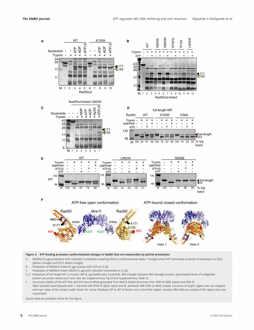
approximately 17-kDa and 19-kDa bands (Fig 3A). We found that
the ATP-dependent trypsin cleavage site maps to K155 on the second
of two signature coupling helices (Williams et al, 2011) connecting
the ATPase N-terminal lobe to the coiled-coil (Supplementary
Table S1, Supplementary Fig S3), as confirmed by partial proteolysis
of a Rad50cd-K155A mutant (Fig 3A). The K155 location suggests
that ATP induces rotation between the N- and C-terminal Rad50
lobes and repositions the coiled-coil domains, increasing K155 flexi-
bility and exposure to solution (Fig 3F). Similar trypsin cleavage
sites are observed using Rad50cd that has been covalently linked by
a short, flexible linker (Rad50cd-linked, Supplementary Fig S1).
Analysis of the L802W Rad50cd-linked protein by partial proteo-
lysis showed that the mutant does not undergo K155 cleavage in the
presence of ATP (Fig 3B). However, further analysis of L802W
showed that the ATP-bound conformation is seen by proteolysis,
but only with non-hydrolysable ATP analog ATPcS (Fig 3C). Thus,
A
C
B
Mre11
Rad50
ATP binding
D
Figure 1. ATP induces conformational changes in Mre11 and Rad50.
A Schematic of Mre11 (blue) and Rad50 (orange, yellow) catalytic domains and conformational changes upon ATP binding.B MR solution structure. Experimental SAXS data of ATP-bound P. furiosus Mre11-Rad50cd-linked in solution closely matches calculated SAXS scattering curves of
MRcd crystal structures in nucleotide-bound states (example shown inset).C Rad50 colored as follows: N-terminal lobe, blue; signature coupling helices, cyan; C-terminal lobe, orange; extended signature motif, magenta; core cavity, green
surface. The cavity is remodeled as Rad50 rotates 35° between nucleotide states. See also Supplementary Movie 1.D Close-up of Rad50 core cavity, basic-switch and core Leu residue changes between nucleotide-free (left) and nucleotide-bound (right) states. Colored as in (C), also
see Supplementary Movie 2.
Rajashree A Deshpande et al ATP regulates MR DNA tethering and end resection The EMBO Journal
ª 2014 The Authors The EMBO Journal 3

B
A
E F
C
Wild-type MRcd + ATP
Scattering angle q (A )-1 Scattering angle q (A )-1 Scattering angle q (A )-1
L802W MRcd + ATP R805E MRcd + ATP
Inte
nsity
Wild-type Rad50cd-linked
Wild-typeRad50cd-linked +ATP
L802W Rad50cd-linked
L802W Rad50cd-linked+ATP
R805E Rad50cd-linked
R805ERad50cd-linked+ATP
D
Figure 2. Rad50 cavity alterations destabilize the ATP-bound state.
A Gel filtration of Rad50cd-linked proteins with or without ATP. Lines across peaks represent molecular weights measured by MALS (right axis); arrows show positionsof dimer peaks. See also Supplementary Fig S1.
B SAXS time-course assays showing scattering curves of MRcd WT, L802W, or R805E variants with 0.5 mM ATP at 65°C, plotted at the indicated time points.C SAXS similarity matrices were made by comparing MRcd variant scattering curves for each time point in the SAXS time-course experiment, measured with ATP at
65°C, compared to the 0 min curve. As indicated by the scale, blue represents very high and red represents very low similarity of curves based on v2 scores.D SAXS analysis of WT and L802W MRcd complexes in the presence of 0.5 mM ATP or ATPcS at 65°C as indicated.E The L802W Rad50cd-linked structure, colored as in Fig 1C, is superimposed on WT Rad50cd-linked, with transparent coloring and cavity (dotted surface). The Trp
substitution overlaps with the WT surface, remodeling the Rad50 core.F ATP-driven conformational states. Superimposition of nucleotide-free Rad50 (blue) with nucleotide-bound Rad50 (gray, left) and nucleotide-free L802W Rad50
(orange, right) using C-terminal domain residues 741–785 for the alignment. N-terminal lobes are shown for clarity.
The EMBO Journal ATP regulates MR DNA tethering and end resection Rajashree A Deshpande et al
The EMBO Journal ª 2014 The Authors4

the L802W mutation does not block ATP binding, but instead desta-
bilizes the ATP-dimerized state, consistent with the SAXS results
showing instability of the ATP-bound compact structure of MRcd
L802W in solution at high temperature (Fig 2).
To examine the effect of Mre11 and the Rad50 coiled-coil, the
solution conformation of Rad50 was also tested using complexes of
MRcd (Supplementary Fig S3) and the full-length MR complex
(Fig 3D). Results with both of these complexes showed that the K63
residue in Rad50 is cleaved by trypsin in the absence of ATP but is
approximately 100% protected by nucleotide (Fig 3F and Supple-
mentary Table S1), indicating solution structures resemble crystal
structures that show that K63 is relatively inaccessible in the ATP-
bound state but exposed in the unbound state. Reduced protection
of the K63 cleavage site in catalytic mutants (S793R and K36A)
confirms the nucleotide specificity of the proteolysis protection
(Fig 3D). In this assay, the L802W mutation reduced the ATP-
dependent protection of the K63 cleavage site in Rad50 (Fig 3E).
Thus proteolysis results with the Rad50 catalytic domains, MRcd,
and full-length MR all show that the L802W mutation destabilizes
the ATP-bound state. The K155 trypsin cleavage site is not observed
in full-length MR as it is protected by Mre11 binding (see Supple-
mentary Fig S3). Notably, Mre11 cleavage by trypsin is also altered
Table 1. X-ray diffraction data collection and refinement statistics
P. furiosus Rad50 P. furiosus Rad50 P. furiosus Rad50 P. furiosus Rad50
L802W (PDB 4NCH) R805E (PDB 4NCI) R805E + ADP/BeF (PDB 4NCJ) R797G (PDB 4NCK)
Data collection
Space group P3221 P212121 P212121 P22121
Cell dimensions
a, b, c (!A) 70.4, 70.4, 282.0 68.0, 69.1, 74.5 83.0, 108.5, 148.7 49.7, 68.1, 203.0
a, b, c (°) 90, 90, 120 90, 90, 90 90, 90, 90 90, 90, 90
Resolution (high res. bin) (!A) 46.1–2.30 (2.44–2.30) 37.2–2.30 (2.44–2.30) 45.4–2.00 (2.09–2.00) 48.2–2.00 (2.12–2.00)
I/!l (high res. bin) 14.2 (2.00) 16.6 (1.96) 16.0 (2.2) 16.4 (1.61)
Rmeas (%) 5.5 (73.2) 4.7 (86.7) 7.9 (70.1) 4.0 (69.5)
CC (1/2) 99.8 (63.9) 99.9 (67.6) 99.8 (78.1) 99.9 (70.6)
Completeness (%) 99.5 (99.2) 99.9 (99.8) 98.0 (100) 99.0 (99.9)
Total observations 203637 116839 705952 298108
Unique observations 37288 16102 90187 47574
Redundancy 5.5 7.3 7.9 6.3
Wilson B-factor 52 56 28 43
Collected at beamlines SSRL11-1 SSRL11-1 ALS 8.3.1 ALS 8.3.1
ALS 12.3.1 ALS 12.3.1 ALS 12.3.1 ALS 12.3.1
Refinement
Resolution (!A) 46.1–2.30 37.2–2.30 45.4–2.00 48.2–2.0
Rwork/Rfree 0.2069/0.2462 0.2015/0.2583 0.2309/0.2618 0.1868/0.2311
Mole per ASU 2 1 8 2
No. of atoms 9446 4949 21267 9577
Macromolecule 4592 2398 10012 4662
Ligand/ions 15 – 128 7
Waters 329 172 846 324
B-factors 57.8 69.9 39 58
Macromolecule 58.3 70.2 39 58.1
Ligand/ions 43.3 – 21.9 106.3
Waters 51.3 65.3 42.7 55
r.m.s. deviations
Bond lengths (A) 0.003 0.003 0.008 0.008
Bond angles (°) 0.68 0.73 1.17 1.07
Ramachandran favored (%) 96 93 97 96
Ramachandran outliers (%) 0.33 0.32 0.87 1.3
Clashscore 3.08 3.58 7.84 5.76
Rajashree A Deshpande et al ATP regulates MR DNA tethering and end resection The EMBO Journal
ª 2014 The Authors The EMBO Journal 5
A
DC
B
E
34
11
17
26
WT K155A
Rad50cd1 2 3 4 5 6 7 8 9 10
155171
- -- + + + +Trypsin
ATP
AD
PAT
PJ S
Nucleotide - -- + + + +
ATP
AD
P
ATPJ S
Rad50cd-linked L802W
- -
34
6
1117
26
43
M 1 2 3 4 5 6 7
171155
- - + + + + + +Trypsin
AM
PAT
PA
DP
Ap5
AAT
PJS
-Nucleotide
130
96
- - + - - - + -- - + -- - - + - - - +- - - +- + + + - + + +- + + +Trypsin
ATPAMPPNP
WT S793R K36A
M 100 24 91 58 100 15 53 49 100 19 29 20
Rad50:
% top band
63
full-length MR
17
26
11
3443
- + +- -
WT
R80
5E
R80
5W
R79
7G
R12
A
ATP
L802
W
Rad50cd-linkedM 1 2 3 4 5 6 7 8 9 10 12 1311
171155
- + + + +Trypsin+ +- -
+ + + ++ +- -
+ + + +
M
WT
- - + -- - - -- + + +Trypsin
ATP -
+- - - +AMPPNP
+-
130
96 63
M 100 25 87 78 58 % top band
L802W
130
96M 100 14 53 78 21
WT
- - + -- - - -- + + +Trypsin
ATP -
+- - - +AMPPNP
+-
- - + -- - - -- + + +Trypsin
ATP -
+- - - +AMPPNP
+-
R805E
130
96M 100 7 91 96 95
full-length
full-length
FATP-free open conformation ATP-bound closed conformation
Rad50 Rad50Mre11
K171 K171RBD
K63 K63K155
K327 K327
linker linker
View 1 View 2
ATPȖS ATPȖS ATPȖS
Figure 3. ATP binding promotes conformational changes in Rad50 that are measurable by partial proteolysis.
A Rad50cd (5 lg) proteolysis with indicated nucleotides revealing distinct conformational states. Triangles show ATP-stimulated products of proteolysis at K155(yellow triangle) and K171 (black triangle).
B Proteolysis of Rad50cd-linked (5 lg) variants with ATP as in (A).C Proteolysis of Rad50cd-linked L802W (5 lg) with indicated nucleotides as in (A).D, E Proteolysis of full-length WT or mutant MR (1 lg Rad50) with nucleotide. Red triangle indicates K63 cleavage product. Quantitated levels of undigested
protein are shown below each lane. Also see Supplementary Fig S3 and Supplementary Table S1.F Structural models of the ATP-free and ATP-bound MRcd generated from Mre11-Rad50 structures from PDB ID 3QKS (open) and PDB ID
3QKU (closed) superimposed with T. maritima MR (PDB ID 3QG5; open) and M. jannaschii MR (PDB ID 3AV0; closed). Locations of tryptic digest sites are mapped,with two views of the closed model shown for clarity. Residues 327 to 357 of Mre11 are in the linker region; residues 394–404 are outside of the region that wascrystallized.
Source data are available online for this figure.
The EMBO Journal ATP regulates MR DNA tethering and end resection Rajashree A Deshpande et al
The EMBO Journal ª 2014 The Authors6
by ATP (Supplementary Fig S3), indicating changes in Mre11 struc-
ture with Rad50 nucleotide binding that experimentally support
structural predictions (Lammens et al, 2011; Lim et al, 2011;
Mockel et al, 2011; Williams et al, 2011).
Rad50 Arg-switch mutations increase ATP-bound dimer stability
Based on Rad50 crystal structures, arginine residues 797 and 805
were hypothesized to be switches for transitions between the open
and closed MR states (Williams et al, 2011). To test this, R797G and
R805E mutations were made in Rad50cd-linked, MRcd, and full-
length MR complexes and analyzed as described above. Similar to
L802W, R797G Rad50cd-linked protein also showed decreased
dimerization with ATP (Supplementary Fig S1); however, the R805E
mutant showed more efficient dimerization than WT Rad50
(Fig 2A). Partial proteolysis of R805E did not show significant differ-
ences from WT Rad50 complexes except that R805E confers higher
protection from trypsin cleavage at K63 in the presence of ATP and
non-hydrolyzable nucleotides in MRcd and in full-length MR, and
also is dominant over the destabilizing effects of the L802W muta-
tion (Fig 3E, Supplementary Fig S3).
Rad50 R805E and L802W mutations alter ATP bindingand hydrolysis
To test the effect of Rad50 cavity and Arg-switch mutations in
more detail, we focused on the R805E and L802W variants with the
greatest impact on ATP-induced dimerization of Rad50. To assess
ATP binding directly, we used full-length MR and measured bind-
ing of a fluorescent ATP derivative, 2!,3!-O-(2,4,6-Trinitrophenyl)ATP (TNP-ATP), as was done for other enzymes (Hiratsuka,
1976). With MR, we found that increasing concentrations of TNP-
ATP quenched the intrinsic fluorescence of tryptophan residues in
Rad50 (likely W782 based on proximity to the bound nucleotide
(Hopfner et al, 2000)) as shown in Fig 4A and B. Both the L802W
and R805E full-length complexes showed equivalent binding to
TNP-ATP, thus the differences observed in ATP-induced conforma-
tional changes are not due to loss of nucleotide binding capacity.
We also confirmed that TNP-ATP exhibited similar effects on the
WT and mutant proteins in the partial proteolysis assay compared
to ATP (Supplementary Fig S4) and that TNP-ATP competes
with ATP in hydrolysis assays with full-length MR (Supplementary
Fig S4).
Analysis of the ATPase activities of the WT and mutant proteins
shows differences in hydrolysis kinetics and also in stimulatory
effects of Mre11 and the coiled-coils on Rad50 catalytic activity
(Fig 4C, Supplementary Fig S4). The full-length MR complex exhibits
lower affinity for ATP but a 100-fold higher kcat for ATP hydrolysis
compared to the Rad50 catalytic domain alone, likely due to
physical attachments between the domains that are mediated by
both Mre11 and the Rad50 coiled-coils. The R805E protein shows
an increase in ATP affinity compared to the WT enzyme (3-fold in
the Rad50cd-linked form and ~100-fold in the full-length protein),
consistent with the higher efficiency of ATP-induced conformational
changes seen with this mutant. The L802W mutant, in contrast, pri-
marily exhibits an increase in kcat relative to the WT protein, which
is most dramatic in the Rad50cd-linked form. In the full-length MR
complex, the kinetic parameters of the L802W complex are more
similar to WT, reflecting the strong effects of the coiled-coil and
Mre11 attachments on ATPase activity.
ATP-dependent changes in Rad50 conformation controlDNA binding
To measure DNA binding by Rad50cd-linked, MRcd, and full-length
MR, we used gel mobility shift assays with double-stranded DNA
substrates (Fig 4D–F). All of the WT proteins formed a large ATP-
dependent complex (I) plus a smaller ATP-independent complex (II)
and exhibited ATP-stimulated DNA binding, extending prior mea-
surements with Rad50 ATPase domains only (Hopfner et al, 2000).
In contrast, L802W Rad50cd-linked and MRcd complexes did not
bind DNA in the presence of ATP but did form a complex with
ATPcS (Fig 4D, E). These Rad50cd-linked results agree with the
K155 partial proteolysis pattern, suggesting that reorganization of
the catalytic domains into the ATP-driven closed conformation
drives formation of the large protein-DNA structures observed in the
gel shift assay. To examine the contribution of the coiled-coils to
DNA binding, full-length proteins were also tested in gel shift
assays, which showed that L802W MR does exhibit ATP-dependent
binding like the WT protein, yet the mobility of complexes is
reduced (Fig. 4F). The Rad50 coiled-coils thus have a stabilizing
effect on Mre11-Rad50 DNA binding. In contrast to L802W, the
R805E Rad50cd-linked and MRcd complexes bound DNA similarly
to WT with ATP, and even more efficiently with ATPcS.Surprisingly, both R805E and R797G full-length MR exhibited ATP-
independent as well as ATP-dependent DNA binding, which is not
observed with the WT enzyme.
To examine the structural basis for the differential abilities of the
Rad50 cavity and Arg-switch variants to form the ATP-bound state,
we solved structures of Rad50 R797G and R805E without nucleotide
and R805E with an ATP mimic, ADP-BeF-Mg2+ (Table 1). These
structures show that the mutations remove R797 and R805, which
act in salt-bridge interactions, without distorting the local fold. In
contrast to the over-rotated state found for L802W, the R805E and
R797G structures show small but significant alteration of overall
subdomain orientation compared to WT: notably both were rotated/
translated (~3°) towards the ATP-bound state.
Interestingly, R797G and L802W share a common feature:
increased flexibility in the D-loop region (residues 827–833) that
immediately follows the Walker B box. In WT Rad50, this loop is
flexible in the absence of ATP and becomes ordered as it packs
with H855 (of the His-loop) in the neighboring Rad50 subunit of
the dimer. In comparison to WT, residues on this loop in the
R797G structure have less-well defined electron density and higher
temperature-factors, and in the L802W structure these residues are
disordered (Supplementary Fig S4). While crystal packing may sta-
bilize the D-loop in the WT structure, increased flexibility of a
loop that becomes ordered in the ATP-bound dimer interface
should destabilize but not block the formation of the ATP-bound
state.
The overall structure of Rad50 R805E bound to ADP-BeF-Mg2+
resembles the WT protein bound to nucleotide. However, a key
difference is seen in the active site with E823, the Walker B
glutamic acid that charges the attacking water and is essential
for ATP hydrolysis, displaced by 2 !A with an additional water
molecule filling the void (Supplementary Fig S4). Similarity
Rajashree A Deshpande et al ATP regulates MR DNA tethering and end resection The EMBO Journal
ª 2014 The Authors The EMBO Journal 7

- -- - --+ + ++- -+ + +-- - --
ATPATPȖS
- WT L802W R805E
Free
Bou
nd
1 2 3 4 5 6 7 8 9 10
MRcd
Free
Bou
nd
WTLWRELW RGRE
- + - - -- + + + +- +ATP
1 2 3 4 5 6 7 8 9 10 1211
MR - -
I
II
F
G
D E MRcd full-length MR
1 2 3 4 5 6 7 8 9 10
- -- - --+ + ++- -+ + +-- - --
ATPATPJS
- Wt L802W R805E
Free
Bound
Rad50cd-linked
I
II
I
II
F /F 0
ȝM TNP-ATP
A[TNP-ATP] ȝM [TNP-ATP] ȝM [TNP-ATP] ȝM
B C
Figure 4. Rad50 catalytic domain mutations affect ATP-dependent DNA binding and hydrolysis.
A ATP binding by full-length MR complexes as measured by quenching of tryptophan fluorescence by TNP-ATP. Shown are emission scans using 290 nm excitation andTNP-ATP as indicated.
B Quantitation of quenching efficiency (F0/F) at 330 nm emission calculated from (A).C ATPase kinetics measurements of Rad50cd-linked and full-length MR proteins (see also Supplementary Fig S4).D Gel-shift assays of Rad50cd-linked (2.5 lM) with a 41-bp DNA duplex substrate.E Gel-shift assays of MRcd complexes (400 nM) with a 41-bp DNA duplex substrate. Protein-DNA complexes are indicated (I and II), with the upper band (I) being
nucleotide-dependent.F Gel-shift assays as in (E) with full-length MR (200 nM).G Crystal structures reveal hydrogen-bonding rearrangements between nucleotide-free WT (left) and R805E Rad50cd-linked (right). Colored as in Fig 1C, with the K54
loop in burgundy and red arrows highlighting major changes. See also Supplementary Fig S4.
Source data are available online for this figure.
The EMBO Journal ATP regulates MR DNA tethering and end resection Rajashree A Deshpande et al
The EMBO Journal ª 2014 The Authors8

between the coordination of active site residues in the R805E
mutant and those in the transition state of MalK (Oldham et al,
2007), a related ABC-ATPase, suggests that this alignment
contributes to more efficient ATP binding and mimics a previously
unseen Rad50 functional intermediate.
Unique features of the R805E structure without ATP suggest how
this mutant binds to ATP more strongly than WT. The R805E muta-
tion disrupts a hydrogen-bonding network of R805, E159, R139,
Y137 and Q142, with a net separation of these residues. This confor-
mational rearrangement alters a neighboring hydrogen bonding net-
work of Y157, R12, D41, D60 and K54, a residue linked to DNA
binding. In the ATP-free state these disruptions displace the loop
containing K54 and D60, toward a solvent-accessible position
(Fig 4G). This shift partially mimics the conformational change that
occurs in WT Rad50 upon ATP binding, where the switch of R12 to
bind nucleotide releases K54 to the protein surface. This Rad50
monomer destabilization may explain the increased stability of the
ATP-induced dimer and, in full-length MR, the displacement of the
K54 loop may be sufficient to form a DNA binding surface between
Rad50 monomers in the complex. Interestingly, deletion of the entire
loop containing K54 (D51–60) eliminates the ATP-stimulated DNA
binding of full-length MR (Supplementary Fig S4), suggesting that
this loop conformation is important for ATP-induced DNA contacts.
DNA end tethering by MR is an ATP-dependent function of Rad50
To test ATP impact on DNA repair functions including DSB end
bridging, we established an end-tethering assay: preincubation of
Mre11-Rad50 with linear DNA containing cohesive ends, followed
by incubation with a DNA ligase. We found that the presence of
full-length MR during preincubation promoted DNA ligation into
multimeric products in an ATP-dependent manner (Fig 5A). Using
full-length Mre11 and Rad50 separately, we found that DNA end
tethering was largely a function of Rad50 (Fig 5B, lanes 8 and 10).
Rad50 alone supports end tethering at a similar concentration to
full-length MR, yet bridging by Mre11 alone required >10-foldhigher concentrations (lane 12). Rad50 DNA end tethering requires
additional domains not present in the catalytic head domains since
Rad50cd-linked, which lacks the Zn-hook and most of the coiled-coil
domains, required 100-fold higher concentrations compared to full-
length Rad50 (Fig 5B,C). The presence of Mre11 also improved
DNA end tethering by the Rad50 catalytic head domains, as seen
with the MRcd complex that supported end ligation at 4-fold higher
concentrations than full-length MR (Fig 5D). DNA end tethering did
not require Mre11 nuclease activity as the nuclease deficient Mre11-
H85L mutant complex supported ligation (Fig 5E). Our results show
that a physical connection between the two Rad50 catalytic
domains, either through the coiled-coils and Zn-hook or through
Mre11, is important for DNA end tethering by MR. The preference
for ATP over AMP-PNP may reflect reduced ability of AMP-PNP to
form the closed complex (see Fig 3E) rather than a requirement for
ATP hydrolysis.
The ATP-bound Rad50 conformation promotes DNA end binding
Analysis of the end tethering ability of the mutants in either MRcd
(Fig 5F) or full-length MR (Fig 5G) showed that L802W Rad50
exhibits similar activity to WT, while R805E and R797G Rad50
variants show higher levels of tethering and strikingly even promote
tethering without ATP (Fig 5G). Thus, efficient end tethering/
bridging is correlated with stable ATP binding and DNA binding.
MR has a general affinity for DNA. To examine specificity for
DNA ends, a circular competitor DNA was included with the idea
that longer occupancy on DNA ends may result in better specificity
for ends over unbroken DNA (Fig 5H). When the substrate was
added before the competitor, WT full-length MR end-bridging was
mostly blocked by the competitor DNA (lane 6), whereas tethering
was completely blocked when the competitor was added before the
substrate (lanes 7, 8). While the L802W mutant behaved similarly
to WT, the R805E mutant tethered ends efficiently with competitor,
even when competitor DNA was added first (Fig 5H, lanes 15, 16).
Thus the R805E mutants are more efficient than WT MR in this
assay and show a preference for DNA with ends, suggesting that the
ATP-bound conformation binds with high specificity to DNA ends.
Importantly, this ligation-mediated tethering assay supports and
extends scanning force microscopy studies of MR complexes
bridging multiple DNA molecules (de Jager et al, 2001); it is
nucleotide-dependent, and is altered by single amino acid changes
in the Rad50 catalytic domain.
The ATP-bound conformation of Rad50 is insufficient for DNAend resection
To test the endonuclease activities of WT and mutant full-length MR,
the complexes were incubated with a 700 bp DNA substrate contain-
ing a fluorescent Cy3 label on one 5! end (Fig 6A). WT MR removes
5! oligonucleotides from this substrate in an ATP-dependent manner
in MgCl2 (Hopkins & Paull, 2008). All mutant complexes showed
activity in this assay. Yet, the L802W mutant was more efficient at 5!strand cleavage compared to WT whereas R805E was reduced in
nuclease activity. Little activity is seen on the 3! strand of linear DNA
with WT MR, but the labeled 3! end was removed efficiently by the
L802W complex (Fig 6B). Interestingly, the coiled-coil and Zn-hook
domains are important for Mre11 endonuclease activity as the WT
MRcd complex fails to exhibit significant nuclease activity in com-
parison to the full-length complex (Fig 6C). The nuclease differences
between MR mutants are not as striking when the complexes are
assayed for 3!–5! exonuclease activity in the presence of manganese
(Fig 6D).
DSB repair by homologous recombination requires extensive
5! end resection to create 3! single-stranded tails, and MR stimu-
lates the helicase-nuclease complex HerA-NurA to carry out long-
range resection of DNA in vitro (Hopkins & Paull, 2008). To test
the effect of the L802W and R805E mutations on this recruit-
ment, we assayed our mutants in a DNA resection assay in
combination with the HerA-NurA complex (Fig 6E). WT
full-length MR increased the degradation of the plasmid substrate
by HerA-NurA 7- to 8-fold, as did the L802W mutant (Fig 6E,
lanes 4–6 and 8–10). In contrast, the R805E mutant failed to
promote HerA-NurA activity (lanes 12–14). Thus stable ATP
binding (i.e. slow hydrolysis) is correlated with stable DNA
binding and stable DNA tethering, but low levels of DNA end
cleavage, as seen with the R805E mutant. In contrast, unstable
ATP binding is associated with unstable DNA binding, low
efficiency DNA tethering and high levels of DNA end cleavage
as seen with the L802W mutant.
Rajashree A Deshpande et al ATP regulates MR DNA tethering and end resection The EMBO Journal
ª 2014 The Authors The EMBO Journal 9

DNA resection requires Rad50 head domain opening followingATP hydrolysis
If DNA end tethering and resection activities oppose each other
and are regulated by the conformational changes that accompany
ATP binding and hydrolysis, it should be possible to regulate
these activities by regulating Rad50 catalytic domain dimerization.
To test this hypothesis, we introduced cysteines into the
Rad50 ATPase domains at positions that would be in close
contact only in the ATP-bound state (D55C and T790C, Fig 7A)
based on the AMP-PNP-bound Rad50 crystal structure (Hopfner
et al, 2000). Using this system, dimerization can be monitored
4kb
8kb
M1 2 3 4 5 6 7 8 9
- -- - - -- +++ ++ - + +- --- +- + + ++ ++
WTWT HL--
Ligase
AMPPNPMR
ATP
Ligase - - + + + + + + + +
ATP
ATP
ATP
- ATP
AM
PP
NP
ATPJS
Ap5
AA
DP
GTP
4kb
8kb
1 2 3 4 5 6 7 8 9 10M
- + - + + + + + + +MR
8kb
4kb
1 2 3 4 5 6 7 8 M
Ligase - + + + + ++
0.1
0.2
0.1
0.5
0.05
MR
+
0.05
MRcd
- -
D E F
1 2 3 4 5 6 7 8 9
Ligase - ++ + + ++
0.1
0.2
RE
+
WT
--
-0.2
0.1
0.2
0.1
0.2
LW
MRcd
WT
WT LW RE
competitor -- (.2) -.5 (.5) --- .5
14 15 161 2 3 4 5 6 7 8 9 10 12 1311M
4kb
8kb
Ligase - + + + ++ ++ +++ ++ ++-
+ - - + ++ ++ +++ ++ ++-MRWT
Ligase + +- +- +- +- + ++
Rad50 - -- --
-----
Mre11
1 2 3 4 5 6 7 8 9 10 1211
4kb
8kb
MR - 0.05 0.10.1-
M
0.050.10.05 0.1
0.11
--- - -
---+
- -1 - --- -- 0.5
13
Ligase - - + + + + + +
5 00 0.05
0.5
1 5 0.05
Rad50cd-linked MR
4kb
8kb
1 2 3 4 5 6 7 8 M
A B C
HG
*
*
**
linea
r lig
atio
n pr
oduc
tsci
rcul
ar
circ
ular
linea
r lig
atio
n pr
oduc
ts
circ
ular
linea
r lig
atio
n pr
oduc
ts
linea
r lig
atio
n pr
oduc
tsci
rcul
ar
linea
r lig
atio
n pr
oduc
tsci
rcul
ar
Ligase
LW RE RGLWRE
+ ++ ++ +++ ++ ++
AMPPNP - +- -+ -- -- - ++MR:
1 2 3 4 5 6 7 8 9 10 1211
4kb
8kb
ATP + -- -- ++- +- --
M13 14 15 16 17 18M
- -- - - ++ ++ - + -- +- + + +
WTWT --
linea
r lig
atio
n pr
oduc
tsci
rcul
ar
(.2).5 (.5) (.2).5 (.5)
Figure 5. DNA end tethering by MR and designed mutants.
A Full-length MR (50 nM) was assayed for DNA end tethering with pCDF-1b plasmid, nucleotide, and E. coli DNA ligase (NAD-dependent) as indicated.B Assays were performed as in (A) with Rad50 (50 nM or as indicated) and Mre11.C Assays were performed as in (A) with Rad50cd-linked.D Assays were performed as in (A) with MRcd.E Assays were performed as in (A) with full-length wt MR and nuclease-deficient MR (Mre11 H85L).F Assays were performed as in (A) with MRcd Rad50 variants as indicated.G Assays were performed as in (A) with full-length MR Rad50 variants.H DNA end tethering assays were performed as in (A) with the indicated addition of supercoiled pCDF-1b competitor DNA.
Data information: Reactions in lanes 4, 6, 10, and 14: substrate added before competitor; reactions in lanes 7, 8, 11, 12, 15, and 16: competitor added before substrate(* indicates positions of competitor DNA). Numbers indicate relative amount of competitor vs. substrate DNA. Ligation products (circular and multimer-linear) areindicated.
Source data are available online for this figure.
The EMBO Journal ATP regulates MR DNA tethering and end resection Rajashree A Deshpande et al
The EMBO Journal ª 2014 The Authors10

5’3’ Cy5
M
46
41
27
1918
14
10
8
1 2 3 4 5
5’ Cy35’
19
14
MWT
LWRELW RGRE
-
41
**
-
A B
ED
4035
30
25
20
16
R797GR805EWT L802W-
*5’
- - - ++ - - ++ - - ++ - - ++ ATP4kb
2kb
WT L802W R805E
HN + - + + +-++-+-
M
MR -- 6.6 13 6.613 13 13136.613
+3.3
+3.3
+3.3
141 2 3 4 5 6 7 8 9 10 12 1311
20
40
60
80
100
0
% s
ubst
rate
rem
aini
ng
--
5’ Cy35’
+MRcdMR
19
14
M
**
1 2 3 4
-- -
RG RE LW WT _
C
M 14 15 16 171 2 3 4 5 6 7 8 9 10 121311
1 2 3 4 5 6 7 8 9 10 12M 11
Figure 6. Rad50 separation-of-function mutations affect MR nuclease activity.
A Endonuclease assay for full-length MR variants (300 and 600 nM) on a 717-bp double-stranded DNA substrate. The shorter oligonucleotides are endonucleolyticproducts, except bands marked with *.
B Nuclease assay as in (A), but with a 41-bp double-stranded DNA substrate.C Nuclease assay as in (A), with WT full-length MR (0.9 lM) and MRcd (1 and 2.5 lM).D Exonuclease assay for full-length MR variants (100 and 200 nM) using a 41-bp double-stranded DNA, labeled on the 50 end of the top strand with 32P (*).E Resection assays with full-length MR (nM concentrations as indicated), HerA and NurA (HN) with a 3.8-kb linearized plasmid.
Source data are available online for this figure.
Rajashree A Deshpande et al ATP regulates MR DNA tethering and end resection The EMBO Journal
ª 2014 The Authors The EMBO Journal 11

A B
ATP
C1
C2
N
C
D55C
T790C
Rad50cd-2C
34
17
26
4356
M - ATP
AM
PA
DP
Ap5
AAT
PJ S
- ATP
AM
PA
DP
Ap5
A
Rad50cd-2C L802WRad50cd-2C14 151 2 3 4 5 6 7 8 9 10 12 1311
C D
8kb
4kb
14 15 16 171 2 3 4 5 6 7 8 9 10 12 1311
WT2C
Ligase + + + + ++ ++ ++++ +- -+-
18
-MR 40 80 80 80 8080 40 40 -408040 - 80
---
--- 8040
H2O
2 - - - - +- +- --++ ++ +---DTT 5 5 0.5 5 0.50.5 0.5 5 0.50.50.50.5 0.5 0.5
+--
0.50.5- 0.50.5
19 20
WT 2C
19
14
1 2 3 4 5 6 7 8 M
WT2C
H2O2 -- - -+DTT 0.5 0.5 5 5 0.5 0.50.50.5
+ -+
E 2C
- + +- + ++ + -MRHN
13 26 26 13 26 26 13 26 26
0.5-
0.5+
5-
DTT
- + +- + ++ + + --- 13 26 26 13 26 26 13 26 26-
0.5-
0.5+
5-
WT
SYBR Green
Southern
1 2 3 4 5 6 7 8 9 10 11 12 13 14 15 16 17 18 19 20
H2O2 ----
Probe for3’ strand
disulfide crosslinked species
N
C
M - - - - - - + +
- - 5 5 0.5 0.5 0.5 0.5- + - + - + - ++ - + - + - + - WT
2CDTT
- - - - + + + + H2O2
ATP
172130
95
72
56
Rad50
Mre11
1 2 3 4 5 6 7 8 9
1X
2X
N1N2
F+ + +- + + +-MR
M
ATPJS
- -
(ȕM
E)
(ȕM
E)
linear ligation products
Figure 7. Cross-linking of Rad50 ATPase domains blocks DNA end resection.
A Crosslinking scheme uses introduced cysteines that are brought close enough to form disulfide bonds when Rad50 dimerizes with ATP.B Crosslinking of Rad50cd-2C variants (3.5 lM) with indicated nucleotides (0.5 mM ATP or 2 mM other nucleotides), separated by SDS–PAGE in the absence of
reducing agent except in lanes marked bME.C Full-length MR or MR-2C proteins (2.35 lM) were cross-linked with H2O2 (2 mM) with DTT (mM) and ATP (0.5 mM) as indicated.D Cross-linked full-length MR and MR-2C (nM) were assayed for DNA end tethering as in Fig 5A with H2O2 (2 mM), DTT (mM) and T4 DNA ligase added as shown.E MR-2C (1.6 lM) and full-length MR (0.9 lM) were tested for endonuclease activity as in Fig 6A following crosslinking with DTT (mM) and H2O2 (2 mM) as indicated.F MR-2C and full-length MR (nM) were cross-linked with DTT (mM) and H2O2 (2 mM) as indicated and assayed for plasmid resection as in Fig 6D. Also see
Supplementary Fig S5.
Source data are available online for this figure.
The EMBO Journal ATP regulates MR DNA tethering and end resection Rajashree A Deshpande et al
The EMBO Journal ª 2014 The Authors12

by disulfide-crosslinking of Rad50 ATPase domains on SDS-PAGEgels in the absence of reducing agents. When the cysteine muta-
tions were made in Rad50cd (Rad50cd-2C), efficient crosslinking
was dependent on ATP (or ATPcS) (Fig 7B), and dimers accu-
mulated during the reaction since the ATPase domains cannot
release following ATP hydrolysis. In these assays, the L802W
mutant also formed the crosslinked species similar to WT Rad50
with ATP or ATPcS. The dinucleotide Ap5A showed a weak
effect in stimulating crosslinking of the Rad50cd complexes
(Fig 7B) but did not mimic ATP in the partial proteolysis or liga-
tion assays (Figs 3C and 5A), thus the adenylate kinase activity
of Rad50 (Bhaskara et al, 2007) does not appear to play a major
role in promoting the conformational changes in Rad50 charac-
terized here.
In full-length MR (MR-2C), disulfide crosslinks were formed in
an ATP-stimulated manner and were promoted by H2O2 oxidation,
although in this case there was also a basal level of ATP-independent
crosslink formation (Fig 7C, lanes 7, 9), consistent with SAXS data
showing flexible MRcd that likely samples a closed state before
becoming fully closed upon ATP binding (Williams et al, 2011).
WT MR did not form crosslinks despite having naturally occurring
cysteines in the Zn-hook region (lanes 6, 8), and Mre11 remained
unaffected under the crosslinking conditions as it has no cysteines.
Next we assessed DNA end tethering by adding the DNA substrate
after MR-2C was crosslinked in the presence of ATP (Fig 7D).
Crosslinked MR-2C (lanes 6, 7) supported end ligation as well as
non-crosslinked MR-2C (lanes 4, 5), and even promoted end ligation
in the presence of closed circular competitor DNA (Supplementary
Fig S5), similar to the R805E mutant, providing more evidence that
the ATP-bound conformation binds with high specificity to DNA
ends.
In contrast to the tethering assay, the MR-2C crosslinked
complexes completely failed to resect DNA under oxidizing condi-
tions (Fig 7E, lane 3). Notably, MR-2C was fully active in this assay
under reducing conditions (lane 4): thus crosslinking of the head
domains is responsible for activity loss in this resection assay. The
2C complexes were also tested in the plasmid resection assay with
HerA-NurA, where it was clear that MR stimulation of HerA-NurA
was abolished under oxidizing conditions but similar to WT under
reducing conditions (Fig 7F, lanes 6, 7). WT MR also exhibited lim-
ited resection of the DNA substrate by itself, which is visible in the
Southern blot (compare lanes 1 and 17), whereas MR-2C did not
exhibit this activity under oxidizing conditions (lane 8), consistent
with the absence of endonuclease activity with crosslinked MR-2C
(Fig 7E). Importantly, ATP is still hydrolyzed by MR-2C when it is
crosslinked (Supplementary Fig S5), thus opening from the
ATP-bound state is required for MR endonuclease activity and
promotion of long-range resection by HerA-NurA, consistent with
SAXS data showing that the Mre11 active site is blocked by Rad50
in solution (Fig 1B) as well as crystallographic evidence (Lim et al,
2011; Mockel et al, 2011).
Rad50 ATP-driven movements regulate DSB repair in vivo
To comprehensively test the separation-of-function mutations, we
examined the effects of the Rad50 mutations in S. cerevisiae,
S. pombe, and in vitro signaling assays to develop an integrated
functional model for ATP-induced conformational changes. First, we
made I1214W and R1217E mutations in the RAD50 gene of S. cerevisiae,
(equivalent to the L802W and R805E mutations in P. furiosus
Rad50). The Rad50 WT and mutant alleles were expressed under
the control of the inducible CUP1 promoter to normalize the expres-
sion levels (Supplementary Fig S6). These strains were exposed to
camptothecin (CPT), which induces DNA strand breaks during repli-
cation by creating topoisomerase I covalent complexes. The strain
expressing the I1214W mutant grew as well on CPT as the strain
expressing WT protein, but the strain expressing the R1217E mutant
grew very poorly on CPT, indicating an inability to repair
endogenous DNA damage by homologous recombination (Fig 8A).
Consistent with this phenotype, a defect in resection was also seen
with the R1217E mutant in an assay for single-strand annealing
which requires extensive resection (25 kb) for repair (Fig 8B).
Lastly, we observed that resection of an HO endonuclease-induced
break was rapid in rad50 strains expressing the WT or I1214W
alleles but that the strain expressing the R1217E allele was similar
to the uncomplemented strain, again indicating a resection defect
associated with R1217E (Supplementary Fig S6D).
We also examined non-homologous end joining in budding
yeast, using two different assays. First, we used a chromosomal
assay in which both precise and imprecise joining products can be
quantitated (Palmbos et al, 2005; Supplementary Fig S6). Unlike
the assay for CPT sensitivity, here both the R1217E and I1214W
mutants showed a robust activity in promoting NHEJ, similar
to the WT complemented strain (Fig 8C). Second, we used
overexpression of the EcoRI restriction endonuclease to generate
DSBs across the genome (Lewis et al, 1999). In this case the
R1217E mutant improved the survival of the complemented rad50
deletion strain significantly more than the WT or I1214W proteins,
indicating a gain-of-function in this case where a massive DSB
load has to be repaired for cell survival. Thus the R1217E mutant
shows a separation-of-function such that the resection activities
are specifically impaired but the end joining activities are intact
and even hyperactive, consistent with the R805E Rad50 mutant
results in vitro. The I1214W mutant shows nearly WT levels of
function in all assays, similar to the Rad50 L802W mutant
in vitro and consistent with the dominant role of homologous
recombination in budding yeast repair of DNA double-strand
breaks.
We also examined the equivalent rad50 alleles in S. pombe to
determine if the relationships between the ATP-driven switches are
evolutionarily conserved in this organism. The I1192W, K1187E,
and R1195E mutations were generated in the rad50 gene of S. pombe
(equivalent to the L802, R797 and R805 residues mutated in
P. furiosus Rad50, respectively). The chromosomal rad50+ locus
was replaced with WT or mutant alleles encoding Rad50 with an
N-terminal TAP-tag, and immunoblotting indicated that the muta-
tions did not affect Rad50 abundance (Supplementary Fig S6).
These mutants were exposed to ionizing radiation (IR) or CPT
(Fig 8E). Similar to the S. cerevisiae results, the K1187E and
R1195E mutants grew very poorly in the presence of DNA damag-
ing agents (Williams et al, 2011) while the I1192W mutant dis-
played a phenotype intermediate between WT and the rad50
strains.
To test the impact of Rad50 conformation on MRN signaling, we
measured phosphorylation of histone H2A following IR to assess
whether the rad50 mutations impair the MRN-dependent function of
Rajashree A Deshpande et al ATP regulates MR DNA tethering and end resection The EMBO Journal
ª 2014 The Authors The EMBO Journal 13

Tel1 (ATM). As both Tel1 and Rad3 (ATR) create phospho-H2A
(cH2A; equivalent to cH2AX in metazoans) in S. pombe (Nakamura
et al, 2004), these assays were performed in rad3+ and rad3D back-
grounds. We found that the large IR-induced increase in cH2A
observed in WT was partially impaired in tel1D and rad3D single
mutants and ablated in the double mutant (Fig 8F, G). The weak
cH2A signal in the rad50D background was eliminated in the
rad50D rad3D strain, supporting prior data establishing that MRN is
% J
oini
ng E
ffici
ency
vecto
rWT
I1214
W
R1217
Eve
ctor
WT
I1214
W
R1217
E0.001
0.01
0.1
1
10 preciseimprecise
CA
B
E
F
0.1 !M CPT 1.0 !M CPT
WTrad50!
TAP-rad50TAP-rad50-I1192WTAP-rad50-K1187ETAP-rad50-R1195E
Untreated 90 Gy IR 270 Gy IR
+
vector
WTI1214WR1217E
untreated 3.6 !M CPT 7.2 !M CPT
0
2
4
WT rad3! tel1! rad50!rad3!tel1!
rad3!rad50!
!H2A
H2A
- + - + - + - + - + - + IR
!H2A
/H2A
- + - + - + - + - + - + - +
0
2
4
!H2A
/H2A
rad3+ rad3! rad3+ rad3!rad3+ rad3!rad50+ rad50-I1192W rad50-K1187E rad50-R1195Erad3+
!H2A
H2A
IR
G
IH
+_ ++ATM
DNAWT MRN
MR(L1211W)N
anti-phospho-p53(ser15)
_ __ _
_
D
fold
vec
tor
vecto
rWT
I1214
W
R1217
E0
1
2
3
4
5 NHEJ efficiency
NHEJ: 2 DSBs
NHEJ: genome-wide DSBs
25kbKK KK
leu2::cshis4::leu28kb 6kb
HO cutKK KK
8kb 2.5kbresection and ligation
K K
3.5kbSSA product
vector
WT
I1214W
R1217E
Galactose
Glucose
vector
WT
I1214W
R1217E
The EMBO Journal ATP regulates MR DNA tethering and end resection Rajashree A Deshpande et al
The EMBO Journal ª 2014 The Authors14

crucial for Tel1 signaling at DSBs (Lee & Paull, 2005; Nakada et al,
2003; You et al, 2005). Similar to rad50D, the rad50-I1192W,
-K1187E, and -R1195E mutations all reduced cH2A in the rad3+
background and nearly eliminated the cH2A signal in the rad3Dbackground. Thus, mutations that impact ATP binding and turnover
in Rad50 severely disrupt MRN functions in DNA repair and Tel1
(ATM) checkpoint signaling in vivo.
To further test the Rad50 mutations for effects on ATM activa-
tion, human MRN was expressed with R1214E and L1211W muta-
tions (pfMR R805E and L802W equivalent, respectively), but we
could only purify the L1211W variant. This complex exhibited
severely reduced activity in stimulating ATM phosphorylation of
p53 in vitro (Fig 8H), consistently indicating that the ATP-induced
conformational changes in Rad50 and Mre11 are critical for ATM
signaling of DNA damage.
Discussion
MR conformations control distinct pathway choices
In comprehensively examining how diverse and sometimes oppos-
ing MR activities are regulated by ATP and by Rad50 ABC-ATPase
conformations, we found that MR complexes that are enhanced for
the ATP-bound conformation show activities critical for DSB repair
by end-joining pathways: reduced ATPase activity, reduced nucle-
ase activities, stronger ATP binding, and more efficient binding
and tethering of dsDNA ends. Conversely, MR complexes defective
for stable ATP binding show activities critical for HR: increased
ATPase activity, hyperactive endonuclease activity, and efficient
end resection. These structure-based mutants achieve separation-
of-function by deliberately altering Rad50 dynamics by shifting the
relative stability of the nucleotide-induced conformational states with
possible implications for other ABC-ATPases (Hopfner & Tainer,
2003).
These data suggest a model for ATP-induced conformational
changes in MR and how they relate to MR biological functions
(Fig 8I). In the ‘open’ conformation (Lammens et al, 2011), MR
binds to DNA but is not-end-specific. When ATP binds to the
catalytic domains, the complex transitions to the ‘closed’ state
(Lim et al, 2011; Mockel et al, 2011; Williams et al, 2011); in this
configuration MR binds DNA with specificity for ends, tethers DNA
ends, promotes end ligation, and activates ATM (Lee et al, 2013),
but cannot cleave DNA. We propose that release from the closed
state that occurs with ATP hydrolysis generates an intermediate
conformation that is competent for DNA end resection and collabo-
ration with other enzymes that perform long-range resection.
Finally, release of nucleotide returns the enzyme to the original
open state.
Role of the Rad50 coiled-coil domain in catalytic domain dimers
Studies in budding yeast have shown that the coiled-coils are impor-
tant for all biological functions of MRX, including HR, NHEJ and
telomere maintenance (Hohl et al, 2011; Hopfner et al, 2002).
Our results show that one distinct role of the coiled-coils is to
mechanically connect the two catalytic domains of Rad50 and
Mre11, leading to enhanced DNA end tethering. Addition of Mre11
to the Rad50 catalytic domains also recovers much of the DNA end
tethering activity in the absence of the coiled-coils, so in this respect
Mre11 dimerization and Rad50 association through the coils are
reinforcing functions. Notably, the crosslinking experiments show
that the isolated Rad50 catalytic domains do not interact in the
absence of ATP, whereas in the full-length complex there is cross-
linking in the absence of nucleotide that can be further stimulated
by the addition of ATP. Thus, in the full-length complex the catalytic
domains can sample the ATP-bound state without ATP, due to the
coiled-coil linkage. Torsional strain applied through the coiled-coils
may aid Mre11 dimer positioning into its active configuration, as
proposed to explain how hook domain removal may alter the juxta-
position of ends within the globular domains (Hohl et al, 2011).
Thus, full-length MR ATPase activity is higher than the Rad50 cata-
lytic domains (a ~12-fold increase in kcat), probably because their
local concentration is increased by the dual connection imposed by
Mre11 and the coiled-coil domains, a conclusion also supported by
recent work showing that the coils promote ATM activation and
Nbs1 binding to MR (Lee et al, 2013).
Regulation of Mre11 nuclease activity by Rad50
Previous results indicate that ATP binding by Rad50 alters Mre11
nuclease activity (Connelly & Leach, 1996; Herdendorf et al, 2011;
Majka et al, 2012; Paull & Gellert, 1999; Trujillo & Sung, 2001). Here
the mutant complexes and the site-specific crosslinking experiments
show definitively that the closed state is incompatible with nuclease
activity in magnesium conditions, in which the MR complex exhibits
Figure 8. Rad50 separation-of-function mutations affect MRN DNA repair functions.
A S. cerevisiae survival assays with rad50 S. cerevisiae strains expressing vector only or WT, I1214W, or R1217E Rad50 proteins.B Diagram of S. cerevisiae SSA assay (left)(Vaze et al, 2002). Survival of rad50 S. cerevisiae strains expressing vector only or WT, I1214W, or R1217E Rad50
proteins before (glucose) or after (galactose) DSB induction at leu2::cs.C Chromosomal NHEJ assay results with S. cerevisiae Rad50 variant strains, characterizing precise versus imprecise NHEJ-mediated joining events. See
Supplementary Fig S6 for assay details. The average of 3 independent experiments is shown, with error bars indicating standard deviation.D NHEJ assay with EcoRI induction, causing genome-wide DSBs. WT, I1214W, or R1217E alleles of Rad50 were expressed in a rad50 strain and survival was
measured after galactose induction of EcoRI for 8 hrs. The average of 5 independent experiments is shown, with the survival of each strain shown relative to therad50 strain that was normalized to 1; error bars indicate standard deviation.
E S. pombe survival assays of Rad50 variant strains.F, G Western blots for cH2A and H2A levels in cell lysates after 90 Gy IR treatment of indicated S. pombe strains.H Stimulation of ATM phosphorylation of p53 by the human MRN complex in vitro. Blots were probed with antibody against phospho-Ser15 of p53.I Model of ATP-induced conformational changes in MR and the functions associated with each state. See text for details.
Source data are available online for this figure.
◂
Rajashree A Deshpande et al ATP regulates MR DNA tethering and end resection The EMBO Journal
ª 2014 The Authors The EMBO Journal 15

5! endonuclease activity as well as promoting the helicase and
nuclease activities of the HerA/NurA complex. In contrast, release
from the closed state (following ATP hydrolysis) promotes both
Mre11 nuclease activity as well as HerA/NurA activity on linear
DNA ends. These results agree generally with experiments
performed with the E. coli SbcC/D complex, the T4 gp46/gp47
complex, and with the human MR complex (Herdendorf et al, 2011;
Paull & Gellert, 1999; Trujillo & Sung, 2001), which showed that
ATP is necessary for resection of dsDNA by MR. Lacking structural
information about intermediates formed during hydrolysis, we
postulate a transient state in which the Mre11 active site gains
access to DNA that is inaccessible in the closed state (Fig 8I).
End tethering and the requirement for ATP
End-tethering assays revealed a surprising specificity for ATP over
the non-hydrolyzable analogs ATPcS and AMP-PNP, despite the fact
that both analogs induced conformational changes in Rad50 that
resembled those induced by ATP (particularly ATPcS). This is not
likely due to a requirement for ATP hydrolysis, since the R805E
mutant promotes end tethering even better than the WT enzyme yet
hydrolyzes ATP more slowly. It is possible that with the coiled-coils
the conformation of MR when bound to ATP might be structurally
distinct from that of the analog-bound complexes and that this
difference could be important for the correct alignment of ends or
access of other enzymes to the ends, even though it is not important
for DNA binding by MR.
Surprisingly, L802W MR complex stimulates some ligation
despite exhibiting a less stable ‘closed’ state. Yet, the L802W mutant
in full-length form is closer to the WT enzyme characteristics than it
is in Rad50cd form, and the short time that the L802W mutant
spends in the closed state may be sufficient for ligation. Alterna-
tively the opening of DNA helices by MR complexes (Cannon et al,
2013) may actually promote intermolecular association of DNA
strands in an end tethering complex that survives even after the pro-
tein has returned to the open state. If so, a more rapid transition
from closed to open state as seen with the L802W mutant would still
be consistent with efficient end ligation.
Rad50 cavity and switch residues act as a communication hub
Structurally, the Rad50 cavity and arginine switches provide a
communication hub between the two ATP binding sites of a single
Rad50 subunit formed by the Walker A/B and signature motifs. They
coordinate the conformational changes, and form an intricate
communication network linking the signature motif to the catalytic
step of the distal ATP binding site (through Walker B motif connec-
tions), and to the catalytic step of the other subunit through the
D-loop and its connections to H855. This communication via the
cavity is conceptually similar to the ‘sectors’ recently shown as
important components of allosteric regulation to connect surface
sites to active sites in dihydrofolate reductase (Reynolds et al, 2011).
We found that cavity mutations impair functions for archaeal,
fission, budding yeast and human MR complexes; thus cavity
regulated conformational plasticity may have key implications for
evolution of distinct biological properties and regulation modes for
Rad50 and other members of the ABC-ATPase superfamily, which
includes mismatch repair ATPase MutS and ABC transporters
(Hopfner & Tainer, 2003). Given segmentation of the ATP binding
cassette into N- and C-terminal halves (Rad50) or assembled modular
domains interacting in transwith effector domains (e.g. Cystic Fibrosis
Transmembrane Conductance Regulator; CFTR, MalK transporters),
cavities of varying size and composition may have evolved to define
distinct emergent biological properties of the ATP binding cassette
superfamily.
The well-characterized CFTR channel also undergoes a rotation
in the ATPase domains upon ATP binding (Kirk & Wang, 2011).
Interestingly, mapping of CFTR small-molecule potentiators that
bind to its ABC-ATPase domain (Moran & Zegarra-Moran, 2005),
such as genistein, identified three probable binding sites with the
most likely (site 2) neighboring the arginine-switch and cavity
regions found in Rad50. Coupling this information with our Rad50
results, showing cavity disruption alters the ABC-ATPase open
versus closed states, suggests that such cavities are druggable;
highlighting a key future challenge to design new small-molecules
to effect biological outcomes controlled by Rad50 and ABC-ATPases
that will have potential for both basic research and therapeutics.
Our results with the separation-of-function mutants in vitro plus
the S. cerevisiae and S. pombe in vivo analyses suggest a molecular
mechanism whereby MR complexes can concomitantly control what
appears to be two opposing functions: DNA end bridging and resec-
tion. While our separation-of-function mutations act as permanent
‘switches’ to make malfunctioning MRN complexes, our results
raise the possibility that during the DNA damage response, regula-
tion of interacting partners and/or post-translational modification of
the MRN complex may temporarily modulate its ATP binding and/
or hydrolysis rates to control pathway outcomes. Indeed, several
phosphorylation sites have been identified in Mre11 and Rad50
(Di Virgilio et al, 2009; Gatei et al, 2011), although effects on bio-
chemical and biological outcomes have yet to be fully elucidated. It
will be intriguing to investigate how such mechanisms may work to
regulate MRN states and control replication fork stability, DSB
repair and signaling in mammalian cells.
Materials and Methods
Plasmids and strains
See Supplementary materials for details.
Protein purification
Full-length and MRcd P. furiosus proteins were purified as described
(Hopkins & Paull, 2008), as were the Rad50cd-linked and Rad50cd
proteins (Williams et al, 2011). See Supplementary materials for
details of gel filtration assays, protein crystallization, and SAXS
determination. All recombinant proteins used in this work in vitro
were derived from P. furiosus, with the exception of the human
MRN complex used in Fig 8.
DNA binding assays
Gel mobility shift assays were performed as previously described
(Lee et al, 2003), with modifications as described in Supplementary
materials.
The EMBO Journal ATP regulates MR DNA tethering and end resection Rajashree A Deshpande et al
The EMBO Journal ª 2014 The Authors16

5! resection assays
Assays shown in Figs 6A–C and 7E were performed as described
(Hopkins & Paull, 2008) with 5 mM MgCl2 and 1 mM ATP except
with a fluorescent substrate (717 bp) made by PCR using primers
TP2108/1621 with Cy3 on the 5! end on the top strand. Assays in
Fig 6B used TP2151 annealed to TP2109. For the assay in Fig 6D,
the substrate consisted of TP74 annealed to TP124 (Paull & Gellert,
2000), with TP74 labeled with 32P at the 5! end. Reactions included
10 nM DNA substrate, 25 mM MOPS pH 7.0, 20 mM Tris pH 8.0,
1 mM MnCl2, 8% glycerol, 80 mM NaCl, 2 mM DTT and 0.5 mM
ATP at 65°C for 1 h. Reactions were stopped with 0.2% SDS and
10 mM EDTA, precipitated with ethanol, dried, dissolved in formam-
ide, heated for 10 min at 100°C and separated on denaturing poly-
acrylamide gels (16 to 20%). After electrophoresis at 40 W for
2.5 h, gels with the TP2108/1621 and TP2151/2109 substrates were
scanned in the Cy3 or Cy5 channel (Typhoon, GE). Gels with TP74/
TP124 were dried and exposed to phosphorimager screens for
analysis.
In reactions with HerA-NurA, 4 ng of PstI-digested pCDF1-b
plasmid (0.16 nM) was incubated with MR and HerA-NurA at 65°Cfor 45 min with 25 mM MOPS pH 7.0, 20 mM Tris pH 8.0, 5 mM
MgCl2, 8% glycerol, 80 mM NaCl, 2 mM DTT and 0.5 mM ATP in a
10 ll reaction volume. The reactions were stopped by 0.2% SDS
and 10 mM EDTA, separated by native agarose gel electrophoresis,
stained with SYBR green (Invitrogen) and analyzed by phosphor-
imager (Typhoon, GE). The same gel was used for non-denaturing
Southern analysis as described (Hopkins & Paull, 2008). A 32P-radio-
labeled RNA probe complementary to the 3! strand was used to
detect 5!-resected products, and the southern blot was analyzed by
phosphorimager (GE).
DNA end tethering assay
The pCDF1-b plasmid was linearized with PstI to generate 4 nt 3!overhangs. 10 ll ligation reactions contained 10 ng linearized DNA
(0.4 nM) in the same buffer as for DNA binding except with 50 mM
NaCl, with incubation at 65°C for 10 min with Mre11, Rad50, or MR
complexes as indicated in figure legends. The temperature was then
lowered to 16°C, and the reactions supplemented with E. coli DNA
ligase buffer and 1,000 units E. coli DNA ligase (NAD-dependent,
Fig 5) or with 10,000 units T4 DNA ligase (Fig 7). Reactions were
further incubated at 16°C for 30 min and stopped by 0.2% SDS and
10 mM EDTA and incubated with 1 lg Proteinase K at 37°C for 45–60 min before separation by native gel electrophoresis, SYBR green
staining, and phosphorimager analysis (Typhoon, GE). In Fig 5H,
the substrate or competitor DNA (pCDF1-b, 0.2! or 0.5! the amount
of substrate DNA) was added to the protein for 10 min before add-
ing competitor or substrate, respectively and further incubation at
65°C for 10 min.
Crosslinking of ‘2C Rad50 complexes
Full-length MR-2C protein complexes were incubated at 65°C for
5 min with 25 mM MOPS pH 7.0, 20 mM Tris pH 8.0, 5 mM MgCl2,
8% glycerol, 80 mM NaCl, 2 mM H2O2, 1 mM ATP, and 0.5 or
5 mM DTT as indicated. The crosslinked protein was diluted upon
addition to the DNA end resection assay reactions (- DTT), reducing
H2O2 concentration to 0.4 mM and DTT to 0.2 mM or 2 mM.
Rad50cd-2C proteins were crosslinked by incubation of 3.5 lMprotein in 25 mM MOPS pH 7.0, 5 mM MgCl2, 120 mM NaCl and
0.5 mM ATP or 2 mM AMP/ADP/ATPcS/Ap5A without H2O2.
Partial proteolysis
Rad50 or MR proteins were incubated with nucleotides at 65°C for
10 min. 0.2 lg trypsin (Promega) was added and incubated at
37°C for 2 h (or 16 h in Fig 3C). The digestion products were
resolved by SDS–PAGE stained with Coomassie. For N-terminal
sequencing, proteins were transferred to PVDF membrane and
stained by Coomassie. The molecular mass was determined by ESI
mass spectrometry analysis of the tryptic digests: Rad50cd (5 lg)was incubated in a buffer containing 25 mM MOPS pH 7.0,
20 mM Tris pH 8.0, 5 mM MgCl2, 8% glycerol, 80 mM NaCl,
2 mM DTT, and nucleotides as indicated (0.5 mM), in a volume
of 10 ll at 65°C for 10 min. The reactions were cooled to 25°C for
5 min, 0.2 lg trypsin (Promega) was added and incubated at 37°Cfor 2 h (or 16 h in Fig 3C).
N-terminal sequencing
Automated Edman-based N-terminal sequencing was performed
using a Procise 492cLc Protein Sequencer (Applied Biosystems)
using standard cycles for PVDF membrane analysis.
ATP hydrolysis and kinetic studies
ATP hydrolysis was measured at 65°C as described (Bhaskara et al,
2007). Values shown in Fig 4C were calculated using non-linear regres-
sion analysis with Graphad Prism. See also Supplementary Fig S4.
ATP binding with TNP-ATP
10 ll reactions contained 200 nM MR with 0–200 lM TNP-
ATP [2!,3!-O-(2,4,6-Trinitrophenyl)adenosine-5!-triphosphate] and
25 mM MOPS pH 7.0, 20 mM Tris pH 8.0, 5 mM MgCl2, 8% glyc-
erol, 80 mM NaCl, 2 mM DTT in a 1,536-well plate (Corning,
#3728). Fluorescence emission spectra were recorded with a Safire2
plate reader (Tecan) using 290 nm excitation for Trp. The quenching
of Trp emission at 330 nm is presented as a Stern-Volmer plot F0/F
versus [TNP-ATP], where F0 and F are the fluorescence intensities
in the absence and presence of quencher.
S. cerevisiae NHEJ assays
S. cerevisiae strains are listed in Supplementary Table S3. The rad50
strain TP3619 was used for the chromsomal NHEJ assay, essentially
as previously described (Palmbos et al, 2005). The rad50 strain
KRY78 was used for the EcoRI NHEJ assay, as previously described
(Lewis et al, 1999). Modifications and further details are in Supple-
mentary materials.
S. cerevisiae SSA assay
The assay was performed essentially as described (Vaze et al, 2002)
with modifications in Supplementary materials.
Rajashree A Deshpande et al ATP regulates MR DNA tethering and end resection The EMBO Journal
ª 2014 The Authors The EMBO Journal 17

S. cerevisiae resection assay
The assay was performed as described (Moncalian et al, 2004) with
modifications in Supplementary materials.
S. pombe DNA damage sensitivity assays and H2Aphosphorylation assay
S. pombe strains were manipulated by standard techniques (Forsburg
& Rhind, 2006) with strains listed in Supplementary Table S2 and
details of growth and signaling assays in Supplementarymaterials.
In vitro kinase assays
MRNcomplexes and dimericATMwere purified as described (Bhaska-
ra et al, 2007; Lee&Paull, 2006). ATMkinase assays contained 50 mM
HEPES, pH 7.5, 50 mM KCl, 5 mM MgCl2, 10% glycerol, 1 mM ATP,
and 1 mM DTT and were performed for 90 min at 30°C in 40 ll asdescribed(Lee&Paull, 2006). SeeSupplementarymaterials for details.
Accession codes
Coordinates and structure factors of the four Rad50 mutant struc-
tures detailed in this manuscript have been deposited in the protein
databank and have the following PDB codes: 4NCH (L802W), 4NCI
(R805E), 4NCJ (R805E + ADP/BeF), and 4NCK (R797G).
Supplementary information for this article is available online:http://emboj.embopress.org
AcknowledgmentsWe thank members of the Paull, Tainer, and Russell laboratories for comments,
Christophe Redon for the cH2A antibody, Antony Carr for the TAP-Rad50
S. pombe strain, and Tom Wilson, Jim Haber, Sang Eun Lee, and Tom Petes for
S. cerevisiae strains. This study was supported by NIH grants CA094008 to
T.P., CA092584 to J.T. and T.P., CA117638 to J.T. and P.R., CA077325 to P.R., and
intramural research program of the US National Institutes of Health (NIH),
National Institute of Environmental Health Sciences (NIEHS) (1Z01ES102765-
01) to R.S.W. The SIBYLS beamline (BL12.3.1) at the Advanced Light Source is
supported by United States Department of Energy program Integrated Diffrac-
tion Analysis Technologies and NIH MINOS R01GM105404. Mass spectroscopy
was performed by the Protein and Metabolite Analysis Facility at UT Austin.
Author contributionsRAD, GJW, and RSW generated reagents, designed and performed experiments,
interpreted results, and helped to write the manuscript. OL and J-HL generated
reagents and performed and analyzed experiments. JK, SC, and GG generated
reagents and contributed to the experiments. PR, JAT, and TTP helped to
design and interpret experiments and edited the manuscript.
Conflict of interestThe authors declare that they have no conflict of interest.
References
Alani E, Padmore R, Kleckner N (1990) Analysis of WT and rad50 mutants of
yeast suggests an intimate relationship between meiotic chromosome
synapsis and recombination. Cell 61: 419 – 436
Bhaskara V, Dupre A, Lengsfeld B, Hopkins BB, Chan A, Lee JH, Zhang X, Gautier
J, Zakian VA, Paull TT (2007) Rad50 adenylate kinase activity regulates DNA
tethering by Mre11/Rad50 complexes. Mol Cell 25: 647 – 661
Cannon B, Kuhnlein J, Yang SH, Cheng A, Schindler D, Stark JM,
Russell R, Paull TT (2013) Visualization of local DNA unwinding by
MRN using single-molecule FRET. Proc Natl Acad Sci U S A 110:
18868 – 18873
Chen L, Trujillo KM, Van Komen S, Roh DH, Krejci L, Lewis LK, Resnick MA,
Sung P, Tomkinson AE (2005) Effect of amino acid substitutions in the
rad50 ATP binding domain on DNA double strand break repair in yeast.
J Biol Chem 280: 2620 – 2627
Connelly JC, Leach DR (1996) The sbcC and sbcD genes of Escherichia coli
encode a nuclease involved in palindrome inviability and genetic
recombination. Genes Cells 1: 285 – 291
Di Virgilio M, Ying CY, Gautier J (2009) PIKK-dependent phosphorylation of
Mre11 induces MRN complex inactivation by disassembly from
chromatin. DNA Repair 8: 1311 – 1320
Doksani Y, Bermejo R, Fiorani S, Haber JE, Foiani M (2009) Replicon
dynamics, dormant origin firing, and terminal fork integrity after
double-strand break formation. Cell 137: 247 – 258
Forsburg SL, Rhind N (2006) Basic methods for fission yeast. Yeast 23: 173 – 183
Gatei M, Jakob B, Chen P, Kijas AW, Becherel OJ, Gueven N, Birrell G, Lee JH,
Paull TT, Lerenthal Y, Fazry S, Taucher-Scholz G, Kalb R, Schindler D,
Waltes R, Dork T, Lavin MF (2011) ATM protein-dependent
phosphorylation of Rad50 protein regulates DNA repair and cell cycle
control. J Biol Chem 286: 31542 – 31556
Herdendorf TJ, Albrecht DW, Benkovic SJ, Nelson SW (2011) Biochemical
characterization of bacteriophage T4 Mre11-Rad50 complex. J Biol Chem
286: 2382 – 2392
Hiratsuka T (1976) Fluorescence properties of 2! (or 3!)-O-(2,4,6-trinitrophenyl)
adenosine 5!-triphosphate and its use in the study of binding to heavy
meromyosin ATPase. Biochim Biophys Acta 453: 293 – 297
Hohl M, Kwon Y, Galvan SM, Xue X, Tous C, Aguilera A, Sung P, Petrini JH
(2011) The Rad50 coiled-coil domain is indispensable for Mre11 complex
functions. Nat Struct Mol Biol 18: 1124 – 1131
Hopfner KP, Craig L, Moncalian G, Zinkel RA, Usui T, Owen BA, Karcher A,
Henderson B, Bodmer JL, McMurray CT, Carney JP, Petrini JH, Tainer JA
(2002) The Rad50 zinc-hook is a structure joining Mre11 complexes in
DNA recombination and repair. Nature 418: 562 – 566
Hopfner KP, Karcher A, Shin DS, Craig L, Arthur LM, Carney JP, Tainer JA
(2000) Structural biology of Rad50 ATPase: ATP-driven conformational
control in DNA double-strand break repair and the ABC-ATPase
superfamily. Cell 101: 789 – 800
Hopfner KP, Tainer JA (2003) Rad50/SMC proteins and ABC transporters:
unifying concepts from high-resolution structures. Curr Opin Struct Biol
13: 249 – 255
Hopkins B, Paull TT (2008) The P. furiosus Mre11/Rad50 complex promotes 5!
strand resection at a DNA double-strand break. Cell 135: 250 – 260
Hura GL, Budworth H, Dyer KN, Rambo RP, Hammel M, McMurray CT, Tainer
JA (2013) Comprehensive macromolecular conformations mapped by
quantitative SAXS analyses. Nat Methods 10: 453 – 454
de Jager M, van Noort J, van Gent DC, Dekker C, Kanaar R, Wyman C (2001)
Human Rad50/Mre11 is a flexible complex that can tether DNA ends. Mol
Cell 8: 1129 – 1135
Kirk KL, Wang W (2011) A unified view of cystic fibrosis transmembrane
conductance regulator (CFTR) gating: combining the allosterism of a
ligand-gated channel with the enzymatic activity of an ATP-binding
cassette (ABC) transporter. J Biol Chem 286: 12813 – 12819
The EMBO Journal ATP regulates MR DNA tethering and end resection Rajashree A Deshpande et al
The EMBO Journal ª 2014 The Authors18

Krogh BO, Symington LS (2004) Recombination proteins in yeast. Annu Rev
Genet 38: 233 – 271
Lamarche BJ, Orazio NI, Weitzman MD (2010) The MRN complex in double-
strand break repair and telomere maintenance. FEBS Lett 584: 3682 – 3695
Lammens K, Bemeleit DJ, Mockel C, Clausing E, Schele A, Hartung S, Schiller
CB, Lucas M, Angermuller C, Soding J, Strasser K, Hopfner KP (2011) The
Mre11:Rad50 structure shows an ATP-dependent molecular clamp in DNA
double-strand break repair. Cell 145: 54 – 66
Lee J-H, Ghirlando R, Bhaskara V, Hoffmeyer MR, Gu J, Paull TT (2003)
Regulation of Mre11/Rad50 by Nbs1: effects on nucleotide-dependent
DNA binding and association with ATLD mutant complexes. J Biol Chem
278: 45171 – 45181
Lee JH, Mand MR, Deshpande RA, Kinoshita E, Yang SH, Wyman C, Paull TT
(2013) Ataxia Telangiectasia-Mutated (ATM) kinase activity is regulated by
ATP-driven conformational changes in the Mre11/Rad50/Nbs1 (MRN)
Complex. J Biol Chem 288: 12840 – 12851
Lee JH, Paull TT (2005) ATM activation by DNA double-strand breaks through
the Mre11-Rad50-Nbs1 complex. Science 308: 551 – 554
Lee JH, Paull TT (2006) Purification and biochemical characterization of
ataxia-telangiectasia mutated and Mre11/Rad50/Nbs1. Methods Enzymol
408: 529 – 539
Lee JH, Paull TT (2007) Activation and regulation of ATM kinase activity in
response to DNA double-strand breaks. Oncogene 26: 7741 – 7748
Lewis LK, Westmoreland JW, Resnick MA (1999) Repair of
endonuclease-induced double-strand breaks in Saccharomyces cerevisiae:
essential role for genes associated with nonhomologous end- joining.
Genetics 152: 1513 – 1529
Lim HS, Kim JS, Park YB, Gwon GH, Cho Y (2011) Crystal structure of the
Mre11-Rad50-ATPgammaS complex: understanding the interplay between
Mre11 and Rad50. Genes Dev 25: 1091 – 1104
Majka J, Alford B, Ausio J, Finn RM, McMurray CT (2012) ATP hydrolysis by
RAD50 protein switches MRE11 enzyme from endonuclease to
exonuclease. J Biol Chem 287: 2328 – 2341
Mockel C, Lammens K, Schele A, Hopfner KP (2011) ATP driven structural
changes of the bacterial Mre11:Rad50 catalytic head complex. Nucleic
Acids Res 40: 914 – 927
Moncalian G, Lengsfeld B, Bhaskara V, Hopfner KP, Karcher A, Alden E, Tainer
JA, Paull TT (2004) The rad50 signature motif: essential to ATP binding
and biological function. J Mol Biol 335: 937 – 951
Moran O, Zegarra-Moran O (2005) A quantitative description of the activation
and inhibition of CFTR by potentiators: Genistein. FEBS Lett 579: 3979 – 3983
Nakada D, Matsumoto K, Sugimoto K (2003) ATM-related Tel1 associates
with double-strand breaks through an Xrs2-dependent mechanism. Genes
Dev 17: 1957 – 1962
Nakamura TM, Du LL, Redon C, Russell P (2004) Histone H2A
phosphorylation controls Crb2 recruitment at DNA breaks, maintains
checkpoint arrest, and influences DNA repair in fission yeast. Mol Cell Biol
24: 6215 – 6230
Oldham ML, Khare D, Quiocho FA, Davidson AL, Chen J (2007) Crystal
structure of a catalytic intermediate of the maltose transporter. Nature
450: 515 – 521
Palmbos PL, Daley JM, Wilson TE (2005) Mutations of the Yku80 C terminus
and Xrs2 FHA domain specifically block yeast nonhomologous end joining.
Mol Cell Biol 25: 10782 – 10790
Paull TT, Gellert M (1998) The 3! to 5! exonuclease activity of Mre 11
facilitates repair of DNA double-strand breaks. Mol Cell 1: 969 – 979
Paull TT, Gellert M (1999) Nbs1 potentiates ATP-driven DNA unwinding and
endonuclease cleavage by the Mre11/Rad50 complex. Genes Dev 13:
1276 – 1288
Paull TT, Gellert M (2000) A mechanistic basis for Mre11-directed DNA
joining at microhomologies. Proc Natl Acad Sci U S A 97: 6409 – 6414
Putnam CD, Hammel M, Hura GL, Tainer JA (2007) X-ray solution scattering
(SAXS) combined with crystallography and computation: defining accurate
macromolecular structures, conformations and assemblies in solution.
Q Rev Biophys 40: 191 – 285
Rambo RP, Tainer JA (2011) Characterizing flexible and intrinsically
unstructured biological macromolecules by SAS using the Porod-Debye
law. Biopolymers 95: 559 – 571
Rambo RP, Tainer JA (2013a) Accurate assessment of mass, models and
resolution by small-angle scattering. Nature 496: 477 – 481
Rambo RP, Tainer JA (2013b) Super-resolution in solution X-ray scattering and
its applications to structural systems biology. Annu Rev Biophys 42: 415 – 441
Reynolds KA, McLaughlin RN, Ranganathan R (2011) Hot spots for allosteric
regulation on protein surfaces. Cell 147: 1564 – 1575
Schlacher K, Christ N, Siaud N, Egashira A, Wu H, Jasin M (2011)
Double-strand break repair-independent role for BRCA2 in blocking stalled
replication fork degradation by MRE11. Cell 145: 529 – 542
Stracker TH, Petrini JH (2011) The MRE11 complex: starting from the ends.
Nat Rev Mol Cell Biol 12: 90 – 103
Tittel-Elmer M, Alabert C, Pasero P, Cobb JA (2009) The MRX complex
stabilizes the replisome independently of the S phase checkpoint during
replication stress. EMBO J 28: 1142 – 1156
Trujillo KM, Sung P (2001) DNA structure-specific nuclease activities in the
Saccharomyces cerevisiae Rad50/Mre11 complex. J Biol Chem 13: 13
Vaze MB, Pellicioli A, Lee SE, Ira G, Liberi G, Arbel-Eden A, Foiani M,
Haber JE (2002) Recovery from checkpoint-mediated arrest after repair
of a double-strand break requires Srs2 helicase. Mol Cell 10: 373 – 385
Williams GJ, Williams RS, Williams JS, Moncalian G, Arvai AS, Limbo O,
Guenther G, SilDas S, Hammel M, Russell P, Tainer JA (2011) ABC ATPase
signature helices in Rad50 link nucleotide state to Mre11 interface for
DNA repair. Nat Struct Mol Biol 18: 423 – 431
Williams RS, Dodson GE, Limbo O, Yamada Y, Williams JS, Guenther G,
Classen S, Glover JN, Iwasaki H, Russell P, Tainer JA (2009) Nbs1 flexibly
tethers Ctp1 and Mre11-Rad50 to coordinate DNA double-strand break
processing and repair. Cell 139: 87 – 99
Williams RS, Moncalian G, Williams JS, Yamada Y, Limbo O, Shin DS,
Groocock LM, Cahill D, Hitomi C, Guenther G, Moiani D, Carney JP,
Russell P, Tainer JA (2008) Mre11 dimers coordinate DNA end bridging
and nuclease processing in double-strand-break repair. Cell 135:
97 – 109
Williams RS, Williams JS, Tainer JA (2007) Mre11-Rad50-Nbs1 is a keystone
complex connecting DNA repair machinery, double-strand break signaling,
and the chromatin template. Biochem Cell Biol 85: 509 – 520
You Z, Chahwan C, Bailis J, Hunter T, Russell P (2005) ATM activation and its
recruitment to damaged DNA require binding to the C terminus of Nbs1.
Mol Cell Biol 25: 5363 – 5379
Rajashree A Deshpande et al ATP regulates MR DNA tethering and end resection The EMBO Journal
ª 2014 The Authors The EMBO Journal 19

![Deprotection, Tethering, and Activation of a One-Legged Metalloporphyrin on a Chemically Active Metal Surface: NEXAFS, Synchrotron XPS, and STM Study of [SAc]P−Mn(III)Cl on Ag(100)](https://static.fdokumen.com/doc/165x107/63458dc903a48733920b69f7/deprotection-tethering-and-activation-of-a-one-legged-metalloporphyrin-on-a-chemically.jpg)