Selective antibody intervention of Toll-like receptor 4 activation through Fc γ receptor tethering
10
Selective Antibody Intervention of Toll-like Receptor 4 Activation through Fc Receptor Tethering Received for publication, December 2, 2013, and in revised form, March 25, 2014 Published, JBC Papers in Press, April 15, 2014, DOI 10.1074/jbc.M113.537936 Limin Shang ‡1 , Bruno Daubeuf ‡ , Martha Triantafilou § , Robin Olden § , Fabien Dépis ‡2 , Anne-Catherine Raby ‡3 , Suzanne Herren ‡ , Anaelle Dos Santos ‡ , Pauline Malinge ‡ , Irene Dunn-Siegrist ¶ , Sanae Benmkaddem , Antoine Geinoz**, Giovanni Magistrelli ‡ , François Rousseau ‡ , Vanessa Buatois ‡ , Susana Salgado-Pires ‡ , Walter Reith**, Renato Monteiro , Jérôme Pugin ¶ , Olivier Leger ‡4 , Walter Ferlin ‡ , Marie Kosco-Vilbois ‡ , Kathy Triantafilou § , and Greg Elson ‡5 From the ‡ NovImmune SA, 14 Chemin des Aulx, 1228 Plan les Ouates, Switzerland, the § Cardiff University School of Medicine, Department of Child Health, University Hospital of Wales, Cardiff, United Kingdom, the ¶ University Hospitals of Geneva, 1211 Geneva, Switzerland, INSERM UMR 699, Faculté de Médecine Paris Diderot, Site Xavier Bichat, 16 Rue Henri Huchard, Paris 75018 Cedex 18, France, and the **Department of Pathology and Immunology, University of Geneva Medical School, Geneva 1211, Switzerland Background: Dysregulated leukocyte activation via Toll-like receptor 4 (TLR4) is central to numerous inflammatory disorders. Results: A novel mechanism of action involving Fc receptor tethering allows anti-TLR4 blocking antibodies to achieve increased potency on inflammatory leukocytes. Conclusion: This novel mechanism of action allows selective targeting of TLR4 activation during inflammation. Significance: The data provide a novel mechanism to dampen TLR4-mediated inflammatory disorders. Inflammation is mediated mainly by leukocytes that express both Toll-like receptor 4 (TLR4) and Fc receptors (FcR). Dys- regulated activation of leukocytes via exogenous and endogenous ligands of TLR4 results in a large number of inflammatory disor- ders that underlie a variety of human diseases. Thus, differentially blocking inflammatory cells while sparing structural cells, which are FcR-negative, represents an elegant strategy when targeting the underlying causes of human diseases. Here, we report a novel tethering mechanism of the Fv and Fc portions of anti-TLR4 block- ing antibodies that achieves increased potency on inflammatory cells. In the presence of ligand (e.g. lipopolysaccharide (LPS)), TLR4 traffics into glycolipoprotein microdomains, forming con- centrated protein platforms that include FcRs. This clustering produces a microenvironment allowing anti-TLR4 antibodies to co-engage TLR4 and FcRs, increasing their avidity and thus sub- stantially increasing their inhibitory potency. Tethering of anti- bodies to both TLR4 and FcRs proves valuable in ameliorating inflammation in vivo. This novel mechanism of action therefore has the potential to enable selective intervention of relevant cell types in TLR4-driven diseases. TLRs 6 are part of a broad range of sensors forming the basis of our immune system’s response against infectious pathogens (1). These receptors recognize pathogen-associated molecular patterns that are invariant among classes of pathogens and well conserved through evolution (2). Furthermore, the TLR family recognizes damage-associated molecular patterns (also known as danger-associated molecular pattern molecules) that initiate and perpetuate immune responses during inflammatory responses (3). TLR4 is an extensively studied member of the TLR family (4). The first identified and best characterized TLR4-activating ligand is lipopolysaccharide (LPS), the major structural compo- nent of the cell wall of Gram-negative bacteria. The ability to react to LPS involves multiple proteins, both soluble and trans- membrane in nature. The glycosylphosphatidylinositol mem- brane-anchored protein CD14 transmits LPS to the soluble protein MD-2 (myeloid differentiation factor-2), allowing for LPS association with TLR4 (5). Interaction of LPS with the MD-2TLR4 complex induces structural rearrangements that trigger the dimerization and migration of TLR4 to lipid rafts with the subsequent formation of “activation clusters” facilitat- ing signal transduction through the TLR4 cytoplasmic domain (6 – 8). TLR4 is expressed on leukocyte subpopulations, such as monocytes, macrophages, dendritic cells, and neutrophils, which play pivotal roles in inflammatory processes (9, 10). In addition, TLR4 is also expressed on structural cells (e.g. epithe- lial cells) and plays a role in tissue homeostasis (11). In addition to TLRs, other receptors act as mediators of inflammation. For example, FcRs are expressed on cells responsible for initiating and maintaining inflammatory responses, such as monocytes, macrophages, dendritic cells, neutrophils, natural killer cells, and B cells. FcRs interact with the Fc portion of IgG. As such, FcRs play a pivotal role in antibody effector functions, including phagocytosis, the release 1 To whom correspondence should be addressed. Tel.: 41-22-593-5192; E-mail: [email protected]. 2 Present address: Dept. of Microbiology and Immunology, Harvard Medical School, 77 Avenue Louis Pasteur, NRB 10, Boston, MA 02115. 3 Present address: Dept. of Infection, Immunity, and Biochemistry, Cardiff Uni- versity School of Medicine, Tenovus Bldg., Heath Park, Cardiff CF14 4XX, United Kingdom. 4 Present address: Protein Engineering and Antibody Technologies, Geneva Research Center, Merck Serono International S.A., 9 Chemin des Mines, 1202 Geneva, Switzerland. 5 Present address: Glenmark Pharmaceuticals SA, 5 Chemin de la Combeta, 2300 La-Chaux-de-Fonds, Switzerland. 6 The abbreviations used are: TLR, Toll-like receptor; FcR, Fc receptor; ITAMi, inhibitory immunoreceptor tyrosine-based activation motif; BAL, bronchoalveolar lavage; CDR, complementarity determining regions; GM1, ganglioside M1. THE JOURNAL OF BIOLOGICAL CHEMISTRY VOL. 289, NO. 22, pp. 15309 –15318, May 30, 2014 © 2014 by The American Society for Biochemistry and Molecular Biology, Inc. Published in the U.S.A. MAY 30, 2014 • VOLUME 289 • NUMBER 22 JOURNAL OF BIOLOGICAL CHEMISTRY 15309
-
Upload
independent -
Category
Documents
-
view
0 -
download
0
Transcript of Selective antibody intervention of Toll-like receptor 4 activation through Fc γ receptor tethering

Selective Antibody Intervention of Toll-like Receptor 4Activation through Fc � Receptor TetheringReceived for publication, December 2, 2013, and in revised form, March 25, 2014 Published, JBC Papers in Press, April 15, 2014, DOI 10.1074/jbc.M113.537936
Limin Shang‡1, Bruno Daubeuf‡, Martha Triantafilou§, Robin Olden§, Fabien Dépis‡2, Anne-Catherine Raby‡3,Suzanne Herren‡, Anaelle Dos Santos‡, Pauline Malinge‡, Irene Dunn-Siegrist¶, Sanae Benmkaddem�,Antoine Geinoz**, Giovanni Magistrelli‡, François Rousseau‡, Vanessa Buatois‡, Susana Salgado-Pires‡,Walter Reith**, Renato Monteiro�, Jérôme Pugin¶, Olivier Leger‡4, Walter Ferlin‡, Marie Kosco-Vilbois‡,Kathy Triantafilou§, and Greg Elson‡5
From the ‡NovImmune SA, 14 Chemin des Aulx, 1228 Plan les Ouates, Switzerland, the §Cardiff University School of Medicine, Departmentof Child Health, University Hospital of Wales, Cardiff, United Kingdom, the ¶University Hospitals of Geneva, 1211 Geneva, Switzerland,�INSERM UMR 699, Faculté de Médecine Paris Diderot, Site Xavier Bichat, 16 Rue Henri Huchard, Paris 75018 Cedex 18, France, and the**Department of Pathology and Immunology, University of Geneva Medical School, Geneva 1211, Switzerland
Background: Dysregulated leukocyte activation via Toll-like receptor 4 (TLR4) is central to numerous inflammatory disorders.Results: A novel mechanism of action involving Fc � receptor tethering allows anti-TLR4 blocking antibodies to achieveincreased potency on inflammatory leukocytes.Conclusion: This novel mechanism of action allows selective targeting of TLR4 activation during inflammation.Significance: The data provide a novel mechanism to dampen TLR4-mediated inflammatory disorders.
Inflammation is mediated mainly by leukocytes that expressboth Toll-like receptor 4 (TLR4) and Fc � receptors (Fc�R). Dys-regulated activation of leukocytes via exogenous and endogenousligands of TLR4 results in a large number of inflammatory disor-ders that underlie a variety of human diseases. Thus, differentiallyblocking inflammatory cells while sparing structural cells, whichare Fc�R-negative, represents an elegant strategy when targetingthe underlying causes of human diseases. Here, we report a noveltethering mechanism of the Fv and Fc portions of anti-TLR4 block-ing antibodies that achieves increased potency on inflammatorycells. In the presence of ligand (e.g. lipopolysaccharide (LPS)),TLR4 traffics into glycolipoprotein microdomains, forming con-centrated protein platforms that include Fc�Rs. This clusteringproduces a microenvironment allowing anti-TLR4 antibodies toco-engage TLR4 and Fc�Rs, increasing their avidity and thus sub-stantially increasing their inhibitory potency. Tethering of anti-bodies to both TLR4 and Fc�Rs proves valuable in amelioratinginflammation in vivo. This novel mechanism of action thereforehas the potential to enable selective intervention of relevant celltypes in TLR4-driven diseases.
TLRs6 are part of a broad range of sensors forming the basisof our immune system’s response against infectious pathogens
(1). These receptors recognize pathogen-associated molecularpatterns that are invariant among classes of pathogens and wellconserved through evolution (2). Furthermore, the TLR familyrecognizes damage-associated molecular patterns (also knownas danger-associated molecular pattern molecules) that initiateand perpetuate immune responses during inflammatoryresponses (3).
TLR4 is an extensively studied member of the TLR family (4).The first identified and best characterized TLR4-activatingligand is lipopolysaccharide (LPS), the major structural compo-nent of the cell wall of Gram-negative bacteria. The ability toreact to LPS involves multiple proteins, both soluble and trans-membrane in nature. The glycosylphosphatidylinositol mem-brane-anchored protein CD14 transmits LPS to the solubleprotein MD-2 (myeloid differentiation factor-2), allowing forLPS association with TLR4 (5). Interaction of LPS with theMD-2�TLR4 complex induces structural rearrangements thattrigger the dimerization and migration of TLR4 to lipid raftswith the subsequent formation of “activation clusters” facilitat-ing signal transduction through the TLR4 cytoplasmic domain(6 – 8). TLR4 is expressed on leukocyte subpopulations, such asmonocytes, macrophages, dendritic cells, and neutrophils,which play pivotal roles in inflammatory processes (9, 10). Inaddition, TLR4 is also expressed on structural cells (e.g. epithe-lial cells) and plays a role in tissue homeostasis (11).
In addition to TLRs, other receptors act as mediators ofinflammation. For example, Fc�Rs are expressed on cellsresponsible for initiating and maintaining inflammatoryresponses, such as monocytes, macrophages, dendritic cells,neutrophils, natural killer cells, and B cells. Fc�Rs interact withthe Fc portion of IgG. As such, Fc�Rs play a pivotal role inantibody effector functions, including phagocytosis, the release
1 To whom correspondence should be addressed. Tel.: 41-22-593-5192;E-mail: [email protected].
2 Present address: Dept. of Microbiology and Immunology, Harvard MedicalSchool, 77 Avenue Louis Pasteur, NRB 10, Boston, MA 02115.
3 Present address: Dept. of Infection, Immunity, and Biochemistry, Cardiff Uni-versity School of Medicine, Tenovus Bldg., Heath Park, Cardiff CF14 4XX,United Kingdom.
4 Present address: Protein Engineering and Antibody Technologies, GenevaResearch Center, Merck Serono International S.A., 9 Chemin des Mines,1202 Geneva, Switzerland.
5 Present address: Glenmark Pharmaceuticals SA, 5 Chemin de la Combeta,2300 La-Chaux-de-Fonds, Switzerland.
6 The abbreviations used are: TLR, Toll-like receptor; Fc�R, Fc � receptor;ITAMi, inhibitory immunoreceptor tyrosine-based activation motif; BAL,
bronchoalveolar lavage; CDR, complementarity determining regions;GM1, ganglioside M1.
THE JOURNAL OF BIOLOGICAL CHEMISTRY VOL. 289, NO. 22, pp. 15309 –15318, May 30, 2014© 2014 by The American Society for Biochemistry and Molecular Biology, Inc. Published in the U.S.A.
MAY 30, 2014 • VOLUME 289 • NUMBER 22 JOURNAL OF BIOLOGICAL CHEMISTRY 15309

of inflammatory mediators, and antibody-dependent cellularcytotoxicity (12). The human Fc�R family includes Fc�RI(CD64), Fc�RIIA (CD32A), Fc�RIIB (CD32B), Fc�RIIC(CD32C), Fc�RIIIA (CD16A), and Fc�RIIIB (CD16B). Thus,Fc�R biology represents a complex effector function systemthat has evolved to be exploited mainly by cells of the immunesystem (13).
We previously reported the characterization of a mouse IgG1anti-human TLR4 antibody, 15C1, that exploits a novelFc�RIIA-binding mechanism (14). In the current study, wedemonstrate that a humanized version (of the original mouseantibody) with an engineered human IgG1� backbone (Hu15C1) engages both Fc�RI and Fc�RIIA, but not Fc�RIII,increasing its inhibitory potency on inflammatory cells to blockTLR4 signaling. The contribution of Fc engagement to theincreased effect is dependent on the clustering of TLR4 withFc�Rs in lipid rafts following agonist ligation and is indepen-dent of signaling through Fc�Rs. The Fc�R contribution alsoincreases the duration of inhibition of TLR4 activity. The ben-efit of this mechanism of action involving TLR4-Fc�R co-en-gagement is demonstrated in vivo in a murine model ofinflammation.
EXPERIMENTAL PROCEDURES
Reagents—Recombinant antibodies were produced in houseusing the Lonza CHO-GS mammalian expression system(Lonza Biologics, Slough, UK). Anti-human Fc�RIIA mAb IV.3was purchased from StemCell Technologies. Ultrapure LPSfrom Salmonella minnesota R595 (Re) and Ultrapure LPS fromEscherichia coli 055:B5 were obtained from List Laboratories.The antibodies used for FRET studies were as follows. Anti-TLR4 mAb HTA125 was obtained from Hycult. Non-blockingCD32 (clone 2E1) and CD64 (clone 1D3) antibodies wereobtained from Acris and Abnova, respectively. The antibodieswere digested using papain into Fab fragments and subse-quently conjugated to either Cy3 or Cy5 using labeling kits fromGE Healthcare. IL-6 and IL-8 ELISA kits were purchased fromR&D Systems or eBioscience, and the mouse cytokine Luminexkit was purchased from Millipore. The RedImune IVIG wasfrom CSL Behring.
Antibody Engineering—Anti-human TLR4 mAb 15C1(mouse IgG1,�) and anti-mouse TLR4 mAb 5E3 (rat IgG2b)have been described previously (14, 15). A chimeric version of5E3 on a mouse IgG2a,� backbone was generated using standardmolecular biology techniques by fusing the VH and VL regionsof 5E3 onto mouse �2a and � constant regions, respectively.15C1 was humanized by CDR grafting and framework optimi-zation. Two human VH and two human VL germ lines wereselected for CDR grafting as follows: 4-28 and 3-66 (VH); L6 andA26 (VL). The humanized 4-28/A26 version of 15C1 on ahuman IgG1,� backbone was selected for further developmentbecause it retained the highest binding affinity for TLR4. Twoamino acids in the CH2 domain were replaced (N325S andL328F) to abrogate binding to both Fc�RIII and complementfactor C1q. The two mutations also increased binding to Fc�RIIand retained high affinity binding to Fc�RI. The final human-ized 15C1 antibody was designated Hu 15C1.
Surface Plasmon Resonance—Affinity and kinetic parameterswere determined on a Biacore 2000 system (GE Healthcare).Recombinant human TLR4-MD-2 and Fc�Rs were acquiredfrom commercial sources (R&D Systems or MyBioSource). Theneonatal Fc receptor (FcRn) was produced in house asdescribed previously (16). Fc�Rs were diluted in 10 mM acetatebuffer at the appropriate pH according to the pH scouting andthen coupled to a CM5 or CM4 sensor chip by amine coupling,following the manufacturer’s instructions (GE Healthcare). An800 –1600-reference unit immobilization signal was reached,depending on the ligand. The FcRn was biotinylated in vivo andcaptured on a streptavidin-coated CM5 chip for orientatedimmobilization of the ligand (16). Five concentrations of Hu15C1 were injected, in duplicate and randomly, on immobilizedreceptors. The concentration range was adapted to theexpected KD for the different ligands. Data were collected at25 °C in HBS EP buffer (GE Healthcare) at a flow rate of 30�l/min. A regeneration scouting was performed when neces-sary, and a 10 mM glycine, pH 3.0, solution was used for Fc�RIand Fc�RIIA 131R receptors. Because IgG binding to FcRnoccurs in a pH-dependent manner, the HBS EP running bufferwas adjusted to pH 6.0 for Hu 15C1 dilution and baseline, asso-ciation, and dissociation steps and to pH 8.0 for regeneration.According to the kinetic profiles, the steady state model wasused for low affinity interactions (Fc�RIIA and Fc�RIIB),whereas the 1:1 Langmuir binding model was applied to higheraffinities (Fc�RI and FcRn). Data were evaluated using BIAe-valuation version 4.1.1 software. A double referencing wasapplied for analysis to subtract buffer signal drift on coatingsurface and nonspecific background signal on a reference chan-nel. All experiments were performed in duplicate.
For TLR4 binding, after dilution in 10 mM acetate, pH 4.5, therecombinant receptor was immobilized on a CM5 sensor chip.IgGs were injected at five concentrations starting from 667 nM,randomly and in duplicate. Hu 15C1 was diluted in HBS EPrunning buffer and injected at 25 °C at a flow rate of 50 �l/min.Regeneration was assessed using a 10 mM glycine, pH 3.0 solu-tion. Data were analyzed, after double subtraction, using the 1:1Langmuir fitting model on the BIAevaluation software.
FACS-based Binding Assay—Antibodies (Mu 15C1 or Hu15C1) were incubated at different concentrations with CHOcells expressing human TLR4 and MD-2. After a washing step,fluorescent anti-15C1 idiotype was added to the cells. Finally,cells were washed and then acquired on a FACSCalibur (BDBiosciences).
Human Macrophage Assay with LPS and Nickel—Monocytesobtained from human blood were cultured with 100 ng/mlM-CSF (Peprotech) for macrophage differentiation. Five dayslater, the differentiated macrophages were incubated with adose range of Hu 15C1 together with ultrapure LPS fromS. minnesota (1, 10, or 100 ng/ml; List Biological Laboratories)or nickel chloride (0.5 mM; Sigma) for 24 h. Supernatants wereharvested for IL-6 ELISA (eBioscience).
U937 Cell Assays—U937 cells were differentiated withmedium containing 25 nM phorbol myristate acetate for 48 h.After differentiation, the cells were pretreated with Hu 15C1 inthe presence or absence of 50 �g/ml IVIG and/or 50 �g/mlanti-Fc�RIIA antibody IV.3 for 30 min. The cells were subse-
Tailoring TLR4 Inhibition
15310 JOURNAL OF BIOLOGICAL CHEMISTRY VOLUME 289 • NUMBER 22 • MAY 30, 2014

quently incubated with 1 ng/ml ultrapure LPS from S. minne-sota for 20 h, and IL-6 in the supernatants was quantified usingthe Quantikine ELISA kit (R&D Systems).
Confocal Microscopy and FRET Analysis—THP-1 cells eitheruntreated or treated with ultrapure LPS from E. coli (100ng/ml) in the presence or absence of different concentrations(0.1, 1, or 10 �g/ml) of Hu 15C1 (or 15C1 D265A), for 30 min or1 h, were rinsed twice in PBS, 0.02% BSA. The cells were fixedand stained with specific detection Fab fragments for TLR4 andFc�Rs directly conjugated to fluorophores, prior to imaging ona Carl Zeiss, Inc. LSM510 META confocal microscope (with anAxiovert 200 fluorescent microscope) using a 1.4 numericalaperture �63 Zeiss objective. The images were analyzed usingLSM 2.5 image analysis software (Carl Zeiss, Inc.). For FRETanalysis, THP-1 cells were labeled with 100 �l of a mixture ofdonor Cy3-conjugated Fab and acceptor-conjugated Cy5 Fabagainst the molecules of interest. FRET was measured using amethod described previously (8).
THP-1 Blue Prolonged Blockade Assay—THP-1 Blue cells(Invivogen) were pretreated with 10 �g/ml Hu 15C1 or 15C1D265A for 30 min. Excess antibody was removed by washingcells twice in PBS. The cells were resuspended in fresh mediumand incubated for 0, 1, or 24 h prior to stimulation with 5 ng/mlultrapure LPS from S. minnesota for 24 h. NF-�B reporter geneactivation, represented by secreted embryonic alkaline phos-phatase activity in the cell culture supernatant, was determinedby colorimetric analysis using photospectrometry at A650 nm.
LPS Lung Instillation—C57BL/6 mice (female, 8 weeks old)from Charles River Laboratories were injected with the indi-cated antibodies intravenously. One hour later, mice were anes-thetized and instilled intranasally (i.n.) with 10 �g of ultrapureLPS from S. minnesota. 24 h post-LPS challenge, mice weresacrificed, and bronchoalveolar lavage (BAL) was performedwith a volume of 500 �l of PBS/BSA (0.2%). Luminex and ELISAwere performed to measure cytokine and antibody concentra-tions in BAL fluid. Antibody concentration in BAL fluid wasmeasured by an in house sandwich ELISA using anti-5E3 idio-type antibody (DA4).
RESULTS
Fc Engagement Promotes the Potency of Hu 15C1—We havepreviously described a mouse anti-human TLR4 mAb, 15C1,with which the observation was made that the engagement ofthe mouse Fc with human Fc�RIIA added neutralization bene-fits (14). To further explore Fc�R assistance, the antibody washumanized. Following conventional CDR grafting of 15C1 intoa human IgG1� backbone, the Fc portion was altered by insert-ing N325S and L328F substitutions within the CH2 domain toeliminate Fc�RIII binding. The modification, although abolish-ing this binding, maintained high affinity for Fc�RI and resultedin intermediate affinities for Fc�RII (Table 1). FcRn binding was
unaffected by the Fc engineering (Table 1). The engineeredantibody was designated Hu 15C1. Humanization did not mod-ify the binding to human TLR4 because Hu 15C1 and the paren-tal murine antibody (Mu 15C1) demonstrated overlappingbinding profiles for human TLR4/MD-2 expressed on CHOcells (Fig. 1A). This maintenance of TLR4 engagement capacityaligned with the results of surface plasmon resonance analysis,in which the dissociation constant (KD) of the parental Mu15C1 was 3.2 nM, whereas that of Hu 15C1 was 2.1 nM forhuman TLR4.
When comparing the ability of Hu 15C1 versus the parentalMu 15C1 to inhibit TLR4 activation on human myeloid cells,Hu 15C1 was more potent (Fig. 1B). The IC50 of Hu 15C1 was 6ng/ml, whereas that of Mu 15C1 was 29 ng/ml. To then inves-tigate whether Fc�R engagement played a role in enhancing theactivity of Hu 15C1, experiments were designed in which thereceptors were blocked individually or in combination. ForFc�RI, a polyclonal preparation of human IgG (IVIG), and forFc�RIIA, the antibody, IV.3, were used. Adding both reagentsallowed the establishment of baseline TLR4 blockade withoutFc�R interaction, which resulted in an IC50 of 2200 ng/ml. Theoccurrence of either Fc�R interaction (when IVIG or IV.3 wasadded) increased the potency of Hu 15C1 �100-fold, with aslightly larger contribution from Fc�RI than Fc�RIIA (IC50 of30 ng/ml in the presence of IVIG alone versus 18 ng/ml in thepresence of IV.3 alone) (Fig. 1C). The availability of both Fc�Rsfurther increased the potency of Hu 15C1 and restored the IC50to 6 ng/ml (Fig. 1C), demonstrating the contribution of bothFc�Rs in enhancing TLR4 inhibition.
Hu 15C1 Inhibits TLR4 Activation by Interfering with Recep-tor Dimerization—Dissecting the mechanism of action, it hasbeen shown that the epitope of 15C1 is made up of at least threesites on human TLR4 (14) between amino acid residues 289 and375 (Fig. 2A). This stretch is part of the interface between thetwo TLR4 molecules that interact upon ligand-induceddimerization (17), an event required for TLR4 activation. Thus,we hypothesized that Hu 15C1 interferes with receptordimerization and therefore would present a non-competitiveprofile. To evaluate this, the TLR4 agonists, nickel, whichdirectly triggers receptor dimerization (18), and LPS were used.As shown in Fig. 2B, Hu 15C1 inhibited nickel-induced IL-6production in a dose-dependent manner. When assessing arange of LPS concentrations (i.e. 1–100 ng/ml), overlappinginhibitory profiles were observed (Fig. 2C). The IC50 values forHu 15C1 were similar when using either nickel or LPS to acti-vate TLR4 (Fig. 2). Taken together, these data demonstrate thatthe mechanism of action by which Hu 15C1 interferes withTLR4 signaling is ligand type- and concentration-independent,as would be expected for a molecule blocking receptordimerization.
TABLE 1Affinity of Hu15C1 to human Fc ReceptorsThe KD values (M) in the table represent the average mean � S.E. from at least three measurements.
Analyte Fc�RI Fc�RIIA 131R Fc�RIIA 131H Fc�RIIB Fc�RIIIA 158V Fc�RIIIA 158F Fc�RIIIB FcRn
M M M M M M M M
Hu 15C1 4.73E � 8 � 0.13 1.51E � 7 � 0.15 3.11E � 7 � 0.37 8.70E � 7 � 0.38 No binding No binding No binding 1.82E � 9 � 0.09
Tailoring TLR4 Inhibition
MAY 30, 2014 • VOLUME 289 • NUMBER 22 JOURNAL OF BIOLOGICAL CHEMISTRY 15311

Blockade of Receptor Dimerization Abrogates Association ofTLR4 with Lipid Rafts—Following ligand-induced activation,TLR4 migrates to lipid rafts, where it clusters with severalmembrane proteins, including Fc�RI and Fc�RIIA (8, 19).However, it is not clear whether receptor dimerization isrequired for migration to lipid rafts or whether dimerizationoccurs postmigration. Using confocal microscopy in conjunc-tion with FRET, we investigated ligand-dependent TLR4 traf-ficking to lipid rafts and the subsequent association of TLR4and Fc�Rs in the presence of varying concentrations of Hu15C1 (Fig. 3). At 10 �g/ml, Hu 15C1 blocked LPS-inducedTLR4 clustering (Fig. 3, A–D). Hu 15C1 at this concentrationalso blocked the migration of TLR4 to lipid rafts (Fig. 3F), andfull inhibition of LPS-stimulated IL-6 secretion was observed(Fig. 3E). The clustering data together with the FRET data sug-gest that TLR4 clustered in lipid rafts. At 0.1 �g/ml, althoughonly partially inhibiting TLR4 clustering (Fig. 3C) and migra-tion to lipid rafts (Fig. 3F), Hu 15C1 completely abrogated LPS-stimulated IL-6 secretion (Fig. 3E). At 0.01 �g/ml, Hu 15C1only reduced the migration of TLR4 to lipid rafts by �30% (Fig.3F), but this concentration was still sufficient to fully block IL-6secretion (Fig. 3E). Further investigating the potential of trimo-lecular interactions of the antibody with TLR4 and Fc�Rswithin the lipid rafts, the Fc�R-non-binding variant of Hu15C1, designated 15C1 D265A, which has a point mutation in
the Fc portion abrogating interaction with Fc�Rs (20), wasemployed. When assessing this variant, at 0.01 and 0.1 �g/ml,again only partial inhibition of TLR4 association with lipid raftswas measured, similar to that observed with Hu 15C1 (Fig. 3F).In contrast, 15C1 D265A was not able to block TLR4 signaling(Fig. 3E). When a higher dose (i.e. 10 �g/ml of 15C1 D265A)was used, both TLR4 association with lipid rafts (Fig. 3F) andTLR4 signaling (Fig. 3E) were completely impaired. Theseresults demonstrate that, depending on the dose, the co-en-gagement of Hu 15C1 with TLR4 and Fc�Rs renders the anti-body capable of inhibiting TLR4 activation even followingligand-dependent TLR4 migration to lipid rafts. Because bothvariants of Hu 15C1, which interfere with TLR4 dimerization,abrogate TLR4 association with lipid rafts at higher concentra-tions, these results suggest that receptor dimerization occursprior to TLR4 transfer to lipid rafts.
Blockade of TLR4 Dimerization Abrogates Ligand-dependentAssociation between Fc�Rs and TLR4 in Lipid Rafts—To fur-ther dissect the interplay between Fc�Rs, TLR4, and lipid raftson the cell surface, we investigated their association in the pres-ence or absence of LPS. FRET analysis revealed Fc�RI to beconstitutively associated with lipid rafts, independently of thepresence of LPS, Hu 15C1, or 15C1 D265A (Fig. 4A). Fc�RI wasnot associated with TLR4 under resting conditions, and neitherHu 15C1 nor 15C1 D265A induced association between TLR4
FIGURE 1. Manipulating the Fc�R binding capacity of anti-human TLR4 mAb, 15C1, does not affect its binding to human TLR4 but increases its potencyin blocking TLR4 activation. A, 15C1 humanization does not affect binding to TLR4. CHO cells expressing human TLR4/MD-2 were used to assess Mu and Hu15C1 binding to TLR4 by flow cytometry. B, 15C1 on an engineered human IgG1,� backbone (Hu 15C1) is more potent than on mouse IgG1,� backbone (Mu 15C1)in blocking IL-6 secretion by U937 cells stimulated with LPS (1 ng/ml). C, both Fc�RIIA and Fc�RI are involved in Hu 15C1-mediated inhibition of LPS (1ng/ml)-induced IL-6 secretion. IV.3 is an Fc�RIIA-specific blocking antibody. IVIG is a polyvalent IgG preparation saturating high affinity Fc�RI. The data shownare representative of four independent experiments. An F test was used to compare the fitted curves of different groups. ***, p � 0.001; **, p � 0.01; ns, notsignificant.
FIGURE 2. 15C1 binds to an epitope involved in TLR4 dimerization and blocks TLR4 activation via different ligands. A, epitope of 15C1 on human TLR4.The area in red depicts the epitope of 15C1 on human TLR4. The area in green represents the MD-2 binding domain. B and C, inhibition by Hu 15C1 of nickel (0.5mM)-stimulated (B) and LPS (1, 10, and 100 ng/ml)-stimulated (C) IL-6 secretion from human macrophages. Samples were analyzed in triplicate and repeatedwith five (B) or nine (C) independent healthy blood donors. Data are presented as the mean � S.E. (error bars). An F test was used to compare the fitted curvesof different groups. ns, not significant.
Tailoring TLR4 Inhibition
15312 JOURNAL OF BIOLOGICAL CHEMISTRY VOLUME 289 • NUMBER 22 • MAY 30, 2014

and Fc�RI. However, association between Fc�RI and TLR4occurred upon LPS stimulation, which was blocked by the addi-tion of Hu 15C1 or 15C1 D265A in a dose-dependent manner(Fig. 4, A and C). Fc�RIIA was not constitutively associated withlipid rafts (Fig. 4B). LPS stimulation induced the associationbetween Fc�RIIA, TLR4, and lipid rafts, whereas either anti-body alone did not. Interestingly, neither antibody affectedLPS-dependent association of Fc�RIIA and lipid rafts, yet bothantibodies blocked Fc�RIIA and TLR4 interaction in a dose-de-pendent manner with no significant difference between the twoantibodies (Fig. 4, B and D). Thus, TLR4 and Fc�Rs cluster inlipid rafts upon LPS exposure, producing a microenvironmentwhereby anti-TLR4 antibodies, such as Hu 15C1, co-engageTLR4 and Fc�Rs.
Next we determined whether engagement by Hu 15C1 stim-ulates signaling through Fc�Rs. To this end, THP-1 cells wereincubated with Hu 15C1 for 30 or 60 min, and the inhibitoryimmunoreceptor tyrosine-based activation motif (ITAMi)pathway (21) was investigated. The Fc-engineered antibody Hu15C1 failed to induce phosphorylation of SHP-1 (Fig. 5). Incontrast, the positive control, anti-CD89 (22), stimulated SHP1phosphorylation at both 30 and 60 min. Thus, activation of the
ITAMi pathway (21) is not implicated in the mechanism ofaction of Hu 15C1.
Co-engagement of TLR4 and Fc�Rs Enhances the Potency andProlongs the Inhibitory Duration of Hu 15C1 on Fc�R-bearingCells—As demonstrated in Fig. 1C and Fig. 3E, engagement ofFc�Rs increased the potency of Hu 15C1 on Fc�R-bearing cells.This mechanism of action suggests that Hu 15C1 would havehigher potency on Fc�R-bearing cells than on Fc�R-negativecells. Consistent with this prediction, Hu 15C1 had a muchhigher potency on HEK293 cells transfected with TLR4�MD-2�Fc�RIIA than on those transfected only with TLR4�MD-2(Fig. 6A). Furthermore, Hu 15C1 and its Fc-null variant, 15C1D265A, had a similar IC50 on Fc�R-negative human umbilicalvein endothelial cells (Fig. 6B), confirming the lack of Fc con-tribution to Hu 15C1 potency on these Fc�R-negative cells.
The beneficial effect afforded by the novel mechanism ofaction of Hu 15C1 was further explored. The Fc�R-bearingTHP-1 Blue reporter cell line was incubated with a dose rangeof either Hu 15C1 or 15C1 D265A for 30 min. After washing toremove unbound antibody, cells were cultured in fresh mediumfor various lengths of time before being subjected to LPS stim-ulation (Fig. 6C). When LPS was added immediately after anti-
FIGURE 3. Blockade of TLR4 dimerization abrogates LPS-dependent TLR4 clustering in lipid rafts. A–D, Hu 15C1 blocks LPS-induced TLR4 clustering onTHP-1 cells. Photomicrographs depict TLR4 expression patterns under resting conditions (A) or post-LPS (100 ng/ml) stimulation alone (B) and in the presenceof 0.1 �g/ml (C) or 10 �g/ml (D) of Hu 15C1. E, blockade of LPS-stimulated IL-6 secretion by Hu 15C1 and 15C1 D265A from THP-1 cells. Data shown arerepresentative of two independent experiments, with each point assessed in duplicate. An F test was used to compare different groups. ***, p � 0.001. F, FRETanalysis of LPS-dependent TLR4 association with lipid rafts in the presence of Hu 15C1 (0.01, 0.1, 1, and 10 �g/ml), 15C1 D265A (0.01, 0.1, 1, and 10 �g/ml), ora human IgG1 isotype control (10 �g/ml). Cells were incubated in the presence or absence of LPS (100 ng/ml) and mAbs for 1 h before being subjected to FRETanalysis. The assessment of the proximity of TLR4 to GM1 was used as a marker for clustering in lipid rafts, whereas that of TLR4 to MHC class I served as abaseline control to provide values for molecular non-association. Data shown represent the mean � S.D. (error bars) of four independent experiments. The datawere analyzed using Student’s t test. The statistical difference between the LPS group and the LPS � mAb groups is shown on individual bars. No difference wasobserved between the Hu 15C1 and 15C1 D265A groups at the concentrations tested. ***, p � 0.001; ns, not significant.
Tailoring TLR4 Inhibition
MAY 30, 2014 • VOLUME 289 • NUMBER 22 JOURNAL OF BIOLOGICAL CHEMISTRY 15313

body incubation, a 10 �g/ml concentration of the Fc-nullmutant, 15C1 D265A, produced maximal blockade, whereasthe effect was reduced at lower concentrations (Fig. 6D). WhenLPS was added 1 h after removing unbound antibody, the inhib-itory effect seen at 10 �g/ml was reduced to less than 40% andreduced to less than 20% when LPS was added at 24 h (Fig. 6D).In contrast, a substantially lower concentration of Hu 15C1 (i.e.0.1 �g/ml) produced maximal blockade when LPS was addedimmediately after antibody incubation. The inhibitory effectwas maintained at more than 80% when LPS was added 1 h laterand more than 70% when LPS was added 24 h after removingunbound antibody. Even at the lowest concentration of Hu15C1 tested (0.01 �g/ml), inhibition was �40% when LPS was
FIGURE 4. Blockade of TLR4 dimerization abrogates its clustering with Fc�Rs but has no effect on Fc�R clustering. A and B, FRET analysis of theassociation of Fc�RI (A) and Fc�RIIA (B) with lipid rafts (GM1) as well as with TLR4 in response to LPS (100 ng/ml), Hu 15C1 (0.1, 1, and 10 �g/ml), and 15C1 D265A(0.1, 1, and 10 �g/ml). Cells were incubated in the presence or absence of LPS and mAbs for 1 h before being subjected to FRET analysis. The assessment of theproximity of Fc�RI and Fc�RIIA to GM1 was used as a marker for clustering in lipid rafts, whereas their proximity to MHC class I served as a baseline control toprovide values for molecular non-association. Data shown represent the mean � S.D. (error bars) of four independent experiments. The data were analyzedwith Student’s t test. The statistical difference between the LPS group and the LPS � mAb groups is shown on individual bars. No difference was observedbetween the Hu 15C1 and 15C1 D265A groups at the concentrations tested. ***, p � 0.001; ns, not significant. C and D, photomicrographs depict the associationof Fc�RI (C) and Fc�RIIA (D) with TLR4 in response to LPS and Hu 15C1. Cells were incubated in the presence or absence of LPS (100 ng/ml) and Hu 15C1 (10�g/ml) for 1 h before being subjected to fluorescence labeling for imaging by confocal microscopy.
FIGURE 5. Lack of Fc�R-mediated ITAMi signaling in response to Hu15C1. THP1 cells were incubated with 10 �g/ml Hu 15C1, 15C1 D265A,positive control (anti-CD89 F(ab)�2, clone A77), or negative control (iso-type control) mAbs for 0, 30, or 60 min. Cell extracts were prepared andsubjected to Western blotting (IB). SHP-1 phosphorylation (pSHP-1) wasused as a marker for ITAMi activation. The data shown represent threeindependent experiments.
Tailoring TLR4 Inhibition
15314 JOURNAL OF BIOLOGICAL CHEMISTRY VOLUME 289 • NUMBER 22 • MAY 30, 2014

added 24 h after antibody removal. These results demonstratethat the Fc-dependent mechanism of action of Hu 15C1 onFc�R-bearing cells not only enhances its inhibitory potency butalso substantially increases the duration of its inhibitory effect.
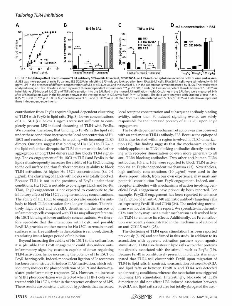
Fc�R Engagement Also Enhances in Vivo Efficacy—Toexplore whether the Fc�R-dependent mechanism of actionafforded additional benefit in TLR4-driven experimental mod-els of inflammation, the previously described rat anti-mouseTLR4 monoclonal antibody (15), 5E3, was reengineered. Toensure Fc�R engagement on mouse cells, the antibody’s Fv por-tion was reconstructed onto a mouse IgG2a,� backbone (5E3mIgG2a), providing high affinity for mouse Fc�RI (13). AD265A variant of 5E3 mIgG2a, which does not bind to any Fc�R,was also generated. In vitro, 5E3 IgG2a was found to be �100-fold more potent than 5E3 D265A in blocking LPS-stimulatedTLR4 activation, demonstrating the potential to exploit thismechanism of action involving Fc�R co-engagement in amurine system (Fig. 7A).
Next, to investigate beneficial effects of Fc�R engagement invivo, these mAbs were used in a mouse model of LPS-inducedacute lung inflammation. Intranasal administration of LPSinduced inflammatory cytokines (i.e. IL-6 and TNF�) in the
bronchoalveolar lavage (BAL) fluid (Fig. 7, B and C). Both 5E3mIgG2a and its D265A variant blocked IL-6 and TNF� in adose-dependent manner. However, 5E3 mIgG2a demonstrateda greater potency in blocking the cytokines as compared withthe Fc�R-null mutant, 5E3 D265A (Fig. 7, B and C). Significantdifferences were observed between the two antibodies at allthree doses, with 5E3 IgG2a being �10-fold more potent thanthe D265A mutant. To confirm that the Fc mutation had notaffected the ability of the 5E3 variant to reach the alveolar space,antibody concentrations in the BAL fluid were measured byELISA. At all doses tested, both mAbs (5E3 mIgG2a and theD265A mutant) reached the lung compartment in a dose-de-pendent manner, and no difference in BAL fluid concentrationswas detected (Fig. 7D). These results indicate that the differ-ence in potency between the two 5E3 variants is due to theirdifferential ability to engage Fc�Rs rather than differences intheir in vivo distribution.
DISCUSSION
In this study, we have demonstrated that the anti-humanTLR4 antibody, Hu 15C1, exploits both Fc�RI and Fc�RIIA toincrease its potency on Fc�R-bearing inflammatory cells. This
FIGURE 6. Fc�R engagement renders Hu 15C1 potent with long lasting inhibitory effects on TLR4- and Fc�R-co-expressing cells but not on Fc�R-negativecells. A, blockade of TLR4 activation by Hu 15C1 in HEK293 cells transfected with TLR4�MD-2 or TLR4�MD-2�Fc�RIIA. Transfected HEK293 cells were incubated in thepresence of 100 ng/ml LPS and Hu 15C1 for 24 h. IL-8 levels in supernatants were measured by ELISA. An F test was used to compare the fitted curves of the inhibitoryprofile of Hu 15C1 with the two different transfected cell lines. ***, p � 0.001. Data shown represent three independent experiments. B, blockade of TLR4 activation byHu 15C1 and 15C1 D265A in human umbilical vein endothelial cells. Cells were incubated in the presence of 100 ng/ml LPS and Hu 15C1 or 15C1 D265A for 24 h. IL-8levels in supernatants were measured by ELISA. An F test was used to compare the fitted curves of different groups. ns, not significant. C and D, Hu 15C1 has a longerlasting inhibitory effect than 15C1 D265A on TLR4 activation in THP-1 Blue cells. The experimental procedure is depicted in C, and the results are shown in D. THP-1 Bluecells were loaded with Hu 15C1 or 15C1 D265A for 30 min, and free mAb in the medium was removed by washing. The cells were then cultured at 37 °C for variousperiods as indicated in the panels (0, 1, or 24 h) and subjected to LPS stimulation (5 ng/ml) for 24 h. Student’s t test was used to compare differences between groups.Results are expressed as the mean � S.E. (error bars). ***, p � 0.001; **, p � 0.01; *, p � 0.05; ns, not significant.
Tailoring TLR4 Inhibition
MAY 30, 2014 • VOLUME 289 • NUMBER 22 JOURNAL OF BIOLOGICAL CHEMISTRY 15315
contribution from Fc�Rs required ligand-dependent clusteringof TLR4 with Fc�Rs in lipid rafts (Fig. 8). Lower concentrationsof Hu 15C1 (i.e. below 1 �g/ml) were not sufficient to com-pletely prevent LPS-induced clustering of TLR4 with Fc�Rs.We consider, therefore, that binding to Fc�Rs in the lipid raftunder these conditions increases the local concentration of Hu15C1 and renders it capable of interacting with incoming TLR4dimers. Our data suggest that binding of Hu 15C1 to TLR4 inthe lipid raft either disrupts the TLR4 dimers or blocks furtheraggregation among TLR4 dimers and thus blocks TLR4 signal-ing. The co-engagement of Hu 15C1 to TLR4 and Fc�Rs in thelipid raft subsequently increases the avidity of Hu 15C1 bindingto the cell surface and thus further increases its ability to blockTLR4 activation. At higher Hu 15C1 concentrations (i.e. 1�g/ml), the clustering of TLR4 with Fc�Rs was totally blocked.Because TLR4 is not in the proximity of Fc�Rs under theseconditions, Hu 15C1 is not able to co-engage TLR4 and Fc�Rs.Thus, Fc�R engagement is not expected to contribute to theinhibitory effect of Hu 15C1 at higher antibody concentrations.The ability of Hu 15C1 to engage Fc�Rs also enables the anti-body to block TLR4 activation for a longer duration. The rela-tively high Fc�RI and Fc�RIIA densities on the surface ofinflammatory cells compared with TLR4 may allow preferentialHu 15C1 binding at lower antibody concentrations. We there-fore speculate that the interaction with Fc�RI and possiblyFc�RIIA provides another means for Hu 15C1 to remain on cellsurfaces when free antibody in the solution is removed, directlytranslating into a longer period of inhibition.
Beyond increasing the avidity of Hu 15C1 to the cell surface,it is plausible that Fc�R engagement could also induce anti-inflammatory signaling cascades capable of down-regulatingTLR4 activation, hence increasing the potency of Hu 15C1 onFc�R-bearing cells. Indeed, monovalent ligation of Fc receptorshas been demonstrated to induce ITAMi activation, which sub-sequently induces the phosphorylation of SHP1 and down-reg-ulates proinflammatory responses (21). However, no increasein SHP1 phosphorylation was detected when THP-1 cells weretreated with Hu 15C1, either in the presence or absence of LPS.These results are consistent with our hypothesis that increased
local receptor concentration and subsequent antibody bindingavidity, rather than Fc-induced signaling events, are solelyresponsible for the increased potency of Hu 15C1 upon Fc�Rengagement.
The Fc�R-dependent mechanism of action was also observedwith an anti-mouse TLR4 antibody, 5E3. Because the epitope of5E3 is also located within a region involved in TLR4 dimeriza-tion (15), this finding suggests that the mechanism could bewidely applicable to TLR4 blocking antibodies directly interfer-ing with receptor dimerization or even more generally to allanti-TLR4 blocking antibodies. Two other anti-human TLR4antibodies, H4 and H52, were reported to block TLR4 activa-tion via an Fc�R-independent mechanism (23). Nevertheless,high antibody concentrations (10 �g/ml) were used in theabove report, which, from our own experience, may mask anypotential contribution from Fc�R engagement. Indeed, anti-receptor antibodies with mechanisms of action involving ben-eficial Fc�R engagement have previously been reported. Forexample, Fc�RIIB engagement has been reported to enhancethe function of an anti-CD40 agonistic antibody targeting cellsco-expressing Fc�RIIB and CD40 (24). The underlying mecha-nism was not clarified in the report. We speculate that the anti-CD40 antibody may use a similar mechanism as described herefor TLR4 to enhance its effects. Additionally, an Fc contribu-tion was recently demonstrated in the mechanism of action ofan anti-CD115 mAb (25).
The clustering of TLR4 upon stimulation has been reportedpreviously (8, 19) and confirmed in this study. In addition to itsassociation with apparent activation partners upon agoniststimulation, TLR4 also clusters in lipid rafts with other proteinsnot directly associated with the stimuli, such as Fc�Rs (19).Because Fc�RI is constitutively present in lipid rafts, it is antic-ipated that TLR4 will cluster with Fc�RI upon migration ofTLR4 to lipid rafts. In contrast, no association between Fc�RIIAand lipid rafts or between Fc�RIIA and TLR4 was detectedunder resting conditions, whereas the association was triggeredfollowing LPS stimulation. Interestingly, blockade of TLR4dimerization did not affect LPS-induced association betweenFc�RIIA and lipid raft structures but totally abrogated the asso-
FIGURE 7. Inhibitory effect of anti-mouse TLR4 antibody 5E3 and its Fc variant, 5E3 D265A, on LPS-induced cytokine secretion both in vitro and in vivo.A, 5E3 was more potent than its Fc variant 5E3 D265A in inhibiting LPS-induced IL-6 secretion from RAW264.7 cells. RAW264.7 cells were stimulated with 10ng/ml LPS in the presence of different concentrations of 5E3 or 5E3 D265A, and the levels of IL-6 in the supernatants were measured by ELISA. The results wereanalyzed using an F test. The data shown represent three independent experiments. ***, p � 0.001. B and C, 5E3 was more potent than its Fc variant 5E3 D265Ain inhibiting LPS-induced IL-6 (B) and TNF� (C) secretion into the BAL fluid in the mouse LPS instillation model. Cytokines in the BAL fluid were measured 24 hafter LPS instillation. Data in the figure are shown as the average mean � S.E. (error bars) (n 10/group). The data were analyzed with Student’s t test (*, p �0.05; **, p � 0.01; ***, p � 0.001). D, concentrations of 5E3 and 5E3 D265A in BAL fluid from mice administered with 5E3 or 5E3 D265A. Data shown representthree independent experiments.
Tailoring TLR4 Inhibition
15316 JOURNAL OF BIOLOGICAL CHEMISTRY VOLUME 289 • NUMBER 22 • MAY 30, 2014

ciation between Fc�RIIA and TLR4. These results suggest thatthe association between Fc�RIIA and lipid rafts requires LPSbut not TLR4. Further studies are necessary in order to eluci-date the precise mechanism by which LPS induces Fc�RIIAmigration to lipid rafts independently of TLR4.
The fact that TLR4 clusters with Fc�Rs upon activation sug-gests that TLR4 activation may provide a platform to facilitateFc�R activation. Indeed, the ability of IgG immune complexesto induce cytokine release in neutrophils and macrophages waslost in TLR4 mutant mice (26), confirming a role for TLR4 inFc�R activation. Furthermore, signaling through Fc�RIII wasshown to modulate TLR4 signaling pathways in human mono-cytes (27). Due to the lack of Fc�RIII binding by Hu 15C1,potential cross-talk was not tested in the current study. On theother hand, such clustering may also enable Fc�Rs to modulateinnate immune responses, which may play a role in preventingthe detrimental effects of uncontrolled inflammation. In sup-port of this, TLR responses have been reported to be enhancedin dendritic cells from Fc receptor � chain-deficient mice (28),and high avidity ligation of Fc�Rs has been shown to dampenTLR signaling under certain circumstances (29). Both reportsdemonstrated a negative role for Fc�Rs in TLR signaling.Although the clustering between TLR4 and Fc�Rs has beenestablished, it is not clear whether the activation of other TLRsinduces similar clustering, and as such, this represents a topicfor future investigational work.
It has been established that TLR4 dimerization and cluster-ing in lipid rafts are critical events in TLR4 signaling. However,the relationship between dimerization and clustering has notbeen well elucidated. LPS-triggered TLR4 dimerization wasreported to be only detected in lipid rafts, and such dimeriza-tion was abrogated by a lipid raft disruptor (30). On the otherhand, artificially forced TLR4 dimerization is by itself sufficientto trigger TLR4 signaling (31, 32). We demonstrate here thatblockade of TLR4 dimerization with Hu 15C1 totally abrogatesTLR4 clustering in lipid rafts, suggesting that TLR4 dimeriza-tion is a prerequisite for TLR4 clustering.
The mechanism of action of Hu 15C1 demonstrates thatTLR4 activation can be modulated even after receptor
dimerization and during receptor clustering in lipid rafts. Thissuggests that Hu 15C1 may be able to disrupt established TLR4dimers. Alternatively, further aggregation of TLR4 in lipid raftsis required after ligand-induced dimerization for maximalTLR4 activation, and Hu 15C1 is able to interfere with thisamplification event. Supporting these concepts, we found thatHu 15C1 could efficiently block TLR4 activation when addedseveral h after ligand exposure.
Dysregulated activation of TLR4 has been implicated innumerous acute inflammatory, chronic inflammatory, andautoimmune disorders, such as infection, asthma, acute lunginjury, ischemia/reperfusion injury, inflammatory bowel dis-ease, graft rejection, transplantation, rheumatoid arthritis, andmultiple sclerosis (33). Because uncontrolled leukocyte activa-tion plays a pivotal role in the maintenance of chronic inflam-mation (34), blocking the activation of these cells is consideredbeneficial to such diseases. On the other hand, TLR4 signalingon epithelial cells has been demonstrated to play a positive rolein tissue homeostasis (11). A prolonged total shutdown of TLR4signaling on epithelial cells may therefore be detrimental totissue repair. Because inflammatory cells express Fc�Rs,whereas epithelial cells do not, the ability to engage Fc�Rsbestows a higher and long lasting potency upon Hu 15C1toward inflammatory cells but a lower and shorter livedpotency toward epithelial cells. This feature provides the pros-pect for Hu 15C1 to preferentially block the dysregulated acti-vation of TLR4 on inflammatory cells while sparing some ben-eficial TLR4 signaling on structural cells. Furthermore, thismechanism highlights events that may occur with other anti-bodies that target cell surface receptors that traffic to lipid rafts.
REFERENCES1. Kumar, H., Kawai, T., and Akira, S. (2011) Pathogen recognition by the
innate immune system. Int. Rev. Immunol. 30, 16 –342. Kawai, T., and Akira, S. (2010) The role of pattern-recognition receptors
in innate immunity: update on Toll-like receptors. Nat. Immunol. 11,373–384
3. Foell, D., Wittkowski, H., and Roth, J. (2007) Mechanisms of disease: a“DAMP” view of inflammatory arthritis. Nat. Clin. Pract. Rheumatol. 3,382–390
FIGURE 8. Hu 15C1 preferentially blocks TLR4 activation on Fc�R-bearing inflammatory cells at lower concentrations. Under resting conditions, lowconcentrations of Hu 15C1 (�1 �g/ml) are not sufficient to block all surface TLR4 molecules. On inflammatory cells, Hu 15C1 accumulates in the lipid raft bybinding to Fc�RI. Upon LPS stimulation, free TLR4 molecules dimerize and migrate to lipid rafts. Anchoring to Fc�RI enables high avidity binding of Hu 15C1 toFc�RI and TLR4 in lipid rafts, which disrupts the TLR4 dimer or the further aggregation of TLR4 dimers.
Tailoring TLR4 Inhibition
MAY 30, 2014 • VOLUME 289 • NUMBER 22 JOURNAL OF BIOLOGICAL CHEMISTRY 15317

4. Kumar, H., Kawai, T., and Akira, S. (2009) Toll-like receptors and innateimmunity. Biochem. Biophys. Res. Commun. 388, 621– 625
5. Shimazu, R., Akashi, S., Ogata, H., Nagai, Y., Fukudome, K., Miyake, K.,and Kimoto, M. (1999) MD-2, a molecule that confers lipopolysaccharideresponsiveness on Toll-like receptor 4. J. Exp. Med. 189, 1777–1782
6. Gay, N. J., and Gangloff, M. (2007) Structure and function of Toll recep-tors and their ligands. Annu. Rev. Biochem. 76, 141–165
7. Park, B. S., Song, D. H., Kim, H. M., Choi, B. S., Lee, H., and Lee, J. O. (2009)The structural basis of lipopolysaccharide recognition by the TLR4-MD-2complex. Nature 458, 1191–1195
8. Triantafilou, M., Miyake, K., Golenbock, D. T., and Triantafilou, K. (2002)Mediators of innate immune recognition of bacteria concentrate in lipidrafts and facilitate lipopolysaccharide-induced cell activation. J. Cell Sci.115, 2603–2611
9. Legein, B., Temmerman, L., Biessen, E. A., and Lutgens, E. (2013) Inflam-mation and immune system interactions in atherosclerosis. Cell Mol. LifeSci. 70, 3847–3869
10. Nemeth, T., and Mocsai, A. (2012) The role of neutrophils in autoimmunediseases. Immunol. Lett. 143, 9 –19
11. Rakoff-Nahoum, S., Paglino, J., Eslami-Varzaneh, F., Edberg, S., and Med-zhitov, R. (2004) Recognition of commensal microflora by Toll-like recep-tors is required for intestinal homeostasis. Cell 118, 229 –241
12. Nimmerjahn, F., and Ravetch, J. V. (2008) Fc� receptors as regulators ofimmune responses. Nat. Rev. Immunol. 8, 34 – 47
13. Bruhns, P. (2012) Properties of mouse and human IgG receptors and theircontribution to disease models. Blood 119, 5640 –5649
14. Dunn-Siegrist, I., Leger, O., Daubeuf, B., Poitevin, Y., Depis, F., Herren, S.,Kosco-Vilbois, M., Dean, Y., Pugin, J., and Elson, G. (2007) Pivotal involve-ment of Fcgamma receptor IIA in the neutralization of lipopolysaccharidesignaling via a potent novel anti-TLR4 monoclonal antibody 15C1. J. Biol.Chem. 282, 34817–34827
15. Daubeuf, B., Mathison, J., Spiller, S., Hugues, S., Herren, S., Ferlin, W.,Kosco-Vilbois, M., Wagner, H., Kirschning, C. J., Ulevitch, R., and Elson,G. (2007) TLR4/MD-2 monoclonal antibody therapy affords protection inexperimental models of septic shock. J. Immunol. 179, 6107– 6114
16. Magistrelli, G., Malinge, P., Anceriz, N., Desmurs, M., Venet, S., Calloud,S., Daubeuf, B., Kosco-Vilbois, M., and Fischer, N. (2012) Robust recom-binant FcRn production in mammalian cells enabling oriented immobili-zation for IgG binding studies. J. Immunol. Methods 375, 20 –29
17. Walsh, C., Gangloff, M., Monie, T., Smyth, T., Wei, B., McKinley, T. J.,Maskell, D., Gay, N., and Bryant, C. (2008) Elucidation of the MD-2/TLR4interface required for signaling by lipid IVa. J. Immunol. 181, 1245–1254
18. Schmidt, M., Raghavan, B., Muller, V., Vogl, T., Fejer, G., Tchaptchet, S.,Keck, S., Kalis, C., Nielsen, P. J., Galanos, C., Roth, J., Skerra, A., Martin,S. F., Freudenberg, M. A., and Goebeler, M. (2010) Crucial role for humanToll-like receptor 4 in the development of contact allergy to nickel. Nat.Immunol. 11, 814 – 819
19. Pfeiffer, A., Bottcher, A., Orso, E., Kapinsky, M., Nagy, P., Bodnar, A.,Spreitzer, I., Liebisch, G., Drobnik, W., Gempel, K., Horn, M., Holmer, S.,Hartung, T., Multhoff, G., Schütz, G., Schindler, H., Ulmer, A. J., Heine,H., Stelter, F., Schütt, C., Rothe, G., Szöllosi, J., Damjanovich, S., andSchmitz, G. (2001) Lipopolysaccharide and ceramide docking to CD14provokes ligand-specific receptor clustering in rafts. Eur. J. Immunol. 31,3153–3164
20. Shields, R. L., Namenuk, A. K., Hong, K., Meng, Y. G., Rae, J., Briggs, J., Xie,D., Lai, J., Stadlen, A., Li, B., Fox, J. A., and Presta, L. G. (2001) Highresolution mapping of the binding site on human IgG1 for Fc � RI, Fc � RII,
Fc � RIII, and FcRn and design of IgG1 variants with improved binding tothe Fc � R. J. Biol. Chem. 276, 6591– 6604
21. Blank, U., Launay, P., Benhamou, M., and Monteiro, R. C. (2009) Inhibi-tory ITAMs as novel regulators of immunity. Immunol. Rev. 232, 59 –71
22. Pfirsch-Maisonnas, S., Aloulou, M., Xu, T., Claver, J., Kanamaru, Y., Ti-wari, M., Launay, P., Monteiro, R. C., and Blank, U. (2011) InhibitoryITAM signaling traps activating receptors with the phosphatase SHP-1 toform polarized “inhibisome” clusters. Sci. Signal. 4, ra24
23. Tsukamoto, H., Fukudome, K., Takao, S., Tsuneyoshi, N., Ihara, H., Ikeda,Y., and Kimoto, M. (2012) Multiple potential regulatory sites of TLR4activation induced by LPS as revealed by novel inhibitory human TLR4mAbs. Int. Immunol. 24, 495–506
24. Li, F., and Ravetch, J. V. (2011) Inhibitory Fc� receptor engagement drivesadjuvant and anti-tumor activities of agonistic CD40 antibodies. Science333, 1030 –1034
25. Haegel, H., Thioudellet, C., Hallet, R., Geist, M., Menguy, T., Le Pogam, F.,Marchand, J. B., Toh, M. L., Duong, V., Calcei, A., Settelen, N., Preville, X.,Hennequi, M., Grellier, B., Ancian, P., Rissanen, J., Clayette, P., Guillen, C.,Rooke, R., and Bonnefoy, J. Y. (2013) A unique anti-CD115 monoclonalantibody which inhibits osteolysis and skews human monocyte differen-tiation from M2-polarized macrophages toward dendritic cells. MAbs 5,736 –747
26. Rittirsch, D., Flierl, M. A., Day, D. E., Nadeau, B. A., Zetoune, F. S., Sarma,J. V., Werner, C. M., Wanner, G. A., Simmen, H. P., Huber-Lang, M. S.,and Ward, P. A. (2009) Cross-talk between TLR4 and FcgammaReceptorIII(CD16) pathways. PLoS. Pathog. 5, e1000464
27. Shalova, I. N., Kajiji, T., Lim, J. Y., Gomez-Pina, V., Fernandez-Ruiz, I.,Arnalich, F., Iau, P. T., Lopez-Collazo, E., Wong, S. C., and Biswas, S. K.(2012) CD16 regulates TRIF-dependent TLR4 response in human mono-cytes and their subsets. J. Immunol. 188, 3584 –3593
28. Chu, C. L., Yu, Y. L., Shen, K. Y., Lowell, C. A., Lanier, L. L., and Hamer-man, J. A. (2008) Increased TLR responses in dendritic cells lacking theITAM-containing adapters DAP12 and FcR�. Eur. J. Immunol. 38,166 –173
29. Wang, L., Gordon, R. A., Huynh, L., Su, X., Park Min, K. H., Han, J., Arthur,J. S., Kalliolias, G. D., and Ivashkiv, L. B. (2010) Indirect inhibition ofToll-like receptor and type I interferon responses by ITAM-coupled re-ceptors and integrins. Immunity 32, 518 –530
30. Wong, S. W., Kwon, M. J., Choi, A. M., Kim, H. P., Nakahira, K., andHwang, D. H. (2009) Fatty acids modulate Toll-like receptor 4 activationthrough regulation of receptor dimerization and recruitment into lipidrafts in a reactive oxygen species-dependent manner. J. Biol. Chem. 284,27384 –27392
31. Medzhitov, R., Preston-Hurlburt, P., and Janeway, C. A., Jr. (1997) A hu-man homologue of the Drosophila Toll protein signals activation of adap-tive immunity. Nature 388, 394 –397
32. Zhang, H., Tay, P. N., Cao, W., Li, W., and Lu, J. (2002) Integrin-nucleatedToll-like receptor (TLR) dimerization reveals subcellular targeting ofTLRs and distinct mechanisms of TLR4 activation and signaling. FEBSLett. 532, 171–176
33. O’Neill, L. A., Bryant, C. E., and Doyle, S. L. (2009) Therapeutic targetingof Toll-like receptors for infectious and inflammatory diseases and cancer.Pharmacol. Rev. 61, 177–197
34. Parihar, A., Eubank, T. D., and Doseff, A. I. (2010) Monocytes and macro-phages regulate immunity through dynamic networks of survival and celldeath. J. Innate Immun. 2, 204 –215
Tailoring TLR4 Inhibition
15318 JOURNAL OF BIOLOGICAL CHEMISTRY VOLUME 289 • NUMBER 22 • MAY 30, 2014