Mitochondrial DNA deletions are associated with non-B DNA conformations

A systematic molecular dynamics study of nearest-neighbor effects on base pair and base pair stepconformations and fluctuations in B-DNARichard Lavery1,*, Krystyna Zakrzewska1, David Beveridge2, Thomas C. Bishop3,
David A. Case4, Thomas Cheatham III5, Surjit Dixit6, B. Jayaram7, Filip Lankas8,
Charles Laughton9, John H. Maddocks10, Alexis Michon1, Roman Osman11,
Modesto Orozco12, Alberto Perez12, Tanya Singh7, Nada Spackova13 and Jiri Sponer13
1Institut de Biologie et Chimie des Proteines, CNRS UMR 5086/Universite de Lyon, 7 passage du Vercors,69367 Lyon, France, 2Department of Chemistry, Wesleyan University, Middletown, CT 06459, 3Center forComputational Science, Tulane University, Lindy Boggs Building Suite 500, New Orleans, LA 70118, 4BioMaPSInstitute and Dept. of Chemistry & Chemical Biology, Rutgers University, 610 Taylor Road, Piscataway, NJ08854-8087, 5Department of Pharmaceutics, University of Utah, SH 201, Salt Lake City, UT 84112, USA,6Zymeworks Inc., 540-1385 W 8th Ave, Vancouver, BC V6H 3V9, Canada, 7Department of Chemistry, IndianInstitute of Technology, Hauz Khas, New Delhi 110016, India, 8Center for Complex Molecular Systems andBiomolecules, Institute of Organic Chemistry and Biochemistry, Flemingovo nam. 2, 166 10 Praha 6, CzechRepublic, 9Centre for Biomolecular Sciences, School of Pharmacy, University of Nottingham, NG7 2RD, UK,10Institut de Mathematiques, Swiss Federal Institute of Technology, CH-1015 Lausanne, Switzerland,11Department of Structural and Chemical Biology, Mount Sinai School of Medicine, New York, NY 10029,USA, 12Joint IRB-BSC Program on Computational Biology, Institute of Research in Biomedicine, Parc Cientıficde Barcelona, Josep Samitier 1-5, Barcelona 08028. Spain and Barcelona Supercomputing Centre, JordiGirona 31, Edifici Torre Girona. Barcelona 08034, and Departament de Bioquımica, Facultat de Biologıa,Avgda Diagonal 647, Barcelona 08028, Spain and 13Institute of Biophysics, Academy of Sciences of theCzech Republic, Kralovopolska 135, 612 65 Brno, Czech Republic
Received August 17, 2009; Revised September 15, 2009; Accepted September 18, 2009
ABSTRACT
It is well recognized that base sequenceexerts a significant influence on the properties ofDNA and plays a significant role in protein–DNA interactions vital for cellular processes.Understanding and predicting base sequenceeffects requires an extensive structural anddynamic dataset which is currently unavailablefrom experiment. A consortium of laboratories wasconsequently formed to obtain this informationusing molecular simulations. This article describesresults providing information not only on all 10unique base pair steps, but also on all possiblenearest-neighbor effects on these steps. Theseresults are derived from simulations of 50–100 nson 39 different DNA oligomers in explicit solventand using a physiological salt concentration.
We demonstrate that the simulations are convergedin terms of helical and backbone parameters. Theresults show that nearest-neighbor effects on basepair steps are very significant, implying thatdinucleotide models are insufficient for predictingsequence-dependent behavior. Flanking basesequences can notably lead to base pair stepparameters in dynamic equilibrium between twoconformational sub-states. Although this studyonly provides limited data on next-nearest-neighbor effects, we suggest that such effectsshould be analyzed before attempting to predictthe sequence-dependent behavior of DNA.
INTRODUCTION
Since the first high-resolution crystal structure of DNAappeared (1), it has become clear that the base sequence
*To whom correspondence should be addressed. Tel: +33 4 72 72 26 37; Fax: +33 4 72 72 26 04; Email: [email protected]
The authors wish it to be known that, in their opinion, the first two authors should be regarded as joint First Authors.
Nucleic Acids Research, 2009, 1–15doi:10.1093/nar/gkp834
� The Author(s) 2009. Published by Oxford University Press.This is an Open Access article distributed under the terms of the Creative Commons Attribution Non-Commercial License (http://creativecommons.org/licenses/by-nc/2.5/uk/) which permits unrestricted non-commercial use, distribution, and reproduction in any medium, provided the original work is properly cited.
Nucleic Acids Research Advance Access published November 6, 2009 by guest on N
ovember 19, 2013
http://nar.oxfordjournals.org/D
ownloaded from

can influence the conformation of the DNA double-helix,leading to significant conformational (and associateddynamic) inhomogeneity that was not foreseen in theidealized model of Watson and Crick (2). It has sincealso become clear that sequence-dependent changes inthe structure and the structural fluctuations of DNA canplay a major role in recognition processes involving DNA.These effects underlie the so-called ‘indirect’ componentof DNA recognition (3–5), which is now known to beimportant in the formation and the stability of manyprotein–DNA complexes and, thus, in the majority ofphysiological processes involving DNA: packaging,repair, replication, gene expression, and so on. Anotable example of this, currently of great interest, isnucleosome positioning on genomic DNA, where,despite the absence of significant direct protein–DNAinteractions, sequence-dependent mechanics can guidebinding and, in turn, are likely to play an important rolein controlling gene expression (6–8).Despite the importance of indirect recognition, this
factor remains difficult to quantify since it requires adetailed knowledge of how base sequence modulates theproperties of DNA. Although crystallography (9–13), andto a lesser extent, NMR spectroscopy (particularly sincethe development of residual dipolar coupling and 31Pchemical shift anisotropy techniques) (14–16), have beeninvaluable in providing a growing database of DNAoligomer structures (17), there are still not enough high-resolution data available to make reliable predictions forsequence effects. Such predictions are also hampered bythe fact that DNA is flexible and certain aspects of itsconformation can be easily deformed by external forces,including those exerted by a crystal lattice (18). Non-local rod-like deformations of DNA are also difficult toanalyze using NMR-derived distance and torsionalrestraints, since they can occur with only small changesin short-range conformational parameters. Lastly, neithercrystallography nor NMR spectroscopy can easily providedetailed information on the sequence-dependent dynamicsof the double helix.For these reasons, it is attractive to use molecular
simulations to make a systematic attempt to analyze theimpact of base sequence. Molecular dynamics applied toDNA has progressed significantly in recent years (19–21)thanks to several factors including better force fields(22,23) and improved treatments of electrostaticinteractions (24–26), combined with explicit solventmodels and increased computer power which allowslonger simulations. Simulations running from the nano-second to the microsecond scale (27–29) are stable andprovide conformational information which, for specificB-DNA oligomers, correlates increasingly well withexperiment (30,31), even if some questions remain con-cerning the quantitative balance of conformational sub-states (32). For further background, we refer two earlierpublications (33,34).The aim of reliably predicting base sequence effects has
not however been achieved. It is not even clear for themoment how sequence effects should be modeled interms of a library of sequence fragments. Early modelsassumed that properties such as bending and flexibility
could be derived from either base pair steps (35,36) orbase pair triplets (37). This choice was largely imposedby a lack of sufficient data for obtaining reliableparameters for models based on more than 10(dinucleotide) or 32 (trinucleotide) sequence fragments,even though available crystallographic data show consid-erable conformational variation for such fragments. Onestudy, based on optimizing base pair stacking energies, hasgone much further than these models and looked at all32 896 octameric sequence fragments (38). The analysisof this work led to the conclusion that the conformationalspace was much less diverse than the sequencespace, many fragments with distinct sequences sharingsimilar structures (39). We can conclude that the bestchoice for a fragment library probably lies at or abovethe tetranucleotide level. Making a systematic study ofconformational and dynamic behavior at this level is theaim of this work.
The results described in this article are the outcome ofa collaboration involving groups from many countries,initially established during a workshop in Switzerlandin 2002 and termed the Ascona B-DNA consortium(ABC). The aim of ABC was to carry out moleculardynamic simulations on a broad enough spectrum ofB-DNA sequences to be able to begin to understand theextent and nature of sequence effects on structure andstructural fluctuations. This aim clearly involved moresimulations than could be easily carried out by a singlegroup and made collaboration logical. It also enabled theparticipating groups, which included many of theresearchers interested in DNA simulations, to cometo a common view of the best way to carry out suchsimulations.
There are two basic ways to build up conformationaland dynamic information on a library of DNA sequencefragments. The first is to place the fragments inside anoligomer with a fixed sequence around the variablefragment. This was the choice of the Sarai group whosimulated all 136 tetranucleotide fragments (WXYZ)within a dodecameric sequence 50-CGCG-WXYZ-CGCG-30 (40,41). This approach needs one oligomer perfragment. A more efficient method is to pack thefragments together within an oligomer with a sequence-repeat identical to the length of the fragments(WXYZWXYZWXYZ. . ..). The advantage of thisapproach is that a single oligomer contains manyfragments (up to four in the case of repeatingtetranucleotides: WXYZ, XYZW, YZWX, ZWXY). Thisis the choice made by the ABC collaboration. However theoligomers are constructed, the aim is to analyze onlythe center of each fragment, a base pair step (that is, adouble-stranded, dinucleotide fragment) for even-lengthfragments and a base pair for odd-length fragments. Ifthe fragments are sufficiently long, changes in basesequence beyond the fragment will have no impact onthe behavior of the central part. In common with ourprevious work, we currently assume that tetranucleotidefragments are ‘sufficiently long’, but this remains to beproved and we return to this question later.
The results of the first round of ABC simulations havebeen described in two earlier publications (33,34). These
2 Nucleic Acids Research, 2009
by guest on Novem
ber 19, 2013http://nar.oxfordjournals.org/
Dow
nloaded from

simulations, which were considerably longer than most ofthose available at that time, led to the discovery of aweakness in the AMBER force field which was over-stabilizing unusual backbone conformations involvingthe a (O30-P-O50-C50) and g (O50-C50-C40-C30) backbonetorsions. These sub-states caused significant deformationsof DNA, including a steadily decreasing twist as moresub-states built up. This problem was subsequently cor-rected with a modification to the parm 99 force field (42)termed BSC0 (22), based on refined quantum chemicalstudies of the relevant part of the DNA phosphodiesterbackbone.
At the end of 2007, the next phase of the ABCsimulations were begun using the modified force field,longer oligomers, physiological salt concentrations andtwo different models for water. Consistent with ourprevious work, the simulations again involved 39 oligomerscontaining all 136 unique tetranucleotide sequences;however, they were run for a longer timescale (50–100 nseach). Due to improving computer power, the simulationswere finished in a few months. The trajectories were thentransferred to a single site (IBCP, Lyon) where analysis wascarried out. This analysis presented a considerable chal-lenge given the amount of data available which requiredalmost a terabyte of storage and comprised almost threemillion conformational snapshots. It also coincided withthe development of a new and faster version of theCurves helical analysis program, termed Curves+, and ofa new tool to process Curves data, termed Canal. Theseprograms are the subject of a recent article (43) and arefreely available (http://gbio-pbil.ibcp.fr/Curves_plus).
Although the data which results from this round ofABC simulations does not answer all the questionsabout how base sequence influences DNA, it doesprovide a systematic view of sequence effects up to thenearest-neighbor level on base pair parameters (that isto say, including all trinucleotide sequences) and on basepair step parameters (that is, including all tetranucleotidesequences). It also gives a glimpse of next-nearest-neighbor effects on base pairs parameters. Given theamount of data to be treated, this publication onlyconsiders DNA structure and dynamics. Full analysis ofparameter correlations and of solvent and counterionbehavior will be treated elsewhere. In addition, much ofthe numerical data we discuss has been placed in the sup-plementary material to make the article more readable.To dig deeper into this data, which has considerableinterest not only for understanding sequence-effects, butalso for developing coarse-grain models of DNA, wepropose to make the original simulations (stripped ofsolvent for ease of storage) and the Curves+ data,usable in Canal, freely available.
Concerning comparison of the results presented herewith experiment, we make two remarks. First, it hasalready been demonstrated that the BSC0 force field,associated with the simulation protocol used in thisstudy, produces results for B-DNA oligomers that are invery good agreement with experiment in terms of confor-mation and flexibility. We refer to recent studies coveringa broad range of oligomeric sequences of B-DNA (as wellas various RNAs, triplex DNA and Z-DNA) (22) and to
a detailed analysis of a microsecond simulation of theDrew–Dickerson oligomer (29). This simulationapproach also shows general trends in sequence-dependent helical parameters for dinucleotides that arein line with experiment. However, we do not expectquantitative agreement at this stage, precisely becausewe demonstrate below that sequence-dependent effectsare probably still significant beyond the nearest-neighborlevel and consequently we do not yet attempt to predictthe behavior of any sequences other than those containedin the present oligomer set.
MATERIALS AND METHODS
The results discussed in this paper are based on moleculardynamics trajectories for 39 double-stranded B-DNAoligomers, each containing 18 base pairs. The sequenceof each oligomer is constructed in the same way: 50-gc-CD-ABCD-ABCD-ABCD-gc-30, where upper case lettersindicate sequences that vary between oligomers and lowercase letters indicate fixed sequences (dashes have beenadded for clarity). Every oligomer therefore contains afour base pair repeating sequence, ABCD, which occursthree and a half times and is used as the name of theoligomer. The full list of the 39 oligomers is given in theSupplementary Table S1.Each oligomer was constructed with a canonical B-
DNA conformation. Simulations were carried out withperiodic boundary conditions within a truncatedoctahedral cell, using the AMBER suite of programs(44) with the parmbsc0 modifications (22) to the parm99force field (42,45). Simulations were run with 150mMKClusing the parameters from Dang (46). The number of ionswas adjusted to ensure a zero net charge for the solute-counterion complex. Counterions were initially placed atrandom within the simulation cell, but at least 5 A fromDNA and at least 3.5 A from one another. The complexwas then solvated with a layer of water at least 10 A thick.Water was modeled using the SPC/E parameters (47), buteight oligomers were also run with the TIP4PEWparameters (48) for comparison purposes. A typical sim-ulation thus involved around 11 500 water moleculesand between 37 000 and 47 000 atoms in total (the largevariation being due to the use of two water models).Electrostatic interactions were treated using the particlemesh Ewald method (49) with a real-space cutoff of 9 Aand cubic B-spline interpolation onto the charge grid witha spacing of �1 A. Lennard-Jones interactions weretruncated at 9 A and the pairlist was built with a bufferregion and a triggered list update whenever a particlemoved more than 0.5 A from the previous update. Initialequilibration, involving energy minimization of thesolvent, then of the solute–solvent system, followed by aslow thermalization, followed the protocol describedearlier (33,34). Production simulations were carried outusing an NPT ensemble and the Berendsen algorithm(50) to control temperature and pressure, with acoupling constant of 5 ps for both parameters. Allchemical bonds involving hydrogen atoms were restrainedusing SHAKE (51), allowing for stable simulations with a2 fs time step. Center of mass motion was removed every
Nucleic Acids Research, 2009 3
by guest on Novem
ber 19, 2013http://nar.oxfordjournals.org/
Dow
nloaded from

5000 steps to avoid kinetic energy building up intranslational motion (52) and to keep the solute centeredin the simulation cell.Each of the 39 oligomers was then simulated for either
50 ns or 100 ns, saving conformational snapshots every1 ps. This led to an initial database of 2.75ms oftrajectories, containing almost 3 million conformationalsnapshots. This dataset (in a compressed format)requires roughly 1 terabyte of storage. A second version,without solvent, requires 30 gigabytes.The first stage of conformational analysis was per-
formed using Curves+, which provides a full set ofhelical, backbone and groove geometry parameters (43).Curves+ uses the commonly agreed ‘Tsukuba’ referenceframe to describe each base (53) and respects theCambridge convention for the names and signs of allhelical parameters (54). Parameters are grouped into fivesets: intra-base pair (intra-BP), (shear, stretch, stagger,buckle, propeller and opening); BP-axis (Xdisp, Ydisp,inclination and tip); inter-BP (shift, slide, rise, tilt, rolland twist); backbone (in the 50!30 direction for eachnucleotide, a P-O50, b O50-C50, g C50-C40, d C40-C30,e C30-O30, z O30-P, the glycosidic angle w and the sugarpucker phase and amplitude); and groove (minor andmajor groove widths and depths). Note that the rise andtwist discussed in this article are the parameters derivedfrom the rotation matrix linking two base pair referenceframes (43). Curves+ also calculates these parameters as atranslation and a rotation around the helical axis, but, inthe case of B-DNA, the differences between the two sets ofparameters is negligible. For further details, we refer toour previous publication (43) (and, notably, to Supple-mentary Figure S1, which is associated with the latter pub-lication and illustrates all the helical parameters).We also remark that when we consider the confor-
mation or the dynamics of a given sequence fragment,we only discuss the conformational parameters connectedwith center of the fragment. Thus, if the fragment containsan odd number of base pairs, we discuss the central basepair in terms of intra-BP, BP-axis and groove dimensions,as well as those parts of the backbone directly associatedwith this base pair (glycosidic torsions and sugar puckers).If the fragment contains an even number of base pairs, wediscuss the central base pair step in terms of the inter-BPparameters and the backbone torsions integral to the basepair step (in the 50-30 direction for each strand: e, z, a, band g).All Curves+ parameters are output in an unformatted
file with a single record for each snapshot in eacholigomer. For the majority of the results presentedbelow, this analysis was limited to the first 50 ns of simu-lation. This produced a new dataset which requires 2.8gigabytes of storage. Rather than using a database asfor earlier versions of the ABC simulations (55), wedecided to develop a new program which would beflexible enough to answer a wide variety of queries onvariable datasets. This program, Canal, is used here toobtain statistical data on all parameters, as well as timeseries, parameter distributions (in the form of histograms)and to search for linear correlations between parameters.Canal can analyze individual base pairs or base pair steps
within the data from a single oligomer trajectory or makea cumulative analysis over many trajectories.
RESULTS
We now discuss the analysis of the 39 oligomers that havebeen studied. We begin with an overview of their behaviorand the results of tests to decide whether or not sufficientconformational sampling has been carried out. We alsolook at the impact of a change in the solvent model. Wethen turn to a discussion of how the dataset can beanalyzed in terms of base sequence. From this startingpoint, we analyze sequence-dependent effects on B-DNAstructure and dynamics by comparing sequence-averagedresults with specific base pairs or base pair steps in asequence-averaged environment, and then with basepairs or base pair steps with specific nearest-neighborsequences. Finally, we look at correlations betweenconformational parameters.
Overall characteristics of the oligomer simulations
We begin considering the question of the convergenceof the results and their stability with respect to changesin modeling the solvent environment. We have madetwo comparisons. For temporal convergence, and foroligomers where 100 ns of simulation was available, wecompared the results averaged over the first block of50 ns with an average over the second block of 50 ns.For solvent effects, and for the oligomers which wererun with two different water models, we compared 50 nsof simulation with each model. The results are veryencouraging, since neither longer simulations, nor thechange from SPC/E to TIP4PEW water had any signifi-cant effect on any of the conformational parameters wehave measured. Both averages and standard deviationsfor all parameters were typically stable to better than0.1 A and 1�. We illustrate this in the case of the AGTColigomer, where 100 ns of simulation with SPC/E waterand 50 ns of simulation with TIP4PEW water were avail-able. Results for the base pair A9 and the central base pairstep A9G10 are presented as histograms in SupplementaryFigure S1 for the most important intra-BP and inter-BPparameters. Supplementary Table S2 gives a comparisonof all parameters. Similar agreement is found whenlooking at parameters averaged along the oligomers,although backbone angles (notable e and z) whichoccupy multiple sub-states (see below) can occasionallyshow differences of a few degrees.
For spatial convergence, that is, the comparison of iden-tical sequence fragments along an oligomer, we looked attwo cases. First, we considered fragments with inversionsymmetry in terms of the sequence and their positionwithin a given oligomer. To take a specific example, thisis the case for the tetranucleotide fragment GTAC in theWatson strand at positions 6!9 and the Crick strand atpositions 13!10 within the oligomer CGTA (whosesequence is GCTACGTACGTACGTAGC). If all confor-mational sub-states within the B-DNA family arethoroughly sampled within 50 ns of simulation, wewould ideally expect to get identical results for these two
4 Nucleic Acids Research, 2009
by guest on Novem
ber 19, 2013http://nar.oxfordjournals.org/
Dow
nloaded from

tetranucleotides. This is indeed the case to within 0.2 Aand 2� for both helical parameters and groove dimensions.All backbone angles also have average values which matchto within 2�, with the exception of e and z, where minordifferences in visiting sub-states increase the differences to4� and 6�, respectively. As shown in Supplementary FigureS2, BII sub-states typically form for a few nanoseconds,but they can persist for ten nanoseconds or more, wewould therefore expect 50 ns to be a minimum in orderto reasonably sample them.
A more severe test involves taking a singletetranucleotide fragment in different positions along anoligomer. These would not necessarily give identicalresults, if end-effects or overall rod-like motions had asignificant influence. We illustrate this test for thetetranucleotide fragment CGTA in positions 5!8,9!12 and 13!16 again in the CGTA oligomer (whosesequence is given above). The results are shown graphi-cally in Supplementary Figure S3 as histograms for themain base pair and base pair step parameters for G andGT in the center of each tetranucleotide fragment. Onceagain, we find very good agreement between the differentpositions. All helical parameters lie within 0.3 A or 1�,groove widths and depths are within 0.4 A and backboneangles are within 3�, with the exception of e and z which,on one strand, reach differences of 9�, for the reasonsmentioned above. We remark that we have included thetetranucleotide fragment in positions 13!16 in this com-parison, although it is not used in the analysis whichfollows, given the chosen exclusion of four base pairs ateither end of each oligomer. We do not, therefore, expectto see any significant end-effects for the results presentedbelow. We can also conclude that, at least for theparameters considered in this work, the presentsimulations can be considered to have converged in50 ns, although a slightly lower precision is achieved forthe e/z torsions than for other backbone, helical or grooveparameters.
Analyzing the dataset in terms of base sequence effects
The 39 oligomers which compose the dataset were care-fully chosen to provide maximum information onsequence-dependent effects. The choice results fromfitting the 136 unique tetranucleotide sequences togetherin the most compact way. To explain this, we begin withbase pairs. All our discussions can be limited to thesequence of a single 50!30 strand, that we have termedthe Watson strand. The two possible base pairs in canon-ical B-DNA are A–T and G–C (where the dash indicatesbase pairing). If we now wish to take nearest-neighboreffects into account, we have to consider four possiblebases on either side of the chosen base pair, leading to atrinucleotide fragment. This results a total of2� 4� 4=32 possible sequences, 16 with a central Aand 16 with a central G. Each of these sequences has acomplementary strand with a central T or C and, conse-quently sequences centered on pyrimidines do notgenerate any new unique trinucleotides.
Similarly, if we start with base pair steps (or dinucleotidefragments), there are unique 10 possibilities. Note that
although there are 4� 4=16 base combinations for abase pair step, there are six pairs of complementarysequences (AA TT, GG CC, AG CT, GA TC, AC GTand CA TG) of which only a single case needs to be con-sidered. If we want to take nearest-neighbor effects intoaccount, we should again consider four possible bases oneither side of each base pair step, making a total of4� 4=16 cases. This is true for the six dinucleotides men-tioned above (AA, GG, AG, GA, AC, CA), however forthe remaining, autocomplementary dinucleotides (AT, TA,CG, GC), there are only 10 unique choices of neighboringbases, for the same reasons of complementarity discussedfor the dinucleotide steps themselves. This leads to a totalof 16� 6+10� 4=136 tetranucleotide fragments. Wenote that the general formulae for calculating the numberof unique N-base pair fragments is 4N/2 when N is odd, and(4N+4N/2)/2 when N is even.Each of the 39 oligonucleotides described in this work
contains between one and four unique tetranucleotidefragments (sequences with higher symmetry, forexample, GGGGGG . . . . or CGCGCG . . . , containfewer unique tetranucleotides than more complexsequences, such as, ACGTACGT. . . . .). Similarly, eachunique tetranucleotide occurs at least three times withinthe oligomer to which it belongs. Descending the scale toshorter fragments, we note that trinucleotide fragmentseach occur in four different oligomers and dinucleotidefragments occur in between 10 and 16 oligomers.To summarize, the 39 oligomer dataset contains all
tetranucleotide fragments in several copies, but, since allcopies belong to a single oligomer, each tetranucleotidehas the same flanking sequence. All trinucleotidefragments exist in several copies and because theyoccur in four different oligomers, each has four distinctflanking sequences, C . . .C, G . . .G, A . . .A and T . . .T(this follows from the ABCDABCD. . . repeatingsequence motif used in our oligomers). We can conse-quently extract complete information of the impact ofnearest-neighbor sequence effects on base pairs (usingtrinucleotide fragments) and on base pair steps (usingtetranucleotide fragments). We can also get a glimpse ofnext-nearest-neighbor effects on base pairs by using thefour distinct pentanucleotide fragments in the dataset foreach given trinucleotide (out of the 16 possible sequenceenvironments). We will however have no information onnext-nearest-neighbor effects on base pair steps. We notein passing that a systematic study at this level would implylooking at all hexanucleotide fragments, of which there are2080 unique cases.
Sequence-averaged results
Table 1 summarizes the conformational parametersaveraged over the 39 oligomers of the dataset and overall accepted base pairs and base pair steps (that isexcluding the four base pairs at either end of eacholigomer). This corresponds to a total of 19.5 milliondata points for each parameter. If we first look at theaverage parameters, we see they describe a canonicalB-DNA state. The base pairs show small averagedeformations aside from a propeller of �11�. They show
Nucleic Acids Research, 2009 5
by guest on Novem
ber 19, 2013http://nar.oxfordjournals.org/
Dow
nloaded from

a weak positive inclination to the helical axis(<Inclin>=6.8�) and are moderately shifted towardsthe major groove (<Xdisp>=�1.4 A). The inter-BPparameters show an average rise of 3.32 A and a twist of32.6�. Note that the average twist is several degrees higherthan that found with the AMBER parm94 or parm99force fields without the recent bsc0 modifications to thebackbone parameters (22). Shift and tilt are close to zero,but there is an overall tendency to negative slide (�0.44 A)and positive roll (3.6�). Backbone angles show that con-ventional states dominate for a/g (gauche�/gauche+) ande/z (trans/gauche�, that is, BI). Taking an average for theentire dataset shows only 1% of non-canonical a/g statesand 15% of BII (that is, e/z gauche�/trans). The averagesugar pucker has a phase of 137� (C10-exo, but close to theboundary with C20-endo) and an amplitude of 40�.If we now look at parameter fluctuations, the standard
deviations of the helical parameters are typically 0.5–1.0 Afor translations, with the largest values for Xdisp andslide, and 5�–10� for rotations, with the largest valuesfor buckle and propeller. For the backbone parameters,the standard deviations are larger, typically 15�–30�, withthe largest values for e (35�), z (49�) and sugar pucker(33�). While the standard deviations again indicate atypical B-DNA state, all parameters show occasional,large deviations from their average values. In manycases, these deviations are connected with temporarybase pair opening. This is reflected by the range of theopening parameter which spans values from �33�
(opening into the minor groove) to +87� (opening intothe major groove). Its asymmetry reflects the greater easeof opening towards the larger groove (56). SupplementaryFigure S4 shows examples of base pair opening in theCGCG oligomer. Base pairs at least two nucleotidesfrom the ends of the oligomers open and closespontaneously, but for short periods. Terminal basepairs can behave similarly, but can also flip completelyopen and remain broken. This is not surprising giventheir low stability, but, as discussed above, it has nodetectable impact on the base pairs we sample.
Rise and twist show large ranges (4.5 A and 76�, respec-tively) reflecting large fluctuations in base pair steps and,similarly local axis bending can reach 20�. If we look at theoverall bending of the oligomers, the average value andstandard deviation are relatively small (20� and 12�,respectively), although spontaneous fluctuations up to40�–50� occur rather regularly. Much larger values areseen for three oligomers, but these are artifacts associatedwith open terminal base pairs.
Backbone torsions show very large fluctuations, andalthough canonical a/g and e/z sub-states dominate,these four torsions, and also b, have ranges of beyond300�. Only w and d fluctuate less, with ranges of 205�
and 133�, respectively, which is explained by theconstraints associated with base stacking and pairingand by sugar ring puckering. Lastly, groove parametersagain show values in line with B-DNA, with a narrowminor groove (6.4 A on average) and a wide majorgroove (12.3 A on average), while the depths are rathersimilar (4.7 A for the minor and 6.2 A for the major).The groove widths have similar standard deviations(below 2 A), but the major groove depth fluctuates twiceas much as that of the minor groove (with standarddeviations of 2 A and 0.8 A, respectively). It is interestingto note that despite the fact that large fluctuations ingroove geometry require backbone distortions overseveral base pairs, such fluctuations do indeed occurwith both grooves covering a range from completelyclosed to 2.5 times their normal widths.
Base pair and base pair-step sequence effects (monoand dinucleotides)
The first step in analyzing sequence effects is to separateA–T and G–C base pairs. When this is done for the entire39 oligomer dataset (leading to 9.85� 106 data pointsfor each base pair), we see only limited effects (seeSupplementary Table S3 for details). As expected, A–Tpairs show larger propeller twist, opening and buckle,because of their weaker hydrogen bonding. Thesedifferences are however limited to changes of 1�–3� inthe average values. Other intra-BP and all BP-axisparameters are virtually identical for the two base pairs.This similarity also applies to the groove and backboneparameters, although there is a distinct difference betweenthe two purines (A, G) and the two pyrimidines (T, C) interms of the glycosidic angle w, which is roughly 10� lessnegative for the purines, and the sugar pucker, withthe purines showing a 15�–20� increase in average phase.
Table 1. Sequence-averaged conformational parameters
Parameter Average SD Range Minimum Maximum
Shear 0.02 0.31 10.5 �4.2 6.3Stretch 0.03 0.12 5.1 �1.4 3.7Stagger 0.09 0.41 6.1 �2.8 3.3Buckle 1.2 12.4 125.0 �65.4 66.2Propeller �11.0 9.3 105.0 �61.6 43.8Opening 2.1 4.6 115.0 �33.3 87.2Xdisp �1.44 0.89 21.0 �12.7 8.3Ydisp 0.02 0.55 18.2 �7.8 10.5Inclination 6.8 5.4 72.0 �20.9 50.8Tip 0.3 5.0 64.0 �35.6 28.4Ax-bend 2.0 1.1 20.0 0.0 20.3Shift �0.05 0.76 9.0 �4.4 4.6Slide �0.44 0.68 8.7 �3.7 5.0Rise 3.32 0.37 4.5 1.4 5.9Tilt �0.3 4.6 58.0 �27.8 28.8Roll 3.6 7.2 82.0 �37.3 44.7Twist 32.6 7.3 76.0 �17.5 60.4a �72.6 15.7 360.0b 169.6 16.2 315.0g 54.8 14.1 355.0d 125.4 19.1 133.0e �162.6 33.3 323.0z �102.8 47.6 314.0w �116.0 19.5 205.0Phase 136.7 33.6 360.0Amplitude 40.0 6.8 67.0
Averages and SD of translational parameters (A) are given to twodecimal places, while those of rotational parameters (�) are given to asingle decimal place.
6 Nucleic Acids Research, 2009
by guest on Novem
ber 19, 2013http://nar.oxfordjournals.org/
Dow
nloaded from

No significant distinction in standard deviations occur atthis level for any of the parameters studied.
We next turn to base pair steps averaging over allpossible flanking sequences The averages and standarddeviations of the inter-BP parameters for the 10 uniquesteps (from 2.2� 106 data points in each case) are showngraphically in Figure 1 and listed numerically in theSupplementary Table S4. Although sequence effects arevisible, these effects are again not very large and significantvariations are mainly limited to a few steps. The YR stepsare the easiest to distinguish (TG, TA and CG, at theright-hand end of each plot in Figure 1), as having lowrise, low twist and high positive roll. These steps also showa much lower proportion of BII states in either strand,whereas RR steps have significant amounts (25%–50%)of BII in the Watson strand and RY steps have more BIIin the Crick strand. Negative rolls occur for RY steps(GC, GT and AT) and AA also has a below average
value. AA and GA steps have the largest values of twist,with averages of 35.3� and 36.7�, respectively. Standarddeviations vary little between steps, with a few exceptionssuch as the more flexible twist and rise of YR steps.However, the average values of the inter-BP parametersfor the 10 base pair steps all fit comfortably within onestandard deviation of the sequence-averaged values,emphasizing that sequence still has a relatively minorimpact at this level.Until now, we have assumed that average values and
standard deviations adequately describe the data we havediscussed. This is obviously only true if the parametershave normal distributions clustered around a singlemean value. At the level of the sequence-averaged results(or at the base pair analysis level), a study of thehistograms of each parameter suggests that this is largelythe case. The main exception involves the backbonetorsions e and z, which mainly occupy the BI state
-1.5
-1
-0.5
0
0.5
1
1.5
CGTATGAAAGATGTGCGAGG
Shift
-10
-8
-6
-4
-2
0
2
4
6
8
CGTATGAAAGATGTGCGAGG
Tilt
-2
-1.5
-1
-0.5
0
0.5
1
CGTATGAAAGATGTGCGAGG
Slide
-10
-5
0
5
10
15
20
CGTATGAAAGATGTGCGAGG
Roll
2.4
2.6
2.8
3
3.2
3.4
3.6
3.8
4
CGTATGAAAGATGTGCGAGG
Rise
15
20
25
30
35
40
45
CGTATGAAAGATGTGCGAGG
Twist
Figure 1. Average values (black circles) and standard deviations (vertical bars) of the inter-BP parameters for the unique base pair steps. In the caseof bimodal distributions (slide and twist), a two-Gaussian fit has been made and the centers of the Gaussians are shown as black triangles.Translational parameters are given in angstroms and rotational parameters in degrees.
Nucleic Acids Research, 2009 7
by guest on Novem
ber 19, 2013http://nar.oxfordjournals.org/
Dow
nloaded from

(trans/gauche�), but, on average, spend roughly 15% oftime in the BII state (gauche�/trans). Other distributions,including twist, roll, a, b and g show shoulders on other-wise Gaussian distributions indicating the possibility ofother states, but these cannot be resolved at this level.When we turn to the results at the level of base pair
steps, this situation changes as shown in Figure 2. Whilethe distributions for shift, rise, tilt and roll are Gaussian inappearance for all base pair steps, those of slide and twistshow bimodal distributions for several steps. This is thecase for GG in terms of slide, while GA, TG and TA showpronounced shoulders. For twist, there are bimodaldistributions for all RY steps (TG, TA, CG), an unusuallybroad distribution for GG and a shoulder for GA. Theseobservations could have two causes: one of two possibleconformational sub-states is selected in function of thebase sequences flanking the given base pair step, or twopossible sub-states are in dynamic equilibrium within agiven oligomer. We will be able to resolve this questionwhen we consider nearest-neighbor sequence effects onbase pair steps, but before this we will consider nearest-neighbors effects on single base pairs.
Nearest-neighbor sequence effects on base pairs(trinucleotide fragments)
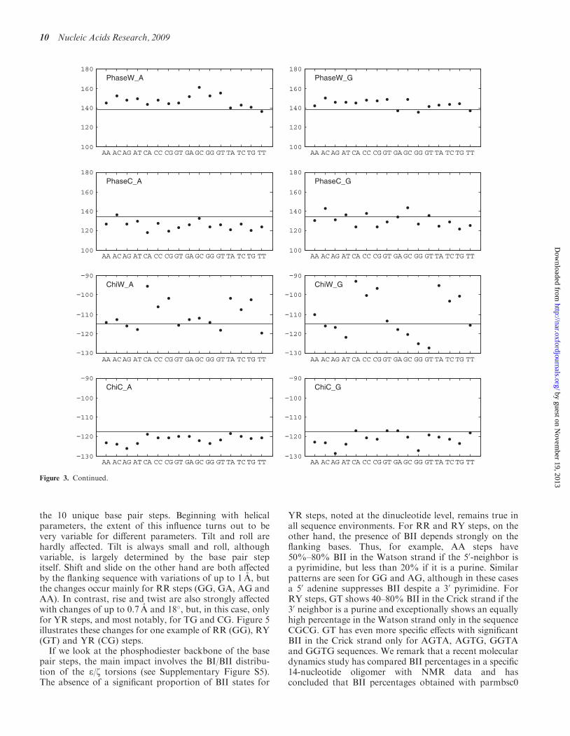
We now consider how flanking base pairs can influence theconformation and fluctuations of a base pair. As discussedabove, this implies considering 32 trinucleotide fragments,16 with a central A–T pair and 16 with a central G–C pair.The average helical and backbone parameters for thesetwo groups are presented graphically in the two columnsof Figure 3. We begin with the central A–T pair.
The left-hand column of Figure 3 shows that theflanking base pairs often produce significant changes inconformation. Sometimes the major influence involvesonly the 50-flanking sequence, as in the case of propeller,which is considerably more negative when the 50 base is apurine, or inclination, which is smallest with adenine asthe 50 neighbor (this base has a similar influence on minorgroove width, data not shown). Sometimes only the 30
neighbor has a marked effect, as in the case of buckle,which is smallest when the 30 neighbor is thymine. Othercases are more complex, as, for example, Xdisp, which isleast negative when an A–T pair is preceded by apyrimidine and followed by a purine. The patterns inFigure 3 enable these different behaviors to be easilyidentified. Similar, but unrelated, effects are seen with acentral G–C pair (right-hand column of Figure 3). Overall,nearest-neighbor effects lead to variations of 1–3 A fortranslational parameters, with the largest changes inXdisp and minor groove width, and to variations of10�–30� for rotational parameters, with the largestchanges in buckle, propeller and w.
To analyze fluctuations, we have looked at nearest-neighbor effects on the standard deviations of the basepair parameters. Here again there are significantchanges, but they rarely follow simple trends. Thelargest changes include Xdisp, sugar phase angles, theglycosidic angles and the minor groove width. We citeone example for each of these parameters. Thus, thestandard deviation of Xdisp is 0.65 A for the sequenceGGC, but becomes 1.05 A after changing the 30 neighborto G. Guanosine sugar pucker fluctuations increase from18� for CGA to 42� in GGG. For glycosidic angles, astandard deviation of 15� for guanine in AGT, increases
5 0 50
0.5
1
5 0 50
0.5
1
0 2 4 60
0.5
1
1.5
50 0 500
0.5
1
1.5
50 0 500
0.5
1
50 0 50 1000
0.5
1
1.5
Shift Slide
Rise Tilt
Roll Twist
Figure 2. Distributions of inter-BP parameters for the unique base pair steps: RR (black), YR (red), RY (blue). See text for a discussion of the stepsshowing bimodal distributions (e.g. RR slide, YR twist). Translational parameters are given in angstroms and rotational parameters in degrees.
8 Nucleic Acids Research, 2009
by guest on Novem
ber 19, 2013http://nar.oxfordjournals.org/
Dow
nloaded from

to 22� for GGA. Similarly for minor groove width, thevalues can change from 1.2 A for CAG or CGG to1.65 A for GAA, GAC or CGT.
We again need to consider if all parameters have normaldistributions at this level of analysis. This turns out to bethe case, with the exception of minor groove width. Asshown in Figure 4, the groove width prefers one of twopossible states, centered on 4 A or 8 A. Some trinucleotidesequences clearly prefer one of these states, such as AAAwhich is (not surprisingly) always narrow, or CAG which
is always wide. Others, such as GAG, exhibit a dynamicequilibrium. These fluctuations are moderately coupled tochanges in inclination and buckle, but not to backbonesub-states, in contrast to the slide and twist equilibria dis-cussed in the next section.
Nearest-neighbor sequence effects on base pair steps(tetranucleotide fragments)
We can now consider whether the flanking base pairs havea significant influence on the structure and dynamics of
AA AC AG AT CA CC CG GT GA GC GG GT TA TC TG TT10
5
0
5
10
15
20
Buckle_A
AA AC AG AT CA CC CG GT GA GC GG GT TA TC TG TT10
5
0
5
10
15
20
Buckle_G
AA AC AG AT CA CC CG GT GA GC GG GT TA TC TG TT20
15
10
5
0Propel_A
AA AC AG AT CA CC CG GT GA GC GG GT TA TC TG TT20
15
10
5
0Propel_G
AA AC AG AT CA CC CG GT GA GC GG GT TA TC TG TT0
1
2
3
4
5
6
Opening_A
AA AC AG AT CA CC CG GT GA GC GG GT TA TC TG TT0
1
2
3
4
5
6
Opening_G
AA AC AG AT CA CC CG GT GA GC GG GT TA TC TG TT3
2.5
2
1.5
1
0.5
0
Xdisp_A
AA AC AG AT CA CC CG GT GA GC GG GT TA TC TG TT3
2.5
2
1.5
1
0.5
0
Xdisp_G
AA AC AG AT CA CC CG GT GA GC GG GT TA TC TG TT0
2
4
6
8
10Inclin_A
AA AC AG AT CA CC CG GT GA GC GG GT TA TC TG TT0
2
4
6
8
10Inclin_G
Figure 3. Average values of intra-BP and backbone parameters for base pairs as a function of the flanking sequences. Left-hand column:trinucleotides centered on adenine (i.e. AAA, AAC, . . .., TAT). Right-hand column: trinucleotides centered on guanine (i.e. AGA, AGC, . . .,TGT). In each plot, the horizontal line indicates the sequence-averaged value of the corresponding parameter. Translational parameters are givenin angstroms and rotational parameters in degrees.
Nucleic Acids Research, 2009 9
by guest on Novem
ber 19, 2013http://nar.oxfordjournals.org/
Dow
nloaded from
the 10 unique base pair steps. Beginning with helicalparameters, the extent of this influence turns out to bevery variable for different parameters. Tilt and roll arehardly affected. Tilt is always small and roll, althoughvariable, is largely determined by the base pair stepitself. Shift and slide on the other hand are both affectedby the flanking sequence with variations of up to 1 A, butthe changes occur mainly for RR steps (GG, GA, AG andAA). In contrast, rise and twist are also strongly affectedwith changes of up to 0.7 A and 18�, but, in this case, onlyfor YR steps, and most notably, for TG and CG. Figure 5illustrates these changes for one example of RR (GG), RY(GT) and YR (CG) steps.If we look at the phosphodiester backbone of the base
pair steps, the main impact involves the BI/BII distribu-tion of the e/z torsions (see Supplementary Figure S5).The absence of a significant proportion of BII states for
YR steps, noted at the dinucleotide level, remains true inall sequence environments. For RR and RY steps, on theother hand, the presence of BII depends strongly on theflanking bases. Thus, for example, AA steps have50%–80% BII in the Watson strand if the 50-neighbor isa pyrimidine, but less than 20% if it is a purine. Similarpatterns are seen for GG and AG, although in these casesa 50 adenine suppresses BII despite a 30 pyrimidine. ForRY steps, GT shows 40–80% BII in the Crick strand if the30 neighbor is a purine and exceptionally shows an equallyhigh percentage in the Watson strand only in the sequenceCGCG. GT has even more specific effects with significantBII in the Crick strand only for AGTA, AGTG, GGTAand GGTG sequences. We remark that a recent moleculardynamics study has compared BII percentages in a specific14-nucleotide oligomer with NMR data and hasconcluded that BII percentages obtained with parmbsc0
AA AC AG AT CA CC CG GT GA GC GG GT TA TC TG TT100
120
140
160
180PhaseW_A
AA AC AG AT CA CC CG GT GA GC GG GT TA TC TG TT100
120
140
160
180PhaseW_G
AA AC AG AT CA CC CG GT GA GC GG GT TA TC TG TT100
120
140
160
180PhaseC_A
AA AC AG AT CA CC CG GT GA GC GG GT TA TC TG TT100
120
140
160
180PhaseC_G
AA AC AG AT CA CC CG GT GA GC GG GT TA TC TG TT130
120
110
100
90ChiW_A
AA AC AG AT CA CC CG GT GA GC GG GT TA TC TG TT130
120
110
100
90ChiW_G
AA AC AG AT CA CC CG GT GA GC GG GT TA TC TG TT130
120
110
100
90ChiC_A
AA AC AG AT CA CC CG GT GA GC GG GT TA TC TG TT130
120
110
100
90ChiC_G
Figure 3. Continued.
10 Nucleic Acids Research, 2009
by guest on Novem
ber 19, 2013http://nar.oxfordjournals.org/
Dow
nloaded from

are incorrect (32). In fact, significant disagreements onlyconcern base pair steps close to the ends of the oligomerstudied by Heddi et al. which are likely to be difficult tocorrectly sample due to end fraying, and an RY step closeto the center of the oligomer (CTGA). For this step, thethree force fields investigated (parmbsc0, parm99 andCHARMM27) all gave low BII percentages in bothstrands, in contrast to the NMR data. This is also thecase for the CTGA tetranucleotide in the present study(Figure 3). This merits further study, but will requiretesting against NMR data on a larger range of basesequences.We finally note that, as mentioned at the dinucleotide
level, the amounts of non-canonical a/g sub-states seenwith parmbsc0 are generally very small (<1%). Afew tetranucleotide steps show moderate percentages(10–25%), but the characteristic lifetimes of these statesare very long (at least tens of nanoseconds) and we
-1
-0.8
-0.6
-0.4
-0.2
0
0.2
0.4
TTTGTCTAGTGGGCGACTCGCCCAATAGACAA
Shift GG
-1.5
-1
-0.5
0
0.5
TTTGTCTAGTGGGCGACTCGCCCAATAGACAA
Slide GG
2.8
3
3.2
3.4
3.6
3.8
TTTGTCTAGTGGGCGACTCGCCCAATAGACAA
Rise GG
20
25
30
35
40
45
TTTGTCTAGTGGGCGACTCGCCCAATAGACAA
Twist GG
TTTGTCTAGTGGGCGACTCGCCCAATAGACAA-1
-0.8
-0.6
-0.4
-0.2
0
0.2
0.4 Shift GT
TTTGTCTAGTGGGCGACTCGCCCAATAGACAA-1.5
-1
-0.5
0
0.5
Slide GT
Figure 5. Average values of inter-BP parameters for the unique base pair steps as a function of the flanking sequences. The three groups of fourplots refer to the helical parameters of GG, an RR step, GT, an RY step and CG, a YR step. In each plot, the two-letter code along the abscissaindicates the 50- and 30-flanking bases and the horizontal line indicates the sequence-averaged value of the parameter. Translational parameters aregiven in angstroms and rotational parameters in degrees.
Figure 4. Minor groove width (A) distributions for A-T base pairs withdifferent flanking sequences: AAA (blue); CAG (green); GAG (red).Data accumulated over 50 ns of simulation.
Nucleic Acids Research, 2009 11
by guest on Novem
ber 19, 2013http://nar.oxfordjournals.org/
Dow
nloaded from

consequently do not have the statistics to understandsequence effects on the timescale of the presenttrajectories.In terms of nearest-neighbor effects on parameter
fluctuations, the effects are relatively small for rise, tiltand roll. They are more significant for shift and slide,where given environments can modify standard deviationsby 60–70%. These changes occur for specific sequencefragments without any easily discernible patterns. Forexample, GT shift fluctuation is unusually low in thefragments AGTC, AGTT, GGTC and GGTT. Similarly,CG shift fluctuation is increased by 30% in the presence ofa 50-adenine, and the same observation applies to CG slidefluctuations. The largest effects involve twist fluctuations,where the standard deviations can double as a function ofthe sequence environment. Some of these effects are rathergeneral, and, for example a 50-C and a 30-A leads to hightwist fluctuations for all RR and RY steps (except TG),while others are very specific and, for example, a 50-T anda 30-G only leads to high twist fluctuations for the AAstep. Supplementary Figure S6 shows the standard
deviations of twist for all base pair steps as a function oftheir environment.
Lastly, we can reanalyze the bistable distributions ofslide and twist seen at the base pair step level to determinewhether they correspond to different sub-states beingfavored by different flanking sequences or to dynamicequilibria for given base pair steps. As for minor groovewidth at the trinucleotide level, both these cases areobserved. Figure 6 shows that the step CG almostexclusively favors a low twist in CCGA and a high twistin ACGT. On the other hand in ACGA is in equilibriumbetween these two states. These differences are linked toBI/BII transitions of the 30 nucleotides in both stands, butmost strongly in this case with the Crick strand, as shownin Figure 7 (see also the time series in SupplementaryFigure S2). Similar results are found for GG, where BI/BII transitions principally induce changes in slide.
Beyond nearest-neighbor effects
The furthest we can go with this dataset in looking atsequence effects is to consider the impact of next-nearest
2.8
3
3.2
3.4
3.6
3.8
TTTGTCTAGTGGGCGACTCGCCCAATAGACAA
Rise GT
20
25
30
35
40
45
TTTGTCTAGTGGGCGACTCGCCCAATAGACAA
Twist GT
-1.5
-1
-0.5
0
0.5
TAGGGCGACGCAATAGACAA
Shift CG
-1.5
-1
-0.5
0
0.5
TAGGGCGACGCAATAGACAA
Slide CG
2.8
3
3.2
3.4
3.6
3.8
TAGGGCGACGCAATAGACAA
Rise CG
20
25
30
35
40
45
TAGGGCGACGCAATAGACAA
Twist CG
Figure 5. Continued.
12 Nucleic Acids Research, 2009
by guest on Novem
ber 19, 2013http://nar.oxfordjournals.org/
Dow
nloaded from

neighbors on base pair parameters. One might expect thatthese effects should be minor and that consideringtrinucleotide sequences (i.e. nearest neighbors) would beenough to fix the conformation of the central base pair.Although our 39 oligomer dataset does not give us a fullview of the pentanucleotide sequences necessary to look atall next-nearest effects, we can compare four differentenvironments (C. . .C, A. . .A, G. . .G, and T. . .T) aroundany given trinucleotide sequence. We have analyzed theseeffects for a selection of four sequence fragments, AAA,GGG, CAT and CGT. The results show that in each casethe central nucleotide pair changes conformationsignificantly as a function of the next-nearest neighbors.To give a few examples, average inclination changesfrom 0� to 7.5� and buckle from 3� to 8� in passing fromAAAAA to CAAAC. Similarly, buckle changes from �2�
to 6.5� and propeller from �4� to �14� in passing fromGGGGG to CGGGC. For less regular sequences, bucklechanges from �6� in ACGTA to 1� in TCGTT, with an
accompanying 4� decrease in inclination. Lastly, Xdsipchanges from �1.8 A in ACGTA to �1.3 A in TCATT,while passing from GCATG to TCATT decreases theinclination from 10� to 4�. Significant changes are alsoseen in glycosidic angles, sugar phase and minor groovewidth. We must conclude that next-nearest-neighboreffects on nucleotide pair conformations cannot beignored, particularly as we have only been able to lookat 4 out of 16 possible next-nearest-neighborenvironments.
CONCLUSION
We have completed a systematic molecular dynamicsstudy of 39 oligomers containing all unique tetranucleo-tide sequences. Simulations were carried out in explicitsolvent, in some cases with two different water models,and with a physiological salt concentration. All oligomerswere simulated for at least 50 ns and many for 100 ns.The use of an improved force field avoided problemswith overpopulated and long-lived a/g sub-states.We can now summarize what can be learnt from theinitial analysis of the results:
. The simulations have converged as far as theconformational properties of the B-DNA duplex areconcerned and are not sensitive to a change from theSPC/E to the TIP4PEW water model. Average param-eter values and their standard deviations correctlyreflect the symmetry that was a design feature of thechosen oligomers and, further, give equivalent valuesfor fragments that have identical sequences, but arenot placed in equivalent positions along the oligomers.The only exceptions to adequate sampling concernopen terminal base pairs and rare a/g backbone states.
. The sequence-averaged structure obtained by simula-tions reflects all the characteristics of B-familyduplexes.
. Sequence-effects at the base or base pair level are rel-atively small with little variation in dynamics.
. Sequence-effects increase at the tri- or tetranucleotidelevel, that is, when we distinguish near-neighborsequence effects on base pairs or base pair steps.Sequence effects at this level concern both the averagevalue and the fluctuations of helical, backbone andgroove parameters.
. Certain parameters, notably twist, slide and minorgroove width show bimodal distributions. Givennearest-neighbor environments can favor a singleconformational sub-state or create a dynamic equilib-rium between two sub-states.
. Studying the sub-set of pentanucleotide sequencescontained in the present oligomer data shows that sig-nificant next-nearest-neighbor effects on base pairparameters are observed.
As a consequence of these observations, it is clear thatpredicting the sequence dependence of DNA structure anddynamics will almost certainly require taking next-nearest-neighbor interactions into account, that is, dealingwith a dataset of penta- or hexanucleotide fragments.
Figure 7. Correlation between C8G9 twist (�, horizontal axis) and z8in the Crick strand (�, vertical axis) within the GAAC oligomer. Dataaccumulated over 50 ns of simulation clearly shows that twist is linkedto BI/BII transitions, with much higher values in BI states (z� 300�).
Figure 6. Distribution of CG twist (degrees) as a function of theflanking sequences: CCGA (blue); ACGT (green); ACGA (red).
Nucleic Acids Research, 2009 13
by guest on Novem
ber 19, 2013http://nar.oxfordjournals.org/
Dow
nloaded from

Predictions will also have to take into account the possi-bility of dynamic equilibria between conformational sub-states. Although the present ABC dataset is the firstbalanced molecular dynamics study with demonstrableconvergence properties, it is still not adapted toconformational predictions. A preliminary study to lookat next-nearest-neighbor effects on base pair stepparameters is now underway.
SUPPLEMENTARY DATA
Supplementary Data are available at NAR Online.
ACKNOWLEDGEMENTS
R.L. and K.Z. thank Christophe Blanchet for his help inhosting the ABC dataset at IBCP, T.E.C. III thanks theCHPC at Utah for the computer time. T.C.B. thanks theLouisiana Optical Network Initiative for providing highperformance computational resources.
FUNDING
The CNRS (to R.L. and K.Z.); the Swiss National ScienceFoundation (to J.H.M.); CHPC funding [NIH R01-GM081411] and also NSF NRAC MCA01S027 (toT.E.C. III), Ministry of Education [AVOZ50040507,AVOZ50040702 and LC06030]; the Academy of Sciences[IAA400040802] and the Grant Agency [203/09/1476] ofthe Czech Republic (to N.S. and J.S.); the J.E. PurkyneFellowship and the Academy of Sciences of the CzechRepublic [grant number Z40550506] and the Ministry ofEducation, Youth and Sports (MSMT) of the CzechRepublic [project number LC512] (to F.L.); NationalInstitutes of Health [grant RR12255 to D.A.C.]; NIH[grant R01GM076356 to T.C.B.]. Funding for openaccess charge: ANR project ALADDIN
Conflict of interest statement. None declared.
REFERENCES
1. Wing,R., Drew,H., Takano,T., Broka,C., Tanaka,S., Itakura,K.and Dickerson,R.E. (1980) Crystal structure analysis of a completeturn of B-DNA. Nature, 287, 755–758.
2. Watson,J.D. and Crick,F.H. (1953) Molecular structure of nucleicacids; a structure for deoxyribose nucleic acid. Nature, 171,737–738.
3. Juo,Z.S., Chiu,T.K., Leiberman,P.M., Baikalov,I., Berk,A.J. andDickerson,R.E. (1996) How proteins recognize the TATA box.J. Mol. Biol., 261, 239–254.
4. Dickerson,R.E. and Chiu,T.K. (1997) Helix bending as a factor inprotein/DNA recognition. Biopolymers, 44, 361–403.
5. Locasale,J.W., Napoli,A.A., Chen,S., Berman,H.M. andLawson,C.L. (2009) Signatures of protein–DNA recognition in freeDNA binding sites. J. Mol. Biol., 386, 1054–1065.
6. Filesi,I., Cacchione,S., De Santis,P., Rossetti,L. and Savino,M.(2000) The main role of the sequence-dependent DNA elasticity indetermining the free energy of nucleosome formation on telomericDNAs. Biophys. Chem., 83, 223–237.
7. Thastrom,A., Lowary,P.T. and Widom,J. (2004) Measurement ofhistone-DNA interaction free energy in nucleosomes. Methods, 33,33–44.
8. Segal,E., Fondufe-Mittendorf,Y., Chen,L., Thastrom,A., Field,Y.,Moore,I.K., Wang,J.P. and Widom,J. (2006) A genomic code fornucleosome positioning. Nature, 442, 772–778.
9. Calladine,C.R. (1982) Mechanics of sequence-dependent stacking ofbases in B-DNA. J. Mol. Biol., 161, 343–352.
10. Yanagi,K., Prive,G.G. and Dickerson,R.E. (1991) Analysis of localhelix geometry in three B-DNA decamers and eight dodecamers.J. Mol. Biol., 217, 201–214.
11. el Hassan,M.A. and Calladine,C.R. (1995) The assessment of thegeometry of dinucleotide steps in double-helical DNA; a new localcalculation scheme. J. Mol. Biol., 251, 648–664.
12. el Hassan,M.A. and Calladine,C.R. (1996) Propeller-twisting ofbase-pairs and the conformational mobility of dinucleotide steps inDNA. J. Mol. Biol., 259, 95–103.
13. Olson,W.K., Gorin,A.A., Lu,X.J., Hock,L.M. and Zhurkin,V.B.(1998) DNA sequence-dependent deformability deduced fromprotein-DNA crystal complexes. Proc. Natl Acad. Sci. USA, 95,11163–11168.
14. Bax,A., Kontaxis,G. and Tjandra,N. (2001) Dipolar couplings inmacromolecular structure determination. Methods Enzymol., 339,127–174.
15. MacDonald,D. and Lu,P. (2002) Residual dipolar couplings innucleic acid structure determination. Curr. Opin. Struct. Biol., 12,337–343.
16. Wu,Z., Delaglio,F., Tjandra,N., Zhurkin,V.B. and Bax,A. (2003)Overall structure and sugar dynamics of a DNA dodecamer fromhomo- and heteronuclear dipolar couplings and 31P chemical shiftanisotropy. J. Biomol. NMR, 26, 297–315.
17. Hays,F.A., Teegarden,A., Jones,Z.J., Harms,M., Raup,D.,Watson,J., Cavaliere,E. and Ho,P.S. (2005) How sequence definesstructure: a crystallographic map of DNA structure andconformation. Proc. Natl Acad. Sci. USA, 102, 7157–7162.
18. Dickerson,R.E., Goodsell,D.S. and Neidle,S. (1994) ‘‘. . . thetyranny of the lattice . . .’’. Proc. Natl Acad. Sci. USA, 91,3579–3583.
19. Norberg,J. and Nilsson,L. (2002) Molecular dynamics applied tonucleic acids. Acc. Chem. Res., 35, 465–472.
20. Cheatham,T.E. III (2004) Simulation and modeling of nucleic acidstructure, dynamics and interactions. Curr. Opin. Struct. Biol., 14,360–367.
21. Orozco,M., Noy,A. and Perez,A. (2008) Recent advances in thestudy of nucleic acid flexibility by molecular dynamics. Curr. Opin.Struct. Biol., 18, 185–193.
22. Perez,A., Marchan,I., Svozil,D., Sponer,J., Cheatham,T.E. III,Laughton,C.A. and Orozco,M. (2007) Refinement of the AMBERforce field for nucleic acids: improving the description of alpha/gamma conformers. Biophys. J., 92, 3817–3829.
23. Perez,A., Lankas,F., Luque,F.J. and Orozco,M. (2008) Towards amolecular dynamics consensus view of B-DNA flexibility. NucleicAcids Res., 36, 2379–2394.
24. York,D.M., Darden,T.A. and Pedersen,L.G. (1993) The effect oflong-range electrostatic interactions in simulations ofmacromolecular crystals—a comparison of the Ewald and truncatedlist methods. J. Chem. Phys., 99, 8345–8348.
25. Darden,T., York,D. and Pedersen,L. (1993) Particle Mesh Ewald –an N.Log(N) method for Ewald sums in large systems. J. Chem.Phys., 98, 10089–10092.
26. Cheatham,T.E., Miller,J.L., Fox,T., Darden,T.A. andKollman,P.A. (1995) Molecular-dynamics simulations on solvatedbiomolecular systems—the particle Mesh Ewald method leads tostable trajectories of DNA, RNA, and proteins. J. Am. Chem. Soc.,117, 4193–4194.
27. Ponomarev,S.Y., Thayer,K.M. and Beveridge,D.L. (2004) Ionmotions in molecular dynamics simulations on DNA. Proc. NatlAcad. Sci. USA, 101, 14771–14775.
28. Varnai,P. and Zakrzewska,K. (2004) DNA and itscounterions: a molecular dynamics study. Nucleic Acids Res., 32,4269–4280.
29. Perez,A., Luque,F.J. and Orozco,M. (2007) Dynamics of B-DNAon the microsecond time scale. J. Am. Chem. Soc., 129,14739–14745.
30. Arthanari,H., McConnell,K.J., Beger,R., Young,M.A.,Beveridge,D.L. and Bolton,P.H. (2003) Assessment of the moleculardynamics structure of DNA in solution based on calculated and
14 Nucleic Acids Research, 2009
by guest on Novem
ber 19, 2013http://nar.oxfordjournals.org/
Dow
nloaded from

observed NMR NOESY volumes and dihedral angles from scalarcoupling constants. Biopolymers, 68, 3–15.
31. Dixit,S.B., Pitici,F. and Beveridge,D.L. (2004) Structure and axiscurvature in two dA6 x dT6 DNA oligonucleotides: comparisonof molecular dynamics simulations with results fromcrystallography and NMR spectroscopy. Biopolymers, 75, 468–479.
32. Heddi,B., Foloppe,N., Oguey,C. and Hartmann,B. (2008)Importance of accurate DNA structures in solution: the Jun–Fosmodel. J. Mol. Biol., 382, 956–970.
33. Ascona B-DNA Consortium. (2004) Molecular dynamicssimulations of the 136 unique tetranucleotide sequences of DNAoligonucleotides. I. Research design and results on d(CpG) steps.Biophys. J., 87, 3799–3813.
34. Ascona B-DNA Consortium. (2005) Molecular dynamicssimulations of the 136 unique tetranucleotide sequences of DNAoligonucleotides. II: sequence context effects on the dynamicalstructures of the 10 unique dinucleotide steps. Biophys. J., 89,3721–3740.
35. Bolshoy,A., McNamara,P., Harrington,R.E. and Trifonov,E.N.(1991) Curved DNA without A–A: experimental estimation ofall 16 DNA wedge angles. Proc. Natl Acad. Sci. USA, 88,2312–2316.
36. Shpigelman,E.S., Trifonov,E.N. and Bolshoy,A. (1993)CURVATURE: software for the analysis of curved DNA.Comput. Appl. Biosci., 9, 435–440.
37. Brukner,I., Sanchez,R., Suck,D. and Pongor,S. (1995) Trinucleotidemodels for DNA bending propensity: comparison of models basedon DNaseI digestion and nucleosome packaging data. J. Biomol.Struct. Dyn., 13, 309–317.
38. Gardiner,E.J., Hunter,C.A., Packer,M.J., Palmer,D.S. andWillett,P. (2003) Sequence-dependent DNA structure: a database ofoctamer structural parameters. J. Mol. Biol., 332, 1025–1035.
39. Gardiner,E.J., Hunter,C.A., Lu,X.J. and Willett,P. (2004) Astructural similarity analysis of double-helical DNA. J. Mol. Biol.,343, 879–889.
40. Arauzo-Bravo,M.J., Fujii,S., Kono,H., Ahmad,S. and Sarai,A.(2005) Sequence-dependent conformational energy of DNA derivedfrom molecular dynamics simulations: toward understanding theindirect readout mechanism in protein-DNA recognition. J. Am.Chem. Soc., 127, 16074–16089.
41. Fujii,S., Kono,H., Takenaka,S., Go,N. and Sarai,A. (2007)Sequence-dependent DNA deformability studied using moleculardynamics simulations. Nucleic Acids Res., 35, 6063–6074.
42. Cheatham,T.E. III, Cieplak,P. and Kollman,P.A. (1999) A modifiedversion of the Cornell et al. force field with improved sugar puckerphases and helical repeat. J. Biomol. Struct. Dyn., 16, 845–862.
43. Lavery,R., Moakher,M., Maddocks,J.H., Petkeviciute,D. andZakrzewska,K. (2009) Conformational analysis of nucleic acids
revisited: Curves+. Nucleic Acids Res., [Epub ahead of print,doi:10.1093/nar/gkp608].
44. Pearlman,D.A., Case,D.A., Caldwell,J.W., Ross,W.S.,Cheatham,T.E. III, DeBolt,S., Ferguson,D., Seibel,G.L. andKollman,P.A. (1995) AMBER, a package of computer programsfor applying molecular mechanics, normal mode analysis,molecular dynamics and free energy calculations to simulate thestructural and energetic properties of molecules. Comp. Phys.Commun., 91, 1–41.
45. Case,D.A., Cheatham,T.E. III, Darden,T., Gohlke,H., Luo,R.,Merz,K.M., Onufriev,A., Simmerling,C., Wang,B. and Woods,R.J.(2005) The Amber biomolecular simulation programs. J. Comput.Chem., 26, 1668–1688.
46. Dang,L.X. (1995) Mechanism and thermodynamics of ionselectivity in aqueous-solutions of 18-crown-6 ether—a moleculardynamics study. J. Am. Chem. Soc., 117, 6954–6960.
47. Berendsen,H.J.C., Grigera,J.R. and Straatsma,T.P. (1987) Themissing term in effective pair potentials. J. Phys. Chem., 91,6269–6271.
48. Horn,H.W., Swope,W.C., Pitera,J.W., Madura,J.D., Dick,T.J.,Hura,G.L. and Head-Gordon,T. (2004) Development of animproved four-site water model for biomolecular simulations:TIP4P-Ew. J. Chem. Phys., 120, 9665–9678.
49. Essmann,U., Perera,L., Berkowitz,M.L., Darden,T., Lee,H. andPedersen,L.G. (1995) A smooth particle mesh Ewald method.J. Chem. Phys., 103, 8577–8593.
50. Berendsen,H.J.C., Postma,J.P.M., van Gunsteren,W.F., DiNola,A.and Haak,J.R. (1984) Molecular dynamics with coupling to anexternal bath. J. Chem. Phys., 81, 3684–3690.
51. Ryckaert,J.P., Ciccotti,G. and Berendsen,H.J.C. (1977) NumericalIntegration of the Cartesian equations of motion of a system withconstraints: molecular dynamics of n-alkanes. J. Comp. Phys., 23,327–341.
52. Harvey,S.C., Tan,R.K.Z. and Cheatham,T.E. III (1998) The flyingice cube: velocity rescaling in molecular dynamics leads to violationof energy equipartition. J. Comput. Chem., 19, 726–740.
53. Olson,W.K., Bansal,M., Burley,S.K., Dickerson,R.E., Gerstein,M.,Harvey,S.C., Heinemann,U., Lu,X.J., Neidle,S., Shakked,Z. et al.(2001) A standard reference frame for the description of nucleicacid base-pair geometry. J. Mol. Biol., 313, 229–237.
54. Dickerson,R.E. (1989) Definitions and nomenclature of nucleic acidstructure components. Nucleic Acids Res., 17, 1797–1803.
55. Dixit,S.B. and Beveridge,D.L. (2006) Structural bioinformatics ofDNA: a web-based tool for the analysis of molecular dynamicsresults and structure prediction. Bioinformatics, 22, 1007–1009.
56. Giudice,E., Varnai,P. and Lavery,R. (2003) Base pair openingwithin B-DNA: free energy pathways for GC and AT pairs fromumbrella sampling simulations. Nucleic Acids Res., 31, 1434–1443.
Nucleic Acids Research, 2009 15
by guest on Novem
ber 19, 2013http://nar.oxfordjournals.org/
Dow
nloaded from
Copyright © 2022 FDOKUMEN