Potential of tropical filamentous cyanobacteria for low-cost ...
The Transcription Factor Ste12 Mediates the RegulatoryRole of the Tmk1 MAP Kinase in Mycoparasitism andVegetative Hyphal Fusion in the Filamentous FungusTrichoderma atrovirideSabine Gruber, Susanne Zeilinger*
Research Area Biotechnology and Microbiology, Institute of Chemical Engineering, Vienna University of Technology, Wien, Austria
Abstract
Mycoparasitic species of the fungal genus Trichoderma are potent antagonists able to combat plant pathogenic fungi bydirect parasitism. An essential step in this mycoparasitic fungus-fungus interaction is the detection of the fungal hostfollowed by activation of molecular weapons in the mycoparasite by host-derived signals. The Trichoderma atroviride MAPkinase Tmk1, a homolog of yeast Fus3/Kss1, plays an essential role in regulating the mycoparasitic host attack, aerial hyphaeformation and conidiation. However, the transcription factors acting downstream of Tmk1 are hitherto unknown. Here weanalyzed the functions of the T. atroviride Ste12 transcription factor whose orthologue in yeast is targeted by the Fus3 andKss1 MAP kinases. Deletion of the ste12 gene in T. atroviride not only resulted in reduced mycoparasitic overgrowth and lysisof host fungi but also led to loss of hyphal avoidance in the colony periphery and a severe reduction in conidial anastomosistube formation and vegetative hyphal fusion events. The transcription of several orthologues of Neurospora crassa hyphalfusion genes was reduced upon ste12 deletion; however, the Dste12 mutant showed enhanced expression ofmycoparasitism-relevant chitinolytic and proteolytic enzymes and of the cell wall integrity MAP kinase Tmk2. Based onthe comparative analyses of Dste12 and Dtmk1 mutants, an essential role of the Ste12 transcriptional regulator in mediatingoutcomes of the Tmk1 MAPK pathway such as regulation of the mycoparasitic activity, hyphal fusion and carbon source-dependent vegetative growth is suggested. Aerial hyphae formation and conidiation, in contrast, were found to beindependent of Ste12.
Citation: Gruber S, Zeilinger S (2014) The Transcription Factor Ste12 Mediates the Regulatory Role of the Tmk1 MAP Kinase in Mycoparasitism and VegetativeHyphal Fusion in the Filamentous Fungus Trichoderma atroviride. PLoS ONE 9(10): e111636. doi:10.1371/journal.pone.0111636
Editor: Stefanie Poggeler, Georg-August-University of Gottingen Institute of Microbiology & Genetics, Germany
Received May 27, 2014; Accepted October 2, 2014; Published October 30, 2014
Copyright: � 2014 Gruber, Zeilinger. This is an open-access article distributed under the terms of the Creative Commons Attribution License, which permitsunrestricted use, distribution, and reproduction in any medium, provided the original author and source are credited.
Data Availability: The authors confirm that all data underlying the findings are fully available without restriction. All relevant data are within the paper and itsSupporting Information files.
Funding: The Vienna Science and Technology Fund WWTF (www.wwtf.at, grant number LS09-036 to SZ) and the Austrian Science Fund FWF (www.fwf.ac.at,grant number V139 to SZ) are acknowledged for funding. The funders had no role in study design, data collection and analysis, decision to publish, or preparationof the manuscript.
Competing Interests: The authors have declared that no competing interests exist.
* Email: [email protected]
Introduction
Mycoparasitic species of the fungal genus Trichoderma are
potent biocontrol agents and promising substitutes for chemical
fungicides as they attack and parasitize plant pathogens, such as
Rhizoctonia spp., Phythium spp., Botrytis cinerea und Fusariumspp. [1]. Mycoparasitic responses are triggered by molecules
released from the host fungus and through physical contact
accomplished through surface located components (e.g. lectins)
[2,3]. As a consequence, Trichoderma inhibits or kills the host by
parasitizing its hyphae thereby employing hydrolytic enzymes like
chitinases, proteases, and glucanases which degrade the host’s cell
wall. Mycoparasitism further includes shaping of infection
structures (coiling response) and the production of antimicrobial
secondary metabolites [4]. In the past years, investigation of
signaling pathways in the potent mycoparasites Trichodermaatroviride and Trichoderma virens showed essential roles of
conserved signaling routes involving G protein-coupled receptors
(GPCRs) and heterotrimeric G proteins, the cAMP pathway and
mitogen-activated protein kinase (MAPK) cascades in regulating
vegetative growth, conidiation, and mycoparasitism-associated
processes (reviewed in [5,6].
MAPK cascades are characterized by a three-tiered signaling
module comprising a MAPK kinase kinase (MAPKKK), a MAPK
kinase (MAPKK) and the MAPK which is hierarchically activated
by dual phosphorylation of conserved threonine and tyrosine
residues [7]. The proposed mechanism of MAPK signaling
comprises the transduction of extracellular and intracellular
signals, thereby often regulating transcription factors by MAPK-
mediated phosphorylation. Fungal MAPKs are involved in
regulating a wide range of processes including cell cycle, stress
response and several essential developmental processes such as
sporulation, mating, hyphal growth, and pathogenicity [8,9]. In
the yeast Saccharomyces cerevisiae, mating and filamentous growth
are controlled by the Fus3 and Kss1 MAPKs, respectively [10].
Despite their distinct activation mechanisms and signaling output,
both MAPKs target the homeodomain transcription factor Ste12,
which acts as a central node in both mating and invasive growth
PLOS ONE | www.plosone.org 1 October 2014 | Volume 9 | Issue 10 | e111636
and that is under complex regulation by several regulatory
proteins and co-factors being tightly controlled by each MAPK.
The Fusp/Kss1 MAPK cascade is highly conserved in filamentous
fungi which, however, in most cases only posses a single Fus3/
Kss1 orthologue [11]. Dmak-2 mutants of the model fungus
Neurospora crassa showed reduced growth rate, derepressed
conidiation, failed to develop protoperithecia, and lacked hyphal
fusion – phenotypes which they share with Dpp-1 mutants missing
the ste12 homologue [12]. In the phytopathogenic fungus
Magnaporthe oryzae, the Fus3/Kss1 homologous MAP kinase
Pmk1 is essential for pathogenicity-related processes. Dpmk1mutants failed to form appressoria and to grow invasively in
plants but still recognized hydrophobic surfaces [13]. Studies from
several phytopathogenic fungi, including appressorium- and non-
appressorium-forming pathogens, necrotrophs and biotrophs,
revealed a conserved role of the Pmk1 MAPK pathway for
regulating plant infection with respective deletion mutants being
affected in pathogenicity-related processes such as appressorium
formation, penetration hyphae differentiation, root attachment
and the production of plant cell wall-degrading enzymes (reviewed
in [9,14]). Concordant with the model of Ste12 being targeted by
the Fus3/Kss1 homologous Pmk1-type MAP kinase, ste12-
deficient mutants of several phytopathognic fungi are either non-
pathogenic or suffer from strongly attenuated virulence (reviewed
in [11,15]).
Similar to other fungal pathogens, the molecular processes
involved in host attack in mycoparasitic fungi are tightly regulated
by conserved signaling pathways. In both, T. atroviride as well as
T. virens, the Pmk1 MAPK homologues Tmk1/TmkA (Tvk1)
play crucial, albeit species-specific, roles in mycoparasitism [16–
18]. T. virens Dtvk1/DtmkA mutants showed secondary metab-
olite production similar to the wild-type and unaltered mycopar-
asitism of R. solani, while antagonism against Sclerotium rolfsiiwas reduced [16,18]. In contrast, deletion of tmk1 in T. atrovirideresulted in mutants with reduced mycoparasitic activity against R.solani and a loss of mycoparasitism of B. cinerea although Dtmk1mutants showed an increased production of antifungal metabolites
such as peptaibols and 6-pentyl-a-pyrone [17].
Although recent comparative genomic analyses revealed struc-
tural conservation of Fus3/Kss1 MAPK cascade components in
taxonomically and biologically diverse fungi [19,20], the available
studies showed remarkable functional differences between fungi
with phytopathogenic and mycoparasitic lifestyles. While in plant
pathogens such as Fusarium oxysporum and Cochliobolus hetero-strophus the expression of extracellular plant-lysing enzymes is
positively regulated by the Pmk1-type MAPK [21,22], Tmk1 and
Tvk1 repress the production of secreted mycoparasitism-relevant
cell wall-lysing chitinases and proteases in the mycoparasites T.atroviride and T. virens [16,17]. A further dissection of the Fus3/
Kss1 MAPK cascade including detailed analyses of factors acting
upstream and downstream of the core MAP kinase and discovery
of the signals originating from the respective hosts will be necessary
for a detailed understanding of this widely conserved signaling
pathway in different fungi.
The objectives of this study were to confirm the presence of a
functional Ste12 homolog in the mycoparasite T. atroviride and to
characterize its function as an assumed central component of the
mycoparasitism-relevant Tmk1 MAPK signaling pathway. To this
end, we deleted the ste12 gene and comparatively analyzed T.atroviride Dste12 and Dtmk1 mutants regarding physiological and
differentiation processes. Furthermore, the role of Ste12 in host
sensing and mycoparasitism of T. atroviride was addressed. Our
study provides the first functional characterization of a Ste12-like
transcription factor in a fungus exhibiting a mycoparasitic lifestyle
and unveils the Tmk1-Ste12 signaling pathway as key player not
only in mycoparasitism but also hyphal avoidance, vegetative
hyphal fusion and carbon source-dependent growth of T.atroviride.
Materials and Methods
Cultivation conditionsT. atroviride strain P1 (ATCC 74058; teleomorph Hypocrea
atroviridis), was used in this study. The parental as well as the
mutant strains Dste12 and Dtmk1-12 [23] were cultivated at 28uCusing a 12 hours light/dark cycle in either rich medium (potato
dextrose agar, PDA, or potato dextrose broth, PDB) (BD Dicfo,
Franklin Lakes, NJ), or minimal medium (MM, containing [g/l]:
MgSO4?7H2O 1, KH2PO4 10, (NH4)2SO4 6, tri-sodium citrate 3,
FeSO4?7H2O 0.005, ZnSO4?2H2O 0.0014, CoCl2?6H2O 0.002,
MnSO4?6H2O 0.0017, glucose or glycerol 10). Cultivations in
liquid medium were either performed in stationary cultures or
shake flask cultures, depending on the respective experiment. For
testing hyphal network formation, liquid stationary cultures were
inoculated with an agar plug from a sporulating culture and
mycelia were harvested from the colony centre and the peripheral
hyphal zone as described [24]. For analyzing chitinase gene
expression and extracellular endo- and exochitinase activities, T.atroviride was inoculated for 20 hours in minimal medium
containing 1% glycerol as a carbon source. Mycelia were then
harvested by filtration and transferred to media containing 1% N-
acetyl-glucosamine (NAG) or 1% colloidal chitin. Mycelia and
culture filtrates were harvested after 5, 14, and 24 hours from
NAG-induced cultures and after 14, 24, 26, and 48 hours from
chitin-induced cultures and stored at 220uC for enzyme assays or
at 280uC for RNA extraction.
Plate confrontation assays with Rhizoctonia solani and Botrytiscinerea as hosts were performed as previously described [3,25].
Pictures were captured from 24 hours until 14 days of growth. For
RNA extraction, cultivations were performed on PDA plates
covered with a sterile cellophane membrane. Trichodermamycelium was harvested from the confrontation zone (5 mm of
the peripheral area) before direct contact between the two fungi
(5 mm distance), at direct contact, and after contact (5 mm
overgrowth). Self-confrontations between the Trichoderma strains
tested served as controls. Mycelia of the T. atroviride parental and
mutant strains were frozen in liquid nitrogen and stored at 280uC.
Enzyme assaysEnzymatic activities of culture supernatants were assayed as
previously described [26] using the substrates p-nitrophenyl N-
acetyl-b-D-glucosaminide for determination of N-acetyl-glucosa-
minidase and 4-nitrophenyl-b-D-N,N9,N0-triacetylchitotriose for
determination of endochitinase activity. Enzyme activity was
measured as U/ml (one unit is defined as the release of 1 mmol of
nitrophenol per minute) relative to total intracellular protein
assessed by Bradford assay (BioRad) and represented as enzyme
activity in U/mg protein.
Molecular techniques and mutant generationIn order to generate T. atroviride Dste12 mutant strains, the
DelsGate deletion construction methodology [27] was applied.
,1 kb of the up- and downstream flanking non-coding regions of
the ste12 gene were amplified and recombined via phage
attachment sites using BP clonase in a ‘‘Donor vector’’ (pDONR)
containing the hph hygromycin B-phosphotransferase-encoding
marker cassette. For amplification of the deletion vector, One Shot
Omnimax 2 T1R Escherichia coli cells (Invitrogen, Carlsbad, CA)
The Trichoderma atroviride Transcription Factor Ste12
PLOS ONE | www.plosone.org 2 October 2014 | Volume 9 | Issue 10 | e111636
were used. The resulting ste12 deletion vector pRAM was
confirmed by PCR and DNA sequencing. In order to generate
stable deletion mutants, the protoplast-based transformation
method was applied as previously described [28]. Transformants
were selected on PDA containing 200 mg/mL hygromycin B.
Mitotically stable transformants were obtained by three rounds of
single spore isolation and homologous integration of the deletion
cassette was confirmed by PCR and Southern analysis [29] (Figure
S1). Loss of ste12 gene expression in the Dste12 mutant was
confirmed by RT-qPCR using the parental and the Dtmk1 strains
as controls.
For complementation, a 5141-kb fragment bearing the ste12gene and its 59 and 39 regulatory regions was amplified from T.atroviride strain P1 by PCR using primers ste12-C-FW and ste12-
C-RV (Table 1) and introduced into the Dste12 mutant by co-
transformation with plasmid p3SR2. Transformants were selected
on acetamide-containing medium and purified by three rounds of
single spore isolation.
RT-qPCR analysisTotal RNA was extracted using the peqGOLD TriFast Solution
(PeqLab, Erlangen, Germany). Frozen mycelia were homogenized
using glass beads by grinding twice for 30 s in a RETSCH
MM301 Ball Mill (Retsch, Haan, Germany). Isolated RNA was
treated with Deoxyribonuclease I (Fermentas, St. Leon-Rot,
Germany), purified with the RNeasy MinElute Cleanup Kit
(Qiagen, Hilden, Germany) and reverse transcribed to cDNA
using the Revert Aid H Minus First Strand cDNA Synthesis Kit
(Fermentas, Vilnius, Lithuania). qPCR was carried out with the
Mastercycler ep realplex real-time PCR system (Eppendorf,
Hamburg, Germany) using iQ SYBR Green Supermix (Bio-
Rad, Hercules, CA).
Relative gene transcript levels were quantified using sar1, act1or tef1 as reference genes [30] and the expression ratios were
calculated according to [31]. For samples derived from chitin- and
N-acetyl-glucosamine-induced cultures, gene expression levels
were normalized to basal expression levels from cultivations on
1% glycerol. All samples were analyzed in three independent
experiments with three replicates in each run.
For expression profiling of genes putatively involved in CAT
formation and hyphal fusion, parental and mutant strains were
grown in stationary liquid cultures with MM containing 1%
glucose, as described in [24] and peripheral and intra hyphal zones
were harvested. mRNA levels were quantified and normalized to
the corresponding signals of tef1 as reference gene [30]. Statistical
analysis was done by relative expression analysis with REST
software using the Pair Wise Fixed Reallocation Randomisation
Test [32].
Expression of ste12 in T. atroviride during the mycoparasitic
interaction with R. solani and during self-confrontation was
analyzed by semi-quantitative RT-PCR. tef1 gene expression was
used as reference. All primer sequences are listed in Table 1.
Biolog phenotype array analysisThe Biolog FF MicroPlate assay (Biolog Inc., Hayward, CA)
which comprises 95 wells with different carbon-containing
compounds and one well with water was used to investigate
growth rates on pre-filled carbon sources. Conidia were collected
from the Trichoderma strains (parental strain, Dtmk1, Dste12) and
used as inoculums as described [33]. Inoculated microplates were
incubated in darkness at 28uC, and OD750 readings determined
after 18, 24, 42, 48, 66, 72, 96 and 168 hours using a microplate
reader (Biolog), which measures the turbidity and reflects mycelia
production on the tested substrate. All analyses were performed in
triplicate. For comparative analysis, values from the 48 hours time
point was used as this was within the linear growth phase of T.atroviride on the majority of carbon sources. Values were
quantitatively illustrated using the Hierarchical Clustering Explor-
er 3 (HCE3) [34]. For all analyses the hierarchical clustering
algorithm with average linkage and Euclidean distance measure
was applied.
Microscopic analysesMicroscopic analyses were mainly performed as described by
[35]. Briefly, 500 ml PDA was spread onto glass slides, inoculated
by placing T. atroviride and R. solani on opposite sides of the glass
slides, and incubated on a moistened filter paper at 28uC in a Petri
dish sealed with Parafilm. After 48–72 hours, the fungal hyphae
were imaged with an inverted T300 microscope (Nikon, Tokyo,
Japan). Images were captured with a Nikon DXM1200F digital
camera and digitally processed using Photoshop CS3 (Adobe, San
Jose, CA, US). For biomimetic assays, sterile nylon 66 fibers
(approximate diameter 14 mm; Nilit, Migdal-Haemek, Israel) were
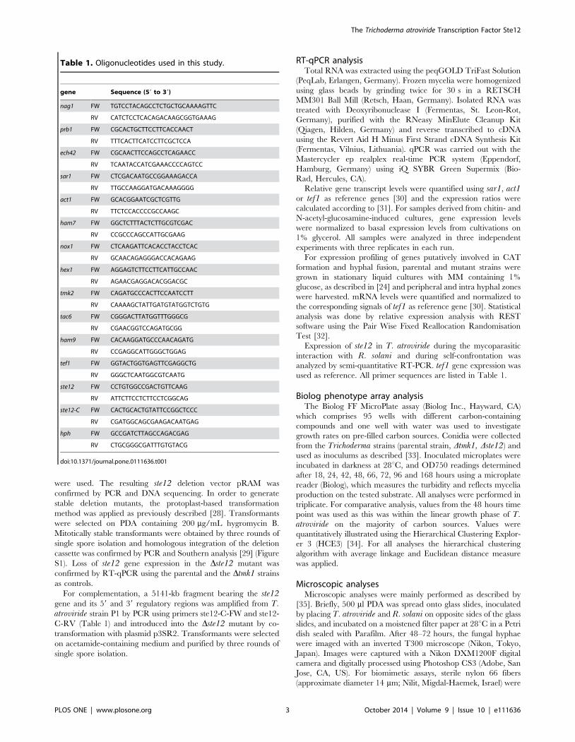
Table 1. Oligonucleotides used in this study.
gene Sequence (59 to 39)
nag1 FW TGTCCTACAGCCTCTGCTGCAAAAGTTC
RV CATCTCCTCACAGACAAGCGGTGAAAG
prb1 FW CGCACTGCTTCCTTCACCAACT
RV TTTCACTTCATCCTTCGCTCCA
ech42 FW CGCAACTTCCAGCCTCAGAACC
RV TCAATACCATCGAAACCCCAGTCC
sar1 FW CTCGACAATGCCGGAAAGACCA
RV TTGCCAAGGATGACAAAGGGG
act1 FW GCACGGAATCGCTCGTTG
RV TTCTCCACCCCGCCAAGC
ham7 FW GGCTCTTTACTCTTGCGTCGAC
RV CCGCCCAGCCATTGCGAAG
nox1 FW CTCAAGATTCACACCTACCTCAC
RV GCAACAGAGGGACCACAGAAG
hex1 FW AGGAGTCTTCCTTCATTGCCAAC
RV AGAACGAGGACACGGACGC
tmk2 FW CAGATGCCCACTTCCAATCCTT
RV CAAAAGCTATTGATGTATGGTCTGTG
tac6 FW CGGGACTTATGGTTTGGGCG
RV CGAACGGTCCAGATGCGG
ham9 FW CACAAGGATGCCCAACAGATG
RV CCGAGGCATTGGGCTGGAG
tef1 FW GGTACTGGTGAGTTCGAGGCTG
RV GGGCTCAATGGCGTCAATG
ste12 FW CCTGTGGCCGACTGTTCAAG
RV ATTCTTCCTCTTCCTCGGCAG
ste12-C FW CACTGCACTGTATTCCGGCTCCC
RV CGATGGCAGCGAAGACAATGAG
hph FW GCCGATCTTAGCCAGACGAG
RV CTGCGGGCGATTTGTGTACG
doi:10.1371/journal.pone.0111636.t001
The Trichoderma atroviride Transcription Factor Ste12
PLOS ONE | www.plosone.org 3 October 2014 | Volume 9 | Issue 10 | e111636
placed on the media before inoculation. For analysis in liquid
cultures, 16106 spores per ml in 250 ml of MM containing 1%
glucose were spread onto the glass slide and pictures were taken at
24 h and 48 h as described above.
Results
Characteristics of the T. atroviride Ste12 homologueThe aim of this study was to characterize the role of the Ste12
transcription factor, which is assumed to act as a central
component of the mycoparasitism-relevant signaling pathway
involving the Tmk1 MAPK in T. atroviride.
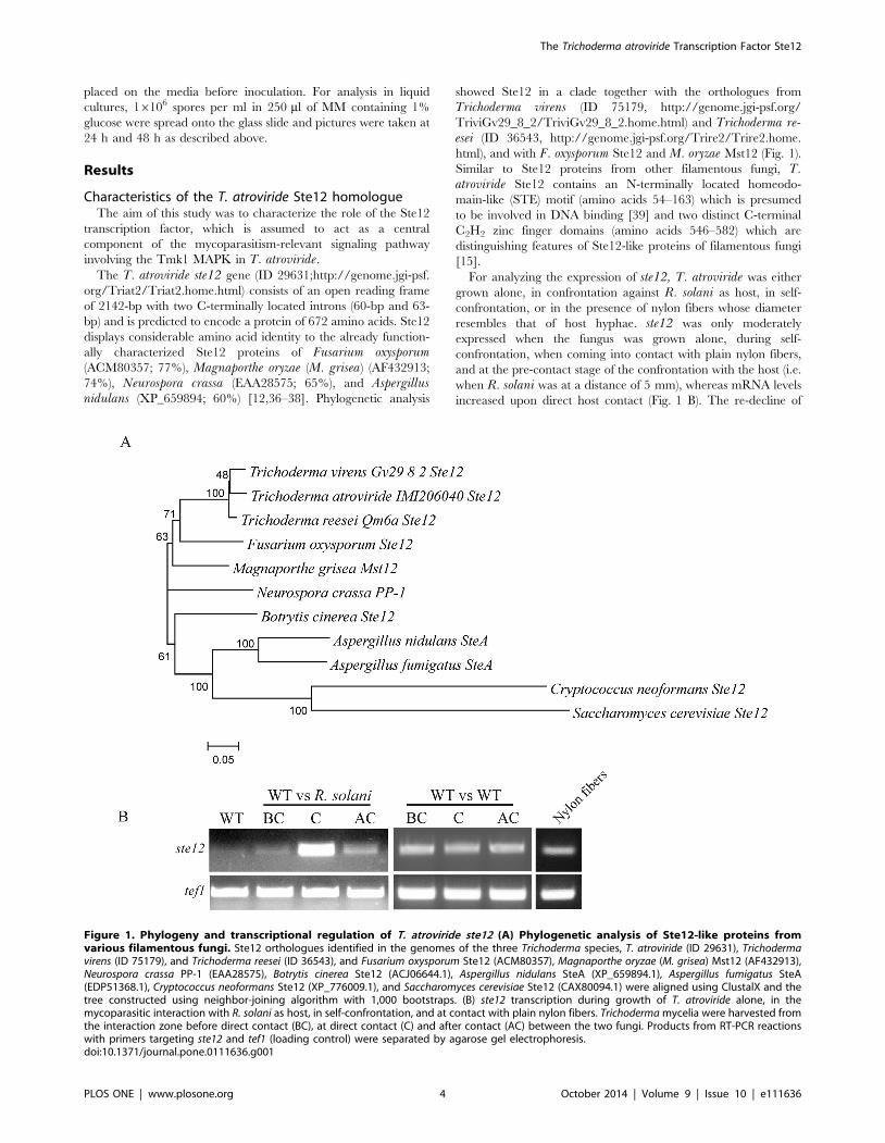
The T. atroviride ste12 gene (ID 29631;http://genome.jgi-psf.
org/Triat2/Triat2.home.html) consists of an open reading frame
of 2142-bp with two C-terminally located introns (60-bp and 63-
bp) and is predicted to encode a protein of 672 amino acids. Ste12
displays considerable amino acid identity to the already function-
ally characterized Ste12 proteins of Fusarium oxysporum(ACM80357; 77%), Magnaporthe oryzae (M. grisea) (AF432913;
74%), Neurospora crassa (EAA28575; 65%), and Aspergillusnidulans (XP_659894; 60%) [12,36–38]. Phylogenetic analysis
showed Ste12 in a clade together with the orthologues from
Trichoderma virens (ID 75179, http://genome.jgi-psf.org/
TriviGv29_8_2/TriviGv29_8_2.home.html) and Trichoderma re-esei (ID 36543, http://genome.jgi-psf.org/Trire2/Trire2.home.
html), and with F. oxysporum Ste12 and M. oryzae Mst12 (Fig. 1).
Similar to Ste12 proteins from other filamentous fungi, T.atroviride Ste12 contains an N-terminally located homeodo-
main-like (STE) motif (amino acids 54–163) which is presumed
to be involved in DNA binding [39] and two distinct C-terminal
C2H2 zinc finger domains (amino acids 546–582) which are
distinguishing features of Ste12-like proteins of filamentous fungi
[15].
For analyzing the expression of ste12, T. atroviride was either
grown alone, in confrontation against R. solani as host, in self-
confrontation, or in the presence of nylon fibers whose diameter
resembles that of host hyphae. ste12 was only moderately
expressed when the fungus was grown alone, during self-
confrontation, when coming into contact with plain nylon fibers,
and at the pre-contact stage of the confrontation with the host (i.e.
when R. solani was at a distance of 5 mm), whereas mRNA levels
increased upon direct host contact (Fig. 1 B). The re-decline of
Figure 1. Phylogeny and transcriptional regulation of T. atroviride ste12 (A) Phylogenetic analysis of Ste12-like proteins fromvarious filamentous fungi. Ste12 orthologues identified in the genomes of the three Trichoderma species, T. atroviride (ID 29631), Trichodermavirens (ID 75179), and Trichoderma reesei (ID 36543), and Fusarium oxysporum Ste12 (ACM80357), Magnaporthe oryzae (M. grisea) Mst12 (AF432913),Neurospora crassa PP-1 (EAA28575), Botrytis cinerea Ste12 (ACJ06644.1), Aspergillus nidulans SteA (XP_659894.1), Aspergillus fumigatus SteA(EDP51368.1), Cryptococcus neoformans Ste12 (XP_776009.1), and Saccharomyces cerevisiae Ste12 (CAX80094.1) were aligned using ClustalX and thetree constructed using neighbor-joining algorithm with 1,000 bootstraps. (B) ste12 transcription during growth of T. atroviride alone, in themycoparasitic interaction with R. solani as host, in self-confrontation, and at contact with plain nylon fibers. Trichoderma mycelia were harvested fromthe interaction zone before direct contact (BC), at direct contact (C) and after contact (AC) between the two fungi. Products from RT-PCR reactionswith primers targeting ste12 and tef1 (loading control) were separated by agarose gel electrophoresis.doi:10.1371/journal.pone.0111636.g001
The Trichoderma atroviride Transcription Factor Ste12
PLOS ONE | www.plosone.org 4 October 2014 | Volume 9 | Issue 10 | e111636
ste12 mRNA levels at the after contact stage, when T. atroviridehad overgrown the host by 5 mm, indicates that this host-induced
up-regulation of ste12 expression is only transient and suggests a
role of Ste12 in the regulation of mycoparasitism-relevant
processes upon direct host contact.
In contrast to Colletotrichum lindemuthianum and Botrytiscinerea, for which alternative splicing of ste12 has been described
[40,41], only full transcripts (data not shown) were found for T.atroviride ste12.
Generation of T. atroviride ste12 deletion andcomplementation mutants
For functional characterization of T. atroviride ste12, we
transformed the linearized ste12 deletion vector into T. atrovirideprotoplasts. Although all of the resulting 20 transformants showed
hygroymcin B-resistance, PCR- and Southern blot-based screen-
ing resulted in only one mutant with homologous integration and
deletion of the ste12 gene in a mitotically stable manner (Figure
S1). Complemented strains were generated by introducing a 5141-
kb fragment bearing the ste12 gene and its 59 and 39 regulatory
regions into the Dste12 mutant. Two complemented transformants
with an ectopically (ste12-C1) and homologously (ste12-C2)
integrated ste12 gene, respectively, were selected and included in
a subset of experiments. The Dste12 and complemented mutants
exhibited growth rates similar to the parental strain on solid
complete medium (PDA). However, deletion of ste12 resulted in
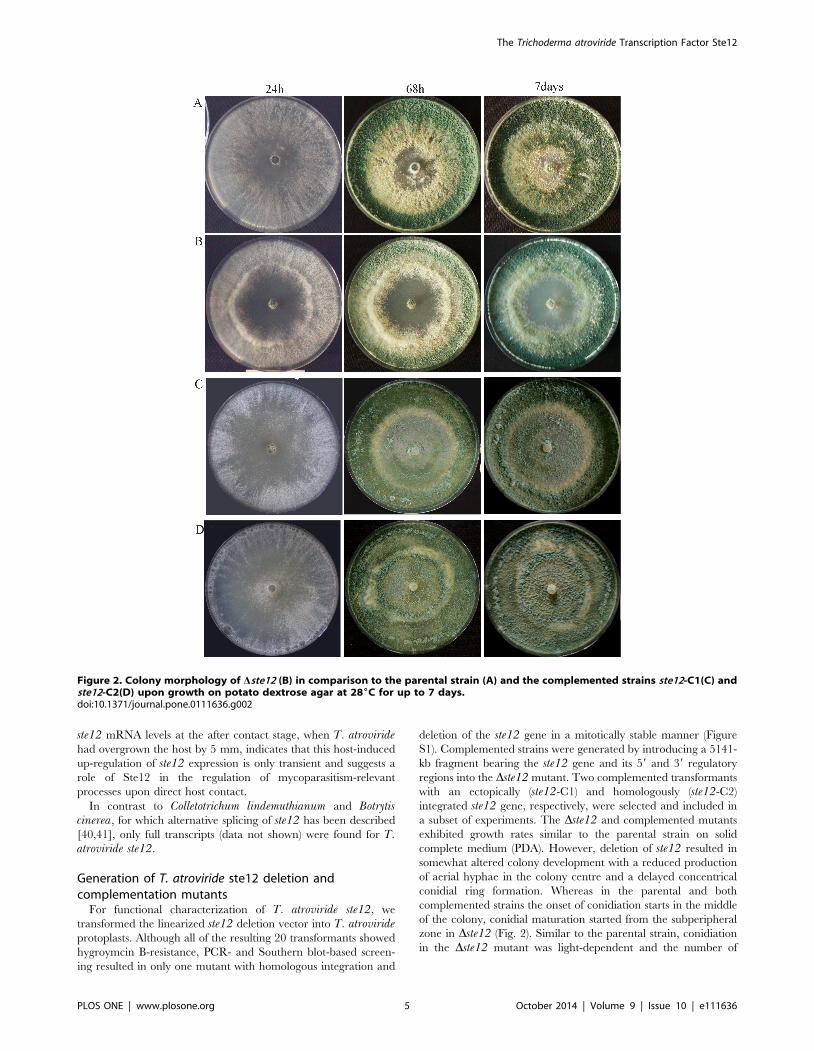
somewhat altered colony development with a reduced production
of aerial hyphae in the colony centre and a delayed concentrical
conidial ring formation. Whereas in the parental and both
complemented strains the onset of conidiation starts in the middle
of the colony, conidial maturation started from the subperipheral
zone in Dste12 (Fig. 2). Similar to the parental strain, conidiation
in the Dste12 mutant was light-dependent and the number of
Figure 2. Colony morphology of Dste12 (B) in comparison to the parental strain (A) and the complemented strains ste12-C1(C) andste12-C2(D) upon growth on potato dextrose agar at 286C for up to 7 days.doi:10.1371/journal.pone.0111636.g002
The Trichoderma atroviride Transcription Factor Ste12
PLOS ONE | www.plosone.org 5 October 2014 | Volume 9 | Issue 10 | e111636
conidia produced by Dste12 was similar to the parental strain
(1.960.3 * 109 and 1.260.2 * 109, respectively).
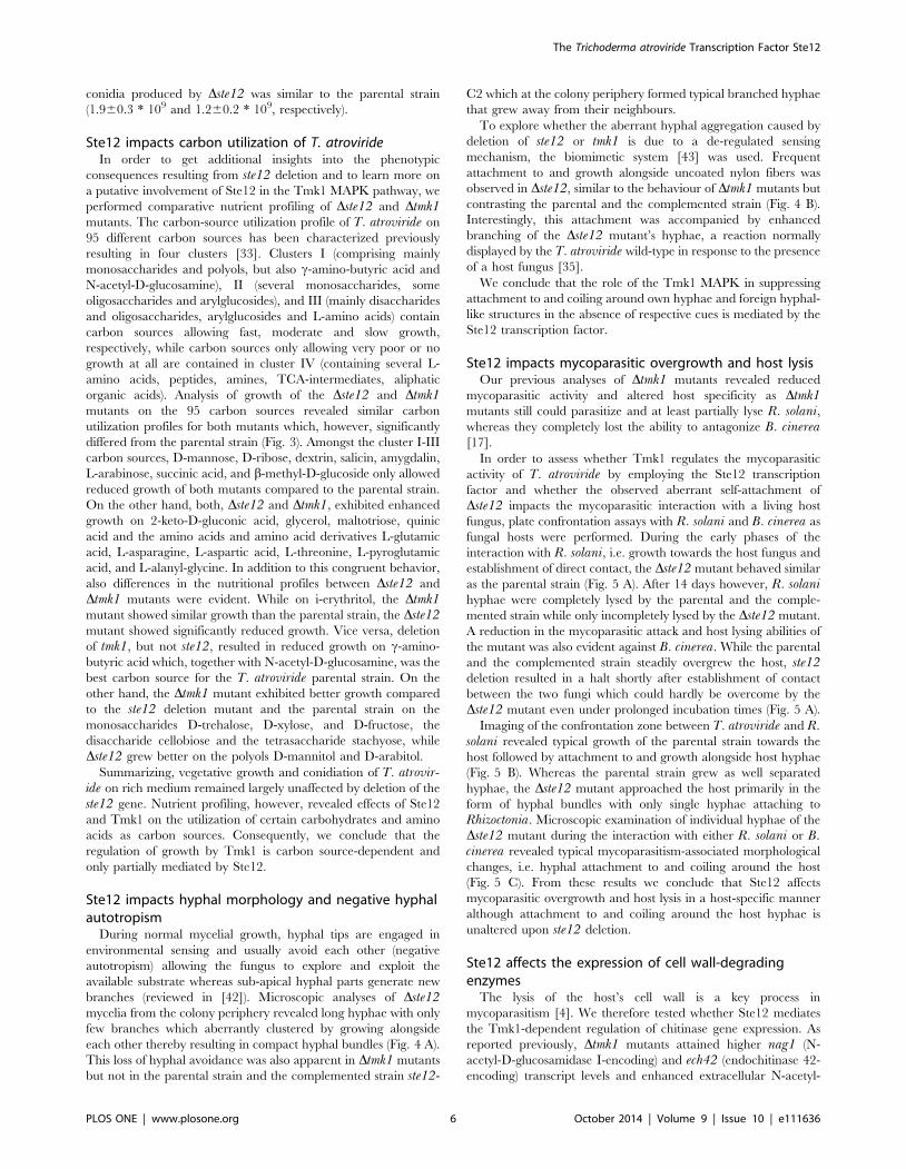
Ste12 impacts carbon utilization of T. atrovirideIn order to get additional insights into the phenotypic
consequences resulting from ste12 deletion and to learn more on
a putative involvement of Ste12 in the Tmk1 MAPK pathway, we
performed comparative nutrient profiling of Dste12 and Dtmk1mutants. The carbon-source utilization profile of T. atroviride on
95 different carbon sources has been characterized previously
resulting in four clusters [33]. Clusters I (comprising mainly
monosaccharides and polyols, but also c-amino-butyric acid and
N-acetyl-D-glucosamine), II (several monosaccharides, some
oligosaccharides and arylglucosides), and III (mainly disaccharides
and oligosaccharides, arylglucosides and L-amino acids) contain
carbon sources allowing fast, moderate and slow growth,
respectively, while carbon sources only allowing very poor or no
growth at all are contained in cluster IV (containing several L-
amino acids, peptides, amines, TCA-intermediates, aliphatic
organic acids). Analysis of growth of the Dste12 and Dtmk1mutants on the 95 carbon sources revealed similar carbon
utilization profiles for both mutants which, however, significantly
differed from the parental strain (Fig. 3). Amongst the cluster I-III
carbon sources, D-mannose, D-ribose, dextrin, salicin, amygdalin,
L-arabinose, succinic acid, and b-methyl-D-glucoside only allowed
reduced growth of both mutants compared to the parental strain.
On the other hand, both, Dste12 and Dtmk1, exhibited enhanced
growth on 2-keto-D-gluconic acid, glycerol, maltotriose, quinic
acid and the amino acids and amino acid derivatives L-glutamic
acid, L-asparagine, L-aspartic acid, L-threonine, L-pyroglutamic
acid, and L-alanyl-glycine. In addition to this congruent behavior,
also differences in the nutritional profiles between Dste12 and
Dtmk1 mutants were evident. While on i-erythritol, the Dtmk1mutant showed similar growth than the parental strain, the Dste12mutant showed significantly reduced growth. Vice versa, deletion
of tmk1, but not ste12, resulted in reduced growth on c-amino-
butyric acid which, together with N-acetyl-D-glucosamine, was the
best carbon source for the T. atroviride parental strain. On the
other hand, the Dtmk1 mutant exhibited better growth compared
to the ste12 deletion mutant and the parental strain on the
monosaccharides D-trehalose, D-xylose, and D-fructose, the
disaccharide cellobiose and the tetrasaccharide stachyose, while
Dste12 grew better on the polyols D-mannitol and D-arabitol.
Summarizing, vegetative growth and conidiation of T. atrovir-ide on rich medium remained largely unaffected by deletion of the
ste12 gene. Nutrient profiling, however, revealed effects of Ste12
and Tmk1 on the utilization of certain carbohydrates and amino
acids as carbon sources. Consequently, we conclude that the
regulation of growth by Tmk1 is carbon source-dependent and
only partially mediated by Ste12.
Ste12 impacts hyphal morphology and negative hyphalautotropism
During normal mycelial growth, hyphal tips are engaged in
environmental sensing and usually avoid each other (negative
autotropism) allowing the fungus to explore and exploit the
available substrate whereas sub-apical hyphal parts generate new
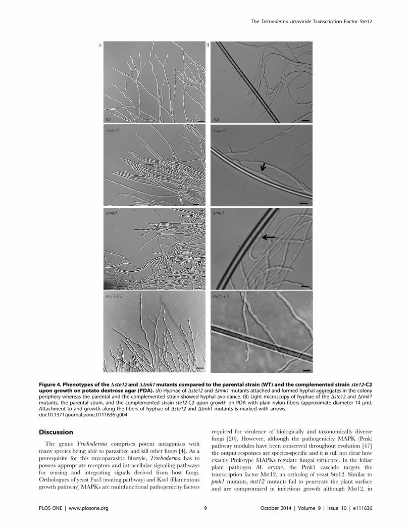
branches (reviewed in [42]). Microscopic analyses of Dste12mycelia from the colony periphery revealed long hyphae with only
few branches which aberrantly clustered by growing alongside
each other thereby resulting in compact hyphal bundles (Fig. 4 A).
This loss of hyphal avoidance was also apparent in Dtmk1 mutants
but not in the parental strain and the complemented strain ste12-
C2 which at the colony periphery formed typical branched hyphae
that grew away from their neighbours.
To explore whether the aberrant hyphal aggregation caused by
deletion of ste12 or tmk1 is due to a de-regulated sensing
mechanism, the biomimetic system [43] was used. Frequent
attachment to and growth alongside uncoated nylon fibers was
observed in Dste12, similar to the behaviour of Dtmk1 mutants but
contrasting the parental and the complemented strain (Fig. 4 B).
Interestingly, this attachment was accompanied by enhanced
branching of the Dste12 mutant’s hyphae, a reaction normally
displayed by the T. atroviride wild-type in response to the presence
of a host fungus [35].
We conclude that the role of the Tmk1 MAPK in suppressing
attachment to and coiling around own hyphae and foreign hyphal-
like structures in the absence of respective cues is mediated by the
Ste12 transcription factor.
Ste12 impacts mycoparasitic overgrowth and host lysisOur previous analyses of Dtmk1 mutants revealed reduced
mycoparasitic activity and altered host specificity as Dtmk1mutants still could parasitize and at least partially lyse R. solani,whereas they completely lost the ability to antagonize B. cinerea[17].
In order to assess whether Tmk1 regulates the mycoparasitic
activity of T. atroviride by employing the Ste12 transcription
factor and whether the observed aberrant self-attachment of
Dste12 impacts the mycoparasitic interaction with a living host
fungus, plate confrontation assays with R. solani and B. cinerea as
fungal hosts were performed. During the early phases of the
interaction with R. solani, i.e. growth towards the host fungus and
establishment of direct contact, the Dste12 mutant behaved similar
as the parental strain (Fig. 5 A). After 14 days however, R. solanihyphae were completely lysed by the parental and the comple-
mented strain while only incompletely lysed by the Dste12 mutant.
A reduction in the mycoparasitic attack and host lysing abilities of
the mutant was also evident against B. cinerea. While the parental
and the complemented strain steadily overgrew the host, ste12deletion resulted in a halt shortly after establishment of contact
between the two fungi which could hardly be overcome by the
Dste12 mutant even under prolonged incubation times (Fig. 5 A).
Imaging of the confrontation zone between T. atroviride and R.solani revealed typical growth of the parental strain towards the
host followed by attachment to and growth alongside host hyphae
(Fig. 5 B). Whereas the parental strain grew as well separated
hyphae, the Dste12 mutant approached the host primarily in the
form of hyphal bundles with only single hyphae attaching to
Rhizoctonia. Microscopic examination of individual hyphae of the
Dste12 mutant during the interaction with either R. solani or B.cinerea revealed typical mycoparasitism-associated morphological
changes, i.e. hyphal attachment to and coiling around the host
(Fig. 5 C). From these results we conclude that Ste12 affects
mycoparasitic overgrowth and host lysis in a host-specific manner
although attachment to and coiling around the host hyphae is
unaltered upon ste12 deletion.
Ste12 affects the expression of cell wall-degradingenzymes
The lysis of the host’s cell wall is a key process in
mycoparasitism [4]. We therefore tested whether Ste12 mediates
the Tmk1-dependent regulation of chitinase gene expression. As
reported previously, Dtmk1 mutants attained higher nag1 (N-
acetyl-D-glucosamidase I-encoding) and ech42 (endochitinase 42-
encoding) transcript levels and enhanced extracellular N-acetyl-
The Trichoderma atroviride Transcription Factor Ste12
PLOS ONE | www.plosone.org 6 October 2014 | Volume 9 | Issue 10 | e111636
The Trichoderma atroviride Transcription Factor Ste12
PLOS ONE | www.plosone.org 7 October 2014 | Volume 9 | Issue 10 | e111636
glucosaminidase (NAGase) and endochitinase activities under
chitinase-inducing conditions [17].
While secreted NAGase activities in N-acetyl-glucosamine-
induced cultures were decreased upon ste12 deletion at all time
points tested, the Dste12 mutant showed elevated extracellular
endochitinase activities compared to the parental strain upon
induction with colloidal chitin (Fig. 6 A). Further analysis at the
transcript level confirmed the enhanced transcription of the ech42gene in the Dste12 mutant after cultivation on colloidal chitin for
36 hours and, unexpectedly, also revealed enhanced nag1 mRNA
levels compared to the parental strain upon cultivation in the
presence of N-acetyl-glucosamine for 14 and 24 hours (Fig. 6 B).
Similar to ech42, the prb1 gene, which encodes a subtilisin-like
serine protease whose over-expression has been shown to improve
the biocontrol activity of T. atroviride [44], can be induced by
chitin. prb1 expression was highest after 36 hours of cultivation in
chitin-containing media in both the Dste12 mutant and the
parental strain with prb1 mRNA levels being ,2-fold enhanced in
the mutant.
Based on the findings that Ste12 negatively regulates the
expression of the cell wall-degrading enzymes tested but positively
affects the mycoparasitic activity of T. atroviride against R. solaniand B. cinerea, we were interested in analyzing the expression of
the mycoparasitism-relevant ech42, nag1 and prb1 genes in direct
confrontation assays. To this end, mycelia from the parental strain
and the Dste12 and Dtmk1 mutants during direct interaction with
R. solani were harvested at the early overgrowth stage which
corresponded to the stage with the most significant differences
between the Dste12 mutant and the parental strain in the
mycoparasitism assays (Fig. 5 A). While in the parental strain all
three tested genes were significantly induced during overgrowth of
R. solani compared to the self confrontation control, only prb1showed a host-induced expression pattern in the Dste12 mutant
with mRNA levels exceeding those of the parental strain by
several-fold. Expression of nag1 was elevated in both, the Dste12and the Dtmk1 mutants, although in a host-independent manner,
i.e. also in the self confrontation control. Similarly, ech42 gene
transcription was independent from host-derived signals in both
mutants with ech42 mRNA levels in the Dtmk1 mutant
significantly exceeding those of the parental strain and of the
Dste12 mutant (Fig. 6 C).
These results suggest that Ste12 and Tmk1 negatively regulate
the expression of genes important for host cell wall degradation
and host lysis in T. atroviride.
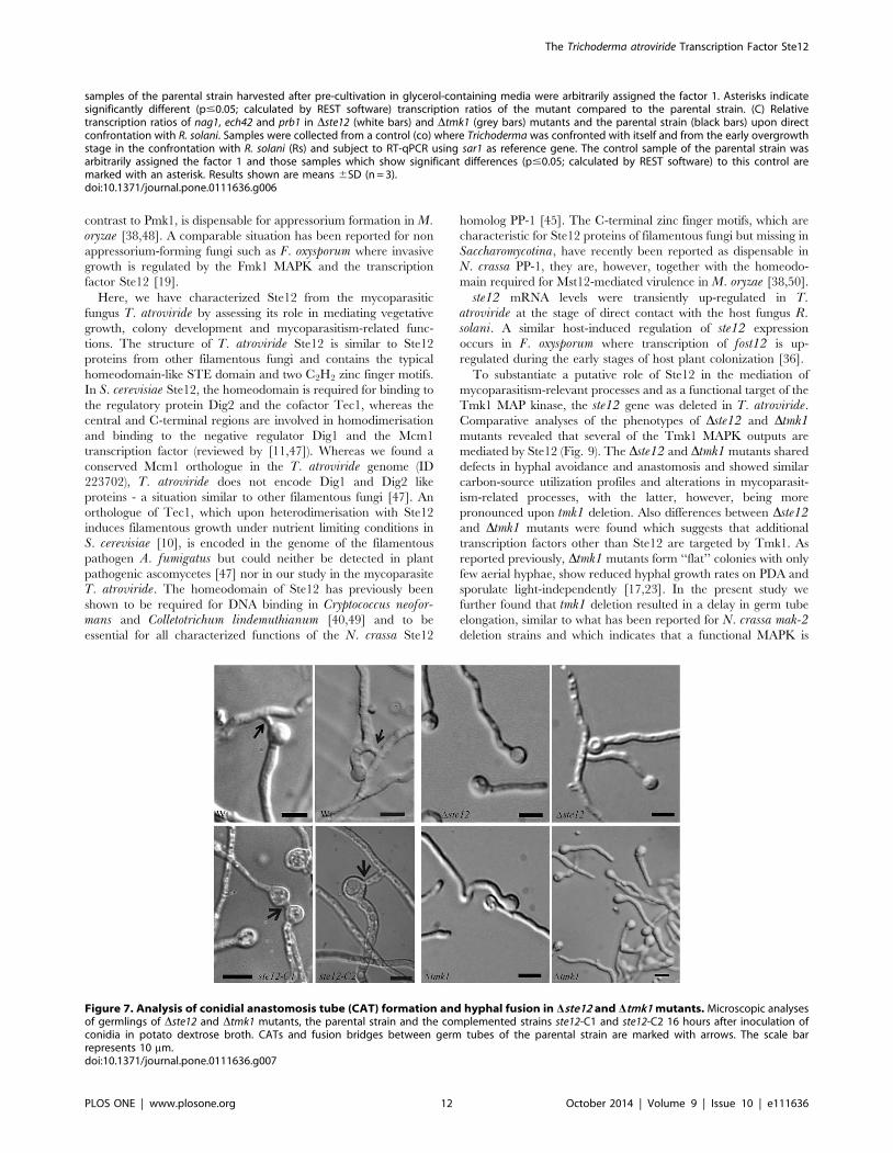
Ste12 mediates the influence of Tmk1 on CAT formationand hyphal fusion
Vegetative hyphal fusion is important for the development of a
mycelial network during colony development in many filamentous
fungi. In addition to fusion of hyphae within a mature colony,
germlings of N. crassa recognize each other shortly after conidial
germination and can fuse via specialized structures, the conidial
anastomosis tubes (CATs) [45]. In N. crassa and F. oxysporum, the
Fus3/Kss1 homologues Mak-2 and Fmk1, respectively, are
required for both CAT and hyphal fusion during vegetative
growth [21,46].
Despite the fact that T. atroviride Dste12 and Dtmk1 mutants
lost negative hyphal autotropism in the colony periphery which
resulted in the observed hyphal aggregation, we were not able to
detect distinct fusions between aggregated hyphae. To further
analyze a putative role of Tmk1 and Ste12 in regulating fusion
processes, the behavior of Dste12 and Dtmk1 germlings was
assessed microscopically. Conidial anastomosis tubes as well as
fusion bridges between germ tubes were frequently observed in the
parental and the complemented strain (Fig. 7). In contrast, CATs
could not be detected in the Dste12 and Dtmk1 mutants and also
fusions between germ tubes were only rarely observed despite
frequent contacts between the germlings. It is worth mentioning
that Dtmk1 conidia showed delayed germ tube formation and
extensive aggregation of Dtmk1 germlings occurred (Fig. 7).
Recent studies in the fungal model N. crassa led to the
identification of target genes being required for cell fusion which
are under control of the Mak-2 MAPK and the Ste12 homolog
PP-1 [45]. To further substantiate our above findings of Tmk1 and
Ste12 playing key roles in fusion processes in T. atroviride, mycelia
of Dste12 and Dtmk1 mutants and the parental strain were
harvested from the internal as well as peripheral zones of the
fungal colony. For gene expression analyses, the respective T.atroviride orthologues (Ta300768, tmk2, hex1, Ta302802,
Ta294940) of the N. crassa fusion genes ham-7 (encoding a
GPI-anchored protein required for activation of the cell wall
integrity MAPK MAK-1), mak-1 (MAPK), hex-1 (involved in
septal plugging), nox-1 (NADPH oxidase), and ham-9 (pleckstrin
domain protein) were identified in the T. atroviride genome
database (http://genome.jgi-psf.org/Triat2/Triat2.home.html) by
BLAST searches. Moreover, the glycosyl-hydrolase 18 (GH18)
subgroup C chitinase-encoding gene tac6 (Ta348129), which plays
a role in hyphal network formation in T. atroviride [24], was
included in the study.
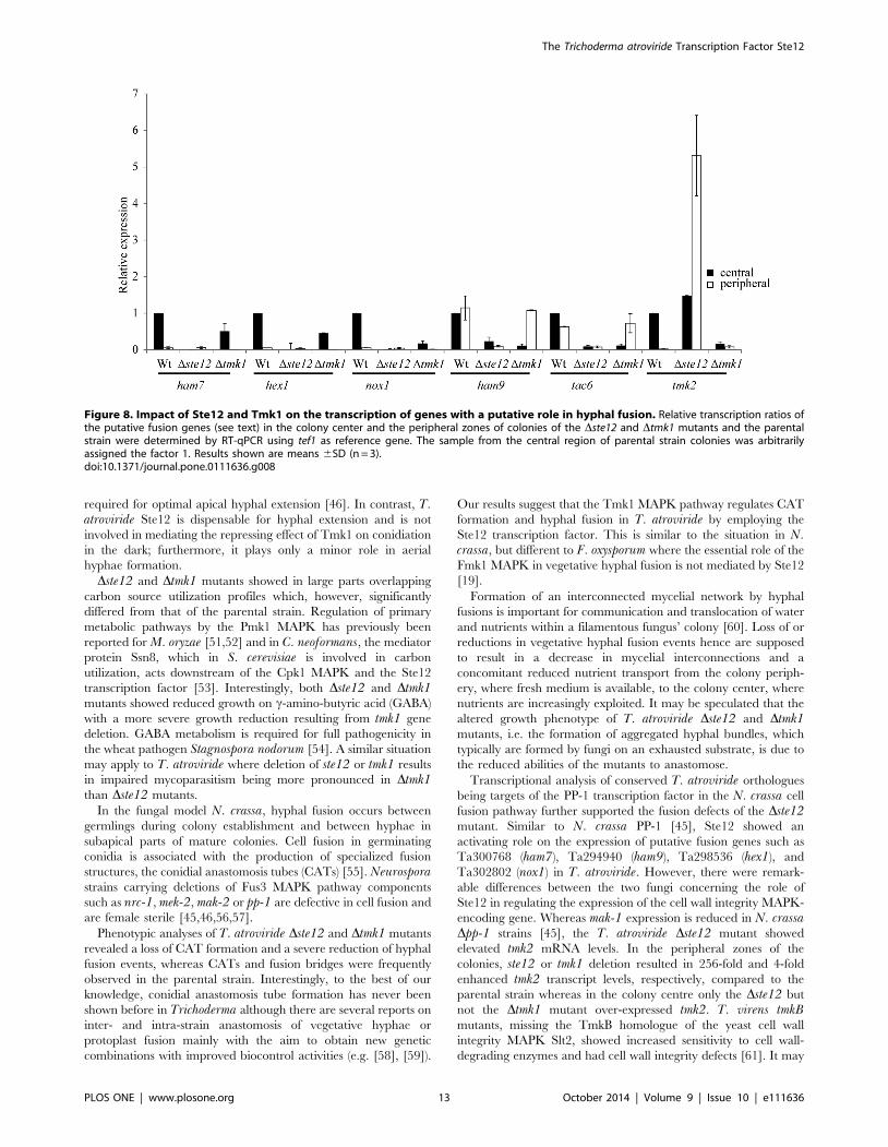
Of the genes tested, Ta300768/ham-7, hex1, Ta302802/nox-1,
tmk2, and tac6 showed a significantly higher transcription in the
center than at the peripheral zone of T. atroviride colonies. This
expression pattern would be indicative of a role of these genes in
hyphal fusion which predominantly takes place in the colony
center (Figure 8). In accordance with tmk1 and ste12 deletion
resulting in a loss of cell fusion, mRNA levels of Ta300768/ham-7,
hex1, Ta302802/nox-1, and tac6 were reduced in both the center
and the periphery of Dste12 and Dtmk1 colonies compared to the
parental strain colony center. Ta294940/ham9 was found to be
similarly transcribed throughout the wild-type colony and the
Dtmk1 peripheral zone but showed heavily reduced expression in
Dste12 colonies. A completely different picture was obtained for
tmk2, which encodes the T. atroviride homolog of the cell wall
integrity pathway MAPK Slt2 of S. cerevisiae and Mak-1 of N.crassa. While deletion of tmk1 was found to result in significantly
reduced tmk2 transcription, Dste12 mutants showed enhanced
tmk2 mRNA levels especially in the peripheral zone of the colony
(Figure 8). These results show that expression of the cell wall
integrity MAPK Tmk2 in T. atroviride is positively regulated by
Tmk1 but negatively affected by Ste12 under the conditions tested
and suggest that the repressing effect of Ste12 is mediated by
upstream components other than Tmk1.
Figure 3. Comparative carbon source utilization profiles of the Dste12 and Dtmk1 mutants and the parental strain (WT). Strains weregrown on 95 carbon sources (FF-plates) using the Biolog Phenotype Microarray system. Hierarchical clustering results are displayed as colouredmosaics attached to a dendrogram according to growth (OD750) after 48 hours of incubation. All data points are the average of three replicates. Thethreshold cut-off for all conditions was set at 0.225 which corresponded to growth of the parental strain on the water control. Red boxes indicatemaximal growth (OD750 0.7–1.3), black boxes medium growth (OD750 0.55–0.7), and green boxes weak growth (OD750 0.2–0.55).doi:10.1371/journal.pone.0111636.g003
The Trichoderma atroviride Transcription Factor Ste12
PLOS ONE | www.plosone.org 8 October 2014 | Volume 9 | Issue 10 | e111636
Discussion
The genus Trichoderma comprises potent antagonists with
many species being able to parasitize and kill other fungi [4]. As a
prerequisite for this mycoparasitic lifestyle, Trichoderma has to
possess appropriate receptors and intracellular signaling pathways
for sensing and integrating signals derived from host fungi.
Orthologues of yeast Fus3 (mating pathway) and Kss1 (filamentous
growth pathway) MAPKs are multifunctional pathogenicity factors
required for virulence of biologically and taxonomically diverse
fungi [20]. However, although the pathogenicity MAPK (Pmk)
pathway modules have been conserved throughout evolution [47]
the output responses are species-specific and it is still not clear how
exactly Pmk-type MAPKs regulate fungal virulence. In the foliar
plant pathogen M. oryzae, the Pmk1 cascade targets the
transcription factor Mst12, an ortholog of yeast Ste12. Similar to
pmk1 mutants, mst12 mutants fail to penetrate the plant surface
and are compromised in infectious growth although Mst12, in
Figure 4. Phenotypes of the Dste12 and Dtmk1 mutants compared to the parental strain (WT) and the complemented strain ste12-C2upon growth on potato dextrose agar (PDA). (A) Hyphae of Dste12 and Dtmk1 mutants attached and formed hyphal aggregates in the colonyperiphery whereas the parental and the complemented strain showed hyphal avoidance. (B) Light microscopy of hyphae of the Dste12 and Dtmk1mutants, the parental strain, and the complemented strain ste12-C2 upon growth on PDA with plain nylon fibers (approximate diameter 14 mm).Attachment to and growth along the fibers of hyphae of Dste12 and Dtmk1 mutants is marked with arrows.doi:10.1371/journal.pone.0111636.g004
The Trichoderma atroviride Transcription Factor Ste12
PLOS ONE | www.plosone.org 9 October 2014 | Volume 9 | Issue 10 | e111636
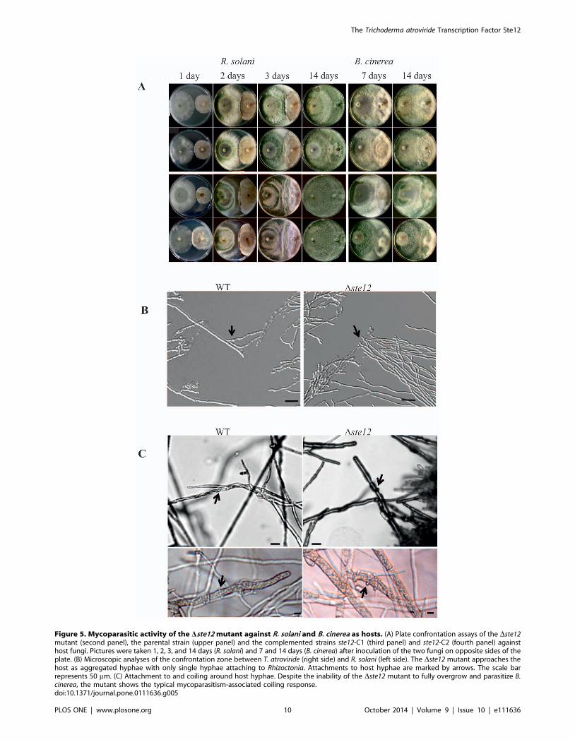
Figure 5. Mycoparasitic activity of the Dste12 mutant against R. solani and B. cinerea as hosts. (A) Plate confrontation assays of the Dste12mutant (second panel), the parental strain (upper panel) and the complemented strains ste12-C1 (third panel) and ste12-C2 (fourth panel) againsthost fungi. Pictures were taken 1, 2, 3, and 14 days (R. solani) and 7 and 14 days (B. cinerea) after inoculation of the two fungi on opposite sides of theplate. (B) Microscopic analyses of the confrontation zone between T. atroviride (right side) and R. solani (left side). The Dste12 mutant approaches thehost as aggregated hyphae with only single hyphae attaching to Rhizoctonia. Attachments to host hyphae are marked by arrows. The scale barrepresents 50 mm. (C) Attachment to and coiling around host hyphae. Despite the inability of the Dste12 mutant to fully overgrow and parasitize B.cinerea, the mutant shows the typical mycoparasitism-associated coiling response.doi:10.1371/journal.pone.0111636.g005
The Trichoderma atroviride Transcription Factor Ste12
PLOS ONE | www.plosone.org 10 October 2014 | Volume 9 | Issue 10 | e111636
Figure 6. Impact of Ste12 on the expression of mycoparasitism-related cell wall-degrading enzymes. (A) Extracellular N-acetyl-glucosaminidase (NAGase) and endochitinase activities in the Dste12 mutant (black bars) and the parental strain (white bars). After pre-cultivation on1% glycerol, mycelial biomass was transferred to 1% N-acetyl-glucosamine-containing media for inducing NAGases and to 1% colloidal chitin-containing media for induction of endochitinases. Culture filtrates were harvested at the indicated time points and determined enzyme activitiesrelated to intracellular total protein. (B) Relative transcription ratios of the chitinase-encoding nag1 and ech42 genes and the prb1 protease-encodinggene in the Dste12 mutant (black bars) and the parental strain (white bars). RT-qPCR analyses were performed 5, 14, and 24 hours after transfer to N-acetyl-glucosamine (nag1) and 14, 24, 36, and 48 hours after transfer to colloidal chitin (ech42, prb1) using act1 as reference gene. Un-induced
The Trichoderma atroviride Transcription Factor Ste12
PLOS ONE | www.plosone.org 11 October 2014 | Volume 9 | Issue 10 | e111636
contrast to Pmk1, is dispensable for appressorium formation in M.oryzae [38,48]. A comparable situation has been reported for non
appressorium-forming fungi such as F. oxysporum where invasive
growth is regulated by the Fmk1 MAPK and the transcription
factor Ste12 [19].
Here, we have characterized Ste12 from the mycoparasitic
fungus T. atroviride by assessing its role in mediating vegetative
growth, colony development and mycoparasitism-related func-
tions. The structure of T. atroviride Ste12 is similar to Ste12
proteins from other filamentous fungi and contains the typical
homeodomain-like STE domain and two C2H2 zinc finger motifs.
In S. cerevisiae Ste12, the homeodomain is required for binding to
the regulatory protein Dig2 and the cofactor Tec1, whereas the
central and C-terminal regions are involved in homodimerisation
and binding to the negative regulator Dig1 and the Mcm1
transcription factor (reviewed by [11,47]). Whereas we found a
conserved Mcm1 orthologue in the T. atroviride genome (ID
223702), T. atroviride does not encode Dig1 and Dig2 like
proteins - a situation similar to other filamentous fungi [47]. An
orthologue of Tec1, which upon heterodimerisation with Ste12
induces filamentous growth under nutrient limiting conditions in
S. cerevisiae [10], is encoded in the genome of the filamentous
pathogen A. fumigatus but could neither be detected in plant
pathogenic ascomycetes [47] nor in our study in the mycoparasite
T. atroviride. The homeodomain of Ste12 has previously been
shown to be required for DNA binding in Cryptococcus neofor-mans and Colletotrichum lindemuthianum [40,49] and to be
essential for all characterized functions of the N. crassa Ste12
homolog PP-1 [45]. The C-terminal zinc finger motifs, which are
characteristic for Ste12 proteins of filamentous fungi but missing in
Saccharomycotina, have recently been reported as dispensable in
N. crassa PP-1, they are, however, together with the homeodo-
main required for Mst12-mediated virulence in M. oryzae [38,50].
ste12 mRNA levels were transiently up-regulated in T.atroviride at the stage of direct contact with the host fungus R.solani. A similar host-induced regulation of ste12 expression
occurs in F. oxysporum where transcription of fost12 is up-
regulated during the early stages of host plant colonization [36].
To substantiate a putative role of Ste12 in the mediation of
mycoparasitism-relevant processes and as a functional target of the
Tmk1 MAP kinase, the ste12 gene was deleted in T. atroviride.
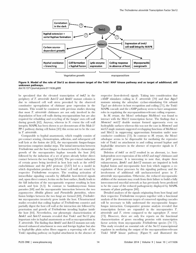
Comparative analyses of the phenotypes of Dste12 and Dtmk1mutants revealed that several of the Tmk1 MAPK outputs are
mediated by Ste12 (Fig. 9). The Dste12 and Dtmk1 mutants shared
defects in hyphal avoidance and anastomosis and showed similar
carbon-source utilization profiles and alterations in mycoparasit-
ism-related processes, with the latter, however, being more
pronounced upon tmk1 deletion. Also differences between Dste12and Dtmk1 mutants were found which suggests that additional
transcription factors other than Ste12 are targeted by Tmk1. As
reported previously, Dtmk1 mutants form ‘‘flat’’ colonies with only
few aerial hyphae, show reduced hyphal growth rates on PDA and
sporulate light-independently [17,23]. In the present study we
further found that tmk1 deletion resulted in a delay in germ tube
elongation, similar to what has been reported for N. crassa mak-2deletion strains and which indicates that a functional MAPK is
samples of the parental strain harvested after pre-cultivation in glycerol-containing media were arbitrarily assigned the factor 1. Asterisks indicatesignificantly different (p#0.05; calculated by REST software) transcription ratios of the mutant compared to the parental strain. (C) Relativetranscription ratios of nag1, ech42 and prb1 in Dste12 (white bars) and Dtmk1 (grey bars) mutants and the parental strain (black bars) upon directconfrontation with R. solani. Samples were collected from a control (co) where Trichoderma was confronted with itself and from the early overgrowthstage in the confrontation with R. solani (Rs) and subject to RT-qPCR using sar1 as reference gene. The control sample of the parental strain wasarbitrarily assigned the factor 1 and those samples which show significant differences (p#0.05; calculated by REST software) to this control aremarked with an asterisk. Results shown are means 6SD (n = 3).doi:10.1371/journal.pone.0111636.g006
Figure 7. Analysis of conidial anastomosis tube (CAT) formation and hyphal fusion in Dste12 and Dtmk1 mutants. Microscopic analysesof germlings of Dste12 and Dtmk1 mutants, the parental strain and the complemented strains ste12-C1 and ste12-C2 16 hours after inoculation ofconidia in potato dextrose broth. CATs and fusion bridges between germ tubes of the parental strain are marked with arrows. The scale barrepresents 10 mm.doi:10.1371/journal.pone.0111636.g007
The Trichoderma atroviride Transcription Factor Ste12
PLOS ONE | www.plosone.org 12 October 2014 | Volume 9 | Issue 10 | e111636
required for optimal apical hyphal extension [46]. In contrast, T.atroviride Ste12 is dispensable for hyphal extension and is not
involved in mediating the repressing effect of Tmk1 on conidiation
in the dark; furthermore, it plays only a minor role in aerial
hyphae formation.
Dste12 and Dtmk1 mutants showed in large parts overlapping
carbon source utilization profiles which, however, significantly
differed from that of the parental strain. Regulation of primary
metabolic pathways by the Pmk1 MAPK has previously been
reported for M. oryzae [51,52] and in C. neoformans, the mediator
protein Ssn8, which in S. cerevisiae is involved in carbon
utilization, acts downstream of the Cpk1 MAPK and the Ste12
transcription factor [53]. Interestingly, both Dste12 and Dtmk1mutants showed reduced growth on c-amino-butyric acid (GABA)
with a more severe growth reduction resulting from tmk1 gene
deletion. GABA metabolism is required for full pathogenicity in
the wheat pathogen Stagnospora nodorum [54]. A similar situation
may apply to T. atroviride where deletion of ste12 or tmk1 results
in impaired mycoparasitism being more pronounced in Dtmk1than Dste12 mutants.
In the fungal model N. crassa, hyphal fusion occurs between
germlings during colony establishment and between hyphae in
subapical parts of mature colonies. Cell fusion in germinating
conidia is associated with the production of specialized fusion
structures, the conidial anastomosis tubes (CATs) [55]. Neurosporastrains carrying deletions of Fus3 MAPK pathway components
such as nrc-1, mek-2, mak-2 or pp-1 are defective in cell fusion and
are female sterile [45,46,56,57].
Phenotypic analyses of T. atroviride Dste12 and Dtmk1 mutants
revealed a loss of CAT formation and a severe reduction of hyphal
fusion events, whereas CATs and fusion bridges were frequently
observed in the parental strain. Interestingly, to the best of our
knowledge, conidial anastomosis tube formation has never been
shown before in Trichoderma although there are several reports on
inter- and intra-strain anastomosis of vegetative hyphae or
protoplast fusion mainly with the aim to obtain new genetic
combinations with improved biocontrol activities (e.g. [58], [59]).
Our results suggest that the Tmk1 MAPK pathway regulates CAT
formation and hyphal fusion in T. atroviride by employing the
Ste12 transcription factor. This is similar to the situation in N.crassa, but different to F. oxysporum where the essential role of the
Fmk1 MAPK in vegetative hyphal fusion is not mediated by Ste12
[19].
Formation of an interconnected mycelial network by hyphal
fusions is important for communication and translocation of water
and nutrients within a filamentous fungus’ colony [60]. Loss of or
reductions in vegetative hyphal fusion events hence are supposed
to result in a decrease in mycelial interconnections and a
concomitant reduced nutrient transport from the colony periph-
ery, where fresh medium is available, to the colony center, where
nutrients are increasingly exploited. It may be speculated that the
altered growth phenotype of T. atroviride Dste12 and Dtmk1mutants, i.e. the formation of aggregated hyphal bundles, which
typically are formed by fungi on an exhausted substrate, is due to
the reduced abilities of the mutants to anastomose.
Transcriptional analysis of conserved T. atroviride orthologues
being targets of the PP-1 transcription factor in the N. crassa cell
fusion pathway further supported the fusion defects of the Dste12mutant. Similar to N. crassa PP-1 [45], Ste12 showed an
activating role on the expression of putative fusion genes such as
Ta300768 (ham7), Ta294940 (ham9), Ta298536 (hex1), and
Ta302802 (nox1) in T. atroviride. However, there were remark-
able differences between the two fungi concerning the role of
Ste12 in regulating the expression of the cell wall integrity MAPK-
encoding gene. Whereas mak-1 expression is reduced in N. crassaDpp-1 strains [45], the T. atroviride Dste12 mutant showed
elevated tmk2 mRNA levels. In the peripheral zones of the
colonies, ste12 or tmk1 deletion resulted in 256-fold and 4-fold
enhanced tmk2 transcript levels, respectively, compared to the
parental strain whereas in the colony centre only the Dste12 but
not the Dtmk1 mutant over-expressed tmk2. T. virens tmkBmutants, missing the TmkB homologue of the yeast cell wall
integrity MAPK Slt2, showed increased sensitivity to cell wall-
degrading enzymes and had cell wall integrity defects [61]. It may
Figure 8. Impact of Ste12 and Tmk1 on the transcription of genes with a putative role in hyphal fusion. Relative transcription ratios ofthe putative fusion genes (see text) in the colony center and the peripheral zones of colonies of the Dste12 and Dtmk1 mutants and the parentalstrain were determined by RT-qPCR using tef1 as reference gene. The sample from the central region of parental strain colonies was arbitrarilyassigned the factor 1. Results shown are means 6SD (n = 3).doi:10.1371/journal.pone.0111636.g008
The Trichoderma atroviride Transcription Factor Ste12
PLOS ONE | www.plosone.org 13 October 2014 | Volume 9 | Issue 10 | e111636
be speculated that the elevated transcription of tmk2 in the
periphery of T. atroviride Dste12 and Dtmk1 mutant colonies is
due to enhanced cell wall stress provoked by the observed
constitutive up-regulation of chitinase gene expression in the
mutants. This would be consistent with previous studies showing
that most T. atroviride chitinases are not only involved in the
degradation of host cell walls during mycoparasitism but are also
required for rebuilding and recycling of the fungus’ own cell wall
during growth [62]. Anyway, whereas in N. crassa the cell wall
integrity MAPK has been shown to act downstream of the Mak-2/
PP-1 pathway during cell fusion [45] this seems not to be the case
in T. atroviride.
Comparable to hyphal anastomosis, which roughly consists of
pre-contact sensing, chemotropism, adhesion, and subsequent cell
wall lysis at the fusion site [63], the mycoparasitic fungus-fungus
interaction comprises similar steps. The initial interaction between
Trichoderma and the host fungus is characterized by chemotropic
growth of the mycoparasites hyphae towards the host [64]
followed by the induction of a set of genes already before direct
contact between the two fungi [65,66]. The pre-contact induction
of certain genes being involved in host lysis such as the ech42endochitinase and the prb1 protease ([3,67] led to a model in
which degradation products of the hosts’ cell wall are sensed by
respective Trichoderma receptors. The resulting activation of
intracellular signaling cascades by diffusible host-derived signals
and, upon direct contact, lectins on the host surface, finally leads to
the full induction of the mycoparasitic response resulting in host
attack and lysis [4,5]. In contrast to basidiomycetous fusion
parasites [68] and the mycoparasitic interaction between the two
zygomycetes Absidia glauca and Parasitella parasitica, where
fusion bridges between the two fungi are formed [69], Trichoder-ma mycoparasites invasively grow inside the host. Ultrastructural
studies revealed that coiling hyphae of Trichoderma constrict and
partially digest the host cell wall at the interaction site followed by
penetration and growth of Trichoderma invading hyphae inside
the host [64]. Nevertheless, our phenotypic characterization of
Dtmk1 and Dste12 mutants revealed that Tmk1 and Ste12 play
important roles in hyphal anastomosis as well as mycoparasitism in
T. atroviride. The finding that Dtmk1 and Dste12 mutants not only
showed enhanced attachment to own and foreign hyphae but also
to hyphal-like plain nylon fibers suggests a repressing role of the
Tmk1 signaling pathway on hyphal attachment in the absence of
respective (host-derived) signals. Taking into consideration that
cAMP stimulates coiling in T. atroviride [70] and that Dtga3mutants missing the adenylate cyclase-stimulating Ga subunit
Tga3 are defective in host recognition and coiling [71], the Tmk1
MAPK cascade and the cAMP pathway seem to have antagonistic
roles in regulating the mycoparasitism-relevant coiling response.
In M. oryzae, the Mcm1 orthologue MoMcm1 was found to
interact with the Mst12 transcription factor. The findings that a
Momcm1 mst12 double mutant formed appressoria even on
hydrophilic surfaces whereas this was not the case in Momcm1 and
mst12 single mutants suggested overlapping functions of MoMcm1
and Mst12 in suppressing appressorium formation under non-
conducive conditions [72]. In contrast to M. oryzae, the Mcm1
othologue seems to be dispensable for mediating the repressive
role of Tmk1 on attachment to and coiling around hyphae and
hyphal-like structures in the absence of respective signals in T.atroviride.
Deletion of tmk1 or ste12 resulted in an aberrant, i.e. host-
independent over-expression of the ech42 and nag1 chitinases and
the prb1 protease. It is interesting to note that, despite these
enhancements, Dtmk1 and Dste12 mutants are impaired in both
hyphal fusion and mycoparasitic host lysis which suggests a co-
regulation of these processes by this signaling pathway and the
involvement of additional still uncharacterized genes in T.atroviride mycoparasitism. Otherwise, the reduced mycoparasitic
abilities of the mutants may result from their failure to build a fully
interconnected mycelial network as has previously been predicted
to be the cause of the reduced pathogenicity displayed by MAPK
mutants of plant pathogens [60].
Detailed analyses of the signals originating from host fungi and
their respective Trichoderma receptors together with a functional
analysis of the downstream targets of conserved signaling cascades
will be necessary to fully understand the mycoparasitic fungus-
fungus interaction. Comparative genome analysis revealed that
transcription factors are expanded in the mycoparasites T.atroviride and T. virens compared to the saprophyte T. reesei[73]. However, there are only few reports on the functional
characterization of genes encoding proteins with transcription
factor activity from mycoparasitic Trichoderma species [74–77].
This study revealed an essential role of the Ste12 transcriptional
regulator in mediating the output of the mycoparasitism-relevant
Tmk1 MAP kinase pathway (Figure 9) and illustrated the
Figure 9. Model of the role of Ste12 as down-stream target of the Tmk1 MAP kinase pathway and as target of additional, stillunknown pathways.doi:10.1371/journal.pone.0111636.g009
The Trichoderma atroviride Transcription Factor Ste12
PLOS ONE | www.plosone.org 14 October 2014 | Volume 9 | Issue 10 | e111636
interconnection between hyphal anastomosis and the mycopar-
asitic activity of T. atroviride.
Supporting Information
Figure S1 Genotypic analysis of Dste12 gene deletionand complementation mutants. (A) PCR analysis of the three
out of 20 hygromycinB-resistant transformants that showed a
stable integration of the ste12 deletion construct after three rounds
of single spore isolation. The primer pair hph-FW and hph-RV
(Table 1) amplified a 560-bp fragment of the integrated hph gene.
(B) Southern hydridization of NcoI-digested DNA from parental
strain (WT) and the three different putative deletion mutants (D, F,
S) with a 2693-bp probe covering 1415-bp of the 59 non-coding
region of the ste12 gene and 1278-bp of the hph selection marker
cassette. The parental strain and transformants F and S show a
1597-bp band indicative of the native ste12 gene, while
transformant D lacks this band and instead shows two bands of
2314-bp and 3360-bp confirming transformant D as a ste12 null
mutant resulting from homologous recombination at the ste12locus. (C) Confirmation of complementation mutants by PCR
using primers ste12-C-FW and ste12-C-RV (Table 1) located
1500-bp 59 and 39, respectively, of the ste12 open reading frame.
This primer pair is expected to amplify a 5141-bp fragment in the
parental strain (lane 5) and a 6159-bp fragment in the Dste12mutant (lanes 1 and 4). The amplification of both fragments in
complementation mutant ste12-C1 (lane 2) confirms ectopic
integration of ste12, whereas the presence of only the 5141-bp
band in complementation mutant ste12-C2 (lane 3) is indicative of
a rescue of the ste12 gene at the homologous locus by replacement
of the deletion construct. (D) RT-PCR with primers ste12-FW and
ste12-RV (Table 1) amplified the expected 320-bp fragment of the
ste12 gene in the parental strain (lane 3), the Dtmk1 mutant (lane
2), and the ste12 complemented strains (lanes 4 and 5) but not in
the Dste12 deletion mutant (lane 1).
(TIF)
Acknowledgments
We thank Markus Omann and Rene Mayer for help with deletion
construct design and mutant generation.
Author Contributions
Conceived and designed the experiments: SZ SG. Performed the
experiments: SG. Analyzed the data: SZ SG. Contributed reagents/
materials/analysis tools: SZ. Wrote the paper: SZ SG.
References
1. Hjeljord L, Tronsmo A (1998) Trichoderma and Gliocladium in Biological
Control: An Overview. In: Harman GE, Kubicek CP, editors. Trichoderma and
Gliocladium. Vol. 2. Enzymes, Biological Control and Commercial Applica-
tions. London: Taylor and Francis Ltd. pp. 131–151.
2. Inbar J, Chet I (1994) A newly isolated lectin from the plant pathogenic fungus
Sclerotium rolfsii: purification, characterization and role in mycoparasitism.
Microbiology 140: 651–657.
3. Zeilinger S, Galhaup C, Payer K, Woo SL, Mach RL et al. (1999) Chitinase
gene expression during mycoparasitic interaction of Trichoderma harzianum
with its host. Fungal Genet Biol 26(2):131–140.
4. Druzhinina IS, Seidl-Seiboth V, Herrera-Estrella A, Horwitz BA, Kenerley CM
et al. (2011) Trichoderma: the genomics of opportunistic success. Nature Rev
Microbiol 9(10):749–759.
5. Zeilinger S, Omann M (2007) Trichoderma biocontrol: signal transduction
pathways involved in host sensing and mycoparasitism. Gene Regul Syst Bio
1:227–234.
6. Mukherjee PK, Horwitz BA, Herrera-Estrella A, Schmoll M, Kenerley CM
(2013) Trichoderma research in the genome era. Annu Rev Phytopathol
51:105–129.
7. Cargnello M, Roux PP (2011) Activation and function of the MAPKs and their
substrates, the MAPK-activated protein kinases. Microbiol Mol Biol Rev
75(1):50–83.
8. Gustin MC, Albertyn J, Alexander M, Davenport K (1998) MAP kinase
pathways in the yeast Saccharomyces cerevisiae. Microbiol Mol Biol Rev
62(4):1264–1300.
9. Xu J-R (1997) MAP Kinases in Fungal Pathogens. Fungal Genet Biol 31(3):137–
152.
10. Madhani HD, Fink GR (1997) Combinatorial control required for the specificity
of yeast MAPK signaling. Science 275(5304):1314–1317.
11. Rispail N, Di Pietro A (2010) The homeodomain transcription factor Ste12:
Connecting fungal MAPK signalling to plant pathogenicity. Commun Integr
Biol 3(4):327–332.
12. Li D, Bobrowicz P, Wilkinson HH, Ebbole DJ (2005) A mitogen-activated
protein kinase pathway essential for mating and contributing to vegetative
growth in Neurospora crassa. Genetics 170(3):1091–1104.
13. Xu JR, Hamer JE (1996) MAP kinase and cAMP signaling regulate infection
structure formation and pathogenic growth in the rice blast fungus Magnaporthegrisea. Genes Dev 10(21):2696–2706.
14. Zhao X, Xu JR (2007) A highly conserved MAPK-docking site in Mst7 is
essential for Pmk1 activation in Magnaporthe grisea. Mol Microbiol 63(3):881–
894.
15. Wong Sak Hoi J, Dumas B (2010) Ste12 and Ste12-like proteins, fungal
transcription factors regulating development and pathogenicity. Euk Cell
9(4):480–485.
16. Mendoza-Mendoza A, Pozo MJ, Grzegorski D, Martinez P, Garcia JM et al.
(2003) Enhanced biocontrol activity of Trichoderma through inactivation of a
mitogen-activated protein kinase. Proc Natl Acad Sci U S A 100(26):15965–
15970.
17. Reithner B, Schuhmacher R, Stoppacher N, Pucher M, Brunner K, Zeilinger S
(2007) Signaling via the Trichoderma atroviride mitogen-activated protein kinase
Tmk1 differentially affects mycoparasitism and plant protection. Fungal Genet
Biol 44(11):1123–1133.
18. Mukherjee PK, Latha J, Hadar R, Horwitz BA (2003) TmkA, a mitogen-
activated protein kinase of Trichoderma virens, is involved in biocontrol
properties and repression of conidiation in the dark. Euk Cell 2(3):446–455.
19. Rispail N, Di Pietro A (2009) Fusarium oxysporum Ste12 controls invasive
growth and virulence downstream of the Fmk1 MAPK cascade. Mol Plant
Microbe Interact 22(7):830–839.
20. Hamel LP, Nicole MC, Duplessis S, Ellis BE (2012) Mitogen-activated protein
kinase signaling in plant-interacting fungi: distinct messages from conserved
messengers. Plant Cell 24(4):1327–1351.
21. Di Pietro A, Garcia-MacEira FI, Meglecz E, Roncero MI (2001) A MAP kinase
of the vascular wilt fungus Fusarium oxysporum is essential for root penetration
and pathogenesis. Mol Microbiol 39(5):1140–1152.
22. Lev S, Horwitz BA (2003) A mitogen-activated protein kinase pathway
modulates the expression of two cellulase genes in Cochliobolus heterostrophusduring plant infection. Plant Cell 15(4):835–844.
23. Zeilinger S (2004) Gene disruption in Trichoderma atroviride via Agrobacterium-
mediated transformation. Curr Genet 45(1):54–60.
24. Gruber S, Vaaje-Kolstad G, Matarese F, Lopez-Mondejar R, Kubicek CP,
Seidl-Seiboth V (2011) Analysis of subgroup C of fungal chitinases containing
chitin-binding and LysM modules in the mycoparasite Trichoderma atroviride.
Glycobiology 21(1):122–133.
25. Lorito M, Mach RL, Sposato P, Strauss J, Peterbauer CK, Kubicek CP (1996)
Mycoparasitic interaction relieves binding of the Cre1 carbon catabolite
repressor protein to promoter sequences of the ech42 (endochitinase-encoding)
gene in Trichoderma harzianum. Proc Natl Acad Sci USA 93(25):14868–14872.
26. Harman GE, Hayes C.K., Lorito M., Broadway R.M., Di Pietro A. et al. (1993)
Chitinolytic enzymes of Trichoderma harzianum: purification of chitobiosidase
and endochitinase. Mol Plant Pathol 83: 313–318.
27. Garcia-Pedrajas MD, Nadal M, Kapa LB, Perlin MH, Andrews DL, Gold SE
(2008) DelsGate, a robust and rapid gene deletion construction method. Fungal
Genet Biol 45(4):379–388.
28. Peterbauer CK, Brunner K, Mach RL, Kubicek CP (2002) Identification of the
N-acetyl-D-glucosamine-inducible element in the promoter of the Trichodermaatroviride nag1 gene encoding N-acetyl-glucosaminidase. Mol Genet Genomics
267(2):162–170.
29. J. Sambrook EFF, Maniatis T (1989) Molecular Cloning: A Laboratory Manual.
(2nd ed)Cold Spring Harbor Laboratory Press, New York.
30. Brunner K, Omann M, Pucher ME, Delic M, Lehner SM et al. (2008)
Trichoderma G protein-coupled receptors: functional characterisation of a
cAMP receptor-like protein from Trichoderma atroviride. Current Genet
54(6):283–299.
31. Pfaffl MW (2001) A new mathematical model for relative quantification in real-
time RT-PCR. Nucleic Acids Res 29(9):e45.
The Trichoderma atroviride Transcription Factor Ste12
PLOS ONE | www.plosone.org 15 October 2014 | Volume 9 | Issue 10 | e111636
32. Pfaffl MW, Horgan GW, Dempfle L (2002) Relative expression software tool
(REST) for group-wise comparison and statistical analysis of relative expressionresults in real-time PCR. Nucleic Acids Res 30(9):e36.
33. Seidl V, Druzhinina IS, Kubicek CP (2006) A screening system for carbon
sources enhancing beta-N-acetylglucosaminidase formation in Hypocrea atrovir-idis (Trichoderma atroviride). Microbiology 152:2003–2012.
34. Seo J, Shneiderman B (2002) Interactively exploring hierarchical clusteringresults [gene identification]. Computer 35(7):80–86.
35. Lu Z, Tombolini R, Woo S, Zeilinger S, Lorito M, Jansson JK (2004) In vivo
study of Trichoderma-pathogen-plant interactions, using constitutive andinducible green fluorescent protein reporter systems. Appl Environ Microbiol
70(5):3073–3081.36. Asuncion Garcia-Sanchez M, Martin-Rodrigues N, Ramos B, de Vega-Bartol JJ,
Perlin MH, Diaz-Minguez JM (2010) Fost12, the Fusarium oxysporum homologof the transcription factor Ste12, is upregulated during plant infection and
required for virulence. Fungal Genet Biol 47(3):216–225.
37. Vallim MA, Miller KY, Miller BL (2000) Aspergillus SteA (sterile12-like) is ahomeodomain-C2/H2-Zn+2 finger transcription factor required for sexual
reproduction. Mol Microbiol 36(2):290–301.38. Park G, Xue C, Zheng L, Lam S, Xu JR (2002) MST12 regulates infectious
growth but not appressorium formation in the rice blast fungus Magnaporthegrisea. Mol Plant Microbe interact 15(3):183–192.
39. Yuan YL, Fields S (1991) Properties of the DNA-binding domain of the
Saccharomyces cerevisiae STE12 protein. Mol Cell Biol 11(12):5910–5918.40. Wong Sak Hoi J, Herbert C, Bacha N, O’Connell R, Lafitte C et al. (2007)
Regulation and role of a STE12-like transcription factor from the plantpathogen Colletotrichum lindemuthianum. Mol Microbiol 64(1):68–82.
41. Schamber A, Leroch M, Diwo J, Mendgen K, Hahn M (2010) The role of
mitogen-activated protein (MAP) kinase signalling components and the Ste12transcription factor in germination and pathogenicity of Botrytis cinerea. Mol
Plant Pathol 11(1):105–119.42. Harris SD (2008) Branching of fungal hyphae: regulation, mechanisms and
comparison with other branching systems. Mycologia 100(6):823–832.
43. Dennis C, Webster J (1971) Antagonistic properties of species-groups ofTrichoderma. Trans Br Mycol Soc 57(1):25–IN23.
44. Flores A, Chet I, Herrera-Estrella A (1997) Improved biocontrol activity ofTrichoderma harzianum by over-expression of the proteinase-encoding gene
prb1. Curr Genet 31(1):30–37.45. Leeder AC, Jonkers W, Li J, Glass NL (2013) Early colony establishment in
Neurospora crassa requires a MAP kinase regulatory network. Genetics
195(3):883–898.46. Pandey A, Roca MG, Read ND, Glass NL (2004) Role of a mitogen-activated
protein kinase pathway during conidial germination and hyphal fusion inNeurospora crassa. Euk Cell 3(2):348–358.
47. Rispail N, Soanes DM, Ant C, Czajkowski R, Grunler A et al. (2009)
Comparative genomics of MAP kinase and calcium-calcineurin signallingcomponents in plant and human pathogenic fungi. Fungal Genet Biol 46(4):287–
298.48. Park JY, Jin J, Lee YW, Kang S, Lee YH (2009) Rice blast fungus (Magnaporthe
oryzae) infects Arabidopsis via a mechanism distinct from that required for theinfection of rice. Plant Physiol 149(1):474–486.
49. Chang YC, Wright LC, Tscharke RL, Sorrell TC, Wilson CF, Kwon-Chung KJ
(2004) Regulatory roles for the homeodomain and C2H2 zinc finger regions ofCryptococcus neoformans Ste12. Mol Microbiol 53(5):1385–1396.
50. Park G, Bruno KS, Staiger CJ, Talbot NJ, Xu JR (2004) Independent geneticmechanisms mediate turgor generation and penetration peg formation during
plant infection in the rice blast fungus. Mol Microbiol 53(6):1695–1707.
51. Jin Q, Li C, Li Y, Shang J, Li D, Chen B, Dong H (2013) Complexity of rolesand regulation of the PMK1-MAPK pathway in mycelium development,
conidiation and appressorium formation in Magnaporthe oryzae. Geneexpression patterns 13(5–6):133–141.
52. Soanes DM, Chakrabarti A, Paszkiewicz KH, Dawe AL, Talbot NJ (2012)
Genome-wide transcriptional profiling of appressorium development by the riceblast fungus Magnaporthe oryzae. PLoS Pathogens 8(2):e1002514.
53. Wang LI, Lin YS, Liu KH, Jong AY, Shen WC (2011) Cryptococcus neoformansmediator protein Ssn8 negatively regulates diverse physiological processes and is
required for virulence. PloS One 6(4):e19162.54. Mead O, Thynne E, Winterberg B, Solomon PS (2013) Characterising the role
of GABA and its metabolism in the wheat pathogen Stagonospora nodorum.
PloS One 8(11):e78368.
55. Roca MG, Arlt J, Jeffree CE, Read ND (2005) Cell biology of conidial
anastomosis tubes in Neurospora crassa. Euk Cell 4(5):911–919.
56. Fleissner A, Simonin AR, Glass NL (2008) Cell fusion in the filamentous fungus,
Neurospora crassa. Methods Mol Biol 475:21–38.
57. Fu C, Iyer P, Herkal A, Abdullah J, Stout A, Free SJ (2011) Identification and
characterization of genes required for cell-to-cell fusion in Neurospora crassa.
Euk Cell 10(8):1100–1109.
58. Furlaneto MC, Pizzirani-Kleiner AA (1992) Intraspecific hybridisation of
Trichoderma pseudokoningii by anastomosis and by protoplast fusion. FEMS
Microbiol Lett 69(2):191–195.
59. Manczinger L, Ferenczy L (1985) Somatic cell fusion of Trichoderma reeseiresulting in new genetic combinations. Appl Microbiol Biotechnol 22(1):72–76.
60. Glass NL, Rasmussen C, Roca MG, Read ND (2004) Hyphal homing, fusion
and mycelial interconnectedness. Trends Microbiol 12(3):135–141.
61. Kumar A, Scher K, Mukherjee M, Pardovitz-Kedmi E et al. (2010) Overlapping
and distinct functions of two Trichoderma virens MAP kinases in cell-wall
integrity, antagonistic properties and repression of conidiation. Biochem Biophys
Res Commun 398(4):765–770.
62. Gruber S, Seidl-Seiboth V (2012) Self versus non-self: fungal cell wall
degradation in Trichoderma. Microbiology 158(Pt 1):26–34.
63. Glass NL, Jacobson DJ, Shiu PK (2000) The genetics of hyphal fusion and
vegetative incompatibility in filamentous ascomycete fungi. Annu Rev Genet
34:165–186.
64. Elad Y, Boyle P, Henis Y (1983) Parasitism of Trichoderma spp. on Rhizoctoniasolani and Sclerotium rolfsii: scanning electron microscopy and fluorescence
microscopy. Phytopathol 73:85–88.
65. Seidl V, Song L, Lindquist E, Gruber S, Koptchinskiy A et al. (2011)
Transcriptomic response of the mycoparasitic fungus Trichoderma atroviride to
the presence of a fungal prey. BMC Genomics 10:567.
66. Atanasova L, Le Crom S, Gruber S, Coulpier F, Seidl-Seiboth V et al. (2013)
Comparative transcriptomics reveals different strategies of Trichodermamycoparasitism. BMC Genomics 14:121.
67. Cortes C, Gutierrez A, Olmedo V, Inbar J, Chet I, Herrera-Estrella A (1998)
The expression of genes involved in parasitism by Trichoderma harzianum is
triggered by a diffusible factor. Mol Gen Genet 260(2–3):218–225.
68. Bauer R, Lutz M, Oberwinkler F (2004) Tuberculina-rusts: a unique
basidiomycetous interfungal cellular interaction with horizontal nuclear transfer.
Mycologia 96(5):960–967.
69. Kellner M, Burmester A, Wostemeyer A, Wostemeyer J (1993) Transfer of
genetic information from the mycoparasite Parasitella parasitica to its host
Absidia glauca. Current Genet 23(4):334–337.
70. Omero C, Inbar J, Rocha-Ramirez V, Herrera-Estrella A, Chet I, Horwitz BA
(1999) G protein activators and cAMP promote mycoparasitic behaviour in
Trichoderma harzianum. Mycol Res 103(12):1637–1642.
71. Zeilinger S, Reithner B, Scala V, Peissl I, Lorito M, Mach RL (2005) Signal
transduction by Tga3, a novel G protein alpha subunit of Trichodermaatroviride. Appl Environ Microbiol 71(3):1591–1597.
72. Zhou X, Liu W, Wang C, Xu Q, Wang Y, Ding S, Xu JR (2011) A MADS-box
transcription factor MoMcm1 is required for male fertility, microconidium
production and virulence in Magnaporthe oryzae. Mol Microbiol 80(1):33–53.
73. Kubicek CP, Herrera-Estrella A, Seidl-Seiboth V, Martinez DA, Druzhinina IS
et al. (2011) Comparative genome sequence analysis underscores mycoparasitism
as the ancestral life style of Trichoderma. Genome Biology 12(4):R40.
74. Casas-Flores S, Rios-Momberg M, Bibbins M, Ponce-Noyola P, Herrera-
Estrella A (2004) BLR-1 and BLR-2, key regulatory elements of photoconidia-
tion and mycelial growth in Trichoderma atroviride. Microbiology 150:3561–
3569.
75. Rubio MB, Hermosa R, Reino JL, Collado IG, Monte E (2009) Thctf1
transcription factor of Trichoderma harzianum is involved in 6-pentyl-2H-
pyran-2-one production and antifungal activity. Fungal Genet Biol 46(1):17–27.
76. Trushina N, Levin M, Mukherjee PK, Horwitz BA (2013) PacC and pH-
dependent transcriptome of the mycotrophic fungus Trichoderma virens. BMC
Genomics 14:138.
77. Peterbauer CK, Litscher D, Kubicek CP (2002) The Trichoderma atroviride seb1(stress response element binding) gene encodes an AGGGG-binding protein
which is involved in the response to high osmolarity stress. Mol Genet Genomics
268(2):223–231.
The Trichoderma atroviride Transcription Factor Ste12
PLOS ONE | www.plosone.org 16 October 2014 | Volume 9 | Issue 10 | e111636
Copyright © 2022 FDOKUMEN