Comparative Life Histories and Ecophysiology of Drosophila melanogaster and D. simulans
Upload
independentCategory
view
0download
0
THE SECRETORY ACTIVITY OF THE PROVENTRICULUS OF DR,OSOPHILA
MELANOGASTER
M. T. M. RIZKI a
Osborn Zoological Laboratory, Ya le University, New Haven, Connecticut
NINE FIQURES
INTRODUOTION
The proventriculus (cardia) of the larva of Drosophila melarzogaster is a pear-shaped region of the anterior midgut which secretes the peritrophic membrane. This membrane, a continuous lining of the midgut and hindgmt, encloses the food in the gut and is excreted in the form of a casing around the faeces. Structural details of the larval proventriculus of D. melarzogaster have been presented by Strasburger ('32). I n a study of the distribution of Golgi-material-and- secretion complexes in various epithelial cells of the midgut of D. melanogaster larvae (ebony mutant), Siang-Hsu ( '47) suggested that the anterior portion of the epithelial wall of the proventriculus secretes the material forming the peri- trophic membrane. He found that cells in this region of the proventriculus contained Golgi bodies and secretory granules which were not present in the remaining cells of the proven- triculus.
During an examination of slides which had been stained by the periodic acid-Schiff reaction to study the polysaccharides of the larval cuticle of Drosoph,iZa willistoni (Rizki, '55), it was noted that the peritrophic membrane and a ring of epi- thelial cells of the proventriculus stained darker than the
Aided by a grant from the Sigma Xi-RESA Research Fund. * Cancer Research Fellow in Biometry.
203 'IHI JOURNAL OF IXPERIXENTAL ZOOLOGY, VOL. 131, NO. 2
MARCH 1956

204 M. T. M. EIZKI
other proventricular cells. The present report deals with the role of these heavily staining cells in the formation of the peritrophic membrane. D . nzelanogaster was considered a more desirable species for this investigation since it is larger, relatively easier to handle, and has been more widely used in developmental genetics than D. willistopzzi.
MATERIALS AND METHODS
The Ore-R wild strain of D. melaizogaster and a sex-linked lethal, 1(1)48J, have been used for the present investigation. Adult flies were placed in bottles containing paper spoons with cream of wheat-molasses food coated with a mixture of yeast and honey. The spoons with eggs were removed every 30 minutes or hourly and replaced with fresh spoons when eggs were timed for fixation. The flies were permitted to lay eggs on the same spoon for longer periods when larvae were desired ; newly hatched larvae were collected from these spoons every hour, after an initial removal of all untimed larvae, and raised in stender dishes or evaporating dishes (50 x 90 mm) covered with petri dishes. The optimum dens- ity of the larvae on the food mas standardized as one larva /em2 area of food. Eggs and larvae were raised at 25°C.
Embryos were fixed every two hours beginning eight hours after egg laying until the time of hatching (22 -+ 3 hr.). First, second and third instar larvae were examined; the larval age given in this paper begins from the time of eclosion. Most of the material was fixed in Carnoy (1 part acetic acid: 3 parts 95% alcohol). Less satisfactory results were ob- tained with Bouin or formalin fixation. Several punctures were made in the larvae aftcr they were immersed in the fixative and the mouth parts were immediately removed to allow better penetration of the fixative as well as the paraffin wax (5456°C.). Some material was also fixed in 2% osmic acid adjusted to pH 7.2 for 15 minutes, transferred to 2% formalin at pH 7.2 for 15 hours and embedded in paraffin. Sections were cut from 3 to 1 2 ~ .

PROVENTRICULUS O F DROSOPHILA 205
The staining method used most extensively in this study is the periodic acid-Schiff reaction (PAS) as described by Hotchkiss ( '48). By this procedure 1,2-glycol groups of polysaccharides are oxidized to form aldehydes which then combine with the Schiff reagent to give a colored product. The acetylation techniques of XcManns and Cason ( '50) have been used to verify the specificity of the PAS reaction for 1,2-glycol groups of polysaccharides. Campbell's ( '29) modification of the chitosan-iodine test was used to demon- strate chitin in the peritrophic membrane. Tyrosine protein was stained by the cytochemical adaptation of the Millon re- action as given by Pollister ('50). Schrader and Leuchten- berger ('50) stained basic groups of proteins with a 0.1 % solution of fast green in 0.1 N HC1. Azure B (0.2 mg/cm2) buffered with potassium acid phthalate to pI3 4.0 has been used to stain ribonucleic acid following the technique of Flax and Himes ( '52).
I n order to study the lipid content of the cells of the pro- ventriculus, larvae were dissected in 6% formalin and allowed to remain in this fixative for 24 hours a t room temperature. These small pieces of tissue mere then embedded in a mix- ture of polyethylene glycol waxes (Rinehart and Abu'l Haj, '51) 1500 and 4000. Sections were cut at 8 p and stained by the propylene glycol-Sudan black €3 method of Chiff elle and Putt ('51). Extraction of lipids was accomplished as fol- lows: Larvae were fixed in weak Bouin and extracted with pyridine according to Baker's ( '46) method. Sections mounted on slides were re-extracted with pyridine for 24 hours at 56-60°C. Sections of embryos fixed in weak Bouin were also extracted by refluxing for 24 hours in a Soxhlet ap- paratus with hot acetone and f o r 24 hours in hot chloroform/ methanol (Pearse, ' 5 3 ) .
Various enzymes have been used in conjunction with some of the above staining procedures. Slides were incubated in 1% a-amylase for one hour at 37°C. Control slides f o r this experiment were placed in the buffer solution consisting of 0.231 KH,PO,, 0.211 Na,HPO, and 0.2N NaCl at pH 6.2

206 M. T. M. RIZKI
(Summer and Somers, '53). The procedures given by Pearse ('53) were followed for the digestions with pepsin, trypsin and ribonuclease with the exception that the concentration of the solution of pepsin was 1 mg/cm3 in 0.01 N HC1. Slides were treated with saliva for 30 minutes at room temperature before staining.
Attempts to interfere with the formation of the peritrophic membrane either by starvation for 24 hours or by feeding larvae on food containing M/5@0 2,4-dinitrophenol for 24 hours have been unsuccessful. However, in both instances the effects of starvation on the general growth of the larvae as compared to their respective controls were evident. The details of experiments concerning the regeneration of the peritrophic membrane in isolated proventriculi will be given in a separate section.
RESULTS
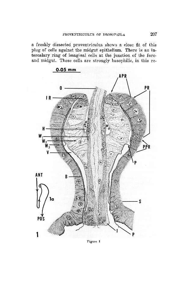
The observations on the proventriculus of the larvae of D. rnelanogaster agree with the desoription given by Stras- burger ('32). The oesophagus is invaginated into a layer of foregut cells which in turn are surrounded by a layer of midgut epithelium (fig. 1). A haemocoel containing haemo- cytes is present between the invaginated oesophagus and the inner layer of proventricular cells. A solid cellular plug is formed by the latter cells; the large lumen pictured in figure 1 is a result of fixation shrinkage. An optical section of
Fig. 1 Camera lucida drawing of a sagittal section of the proventricuIus of a D. rnelanogaster larva (72 hrs.). Section stained with fast green. APR, cells anterior to the peritrophic membrane-forming ring; B, striated border j H, haemo- coel with haemocytes; I, cuticular intima of the foregut invagination; IR, imaginal ring; 0, oesophagus; P, peritrophic membrane; PPR, region of the midgut posterior to the peritrophic membrane-forming ring ; PR, cells which secrete the peritrophio membrane-forming substance; S, sphincter; V, vault; W,, wall of the oesophagus with a c~.~ticular lining; W,, layer of the recurrent section of the invaginated foregut with alveolar cells; W,, wall of the midgut, cardia. la outlines the anterior-posterior course of the foregut-midgut in the proventrieulus corresponding to the right half of figure 1. The lower half of the invaginated region of the foregut shaped like a manubrium gives a probing action through the orifice of the gut at the level of the sphincter.
PROVENTRIOULUS O F DROSOPH.ILA 207
a freshly dissected proventriculus shows a close fit of this plug of cells against the midgut epithelium. There is an in- tercalary ring of imaginal cells at the junction of the fore- and midgut. These cells are strongly basophilic, in this re-
Figure 3

208 M. T. M. RIZKI
spect resembling the cells of the other imaginal disks. The two anteriormost cells of the midgut (cardia) following this imaginal ring are morphologically different from the subse- quent cells of the midgut epithelium; they either possess no striated border o r the border is extremely reduced so that it is not detectable by the methods employed in the present study. These cells will be referred to as APR cells, or the cells which are anterior to the ring of cells secreting the polysac- charide component of the peritrophic membrane. The APR cells are followed by four large cells which stain heavily with the periodic acid-Schiff reagent (PAS). R.emova1 of glycogen from the larva by digestion with saliva or amylase increases the staining difference between these four cells, labelled PR cells, and the other proventricular cells as can be seen in a striking manner in figures 4 and 5. Whole mounts of the pro- ventriculus which have been treated with amylase followed by PAS demonstrated very well that this ring which has such a strong affinity for PAS is four cells in width (figs. 2 and 3). Tangential sections of the proventriculus may contain more or less than four deeply stained cells. Two or three cells posterior to the PR cells ( P P R cells) are morphologically indistinguishable from the PR cells, but the intensity of the PAS reaction in these cells is comparable to that of the APR cells. These differences in staining intensity with the PAS reaction after digestion with amylase have been observed in embryos from 18 hours and throughout larval life. Embryos have been examined at two hour intervals from the eighth hour of development unt-il the time of hatching. Structurally the proventriculus is a well-formed organ by 12 hours of de- velopment (Poulson, '50), but no evidence of the PAS-pos- itive secretion in the PR cells or the peritrophic membrane was noted until 18 hours of development.
Material fixed in osmic acid gave the most satisfactory cytoplasmic details. I n unstained, 3 p sections of the pro- ventriculus, all of the cells in the anterior three-fourths of the cardia (APR, PR, PPR) contain osmiop1iilic bodies in the form of threads and globules as described by Siang-Hsu

PROVENTRICULUS O F DROSOPHILA 209
('47). With phase contrast the cytoplasm of these cells shows many large inclusions while the cytoplasm of the remaining proventricular cells is smooth in appearance (figs. 6 and 7). When these sections are placed in turpentine oil to remove the osmic acid, then treated with a-amylase and the PAS reagent, the PR cells stain heavily in contrast to the ,4PR and PPR cells. Observed under phase contxast this same section demonstrates the presence of numerous cyt.oplasmic inclusions in the APR, PR and PPR cells.
Although the osmic acid-fixed material was excellent for preserving cytoplasmic detail as well as the morphological appearance of the entire proventriculus, the faint brownish background which proved unremovable is not desirable for cytochemical staining. For all of the staining reactions dis- cussed in the succeeding section Carnoy-fixed material has been used.
There is an accumulation of a homogeneous, PAS-positive substance at the striated borders of the PR cells. The first appearance of peritrophic membrane in the gut of Drosoplda is found at the anterior level of the PPR cells. The PAS- positive peritrophic membrane is continuous with the secre- tory product of the PR cells, a morphological picture sugges- tive of the formation of peritrophic membrane from the latter. The peritrophic membrane in DrosophiZa gives a positive chitosan test demonstrating the presence of chitin. Since purified chitin is a polymer of N-acetylglucosamine residues joined by p-glycosidic ether linkages, no reaction with the periodic acid-Schiff reagent would be expected. The consistent parallel positive PAS reaction with a positive chitosan test in cuticle (Rizki, ' 5 5 ) and peritrophic membrane indicates that there may be other nonacetylated polysaccharides asso- ciated with chitin, or isolation and fixation of chitin alter the native state of this structural polysaccharide. Since the chitosan test requires treatment of the material to be tested with boiling saturated KOH, no cellular detail is pre- served. Therefore this reaction cannot be used to identify the nature of the PAS-positive material in the PR cells or

210 M. T. M. RIZICI
TABLE 1
Staining reactions of the proventriculus
TRBATYENT a PERITROPHIC APR AND XCEMBRANE pE CELLS PPR CELLS
Periodic acid-Schiff reagent (PAS) vs vs PAS control Acetylation + PAS Acetylation + KOH + PAS Amylase control + PAS Amylase + PAS Saliva + PAS Pepsin control + PAS Pepsin + PAS
1 mg enz./ml in 0.01 N HCI at 37°C. for 1 hr.
Pepsin control + fast green Pepsin + fast green Pepsin control + Millon Pepsin + Millon Trypsin control + PAS Trypsin + PAS
0.1 mg enz./ml 0.05 M phosphate, pH 6, 37°C. for 1 hr.
Trypsin control + fast green Trypsin + fast green
V8
vs vs vs
vs W
W
I B
vs
vs
VB
vs vs vs
-78
VS
W
s vw VS
VS
S
vw vw
8
W
vs W
8
vw S
W
4 vs vs 1 S €4
Ribonuclease control + azure B 1 8 S
Ribonuclease + azure B
Sudan black B (SBB) Lipid extraction + SBB Lipid extraction + PAS Lipid extraction + amylase + PAS
0.2 rng enz./ml for 2 hrs., 56°C.
'vs, very strong; s, strong; w, weak; vw, very weak; -, negative. Control is the buffer solution without enzyme. Ribonuclcase obtained from the
Worthington Biochemical Laboratory, Freehold, N. J. ; other enzymes from the Nutritional Biochemical Corp., Cleveland, Ohio.
In the vicinity of the PPR cells.

PROVENTRICULUS O F DROSOPHILA 211
to study the secretion found in the lumen beside these cells. Under these circumstances other cytochemical stains have been employed to further explore the nature of the secretory activity of the PB cells.
The results of the enzymatic digestions and staining tech- niques are summarized in table 1. The YR cells are differen- tiated from the APR and PPR cells only by the PAS reac- tion; none of the other procedures which are specific for proteins, lipids and ribonucleic acid demonstrated any dif- ferences between these groups of cells. The PAS-positive material in the PR cells is not removed by saliva, amylase, pepsin or trypsin. Furthermore, removal of lipids from sec- tions of embryos did not affect the PAS staining of the PR cells. The PAS-positive material of the peritrophic mem- brane in the vicinity of the proventricular cells and the larval endocuticle, which is a chitinous structure, behaved the same as the secretion of the PR cells.
Although the Millon reaction was not detectable in the peri- trophic membrane at the level of the PPR cells and in the vault of the proventriculus, it shows a positive Millon reaction below the level of the sphincter and in the rest of the midgut. Similarly the membrane stains with Sudan black B in re- gions of the gut other tha.n the proventriculus. The staining differences between the peritrophic membrane in the proven- triculus and in the subsequent regions of the gut may be due to protein and lipid inclusions during digestion.
Lethal 1(1)48J Hemizygous 1 (1)48J embryos show abiiormalities of the
muscles and midgut as well as other structures. This sex- linked embryonic lethal, roughly localized between the cut and vermillion loci, has been studied by Dr. D. F. Poulson in detail (personal communication). The abnormality of the proventriculus of 1(1)4sJ embryos is of especial interest to the present study. Sections of embryos 22-24 hours old were examined. In all of the lethal embryos an enormous mass of PAS-positive material was found in the lumen of the proven-

212 M. T. M. BIZKI
triculus specifically in the vicinity of the PR cells (figs. 8 and 9). PAS-positive granules can be seen in the PR cells of those embryos and the PR cells can readily be distinguished from the neighboring cells by their secretory contents. In some em- bryos peritrophic membrane is secreted and extends a short distance in the gut, while in other instances no sign of peri- trophic membrane is found. The membrane when formed is unusually irregular and thick as compared to the normal. Melanosis of the peritrophic membrane and the secretory mass often occurs in late embryos.
One aspect of the 1(1)48J syndrome is an upset in the mech- anism leading to the formation of normal peritrophic mem- brane. The accumulation of the substance forming peritrophic membrane in the lethal embryos is clearly attributable to the PR cells. However, the continuous formation of peritrophic membrane involves other activities of the proventriculus as will be discussed later.
Experiments on the regeneration of peritrophic membraw
The morphological observations on the proventriculus of D. rnelarzogaster demonstrate that the PR cells secrete a poly- saccharide component of the peritrophic membrane, but they do not exclude the possibility that the secretion of the pro- ventricular cells anterior and posterior to this region also participates in the process of peritrophic membrane for- mation. The isolation of small segments of the proventriculus and other regions of the anterior midgut and implantation of these tissues into adult hosts using the method of Bodenstein ('43) would be a desirable approach to localize the secretory centers participating in the formation of the peritrophic mem- brane. Unfortunately attempts to implant and recover tissue fragments consisting of PR or PPR cells alone proved tech- nically difficult. Therefore as an alternative the alimentary tracts of early third instar larvae were removed in Wadding- ton's solution and divided into two sections by cutting the proventriculus a t the anterior level of the sphincter. Thus
PROVENTRICULUS O F DROSOPHILA 213
one segment included the anterior proventriculus with the PR and PPR cells while the other segment consisted of the remaining proventricular cells, the caecae and the conical portion of the anterior midgut. These pieces of the gut were then implanted into adult males (2-3 days old). The implants were allowed to remain in t.he hosts from 48 to 72 hours, at the end of which the hosts were etherized and the implants removed and fixed in Carnoy. P a r d n sections were treated with amylase and stained with PAS. Whole mounts of some implants were also prepared (29 implants were recovered from a total of 70 paired implants).
The peritrophic membrane was carefully removed from the alimentary tract by gentle pulling before the gut was cut in two parts. To test for complete removal of peritrophic membrane 10 similar operations were performed and the guts immediately fixed. Examination of parailin sections stained with amylase-PAS confirmed the complete removal of peri- trophic membrane in these experiments.
An accumulation of PAS-positive materid was found in the lumen of all of the implanted anterior pieces of the midgut. I n addition to this accumulation several implanted proven- triculi contained peritrophic membrane. The PR cells retain a strong affinity for PAS and often the PAS-positive secre- tion can be seen flowing out of these cells into the lumen of the proventriculus. The implants devoid of anterior pro- ventricular cells failed to show any accumulation of PAS- positive material in the lumen o r any peritrophic membrane.
nISCUSSION
There are two principal modes of peritrophic membrane formation as described by WigglesmTorth ( '30, '50). Peri- trophic membrane may be the result of delamination of the surface of the midgut epithelium of different regions, or, as in the case of the Drosophila larva, it may be a continuous tube secreted by cells in the anterior midgut epithelium. Wig- glesworth ( '29) described the latter type of membrane forma- tion in the proventriculus of the adult tsetse-fly by an elab-

214 M. T. M. RIZKI
orate mechanism involving the structure of this organ. The peritrophic membrane-forming substance is poured into the lumen of the proventriculus by a pad of epithelial cells. This secretion is then pressed to form a thin membrane as it is forced through a press mechanism consisting of the closely apposed walls of the invaginated foregut and the midgut. A similar mechanism of membrane formation has been found in many orders of insects, the most specialized occurring in the Diptera (Wigglesworth, '30). Differentia- tion of the cells secreting the peritrophic membrane-forming substance is clearest in species with a highly efficient press.
The peritrophic membrane in the Drosophila larva is pro- duced by analogous structures involving the activities of a secretory region and a press mechanism displaying muscular contractions of the proventricular walls. Reduced basophilia is found in the muscles of 1(1)48J (Poulson, unpublished). The abnormal accumulation of PA S-positive substance in the proventriculi of the lethal embryos may be due to a failure of the musculature of the press mechanism. Occasional short pieces of peritrophic membrane occurring in some of the lethal embryos are imperfectly formed and not uniform in thickness, a further indication of an inefficient press mech- anism. Similar difficulties are apparently encountered in the implanted proventriculi which invariably accumulate the peri- trophic membrane-forming substance but lack continuous peritrophic membrane formation. Since the isolated proven- triculi are no longer anchored by attachment to the foregut and midgut, the mechanical advantage resulting from peri- stalsis of the gut is lost to the proventricular press. Peri- stalsis apparently also adds a pulling action on the membrane passing out of the proventriculus. Furthermore in isolated proventriculi the sphincter of the midgut has been removed and the probing of the formed membrane through the orifice of the midgut sphincter by the foregut invagination is no longer in operation. Nevertheless the implantation experi- ments clearly demonstrate that the faculty of peritrophic

PROVENTRICULUS OF DROSOPHILA 215
membrane formation resides in the proventriculus of the Drosophila larva.
Chitin has been reported in all examples of peritrophic membrane which have been investigated (Waterhouse, '53 ; Wigglesworth, '30). It is a long chain polysaccharide chem- ically defined as anhydro-N-acetylglucosamine. Taking this chemical composition as a clue a logical procedure would be to seek a cytological source of polysaccharide in the region where peritrophic membrane apparently originates, and then establish the identity of this cellular polysaccharide with the membrane. The periodic acid-Schiff reagent (PAS) can be utilized with proper precautions to stain intracellular chitin- polysaccharide or its immediate precursors, where the chito- san test is not applicable. PAS-positive material is of wide occurrence in the Drosophila larva ; after the removal of gly- cogen, a strong positive reaction is found in the striated borders of the midgut epithelium, ingested material in the lumen of the gut, endocuticle, peritrophic membrane, and a ring of cells (PR) in the cardia. The likelihood of the striated borders of the midgut epithelium as a source of peritrophic membrane-forming substance is ruled out by the implanta- tion experiments which demonstrate that this substance is found only in the presence of PR cells regardless of the in- clusion of striated borders in the implanted tissue. I% situ accumulation of PAS-positive material is found in the pro- ventricular lumen corresponding to the level of the ring of PR cells and the peritrophic membrane emerges from this material immediately following these cells.
In addition to these morphological relationships there is a parallel cytochemical behavior of the intracellular secretion of the PR cells and the peritrophic membrane in this vicinity. The PAS-positive staining of these structures is not due to glycogen, protein or ribonucleic acid since it is not removable with a-amylase, saliva, pepsin, trypsin or ribonuclease. The intensity of the PAS reaction was unaffected by lipid extrac- tions, thus excluding the presence of a lipoprotein. If the 1,2-glycol groups stained by PAS were associated propor-

216 M. T. M. RIZKI
tionately with proteins, nucleic acid or lipid, a differentiation of PR cells from the neighboring cells might be expected with some of the stains which have been employed (hlillon, fast green for proteins; Azure B for ribonucleic acid; oil red 0, Sudan black B for lipids). We may conclude that the PAS staining indicates the presence of a polysaccharide in the PB cells and the peritrophic membrane. Since the latter is a chitinous structure (chitosan) as mentioned earlier, in this instance the PAS is staining a chitin-polysaccharide. The immediate precursors of this chitin-polysaccharide are the secretion in the lumen and the secretory material in the PR cells. The intracellular PAS-positive material (Carnoy and osmic fixation) is granular, and the precursor in the lumen is a homogeneous plastic substance which is then molded and condensed into a membrane, the polymerized form.
In the absence of any knowledge about the biosynthesis of insect chitin, no suggestions can be made regarding the chem- ical structure of the intra- and extracellular precursors which are polymerized to form peritrophic membrane. Leloir and Cardini ( '53) have reported the presence of an enzyme com- plex in extracts of Neurospora crassa whereby acetylgluco- samine-I-phosphate can be synthesized from hexose-6-phos- phate. Theoretically anhydyo-N-acetylglucosamine can result from the polymerization of the former compound. Such a synthetic chain requires a phosphorylating mechanism similar to the one in glycogenesis : glucose-1-phosphate + glycogen + phosphate. The secretory actiT-ity noted in the APR and PPR cells may be correlated with the enzymes participating in the polymerization of the peritrophic membrane.
SUMMSRY
1. In Drosophda naelarzogaster a ring of cells located in the anterior region of the proventrieulus (cardia) begins to produce the peritrophic membrane at 18 hours of embryonic development and continues to secrete the substance forming this membrane throughout larval life. These cells (PR cells) stain more heavily by the periodic acid-Schiff (PAS) reagent

PR-OVENTRICULUS O F DROSOPHILA 217
for polysaccharides than the other proventricular cells, but are not differentiated by stains for proteins, lipids or ribo- nucleic acid. The PAS-positive material in these cells, in the secretion in the lumen of the proventriculus, and in the peri- trophic membrane itself is not removable by amylase, saliva, pepsin, trypsin, ribonuclease or lipid extraction.
Since the peritrophic membrane contains chitin (chito- san test), it may be concluded that the PAS-positive intra- and extracellular secretions of the PR cells are immediate precursors of chitin-polysaccharide.
The implantation of isolated proventriculi and adjacent regions of the midgut into adult hosts has further demon- strated that the faculty of peritrophic membrane production resides in the proventriculus, and the presence of the PI1 cells is necessary for the secretion of polysaccharides forming this membrane.
The above conclusions are further supported by the observations on lethal 1( 1)48J embryos which accumulate PAS-positive secretion from the PR cells in the lumen of the proventriculus. Abnormalities in the formation of the peri- trophic membrane can be assigned to disfunction of the mus- cles and the press mechanism to mold the plastic secretion before polymerization into a uniform membrane.
2.
3.
4.
ACKNOWLEDGMENTS
The author wishes to thank Mrs. R. M. Rizki for technical assistance. He is also thankful to Dr. D. F. Poulson for pro- viding the 1(1)48J stock, and for his helpful criticism of the manuscript.
LITERATURE CITED
BAKER, J. R. 1946 The histochemical recognition of lipine. Quart J. Micr. Sci., 87: 409470.
BODENSTEIN, D. 1943 Factors influencing growth and metamorphosis of the salivary glands in Drosophila. Biol. Bull., 84: 13-33.
CAMPBELL, F. L. The detection and estimation of insect chitin; and the irrelation of chitinization to hardness and pigmentation of the cuticula of the American cockroach, PeripZaneta americana. Ann. Eat. SOC. Amer., 22: 401-426.
1929

218 M. T. M. RIZKI
CHIFFELLE, T. L., AND F. A. PUTT Propylene and ethylene glycol as solvents for Sudan I V and Sudan black B. Stain Tech., 66: 51-59.
FLAX, M. H., AND M. H. HINES 1952 Microspectrophotometric analysis of metachromatic staining of nueleic acids. Physiol. Zool., 35 : 297-311.
HOTCHKISS, R. D. A microchemical reaction resulting in the staining of polysaccharide structures in fixed tissue preparations. Arch. of Bio- ehem., 16: 131-141.
LELOIR, L. F., AND C. E. CARDINI 1953 The biosynthesis of glucosamine. Bio- chem. et Biophys. Aeta, 12: 15-22.
MUMANUS, J. F. A., AND J. E. CASON 1950 Carbohydrate histochemistry studied by acetylation techniques. J. Exp. Med., 91 : 651-654.
PEARSE, A. Q. E. 1953 Histoehemistry, theoretical and applied. Little, Brown and Co., Boston.
POLLISTER, A. W. 1950 Quelques mhthodes de cytologie chimique quantitative. Rev. d’Hematol., 5: 527-554.
POULSON, D. F. 1950 Histogenesis, organogenesis, and differentiation in the embryo of Drosophila melanogaster Meigen; in Demerec, M., Ed., Biology of Drosophila. John Wiley & Sons, he. , N. Y.
An improved method for histo- logic demonstration of acid mucopolysaccharides in tissues. Arch. Path.,
RIZKI, M. T. M. Hereditary melanotic sclerosis in Drosophila willistoni. J. Exp. Zool., 288 :591-610.
SCHRADER, F., AND C. LELJCHTENRERGER 1950 A cytochemical analysis of the functional interrelations of various cell structures in Arwelizis albo- punctatus (de Geer). Esp. Cell Res., 1: 421-452.
SIANG-HSU, W. 1947 On the cytoplasmic elements in the midgut epithelium of the larvae of Drosophila mclanogaster Meigen. J. Morph., 80: 161-194.
STRASBURGER, M. 1932 Bau, Funktion und Variabilitit des Darmtraktus von Drosophila melanogaster Meigen. Zeit. f. wiss. Zool., 140 : 539-649.
SUMNEB, J. B., AXD G. F. SOMERS 1953 Chemistry and methods of enzymes. 3rd Ed. Academic Press, New York.
WATERHOUSE, D. F. 1953 The occurrence and significance of the peritrophic membrane, with special reference to adult Lepidoptera and Diptera.
WIGGLESWORTH, V. B. 1929 Digestion in the tsetse-fly: A study of structure and function. Parasitology, 81 : 288-321. 1930 The formation of the peritrophic membrane in insects, with
special reference to the larvae of mosquitoes. Quart. J. Mier. Sci., 73: 593-616. 1950 The principles of insect physiology. 4th Ed. Methuen & Co.,
Ltd., London.
1951
1948
RINEHART, J. F., AND 8. I(. ABU’L HAJ 1951
5.3: 189-194. 1955
Aust. J. ZOO^., 2: 299-318.

PLATE

PLATE 1
EXPLANATION OF FIGURES
(Photomicrographs)
2 Whole mount of the anterior midgut of a Drosophila larva (early third instar) fixed in Cranoy, treated with a-amylase, and stained with the PAS reagent. The region of the proventriculus, PR, which is heavily stained is the ring of cells secreting the substance forming the periti ophic membrane. The negative-PAS rcaction of the nuclei facilitates counting of the cells. This band is 4 cells in width; the anteriormost cells a re out of foeus due t o the curvature of the pro- ventriculus. I n the surface view the ends of these spindle-shaped cells are wedged together; thus depending upon the plane of the cut in section, different numbers and different parts of the cells are seen. The peritrophir membrane and the foregut invagination have been removed from this specimen.
Optical section of a whole mount of a proventriculus (Carnoy-amylase-PAS), In addition to the deeply stained P R cells, thrre is a strong PAS reaction of the yeritrophic membrane, P, in the lumen clcliinited by the reflected surface of the foregut invagination and the midgut epithelium. The pcritrophic membrane is folded as it passes through the narrow orifice of the sphincter into the midgut which has been sovered from the proventriculus. The light region anterior t o the PR corresponds to the APR cells, and the proventricular lumen in this region is devoid of peritrophic membrane. One gastric caecum is attached, the other thrcc having been removed.
Frontal section of a third iiistar larva with the provcntriculus in sagittal view (Carnoy-8 + section-saliva-PAS). Kote the strong staining reaction of the P R cells and the cuticle, C. Most of the tissues are outlined by a heavier reaction than the general background staining, but the striated borders give a strong PAS reac- tion as can be seen in sections of various irgions of the gut. The deeply stained hodies in the lower region of the foregut invagination are ingested yeast cells.
A region of the proventriculus in figure 4 under oil immersion t o show the differences in intensity of the P A S reaction in the PR, APR, and P P R cells. Kote the lieavily stained granules in the P R cells and the peritrophic membrane, P. The intercellular spaces a re due to shrinkage of the cytoplasm resulting from fixation; the anuclear segments of the P R cells are the ends of the spindles as shown in figure 2.
6 Region of a sxgittal section of the proventriculus fixed in osmic acid- formalin. 3 p, uiistained section mounted in refractive index oil 1.460.
7 The section in figure 6 under phase contrast. l’he cytoplasm of the APR, PR, and P P R cells contains large granules in contrast to tlic smoother appearance of the reniainiiig proventricular cells. Thus the anterior region of the provcntricu- lus, designated ?J? arrows, can be differentiated from the posterior region on the basis of secretory activity as described by Siang-Hsu ( ’47). This region is sub- divided into three sections by tlic PAS reaction, the middle section alone, PR, secreting the polysaccharides incorporated in the peritrophic membrane.
Consecutive sections (oblique) through the proventriculus of a 1(1)48J mutant embrpo showing the accumulation of PAS-positive material in the lumen and the heavily stained P R cells. I n figuir 9 the secrction can he seen flowing out of the PR cells, X.
3
4
5
8 and 9
220

PROVEKTRICULUS OF DROSOPJIJLA M. T. 31. IIIZKI
PLATE 1
221
Copyright © 2022 FDOKUMEN