Curcumin enhances parental reproductive lifespan and progeny viability in Drosophila melanogaster
14
Curcumin enhances parental reproductive lifespan and progeny viability in Drosophila melanogaster K. T. Chandrashekara & Sonam Popli & M. N. Shakarad Received: 25 February 2013 /Accepted: 4 August 2014 # American Aging Association 2014 Abstract Organismal lifespan is a complex trait that is governed by both its genetic makeup as well as the environmental conditions. The improved socioeconom- ic condition of humans has led to many lifestyle changes that in turn have altered the demography that includes postponement of procreation. Late age progeny is shown to suffer from many congenital diseases. Hence, there is a need to identify and evaluate natural molecules that could enhance reproductive health span. We have used the well-established model organism, Drosophila melanogaster , and ascertained the consequence of diet supplementation with curcumin. Flies reared on curcumin-supplemented diet had significantly higher lifespan. The progeny of flies reared on curcumin had a higher viability. The activity of a key mitochondrial enzyme—aconitase was significantly higher in flies reared on curcumin-supplemented diet. The results sug- gest that curcumin can not only correct a key step in the citric acid cycle and help in the release of additional energy but also permanently correct developmental and morphogenetic processes. Keywords Curcumin . Aconitase . Glycogen . Triglycerides . pAkt/Akt . CG9510 . AKHR . Drosophila Abbreviations DR Diet restriction SM Standard banana-jaggery medium SLC Standard laboratory conditions RT Room temperature PC Parental fly cage FC Progeny fly cage F A Progeny raised on SM F B Progeny raised on SM supplemented with 5 μM curcumin F C Progeny raised on SM supplemented with 10 μM curcumin F D Progeny raised on SM supplemented with 25 μM curcumin F E Progeny raised on SM supplemented with 50 μM curcumin Introduction Prolonged reproductive lifespan is an important fitness trait in all species engaged in repeated reproduction. However, there is a progressive decline in the functional AGE (2014) 36:9702 DOI 10.1007/s11357-014-9702-8 Electronic supplementary material The online version of this article (doi:10.1007/s11357-014-9702-8) contains supplementary material, which is available to authorized users. K. T. Chandrashekara : M. N. Shakarad (*) Evolutionary Biology Laboratory, Department of Zoology, University of Delhi, New Delhi 110007, India e-mail: [email protected] S. Popli Gut Biology Laboratory, Department of Zoology, University of Delhi, New Delhi 110007, India Present Address: K. T. Chandrashekara Institution of Excellence, University of Mysore, Manasagangotri, Mysore 570006, India
-
Upload
independent -
Category
Documents
-
view
4 -
download
0
Transcript of Curcumin enhances parental reproductive lifespan and progeny viability in Drosophila melanogaster
Curcumin enhances parental reproductive lifespanand progeny viability in Drosophila melanogaster
K. T. Chandrashekara & Sonam Popli &M. N. Shakarad
Received: 25 February 2013 /Accepted: 4 August 2014# American Aging Association 2014
Abstract Organismal lifespan is a complex trait that isgoverned by both its genetic makeup as well as theenvironmental conditions. The improved socioeconom-ic condition of humans has led to many lifestyle changesthat in turn have altered the demography that includespostponement of procreation. Late age progeny isshown to suffer from many congenital diseases. Hence,there is a need to identify and evaluate natural moleculesthat could enhance reproductive health span. We haveused the well-established model organism, Drosophilamelanogaster, and ascertained the consequence of dietsupplementation with curcumin. Flies reared oncurcumin-supplemented diet had significantly higherlifespan. The progeny of flies reared on curcumin hada higher viability. The activity of a key mitochondrialenzyme—aconitase was significantly higher in flies
reared on curcumin-supplemented diet. The results sug-gest that curcumin can not only correct a key step in thecitric acid cycle and help in the release of additionalenergy but also permanently correct developmental andmorphogenetic processes.
Keywords Curcumin . Aconitase . Glycogen .
Triglycerides . pAkt/Akt .CG9510 .AKHR .Drosophila
AbbreviationsDR Diet restrictionSM Standard banana-jaggery mediumSLC Standard laboratory conditionsRT Room temperaturePC Parental fly cageFC Progeny fly cageFA Progeny raised on SMFB Progeny raised on SM supplemented with
5 μM curcuminFC Progeny raised on SM supplemented with
10 μM curcuminFD Progeny raised on SM supplemented with
25 μM curcuminFE Progeny raised on SM supplemented with
50 μM curcumin
Introduction
Prolonged reproductive lifespan is an important fitnesstrait in all species engaged in repeated reproduction.However, there is a progressive decline in the functional
AGE (2014) 36:9702DOI 10.1007/s11357-014-9702-8
Electronic supplementary material The online version of thisarticle (doi:10.1007/s11357-014-9702-8) contains supplementarymaterial, which is available to authorized users.
K. T. Chandrashekara :M. N. Shakarad (*)Evolutionary Biology Laboratory, Department of Zoology,University of Delhi,New Delhi 110007, Indiae-mail: [email protected]
S. PopliGut Biology Laboratory, Department of Zoology, Universityof Delhi,New Delhi 110007, India
Present Address:K. T. ChandrashekaraInstitution of Excellence, University of Mysore,Manasagangotri, Mysore 570006, India
capacity of physiological processes in all the livingsystems (Yarian et al. 2006)—a process called aging.Changing human lifestyle since the late twentieth cen-tury has led to postponement of procreation. Acrossbiological systems, reproduction at later ages is shownto be on the declining trend. Further, children born toparents of older ages are shown to suffer from manycongenital diseases. It is uncertain that the progressmade in saving lives is due to improved health in ad-vanced age and, hence, an important topic for research(Vaupel 2010). Furthermore, there is a need to addressnot only the problems associated with reproduction atlater ages and suggest potential remedies but alsosuggest possible ways of producing viable, healthy,and fertile progeny at older ages as offspring viabilityis shown to decline with increasing parental age inDrosophila (Hercus and Hoffmann 2000; Kern et al.2001). Fortunately, a sizable fraction of many signalingpathways regulating aging in short-lived organisms isshown to extend lifespan in mammals as well (Kenyon2010). However, how different pathways and processesact together to promote or retard the aging process is notentirely clear (Sahin and DePinho 2010). Drosophilamelanogaster has been in the forefront of research onaging interventions. Our focus here is to utilize theinformation from studies that have attempted to under-stand the dynamics of life-history traits through dietarymanipulations (Tatar et al. 2014) in this model organismand elaborate further as direct genetical interventions inhumans are not possible due to technical and ethicalreasons (Lee et al. 2010). A number of studies in therecent past have shown the beneficial effects ofnutraceuticals—secondary compounds produced byplants in promoting healthy aging in invertebrate modelsthrough reduced oxidative damage (Dong et al. 2012).Aloe vera (Chandrashekara and Shakarad 2011), blue-berry (Peng et al. 2012), and green tea (Li et al. 2007,2008) extract supplementation of fly diet increased thelifespan through increased activity of antioxidant en-zymes—superoxide dismutase and catalase. Spermidinesupplementation of ordinary fly food increased the meanlifespan of flies up to 30 % (Eisenberg et al. 2009).Curcumin supplementation of sucrose-yeast diets ex-tended the fly lifespan through upregulation of superox-ide dismutase (Shen et al. 2012) and protection againstoxidative stress (Lee et al. 2010; Soh et al. 2013).
It is evident that the primary mode of action ofnutraceuticals seems to be through scavenging of reac-tive oxygen species from the mitochondria and other
cellular sources. In a study that attempted to assess thedamage to (mitochondrial) proteins during the course ofaging in flies, it was found that only aconitase activitywas reduced due to its increased carbonylation andoxidative damage. Further, supplementation of fly dietwith fluoroacetate—a competitive inhibitor of aconitaseactivity—reduced the lifespan of flies in a dose-dependent manner (Das et al. 2001), thus establishingthe causal link between aconitase activity and longevity.Aconitase is an important protein in the mitochondrialmatrix. Furthermore, mitochondrial protein density wasshown to increase by 25 % in flies under diet restriction(DR), demonstrating the importance of the mitochon-drial electron transport chain function in the lifespanextension upon DR in Drosophila (Zid et al. 2009).However, dietary restrictions are shown to genuinelyextend lifespan in flies (Grandison et al. 2009) at a costto reproduction (Partridge et al. 2005), supporting theadaptive response hypothesis involving a shift of re-sources during short periods of famine away from re-production toward increased somatic maintenance(Shanley and Kirkwood 2000). If indeed the resourceshifting adaptive hypothesis is true, then it is possiblethat the lifespan extension by nutraceuticals could alsobe due to diversion of energy reserves from reproduc-tion to somatic maintenance. Unfortunately, none of thestudies that have used nutraceutical supplementationhave ascertained their effect on fecundity, and it isentirely possible that the fecundity of the flies wassignificantly reduced. Alternatively, it is possible thatthe mitochondrial activity is increased, thus makingadditional energy available for somatic maintenanceand/or reproduction. Through this research, we set outto test these two not necessarily mutually exclusivehypotheses by supplementing larval diet with curcumin.Curcumin supplementation in larval diet significantlyincreased pre-adult viability, adult longevity, fertility,and vertical climbing ability. The increased adult life-history trait values were associated with increased levelsof glycogen and pAkt and aconitase activity but de-creased CG9510, AKHR, and tryglyceride levels.
Materials and methods
Fly husbandry
Wild-type Canton S strain ofD. melanogaster flies usedin this study were obtained from the National
9702, Page 2 of 14 AGE (2014) 36:9702
Drosophila Stock Center at University of Mysore, My-sore, Karnataka, India. Flies were maintained on stan-dard banana-jaggery medium (SM) (Chandrashekaraand Shakarad 2011) under standard laboratory condi-tions of 24±1 °C temperature, 75±5 % relative humid-ity, and 12:12 L:D cycle (SLC). Flies weremaintained ina 2 week discrete generation cycle for ten generationsbefore being used in this study. The adult density wasregulated at about 100 flies per half-pint bottle with25 mL of SM. There were a total of ten bottles. Fliesfrom ten bottles were combined into a single breedingcage, hereafter referred to as parental cage (PC).
Preparation of curcumin-supplemented diet
A total of 2.5 L of SM was prepared following theprocedure of Chandrashekara and Shakarad (2011) andsplit into five batches of 500 mL each. For the controlgroup, SM was poured into the bottles. For thecurcumin-supplemented media, either 5, 10, 25, or50 μM of curcumin obtained from Sigma-Aldrich,USA, was added and mixed thoroughly just beforepouring into the bottles. All bottles were plugged withnonadsorbent cotton andmedia was allowed to set underroom temperature. All other laboratory reagents used inthis study were purchased fromMerck, unless otherwisementioned.
Generation of assay flies and egg to adult viability assay
The eggs obtained from PC on a laying plate weredispensed into different treatment media bottles at adensity of ∼100 eggs/bottle with 45 mL media. Tenbottles each for the five treatment groups of SM,SM+5, 10, 25, or 50 μM curcumin were set up. Allbottles were incubated at SLC till the emergence offlies. The emerging flies were sorted based on gen-der under light diethyl ether anesthesia and held asvirgins in holding bottles containing SM till beingused in further assays. The flies emerging fromdifferent treatments are designated as FA, FB, FC,FD, and FE, to indicate progeny raised on SM andSM with 5, 10, 25, or 50 μM of curcumin, respec-tively. All assays were carried out on SM usingmated flies. The total number of flies that emergedfrom each bottle was used to calculate the viabilityof flies emerging from each treatment group.
Longevity assay of actively reproducing progeny (F)flies
The longevity of reproducing female and male FA, FB,FC, FD, and FE flies was assessed in mixed sex cultures(Handa et al. 2014). The flies were sorted into vialscontaining ∼4 mL SM under light diethyl etheranesthetization. A density of four females and four maleswas maintained per vial. Ten vials were set up per treat-ment. Three replicate sets were assayed over a period of1 year. In total, 30 vials of four mixed sex pairs wereassayed per treatment. Census was carried out on a dailybasis. The surviving flies were transferred to fresh SMvials every alternate day. The dead flies were aspiratedout, gender identified, and recorded. The census wascontinued till the death of the last fly. In total, 600 malesand 600 females were used for the longevity assay.
Fertility assay
Five hundred virgin female and 500 virgin male flies fromthe respective holding bottles of the five treatment (FA, FB,FC, FD, and FE) groups were recombined into five inde-pendent progeny breeding cages (FCs) and allowed tomate and age. The flies in these five FCs were maintainedon SM and were given fresh food every alternate day. Theidentity and purity of the FCwas strictly observed. Fertilityof the aging progeny (F) flies was assayed at 3, 10, 20, 30,40, and 50 day adult ages. On the designated age, eggswere collected on a laying plate from FCs and dispensedinto ten bottles each, at a density of 100 eggs/bottle with45 mL SM. All the 50 bottles were incubated at SLC. Thenumber of emerging flies from each of the ten bottles forprogeny groups FA, FB, FC, FD, and FE was recorded. Theeffective fertility was calculated as the average of the sumtotal of adults that emerged. Since each of the ten replicatebottles per treatment had 100 exact eggs dispensed, theaverage is represented as percent fertility.
Vertical climbing assay
The ability to move against gravity and climb is sug-gested to indicate the level of physical fitness of testanimals (Lee et al. 2010). Vertical climbing ability ofmale flies that emerged from different treatment bottleswas assessed. Twenty male flies per treatment groupwere collected and transferred to the empty, 0–15 cmgraduated vial. The vial was gently tapped and placed invertical position. The number of flies that crossed the
AGE (2014) 36:9702 Page 3 of 14, 9702
15 cm mark in 30 s was counted. Three trials wereconducted on each set of 20 flies. The vertical climbingwas assessed on 3, 10, 20, 30, 40, and 50 day adult ages.The data is expressed as the percentage of flies thatcrossed the 15 cm mark.
Larval feeding rate assay
The eggs obtained from PC were transferred at a densityof 50 eggs/6 mL SM and allowed to develop till earlythird instar. The early third instar larvae were removedfrom the SM vials and used in the feeding rate assay.The larvae were individually transferred to an assaypetri plate of 5 cm diameter containing 10 mL of eitherliquid SM (SM without agar) or liquid SM supplement-ed with 10 μM of curcumin and allowed 5 s for accli-mation. The feeding rate was measured as the meannumber of sclerite retractions in two consecutive 10 sintervals. The average of the two rates was taken as thefeeding rate of that larva. Twenty larvae were assayedfor each of the two treatment groups. The feeding rateassays were replicated four times. A total of 160 larvaewere assayed for feeding rate.
Egg to adult development time, dry weight, and lipidcontent assay
The egg to adult development timewas assessed in the SMand SM supplemented with 10 μM of curcumin. Eggsobtained from PC on a laying plate were collected with thehelp of a moistened brush and dispensed into 10 cm (L)×2.5 cm (D) glass vials containing 6 mL either SM or SMsupplemented with 10 μM of curcumin at a density of 50eggs per vial. Twenty such vials were set up per treatmentgroup and incubated at SLC.Once the pupae darkened, thevials were checked every 4 h and any eclosed adults wereremoved and sexed and the time of their eclosion wasrecorded. The four hourly checks were continued untilthree consecutive days passed with no eclosion recordedfrom any vial. The egg to adult development time wascalculated by taking midpoint of the egg laying window asthe 0th hour and the midpoint of two successive fourhourly checks as the time of emergence.
The flies from development time assay, on emer-gence, were etherized and sorted based on gender intogroups of ten individuals and transferred to prelabeledclean empty dry vials, dried at 70 °C for 36 h, andweighed to the nearest microgram using a microbalanceagain to obtain dry weight. The dry flies were
transferred to corresponding prelabeled 1.5 mLEppendroff microcenrtifuge tubes containing 1.2 mLdiethyl ether and put on a gel rocker set to 2,000 rpmto extract ether soluble lipids. The lipids were extractedover 36–40 h with two ether changes at 12 h intervals.After the last ether change, the flies were washed inexcess of ether, dried for 2 h at room temperature, andweighed to obtain lipid-free weight of flies. The dryweight and lipid-free dry weight were used to obtainthe lipid content in the flies. Five replicate vials per sexper treatment were set up. The development time, dryweight, and lipid content assay were replicated fourtimes.
Estimation of energy content and enzyme activity
Protein preparation
Twenty female flies were collected from each treatment.Protein extraction from the flies was conducted by fol-lowing a standard protocol (Clark and Keith 1988) withmodifications. In brief, flies were homogenized in100 μL of protein extraction buffer (0.01 M Na2PO4,1 mM EDTA, pH 7.4). The homogenates were centri-fuged at 3,000 rpm for 6 min at 4 °C. Twenty-fivemicroliters of the supernatant was added to 1.5 mL ofBradford reagent (Sigma). Absorbance was read at600 nm. A calibration curve was obtained using bovineserum albumin (BSA) as a standard.
Estimation of glycogen
Flies were killed by rapid immersion in >95 °C waterwithout prior modification to their behavior so as todenature enzymes and prevent postmortem degradationof glycogen. Animals were then stored overnight at−70 °C. The tissue from 20 female flies was pooledfor the assay of glycogen. Glycogen levels were deter-mined as described by using a standard protocol(Palanker et al. 2009) with minormodifications. In brief,flies were homogenized in ice-cold homogenizationbuffer (20 mM HEPES, 0.2 % sodium azide, 0.2 mMPMSF, and 1 mM EDTA, pH 7.0) and centrifuged at17,000 rpm for 3 min at 4 °C, and 25 μL of thesupernatant was incubated with 100 μL glucose reagent(Sigma) to measure the glucose level, or 100 μL glucosereagent and 0.5 U amyloglucosidase (Sigma) to measurethe glycogen level. The samples were incubated at 37 °Cfor 30 min and the absorbance was measured at 540 nm.
9702, Page 4 of 14 AGE (2014) 36:9702
Glucose and glucose plus glycogen amounts were de-termined using a standard curve and were normalized tothe amount of protein. The amount of glycogen wasdetermined by subtracting the amount of glucose fromthe glucose plus glycogen level. Each sample wasassayed thrice and the mean used in the statisticalanalysis.
Triglyceride measurements
Triglyceride levels were determined by using a standardprotocol (Palanker et al. 2009) with minor modifica-tions. Twenty female flies were homogenized in100 μL phosphate buffered saline (PBS) and 0.5 %Tween 20 and immediately incubated at 70 °C for5 min. The heat-treated homogenate was centrifuged,and 25 μL supernatant was incubated with TriglycerideReagent (Sigma) for 30min at 37 °C. Samples were thenincubated with Free Glycerol Reagent (Sigma) for 5 minat 37 °C and assayed at 525 nm using a Beckman DU640 spectrophotometer. Triglyceride levels were nor-malized to protein amounts in each homogenate andexperiments were repeated three times.
Measurement of aconitase activity
Since the fat bodies are known to impede in the enzymeassays, we have used mated male flies for assayingaconitase activity. We followed the protocol of Daset al. (2001) to measure the aconitase activity. Theactivity of aconitase (EC 4.2.1.3) is measured on thebasis of the conversion of citrate into α-oxoglutarate inassociation with the reduction of NADP, as described byKennedy et al. (1983). The mitochondrial pellets(obtained by following the methods of Das et al. 2001)were sonicated four times in a Branson 2200 sonicatorfor 30 s each in a buffer consisting of 154 mMTris-HCl,pH 7.4, and 5 mM citrate. The reaction mixtureconsisted of 25 μg protein in 27 mM Tris-HCl, pH7.4, 5 mM sodium citrate, 0.2 mM NADP, 0.6 mMMnCl2, and 1 U of isocitrate dehydrogenase. One unitof enzyme activity is defined as the amount of enzymethat catalyzes the formation of 1 μmol of isocitrate fromcitrate per min at pH 7.4 at 30 °C. The formation ofNADPH was followed over time at 30 °C at a wave-length of 340 nm using a Beckman DU 640 spectropho-tometer (Das et al. 2001).
Estimation of pAkt/Akt
An equal number of control and curcumin-treatedflies were stored in prechilled PBS pH 7.4 con-taining protease inhibitors (Sigma) at −20 °C tillfurther use. Extracts were prepared by homogeni-zation (Kimble/Kontes Motor Cordless 749540-0000) followed by centrifugation at 4,000 rpmfor 5 min at 4 °C. Supernatants were transferredto fresh tubes and quantified using Bradford re-agent (Sigma). An equal amount of extracts fromthe control and curcumin treated flies was run onSDS-PAGE at 150 V for 45 min. Gel was trans-ferred to nitrocellulose membrane (Amersham)using Mini Trans-Blot cell apparatus (Bio-Rad)for 2 h at 50 V. For Western blotting, the mem-brane was incubated with PBS containing 3 %BSA followed by overnight incubation at 4 °Cwith primary antibodies anti-rabbit Akt (1:1,000)from Thermo Scientific and anti-rabbit pAkt(1:1,000) donated by Dr. Rajesh Rajaiah of Mary-land University. The following day, the membranewas washed thrice with PBS containing 0.01 % ofTween-20 followed by incubation with secondaryanti-rabbit HRP 1:1,000 (Sigma) for 1 h at RT.Thereafter, the membrane was again washed withPBS containing 0.01 % of Tween-20 prior to de-velopment. Blot was developed using Luminol re-agent (Millipore) as per the manufacturer’s instruc-tions. The relative band intensity ratio (wherevermentioned) was calculated using NIH ImageJsoftware.
Total RNA isolation and semiconservative PCRof AKHR, CG9510, and RPL32
Total RNA isolation was done from an equal number ofcontrol and curcumin-treated flies. cDNA was freshlyprepared from quantified RNA as per manual’s instruc-tions (Thermo Scientific). PCR was performed usingAKHR fwd: 5′-TCCATCACCGTGTACAGCAT-3′and AKHR rev: 5′-GAGCGATATGCAGACCATCA-3′ and CG9510 fwd: 5′-GGTTGTACGACACAGTGAAG-3′ and CG9510 rev: 5′-CTGGCGTCCAAATCATCC-3′ (Iijima et al. 2009; Heinrichsen et al. 2013).Normalization was done using RPL32 gene with fwd:5′-CCGCTTCAAGGGACAGTATC-3′ and rev: 5′-GACAATCTCCTTGCGCTTCT-3′ primers.
AGE (2014) 36:9702 Page 5 of 14, 9702
Statistical analyses
In all cases other than survival function analysis, themeans were used as the units of analysis. One-wayanalysis of variance was used to assess the significanceof difference between means. The difference amongtreatment means was compared using Tukey-Kramerminimum significant difference (MSDα0.05) test (Sokaland Rohlf 1995). The difference between adult survivalcurves was analyzed using Kaplan-Meier log-rank test(Fisher and van Belle 1993). Graphical presentations ofall results except survival probability values are indicat-ed as mean+SEM.
Results
Egg to adult viability
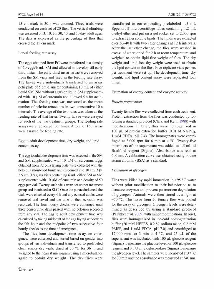
There was a significant main effect of treatment on theegg to adult viability (F4, 10=4.2797, p=0.0383). Theviability was lowest in the SM when compared to theSM supplemented with different concentrations ofcurcumin. Further, supplementing SM with 10 μM ofcurcumin resulted in a significantly higher viabilitywhen compared to the viability in SM but not signifi-cantly different from the viability in SM supplementedwith other concentrations of curcumin (Fig. 1).
Longevity of actively reproducing progeny (F) flies
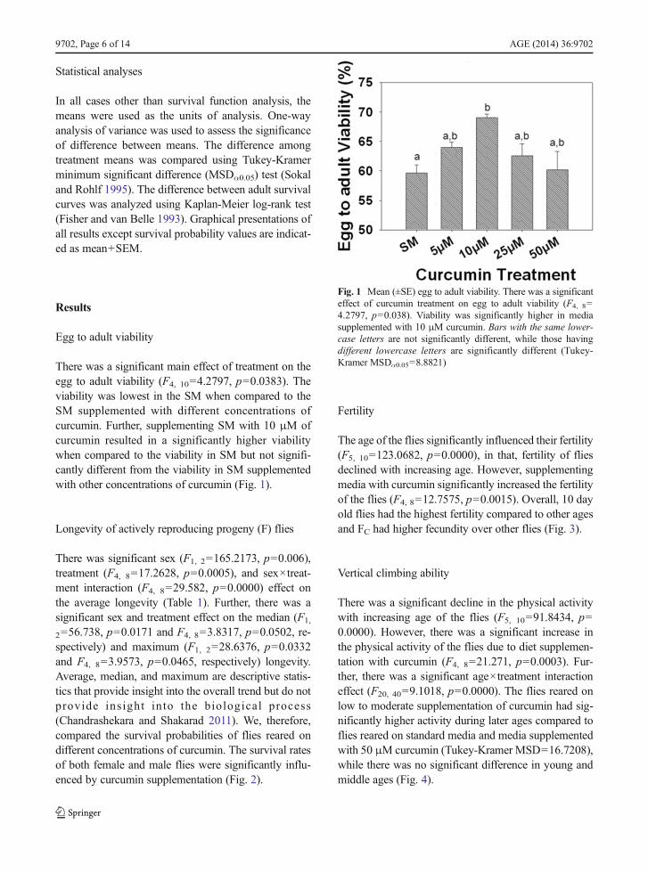
There was significant sex (F1, 2=165.2173, p=0.006),treatment (F4, 8=17.2628, p=0.0005), and sex×treat-ment interaction (F4, 8=29.582, p=0.0000) effect onthe average longevity (Table 1). Further, there was asignificant sex and treatment effect on the median (F1,
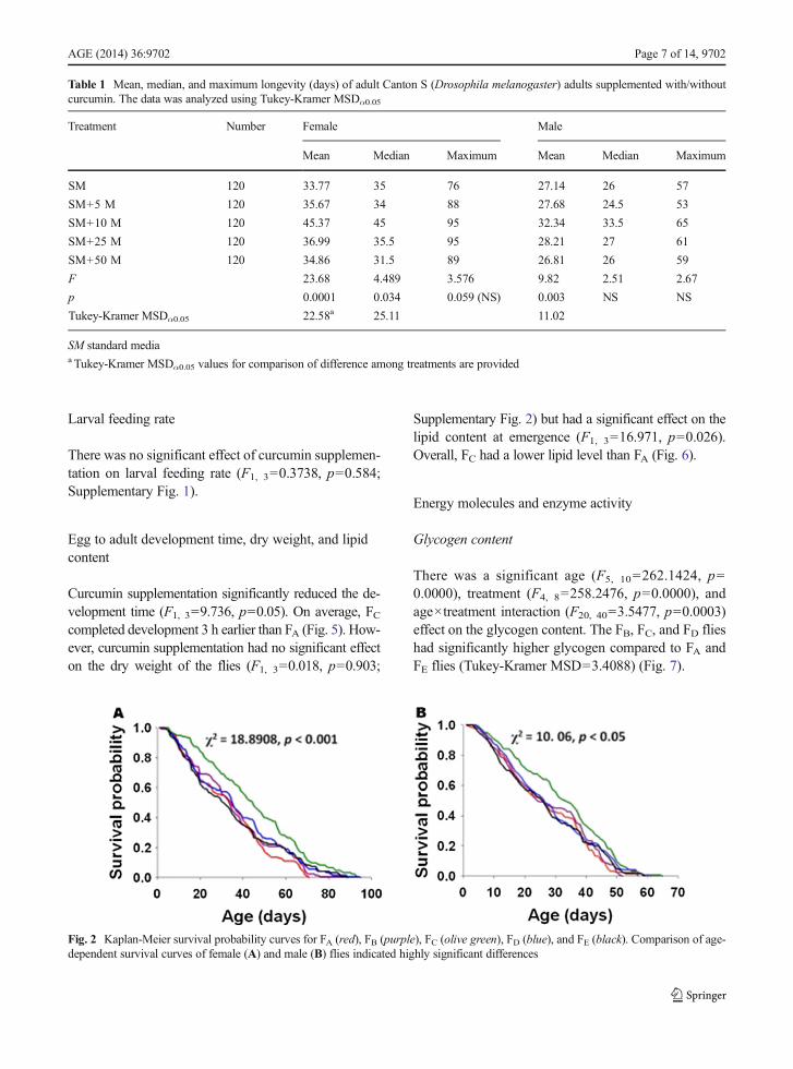
2=56.738, p=0.0171 and F4, 8=3.8317, p=0.0502, re-spectively) and maximum (F1, 2=28.6376, p=0.0332and F4, 8=3.9573, p=0.0465, respectively) longevity.Average, median, and maximum are descriptive statis-tics that provide insight into the overall trend but do notprovide insight into the biological process(Chandrashekara and Shakarad 2011). We, therefore,compared the survival probabilities of flies reared ondifferent concentrations of curcumin. The survival ratesof both female and male flies were significantly influ-enced by curcumin supplementation (Fig. 2).
Fertility
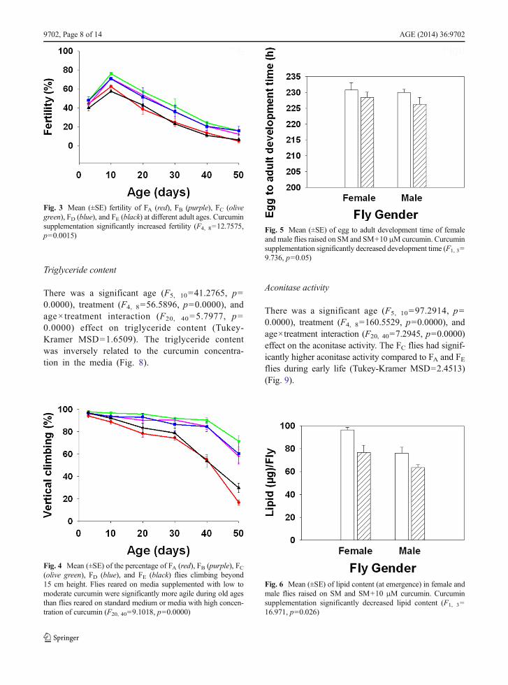
The age of the flies significantly influenced their fertility(F5, 10=123.0682, p=0.0000), in that, fertility of fliesdeclined with increasing age. However, supplementingmedia with curcumin significantly increased the fertilityof the flies (F4, 8=12.7575, p=0.0015). Overall, 10 dayold flies had the highest fertility compared to other agesand FC had higher fecundity over other flies (Fig. 3).
Vertical climbing ability
There was a significant decline in the physical activitywith increasing age of the flies (F5, 10=91.8434, p=0.0000). However, there was a significant increase inthe physical activity of the flies due to diet supplemen-tation with curcumin (F4, 8=21.271, p=0.0003). Fur-ther, there was a significant age×treatment interactioneffect (F20, 40=9.1018, p=0.0000). The flies reared onlow to moderate supplementation of curcumin had sig-nificantly higher activity during later ages compared toflies reared on standard media and media supplementedwith 50 μM curcumin (Tukey-Kramer MSD=16.7208),while there was no significant difference in young andmiddle ages (Fig. 4).
Fig. 1 Mean (±SE) egg to adult viability. There was a significanteffect of curcumin treatment on egg to adult viability (F4, 8=4.2797, p=0.038). Viability was significantly higher in mediasupplemented with 10 μM curcumin. Bars with the same lower-case letters are not significantly different, while those havingdifferent lowercase letters are significantly different (Tukey-Kramer MSDα0.05=8.8821)
9702, Page 6 of 14 AGE (2014) 36:9702
Larval feeding rate
There was no significant effect of curcumin supplemen-tation on larval feeding rate (F1, 3=0.3738, p=0.584;Supplementary Fig. 1).
Egg to adult development time, dry weight, and lipidcontent
Curcumin supplementation significantly reduced the de-velopment time (F1, 3=9.736, p=0.05). On average, FCcompleted development 3 h earlier than FA (Fig. 5). How-ever, curcumin supplementation had no significant effecton the dry weight of the flies (F1, 3=0.018, p=0.903;
Supplementary Fig. 2) but had a significant effect on thelipid content at emergence (F1, 3=16.971, p=0.026).Overall, FC had a lower lipid level than FA (Fig. 6).
Energy molecules and enzyme activity
Glycogen content
There was a significant age (F5, 10=262.1424, p=0.0000), treatment (F4, 8=258.2476, p=0.0000), andage×treatment interaction (F20, 40=3.5477, p=0.0003)effect on the glycogen content. The FB, FC, and FD flieshad significantly higher glycogen compared to FA andFE flies (Tukey-Kramer MSD=3.4088) (Fig. 7).
Table 1 Mean, median, and maximum longevity (days) of adult Canton S (Drosophila melanogaster) adults supplemented with/withoutcurcumin. The data was analyzed using Tukey-Kramer MSDα0.05
Treatment Number Female Male
Mean Median Maximum Mean Median Maximum
SM 120 33.77 35 76 27.14 26 57
SM+5 M 120 35.67 34 88 27.68 24.5 53
SM+10 M 120 45.37 45 95 32.34 33.5 65
SM+25 M 120 36.99 35.5 95 28.21 27 61
SM+50 M 120 34.86 31.5 89 26.81 26 59
F 23.68 4.489 3.576 9.82 2.51 2.67
p 0.0001 0.034 0.059 (NS) 0.003 NS NS
Tukey-Kramer MSDα0.05 22.58a 25.11 11.02
SM standard mediaa Tukey-Kramer MSDα0.05 values for comparison of difference among treatments are provided
Fig. 2 Kaplan-Meier survival probability curves for FA (red), FB (purple), FC (olive green), FD (blue), and FE (black). Comparison of age-dependent survival curves of female (A) and male (B) flies indicated highly significant differences
AGE (2014) 36:9702 Page 7 of 14, 9702
Triglyceride content
There was a significant age (F5, 10=41.2765, p=0.0000), treatment (F4, 8=56.5896, p=0.0000), andage× treatment interaction (F20, 40=5.7977, p=0.0000) effect on triglyceride content (Tukey-Kramer MSD=1.6509). The triglyceride contentwas inversely related to the curcumin concentra-tion in the media (Fig. 8).
Aconitase activity
There was a significant age (F5, 10=97.2914, p=0.0000), treatment (F4, 8=160.5529, p=0.0000), andage×treatment interaction (F20, 40=7.2945, p=0.0000)effect on the aconitase activity. The FC flies had signif-icantly higher aconitase activity compared to FA and FEflies during early life (Tukey-Kramer MSD=2.4513)(Fig. 9).
Fig. 3 Mean (±SE) fertility of FA (red), FB (purple), FC (olivegreen), FD (blue), and FE (black) at different adult ages. Curcuminsupplementation significantly increased fertility (F4, 8=12.7575,p=0.0015)
Fig. 4 Mean (±SE) of the percentage of FA (red), FB (purple), FC(olive green), FD (blue), and FE (black) flies climbing beyond15 cm height. Flies reared on media supplemented with low tomoderate curcumin were significantly more agile during old agesthan flies reared on standard medium or media with high concen-tration of curcumin (F20, 40=9.1018, p=0.0000)
Fig. 5 Mean (±SE) of egg to adult development time of femaleand male flies raised on SM and SM+10 μM curcumin. Curcuminsupplementation significantly decreased development time (F1, 3=9.736, p=0.05)
Fig. 6 Mean (±SE) of lipid content (at emergence) in female andmale flies raised on SM and SM+10 μM curcumin. Curcuminsupplementation significantly decreased lipid content (F1, 3=16.971, p=0.026)
9702, Page 8 of 14 AGE (2014) 36:9702

pAkt/Akt ratio
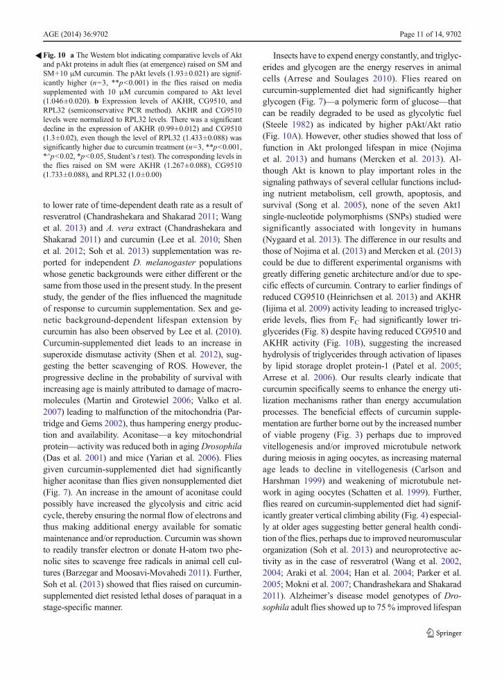
The pAkt/Akt ratio was significantly higher in adult fliesthat emerged from media supplemented with 10 μMcurcumin (FC) than those that emerged from SM (FA)(Fig. 10A).
AKHR and CG9510 expression
There was a significant decrease in the expression levelsof AKHR and CG9510 levels in FC flies as compared toFA flies (Fig. 10B).
Discussion
Lifespan—the time lag between birth and death of anorganism—is an important evolutionary adaptation(Carey 2003). Several studies in the past have success-fully enhanced the lifespan of model organisms bydietary supplementation with reactive oxygen species(ROS)-scavenging substances (Howitz et al. 2003;Wood et al. 2004). However, in most diet manipulationstudies (Chippindale et al. 1993; Chapman andPartridge 1996; Tu and Tatar 2003) barring that ofChandrashekara and Shakarad (2011), the correlatedresponse to increased longevity has been the reducedfecundity. Any manipulation that reduces fecundity isgoing to affect the fitness of the organism(s). Further, thecurrent lifestyle changes demand that we identify and
evaluate molecules that could potentially increase repro-ductive health span. In the present study, low to moder-ate curcumin supplementation ofD. melanogaster larvaldiet significantly increased lifespan. The increasedlifespan was not due to increased body weight or energylevels (Supplementary Fig. 2 and Fig. 6) or decreasedfertility; in contrast, FC flies had higher fertility (Fig. 3).Soh et al. (2013) also reported increased fertility in fliesfed with curcumin during larval stages. Lifespan exten-sion by curcumin is associated with lower time-dependent death rate (Fig. 2). Lifespan extension due
Fig. 7 Mean (±SE) of glycogen content per female fly at differentadult ages in FA (red), FB (purple), FC (olive green), FD (blue), andFE (black). Curcumin supplementation significantly increased gly-cogen content (F4, 8=258.2476, p=0.0000)
Fig. 8 Mean (±SE) of triglycerides content per female fly atdifferent adult ages in FA (red), FB (purple), FC (olive green), FD(blue), and FE (black). Curcumin supplementation significantlydecreased triglycerides content (F4, 8=56.5896, p=0.0000)
Fig. 9 Mean (±SE) of aconitase content per milligram of proteinat different adult ages in FA (red), FB (purple), FC (olive green), FD(blue), and FE (black). Curcumin supplementation significantlyincreased aconitase content (F4, 8=160.5529, p=0.0000)
AGE (2014) 36:9702 Page 9 of 14, 9702
9702, Page 10 of 14 AGE (2014) 36:9702
to lower rate of time-dependent death rate as a result ofresveratrol (Chandrashekara and Shakarad 2011; Wanget al. 2013) and A. vera extract (Chandrashekara andShakarad 2011) and curcumin (Lee et al. 2010; Shenet al. 2012; Soh et al. 2013) supplementation was re-ported for independent D. melanogaster populationswhose genetic backgrounds were either different or thesame from those used in the present study. In the presentstudy, the gender of the flies influenced the magnitudeof response to curcumin supplementation. Sex and ge-netic background-dependent lifespan extension bycurcumin has also been observed by Lee et al. (2010).Curcumin-supplemented diet leads to an increase insuperoxide dismutase activity (Shen et al. 2012), sug-gesting the better scavenging of ROS. However, theprogressive decline in the probability of survival withincreasing age is mainly attributed to damage of macro-molecules (Martin and Grotewiel 2006; Valko et al.2007) leading to malfunction of the mitochondria (Par-tridge and Gems 2002), thus hampering energy produc-tion and availability. Aconitase—a key mitochondrialprotein—activity was reduced both in aging Drosophila(Das et al. 2001) and mice (Yarian et al. 2006). Fliesgiven curcumin-supplemented diet had significantlyhigher aconitase than flies given nonsupplemented diet(Fig. 7). An increase in the amount of aconitase couldpossibly have increased the glycolysis and citric acidcycle, thereby ensuring the normal flow of electrons andthus making additional energy available for somaticmaintenance and/or reproduction. Curcumin was shownto readily transfer electron or donate H-atom two phe-nolic sites to scavenge free radicals in animal cell cul-tures (Barzegar and Moosavi-Movahedi 2011). Further,Soh et al. (2013) showed that flies raised on curcumin-supplemented diet resisted lethal doses of paraquat in astage-specific manner.
Insects have to expend energy constantly, and triglyc-erides and glycogen are the energy reserves in animalcells (Arrese and Soulages 2010). Flies reared oncurcumin-supplemented diet had significantly higherglycogen (Fig. 7)—a polymeric form of glucose—thatcan be readily degraded to be used as glycolytic fuel(Steele 1982) as indicated by higher pAkt/Akt ratio(Fig. 10A). However, other studies showed that loss offunction in Akt prolonged lifespan in mice (Nojimaet al. 2013) and humans (Mercken et al. 2013). Al-though Akt is known to play important roles in thesignaling pathways of several cellular functions includ-ing nutrient metabolism, cell growth, apoptosis, andsurvival (Song et al. 2005), none of the seven Akt1single-nucleotide polymorphisms (SNPs) studied weresignificantly associated with longevity in humans(Nygaard et al. 2013). The difference in our results andthose of Nojima et al. (2013) and Mercken et al. (2013)could be due to different experimental organisms withgreatly differing genetic architecture and/or due to spe-cific effects of curcumin. Contrary to earlier findings ofreduced CG9510 (Heinrichsen et al. 2013) and AKHR(Iijima et al. 2009) activity leading to increased triglyc-eride levels, flies from FC had significantly lower tri-glycerides (Fig. 8) despite having reduced CG9510 andAKHR activity (Fig. 10B), suggesting the increasedhydrolysis of triglycerides through activation of lipasesby lipid storage droplet protein-1 (Patel et al. 2005;Arrese et al. 2006). Our results clearly indicate thatcurcumin specifically seems to enhance the energy uti-lization mechanisms rather than energy accumulationprocesses. The beneficial effects of curcumin supple-mentation are further borne out by the increased numberof viable progeny (Fig. 3) perhaps due to improvedvitellogenesis and/or improved microtubule networkduring meiosis in aging oocytes, as increasing maternalage leads to decline in vitellogenesis (Carlson andHarshman 1999) and weakening of microtubule net-work in aging oocytes (Schatten et al. 1999). Further,flies reared on curcumin-supplemented diet had signif-icantly greater vertical climbing ability (Fig. 4) especial-ly at older ages suggesting better general health condi-tion of the flies, perhaps due to improved neuromuscularorganization (Soh et al. 2013) and neuroprotective ac-tivity as in the case of resveratrol (Wang et al. 2002,2004; Araki et al. 2004; Han et al. 2004; Parker et al.2005; Mokni et al. 2007; Chandrashekara and Shakarad2011). Alzheimer’s disease model genotypes of Dro-sophila adult flies showed up to 75% improved lifespan
�Fig. 10 a The Western blot indicating comparative levels of Aktand pAkt proteins in adult flies (at emergence) raised on SM andSM+10 μM curcumin. The pAkt levels (1.93±0.021) are signif-icantly higher (n=3, **p<0.001) in the flies raised on mediasupplemented with 10 μM curcumin compared to Akt level(1.046±0.020). b Expression levels of AKHR, CG9510, andRPL32 (semiconservative PCR method). AKHR and CG9510levels were normalized to RPL32 levels. There was a significantdecline in the expression of AKHR (0.99±0.012) and CG9510(1.3±0.02), even though the level of RPL32 (1.433±0.088) wassignificantly higher due to curcumin treatment (n=3, **p<0.001,*^p<0.02, *p<0.05, Student’s t test). The corresponding levels inthe flies raised on SM were AKHR (1.267±0.088), CG9510(1.733±0.088), and RPL32 (1.0±0.00)
AGE (2014) 36:9702 Page 11 of 14, 9702
and locomotor activity and reduced neurotoxicity whenfed with curcumin-supplemented diet throughout adultlife (Caesar et al. 2012). The improved health conditionof the flies given curcumin-supplemented diet is alsoreflected by increased egg to adult viability. An inde-pendent study using flies with different genetic back-grounds and different diet compositions also reportedthat curcumin supplementation had no detrimental effecton egg-hatching rate (Soh et al. 2013).
In conclusion, our results suggests that curcumin supple-mentation not only enhances ROS scavenging (Barzegarand Moosavi-Movahedi 2011; Shen et al. 2012), neuropro-tection (Caesar et al. 2012), and neuromuscular organization(Soh et al. 2013) but also increases the efficiency of en-zymes involved in energy metabolism and, thus, makesadditional energy available for somatic maintenance and/or reproduction, truly making it the sanjeevani—infusinglife not only into those that are fed with curcumin but intotheir progeny that are raised on curcumin-unsupplementeddiet as well.
Acknowledgments We thank two anonymous referees for theirhelpful comments on the earlier versions of the manuscript andsuggesting additional experiments on pAkt/Akt ratio and fat me-tabolism related genes. We also thank Dr. Rajesh Rajaiah, Depart-ment of Microbiology and Immunology, University of Maryland,USA, for providing us the pAKTantibody; Dr. Rajagopal Raman,Gut Biology Laboratory, Department of Zoology, University ofDelhi; Dr. K. Natarajan, Ambedkar Centre for Biomedical Re-search, University of Delhi for providing the chemicals, buffers,and instruments required in Akt/pAkt, AKHR, and CG9510West-ern blot and PCR experiments; and Ms. Nalini Mishra and numer-ous research trainees for their assistance in the research.MS thanksthe University of Delhi and Council of Scientific and IndustrialResearch, Government of India, for financial assistance. SP thanksthe University Grants Commission, Government of India, forDSKPDF fellowship.
Conflict of interest The authors declare no conflict of interest.
Author contributions K.T.C., S.P., and M.S. designed the re-search; K.T.C and S.P. performed the research; and K.T.C., S.P.,and M.S. analyzed the data and wrote the paper.
References
Araki T, Sasaki Y, Milbrandt J (2004) Increased nuclear NADbiosynthesis and SIRT1 activation prevent axonal degenera-tion. Science 305:1010–1013
Arrese EL, Soulages JL (2010) Insect fat body: energy, metabo-lism, and regulation. Annu Rev Entomol 55:207–255
Arrese EL, Patel RT, Soulages JL (2006) The main triglyceride-lipase from the insect fat body is an active phospholipaseA(1): identification and characterization. J Lipid Res 47:2656–2667
Barzegar A, Moosavi-Movahedi AA (2011) Intracellular ROSprotection efficiency and free radical-scavenging activity ofcurcumin. PLoS ONE 6(10):e26012. doi:10.1371/journal.pone.0026012
Caesar I, JonsonM,Nilsson KPR, Thor S, Hammarström P (2012)Curcumin promotes A-beta fibrillation and reduces neurotox-icity in transgenic Drosophila. PLoS ONE 7(2):e31424. doi:10.1371/journal.pone.0031424
Carey JR (2003) Life span: a conceptual overview. In: Carey JR,Tuljapurkar S (eds) Life span: evolutionary, ecological, anddemographic perspectives. Population Council andDevelopment Review 29(Suppl):1–18
Carlson KA, Harshman LG (1999) Extended longevity lines ofDrosophila melanogaster: characterization of oocyte stagesand ovariole numbers as a function of age and diet. J GerontolA Biol 54:B432–B440
Chandrashekara KT, ShakaradMN (2011) Aloe vera or resveratrolsupplementation in larval diet delays adult aging in the fruitfly,Drosophila melanogaster. J Gerontol A Biol Sci Med Sci66(9):965–971
Chapman T, Partridge L (1996) Female fitness in Drosophilamelanogaster: an interaction between the effect of nutritionand of encounter rate withmales. Proc R Soc Lond BBiol Sci263:755–759
Chippindale AK, Leroi AM, Kim SB, Rose MR (1993)Phenotypic plasticity and selection in Drosophila life historyevolution. 1. Nutrition and the cost of reproduction. J EvolBiol 6:171–193
Clark AG, Keith LE (1988) Variation among extracted lines ofDrosophila melanogaster in triacylglycerol and carbohydratestorage. Genetics 119:595–607
Das N, Levine RL, Orr WC, Sohal RS (2001) Selectivity ofprotein oxidative damage during aging in Drosophilamelanogaster. Biochem J 360:209–216
Dong Y, Guha S, Sun X, Cao M, Wang X, Zou S (2012)Nutraceutical interventions for promoting healthy aging ininvertebrate models. Oxidative Med Cell Longev. doi:10.1155/2012/718491
Eisenberg T, Knauer H, Schauer A, Büttner S, Ruckenstuhl C,Carmona-Gutierrez D, Ring J, Schroeder S, Magnes C,Antonacci L, Fussi H, Deszcz L, Hartl R, Schraml E,Criollo A, Megalou E, Weiskopf D, Laun P, Heeren G,Breitenbach M, Grubeck-Loebenstein B, Herker E,Fahrenkrog B, Fröhlich KU, Sinner F, Tavernarakis N,Minois N, Kroemer G, Madeo F (2009) Induction of autoph-agy by spermidine promotes longevity. Nat Cell Biol 11(11):1305–1314
Fisher LD, van Belle G (1993) Biostatistics: a methodology forhealth sciences. Wiley, New York, pp 786–843
Grandison RC, Piper MD, Partridge L (2009) Amino-acid imbal-ance explains extension of lifespan by dietary restriction inDrosophila. Nature 462:1061–1064
Han YS, Zheng WH, Bastianetto S, Chabot JG, Quirion R(2004) Neuroprotective effects of resveratrol againstbeta-amyloid-induced neurotoxicity in rat hippocampalneurons: involvement of protein kinase C. Br JPharmacol 141:997–1005
9702, Page 12 of 14 AGE (2014) 36:9702
Handa J, Chandrashekara KT, Kashyap K, Sageena G, ShakaradMN (2014) Gender based disruptive selection maintainsbody size polymorphism in Drosophila melanogaster. JBiosci 39:609–620. doi:10.1007/s12038-014-9452-x
Heinrichsen ET, Zhang RJE, Ngo J, Diop S, Bodmer R, JoinerWJ,Metallo CM, Haddad GG (2013) Metabolic and transcrip-tional response to a high-fat diet inDrosophila melanogaster.Mol Metabol 3:42–54. doi. 10.1016/j.molmet.2013.10.003
Hercus MJ, Hoffmann AA (2000) Maternal and grandmaternalage influence offspring fitness in Drosophila. Proc R SocLond B 267:2105–2110
Howitz KT, Bitterman KJ, Cohen HY, Lamming DW, Lavu S,Wood JG, Zipkin RE, Chung P, Kisielewski A, Zhang LL,Scherer B, Sinclair DA (2003) Small molecule activators ofsirtuins extend Saccharomyces cerevisiae lifespan. Nature425:191–196
Iijima K, Zhao L, Shenton C, Iijima-Ando K (2009) Regulation ofenergy stores and feeding by neuronal and peripheral CREBactivity in Drosophila. PLoS ONE 4(12):e8498. doi:10.1371/journal.pone.0008498
KennedyMC, EmptageMH, Dreyer JL, Beinert H (1983) The roleof iron in activation-inactivation of aconitase. J Biol Chem258:11098–11105
Kenyon CJ (2010) The genetics of aging. Nature 464:504–512Kern S, AckermannM, Stearns SC, Kawecki TJ (2001) Decline in
offspring viability as a manifestation of aging in Drosophilamelanogaster. Evolution 55(9):1822–1831
Lee KS, Lee BS, Semnani S, Avanesian A, Um CY, Jeon HJ,Seong KM, Yu K, Min KJ, Jafari M (2010) Curcumin ex-tends lifespan, improves health span, and modulates theexpression of age-associated aging genes in Drosophilamelanogaster. Rejuvenation Res 13(5):561–570
Li YM, Chan HYE, Huang Y, Chen ZY (2007) Green tea cate-chins upregulate superoxide dismutase and catalase in fruitflies. Mol Nutri Food Res 51(5):546–554
Li YM, Chan HYE, Yao XQ, Huang Y, Chen ZY (2008) Green teacatechins and broccoli reduce fat-induced mortality inDrosophila melanogaster. J Nutri Biochem 19(6):376–383
Martin I, Grotewiel MS (2006) Oxidative damage and age relatedfunctional declines. Mech Ageing Dev 127(5):411–423
Mercken EM, Crosby SD, Lamming DW, JeBailey L, Krzysik-Walker S, Villareal DT, Capri M, Franceschi C, Zhang Y,Becker K, Sabatini DM, de Cabo R, Fontana L (2013)Calorie restriction in humans inhibits the PI3K/AKT pathwayand induces a younger transcription profile. Aging Cell 12:645–651
Mokni M, Elkahoui S, Limam F, Amri M, Aouani E (2007) Effectof resveratrol on antioxidant enzyme activities in the brain ofhealthy rat. Neurochem Res 32:981–987
Nojima A, Yamashita M, Yoshida Y, Shimizu I, Ichimiya H,Kamimura N, Kobayashi Y, Ohta S, Ishii N, Minamino T(2013) Haploinsufficiency of Akt1 prolongs the lifespan ofmice. PLoS ONE 8(7):e69178
Nygaard M, Soerensen M, Flachsbart F, Mengel-From J, Tan Q,Schreiber S, Nebel A, Christensen K, Christiansen L (2013)Akt1 fails to replicate as a longevity-associated gene inDanish and German nonagenarians and centenarians. Eu JHuman Genet 21:574–577
Palanker L, Tennessen JM, Lam G, Thummel CS (2009)Drosophila HNF4 regulates lipid mobilization and beta-oxi-dation. Cell Metab 9(3):228–239
Parker JA, Arango M, Abderrahmane S, Lambert E, Tourette C,Catoire H, Néri C (2005) Resveratrol rescues mutantpolyglutamine cytotoxicity in nematode and mammalianneurons. Nat Genet 37:349–350
Partridge L, Gems D (2002) Mechanisms of ageing: public orprivate? Nature Rev Genet 3:165–175
Partridge L, Piper MDW, Mair W (2005) Dietary restriction inDrosophila. Mech Ageing Dev 126:938–950
Patel RT, Soulages JL, Hariharasundaram B, Arrese EL (2005)Activation of the lipid droplet controls the rate of lipolysis oftriglycerides in the insect fat body. J Biol Chem 280:22624–22631
Peng C, Zuo Y, Kwan KM, Liang Y, Ma KY, Chan HYE,Huang Y, Yu H, Chen ZY (2012) Blueberry extractprolongs lifespan of Drosophila melanogaster. ExpGerontol 47(2):170–178
Sahin E, DePinho RA (2010) Linking functional decline of telo-meres, mitochondria and stem cells during aging. Nature 464:520–528
Schatten H, Chakrabarti A, Hedrick J (1999) Centrosome andmicrotubule instability in aging Drosophila cells. J CellBiochem 74:229–241
Shanley DP, Kirkwood TBL (2000) Calorie restriction and aging:a life-history analysis. Evolution 54(3):740–750
Shen LR, Xiao F, Yuan P, Chen Y, Gao QK, Parnell LD, MeydaniM, Ordovas JM, Li D, Lai CQ (2012) Curcumin-supplemented diets increase superoxide dismutase activityand mean lifespan in Drosophila. Age. doi:10.1007/s11357-012-9438-2
Soh JW, Marowsky N, Nichols TJ, Rahman AM, Miah T, Sarao P,Khasawneh R, Unnikrishan A, Heydari AR, Silver RB,Arking R (2013) Curcumin is an early-acting stage specificinducer of extended functional longevity in Drosophila. ExpGerontol 48:229–239
Sokal RR, Rohlf JF (1995) Biometry: the principles and practicesof statistics in biological sciences. WH Freeman, New York,pp 179–450
Song G, Ouyang G, Bao S (2005) The activation of Akt/PKBsignaling pathway and cell survival. J Cell Mol Med 9:59–71
Steele JE (1982) Glycogen-phosphorylase in insects. InsectBiochem 12:131–147
Tatar M, Post S, Yu K (2014) Nutrient control of Drosophilalongevity. TEM. http://dx.doi.org/10.1016/j.tem.2014.02.006
Tu MP, Tatar M (2003) Juvenile diet restriction and the aging andreproduction of adult Drosophila melanogaster. Aging Cell2:327–333
Valko M, Leibfritz D, Moncol J, Cronin MT, Mazur M, Telser J(2007) Free radicals and antioxidants in normal physiologicalfunctions and human disease. Int J BiochemCell B 39(1):44–84
Vaupel JW (2010) Biodemography of human aging. Nature 464:536–542
Wang Q, Xu J, Rottinghaus GE, Simonyai A, Lubahn D, Sun GY,Sun AY (2002) Resveratrol protects against global cerebralischemic injury in gerbils. Brain Res 958:439–447
Wang Q, Yu S, Simonyai A, Rottinghaus GE, Sun GY, Sun AY(2004) Resveratrol protects against neurotoxicity induced bykainic acid. Neurochem Res 29:2105–2112
Wang C, Wheeler CT, Alberico T, Sun X, Seeberger J, Laslo M,Spangler E, Kern B, de Cabo R, Zou S (2013) The effect of
AGE (2014) 36:9702 Page 13 of 14, 9702
resveratrol on lifespan depends on both gender and dietarynutrient composition in Drosophila melanogaster. Age 35:69–81. doi:10.1007/s11357-011-9332-3
Wood JG, Rogina B, Lavu S, Howitz K, Helfand SL, TatarM, Sinclair D (2004) Sirtuin activators mimic caloricrestriction and delay ageing in metazoans. Nature 430:686–689
Yarian CS, Toroser D, Sohal RS (2006) Aconitase is the mainfunctional target of aging in the citric acid cycle of kidneymitochondria from mice. Mech Age Dev 127:79–84
Zid BM, Rogers AN, Katewa SD, Vargas MA, Kolipinski MC, LuTA, Benzer S, Kapahi P (2009) 4E-BP extends lifespan upondietary restriction by enhancing mitochondrial activity inDrosophila. Cell 139:149–160
9702, Page 14 of 14 AGE (2014) 36:9702