Genetic interactions between Drosophila melanogaster menin and Jun/Fos
Upload
independentCategory
view
2download
0
Published: May 09, 2011
r 2011 American Chemical Society 4974 dx.doi.org/10.1021/es104216b | Environ. Sci. Technol. 2011, 45, 4974–4979
ARTICLE
pubs.acs.org/est
Acute and Chronic Toxicity Effects of Silver Nanoparticles(NPs) on Drosophila melanogasterAles Panacek,† Robert Prucek,† Dana Safarova,‡ Milan Dittrich,§ Jana Richtrova,†
Katerina Benickova,† Radek Zboril,† and Libor Kvitek*,†
†Regional Centre of Advanced Technologies and Materials, Department of Physical Chemistry, Faculty of Science,Palacky University, 17. Listopadu 12, 77146 Olomouc, Czech Republic‡Department of Cell Biology and Genetics, Faculty of Science, Palacky University, Slechtitelu 11, 78371 Olomouc, Czech Republic§Department of Pharmaceutical Technology, Faculty of Pharmacy, Charles University, Heyrovskeho 1203,50005 Hradec Kralove, Czech Republic
bS Supporting Information
’ INTRODUCTION
Nanoparticles (NPs) and their unique properties are currentlynot only the subject of the scientific research, but they are beingimplemented and used in chemical industry and everyday humanlife as commercial products. Various nanomaterials and nano-technologies are now commonly used, for example, in areasincluding electrical engineering, construction, cosmetics, food,healthcare, and disinfection preparations.1�3 On the other hand,the research and knowledge of side effects or adverse physical andchemical interactions of nanomaterials with living organisms andthe environment is not as far advanced as compared to basic andapplied research of nanomaterials production. Nanoparticles ofvarious compounds can normally occur in nature, however,engineered nanomaterials are not of natural origin, and theycan, contrary to their bulk form or to normally occurrednanoparticles, induce totally different interactions within livingorganisms.
Silver NPs, which are ranked among the most intensivelystudied nanomaterials, are used in various commercial productsas a disinfection agent due to their extraordinary antimicrobial
properties. Silver NPs are increasingly becoming a part of oureveryday life, which considerably increases the risk of contam-ination of the environment. Hence, it is necessary to pay a greaterattention to the ecotoxicological properties of silver NPs. In thepast few years, several studies have been published dealing withthe ecotoxicity of silver NPs, particularly to aquatic organismssuch as crustaceans,4,5 protozoan ciliate,6 fish,4,7�10 or algae,11
and also to soil organisms12,13 and plants.14 However, theresearch in this area is still at the beginning and it is necessaryto further extend the knowledge on the ecotoxicity of Ag NPs inmore comprehensive studies.
The fruit fly (Drosophila melanogaster), a commonly occurringorganism in nature, is characterized by a short life cycle withdistinct developmental stages. Thanks to its easy manipulationand cultivation, numerous offspring and possibility to induce
Received: December 17, 2010Accepted: April 28, 2011Revised: April 24, 2011
ABSTRACT: The use of nanoscaled materials is rapidlyincreasing, however, their possible ecotoxicological effects arestill not precisely known. This work constitutes the first com-plex study focused on in vivo evaluation of the acute and chronictoxic effects and toxic limits of silver nanoparticles (NPs) on theeukaryotic organism Drosophila melanogaster. For the purposeof this study, silver NPs were prepared in the form of soliddispersion using microencapsulation method, where mannitolwas used as an encapsulation agent. This newly prepared soliddispersion with a high concentration of silver NPs was exploitedto prepare the standardDrosophila culture medium at a silver concentration range from 10mg 3 L
�1 to 100 mg 3 L�1 of Ag in the case
of the acute toxicity testing and at a concentration equal to 5mg 3 L�1 in the case of the chronic toxicity testing. The acute toxic effect
of silver NPs onDrosophila melanogaster was observed for the silver concentration equal to 20 mg 3 L�1. At this silver concentration,
50% of the tested flies were unable to leave the pupae, and they did not finish their developmental cycle. Chronic toxicity of silverNPs was assessed by a long-term exposure of overall eight filial generations of Drosophila melanogaster to silver NPs. The long-termexposure to silver NPs influenced the fertility ofDrosophila during the first three filial generations, nevertheless the fecundity of fliesin subsequent generations consequently increased up to the level of the flies from the control sample due to the adaptability of flies tothe silver NPs exposure.
4975 dx.doi.org/10.1021/es104216b |Environ. Sci. Technol. 2011, 45, 4974–4979
Environmental Science & Technology ARTICLE
mutations, it is commonly used as a model organism for manybiological processes including toxicity testing. Drosophila mela-nogaster has been exploited in testing the toxicity of severalnanomaterials such as carbon nanotubes,15 cerium oxide NPs,16
or silver NPs.12,13 In the case of toxicity testing of silver NPs withrespect to Drosophila melanogaster, the molecular mechanism oftoxic action was investigated by Ahamed and co-workers.12 Thisstudy revealed that silver NPs at high silver concentrations equalto 50 and 100 mg 3 L
�1 of Ag induce heat shock protein 70,oxidative stress and apoptosis in Drosophila melanogaster. On theother hand, dose-dependent study evaluating acute and chronictoxic limits of silver NPs to Drosophila melanogaster, have notbeen investigated yet. Therefore, the subject of this study was todetermine the acute toxic limits and chronic toxicity effects ofsilver NPs to developmental stages ofDrosophila melanogaster. Inthe case of acute toxicity testing, numbers of larvae, pupae, andhatched adult individuals have been determined together withthe phenotype changes in theDrosophila organism. In the case ofchronic toxicity testing, the fecundity of adult flies and hatchingof individuals have been observed.
’EXPERIMENTAL SECTION
Preparation of the Solid Dispersion of Silver NPs. For thepurpose of toxicity testing of silver NPs at high silver concentra-tions, stable and highly concentrated silver NPs dispersion had tobe prepared. Therefore, a solid dispersion of silverNPs was foundto be the most suitable form to achieve the goal of this study.Primarily an aqueous dispersion of silver NPs was synthesized
by the well established modified Tollens process,17 and conse-quently, it was concentrated following the previously publisheddialysis based procedure.6 Briefly, aqueous dispersion of silverNPs has been prepared by the reduction of complex cation[Ag(NH3)2]
þ by D-maltose in alkaline media.17 Utilizing thisprocedure, nearly monodisperse 29( 4 nm sized spherical silverNPs were prepared as determined by DLS method using aZetasizer Nano ZS (Malvern, U.K.). The size and morphologyof the prepared silver NPs have been verified by the TEMmethod using a JEM 2010 (Jeol, Japan) (Supporting InformationFigure S1). The zeta potential of the prepared silver NPs was(�29( 5)mV as determined by electrophoretic technique usinga Zetasizer Nano ZS instrument.The initially prepared silver NPs dispersion with a concentra-
tion of silver equal to 108 mg 3 L�1 was subsequently concen-
trated by the 7 dialysis tubing membranes containing 1 g of thesuperabsorbing copolymer polyacrylate-polyalcohol during 48 hto a final silver concentration of 270 mg 3 L
�1 as determined byatomic absorption spectrometry method (AAS).
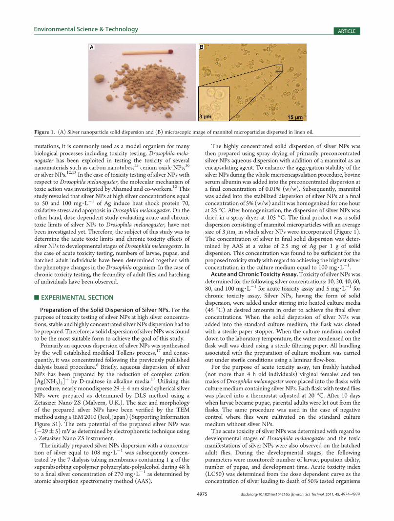
The highly concentrated solid dispersion of silver NPs wasthen prepared using spray drying of primarily preconcentratedsilver NPs aqueous dispersion with addition of a mannitol as anencapsulating agent. To enhance the aggregation stability of thesilver NPs during the whole microencapsulation procedure, bovineserum albumin was added into the preconcentrated dispersion ata final concentration of 0.01% (w/w). Subsequently, mannitolwas added into the stabilized dispersion of silver NPs at a finalconcentration of 5% (w/w) and it was homogenized for one hourat 25 �C. After homogenization, the dispersion of silver NPs wasdried in a spray dryer at 105 �C. The final product was a soliddispersion consisting of mannitol microparticles with an averagesize of 3 μm, in which silver NPs were incorporated (Figure 1).The concentration of silver in final solid dispersion was deter-mined by AAS at a value of 2.5 mg of Ag per 1 g of soliddispersion. This concentration was found to be sufficient for theproposed toxicity study with regard to achieving the highest silverconcentration in the culture medium equal to 100 mg 3 L
�1.Acute andChronic Toxicity Assay.Toxicity of silver NPs was
determined for the following silver concentrations: 10, 20, 40, 60,80, and 100 mg 3 L
�1 for acute toxicity assay and 5 mg 3 L�1 for
chronic toxicity assay. Silver NPs, having the form of soliddispersion, were added under stirring into heated culture media(45 �C) at desired amounts in order to achieve the final silverconcentrations. When the solid dispersion of silver NPs wasadded into the standard culture medium, the flask was closedwith a sterile paper stopper. When the culture medium cooleddown to the laboratory temperature, the water condensed on theflask wall was dried using a sterile filtering paper. All handlingassociated with the preparation of culture medium was carriedout under sterile conditions using a laminar flow-box.For the purpose of acute toxicity assay, ten freshly hatched
(not more than 4 h old individuals) virginal females and tenmales of Drosophila melanogaster were placed into the flasks withculture medium containing silver NPs. Each flask with tested flieswas placed into a thermostat adjusted at 20 �C. After 10 dayswhen larvae became pupae, parental adults were let out from theflasks. The same procedure was used in the case of negativecontrol where flies were cultivated on the standard culturemedium without silver NPs.The acute toxicity of silver NPs was determined with regard to
developmental stages of Drosophila melanogaster and the toxicmanifestations of silver NPs were also observed on the hatchedadult flies. During the developmental stages, the followingparameters were monitored: number of larvae, pupation ability,number of pupae, and development time. Acute toxicity index(LC50) was determined from the dose dependent curve as theconcentration of silver leading to death of 50% tested organisms
Figure 1. (A) Silver nanoparticle solid dispersion and (B) microscopic image of mannitol microparticles dispersed in linen oil.
4976 dx.doi.org/10.1021/es104216b |Environ. Sci. Technol. 2011, 45, 4974–4979
Environmental Science & Technology ARTICLE
(dead organismmeans not hatched individuals). The toxic effectsof silver NPs were also monitored on adult flies where thenumber of adults, the number of females and males, and changesin the phenotype of adult flies were considered. All obtainedresults including the toxic effects of silver NPs on Drosophilamelanogaster were compared with those obtained in the case ofnegative control.Chronic toxicity assay was initiated by placing ten freshly
hatched (not more than 4 h old individuals) virginal females andten males of Drosophila melanogaster into the flasks with culturemedia containing silver NPs with a concentration of 5 mg 3 L
�1 ofAg. This generation of flies entitled as “parental generation, P”gave rise to the first filial generation, F1 generation. Conse-quently, the parental generation was let out from the flasks after10 days. After the hatching of new imagoes from F1 generation,ten randomly selected freshly hatched females and ten maleswere transferred into the flasks with fresh culture media contain-ing silver NPs where the individuals from F1 generation gave riseto the new generation, F2 generation. This procedure wasrepeated until F8 generation.All the experiments evaluating the toxicity of silver NPs on
Drosophila melanogaster were performed in parallel three flasksand were repeated three times. Thus, the acquired results areexpressed as the average values of nine toxicity assays in total.
’RESULTS AND DISCCUSSION
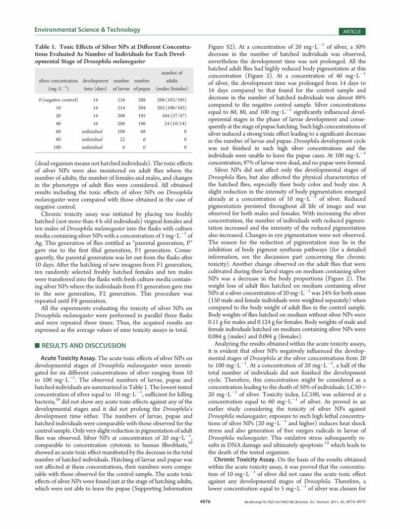
Acute Toxicity Assay.The acute toxic effects of silver NPs ondevelopmental stages of Drosophila melanogaster were investi-gated for six different concentrations of silver ranging from 10to 100 mg 3 L
�1. The observed numbers of larvae, pupae andhatched individuals are summarized in Table 1. The lowest testedconcentration of silver equal to 10 mg 3 L
�1, sufficient for killingbacteria,18 did not show any acute toxic effects against any of thedevelopmental stages and it did not prolong the Drosophila'sdevelopment time either. The numbers of larvae, pupae andhatched individuals were comparable with those observed for thecontrol sample. Only very slight reduction in pigmentation of adultflies was observed. Silver NPs at concentration of 20 mg 3L
�1,comparable to concentration cytotoxic to human fibroblasts,19
showed an acute toxic effect manifested by the decrease in the totalnumber of hatched individuals. Hatching of larvae and pupae wasnot affected at these concentrations, their numbers were compa-rable with those observed for the control sample. The acute toxiceffects of silver NPs were found just at the stage of hatching adults,which were not able to leave the pupae (Supporting Information
Figure S2). At a concentration of 20 mg 3L�1 of silver, a 50%
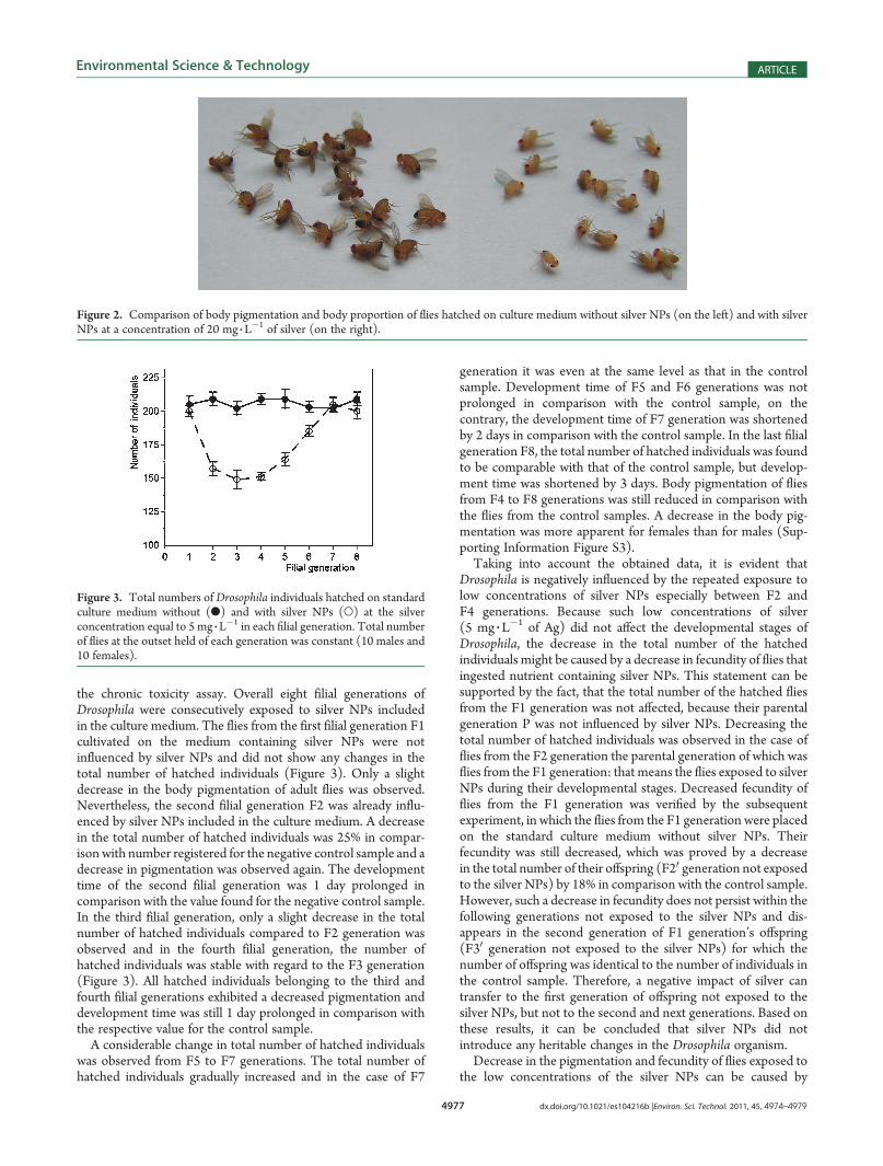
decrease in the number of hatched individuals was observed,nevertheless the development time was not prolonged. All thehatched adult flies had highly reduced body pigmentation at thisconcentration (Figure 2). At a concentration of 40 mg 3L
�1
of silver, the development time was prolonged from 14 days to16 days compared to that found for the control sample anddecrease in the number of hatched individuals was almost 88%compared to the negative control sample. Silver concentrationsequal to 60, 80, and 100 mg 3L
�1 significantly influenced devel-opmental stages in the phase of larvae development and conse-quently at the stage of pupae hatching. Such high concentrations ofsilver induced a strong toxic effect leading to a significant decreasein the number of larvae and pupae. Drosophila development cyclewas not finished in such high silver concentrations and theindividuals were unable to leave the pupae cases. At 100 mg 3L
�1
concentration, 97% of larvae were dead, and no pupae were formed.Silver NPs did not affect only the developmental stages of
Drosophila flies, but also affected the physical characteristics ofthe hatched flies, especially their body color and body size. Aslight reduction in the intensity of body pigmentation emergedalready at a concentration of 10 mg 3 L
�1 of silver. Reducedpigmentation persisted throughout all life of imago and wasobserved for both males and females. With increasing the silverconcentration, the number of individuals with reduced pigmen-tation increased and the intensity of the reduced pigmentationalso increased. Changes in eye pigmentation were not observed.The reason for the reduction of pigmentation may lie in theinhibition of body pigment synthesis pathways (for a detailedinformation, see the discussion part concerning the chronictoxicity). Another change observed on the adult flies that werecultivated during their larval stages on medium containing silverNPs was a decrease in the body proportions (Figure 2). Theweight loss of adult flies hatched on medium containing silverNPs at a silver concentration of 20mg 3L
�1 was 24% for both sexes(150 male and female individuals were weighted separately) whencompared to the body weight of adult flies in the control sample.Body weights of flies hatched on medium without silver NPs were0.11 g for males and 0.124 g for females. Body weights of male andfemale individuals hatched on medium containing silver NPs were0.084 g (males) and 0.094 g (females).Analyzing the results obtained within the acute toxicity assays,
it is evident that silver NPs negatively influenced the develop-mental stages of Drosophila at the silver concentrations from 20to 100 mg 3 L
�1. At a concentration of 20 mg 3 L�1, a half of the
total number of individuals did not finished the developmentcycle. Therefore, this concentration might be considered as aconcentration leading to the death of 50% of individuals: LC50 =20 mg 3 L
�1 of silver. Toxicity index, LC100, was achieved at aconcentration equal to 60 mg 3 L
�1 of silver. As proved in anearlier study considering the toxicity of silver NPs againstDrosophila melanogaster, exposure to such high lethal concentra-tions of silver NPs (20 mg 3 L
�1 and higher) induces heat shockstress and also generation of free oxygen radicals in larvae ofDrosophila melanogaster. This oxidative stress subsequently re-sults in DNA damage and ultimately apoptosis 12 which leads tothe death of the tested organism.Chronic Toxicity Assay. On the basis of the results obtained
within the acute toxicity assay, it was proved that the concentra-tion of 10 mg 3 L
�1 of silver did not cause the acute toxic effectagainst any developmental stages of Drosophila. Therefore, alower concentration equal to 5 mg 3 L
�1 of silver was chosen for
Table 1. Toxic Effects of Silver NPs at Different Concentra-tions Evaluated As Number of Individuals for Each Devel-opmental Stage of Drosophila melanogaster
silver concentration
(mg 3 L�1)
development
time (days)
number
of larvae
number
of pupae
number of
adults
(males/females)
0 (negative control) 14 216 208 208 (103/105)
10 14 214 204 203 (100/103)
20 14 208 195 104 (57/47)
40 16 206 196 24 (10/14)
60 unfinished 108 68 0
80 unfinished 22 6 0
100 unfinished 6 0 0
4977 dx.doi.org/10.1021/es104216b |Environ. Sci. Technol. 2011, 45, 4974–4979
Environmental Science & Technology ARTICLE
the chronic toxicity assay. Overall eight filial generations ofDrosophila were consecutively exposed to silver NPs includedin the culture medium. The flies from the first filial generation F1cultivated on the medium containing silver NPs were notinfluenced by silver NPs and did not show any changes in thetotal number of hatched individuals (Figure 3). Only a slightdecrease in the body pigmentation of adult flies was observed.Nevertheless, the second filial generation F2 was already influ-enced by silver NPs included in the culture medium. A decreasein the total number of hatched individuals was 25% in compar-ison with number registered for the negative control sample and adecrease in pigmentation was observed again. The developmenttime of the second filial generation was 1 day prolonged incomparison with the value found for the negative control sample.In the third filial generation, only a slight decrease in the totalnumber of hatched individuals compared to F2 generation wasobserved and in the fourth filial generation, the number ofhatched individuals was stable with regard to the F3 generation(Figure 3). All hatched individuals belonging to the third andfourth filial generations exhibited a decreased pigmentation anddevelopment time was still 1 day prolonged in comparison withthe respective value for the control sample.A considerable change in total number of hatched individuals
was observed from F5 to F7 generations. The total number ofhatched individuals gradually increased and in the case of F7
generation it was even at the same level as that in the controlsample. Development time of F5 and F6 generations was notprolonged in comparison with the control sample, on thecontrary, the development time of F7 generation was shortenedby 2 days in comparison with the control sample. In the last filialgeneration F8, the total number of hatched individuals was foundto be comparable with that of the control sample, but develop-ment time was shortened by 3 days. Body pigmentation of fliesfrom F4 to F8 generations was still reduced in comparison withthe flies from the control samples. A decrease in the body pig-mentation was more apparent for females than for males (Sup-porting Information Figure S3).Taking into account the obtained data, it is evident that
Drosophila is negatively influenced by the repeated exposure tolow concentrations of silver NPs especially between F2 andF4 generations. Because such low concentrations of silver(5 mg 3 L
�1 of Ag) did not affect the developmental stages ofDrosophila, the decrease in the total number of the hatchedindividuals might be caused by a decrease in fecundity of flies thatingested nutrient containing silver NPs. This statement can besupported by the fact, that the total number of the hatched fliesfrom the F1 generation was not affected, because their parentalgeneration P was not influenced by silver NPs. Decreasing thetotal number of hatched individuals was observed in the case offlies from the F2 generation the parental generation of which wasflies from the F1 generation: that means the flies exposed to silverNPs during their developmental stages. Decreased fecundity offlies from the F1 generation was verified by the subsequentexperiment, in which the flies from the F1 generation were placedon the standard culture medium without silver NPs. Theirfecundity was still decreased, which was proved by a decreasein the total number of their offspring (F20 generation not exposedto the silver NPs) by 18% in comparison with the control sample.However, such a decrease in fecundity does not persist within thefollowing generations not exposed to the silver NPs and dis-appears in the second generation of F1 generation’s offspring(F30 generation not exposed to the silver NPs) for which thenumber of offspring was identical to the number of individuals inthe control sample. Therefore, a negative impact of silver cantransfer to the first generation of offspring not exposed to thesilver NPs, but not to the second and next generations. Based onthese results, it can be concluded that silver NPs did notintroduce any heritable changes in the Drosophila organism.Decrease in the pigmentation and fecundity of flies exposed to
the low concentrations of the silver NPs can be caused by
Figure 2. Comparison of body pigmentation and body proportion of flies hatched on culture medium without silver NPs (on the left) and with silverNPs at a concentration of 20 mg 3 L
�1 of silver (on the right).
Figure 3. Total numbers of Drosophila individuals hatched on standardculture medium without (b) and with silver NPs (O) at the silverconcentration equal to 5 mg 3 L
�1 in each filial generation. Total numberof flies at the outset held of each generation was constant (10 males and10 females).
4978 dx.doi.org/10.1021/es104216b |Environ. Sci. Technol. 2011, 45, 4974–4979
Environmental Science & Technology ARTICLE
oxidative and heat stress, which are induced by silver NPs.9 Thesestress conditions induced during the developmental stages ofDrosophila can be the reason for disturbance of metabolicsynthesis pathways of biogenic amines and several hormonesregulating Drosophila reproduction. It has been demonstratedthat the metabolic systems involved in the stress reaction inDrosophila are those of dopamine (DA), octopamine (OA),juvenile hormone (JH), and ecdysteroids.20�28 Metabolic synth-esis of biogenic amines such as DA and OA is important forfurther metabolic processes and also for regulation of gonado-tropins secretion. Since DA is, beside all, the major precursor fora melanization of body cuticle,29 affecting its metabolic synthesisvia oxidative and heat shock stress induced by silver NPs can leadto the disturbance of metabolic synthesis of melanine which mayresult in a decrease in the pigmentation of adult flies. The JHmetabolic system in females was shown to respond to stress witha decrease in JH degradation,30,31 and the ecdysteroid systemresponded to heat stress with an increase in 20-hydroxyecdysone(20HE) levels.32 A dramatic fertility decrease was the result of thechanges in the JH and 20HE metabolic systems.30 Another workreported that heat stress in wild-type females of Drosophilaresults in oocyte maturation delays, degradation of early vitello-genic egg chambers, inhibition of yolk protein gene expression infollicle cells, and accumulation of mature oocytes.21
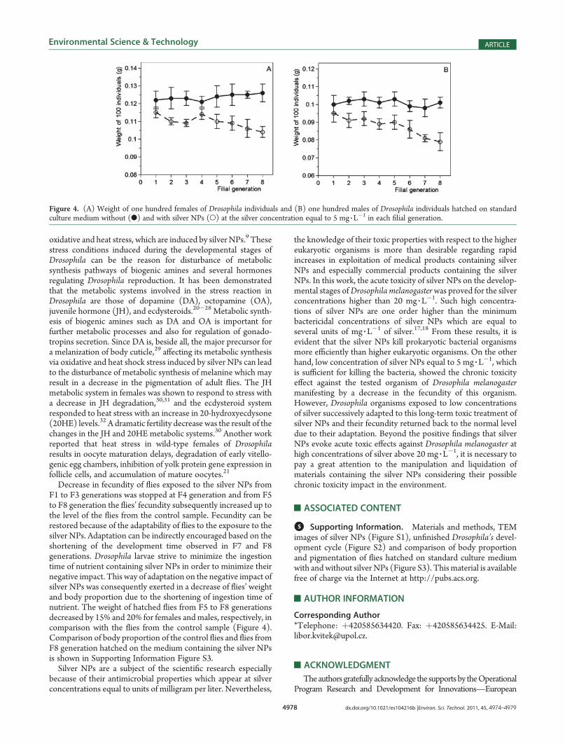
Decrease in fecundity of flies exposed to the silver NPs fromF1 to F3 generations was stopped at F4 generation and from F5to F8 generation the flies’ fecundity subsequently increased up tothe level of the flies from the control sample. Fecundity can berestored because of the adaptability of flies to the exposure to thesilver NPs. Adaptation can be indirectly encouraged based on theshortening of the development time observed in F7 and F8generations. Drosophila larvae strive to minimize the ingestiontime of nutrient containing silver NPs in order to minimize theirnegative impact. This way of adaptation on the negative impact ofsilver NPs was consequently exerted in a decrease of flies’ weightand body proportion due to the shortening of ingestion time ofnutrient. The weight of hatched flies from F5 to F8 generationsdecreased by 15% and 20% for females andmales, respectively, incomparison with the flies from the control sample (Figure 4).Comparison of body proportion of the control flies and flies fromF8 generation hatched on the medium containing the silver NPsis shown in Supporting Information Figure S3.Silver NPs are a subject of the scientific research especially
because of their antimicrobial properties which appear at silverconcentrations equal to units of milligram per liter. Nevertheless,
the knowledge of their toxic properties with respect to the highereukaryotic organisms is more than desirable regarding rapidincreases in exploitation of medical products containing silverNPs and especially commercial products containing the silverNPs. In this work, the acute toxicity of silver NPs on the develop-mental stages ofDrosophila melanogasterwas proved for the silverconcentrations higher than 20 mg 3 L
�1. Such high concentra-tions of silver NPs are one order higher than the minimumbactericidal concentrations of silver NPs which are equal toseveral units of mg 3 L
�1 of silver.17,18 From these results, it isevident that the silver NPs kill prokaryotic bacterial organismsmore efficiently than higher eukaryotic organisms. On the otherhand, low concentration of silver NPs equal to 5 mg 3 L
�1, whichis sufficient for killing the bacteria, showed the chronic toxicityeffect against the tested organism of Drosophila melanogastermanifesting by a decrease in the fecundity of this organism.However, Drosophila organisms exposed to low concentrationsof silver successively adapted to this long-term toxic treatment ofsilver NPs and their fecundity returned back to the normal leveldue to their adaptation. Beyond the positive findings that silverNPs evoke acute toxic effects against Drosophila melanogaster athigh concentrations of silver above 20 mg 3 L
�1, it is necessary topay a great attention to the manipulation and liquidation ofmaterials containing the silver NPs considering their possiblechronic toxicity impact in the environment.
’ASSOCIATED CONTENT
bS Supporting Information. Materials and methods, TEMimages of silver NPs (Figure S1), unfinished Drosophila’s devel-opment cycle (Figure S2) and comparison of body proportionand pigmentation of flies hatched on standard culture mediumwith and without silverNPs (Figure S3). Thismaterial is availablefree of charge via the Internet at http://pubs.acs.org.
’AUTHOR INFORMATION
Corresponding Author*Telephone: þ420585634420. Fax: þ420585634425. E-Mail:[email protected].
’ACKNOWLEDGMENT
Theauthors gratefully acknowledge the supports by theOperationalProgram Research and Development for Innovations—European
Figure 4. (A) Weight of one hundred females of Drosophila individuals and (B) one hundred males of Drosophila individuals hatched on standardculture medium without (b) and with silver NPs (O) at the silver concentration equal to 5 mg 3 L
�1 in each filial generation.

4979 dx.doi.org/10.1021/es104216b |Environ. Sci. Technol. 2011, 45, 4974–4979
Environmental Science & Technology ARTICLE
Social Fund (project CZ.1.05/2.1.00/03.0058). The authors alsogratefully acknowledge the supports by the Ministry of Education,Youth and Sports of the Czech Republic (MSM6198959218,MSM6198959201, MSM0021620822), Czech Science Foundation(GAP304/10/1316), andAcademyof Sciences of theCzechRepublic(KAN115600801).
’REFERENCES
(1) Chen, X.; Schluesener, H. J. Nanosilver: A nanoproduct inmedical application. Toxicol. Lett. 2008, 176 (1), 1–12.(2) Lee, H. Y.; Park, H. K.; Lee, Y. M.; Kim, K.; Park, S. B. A practical
procedure for producing silver nanocoated fabric and its antibacterialevaluation for biomedical applications. Chem. Commun. 2007, 28,2959–2961.(3) Vigneshwaran, N.; Kathe, A. A.; Varadarajan, P. V.; Nachane,
R. P.; Balasubramanya, R. H. Functional finishing of cotton fabrics usingsilver nanoparticles. J. Nanosci. Nanotechnol. 2007, 7 (6), 1893–1897.(4) Griffitt, R. J.; Luo, J.; Gao, J.; Bonzongo, J. C.; Barber, D. S.
Effects of particle composition and species on toxicity of metallicnanomaterials in aquatic organisms. Environ. Toxicol. Chem. 2008, 27(9), 1972–1978.(5) Gaiser, B. K.; Fernandes, T. F.; Jepson, M.; Lead, J. R.; Tyler,
C. R.; Stone, V. Assessing exposure, uptake and toxicity of silver andcerium dioxide nanoparticles from contaminated environments. Environ.Health 2009, 8, S2.(6) Kvitek, L.; Vanickova, M.; Panacek, A.; Soukupova, J.; Dittrich,
M.; Valentova, E.; Prucek, R.; Bancirova, M.; Milde, D.; Zboril, R. Initialstudy on the toxicity of silver nanoparticles (NPs) against Parameciumcaudatum. J. Phys. Chem. C 2009, 113 (11), 4296–4300.(7) Chae, Y. J.; Pham, C. H.; Lee, J.; Bae, E.; Yi, J.; Gu, M. B.
Evaluation of the toxic impact of silver nanoparticles on Japanesemedaka (Oryzias latipes). Aquat. Toxicol. 2009, 94 (4), 320–327.(8) Laban, G.; Nies, L. F.; Turco, R. F.; Bickham, J. W.; Sepulveda,
M. S. The effects of silver nanoparticles on fathead minnow (Pimephalespromelas) embryos. Ecotoxicology 2010, 19 (1), 185–195.(9) Bilberg, K.; Malte, H.; Wang, T.; Baatrup, E. Silver nano-
particles and silver nitrate cause respiratory stress in Eurasian perch(Perca fluviatilis). Aquat. Toxicol. 2010, 9 (2), 159–165.(10) Scown, T. M.; Santos, E. M.; Johnston, B. D.; Gaiser, B.;
Baalousha, M.; Mitov, S.; Lead, J. R.; Stone, V.; Fernandes, T. F.; Jepson,M.; van Aerle, R.; Tyler, C. R. Effects of aqueous exposure to silvernanoparticles of different sizes in rainbow trout.Toxicol. Sci. 2010, 115 (2),521–534.(11) Navarro, E.; Piccapietra, F.; Wagner, B.; Marconi, F.; Kaegi, R.;
Odzak, N.; Sigg, L.; Behra, R. Toxicity of silver nanoparticles to Chlamy-domonas reinhardtii. Environ. Sci. Technol. 2008, 42 (23), 8959–8964.(12) Ahamed, M.; Posgai, R.; Gorey, T. J.; Nielsen, M.; Hussain,
S. M.; Rowe, J. J. Silver nanoparticles induced heat shock protein 70,oxidative stress and apoptosis in Drosophila melanogaster. Toxicol. Appl.Pharmacol. 2010, 242 (3), 263–269.(13) Posgai, R.; Ahamed, M.; Hussain, S. M.; Rowe, J. J.; Nielsen,
M. G. Inhalation method for delivery of nanoparticles to the Drosophilarespiratory system for toxicity testing. Sci. Total Environ. 2009, 408 (2),439–443.(14) Kumari, M.; Mukherjee, A.; Chandrasekaran, N. Genotoxicity
of silver nanoparticles in Allium cepa. Sci. Total Environ. 2009, 407 (19),5243–5246.(15) Liu, X. Y.; Vinson, D.; Abt, D.; Hurt, R. H.; Rand, D. M.
Differential Toxicity of Carbon Nanomaterials in Drosophila: LarvalDietary Uptake Is Benign, but Adult Exposure Causes LocomotorImpairment and Mortality. Environ. Sci. Technol. 2009, 43 (16),6357–6363.(16) Cohen, C. A.; Katfakis, J. A.; Kurnick, M. D.; Hockey, K. S.;
Rzigalinski, B. A. Cerium oxide nanoparticles reduce free radical-mediated toxicity in drosophila melanogaster. Free Radic. Biol. Med.2007, 43, S68–S68.
(17) Panacek, A.; Kvitek, L.; Prucek, R.; Kolar, M.; Vecerova, R.;Pizurova, N.; Sharma, V. K.; Nevecna, T.; Zboril, R. Silver colloidnanoparticles: Synthesis, characterization, and their antibacterial activity.J. Phys. Chem. B 2006, 110 (33), 16248–16253.
(18) Kvitek, L.; Panacek, A.; Soukupova, J.; Kolar, M.; Vecerova, R.;Prucek, R.; Holecova, M.; Zboril, R. Effect of surfactants and polymerson stability and antibacterial activity of silver nanoparticles (NPs).J. Phys. Chem. C 2008, 112 (15), 5825–5834.
(19) Panacek, A.; Kolar, M.; Vecerova, R.; Prucek, R.; Soukupova, J.;Krystof, V.; Hamal, P.; Zboril, R.; Kvitek, L. Antifungal activity of silvernanoparticles againstCandida spp.Biomaterials2009, 30 (31), 6333–6340.
(20) Rauschenbach, I. Y.; Shumnaya, L. V.; Khlebodarova, T. M.;Chentsova, N. A.; Grenback, L. G. Role of phenol oxidases and thyrosinehydroxylase in control of dopamine content in Drosophila-Virilis undernormal conditions and heat-stress. J. Insect Physiol. 1995, 41 (3),279–286.
(21) Gruntenko, N. E.; Bownes, M.; Terashima, J.; Sukhanova,M. Z.; Raushenbach, I. Y. Heat stress affects oogenesis differently inwild-type Drosophila virilis and a mutant with altered juvenile hormoneand 20-hydroxyecdysone levels. Insect Mol. Biol. 2003, 12 (4), 393–404.
(22) Gruntenko, N. E.; Karpova, E. K.; Adonyeva, N. V.; Chentsova,N. A.; Faddeeva, N. V.; Alekseev, A. A.; Rauschenbach, I. Y. Juvenilehormone, 20-hydroxyecdysone and dopamine interaction in Drosophilavirilis reproduction under normal and nutritional stress conditions.J. Insect Physiol. 2005, 51 (4), 417–425.
(23) Neckameyer, W. S.; Weinstein, J. S. Stress affects dopaminergicsignaling pathways inDrosophilamelanogaster. Stress 2005, 8 (2), 117–131.
(24) Gruntenko, N. E.; Karpova, E. K.; Alekseev, A. A.; Chentsova,N. A.; Bogomolova, E. V.; Bownes, M.; Rauschenbach, I. Y. Effects ofoctopamine on reproduction, juvenile hormone metabolism, dopamine,and 20-hydroxyecdysone contents in Drosophila. Arch. Insect Biochem.Physiol. 2007, 65 (2), 85–94.
(25) Rauschenbach, I. Y.; Chentsova, N. A.; Alekseev, A. A.;Gruntenko, N. E.; Adonyeva, N. V.; Karpova, E. K.; Komarova, T. N.;Vasiliev, V. G.; Bownes, M. Dopamine and octopamine regulate20-hydroxyecdysone level in vivo in Drosophila. Arch. Insect Biochem.Physiol. 2007, 65 (2), 95–102.
(26) Gruntenko, N. E.; Rauschenbach, I. Y. Interplay of JH, 20E andbiogenic amines under normal and stress conditions and its effect onreproduction. J. Insect Physiol. 2008, 54 (6), 902–908.
(27) Bogomolova, E. V.; Adon’eva, N. V.; Alekseev, A. A.; Gruntenko,N. E.; Rauschenbach, I. Y. Effect of gonadotropins on dopaminemetabolism in mature Drosophila females. Dokl. Biochem. Biophys.2009, 427 (1), 179–181.
(28) Gruntenko, N. E.; Karpova, E. K.; Chentsova, N. A.; Adonyeva,N. V.; Rauschenbach, I. Y. 20-hydroxyecdysone and juvenile hormoneinfluence tyrosine hydroxylase activity in Drosophila females undernormal and heat stress conditions. Arch. Insect Biochem. Physiol. 2009, 72(4), 263–272.
(29) Walter, M. F.; Zeineh, L. L.; Black, B. C.;McIvor,W. E.;Wright,T. R. F.; Biessmann, H. Catecholamine metabolism and in vitroinduction of premature cuticle melanization in wild type and pigmenta-tion mutants of Drosophila melanogaster. Arch. Insect Biochem. Physiol.1996, 31 (2), 219–233.
(30) Rauschenbach, I. Y.; Gruntenko, N. E.; Khlebodarova, T. M.;Mazurov, M. M.; Grenback, L. G.; Sukhanova, M. J.; Shumnaja, L. V.;Zakharov, I. K.; Hammock, B. D. The role of the degradation system ofthe juvenile hormone in the reproduction of Drosophila under stress.J. Insect Physiol. 1996, 42 (8), 735–742.
(31) Rauschenbach, I. Y.; Khlebodarova, T. M.; Chentsova, N. A.;Gruntenko, N. E.; Grenback, L. G.; Yantsen, E. I.; Filipenko, M. L.Metabolism of the juvenile-hormone in Drosophila adults under normalconditions and heat-stress—Genetic and biochemical aspects. J. InsectPhysiol. 1995, 41 (2), 179–189.
(32) Hirashima, A.; Rauschenbach, I. Y.; Sukhanova,M. J. Ecdysteroidsin stress responsive and nonresponsive Drosophila virilis lines under stressconditions. Biosci. Biotechnol. Biochem. 2000, 64 (12), 2657–2662.
Copyright © 2022 FDOKUMEN