Ferritin is required at multiple stages during the embryonic development of Drosophila melanogaster
25
1 TITLE 1 2 Ferritin is required at multiple stages during the embryonic development of Drosophila melanogaster. 3 4 AUTHORS 5 6 Nicanor González-Morales 1, 2 , Fanis Missirlis 1, 3 , Miguel Ángel Mendoza-Ortíz 1 , Liisa M. Blowes 1, 4 7 and Juan R. Riesgo-Escovar 1* 8 9 AFFILIATIONS 10 11 1 Departamento de Neurobiología del Desarrollo y Neurofisiología, Instituto de Neurobiología, 12 Universidad Nacional Autónoma de México, Campus UNAM Juriquilla, Querétaro, 76230, México. 13 2 CNRS, Institut de Biologie Valrose, iBV, UMR 7277, Nice, 06100, France. 14 3 Departmento de Fisiología, Biofísica y Neurociencias, Centro de Investigación y de Estudios 15 Avanzados del Instituto Politécnico Nacional, Avenida IPN 2508, Zacatenco, México, Distrito Federal, 16 07360, México. 17 4 School of Biological and Chemical Sciences, Queen Mary University of London, London, E1 4NS, 18 UK. 19 20 *corresponding author: [email protected] 21 22 PeerJ PrePrints | http://dx.doi.org/10.7287/peerj.preprints.301v1 | CC-BY 4.0 Open Access | received: 24 Mar 2014, published: 24 Mar 2014 PrePrints
Transcript of Ferritin is required at multiple stages during the embryonic development of Drosophila melanogaster

1
TITLE 1
2
Ferritin is required at multiple stages during the embryonic development of Drosophila melanogaster. 3
4
AUTHORS 5
6
Nicanor González-Morales1, 2, Fanis Missirlis1, 3, Miguel Ángel Mendoza-Ortíz1, Liisa M. Blowes1, 4 7
and Juan R. Riesgo-Escovar1* 8
9
AFFILIATIONS 10
11 1 Departamento de Neurobiología del Desarrollo y Neurofisiología, Instituto de Neurobiología, 12
Universidad Nacional Autónoma de México, Campus UNAM Juriquilla, Querétaro, 76230, México. 13 2 CNRS, Institut de Biologie Valrose, iBV, UMR 7277, Nice, 06100, France. 14 3 Departmento de Fisiología, Biofísica y Neurociencias, Centro de Investigación y de Estudios 15
Avanzados del Instituto Politécnico Nacional, Avenida IPN 2508, Zacatenco, México, Distrito Federal, 16
07360, México. 17 4 School of Biological and Chemical Sciences, Queen Mary University of London, London, E1 4NS, 18
UK. 19
20
*corresponding author: [email protected] 21
22
PeerJ PrePrints | http://dx.doi.org/10.7287/peerj.preprints.301v1 | CC-BY 4.0 Open Access | received: 24 Mar 2014, published: 24 Mar 2014
PrePrin
ts
2
ABSTRACT 23
24
In Drosophila, iron is stored in the endomembrane system of cells inside a protein cavity formed by 24 25
ferritin subunits of two types (Fer1HCH and Fer2LCH) in a 1:1 stoichiometry. Ferritin accumulates in 26
the midgut, nervous system, hemolymph and nephrocytes of Drosophila larvae. Here, we show that 27
mutation of either ferritin gene product or deletion of both genes resulted in a similar set of phenotypes 28
of embryonic lethality, ranging from non-deposition of cuticle to developmental defects associated with 29
germ band retraction, dorsal closure and head involution. Maternal contribution of ferritin, which 30
varied reflecting the mother’s iron stores, is used in early development, but zygotic ferritin mutants 31
died with ectopic apoptotic events and disrupted intestinal morphology. The embryonic nervous system 32
of ferritin mutants presented ventral nerve cord disruptions, misguided axonal projections and brain 33
malformations. Finally, ferritin accumulation was also observed in embryonic hemocytes. One ferritin 34
mutant showed no hemocyte ferritin accumulation and this expression was also lost by genetic 35
inhibition of the secretory pathway. Our work suggests that insect ferritin functions in iron storage, 36
intercellular iron transport and protection from oxidative stress at multiple times during the embryonic 37
development of Drosophila melanogaster. 38
PeerJ PrePrints | http://dx.doi.org/10.7287/peerj.preprints.301v1 | CC-BY 4.0 Open Access | received: 24 Mar 2014, published: 24 Mar 2014
PrePrin
ts

3
INTRODUCTION 39
40
Iron is the most abundant transition metal on earth, commonly found at the active sites of proteins in 41
the form of heme or iron-sulfur clusters or as mono-nuclear and di-nuclear iron (Sheftel et al. 2012). 42
Because of the high reactivity between iron and oxygen, iron has become a key player in aerobic 43
metabolism but also catalyzes oxidative stress when present in excess. Therefore, iron concentration 44
within subcellular compartments and in extracellular fluids is tightly regulated (Cabantchik 2014). The 45
Divalent Metal Transporter 1 (DMT1) is responsible for cellular iron uptake (Gunshin et al. 1997). 46
Ferritin participates in iron homeostasis as the main iron storage complex in both prokaryotes and 47
eukaryotes (Harrison & Arosio 1996). The major form of ferritin in vertebrate animals is cytosolic and 48
consists of 24 subunits of H and L protein chains that assemble into a cage-like structure that sequesters 49
up to 4,500 atoms of iron in its interior core. The H chain contains a ferroxidase center necessary for 50
iron internalization while the L chain contains acidic groups exposed in the interior surface of 51
holoferritin facilitating iron mineralization (Santambrogio et al. 1993). Ferritin genes are regulated 52
during translation by the binding of an iron regulatory protein (IRP) to an iron responsive element 53
(IRE) located in the 5' untranslated region of the mRNA (Pantopoulos et al. 2012). The discovery in 54
mice of ferritin receptors Scara5 (Li et al. 2009) and Tim2 (Todorich et al. 2008) has lead to the idea 55
that ferritin might be involved in iron transport (Meyron-Holtz et al. 2011), however this idea remains 56
controversial (Kell & Pretorius 2014). 57
58
In insects, ferritin shells have an H12L12 organization due to inter- and intra- subunit disulfide-bonds 59
ensuring protein folding and assembly (Hamburger et al. 2005). Ferritin intracellular localization in 60
most insects is directed to the endoplasmic reticulum and the Golgi (Missirlis et al. 2006; Missirlis et 61
al. 2007). The Drosophila melanogaster genome encodes for three ferritin genes: Ferritin 1 heavy 62
chain homologue (Fer1HCH) and Ferritin 2 light chain homologue (Fer2LCH) together produce the 63
major ferritin complex (Georgieva et al. 2002; Georgieva et al. 1999; Mandilaras et al. 2013), whereas 64
Ferritin 3 heavy chain homologue (Fer3HCH) encodes the mitochondrial ferritin, which is 65
predominantly expressed in testis (Kumar et al. 2011; Missirlis et al. 2006). The Drosophila Iron 66
Regulatory Protein-1A (IRP-1A), in its iron-sulfur cluster depleted modality, binds IREs thereby 67
regulating the translation of a subset of Fer1HCH mRNA transcripts (Lind et al. 2006). The sole 68
Drosophila DMT1 homolog (Folwell et al. 2006) was originally isolated as a gustatory mutant named 69
PeerJ PrePrints | http://dx.doi.org/10.7287/peerj.preprints.301v1 | CC-BY 4.0 Open Access | received: 24 Mar 2014, published: 24 Mar 2014
PrePrin
ts

4
Malvolio (Mvl) (D'Souza et al. 1999). Previous work has shown that ferritin is required for embryonic 70
and larval development (Missirlis et al. 2007; Tang & Zhou 2013) and that the ferroxidase center of the 71
H chain is essential (Missirlis et al. 2007), but the specific phenotypes of the ferritin mutants have not 72
been studied to date. Drosophila ferritin has been proposed to function as an external source of iron (Li 73
2010; Nichol et al. 2002; Tang & Zhou 2013; Zhou et al. 2007) but functional analysis in support of 74
this proposition exists only for the secreted ferritin of ticks (Galay et al. 2013; Galay et al. 2014; 75
Hajdusek et al. 2009). 76
77
Here, we analyze Fer1HCH and Fer2LCH mutant embryonic phenotypes. We propose that key 78
functions of the ferritin subunits are likely mediated through the ferritin complex, as their respective 79
mutant phenotypes are indistinguishable during embryogenesis. We show that ferritin mutant 80
phenotypes can be enhanced when embryos are deprived of or have a reduced ferritin maternal 81
contribution, by induction of germ line clones or by limiting iron uptake in parental diets, respectively. 82
A severe central nervous system (CNS) defect, ectopic apoptosis and intestinal damage are seen in 83
embryos dying at later stages of development. Blocking the intracellular secretory pathway during 84
embryogenesis results in the mislocalisation of ferritin. We hypothesize that failure of ferritin transport 85
in one of the mutants tested contributes to the resulting phenotype and lethality. 86
87
88
RESULTS AND DISCUSSION 89
90
Pleiotropic phenotypes of ferritin mutants revealed from embryonic cuticle preparations 91
92
To characterize the embryonic lethal phenotype of ferritin mutants we analyzed the cuticles of 93
previously described transposon-induced loss-of-function alleles Fer1HCH451 and Fer2LCH35 94
(Missirlis et al. 2007; Tang & Zhou 2013), a null mutation for both ferritin genes Df(3R)Fer (Gutierrez 95
et al. 2013), a GFP-trap line Fer1HCHG188 (Missirlis et al. 2007) and also a new allele described here 96
for the first time Fer2LCHΔ17, which fails to complement Fer1HCH451, Fer1HCHG188 and Fer2LCH35 97
(see Materials and Methods). Cuticle preparations of ferritin mutants revealed examples of 98
developmental arrest during the key embryonic processes of germ band extension and retraction, dorsal 99
closure and head involution (Figure 1). Nevertheless, quantification of phenotypes suggests that most 100
PeerJ PrePrints | http://dx.doi.org/10.7287/peerj.preprints.301v1 | CC-BY 4.0 Open Access | received: 24 Mar 2014, published: 24 Mar 2014
PrePrin
ts

5
mutant embryos presented either a normal cuticle (~75%) or no cuticle at all (~15%). These phenotypes 101
suggested that ferritin is required multiple times during development, consistent with differential 102
requirements for iron to support the metabolic shifts that occur during development (Tennessen et al. 103
2014). Notably, a number of iron sulfur cluster proteins are induced during the final stages of 104
embryogenesis to support aerobic glycolysis. 105
106
A maternal contribution of ferritin is utilized during early embryonic development 107
108
Ferritin is maternally contributed (Missirlis et al. 2007). We therefore wondered if our analysis of 109
zygotic mutants could miss early requirements for ferritin fulfilled by this maternal contribution. To 110
analyze how maternal ferritin functioned during embryogenesis we followed two strategies: curtailing 111
iron availability in mothers and generating female germ line clones without ferritin. One way of 112
reducing ferritin expression in adults is to add an iron-specific chelator in their diet (Gutierrez et al. 113
2013; Missirlis et al. 2006). We hypothesized that reduced overall ferritin levels would result in 114
decreased ferritin maternal contribution and a more severe embryonic phenotype. Homozygous mutant 115
embryos derived from heterozygous adults grown with food containing 200µM Bathophenantrholine 116
Sulfate (BPS) showed a doubling of incidence for the no cuticle phenotype (from ~15% to ~30%; 117
Figure 1). Lack of cuticle deposition was likely due to early embryonic death before epidermal 118
differentiation, although we have not excluded a particular requirement for iron in the differentiation of 119
the epidermis, which could provide an alternative explanation for the same phenotypic outcome. Late 120
embryonic phenotypes also became more frequent: U-shaped embryos, indicative of a failure to retract 121
the germ band, augmented from ~2% to ~4%, and embryos failing to complete dorsal closure increased 122
from ~1% to ~4% (Figure 1F; BPS columns). At the same time, the percentage of “wild type” cuticles 123
decreased. This evidence shows that the extent of ferritin maternal contribution is at least partially 124
regulated in flies via limited iron availability. We note that the changes in phenotypic classes 125
abundances were consistently similar in Fer1HCH451, Fer2LCH35, and Df(3R)Fer. Feeding extra iron 126
to adults did not result in a rescue of the embryonic phenotype of the mutant offspring (Figure 1F, 127
FAC), even though total levels of ferritin in mothers were increased, showing that maternal ferritin 128
contribution was not sufficient to fully rescue embryogenesis. 129
130
To further explore whether maternal ferritin was partially rescuing zygotic ferritin mutants, especially 131
PeerJ PrePrints | http://dx.doi.org/10.7287/peerj.preprints.301v1 | CC-BY 4.0 Open Access | received: 24 Mar 2014, published: 24 Mar 2014
PrePrin
ts
6
during early development, we generated homozygous mutant germ line clones for the ferritin genes. 132
Most germ line clones had no cuticle or bore cuticles with defects. The ‘no cuticle’ phenotype 133
percentage changed spectacularly from ~15% to ~80% (Figure 1F). Heterozygous embryos without 134
maternal contribution developed normally into adults (data not shown). We hypothesize that this rescue 135
was due to zygotic ferritin genes being overexpressed as a response to lack of maternal ferritin and that 136
iron in these embryos was delivered to the developing oocyte by other means. 137
138
Central Nervous System (CNS) phenotypes of ferritin mutants 139
140
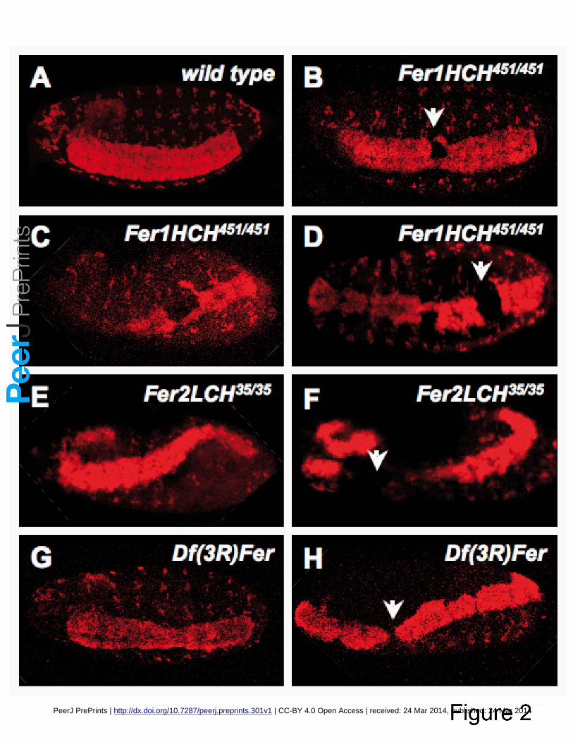
The majority of zygotic ferritin mutant embryos derived from heterozygous mothers died at the final 141
stages of embryogenesis with an apparently normal cuticle. Therefore, we sought to study internal 142
tissues that might be affected by lack of ferritin. Since ferritin protein in first instar larvae is 143
concentrated in the intestine and the CNS (Mehta et al. 2009), we asked whether the CNS developed 144
normally in ferritin mutants. We performed immunofluorescence with antibodies against neuronal 145
markers. The application of an antibody against Elav, which marks all neuronal nuclei, revealed that 146
ferritin mutants harbor holes in the abdominal segments of the CNS (Figure 2). More severe 147
phenotypes were also present, albeit in fewer embryos, including aberrant condensation of the CNS, 148
twisted CNS, and complete lack of parts of the brain and peripheral nervous system (PNS). 149
Importantly, and consistent with our analysis of the cuticle phenotypes discussed above, these 150
phenotypes were observed with both ferritin alleles and with the 2.2 kb genomic deletion that 151
specifically deletes both Fer1HCH and Fer2LCH (Figure 2). We interpret this to mean that, in wild 152
type embryos individual ferritin subunits function in concert to assemble the ferritin complex and do 153
not carry out subunit-specific functions. 154
155
It was previously reported that in adult flies, RNA interference in subsets of neurons against Fer2LCH 156
but not against Fer1HCH disrupted circadian rhythms (Mandilaras & Missirlis 2012). Furthermore, 157
some cell types, including commonly used cell culture lines (Metzendorf et al. 2009; Missirlis et al. 158
2003a), only express Fer1HCH and not Fer2LCH. Finally, overexpression of either Fer1HCH or 159
Fer2LCH or both subunits simultaneously in Drosophila glia (Kosmidis et al. 2011) or neurons (Wu et 160
al. 2012b) resulted in qualitatively different responses. Thus, the question of how each cell type 161
regulates the two ferritin genes and subunits and whether they always act in concert, as the analysis of 162
PeerJ PrePrints | http://dx.doi.org/10.7287/peerj.preprints.301v1 | CC-BY 4.0 Open Access | received: 24 Mar 2014, published: 24 Mar 2014
PrePrin
ts

7
mutant embryonic phenotypes suggests, requires further investigation. 163
164
Further markers were used in embryos homozygous for Df(3R)Fer. We tested whether neuronal axons 165
were projecting normally using ɑ-BP102, which stains axons. BP102-dependent fluorescence revealed 166
that CNS axons are frequently misguided (Figure 3A, D). The developing CNS consists of at least four 167
types of cells: neuroblasts, ganglion mother cells, neurons and glia. Neuroblasts give rise to ganglion 168
mother cells, and these, in turn, give rise to neurons and glia (Biffar & Stollewerk 2014). In order to 169
test whether neuroblasts were affected in ferritin mutant embryos, we performed antibody staining with 170
Deadpan and Evenskipped (Eve) antibodies (Boone & Doe 2008; Kohwi et al. 2013). Our preparations 171
with antibodies against Deadpan, which marks all neuroblasts, indicate that CNS defects are already 172
present within neuroblast cell lineages, including misplaced neuroblasts (Figure 3B, E). As expected for 173
early CNS defects, Eve positive neuroblasts were also heavily misplaced (Figure 3C, F). In conclusion, 174
the developing CNS of embryos lacking the ferritin genes is affected from the time neuroblast cell 175
lineages are specified giving rise to contorted and aberrant CNS in late embryos. 176
177
A ferritin enhancer trap shows expression in the CNS 178
179
Fer1HCH451 carries a LacZ element that serves as reporter of Fer1HCH expression. To test whether this 180
reporter recapitulates known changes in Fer1HCH expression, we monitored LacZ activity in the 181
anterior midgut upon iron feeding of larvae, as occurs for endogenous Fer1HCH (Figure S1). Iron-182
dependent induction of expression was confirmed with this reporter and we therefore used LacZ 183
detection to monitor Fer1HCH expression in the embryo. LacZ driven from Fer1HCH451 is strongly 184
expressed in the neuroectoderm (Figure 4A). The co-localization between the neuronal marker Elav 185
and the β-galactosidase reporter was also observed in homozygous mutant embryos with a disrupted 186
CNS (Figure 4B). We note that the use of the Elav-Gal4 driver to silence either Fer1HCH or Fer2LCH 187
resulted in viable adults with disturbed circadian behavior (Mandilaras & Missirlis 2012) and apparent 188
neurodegeneration (Tang & Zhou 2013). RNA interference is known to cause reduced expression but 189
not complete silencing of its targets, which may explain why the RNAi flies survived to adulthood. In 190
addition, overexpression of ferritin subunits with Elav-Gal4 failed to rescue their respective mutants 191
(Tang & Zhou 2013). Nevertheless, our findings suggest that ferritin is expressed in the embryonic 192
CNS and is required for its development. 193
PeerJ PrePrints | http://dx.doi.org/10.7287/peerj.preprints.301v1 | CC-BY 4.0 Open Access | received: 24 Mar 2014, published: 24 Mar 2014
PrePrin
ts
8
194
Ectopic apoptotic activation in ferritin mutants 195
196
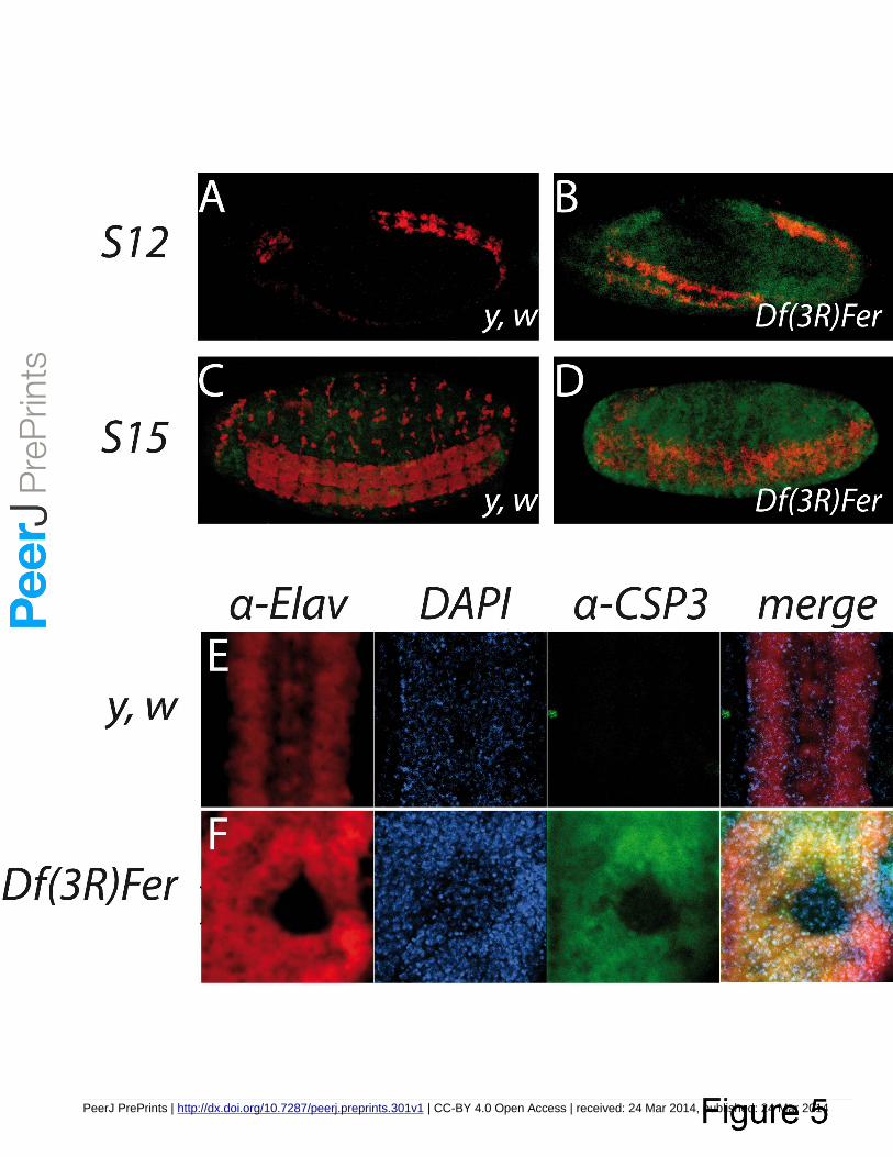
Several links have been drawn in the last decade between iron metabolism and apoptosis (Gambis et al. 197
2011; Kosmidis et al. 2011; Pham et al. 2004). We hypothesized that the disrupted CNS would result 198
following cell death by an apoptotic mechanism. To test the hypothesis we used an antibody that 199
recognizes solely the cleaved, activated caspases, and has been used in flies to mark apoptotic cells 200
(Denton et al. 2008). In contrast to control embryos at stage 12 where there was no apoptotic signal 201
detected (Figure 5A), ectopic apoptotic activation appeared in the neuroectoderm region in mutant 202
embryos (Figure 5B). By stage 15 of embryogenesis control embryos have a weak and restricted 203
apoptotic signal (Figure 5C) whereas in the mutant embryos this signal was massive and covered most 204
of the embryo (Figure 5D). Similar widespread immunoreactivity against activated caspase was also 205
observed with Fer1HCH451 and Fer2LCH35 homozygous mutant embryos (data not shown). Thus, the 206
early apoptotic activation seen in ferritin mutants, mainly restricted to the neuroectoderm region, 207
suggests that the CNS may be more susceptible to the lack of the ferritin complex, although, ultimately, 208
many other tissues become affected. 209
210
The Drosophila DMT1 homolog, Malvolio, is upregulated in ferritin mutants 211
212
Mutations in Mvl result in reduced iron within intestinal iron storage cells and in the whole body 213
(Bettedi et al. 2011). Mvl mutants can suppress both intestinal iron accumulation resulting from ferritin 214
(Tang & Zhou 2013) and Multicopper Oxidase-1 (MCO1) (Lang et al. 2012) misregulation. MCO1 215
catalyzes the oxidation of ferrous iron (Fe2+) to ferric iron (Fe3+) and is thought to participate in cellular 216
iron export in insects (Lang et al. 2012). In view of the above findings, we used Mvl97f, a P-element 217
insertion mutant carrying LacZ reporter gene activity (Rodrigues et al. 1995) to assess β-Galactosidase 218
activity staining. In control embryos, LacZ showed a very restricted pattern of Mvl expression 219
consistent with previous studies (Kumar et al. 2011; Rodrigues et al. 1995), however in the absence of 220
ferritin, Mvl-driven LacZ is upregulated (Figure 6A). We hypothesize than in the absence of functional 221
ferritin, iron depleted cells upregulate Mvl. Strikingly, the addition of one copy of Mvl97f hypomorphic 222
allele into a ferritin-depleted embryo resulted in the appearance of melanotic spots, which were never 223
observed in Mvl97f or in ferritin homozygous mutants alone (Figure 6B, C). The interaction between 224
PeerJ PrePrints | http://dx.doi.org/10.7287/peerj.preprints.301v1 | CC-BY 4.0 Open Access | received: 24 Mar 2014, published: 24 Mar 2014
PrePrin
ts
9
DMT1 and ferritin is reminiscent of the recent finding that the mammalian ferritin receptor SCARA5 is 225
likewise up regulated in the absence of transferrin receptor (Li et al. 2009). 226
227
Ferritin expression, localization and trafficking during development 228
229
Fer1HCHG188 is a mutant Fer1HCH allele, shown to generate a chimeric GFP-Fer1HCH that faithfully 230
mimics endogenous Fer1HCH pattern in heterozygous condition (Gutierrez et al. 2010; Mehta et al. 231
2009; Missirlis et al. 2007; Uhrigshardt et al. 2013). During stages 16-17 of embryonic development 232
GFP tagged Fer1HCH is present in hemocytes (Figure 7A). Hemocytes are large cells that are loosely 233
associated with peripheral tissues and can circulate in the hemolymph, where they function as both 234
phagocytic and immune cells (Evans & Wood 2011). In order to confirm that the large, ferritin-235
accumulating cells were actually hemocytes we used Cg-Gal4 line to drive expression, exclusively in 236
hemocytes, of a nuclearRFP in Fer1HCHG188/+ embryos (Figure 7A). GFP-Fer1HCH is present in the 237
same cells as Cg-nRFP, but not in FB-nRFP (Figure 7B), where mRNA expression is seen (Missirlis et 238
al. 2007). The GFP tag is thought to block the correct function of ferritin in homozygous Fer1HCHG188 239
embryos, because if all the H-subunits carry a GFP tag, embryonic development fails (Missirlis et al. 240
2007) and even Fer1HCHG188/+ flies show a mild reduction in iron accumulation within ferritin 241
(Gutierrez et al. 2013). We noticed that homozygous mutant Fer1HCHG188 embryos showed a 242
dramatically reduced expression of GFP-Fer1HCH than similarly staged heterozygous Fer1HCHG188 243
embryos (Figure 7E, F). Lower and restricted GFP-Fer1HCH expression in homozygous Fer1HCHG188 244
embryos, mainly seen in the intestinal region (Figure 7F), suggested either degradation of the mutant 245
protein or that mutant, non-functional heteropolymers (composed exclusively of GFP-Fer1HCH and 246
Fer2LCH subunits) would not be trafficked. Furthermore, we noted that the intestinal morphology of 247
homozygous Fer1HCHG188 was affected (Figure 7F). To test further whether ferritin is delivered to 248
hemocytes from other embryonic tissues and whether ferritin trafficking can be observed, we blocked 249
the intracellular secretory pathway by means of a lethal mutation in Sec23, sec23j13C8, a P-element 250
insertion in the 5’ UTR of sec23, expected to eliminate or severely attenuate gene function (Abrams & 251
Andrew 2005). If ferritin is indeed transported during embryogenesis, blocking the secretory pathway 252
will impede its exit from the cell where it was originally transcribed. Ferritin was not detected in 253
hemocytes of sec23j13C8/j13C8 mutants; rather, GFP-Fer1HCH aggregates were detected mainly around 254
the midgut (Figure 7D) in a similar expression pattern as observed in homozygous Fer1HCHG188 255
PeerJ PrePrints | http://dx.doi.org/10.7287/peerj.preprints.301v1 | CC-BY 4.0 Open Access | received: 24 Mar 2014, published: 24 Mar 2014
PrePrin
ts
10
mutants. Thus, in embryos impaired in the secretory pathway hemocytes fail to accumulate ferritin, 256
suggesting that its source in wild type embryos may come from hemolymph ferritin. Taken together, 257
our observations suggest that ferritin traffics between tissues during embryonic development and that 258
the hemocytes could play a key role during this trafficking process, by importing ferritin from one 259
tissue and delivering it to another. Such a communicating role for hemocytes has been previously 260
suggested in the context of tissue communication in the innate immune response (Wu et al. 2012a). 261
262
Conclusions 263
264
Insect embryos must course through development with limited amounts of iron, provided during 265
oogenesis by the mother in part via ferritin. It seems reasonable to assume that tissues developing at 266
different rates and with different metabolic states present different iron requirements. Therefore, iron 267
storage and transport are of vital importance for normal development. Ferritin appears to be serving 268
both functions in Drosophila. 269
270
271
EXPERIMENTAL PROCEDURES 272
273
Fly stocks 274
275
As a control strain y,w flies were used. Fer1HCH451 and Fer2LCH35 are P(ry[+t7.2]=PZ) insertion 276
alleles generated during a large-scale mutagenesis screen (Spradling et al. 1999), they have been 277
partially characterized elsewhere (Missirlis et al. 2007) and they were obtained from the Bloomington 278
Drosophila Stock Center (BDSC) stock numbers #11497 and #11483, respectively. Fer2LCHΔ17 was 279
generated from an imprecise excision of Fer2LCHEP1059 (described in Flybase) and interferes with 280
expression of both genes, as confirmed by complementation crosses. Df(3R)Fer was a gift from Alexis 281
Gambis, Bertrand Mollereau and Hermann Steller and is a 2,2 kb deletion disrupting specifically 282
Fer1HCH and Fer2LCH (Gutierrez et al. 2013). To generate germline clones, Fer1HCH451 and 283
Fer2LCH35 were recombined unto FRT82 containing chromosomes (Xu & Rubin 1993). Fer1HCHG188 284
is a protein trap line and has been extensively described elsewhere (Missirlis et al. 2007). Mvl97f is an 285
homozygous viable a P(lacW) insertion was obtained from BDSC #5151 (Rodrigues et al. 1995). 286
PeerJ PrePrints | http://dx.doi.org/10.7287/peerj.preprints.301v1 | CC-BY 4.0 Open Access | received: 24 Mar 2014, published: 24 Mar 2014
PrePrin
ts

11
Sec23j13c8 mutant is a P(lacW) insertion within the 5'UTR of Sec23 (Abrams & Andrew 2005), BDSC 287
stock #10218. Cg-Gal4 (BDSC stock #7011) was used to drive expression in the hemocytes, drm-Gal4 288
(BDSC stock #7098) in embryonic gut and scattered cells around the epidermis, FB-Gal4 in the fat 289
bodies (Missirlis et al. 2006; Missirlis et al. 2003b). In cases where recombinant or double balanced 290
stocks were needed they were generated following conventional crossing schemes. 291
292
Iron diets 293
294
Flies were raised for 3 successive generations on standard medium supplemented with 200 μM 295
Bathophenanthrolinedisulfonic acid disodium salt (SIGMA #B1375) referred to as BPS in the text or 296
with 1 mM ammonium iron (III) citrate (SIGMA #F5859) referred to as FAC. Adults were used for 297
embryo collections. Protein extracts from female adults were also analyzed by non-reducing SDS-298
PAGE, confirming the differential accumulation of ferritin in flies raised on the respective diets (data 299
not shown). 300
301
Immunohistochemistry and confocal imaging 302
303
Following dechorionation with a commercial bleach solution, embryos from overnight collections were 304
devitellinized and fixed in a 1:1 mixture of heptane and 36% formaldehyde for 5 minutes and then 305
washed in methanol. Embryos were then stored at -20 C or rehydrated, and used for staining. Primary 306
antibodies used were: rat α-Elav 1:100, mouse α-BP102 1:100, mouse α-Eve 1:100 (Developmental 307
Studies Hybridoma Bank); α-activated Caspase 3 1:100 (Cell Signaling); and rat α-Deadpan 1:2, a gift 308
from Cheng-Yu Lee. Secondary antibodies used were: Alexa flour 546 α-rat 1:100 (Santa Cruz 309
Biotechnology), Cy5 α-mouse 1:1000, Cy3 α-mouse 1:1000, FITC α-rabbit 1:1000 (Zymax). Signal 310
from α-deadpan staining was increased with the ABC kit from Vectastain. A 510 Meta confocal 311
microscope (Zeiss) was used for fluorescent imaging, and images were processed with Zeiss software 312
and ImageJ. 313
314
Cuticle preparations and X-Gal staining 315
316
Embryos were collected in agar containing plates for 12 hours and incubated for another 36 hours at 317
PeerJ PrePrints | http://dx.doi.org/10.7287/peerj.preprints.301v1 | CC-BY 4.0 Open Access | received: 24 Mar 2014, published: 24 Mar 2014
PrePrin
ts
12
25°C. Viable first instar larvae were removed from cultures. The cuticles of unhatched (dead) embryos 318
were dechorionated and mounted in Hoyer's medium and incubated for 24 hours at 50°C to digest soft 319
tissues. Resulting cuticles were then viewed and photographed with dark field optics in a compound 320
microscope (Nikon). For X-Gal staining embryos were fixed and stained with X-Gal using standard 321
procedures. Both controls and experimental embryos were incubated in parallel for the same amount of 322
time to allow for direct comparisons. 323
324
325
ACKNOWLEDGEMENTS 326
327
We thank María Teresa Peña-Rangel for expert technical assistance in the course of this project, Nydia 328
Hernández-Rios for assistance with the use of the confocal microscope. We also acknowledge Cheng-329
Yu Lee for sending the ɑ-deadpan antibody and Alexis Gambis, Bertrand Mollereau, Hermann Steller 330
for sharing the Df(3R)Fer fly stock prior to publication. 331
332
333
334
PeerJ PrePrints | http://dx.doi.org/10.7287/peerj.preprints.301v1 | CC-BY 4.0 Open Access | received: 24 Mar 2014, published: 24 Mar 2014
PrePrin
ts

13
FIGURE LEGENDS 335
336
Figure 1. Ferritin mutants result in a variety of cuticle phenotypes quantified by different colors. 337
Examples are shown of (A) wild type cuticle (green), (B) head involution defects (red), (C) dorsal 338
closure defects (yellow), (D) germ band retraction defects (orange), (E) no cuticle deposition (blue). (F) 339
Percentages of cuticular phenotypes of ferritin mutants. An enhancement of the earlier phenotypes was 340
seen in mutant embryos whose mothers were fed BPS. 80% of embryos derived from ferritin mutant 341
germline clones failed to develop cuticle. C: normal diet, FAC: high iron diet, BPS: low iron diet, GL: 342
germline clones, n: number of cuticles examined. 343
344
Figure 2. Ferritin mutants result in CNS phenotypes revealed by ɑ-Elav immunofluorescence. All 345
embryos were oriented with anterior to the left and were visualized from a ventrolateral view. (A) 346
control y, w embryo, (B-D) three examples of Fer1HCH451, (E, F) Fer1HCH35 and (G,H) Df(3R)Fer 347
homozygous mutant embryos. The CNS appears twisted (C, E, H) and holes are seen within the ventral 348
nerve cord (white arrows, B, D, F, H) in mutant embryos from all three genotypes. 349
350
Figure 3. In Df(3R)Fer homozygous mutant embryos the CNS appears contorted and with gaps in its 351
organization. (A) Axons of the brain and ventral nerve cord have a stereotyped pattern in normal 352
development. (B, C) Two different neuroblast populations, marked by Dpn and Eve, respectively, show 353
a characteristic spatial organization in control y, w embryos. In homozygous Df(3R)Fer mutants, (D) 354
the axons form but are disorganized and (E, F) the neuroblast populations are misplaced. 355
356
Figure 4. Fer1HCH451 lacZ enhancer trap is expressed in the embryonic CNS. Using an antibody 357
against β-Galactosidase (green) and an antibody against the neuronal marker Elav (red), colocalization 358
is observed in (A) heterozygous Fer1HCH451/+ and (B) homozygous Fer1HCH451 embryos. 359
360
Figure 5. Ferritin mutants cause apoptosis in the CNS and other tissues. Whole embryos were treated 361
with an α-CSP3act marking apoptotic cells (green), and an α-Elav marking neurons (red). (A) Absence 362
of staining for CSP3act in stage 12 y, w embryos, whereas (B) apoptotic markers appear in 363
homozygous Df(3R)Fer embryos from stage 12 onwards mostly restricted to the neurogenic region. C) 364
At stage 15 limited apoptotic events are seen in y, w embryos. D) At stage 15 homozygous Df(3R)Fer 365
PeerJ PrePrints | http://dx.doi.org/10.7287/peerj.preprints.301v1 | CC-BY 4.0 Open Access | received: 24 Mar 2014, published: 24 Mar 2014
PrePrin
ts

14
embryos apoptosis can be observed in all embryonic tissues. (E) Higher magnification of a control 366
ventral nerve cord and (F) a hole in the ventral nerve cord. DAPI was used to mark nuclei (blue), α-367
Elav for neurons (red) and α-CSP3act (green) for apoptosis. 368
369
Figure 6. Genetic interaction between ferritin and the DMT1 homolog Mvl. (A) The Mvl97f-LacZ line 370
shows a spatially restricted expression pattern for Mvl, mainly in head region, the brain, and a 371
segmental repeated pattern. (B) In a Fer2LCH∆17 mutant background, Mvl97f-LacZ expression is 372
induced. Black arrows denote the head region, white asterisk the embryonic brain, red asterisk the 373
ventral nerve cord, and red arrows mark the segmented expression pattern. Cuticle preparations of (C) a 374
homozygous Fer2LCH∆17 embryo and (D) a double mutant Mvl97f, Fer2LCH∆17. (E) Quantification of 375
the appearance of melanotic spots in the cuticle following the introduction of one or two Mvl97f alleles 376
into a ferritin mutant background. 377
378
Figure 7. Ferritin accumulation in embryos with different genetic backgrounds. Ferritin protein was 379
visualized in all embryos from the Fer1HCHG188 GFP trap line. In stage 17 heterozygous 380
Fer1HCHG188/+ embryos that successfully complete development, ferritin mainly accumulates in the 381
midgut and in hemocytes. (A) The GFP-Fer1HCH signal is found in hemocytes marked by cg>nRFP, 382
but not (B) in the fat bodies marked by FB>nRFP. (C, D) Blocking the secretory pathway using a 383
homozygous mutant Sec23j13C8 background reveals that ferritin is absent from the hemocytes, where it 384
normally resides, but accumulates in intestine. (E, F) Fer1HCHG188 homozygous embryos, which die 385
like other ferritin mutants, also show ferritin accumulation in the intestine. These embryos also lose 386
drm>nRFP staining suggesting intestinal disruption and lack ferritin accumulation in hemocytes 387
suggesting ferritin trafficking defects. 388
389
Figure S1. Fer1HCH451 is a functional ferritin enhancer trap line. β-Galactosidase expression is 390
normally restricted to the iron region in the larval midgut but if the expression is enhanced in the 391
anterior midgut (AMG) when iron fed, as occurs in wild type larve (Mehta et al. 2009). 392
393
PeerJ PrePrints | http://dx.doi.org/10.7287/peerj.preprints.301v1 | CC-BY 4.0 Open Access | received: 24 Mar 2014, published: 24 Mar 2014
PrePrin
ts
15
REFERENCES 394
395 Abrams EW, and Andrew DJ. 2005. CrebA regulates secretory activity in the Drosophila salivary 396
gland and epidermis. Development 132:2743-2758. 397 Bettedi L, Aslam MF, Szular J, Mandilaras K, and Missirlis F. 2011. Iron depletion in the intestines of 398
Malvolio mutant flies does not occur in the absence of a multicopper oxidase. J Exp Biol 399 214:971-978. 400
Biffar L, and Stollewerk A. 2014. Conservation and evolutionary modifications of neuroblast 401 expression patterns in insects. Dev Biol 388:103-116. 402
Boone JQ, and Doe CQ. 2008. Identification of Drosophila type II neuroblast lineages containing 403 transit amplifying ganglion mother cells. Dev Neurobiol 68:1185-1195. 404
Cabantchik ZI. 2014. LABILE IRON IN CELLS AND BODY FLUIDS . Physiology, Pathology and 405 Pharmacology. Frontiers in Pharmacology 5. 406
D'Souza J, Cheah PY, Gros P, Chia W, and Rodrigues V. 1999. Functional complementation of the 407 malvolio mutation in the taste pathway of Drosophila melanogaster by the human natural 408 resistance-associated macrophage protein 1 (Nramp-1). J Exp Biol 202:1909-1915. 409
Denton D, Mills K, and Kumar S. 2008. Methods and protocols for studying cell death in Drosophila. 410 Methods Enzymol 446:17-37. 411
Evans IR, and Wood W. 2011. Drosophila embryonic hemocytes. Curr Biol 21:R173-174. 412 Folwell JL, Barton CH, and Shepherd D. 2006. Immunolocalisation of the D. melanogaster Nramp 413
homologue Malvolio to gut and Malpighian tubules provides evidence that Malvolio and 414 Nramp2 are orthologous. J Exp Biol 209:1988-1995. 415
Galay RL, Aung KM, Umemiya-Shirafuji R, Maeda H, Matsuo T, Kawaguchi H, Miyoshi N, Suzuki H, 416 Xuan X, Mochizuki M, Fujisaki K, and Tanaka T. 2013. Multiple ferritins are vital to 417 successful blood feeding and reproduction of the hard tick Haemaphysalis longicornis. J Exp 418 Biol 216:1905-1915. 419
Galay RL, Umemiya-Shirafuji R, Bacolod ET, Maeda H, Kusakisako K, Koyama J, Tsuji N, Mochizuki 420 M, Fujisaki K, and Tanaka T. 2014. Two kinds of ferritin protect ixodid ticks from iron 421 overload and consequent oxidative stress. PLoS One 9:e90661. 422
Gambis A, Dourlen P, Steller H, and Mollereau B. 2011. Two-color in vivo imaging of photoreceptor 423 apoptosis and development in Drosophila. Dev Biol 351:128-134. 424
Georgieva T, Dunkov BC, Dimov S, Ralchev K, and Law JH. 2002. Drosophila melanogaster ferritin: 425 cDNA encoding a light chain homologue, temporal and tissue specific expression of both 426 subunit types. Insect Biochem Mol Biol 32:295-302. 427
Georgieva T, Dunkov BC, Harizanova N, Ralchev K, and Law JH. 1999. Iron availability dramatically 428 alters the distribution of ferritin subunit messages in Drosophila melanogaster. Proc Natl 429 Acad Sci U S A 96:2716-2721. 430
Gunshin H, Mackenzie B, Berger UV, Gunshin Y, Romero MF, Boron WF, Nussberger S, Gollan JL, 431 and Hediger MA. 1997. Cloning and characterization of a mammalian proton-coupled 432 metal-ion transporter. Nature 388:482-488. 433
Gutierrez L, Sabaratnam N, Aktar R, Bettedi L, Mandilaras K, and Missirlis F. 2010. Zinc 434 accumulation in heterozygous mutants of fumble, the pantothenate kinase homologue of 435 Drosophila. FEBS Lett 584:2942-2946. 436
Gutierrez L, Zubow K, Nield J, Gambis A, Mollereau B, Lazaro FJ, and Missirlis F. 2013. Biophysical 437 and genetic analysis of iron partitioning and ferritin function in Drosophila melanogaster. 438
PeerJ PrePrints | http://dx.doi.org/10.7287/peerj.preprints.301v1 | CC-BY 4.0 Open Access | received: 24 Mar 2014, published: 24 Mar 2014
PrePrin
ts

16
Metallomics 5:997-1005. 439 Hajdusek O, Sojka D, Kopacek P, Buresova V, Franta Z, Sauman I, Winzerling J, and Grubhoffer L. 440
2009. Knockdown of proteins involved in iron metabolism limits tick reproduction and 441 development. Proc Natl Acad Sci U S A 106:1033-1038. 442
Hamburger AE, West AP, Jr., Hamburger ZA, Hamburger P, and Bjorkman PJ. 2005. Crystal 443 structure of a secreted insect ferritin reveals a symmetrical arrangement of heavy and light 444 chains. J Mol Biol 349:558-569. 445
Harrison PM, and Arosio P. 1996. The ferritins: molecular properties, iron storage function and 446 cellular regulation. Biochim Biophys Acta 1275:161-203. 447
Kell DB, and Pretorius E. 2014. Serum ferritin is an important inflammatory disease marker, as it 448 is mainly a leakage product from damaged cells. Metallomics. 449
Kohwi M, Lupton JR, Lai SL, Miller MR, and Doe CQ. 2013. Developmentally regulated subnuclear 450 genome reorganization restricts neural progenitor competence in Drosophila. Cell 152:97-451 108. 452
Kosmidis S, Botella JA, Mandilaras K, Schneuwly S, Skoulakis EM, Rouault TA, and Missirlis F. 2011. 453 Ferritin overexpression in Drosophila glia leads to iron deposition in the optic lobes and 454 late-onset behavioral defects. Neurobiol Dis 43:213-219. 455
Kumar S, Konikoff C, Van Emden B, Busick C, Davis KT, Ji S, Wu LW, Ramos H, Brody T, 456 Panchanathan S, Ye J, Karr TL, Gerold K, McCutchan M, and Newfeld SJ. 2011. FlyExpress: 457 visual mining of spatiotemporal patterns for genes and publications in Drosophila 458 embryogenesis. Bioinformatics 27:3319-3320. 459
Lang M, Braun CL, Kanost MR, and Gorman MJ. 2012. Multicopper oxidase-1 is a ferroxidase 460 essential for iron homeostasis in Drosophila melanogaster. Proc Natl Acad Sci U S A 461 109:13337-13342. 462
Li JY, Paragas N, Ned RM, Qiu A, Viltard M, Leete T, Drexler IR, Chen X, Sanna-Cherchi S, 463 Mohammed F, Williams D, Lin CS, Schmidt-Ott KM, Andrews NC, and Barasch J. 2009. 464 Scara5 is a ferritin receptor mediating non-transferrin iron delivery. Dev Cell 16:35-46. 465
Li S. 2010. Identification of iron-loaded ferritin as an essential mitogen for cell proliferation and 466 postembryonic development in Drosophila. Cell Res 20:1148-1157. 467
Lind MI, Missirlis F, Melefors O, Uhrigshardt H, Kirby K, Phillips JP, Soderhall K, and Rouault TA. 468 2006. Of two cytosolic aconitases expressed in Drosophila, only one functions as an iron-469 regulatory protein. J Biol Chem 281:18707-18714. 470
Mandilaras K, and Missirlis F. 2012. Genes for iron metabolism influence circadian rhythms in 471 Drosophila melanogaster. Metallomics 4:928-936. 472
Mandilaras K, Pathmanathan T, and Missirlis F. 2013. Iron absorption in Drosophila melanogaster. 473 Nutrients 5:1622-1647. 474
Mehta A, Deshpande A, Bettedi L, and Missirlis F. 2009. Ferritin accumulation under iron scarcity in 475 Drosophila iron cells. Biochimie 91:1331-1334. 476
Metzendorf C, Wu W, and Lind MI. 2009. Overexpression of Drosophila mitoferrin in l(2)mbn cells 477 results in dysregulation of Fer1HCH expression. Biochem J 421:463-471. 478
Meyron-Holtz EG, Moshe-Belizowski S, and Cohen LA. 2011. A possible role for secreted ferritin in 479 tissue iron distribution. J Neural Transm 118:337-347. 480
Missirlis F, Holmberg S, Georgieva T, Dunkov BC, Rouault TA, and Law JH. 2006. Characterization of 481 mitochondrial ferritin in Drosophila. Proc Natl Acad Sci U S A 103:5893-5898. 482
Missirlis F, Hu J, Kirby K, Hilliker AJ, Rouault TA, and Phillips JP. 2003a. Compartment-specific 483 protection of iron-sulfur proteins by superoxide dismutase. J Biol Chem 278:47365-47369. 484
PeerJ PrePrints | http://dx.doi.org/10.7287/peerj.preprints.301v1 | CC-BY 4.0 Open Access | received: 24 Mar 2014, published: 24 Mar 2014
PrePrin
ts

17
Missirlis F, Kosmidis S, Brody T, Mavrakis M, Holmberg S, Odenwald WF, Skoulakis EM, and Rouault 485 TA. 2007. Homeostatic mechanisms for iron storage revealed by genetic manipulations and 486 live imaging of Drosophila ferritin. Genetics 177:89-100. 487
Missirlis F, Rahlfs S, Dimopoulos N, Bauer H, Becker K, Hilliker A, Phillips JP, and Jackle H. 2003b. A 488 putative glutathione peroxidase of Drosophila encodes a thioredoxin peroxidase that 489 provides resistance against oxidative stress but fails to complement a lack of catalase activity. 490 Biol Chem 384:463-472. 491
Nichol H, Law JH, and Winzerling JJ. 2002. Iron metabolism in insects. Annu Rev Entomol 47:535-492 559. 493
Pantopoulos K, Porwal SK, Tartakoff A, and Devireddy L. 2012. Mechanisms of mammalian iron 494 homeostasis. Biochemistry 51:5705-5724. 495
Pham CG, Bubici C, Zazzeroni F, Papa S, Jones J, Alvarez K, Jayawardena S, De Smaele E, Cong R, 496 Beaumont C, Torti FM, Torti SV, and Franzoso G. 2004. Ferritin heavy chain upregulation by 497 NF-kappaB inhibits TNFalpha-induced apoptosis by suppressing reactive oxygen species. 498 Cell 119:529-542. 499
Rodrigues V, Cheah PY, Ray K, and Chia W. 1995. malvolio, the Drosophila homologue of mouse 500 NRAMP-1 (Bcg), is expressed in macrophages and in the nervous system and is required 501 for normal taste behaviour. EMBO J 14:3007-3020. 502
Santambrogio P, Levi S, Cozzi A, Rovida E, Albertini A, and Arosio P. 1993. Production and 503 characterization of recombinant heteropolymers of human ferritin H and L chains. J Biol 504 Chem 268:12744-12748. 505
Sheftel AD, Mason AB, and Ponka P. 2012. The long history of iron in the Universe and in health 506 and disease. Biochim Biophys Acta 1820:161-187. 507
Spradling AC, Stern D, Beaton A, Rhem EJ, Laverty T, Mozden N, Misra S, and Rubin GM. 1999. The 508 Berkeley Drosophila Genome Project gene disruption project: Single P-element insertions 509 mutating 25% of vital Drosophila genes. Genetics 153:135-177. 510
Tang X, and Zhou B. 2013. Ferritin is the key to dietary iron absorption and tissue iron 511 detoxification in Drosophila melanogaster. FASEB J 27:288-298. 512
Tennessen JM, Bertagnolli NM, Evans J, Sieber MH, Cox J, and Thummel CS. 2014. Coordinated 513 Metabolic Transitions During Drosophila Embryogenesis and the Onset of Aerobic 514 Glycolysis. G3 (Bethesda). 515
Todorich B, Zhang X, Slagle-Webb B, Seaman WE, and Connor JR. 2008. Tim-2 is the receptor for H-516 ferritin on oligodendrocytes. J Neurochem 107:1495-1505. 517
Uhrigshardt H, Rouault TA, and Missirlis F. 2013. Insertion mutants in Drosophila melanogaster 518 Hsc20 halt larval growth and lead to reduced iron-sulfur cluster enzyme activities and 519 impaired iron homeostasis. J Biol Inorg Chem 18:441-449. 520
Wu SC, Liao CW, Pan RL, and Juang JL. 2012a. Infection-induced intestinal oxidative stress triggers 521 organ-to-organ immunological communication in Drosophila. Cell Host Microbe 11:410-522 417. 523
Wu Z, Du Y, Xue H, Wu Y, and Zhou B. 2012b. Aluminum induces neurodegeneration and its toxicity 524 arises from increased iron accumulation and reactive oxygen species (ROS) production. 525 Neurobiol Aging 33:199 e191-112. 526
Xu T, and Rubin GM. 1993. Analysis of genetic mosaics in developing and adult Drosophila tissues. 527 Development 117:1223-1237. 528
Zhou G, Kohlhepp P, Geiser D, Frasquillo Mdel C, Vazquez-Moreno L, and Winzerling JJ. 2007. Fate 529 of blood meal iron in mosquitoes. J Insect Physiol 53:1169-1178. 530
PeerJ PrePrints | http://dx.doi.org/10.7287/peerj.preprints.301v1 | CC-BY 4.0 Open Access | received: 24 Mar 2014, published: 24 Mar 2014
PrePrin
ts

18
531 532
PeerJ PrePrints | http://dx.doi.org/10.7287/peerj.preprints.301v1 | CC-BY 4.0 Open Access | received: 24 Mar 2014, published: 24 Mar 2014
PrePrin
ts

PeerJ PrePrints | http://dx.doi.org/10.7287/peerj.preprints.301v1 | CC-BY 4.0 Open Access | received: 24 Mar 2014, published: 24 Mar 2014
PrePrin
ts
PeerJ PrePrints | http://dx.doi.org/10.7287/peerj.preprints.301v1 | CC-BY 4.0 Open Access | received: 24 Mar 2014, published: 24 Mar 2014
PrePrin
ts

PeerJ PrePrints | http://dx.doi.org/10.7287/peerj.preprints.301v1 | CC-BY 4.0 Open Access | received: 24 Mar 2014, published: 24 Mar 2014
PrePrin
ts

PeerJ PrePrints | http://dx.doi.org/10.7287/peerj.preprints.301v1 | CC-BY 4.0 Open Access | received: 24 Mar 2014, published: 24 Mar 2014
PrePrin
ts
PeerJ PrePrints | http://dx.doi.org/10.7287/peerj.preprints.301v1 | CC-BY 4.0 Open Access | received: 24 Mar 2014, published: 24 Mar 2014
PrePrin
ts

PeerJ PrePrints | http://dx.doi.org/10.7287/peerj.preprints.301v1 | CC-BY 4.0 Open Access | received: 24 Mar 2014, published: 24 Mar 2014
PrePrin
ts

PeerJ PrePrints | http://dx.doi.org/10.7287/peerj.preprints.301v1 | CC-BY 4.0 Open Access | received: 24 Mar 2014, published: 24 Mar 2014
PrePrin
ts