Histone Methyltransferase DOT1L Drives Recovery of Gene Expression after a Genotoxic Attack
12
Histone Methyltransferase DOT1L Drives Recovery of Gene Expression after a Genotoxic Attack Valentyn Oksenych 1.¤a , Alexander Zhovmer 1.¤b , Salim Ziani 1 , Pierre-Olivier Mari 2 , Jitka Eberova 1 , Tiziana Nardo 3 , Miria Stefanini 3 , Giuseppina Giglia-Mari 2 , Jean-Marc Egly 1 , Fre ´de ´ ric Coin 1 * 1 IGBMC, Department of Functional Genomics and Cancer, CNRS/INSERM/Universite ´ de Strasbourg, C. U. Strasbourg, France, 2 Universite ´ de Toulouse, UPS, IPBS, Toulouse, France, 3 Istituto di Genetica Molecolare, Consiglio Nazionale delle Ricerche, Pavia, Italy Abstract UV-induced DNA damage causes repression of RNA synthesis. Following the removal of DNA lesions, transcription recovery operates through a process that is not understood yet. Here we show that knocking-out of the histone methyltransferase DOT1L in mouse embryonic fibroblasts (MEF DOT1L ) leads to a UV hypersensitivity coupled to a deficient recovery of transcription initiation after UV irradiation. However, DOT1L is not implicated in the removal of the UV-induced DNA damage by the nucleotide excision repair pathway. Using FRAP and ChIP experiments we established that DOT1L promotes the formation of the pre-initiation complex on the promoters of UV-repressed genes and the appearance of transcriptionally active chromatin marks. Treatment with Trichostatin A, relaxing chromatin, recovers both transcription initiation and UV- survival. Our data suggest that DOT1L secures an open chromatin structure in order to reactivate RNA Pol II transcription initiation after a genotoxic attack. Citation: Oksenych V, Zhovmer A, Ziani S, Mari P-O, Eberova J, et al. (2013) Histone Methyltransferase DOT1L Drives Recovery of Gene Expression after a Genotoxic Attack. PLoS Genet 9(7): e1003611. doi:10.1371/journal.pgen.1003611 Editor: Bjo ¨ rn Schumacher, Cologne Excellence Cluster for Cellular Stress Responses in Aging Associated Diseases, Germany Received February 5, 2013; Accepted May 18, 2013; Published July 4, 2013 Copyright: ß 2013 Oksenych et al. This is an open-access article distributed under the terms of the Creative Commons Attribution License, which permits unrestricted use, distribution, and reproduction in any medium, provided the original author and source are credited. Funding: Research for this paper was found by the French National Research Agency (ANR ChromaRep) and the European Research Council (ERC TransReact). The funders had no role in study design, data collection and analysis, decision to publish, or preparation of the manuscript. Competing Interests: The authors have declared that no competing interests exist. * E-mail: [email protected] . These authors contributed equally to this work. ¤a Current address: Department of Genetics, Harvard Medical School, Children’s Hospital, Immune Disease Institute, Howard Hughes Medical Institute, Boston, Massachusetts, United States of America. ¤b Current address: Memorial Sloan-Kettering Cancer Center, The Rockefeller Research Laboratories, New York, New York, United States of America. Introduction Short-wave UV light is a significant source of mutagenic and cytotoxic DNA damage. UV irradiation induces two major types of DNA lesions; the cis-syn cyclobutane-pyrimidine dimers (CPD) and the pyrimidine (6-4) pyrimidone photoproducts (6-4PP) [1]. Through the deformation of the DNA structure, these lesions have repressive effect on various nuclear processes including replication and transcription. As a matter of fact, the removal of these lesions is a priority for the cell and takes place at the expense of fundamental cellular processes that are paused to circumvent the risks of mutations that may lead to cancer. The molecular mechanism underlying transcription inhibition and recovery is not understood yet but it includes proteins such as CSB, a member of the SWI2/SNF2 family of chromatin remodeling proteins, which promote transcription re-initiation at the promoters of UV- repressed genes [2,3]. UV lesions are removed from DNA by the nucleotide excision repair (NER) mechanism through two sub-pathways. The general global genome repair (GG-NER) removes DNA damage from the entire genome, while the transcription-coupled repair (TC-NER) corrects lesions located on actively transcribed genes [4]. In TC- NER, an elongating RNA polymerase II (RNA Pol II) stalled by a lesion triggers efficient repair of the cytotoxic damage that blocks transcription, while lesion elsewhere in the genome are detected by the XPC/hHR23B complex for GG-NER [5]. Then, both sub- pathways funnel into a common process involving XPA, RPA, TFIIH, XPG and XPF-ERCC1 to excise damaged oligonucleo- tides from DNA. Post-translational histone modifications modulate promoter activity. Histone acetylation, phosphorylation, ubiquitination, and methylation dictate the transcriptional fate of any given locus [6]. Inactive heterochromatin is associated with high levels of methylation at H3K9, H3K27, and H4K20 residues and low levels of acetylation, while actively transcribed euchromatin shows a high level of acetylation of H4K16 and H4K20 and methylation of H3K4, H3K36, and H3K79 residues [7,8]. The dot1 gene ( disruptor of telomeric silencing-1), also called kmt4 (lysine methyltransferase-4), encodes a protein that exclusively methylates lysine 79 of histone H3 (H3K79) [9,10,11]. Unlike most modified histone residues that are located within the N- terminal tail, H3K79 is found within the globular core of the histone octamer [12]. The Dot1 protein is the only histone lysine methyltransferase that does not contain the conserved SET domain but exhibits a methyltransferase fold that is responsible for its activity [13,14]. In mammals, several studies have shown that DOT1L (the Dot1 homolog) exists in a complex that trimethylates H3K79 and that contains several myeloid/lymphoid or mix-lineage leukemia fusion partners, such as MLLT1, 2, 3 or 10 [15]. More recently, DOT1L was shown to be involved in cell PLOS Genetics | www.plosgenetics.org 1 July 2013 | Volume 9 | Issue 7 | e1003611
-
Upload
independent -
Category
Documents
-
view
1 -
download
0
Transcript of Histone Methyltransferase DOT1L Drives Recovery of Gene Expression after a Genotoxic Attack

Histone Methyltransferase DOT1L Drives Recovery ofGene Expression after a Genotoxic AttackValentyn Oksenych1.¤a, Alexander Zhovmer1.¤b, Salim Ziani1, Pierre-Olivier Mari2, Jitka Eberova1,
Tiziana Nardo3, Miria Stefanini3, Giuseppina Giglia-Mari2, Jean-Marc Egly1, Frederic Coin1*
1 IGBMC, Department of Functional Genomics and Cancer, CNRS/INSERM/Universite de Strasbourg, C. U. Strasbourg, France, 2 Universite de Toulouse, UPS, IPBS,
Toulouse, France, 3 Istituto di Genetica Molecolare, Consiglio Nazionale delle Ricerche, Pavia, Italy
Abstract
UV-induced DNA damage causes repression of RNA synthesis. Following the removal of DNA lesions, transcription recoveryoperates through a process that is not understood yet. Here we show that knocking-out of the histone methyltransferaseDOT1L in mouse embryonic fibroblasts (MEFDOT1L) leads to a UV hypersensitivity coupled to a deficient recovery oftranscription initiation after UV irradiation. However, DOT1L is not implicated in the removal of the UV-induced DNAdamage by the nucleotide excision repair pathway. Using FRAP and ChIP experiments we established that DOT1L promotesthe formation of the pre-initiation complex on the promoters of UV-repressed genes and the appearance of transcriptionallyactive chromatin marks. Treatment with Trichostatin A, relaxing chromatin, recovers both transcription initiation and UV-survival. Our data suggest that DOT1L secures an open chromatin structure in order to reactivate RNA Pol II transcriptioninitiation after a genotoxic attack.
Citation: Oksenych V, Zhovmer A, Ziani S, Mari P-O, Eberova J, et al. (2013) Histone Methyltransferase DOT1L Drives Recovery of Gene Expression after aGenotoxic Attack. PLoS Genet 9(7): e1003611. doi:10.1371/journal.pgen.1003611
Editor: Bjorn Schumacher, Cologne Excellence Cluster for Cellular Stress Responses in Aging Associated Diseases, Germany
Received February 5, 2013; Accepted May 18, 2013; Published July 4, 2013
Copyright: � 2013 Oksenych et al. This is an open-access article distributed under the terms of the Creative Commons Attribution License, which permitsunrestricted use, distribution, and reproduction in any medium, provided the original author and source are credited.
Funding: Research for this paper was found by the French National Research Agency (ANR ChromaRep) and the European Research Council (ERC TransReact).The funders had no role in study design, data collection and analysis, decision to publish, or preparation of the manuscript.
Competing Interests: The authors have declared that no competing interests exist.
* E-mail: [email protected]
. These authors contributed equally to this work.
¤a Current address: Department of Genetics, Harvard Medical School, Children’s Hospital, Immune Disease Institute, Howard Hughes Medical Institute, Boston,Massachusetts, United States of America.¤b Current address: Memorial Sloan-Kettering Cancer Center, The Rockefeller Research Laboratories, New York, New York, United States of America.
Introduction
Short-wave UV light is a significant source of mutagenic and
cytotoxic DNA damage. UV irradiation induces two major types
of DNA lesions; the cis-syn cyclobutane-pyrimidine dimers (CPD)
and the pyrimidine (6-4) pyrimidone photoproducts (6-4PP) [1].
Through the deformation of the DNA structure, these lesions have
repressive effect on various nuclear processes including replication
and transcription. As a matter of fact, the removal of these lesions
is a priority for the cell and takes place at the expense of
fundamental cellular processes that are paused to circumvent the
risks of mutations that may lead to cancer. The molecular
mechanism underlying transcription inhibition and recovery is not
understood yet but it includes proteins such as CSB, a member of
the SWI2/SNF2 family of chromatin remodeling proteins, which
promote transcription re-initiation at the promoters of UV-
repressed genes [2,3].
UV lesions are removed from DNA by the nucleotide excision
repair (NER) mechanism through two sub-pathways. The general
global genome repair (GG-NER) removes DNA damage from the
entire genome, while the transcription-coupled repair (TC-NER)
corrects lesions located on actively transcribed genes [4]. In TC-
NER, an elongating RNA polymerase II (RNA Pol II) stalled by a
lesion triggers efficient repair of the cytotoxic damage that blocks
transcription, while lesion elsewhere in the genome are detected by
the XPC/hHR23B complex for GG-NER [5]. Then, both sub-
pathways funnel into a common process involving XPA, RPA,
TFIIH, XPG and XPF-ERCC1 to excise damaged oligonucleo-
tides from DNA.
Post-translational histone modifications modulate promoter
activity. Histone acetylation, phosphorylation, ubiquitination,
and methylation dictate the transcriptional fate of any given locus
[6]. Inactive heterochromatin is associated with high levels of
methylation at H3K9, H3K27, and H4K20 residues and low
levels of acetylation, while actively transcribed euchromatin shows
a high level of acetylation of H4K16 and H4K20 and methylation
of H3K4, H3K36, and H3K79 residues [7,8].
The dot1 gene (disruptor of telomeric silencing-1), also called
kmt4 (lysine methyltransferase-4), encodes a protein that exclusively
methylates lysine 79 of histone H3 (H3K79) [9,10,11]. Unlike
most modified histone residues that are located within the N-
terminal tail, H3K79 is found within the globular core of the
histone octamer [12]. The Dot1 protein is the only histone lysine
methyltransferase that does not contain the conserved SET
domain but exhibits a methyltransferase fold that is responsible
for its activity [13,14]. In mammals, several studies have shown
that DOT1L (the Dot1 homolog) exists in a complex that
trimethylates H3K79 and that contains several myeloid/lymphoid
or mix-lineage leukemia fusion partners, such as MLLT1, 2, 3 or
10 [15]. More recently, DOT1L was shown to be involved in cell
PLOS Genetics | www.plosgenetics.org 1 July 2013 | Volume 9 | Issue 7 | e1003611

cycle progression, the control of the differentiation of pluripotent
cells [16] and leukemogenesis [17].
In addition to these roles in fundamental cellular processes,
several lines of evidence suggest that DOT1L plays an important
role in genomic stability. DOT1L has been reported to favor the
recruitment of double strand breaks sensor 53BP1 to DNA lesions
[18]. Dot1 is known to be required for the activation of the RAD9/
RAD53 checkpoint function by UV and c-radiation [19,20,21].
Dot1 also plays a role in the yeast cellular response to UV damage
but its specific function in this mechanism is unclear [22].
Here, we provide evidence that the mammalian DOT1L
protein is required to re-initiate transcription after UV irradiation.
Knocking-out of DOT1L results in high sensitivity to UV
irradiation in mouse embryonic fibroblasts (MEF), but preserves
an accurate repair of (6-4)PP lesions. Instead, MEFDOT1L are
unable to recover transcription of constitutively expressed genes
after UV irradiation. Using fluorescence recovery after photo-
bleaching (FRAP), we have shown that DOT1L regulates the
recruitment of RNA Pol II to chromatin after UV irradiation.
Applying chromatin immunoprecipitation assay, we additionally
revealed that DOT1L triggers the formation of the pre-initiation
complex (PIC) to the promoters of UV-repressed genes, as well as
the appearance of active transcriptional marks on histones.
Altogether, our results highlight a new role for DOT1L in the
maintenance of an open chromatin structure in order to reactivate
the formation of the transcription machinery on the promoter of
constitutive genes after a genotoxic attack.
Results
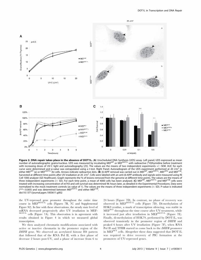
DOT1L deficient mammalian cells are UV-sensitiveTo investigate the role of DOT1L in the repair of UV-induced
DNA damage, we used knocked-down MEFDOT1L cells carrying a
homozygous gene trap insertion in Dot1l [23]. Together with an
absence of Dot1l protein expression, the mono- and tri-methylation
of H3K79 were strongly reduced in MEFDOT1L (Figure 1A). In a
UV-C survival assay, MEFDOT1L cells were more sensitive to
irradiation as compared to control MEFWT but less than MEFXPG,
knocked-out for the NER factor XPG and deficient both for GG-
and TC-NER [24] (Figure 1B). However, MEFDOT1L showed
similar UV-C sensitivity than the MEFCSB cells, knocked-out for the
CSB protein involved only in TC-NER. Note that tri-methylation of
H3K79 was similar in MEFWT, MEFXPG and MEFCSB (Supple-
mental Figure S1A). Knocking-down of DOT1L expression in
HeLa cells using siRNA (Supplemental Figure S1B) recapitulated
the mild UV-sensitivity of the MEF cells compared to the high UV-
sensitivity induced by the knocked-down of the NER factor XPA
(Supplemental Figures S1B–C). These data indicate that DOT1L
deficiency induces UV-sensitivity in mammalian cells.
We further investigated whether DOT1L affected UV-C
survival by sustaining the repair of UV-induced DNA damage.
We performed unscheduled DNA synthesis assay (UDS), which is
mainly a measure of the GG-NER efficiency [25]. The UDS of
MEFDOT1L was identical to that of the MEFWT (Figure 2A). We
also used an assay based on immunofluorescence coupled to
quantification of DNA lesions directly in cell nucleus to measure
the removal of the (6-4)PPs, the main UV-induced DNA lesions
(see experimental procedures). MEFXPG cells showed low removal
of (6-4)PP along the time course of repair, compared to MEFWT or
MEFCSB in which lesions were rapidly removed (Figures 2B).
When measured in MEFDOT1L cells, the removal rate of (6-4)PP
lesions was identical to that of MEFWT (Figures 2B), which implied
that the absence of DOT1L does not impair GG-NER.
To determine whether MEFDOT1L were able to perform TC-
NER, we performed two sets of experiments. First we measured
cell survival following incubation with Ecteinascidin 743 (et743),
an anti-tumor drug that shows cytotoxicity effect only on human
TC-NER proficient cells [26]. In our experimental conditions,
knocking-down of the TC-NER factor CSB in MEF cells resulted in
et743 resistance, compared to MEFWT cells (Figure 2C). In contrast,
MEFDOT1L cells showed sensitivity to et743 to a level equivalent to
that of MEFWT cells (Figure 2C). Next, we performed a host cell
reactivation assay [27]. We employed a dual GFP/RFP readout
with a UV-damaged plasmid (600 J/m2, 3 Kb) containing a GFP-
tagged reporter and an undamaged plasmid containing an RFP-
tagged reporter transiently transfected into MEF cells. Recovery of
GFP-reporter expression, 12 hours post-transfection, was efficient
in both MEFWT (Figure 3, panels a–f) and MEFDOT1L (Figure 3,
panels g–l) cells but not in the TC-NER deficient MEFCSB cells
(Figure 3, m–r). Overall, these results suggest that the absence of
DOT1L induces sensitivity to UV irradiation that is not the
consequence of a defect in GG- or TC-NER.
DOT1L promotes transcription recovery after UVirradiation
Next we aimed to determine the global RNA synthesis of MEF
cells after irradiation using the recovery of RNA synthesis (RRS)
assay [25]. Cells were UV-irradiated with 10 or 20 J/m2 and
incubated with radioactive [3H]uridine during a 30 minutes pulse
performed before and 24 hours after irradiation. Mock-treated
MEFWT and MEFDOT1L cells showed similar levels of RNA
synthesis, visualized by equivalent number of black dots in their
nuclei (around 100 dots/nucleus, Figure 4A and 4B, compare
panels a and c). In contrast, we observed a marked deficiency in
RNA synthesis in MEFDOT1L cells, 24 hours after UV-C
treatment, as compared to MEFWT (Figure 4A and 4B, compare
panels b and d). We estimated the residual transcription activity in
the MEFDOT1L cells to 30% of that of the mock-treated cells,
24 hours after irradiation with 20 J/m2.
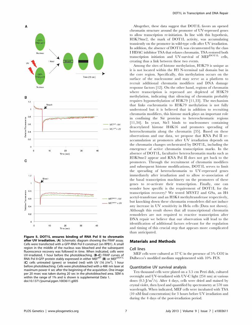
DOT1L ensures RNA Pol II binding to chromatin after UVirradiation
To unveil the molecular mechanisms that led to the inhibition of
transcription in MEFDOT1L cells after UV irradiation, we
examined live-cell protein mobility of RNA Pol II by fluorescence
recovery after photobleaching (FRAP). In brief, a small region in
the middle of the nucleus was bleached and the subsequent
Author Summary
Through the deformation of the genomic DNA structure,UV-induced DNA lesions have repressive effect on variousnuclear processes including replication and transcription.As a matter of fact, the removal of these lesions is a priorityfor the cell and takes place at the expense of fundamentalcellular processes that are paused to circumvent the risksof mutations that may lead to cancer. The molecularmechanism underlying transcription inhibition and recov-ery is not clearly understood and appears more compli-cated than anticipated. Here we analyzed the process oftranscription recovery after UV-irradiation and found that itdepends on DOT1L, a histone methyltransferase thatpromotes the reformation of the transcription machineryat the promoters of UV-repressed genes. Our discoveryshows that transcription recovery after a genotoxic attackis an active process under the control of chromatinremodelling enzymes.
DOT1L in Transcription and DNA Repair
PLOS Genetics | www.plosgenetics.org 2 July 2013 | Volume 9 | Issue 7 | e1003611

fluorescence recovery was measured in time (Figure 5A). For that
purpose, the largest RNA Pol II subunit RPB1 was fused with GFP
and expressed either in MEFWT or MEFDOT1L. In these
conditions, we found an equivalent mobility of RNA Pol II in
mock-treated and UV-irradiated (UV-C, 16 J/m2) MEFWT
(Figure 5B and Supplemental Data S1) (T-test = 3.4E-2). In
marked contrast, FRAP analysis of UV-damaged MEFDOT1L cells
revealed a significant increase in fluorescence recovery when
compared to mock-treated cells (Figure 5C and Supplemental
Data S1) (T-test = 1.4E-5), indicating that a fraction of RNA Pol II
became mobile in the absence of DOT1L, after UV irradiation.
Defect in transcription initiation after UV irradiation incells depleted of DOT1L
We next wondered whether DOT1L was required to mobilize
RNA pol II either during the initiation or elongation steps of
transcription. For this purpose, we examined the step of transition
from initiation to elongation by the RNA Pol II in vivo on an
endogenous mouse gene. Briefly, we reversibly blocked gene
transcription by incubating cells with the P-TEFb inhibitor DRB
(5,6-dichloro-1-b-D-ribobenzimidazole), which inhibited the tran-
sition from initiation to elongation but did not block elongation of
ongoing mRNA transcripts [28] (Figure 6A). Following the
removal of DRB, RNA Pol II was released from promoter-
proximal regions and the level of newly synthesized pre-mRNA had
been measured owing to the presence of exons and introns. We
measured the transition from initiation to elongation on the Utrophin
gene that possesses a very short Exon1 (170 bp). We estimated that
an average of one transcription blocking lesion (CPD or (6-4)PP) was
created per 5 kb of DNA at 20 J/m2 [29], indicating that less than
5% of cells harbor a UV-lesion in this exon following a dose of 15 J/
m2 of UV-C. Therefore, any significant inhibition of transcription
initiation cannot be explained by the blockage of RNA Pol II in
front of a lesion in Exon 1.
We treated MEF cells for 3 hours with DRB and extracted
RNA at 10 minutes intervals after the removal of the drug
(Figure 6B). Next, we performed RT-PCR using primers spanning
Exon1-Intron1 junctions to detect newly synthesized pre-mRNA of
Utrophin gene. In the absence of genotoxic stress, MEFWT and
MEFDOT1L were both able to recover transcription of the Exon1
region within 10 to 20 minutes after DRB removal (Figure 6C),
indicating that the transcriptional initiation rate in MEFWT and
MEFDOT1L was identical, in absence of a genotoxic attack. Then, we
irradiated MEF cells with UV-C (15 J/m2) after the DRB treatment
(Figure 6B). In these experimental conditions, MEFWT were able to
recover transcription of the Exon1 within 60 minutes after removal
of DRB and UV-treatment, while MEFDOT1L showed no recovery
of Exon1 transcription even after 80 minutes (Figure 6D).
Since DOT1L was shown to be involved in chromatin
remodeling, we next tested whether chromatin relaxation could
overcome the absence of DOT1L. For that purpose, we treated
MEFDOT1L with Trichostatin A (TSA, 20 nM), a class I histone
deacetylase (HDAC) inhibitor that relaxed chromatin (Figure 6B).
Following TSA treatment, we observed a recovery of Exon1 pre-
mRNA expression in MEFDOT1L, which peaked between 30 to
40 minutes after UV irradiation (Figure 6E) and paralleled the
transcription of Exon1 in MEFWT cells. Together with this
recovery, pre-treatment of MEFDOT1L with TSA (10 nM) induced
a potent recovery of UV survival (Figure 6F) suggesting that
transcription inhibition in MEFDOT1L was indeed responsible for
the UV-sensitivity of these cells. Pre-treatment of the TC-NER
deficient MEFCSB with TSA did not modify their UV-sensitivity
(Figure 6F). Altogether, these data suggested that DOT1L allowed
RNA Pol II transcription re-initiation after UV irradiation.
Repressive chromatin marks in cells depleted of DOT1Lafter UV irradiation
The above data prompted us to perform a detailed analysis of
the PIC formation on the promoter of UV-repressed genes
throughout the time, after irradiation. We studied the promoter of
the several housekeeping genes such as DHFR (dihydrofolate
reductase), B2M (beta-2-microglobulin) or KLF7 (Kruppel-like
factor 7) that we used as models for assembly/disassembly of the
PIC after UV-irradiation. Using chromatin immunoprecipitation
(ChIP), we observed a slight decrease in both RNA Pol II
(Figures 7B and Supplemental Figure S2) and basal transcription
factors (Figure 7C) occupancy at these promoters in UV-irradiated
MEFs, 2 hours post-UV irradiation. The transcription machinery
started to re-assemble on the promoter between 6 and 10 hours
after UV irradiation and the steady state level of mRNA did not
vary significantly with time in the wild-type situation (Figure 7A).
In contrast, the basal transcription machinery did not re-form on
Figure 1. DOT1L is involved in UV-survival in mammalian cells.(A) Fifty mg of whole cell extracts from MEFWT or MEFDOT1L cells wereresolved by SDS-PAGE and Western-blotted for DOT1L. Asteriskindicates an unspecific cross-reacting band. Ten mg of fractions fromhistone acid-extraction performed on MEFWT or MEFDOT1L cells wereresolved by SDS-PAGE and Western-blotted for H3, H3K79me1 orH3K79me3. (B) MEFWT, MEFDOT1L, MEFXPG and MEFCSB cells were testedin a UV-survival assay. Cells were treated with increasing dose of UV-Cand cell survival was determined 96 hours later, as detailed in theExperimental Procedures. Data were normalized to the mock treatmentcontrols (as value of 1). The values are the means of three independentexperiments (6 SD). P-value was extrapolated for MEFDOT1L/MEFWT orMEFDOT1L/MEFXPG and indicated on the graph (***,0.005).doi:10.1371/journal.pgen.1003611.g001
DOT1L in Transcription and DNA Repair
PLOS Genetics | www.plosgenetics.org 3 July 2013 | Volume 9 | Issue 7 | e1003611
the UV-repressed gene promoter throughout the entire time
course in MEFDOT1L cells (Figures 7B, 7C and Supplemental
Figure S2). In line with these observations, the steady state level of
mRNA decreased progressively after UV irradiation in MEF-DOT1L cells (Figure 7A). This observation is in agreement with
results obtained in Figure 4 in which we measured global
transcription.
We then analyzed chromatin modifications associated with
active or inactive chromatin in the promoter region of the
DHFR gene. We observed an acetylated histone H4 pattern
that followed that of the RNA Pol II, with a first phase of
decrease 2 hours post-UV, and a phase of increase from 6 to
24 hours (Figure 7D). In contrast, no phase of recovery was
observed in MEFDOT1L cells (Figure 7D). Di-methylation of
H3K9 residue, a mark of transcription silencing, was stable in
MEFWT throughout the time course after UV-treatment, while
it increased just after irradiation in MEFDOT1L (Figure 7E).
Finally, di-methylation of H3K79, performed by DOT1L, was
observed transiently in the promoter region of DHFR and
peaked 6 hours after UV irradiation (Figure 7F), when RNA
Pol II and TFIIB started to come back to the DHFR promoter
in MEFWT cells. Altogether these data suggested that DOT1L
was required to drive recovery of PIC formation at the
promoters of UV-repressed genes.
Figure 2. DNA repair takes place in the absence of DOT1L. (A) Unscheduled DNA Synthesis (UDS) assay. Left panel; UDS expressed as meannumber of autoradiographic grains/nucleus. UDS was measured by incubating MEFWT or MEFDOT1L with radioactive [3H]thymidine before treatmentwith increasing doses of UV-C light and autoradiography [25]. The values are the means of two independent experiments +/2SEM. AUC for eachcurve were determined and p-value was extrapolated using a t-test. Right Panel; Autoradiogram of the UDS experiment performed at 20 J/m2 ineither MEFWT (a) or MEFDOT1L (b) cells. Arrows indicate radioactive dots. (B) (6-4)PP removal was carried out in MEFWT, MEFDOT1L, MEFXPG and MEFCSB,harvested at different time points after UV irradiation at 20 J/m2. Cells were labeled with an anti-(6-4)PP antibody and signals were measured using INCell 1000 analyzer (GE Healthcare). Graph represents the % of lesions removed from the genome at different time points. The values are the means ofthree independent experiments (6 SD). For each time point, a mean of 4000 cells has been analysed. (C) MEFWT, MEFDOT1L and MEFCSB cells weretreated with increasing concentration of et743 and cell survival was determined 96 hours later, as detailed in the Experimental Procedures. Data werenormalized to the mock treatment controls (as value of 1). The values are the means of three independent experiments (6 SD). P-value is indicated(***,0.005) and was determined between MEFDOT1L and either MEFCSB.doi:10.1371/journal.pgen.1003611.g002
DOT1L in Transcription and DNA Repair
PLOS Genetics | www.plosgenetics.org 4 July 2013 | Volume 9 | Issue 7 | e1003611

Discussion
Transcription inhibition and the subsequent recovery that
operate after a genotoxic attack are thought to limit the risks that
lesions represent for the genome. The molecular mechanisms that
are responsible for the turn-off/turn-on of transcription after DNA
damage are not well understood. Here we show that the
methyltransferase DOT1L is required for the re-initiation of
transcription by triggering re-formation of the transcription
machinery at the promoter of UV-repressed genes. Our data
supports the hypothesis that transcription recovery after a
genotoxic attack is an active process involving specific actors
insuring not only the repair of the DNA but also the recovery of
fundamental cellular processes such as transcription.
DOT1L participates in UV-survival but not in DNA repairOur study demonstrated that disruption of DOT1L caused
hypersensitivity to UV irradiation in mammalian cells. There are
several potential mechanisms that could explain the increased
sensitivity to UV irradiation conferred by DOT1L depletion. If it
was directly involved in DNA repair, its absence may results in
increased levels of DNA damage, leading to cellular apoptosis. A
function for yeast DOT1 in GG-NER has been described recently
[30]. However, we did not find any DNA repair defect associated
with the absence of DOT1L in mammalian cells. Indeed, MEF
cells depleted of DOT1L exhibited normal UDS level, an assay
that mainly measured GG-NER. Furthermore, these cells repaired
(6-4)PP, the best UV-induced NER substrate, at the same rate
than wild-type cells. To show that MEFDOT1L cells were also
proficient in TC-NER, we used two strategies. First we made use
of the sensitivity of cells to the drug et743, a natural marine
product isolated from the Caribbean see squirt. Antiproliferative
activity of et743 was shown to be dependent on active TC-NER
[26]. In our experimental conditions, we observed that MEFs
knockdown for CSB, one of the two TC-NER specific factors,
exhibited a higher resistance to et743 treatment than MEFWT,
confirming previous observation obtained with patient cells [26].
Using et743 on MEFDOT1L cells, we observed that these cells were
as sensitive to treatment as MEFWT. In a second set of
experiments, we measured the capacity of MEFDOT1L to drive
the recovery of a reporter expression construct previously exposed
to UV-irradiation. In mouse-derived cells, the reactivation
depends both on efficient GG- and TC-NER activities as
illustrated by the absence of transcription recovery in MEFCSB
cells observed both in our study and in other reports [31].
However, MEFDOT1L showed a full recovery of reporter
expression. Finally, the treatment of MEFDOT1L with TSA
induced a recovery of UV-survival while treatment of MEFCSB
did not. We concluded from these cellular observations summa-
rized in Table 1 that it was unlikely that UV-irradiation sensitivity
in DOT1L-deficient mammalian cells was due to a GG-NER or
TC-NER defect.
DOT1L controls transcription recovery after UVirradiation
Alternatively, DOT1L may serve to reactivate global mRNA
synthesis after UV irradiation. Transcriptional arrest has been
shown to lead to a highly cytotoxic cellular response to stress [5].
This response has multiple causes and is likely not only the result
of DNA lesions that block RNA Pol II in elongation. Previous
studies have challenged the relationship between efficient repair of
a lesion in the transcribed strand of active genes and the
restoration of DNA damage inhibited transcription. For instance,
cells carrying mutations in CSB were unable to recover lesion-
inhibited transcription while they efficiently repaired acetylamino-
fluorene lesions in transcriptionally active genes [32]. In addition,
CSB was shown to accumulate at the promoters of UV-repressed
genes, where it stimulated the recovery of transcription indepen-
dently of the presence of lesions [3]. This finding led to the
Figure 3. Host cell reactivation assay in MEF cells. MEFWT,MEFDOT1L and MEFCSB were transfected with mock-treated (panels a–c,g–i, m–o) or irradiated (UV-C, 600 J/m2) (panels d–f, j–l, p–r) pEGFPreporter plasmid in combination with a non-irradiated pRFP reporterplasmid used as control. Twelve hours post transfection, GFP and RFPexpression were determined. A ratio of 3/1 (pEGFP/pRFP) was usedduring transfection to ensure that any cell expressing the RFP expressesalso the GFP. P-value was extrapolated using t-test (**,0.05,***,0.005).doi:10.1371/journal.pgen.1003611.g003
DOT1L in Transcription and DNA Repair
PLOS Genetics | www.plosgenetics.org 5 July 2013 | Volume 9 | Issue 7 | e1003611

hypothesis that removal of transcription blocking lesions was
insufficient to restore transcription after DNA damage and that in
addition, chromatin changes in the promoters of UV-repressed
genes may be required. We performed RNA-sequencing on
MEFWT and MEFDOT1L after UV-irradiation (unpublished Data).
However, RNA-sequencing measures steady-state level of individ-
ual mRNA but does not provide information on the de novo RNA
activity. To measured the global level of newly synthetized RNA
we used the RRS assay and we observed a general inhibition of de
novo RNA synthesis in cells depleted of DOT1L; 30% of residual
transcription activity was detected 24 hours post irradiation (20 J/
m2). This inhibition was confirmed at the single gene level since
DHFR showed a similar level of inhibition.
DOT1L allow PIC formation on UV-repressed genepromoters
Early steps of mRNA expression include formation of PIC,
transcription initiation and escape of RNA Pol II from the promoter
to the elongation step. Using an assay that measured the
transcription of the newly synthetized first exon of Utrophin gene
model in vivo, we demonstrated that the transition from initiation to
elongation was deficient in the absence of DOT1L after UV. We
further analysed both PIC occupancy and the chromatin modifi-
cations on the promoter of a UV-repressed gene after irradiation
and have shown that this promoter was temporally depleted of basal
transcription factors in the first hours after irradiation in wild-type
cells. Six to ten hours after UV irradiation, when DNA repair took
place, the PIC occupancy recovered completely in the wild-type
situation. In the absence of DOT1L, RNA Pol II and TFIIB did not
get back to the promoter of our UV-repressed gene model, and
heterochromatin marks such as methylation of the H3K9 residue
appeared. FRAP experiment using GFP-tagged RPB1 subunit of
RNA Pol II showed that a fraction of RNA Pol II became mobile
after UV-irradiation in the absence of DOT1L, indicating that
dissociation of RNA Pol II from chromatin of UV-repressed genes
after irradiation in the absence of DOT1L is a broad phenomenon.
Figure 4. Deficient transcription recovery after UV irradiation in the absence of DOT1L. (A) Recovery of RNA synthesis assay (RRS). Themean numbers of auto-radiographic grains per nucleus of mock treated or UV-irradiated cells (10 or 20 J/m2) from two independent experiments areexpressed (6 SEM, measured on at least 150 cells). Under the graph, the results are expressed as percentage of grains per nucleus relative to mocktreated cells. (B) Autoradiogram of an RRS experiment. Twenty four hours after UV irradiation (20 J/m2), MEFWT (panels a–b) or MEFDOT1L (panels c–d)cells were pulse labeled 30 minutes with [3H]uridine followed by autoradiography. P-value was extrapolated using t-test (**,0.05, ***,0.005).doi:10.1371/journal.pgen.1003611.g004
DOT1L in Transcription and DNA Repair
PLOS Genetics | www.plosgenetics.org 6 July 2013 | Volume 9 | Issue 7 | e1003611
Altogether, these data suggest that DOT1L favors an opened
chromatin structure around the promoter of UV-repressed genes
to allow transcription re-initiation. In line with this hypothesis,
H3K79me2, the mark of DOT1L activity, was accumulating
transiently on the promoter in wild-type cells after UV irradiation.
In addition, the absence of DOT1L was circumvented by the class
I HDAC inhibitor TSA that relaxes chromatin. TSA restored both
transcription initiation and UV-survival of MEFDOT1L cells,
creating thus a link between these two events.
Among the sites of histone methylation, H3K79 is unique as
it is not located within the H3 N-terminal tail domain but in
the core region. Specifically, this methylation occurs on the
surface of the nucleosome and may serve as a platform to
recruit additional chromatin modifiers and DNA damage
response factors [12]. On the other hand, regions of chromatin
where transcription is repressed are depleted of H3K79
methylation, indicating that silencing of chromatin probably
requires hypomethylation of H3K79 [11,33]. The mechanism
that links euchromatin to H3K79 methylation is not fully
understood but it is believed that in addition to recruiting
chromatin modifiers, this histone mark plays an important role
in confining the Sir proteins to heterochromatic regions
[11,34]. In yeast, Sir3 binds to nucleosomes containing
deacetylated histone H4K16 and promotes spreading of
heterochromatin along the chromatin [35]. Based on these
observations and our data, we propose that RNA Pol II re-
accumulation at promoters after UV irradiation depends on
the chromatin changes orchestrated by DOT1L, including the
emergence of active chromatin transcription marks. In the
absence of DOT1L, facultative heterochromatin marks such as
H3K9me2 appear and RNA Pol II does not get back to the
promoters. Through the recruitment of chromatin modifiers
and subsequent histone modifications, DOT1L serves to limit
the spreading of heterochromatin to UV-repressed genes
immediately after irradiation and to allow re-association of
the basal transcription machinery on the promoters of these
genes to re-activate their transcription. Finally, one can
wonder how specific is the requirement of DOT1L for the
transcription recovery? We tested MYST2 and G9a, an H4
acetyl-transferase and an H3K4 methyltransferase respectively,
but knocking down these chromatin remodelers did not induce
any increase in UV sensitivity in Hela cells (Data not shown).
Although this result shows that all transcriptional chromatin
remodelers are not required to reactive transcription after
DNA repair we believe that our observation will lead to the
identification of additional factors relevant for the regulation
and timing of this crucial step that appears more complicated
than anticipated.
Materials and Methods
Cell linesMEF cells were cultured at 37uC in the presence of 5% CO2 in
Dulbecco’s modified medium supplemented with 10% FCS.
Quantitative UV survival analysisTen thousand cells were plated on a 3.5 cm Petri dish, cultured
overnight and UV-irradiated with UV-C light (254 nm) at various
doses (0.5 J/m2/s). After 4 days, cells were dried and stained by
crystal violet, then lysed and quantified by spectrometry at 570 nm
wavelength. When indicated, MEF cells were incubated with TSA
(10 nM final concentration) for 3 hours before UV irradiation and
during the 4 days of the post-irradiation period.
Figure 5. DOT1L ensures binding of RNA Pol II to chromatinafter UV irradiation. (A) Schematic diagram showing the FRAP assay.Cells were transfected with a GFP-RNA Pol II construct (on RPB1). A smallregion in the middle of the nucleus was bleached and the subsequentfluorescence recovery was followed in time. When indicated, cells wereUV-irradiated, 1 hour before the photobleaching. (B–C) FRAP curves ofRNA Pol II-GFP protein stably expressed in either MEFWT (B) or MEFDOT1L
(C) cells untreated (green) or treated (red) with UV (16 J/m2), 1 hourbefore photobleaching. Cells were photobleached with a 488 nm laser atmaximum power 4 sec after the beginning of the acquisition. One imageper 20 msec was taken during 20 sec in the photobleached area. SEM iswithin the range of 1% and is shown is supplemental Data 1.doi:10.1371/journal.pgen.1003611.g005
DOT1L in Transcription and DNA Repair
PLOS Genetics | www.plosgenetics.org 7 July 2013 | Volume 9 | Issue 7 | e1003611

Figure 6. Inhibition of the initiation-to-elongation transition phase in the absence of DOT1L, after UV irradiation. (A) Schematicrepresentation of measuring the initiation-to-elongation transition rate of RNA Pol II transcription in vivo on endogenous genes. We reversiblyblocked gene transcription by incubating cells with DRB. Cells are depleted of their pre-mRNA pool within few hours of incubation with the drug [28].Following the chase of DRB, RNA Pol II is released from promoter-proximal regions and newly synthesized pre-mRNA appear; the level of pre-mRNA ismeasured using oligonucleotides targeting respectively the exon/intron junctions of the gene. (B) Schematic diagram showing the experimentalapproach used to measure the rate of initiation-to-elongation transition phase by RNA Pol II after UV irradiation. MEF cells were treated with DRB for3 hours before chase and addition of fresh medium at t = 0 hour. When indicated, cells were UV irradiated at t = 0 hour, before the addition of freshmedium or treated with TSA for 12 hours before the addition of DRB. (C) Expression levels of the newly synthesized Exon1 of the Utrophin gene inMEFWT and MEFDOT1L cells treated with 100 mM of DRB for 3 hours before the addition of fresh medium. The cells were harvest at intervals of10 minutes for RNA isolation and qRT-PCR was performed using oligonucleotides targeting respectively the Exon1 and Intron1 of the gene. Relativeexpression values compared to mock treated cells are plotted against time (6SD). AUC was determined and p-value was extrapolated using t-test. (D)Expression levels of the newly synthesized Exon1 of the Utrophin gene in either MEFWT or MEFDOT1L irradiated with UV-C (15 J/m2) after treatment asin (C). AUC was determined and p-value was extrapolated using t-test. (E) Expression levels of the newly synthesized Exon1 of the Utrophin gene ineither MEFWT or MEFDOT1L treated with TSA (20 nM) for 12 hours before addition of DRB for 3 hours and UV-C irradiation (15 J/m2). TSA wasmaintained in the medium during DRB treatment and time course. AUC was determined and p-value was extrapolated using t-test. (F) MEFWT, MEFCSB
and MEFDOT1L were irradiated with increasing doses of UV-C light. Cell survival was determined 96 hours later, as detailed in the ExperimentalProcedures. Data were normalized to the mock treated controls (as value of 1). Means of three independent experiments are shown (6 SD). Whenindicated, cells were treated with TSA (10 nM), 12 hours before UV irradiation, and TSA was maintained for the time of the experiment.doi:10.1371/journal.pgen.1003611.g006
DOT1L in Transcription and DNA Repair
PLOS Genetics | www.plosgenetics.org 8 July 2013 | Volume 9 | Issue 7 | e1003611

Immunofluorescent-based DNA lesion quantificationFive thousand MEF cells were plated in 96 well plates
(OptiPlates-96, Perkin Elmer). Twenty-four hours later, cells were
UV-irradiated with UV-C lamp (10 J/m2) and recovered in fresh
medium for the indicated period of time at 37uC, 5% CO2.
Immuno-labeling of (6-4)PP was performed using mouse 64M-2
antibodies. DNA was denatured with 2 M HCl for 30 minutes at
RT and blocked in 10% BSA in PBS for 15 minutes prior to
labeling. (6-4)PP lesions were quantified using an IN Cell Analyser
1000 imaging system (GE Healthcare) and the percentage of (6-
4)PP removal was determined (100% represents the amount of
lesions determined just after UV irradiation).
Figure 7. Pre-initiation complex assembly and chromatin modification after UV irradiation. (A) Determination of the relative mRNAexpression level of DHFR gene in MEFWT and MEFDOT1L after UV irradiation (10 J/m2) performed after reverse transcription with random hexameres.The values are the means of three independent experiments (6 SD). AUC was determined and p-value was extrapolated using t-test. (B–F) Time-dependent occupancy of RNA Pol II (B), TFIIB (C), H4Ac (D), H3K9me2 (E) and H3K79me2 (F) at the promoter of the DHFR gene following UVirradiation (10 J/m2). Soluble chromatin was prepared from MEFWT and MEFDOT1L cells at indicated time points after UV-C treatment and subjected toChIP assay using the indicated antibodies. Real-time PCR using specific primers was performed to test the relative enrichment at the proximalpromoter of the DHFR gene. The results are expressed as folds of enrichment relative to the untreated cells. The values are the means of a triplicateexperiment (6 SD). P-value was extrapolated using t-test (**,0.05, ***,0.005).doi:10.1371/journal.pgen.1003611.g007
DOT1L in Transcription and DNA Repair
PLOS Genetics | www.plosgenetics.org 9 July 2013 | Volume 9 | Issue 7 | e1003611

Unscheduled DNA synthesis (UDS) and transcriptionrecovery after UV irradiation (RRS)
UDS was determined by counting the number of grains on at
least 50 non-S-phase cells in autoradiographic preparations of
cultures incubated for 3 h after UV-irradiation in medium
containing 3H-thymidine (3H-TdR, Amersham, specific activity
25 Ci/mmol) [25]. In RRS, mock-treated or UV-irradiated cells
(10 or 20 J/m2) were pulse labeled with 5 mCi/ml of [3H]uridine
(PerkinElmer Life and Analytical Sciences, Boston MA 02118
USA) for 30 minutes, 24 h post-irradiation. Cells were fixed and
auto-radiographied.
Fluorescence recovery after photobleaching (FRAP)Cell lines stably expressing GFP-RPB1a-amaR were generated by
transfection of MEFWT or MEFDOT1L with 1 ug of pAT7h1a-amaR
[36] using FuGENE6 (Roche Diagnostics, Mannheim, Germany).
One day after transfection, cells expressing GFP-RPB1a-amaR were
selected by overnight incubation with 20 ug/ml of alpha-amanitin.
Three days prior to microscopy experiments, cells were seeded
onto 24 mm diameter coverslips. Imaging and FRAP were
performed on a Zeiss LSM 710 meta confocal laser scanning
microscope (Zeiss, Oberkochen, FRG).
FRAP analysis was performed at high time resolution as
previously described [37]. Briefly, a strip spanning the nucleus was
photo-bleached for 20 ms at 100% laser intensity (laser current set
at 6.1 A). We monitored the recovery of fluorescence in the strip
every 20 msec for 20 sec at 0.5% of laser intensity. Twenty
independent measurements were performed and the average
values were used for every mobility curve. Mobility curves show
relative fluorescence (fluorescence post-bleach divided by fluores-
cence pre-bleach) plotted against time. Error bars included in all
the plotted FRAP data represent the SEM. Whenever two distinct
FRAP curves were not easily dissociable, the statistical significance
of their difference was checked by using Student’s t-test (two-
sample, two-tailed) within an appropriate time window: right after
the photobleaching when evaluating mobility differences or after
complete recovery when immobile fractions were being evaluated.
Initiation-elongation transition assay in vivoWe grew cells overnight on 60 mm plates to 70–80%
confluency and treated them with 100 mM of 5,6-Dichlorobenzi-
midazole 1-b-D-ribofuranoside (DRB) (Sigma) in culture medium
for 3 hours. The cells were washed twice with PBS and incubated
in fresh medium for various periods of time and RNA was
extracted. Trichostatin A (TSA) (Sigma) was used at a concentra-
tion of 20 nM and was added 12 hours before treatment with
DRB and maintained during the time of the experiment. When
indicated, cells were UV-irradiated (15 J/m2) after the DRB
treatment.
Reverse transcription and real-time quantitative PCRcDNA synthesis was performed by using hexamere and AMV
reverse transcriptase (Sigma; St. Louis, MO). Real-time quantita-
tive PCR was done with the FastStart DNA Master SYBR Green
kit and the Lightcycler apparatus (Roche Diagnostic; Basel,
Switzerland). Primer sequences are available upon request.
ChIPCells were crosslinked with a 1% formaldehyde solution for
10 minutes at RT. Crosslinking was stopped by addition of glycine
at 125 mM final concentration. Samples were sonicated to
generate 500 bp DNA fragments. For immunoprecipitations,
100 mg of chromatin extract was pre-cleared for 2 hours with
50 ml of protein G-sepharose before addition of the indicated
antibodies. Then, 2 mg of antibody was added to the reactions and
incubated over night at 4uC in the presence of 50 ml of protein A/
G beads. After serial washings, the immunocomplexes were eluted
twice for 10 minutes at 65uC and crosslinking was reversed by
adjusting to 200 mM NaCl and incubating 5 hours at 65uC.
Further proteinase-K digestion was performed for 2 hours at
42uC. DNA was purified using Quiagen columns (QIAquick PCR
Purification Kit). Immunoprecipitated DNA was quantified by
real-time PCR. Primer sequences are available upon request.
Host-cell reactivation assayThe pEGFP-reporter construct was purchased from Clontech.
The pEGFP-reporter vector was UV-irradiated (254 nm, 600 J/
m2) at a concentration of 1 mg/ml in 10 mM Tris-HCl (pH 8.0)
and 1 mM EDTA. MEF cells were transfected with 3 mg of
pEGFP-reporter and 1 mg of unirradiated pRED-reporter in a six-
well plate at a confluence of 95% using X-tremeGENE 9 DNA
Transfection Reagent (Roche). A 3/1 ratio was used to ensure that
every cell expressing RFP protein expresses GFP in non-irradiated
condition. After 12 hours of incubation, GFP and RFP were
detected by reverse microscopy.
AntibodiesPrimary antibodies (the final dilutions are indicated in
parentheses) used were anti-6–4PP (Cosmobio; 64M-2, dilution
1/500), anti-TFIIB C-18 (Santa Cruz, sc-225), anti-H3K79me1
(Abcam, ab2886) (1/1.000), anti-H3K79me3 (Abcam, ab2621) (1/
1.000), anti Histone H3 (Abcam, ab1791), anti-GFP (Clinisciences,
Table 1. Cellular characteristics of MEF cells.
Cell lines Cell survival to UV-CRCS1 following TSAtreatment
Cell survival toet743 UDS (6-4)PP removal RRS HCRA3
MEFWT + + 2 + + + +
MEFDOT1L 2 + 2 + + 2 +
MEFCSB 2 22 + + + 22 2
MEFXPG – ND ND ND 24 ND ND
1- Recovery of Cell Survival.2- As observed in [38].3- Host Cell Reactivation Assay.4- As observed in [39].The cellular characteristics of MEF cells studied in this report are summarized. In bold we highlighted the characteristics of MEFDOT1L that differ from MEFCSB.doi:10.1371/journal.pgen.1003611.t001
DOT1L in Transcription and DNA Repair
PLOS Genetics | www.plosgenetics.org 10 July 2013 | Volume 9 | Issue 7 | e1003611

TP-401) and anti-DOT1L (Novus Biologicals, NB100-40845).
Anti-Acetylated H4 is a mouse monoclonal antibodies produced at
the IGBMC.
StatisticsWhen required, the trapezoid rule was used to estimate the area
under the curve (AUC). Student’s t-test was used to assess whether
the mean AUC (graph curves) or mean values from triplicate (bar
graphs) were statistically significant. A P-value of 0.05 or less was
considered as significant.
Supporting Information
Data S1 Data of the Strip-FRAP curves of RNA Pol II-GFP
protein stably expressed in either MEFWT or MEFDOT1L cells
untreated (2UV) or treated (+UV) with UV (16 J/m2), 1 hour
before photobleaching. Cells were photobleached with a 488 nm
laser at maximum power 4 sec after the beginning of the
acquisition. One image per 20 msec was taken during 40 sec in
the photobleached area. Twenty independent measurements were
performed and the average values were used for every mobility
curve. SEM is indicated.
(XLSX)
Figure S1 Knocked-down of DOT1L induces UV sensitivity. (A)
Ten mg of fractions from histone acid-extraction performed on
MEFWT, MEFDOT1L, MEFXPG or MEFCSB cells were resolved by
SDS-PAGE and Western-blotted for H3 or H3K79me3. (B) Total
lysates were prepared from HeLa cells treated with control
(siCTL), XPA (siXPA) or DOT1L (siDOT1L) siRNA. DOT1L,
XPA and bTubulin were detected following SDS-PAGE and
Western-blotting. siRNA smart pools are from Dharmacon. (C)
HeLa cells treated with siCTL, siXPA or siDOT1L were
irradiated with increasing doses of UV-C light. Cell survival was
determined 96 h later, as detailed in the Experimental Procedures.
Data were normalized to the mock treatment controls (as value of
1). The values are the means of three independent experiments
+/2 SD.
(PDF)
Figure S2 RNA pol II occupancy at housekeeping genes. (A–B)
Time-dependent occupancy of RNA Pol II at the promoter of the
B2M (A) or KLF7 (B) genes following UV irradiation (10 J/m2).
Soluble chromatin was prepared from MEFWT and MEFDOT1L
cells at indicated time points after UV-C treatment and subjected
to ChIP assay. Real-time PCR using specific primers was
performed to test the relative enrichment at the proximal
promoters. The results are expressed as % of inputs. The values
are the means of a triplicate experiment (6 SD).
(PDF)
Acknowledgments
We thank PharmaMar for providing et743. We are grateful to Alain
Sarasin for discussions and Zita Nagy for her critical reading. We thank C.
Vakoc for providing MEF cells.
Author Contributions
Conceived and designed the experiments: MS GG-M J-ME FC. Performed
the experiments: VO AZ P-OM SZ JE TN. Analyzed the data: MS GG-M
FC. Wrote the paper: FC.
References
1. Friedberg EC, Aguilera A, Gellert M, Hanawalt PC, Hays JB, et al. (2006) DNA
repair: from molecular mechanism to human disease. DNA Repair (Amst) 5:
986–996.
2. Citterio E, Van Den Boom V, Schnitzler G, Kanaar R, Bonte E, et al. (2000)
ATP-dependent chromatin remodeling by the Cockayne syndrome B DNA
repair-transcription-coupling factor. Mol Cell Biol 20: 7643–7653.
3. Proietti-De-Santis L, Drane P, Egly JM (2006) Cockayne syndrome B protein
regulates the transcriptional program after UV irradiation. Embo J 25: 1915–
1923.
4. Hanawalt PC (2002) Subpathways of nucleotide excision repair and their
regulation. Oncogene 21: 8949–8956.
5. Fousteri M, Mullenders LH (2008) Transcription-coupled nucleotide excision
repair in mammalian cells: molecular mechanisms and biological effects. Cell
Res 18: 73–84.
6. Shilatifard A (2006) Chromatin modifications by methylation and ubiquitina-
tion: implications in the regulation of gene expression. Annu Rev Biochem 75:
243–269.
7. Kouzarides T (2007) Chromatin modifications and their function. Cell 128:
693–705.
8. Campos EI, Reinberg D (2009) Histones: annotating chromatin. Annu Rev
Genet 43: 559–599.
9. Feng Q, Wang H, Ng HH, Erdjument-Bromage H, Tempst P, et al. (2002)
Methylation of H3-lysine 79 is mediated by a new family of HMTases without a
SET domain. Curr Biol 12: 1052–1058.
10. Lacoste N, Utley RT, Hunter JM, Poirier GG, Cote J (2002) Disruptor of
telomeric silencing-1 is a chromatin-specific histone H3 methyltransferase. J Biol
Chem 277: 30421–30424.
11. van Leeuwen F, Gafken PR, Gottschling DE (2002) Dot1p modulates silencing
in yeast by methylation of the nucleosome core. Cell 109: 745–756.
12. Luger K, Mader AW, Richmond RK, Sargent DF, Richmond TJ (1997) Crystal
structure of the nucleosome core particle at 2.8 A resolution. Nature 389: 251–
260.
13. Min J, Feng Q, Li Z, Zhang Y, Xu RM (2003) Structure of the catalytic domain
of human DOT1L, a non-SET domain nucleosomal histone methyltransferase.
Cell 112: 711–723.
14. Sawada K, Yang Z, Horton JR, Collins RE, Zhang X, et al. (2004) Structure of
the conserved core of the yeast Dot1p, a nucleosomal histone H3 lysine 79
methyltransferase. J Biol Chem 279: 43296–43306.
15. Nguyen AT, Zhang Y (2011) The diverse functions of Dot1 and H3K79
methylation. Genes Dev 25: 1345–1358.
16. Onder TT, Kara N, Cherry A, Sinha AU, Zhu N, et al. (2012) Chromatin-
modifying enzymes as modulators of reprogramming. Nature 483: 598–602.
17. Okada Y, Feng Q, Lin Y, Jiang Q, Li Y, et al. (2005) hDOT1L links histonemethylation to leukemogenesis. Cell 121: 167–178.
18. Huyen Y, Zgheib O, Ditullio RA, Jr., Gorgoulis VG, Zacharatos P, et al. (2004)
Methylated lysine 79 of histone H3 targets 53BP1 to DNA double-strand breaks.Nature 432: 406–411.
19. Giannattasio M, Lazzaro F, Plevani P, Muzi-Falconi M (2005) The DNA
damage checkpoint response requires histone H2B ubiquitination by Rad6-Bre1
and H3 methylation by Dot1. J Biol Chem 280: 9879–9886.
20. Wysocki R, Javaheri A, Allard S, Sha F, Cote J, et al. (2005) Role of Dot1-dependent histone H3 methylation in G1 and S phase DNA damage checkpoint
functions of Rad9. Mol Cell Biol 25: 8430–8443.
21. Toh GW, O’Shaughnessy AM, Jimeno S, Dobbie IM, Grenon M, et al. (2006)Histone H2A phosphorylation and H3 methylation are required for a novel
Rad9 DSB repair function following checkpoint activation. DNA Repair (Amst)5: 693–703.
22. Bostelman LJ, Keller AM, Albrecht AM, Arat A, Thompson JS (2007)
Methylation of histone H3 lysine-79 by Dot1p plays multiple roles in the
response to UV damage in Saccharomyces cerevisiae. DNA Repair (Amst) 6:383–395.
23. Steger DJ, Lefterova MI, Ying L, Stonestrom AJ, Schupp M, et al. (2008)
DOT1L/KMT4 recruitment and H3K79 methylation are ubiquitously coupledwith gene transcription in mammalian cells. Mol Cell Biol 28: 2825–2839.
24. Shiomi N, Mori M, Kito S, Harada YN, Tanaka K, et al. (2005) Severe growth
retardation and short life span of double-mutant mice lacking Xpa and exon 15of Xpg. DNA Repair (Amst) 4: 351–357.
25. Stefanini M, Giliani S, Nardo T, Marinoni S, Nazzaro V, et al. (1992) DNA
repair investigations in nine Italian patients affected by trichothiodystrophy.
Mutat Res 273: 119–125.
26. Takebayashi Y, Pourquier P, Zimonjic DB, Nakayama K, Emmert S, et al.(2001) Antiproliferative activity of ecteinascidin 743 is dependent upon
transcription-coupled nucleotide-excision repair. Nat Med 7: 961–966.
27. Carreau M, Eveno E, Quilliet X, Chevalier-Lagente O, Benoit A, et al. (1995)Development of a new easy complementation assay for DNA repair deficient
human syndromes using cloned repair genes. Carcinogenesis 16: 1003–1009.
28. Singh J, Padgett RA (2009) Rates of in situ transcription and splicing in largehuman genes. Nat Struct Mol Biol 16: 1128–1133.
29. Coin F, Oksenych V, Mocquet V, Groh S, Blattner C, et al. (2008) Nucleotide
excision repair driven by the dissociation of CAK from TFIIH. Mol Cell 31: 9–
20.
DOT1L in Transcription and DNA Repair
PLOS Genetics | www.plosgenetics.org 11 July 2013 | Volume 9 | Issue 7 | e1003611

30. Tatum D, Li S (2011) Evidence that the histone methyltransferase Dot1
mediates global genomic repair by methylating histone H3 on lysine 79. J Biol
Chem 286: 17530–17535.
31. Andressoo JO, Mitchell JR, de Wit J, Hoogstraten D, Volker M, et al. (2006) An
Xpd mouse model for the combined xeroderma pigmentosum/Cockayne
syndrome exhibiting both cancer predisposition and segmental progeria. Cancer
Cell 10: 121–132.
32. van Oosterwijk MF, Versteeg A, Filon R, van Zeeland AA, Mullenders LH
(1996) The sensitivity of Cockayne’s syndrome cells to DNA-damaging agents is
not due to defective transcription-coupled repair of active genes. Mol Cell Biol
16: 4436–4444.
33. Ng HH, Ciccone DN, Morshead KB, Oettinger MA, Struhl K (2003) Lysine-79
of histone H3 is hypomethylated at silenced loci in yeast and mammalian cells: a
potential mechanism for position-effect variegation. Proc Natl Acad Sci U S A
100: 1820–1825.
34. Ng HH, Feng Q, Wang H, Erdjument-Bromage H, Tempst P, et al. (2002)
Lysine methylation within the globular domain of histone H3 by Dot1 is
important for telomeric silencing and Sir protein association. Genes Dev 16:
1518–1527.35. Johnson A, Li G, Sikorski TW, Buratowski S, Woodcock CL, et al. (2009)
Reconstitution of heterochromatin-dependent transcriptional gene silencing.
Mol Cell 35: 769–781.36. Nguyen VT, Giannoni F, Dubois MF, Seo SJ, Vigneron M, et al. (1996) In vivo
degradation of RNA polymerase II largest subunit triggered by alpha-amanitin.Nucleic Acids Res 24: 2924–2929.
37. Giglia-Mari G, Miquel C, Theil AF, Mari PO, Hoogstraten D, et al. (2006)
Dynamic interaction of TTDA with TFIIH is stabilized by nucleotide excisionrepair in living cells. PLoS Biol 4: e156.
38. van der Horst GT, van Steeg H, Berg RJ, van Gool AJ, de Wit J, et al. (1997)Defective transcription-coupled repair in Cockayne syndrome B mice is
associated with skin cancer predisposition. Cell 89: 425–435.39. Harada YN, Shiomi N, Koike M, Ikawa M, Okabe M, et al. (1999) Postnatal
growth failure, short life span, and early onset of cellular senescence and
subsequent immortalization in mice lacking the xeroderma pigmentosum groupG gene. Mol Cell Biol 19: 2366–2372.
DOT1L in Transcription and DNA Repair
PLOS Genetics | www.plosgenetics.org 12 July 2013 | Volume 9 | Issue 7 | e1003611


![Anguilla anguilla L. Biochemical and Genotoxic Responses to Benzo[ a]pyrene](https://static.fdokumen.com/doc/165x107/631d4597f26ecf94330a787a/anguilla-anguilla-l-biochemical-and-genotoxic-responses-to-benzo-apyrene.jpg)