The role of KRAS rs61764370 in invasive epithelial ovarian cancer: implications for clinical testing
Upload
independentCategory
view
4download
0
GENES, CHROMOSOMES & CANCER 49:1024–1034 (2010)
Mutated KRAS Results in Overexpression of DUSP4,a MAP-Kinase Phosphatase, and SMYD3, a HistoneMethyltransferase, in Rectal Carcinomas
Jochen Gaedcke,1† Marian Grade,1† Klaus Jung,2 Jordi Camps,3 Peter Jo,1 Georg Emons,1 Anastasia Gehoff,4
Ulrich Sax,5 Markus Schirmer,6 Heinz Becker,1 Tim Beissbarth,2 Thomas Ried,3* and B. Michael Ghadimi1*
1Departmentof General and Visceral Surgery,University Medicine,G˛ttingen,Germany2Departmentof Medical Statistics,University Medicine,G˛ttingen,Germany3Section of Cancer Genomics,Genetics Branch,Center for Cancer Research,National Cancer Institute,National Institutes of Health,Bethesda,MD4Institute of Pathology,Pathology Nord-Hessen,Kassel,Germany5Departmentof Medical Informatics,University Medicine,G˛ttingen,Germany6Departmentof Clinical Pharmacology,University Medicine,G˛ttingen,Germany
Mutations of the KRAS oncogene are predictive for resistance to treatment with antibodies against the epithelial growth factor
receptor in patients with colorectal cancer. Overcoming this therapeutic dilemma could potentially be achieved by the intro-
duction of drugs that inhibit signaling pathways that are activated by KRAS mutations. To identify comprehensively such signal-
ing pathways, we profiled pretreatment biopsies and normal mucosa from 65 patients with locally advanced rectal cancer—30
of which carried mutated KRAS—using global gene expression microarrays. By comparing all tumor tissues exclusively to
matched normal mucosa, we could improve assay sensitivity, and identified a total of 22,297 features that were differentially
expressed (adjusted P-value <0.05) between normal mucosa and cancer, including several novel potential rectal cancer genes.
We then used this comprehensive description of the rectal cancer transcriptome as the baseline for identifying KRAS-depend-
ent alterations. The presence of activating KRAS mutations is significantly correlated to an upregulation of 13 genes (adjusted
P-value <0.05), among them DUSP4, a MAP-kinase phosphatase, and SMYD3, a histone methyltransferase. Inhibition of the
expression of both genes has previously been shown using the MEK1-inhibitor PD98059 and the antibacterial compound
Novobiocin, respectively. These findings suggest a potential approach to overcome resistance to treatment with antibodies
against the epithelial growth factor receptor in patients with KRAS-mutant rectal carcinomas. VVC 2010 Wiley-Liss, Inc.
INTRODUCTION
The introduction of therapeutic antibodies for
cancer treatment was a first step toward the imple-
mentation of targeted therapies, and, consequently,
an important milestone toward the realization of
individualized treatment. The most heralded target
for a rational therapy of patients with colorectal can-
cer was an antibody against the EGF receptor,
Cetuximab. The gene that encodes this protein
maps to 7p, which is subject to recurrent genomic
amplification in CRC (Platzer et al., 2002). Treat-
ment with Cetuximab leads to higher response
rates and to a significant prolongation of the pro-
gression-free interval in metastatic colorectal can-
cer. However, recent evidence strongly suggests
that treatment failure in patients receiving chemo-
therapy in combination with anti-EGFR antibodies
is caused by activating mutations of the KRASproto-oncogene (Lievre et al., 2006, 2008; Di Fiore
et al., 2007; Karnoub and Weinberg, 2008). Muta-
tions of this gene occur in 35–45% of all colorectal
cancers (Brink et al., 2003; Baldus et al., 2010), and
result in the continuous activation of the KRAS sig-
naling pathway, now independent of EGFR-de-
pendent stimulation. Therefore, targets other than
EGFR are currently pursued for the treatment of
patients with KRAS mutated colorectal cancer.
Alternatively, one could envision that drugs that
counteract the effect of mutant KRAS or its down-
stream targets and would thus overcome the resist-
ance of KRAS mutant tumors to EGFR inhibitors,
could evolve as valuable treatment options.
Additional Supporting Information may be found in the onlineversion of this article.
yJochen Gaedcke andMarian Grade contributed equally to this work.
Supported by the National Institutes of Health; National CancerInstitute; Deutsche Forschungsgemeinschaft, Grant number: KFO 179.
*Correspondence to: Michael Ghadimi, University Medical Cen-ter, Department of General and Visceral Surgery, Robert-Koch-Str. 40,Gottingen 37075, Germany. E-mail: [email protected] or ThomasRied, Section of Cancer Genomics, National Cancer Institute,National Institutes of Health, Bethesda, MD 20892. E-mail: [email protected]
Received 18 June 2010; Accepted 2 July 2010
DOI 10.1002/gcc.20811
Published online 19 August 2010 inWiley Online Library (wileyonlinelibrary.com).
VVC 2010 Wiley-Liss, Inc.

We therefore aimed to analyze systematically
and comprehensively the influence of KRAS muta-
tions on the rectal cancer transcriptome. Toward
this goal, we performed whole genome expression
profiling of locally advanced rectal cancers, for
which the respective KRAS mutation status had
recently been analyzed (Gaedcke et al., 2010). We
focused exclusively on rectal carcinomas and nor-
malized gene expression levels for all carcinomas
to matched normal mucosa biopsies. We defined
these two criteria in an attempt to reduce the noise
induced by the idiosyncrasies of individual patient
samples and by differences as a consequence of
the anatomical location. We hypothesized that the
delineation of a ‘‘KRAS signature,’’ and with it a
comprehensive and definitive description of the
rectal cancer transcriptome will lead to the identifi-
cation of novel critical pathways and potential tar-
get genes, and hence unexplored potential
alternative therapeutic strategies.
MATERIALS AND METHODS
Selection of Patients, Sample Ascertainment, and
RNA Isolation
Sixty-five patients with rectal adenocarcinomas
were included in this study (Supporting Information
Table 1). All tumors were located within 12 cm
from the anocutaneous verge, and diagnosed as
locally advanced stages of the disease (UICC II/
III). From each patient we collected pretreatment
tumor biopsies adhering to the guidelines set by the
local ethical review board. Biopsies were immedi-
ately stored in RNAlater (Qiagen, Hilden, Ger-
many). Using a second forceps normal rectal
mucosa biopsies were obtained from all 65 patients
at a minimum distance of 3 cm from the tumor site.
Subsequently, RNA was isolated using TRIzol
(Invitrogen, Carlsbad, CA) following standard pro-
cedures as previously described (Grade et al.,
2006, 2007). Nucleic acid quantity, quality, and
purity were determined using a spectrophotome-
ter (Nanodrop, Rockland, DE) and a 2100 Bioa-
nalyzer (Agilent Technologies, Palo Alto, CA).
RNA samples with an RNA Integrity Number
(RIN) of �5 were included.
Gene Expression Profiling
Expression profiling was performed as previ-
ously described (Grade et al., 2010). Briefly, 1 lgof total RNA was labeled with Cy3 using the
Low RNA Input Fluorescent Linear Amplifica-
tion Kit according to the manufacturer’s recom-
mendations (Agilent Technologies, Santa Clara,
CA). Quantity and efficiency of the labeled
amplified cRNA were determined using the
NanoDrop ND-1000 UV-VIS Spectrophotometer
version 3.2.1. Subsequently, 1.5 lg of Cy3-labeled
cRNA was hybridized to an oligonucleotide-based
Whole Human Genome Microarray (4x44K, Agi-
lent Technologies) and incubated at 65�C for 17 h.
Slides were washed and scanned using an Agilent
G2565BA scanner. Raw data were extracted
using the Feature Extraction software version 9.1
(Agilent Technologies).
Data Normalization and Processing
Statistical analyses were performed with the
free software R (version 2.8, www-r-project.org).
The R-package ‘‘limma’’ (www.bioconductor.org)
was used for data normalization and identification
of differentially expressed genes. Raw expression
data from all 130 microarrays were log2-trans-
formed and quantile normalized (Bolstad et al.,
2003). Features that showed in 90% of all arrays
an expression that was lower than the average
‘‘Dark Corner’’ values were removed.
Statistical Analysis and Pathway Information
Genes with significantly different expression
level ratios between tumor and mucosa samples
were identified using the Limma method (Smyth,
2004). To control for multiple testing, raw P values
were adjusted using the method of Benjamini and
Hochberg (1995). Genes were regarded as differ-
entially expressed when the adjusted P value was
smaller than 0.05. For a more stringent assessment
we applied additional filter criteria: a twofold
change in expression and a ‘‘tumor marker’’ crite-
rion (the lowest expression of a given feature in
the tumor samples always had to be higher than
the highest expression in all of the matched normal
mucosae, or vice versa (in the following referred to
as Min/Max criterion)).
Both gene lists were screened for known inter-
actions and involvement in biological networks
using the software package Ingenuity Pathway
Analysis (IPA; Ingenuity, Mountain View, CA).
The genes showing a twofold change in expres-
sion were queried as to their enrichment at cer-
tain chromosomal locations.
From statistical theory it is anticipated that the
analysis of paired tumor and mucosa samples
from the same patients is more powerful than a
similar analysis with unpaired tumor and mucosa
KRAS INDUCED GENE EXPRESSION IN RECTAL CANCER 1025
Genes, Chromosomes & Cancer DOI 10.1002/gcc

samples from different patients (Fisher, 1925).
To demonstrate further the superiority of a
paired tumor and mucosa samples in our data, we
performed some random sampling experiments.
In each run, 30 patients were randomly chosen
from our studied collective and their tumor sam-
ples were compared with their related mucosa
samples. In the same run the tumor samples from
the 30 selected patients were also compared with
30 randomly chosen unrelated mucosa samples
(Supporting Information Fig. 1).
The potential of differentially expressed genes
detected between tumors with and without a KRASmutation to distinguish between those two groups
was evaluated using discriminant analysis within a
Leave-One-Out-Cross-Validation (LOOCV).
Semi-Quantitative Real-time PCR
The mRNA expression levels of distinct genes
were validated by semi-quantitative real-time PCR
(qPCR) using iQTM SYBRVR Green Supermix
(BIO-RAD Laboratories, Hercules, CA). Gene-spe-
cific primers were designed using Primer3 (http://
frodo.wi.mit.edu/) and obtained from MWG Bio-
tech AG (Ebersberg, Germany). All nucleotides
were optimized according to standard protocols and
shown to produce single amplicons and no primer-
dimer artifacts. The efficiency of amplification was
validated using LinRegPCR (http://www.gene-
quantification.de/download.html#linregpcr). Corre-
sponding primer sequences are listed in Supporting
Information Table 2.
Briefly, total RNA was reverse-transcribed into
cDNA using Superscript III Reverse Transcriptase
(Invitrogen, Carlsbad, CA) and subsequently
diluted 1:5. Triplicate quantifications were per-
formed for each gene in an iCycler (Bio-Rad Lab-
oratories GmbH, Munich, Germany), and each
data point was calculated as the median of the
three measured CT values. Relative mRNA levels
were calculated and normalized to the expression
levels of OTUB1, FBXL12, and RAB35 using the
DDCt technique. These genes were specifically
chosen because their expression levels were stable
among all samples. A detailed protocol can be
found at www.riedlab.nci.nih.gov/protocols.asp.
Analysis of KRAS Status
KRAS mutation status was assessed by Sanger
sequencing of DNA extracted from tumor biopsies.
Analysis included exons 1, 2, and 3 as reported previ-
ously (Gaedcke et al., 2010). Gene expression pro-
files and KRAS mutation status were analyzed from
identical biopsies (Supporting Information Table 1).
RESULTS
The signaling pathway governed by the onco-
gene KRAS is crucially involved in tumorigenesis.
In addition, there is sound evidence that mutations
in the KRAS oncogene determine response to treat-
ments that target the MAP kinase pathway, a prom-
ising molecular target for individualized therapy.
To identify downstream pathways of mutated
KRAS that could explain resistance to MAP-kinase
pathway inhibition, we first created a baseline for
the systematic exploration of the consequences of
activated ras signaling by comprehensively catalo-
guing transcriptional alterations in 65 rectal carcino-
mas for which we had previously established KRASmutation status. To account for potential differen-
ces between rectal and colonic carcinomas, includ-
ing different therapeutic regimen, we concentrated
in this study exclusively on rectal cancer, and we
compensated for inter-patient transcriptional differ-
ences by normalizing changes in the tumor tran-
scriptome to patient-matched normal mucosa.
The Rectal Cancer Transcriptome: Differentially
Expressed Genes
We profiled a series of 65 locally advanced rectal
cancers. In contrast to previously performed micro-
array analyses of rectal carcinomas (Alon et al.,
1999; Zou et al., 2002; Friederichs et al., 2005;
Bianchini et al., 2006) matched normal rectal mu-
cosa samples were used for comparison, to increase
the power of the tests due to likely smaller
Figure 1. Principal component plot representing tumor andmucosa samples from 65 patients.
1026 GAEDCKE ET AL.
Genes, Chromosomes & Cancer DOI 10.1002/gcc
variances. As expected, using paired samples
yielded in significantly more differentially
expressed genes than using unpaired samples (P <0.01, Supporting Information Fig. 1). Data were
normalized and filtered as described in Materials
and Methods. Of the 29,149 remaining features
22,297 were differentially expressed according to
the FDR-adjusted P values; they allowed a clear
separation between tumors and matched normal
mucosa samples (Fig. 1). To increase further the
biological relevance of differentially expressed
genes, we applied the additional filter criteria of
twofold difference in expression which reduced
the number to 3,174 genes.
Since we have previously shown that a dispro-
portional number of deregulated genes maps to
specific chromosomes (Grade et al., 2007), we
now explored whether this observation holds true
when individual tumors were compared with
matched normal mucosa. Of the 3,174 differen-
tially expressed genes (false discovery rate
adjusted P value <0.05 and a >2-fold difference
in expression), 3,136 had a defined chromosomal
location. One thousand six hundred seventy one
genes were downregulated and 1,503 were upreg-
ulated in the tumors. To compare the observed
percentage of differentially expressed genes per
chromosome with the percentage of genes
expected to be differentially expressed by
chance, we calculated 1,000 random distributions
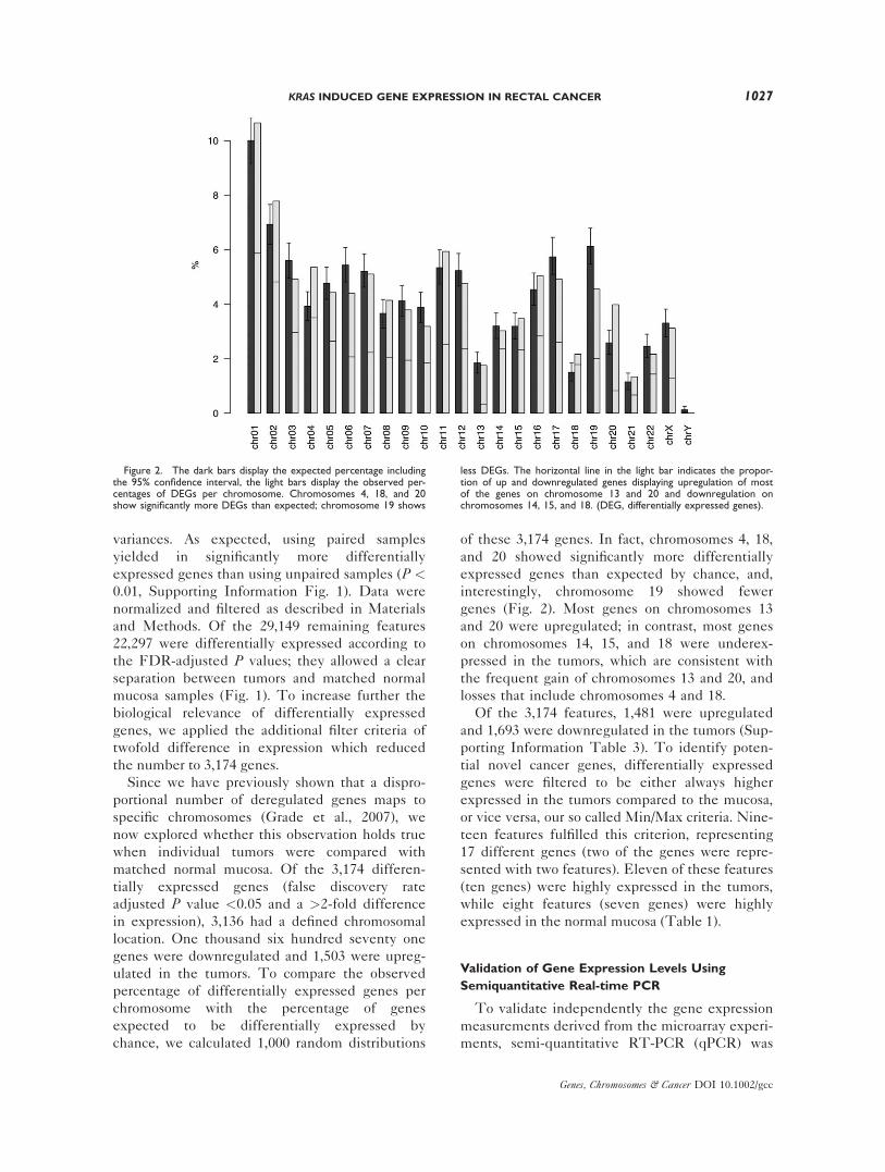
of these 3,174 genes. In fact, chromosomes 4, 18,
and 20 showed significantly more differentially
expressed genes than expected by chance, and,
interestingly, chromosome 19 showed fewer
genes (Fig. 2). Most genes on chromosomes 13
and 20 were upregulated; in contrast, most genes
on chromosomes 14, 15, and 18 were underex-
pressed in the tumors, which are consistent with
the frequent gain of chromosomes 13 and 20, and
losses that include chromosomes 4 and 18.
Of the 3,174 features, 1,481 were upregulated
and 1,693 were downregulated in the tumors (Sup-
porting Information Table 3). To identify poten-
tial novel cancer genes, differentially expressed
genes were filtered to be either always higher
expressed in the tumors compared to the mucosa,
or vice versa, our so called Min/Max criteria. Nine-
teen features fulfilled this criterion, representing
17 different genes (two of the genes were repre-
sented with two features). Eleven of these features
(ten genes) were highly expressed in the tumors,
while eight features (seven genes) were highly
expressed in the normal mucosa (Table 1).
Validation of Gene Expression Levels Using
Semiquantitative Real-time PCR
To validate independently the gene expression
measurements derived from the microarray experi-
ments, semi-quantitative RT-PCR (qPCR) was
Figure 2. The dark bars display the expected percentage includingthe 95% confidence interval, the light bars display the observed per-centages of DEGs per chromosome. Chromosomes 4, 18, and 20show significantly more DEGs than expected; chromosome 19 shows
less DEGs. The horizontal line in the light bar indicates the propor-tion of up and downregulated genes displaying upregulation of mostof the genes on chromosome 13 and 20 and downregulation onchromosomes 14, 15, and 18. (DEG, differentially expressed genes).
KRAS INDUCED GENE EXPRESSION IN RECTAL CANCER 1027
Genes, Chromosomes & Cancer DOI 10.1002/gcc

performed for 13 of the 17 different genes in 10
tumors and 10 matched mucosa samples. As shown
in Figure 3, the differential expression levels were
confirmed for all genes analyzed with both meth-
ods. To assess the correlation of microarray data
and qPCR results Pearson’s correlation coefficient
R was calculated using the fold changes between
tumor and matched mucosa. We found a highly
significant correlation between both techniques
(Pearson’s r ¼ 0.98, P < 0.01; Fig. 3).
The Rectal Cancer Transcriptome: Biological
Networks
We then interrogated in which networks or
pathways these 17 genes operate using the soft-
ware package IPA. Strikingly, 14 of the genes
clustered together in one particular network,
which was connected through CEBPA and NFkBcomplex, which are transcription factors, GDF 15and TNF, both of which are BMP superfamily
members, and ERK and JNK (also known as
MAPK1 and MAPK8), two components of the
MAP Kinase pathway. Furthermore, TGFB1 was
a central gene within the network (Fig. 4).
For a more global analysis of the differentially
expressed genes, we expanded the IPA analysis
using genes that passed the filter criteria based on
a fold change larger than two (n ¼ 3,174). As
expected a large number of networks emerged.
The top five, based on P value, were functionally
associated to cell cycle, cell mediated immune
response, cell-cell signaling, tumor and organ mor-
phology, and, most prominently, cancer. The most
outstanding intersections were centered on IL-6(P ¼ 10�41), MMP3 and KRAS (P ¼ 10�38), NR3C1(P ¼ 10�36), BRCA1 and CDKN2A (P ¼ 10�36), as
well as ERK and TRIB3 (P ¼ 10�34). The most rel-
evant functions described for the differentially
expressed genes included tumorigenesis (P ¼10�61), cancer (P ¼ 10�58), neoplasia (P ¼ 10�57),
genetic disorders (P ¼ 10�42), and colorectal cancer
(P ¼ 10�39) (Supporting Information Fig. 2).
Effect of KRAS Mutation Status on the Rectal
Cancer Transcriptome
After we had now carefully annotated the tran-
scriptional changes associated with rectal cancer, we
aimed to identify the consequences of KRAS muta-
tions on the rectal cancer transcriptome. This analysis
is relevant because (i) activating mutations are
known to play a fundamental role in carcinogenesis,
(ii) KRAS status is used for stratification of anti-
EGFR therapy with Cetuximab and, (iii) it is of
TABLE 1. Expression Ratio for Genes of Interest: Features Fulfilling the Min >< Max Criteria
Systematic name Gene name DescriptionLog2 FC tumorvs. mucosa P Map
NM_013227 ACAN Aggrecan 3.07 6.60E-36 15q26.1NM_004673 ANGPTL1 Angiopoietin-like 1 �2.66 1.22E-37 1q25.2NM_018270 ANO5 Anoctamin 5 �2.9 3.78E-32 11p14.3NM_018270 C20orf20 Chromosome 20 open
reading frame 201.67 1.05E-33 20q13.33
NM_018140 CEP72 Centrosomal protein 72kDa 1.95 5.92E-35 5p15.33NM_021101 CLDN1 Claudin 1 4.29 5.01E-48 3q28-q29NM_015036 ENDOD1 Endonuclease domain containing 1 �1.91 6.00E-37 11q21NM_001079675 ETV4 Ets variant 4 3.25 1.26E-41 17q21NM_001079675 ETV4 Ets variant 4 2.97 9.72E-39 17q21NM_001445 FABP6 Fatty acid binding protein 6, ileal 4.82 4.94E-43 5q33.3-q34NM_000148 FUT1 Fucosyltransferase 1 2.49 2.00E-35 19q13.3NM_003641 IFITM1 Interferon induced transmembrane
protein 1 (9–27)2.82 1.36E-38 11p15.5
NM_021034 IFITM3 Interferon induced transmembraneprotein 3 (1–8U)
2.68 2.72E-38 11p15.5
NM_006790 MYOT Myotilin �1.91 3.97E-41 5q31NM_005012 ROR1 Receptor tyrosine kinase-like
orphan receptor 1�1.67 9.35E-38 1p32-p31
NM_005012 ROR1 Receptor tyrosine kinase-likeorphan receptor 1
�1.81 1.32E-35 1p32-p31
NM_003759 SLC4A4 Solute carrier family 4, sodiumbicarbonate cotransporter, member 4
�4.81 8.58E-30 4q21
NM_006714 SMPDL3A Sphingomyelin phosphodiesterase,acid-like 3A
�2.53 3.09E-34 6q22.31
NM_021158 TRIB3 Tribbles homolog 3 (Drosophila) 3.51 3.07E-41 20p13-p12.2
FC, fold change.
1028 GAEDCKE ET AL.
Genes, Chromosomes & Cancer DOI 10.1002/gcc

clinical importance to identify strategies to overcome
the resistance against such antibody-based treatment.
Unsupervised clustering did not result in sep-
aration according to the KRAS mutation status,
however, we identified a set of 13 genes that
were differentially expressed between the two
groups, based on an adjusted P value smaller
than 0.05. These genes are: COPZ1, LEMD1,S100A14, RDHE2, WDR51B, SMYD3, MYBPC1,TEGT, DUSP4, SERPINB1, TCP10L, GOLPH3L,and CACNA1C. Interestingly, KRAS mutation
caused upregulation of all of these genes (Fig.
5 and Table 2). The potential of these differ-
entially expressed genes to distinguish tumors
with and without mutations was evaluated using
a Leave-One-Out-Cross-Validation (LOOCV).
With that estimate we achieved a test accuracy
of 96.9% (sensitivity 93.3%; specificity 97.1%).
Of those 13 genes, only one, DUSP4, had been
previously reported to be linked to KRAS and
the MAPK pathway.
DISCUSSION
Activating mutations of the KRAS oncogene
play an important role in colorectal carcinogene-
sis. Mutations of this gene result in the GTP-
Figure 3. (A) Comparison of log2 FC between qPCR and gene expression array (B) Correlationbetween log fold changes of qPCR measurements versus those of microarray features. Plot represents13 of the detected tumor markers. Microarray fold changes deviate from those in Tables 1 and 2because they were derived from only nine patients.
KRAS INDUCED GENE EXPRESSION IN RECTAL CANCER 1029
Genes, Chromosomes & Cancer DOI 10.1002/gcc

dependent activation of the MAPK pathway,
which, in turn impairs cell differentiation and ap-
optosis, and increases cell proliferation.
Furthermore, KRAS mutations have implica-
tions above and beyond basic tumor biology
because successful targeting of the EGFR axis
using Cetuximab depends on the maintenance
of wild type KRAS (Lievre et al., 2006, 2008;
Di Fiore et al., 2007; De Roock et al., 2008;
Karapetis et al., 2008). Nevertheless, nothing is
known about the transcriptional differences
between KRAS mutant and wild-type tumors in
rectal carcinomas and their impact on the whole
transcriptome. In an attempt to identify such dif-
ferences, we assessed KRAS mutation status and
its consequences on the cancer transcriptome by
analyzing 65 locally advanced rectal cancers and
their corresponding normal mucosa. With this
considerably large dataset, we were in a position
Figure 4. Ingenuity pathway analysis reveals the close relationship between 14 of 17 of most relevantgenes in rectal cancer.
Figure 5. Differentially expressed genes between KRAS wild typeand mutant rectal carcinomas.
1030 GAEDCKE ET AL.
Genes, Chromosomes & Cancer DOI 10.1002/gcc

to screen for KRAS mutation dependent transcrip-
tional consequences on downstream targets.
Impact of KRAS on the Rectal Cancer
Transcriptome
Forty-seven percent of rectal carcinomas in our
dataset revealed activating KRAS mutations
(Gaedcke et al., 2010). Our data are therefore
congruent with published data on the prevalence
of the mutations. Comparison of KRAS mutant
and wild-type rectal cancers revealed 13 differen-
tially expressed genes which were always, and
with high-fold and high-significance differentially
expressed between tumors with and without
mutations. All genes were upregulated in the mu-
tant tumors. Relatively little is known about most
of these genes. Only for one of the upregulated
genes an association to the MAPK pathway
had been reported previously: MAP kinase phospha-tase 2 (DUSP4) has previously been reported to be
upregulated in various cancer types (Yip-Schneider
et al., 2001; Wang et al., 2003). Our own data con-
firm the significant upregulation of DUSP4 in rectal
cancer (P ¼ 10�21).
Khambata-Ford et al. (2007) investigated the
impact of DUSP4 expression levels on outcome of
patients with metastatic colorectal cancer. Gene
expression profiling from 80 patients with metastatic
colorectal carcinomas enrolled in a Cetuximab
monotherapy trial revealed DUSP4 as one of the top
resistance markers. Since KRAS mutations are
currently considered as some of the most relevant
resistance markers for treatment failure, overexpres-
sion of DUSP4 within the same group confirms the
finding of a mutation dependent regulation. Lung
cancers with EGFR mutations respond well to
Cetuximab, and it was recently shown that DUSP4is downregulated in those tumors. The overexpres-
sion of DUSP4 in rectal cancer in the presence of
KRAS mutations which are resistant to Cetuximab is
therefore a possible explanation for the mode of
action. DUSP4 expression levels could therefore
serve as biomarkers for treatment stratification thera-
pies with Cetuximab.
In cDNA microarray analysis, the gene LEMD1(LEM domain-containing 1) has previously been
found to be upregulated in colorectal cancer and
was shown to be a member of the cancer-testis
antigens (Yuki et al., 2004). TEGT is a regulator
of apoptosis (Grzmil et al., 2006), SERPINB1 was
reported to be upregulated in oral cancer (Tseng
et al., 2009) and SMYD3, a histone methyltrans-
ferase, is involved in the proliferation of cancer
cells (Hamamoto et al., 2004, 2006; Zou et al.,
2009). Nine of the thirteen genes showed connec-
tions when analyzed with IPA which suggests a
functional relationship between these genes and
could explain why they are jointly deregulated as
a consequence of KRAS mutation (Supporting In-
formation Fig. 3).
We queried the relevance of identifying KRAS-related genes for clinical considerations. For
instance, if resistance to Cetuximab as a conse-
quence of KRAS mutation depends on KRASregulated genes one could hypothesize that
TABLE 2. Expression Ratio for Genes of Interest: Genes Being Differentially Expressed Between KRAS WT and KRAS Mutant
Systematic name Gene name Descriptionlog2 FC WTvs. mutant P Map
NM_000719 CACNA1C Calcium channel, voltage dependent,L type, alpha 1C subunit
1.2 2.16E-05 12p13.3
NM_003217 TEGT Testis enhanced gene transcript(BAX-inhibitor-1)
0.54 4.75E-06 12q12-q13
NM_016057 COPZ1 Coatomer protein complex,subunit zeta 1
0.52 7.25E-07 12q13.2-q13.3
NM_172240 WDR51B WD repeat domain 51B 0.6 2.21E-06 12q21.33NM_206819 MYBPC1 Myosin binding protein C, slow type 0.68 3.86E-06 12q23.2NM_020672 S100A14 S100 calcium binding protein A14 0.94 1.02E-06 1q21.3NM_018178 GOLPH3L Golgi phosphoprotein 3 like 0.57 1.73E-05 1q21.3NM_001001552 LEMD1 LEM domain containing 1 2.02 7.58E-07 1q32.1NM_022743 SMYD3 SET and MYND domain containing 3 0.49 3.40E-06 1q44NM_144659 TCP10L T-complex 10 (mouse)-like 0.52 1.57E-05 21q22.11NM_030666 SERPINB1 Serpin peptidase inhibitor, clade
B (ovalbumin), member 10.56 1.32E-05 6p25
NM_001394 DUSP4 Dual specificity phosphatase 4 0.9 1.10E-05 8p12-p11NM_138969 RDHE2 Epidermal retinal dehydrogenase 2 1.26 1.22E-06 8q12.1
FC, Fold Change
KRAS INDUCED GENE EXPRESSION IN RECTAL CANCER 1031
Genes, Chromosomes & Cancer DOI 10.1002/gcc

transcriptional modification of these genes would
restore the sensitivity of colorectal carcinomas to
Cetuximab. DUSP4 is a good example because
low levels of DUSP4 sensitize tumors to Cetuxi-
mab and decreasing DUSP4 levels using the
agent PD98059 could therefore be used in treat-
ment of KRAS mutated tumors in combination
with Cetuximab (Yip-Schneider et al., 2001).
Another potential target for such an intervention
would be SMYD3, another one of the differen-
tially expressed genes in our dataset, because the
drug Novobiocin lowers the expression level of
this gene (Luo et al., 2009).
Identification of Novel Rectal Cancer Tumor
Markers
The most stringent criteria to select differen-
tially expressed genes was introduced to reveal
new tumor markers (Min >< Max rule). This
rule filtered genes that are always higher
expressed in any of the tumors compared with all
mucosa samples, or vice versa. Of the 19 features
identified 11 were higher and eight were lower
expressed in the tumor. The expression levels of
13 of these genes were validated using qPCR. As
in previous validation experiments, the results
between arrays and qPCR were extremely repro-
ducible (r ¼ 0.98) attesting to the robustness of
either methodology. Within the validated genes
ETV4 (Liu et al., 2007), ROR1 (Katoh, 2005) or
CLDN1 (Kinugasa et al., 2007; Huo et al., 2009),
C20orf20 (Cai et al., 2003; Carvalho et al., 2009),
and FUT1 (Hallouin et al., 1999) have already
been linked to colorectal cancer. Others are
known to play a role in carcinogenesis in general,
such as TRIB3 (Du et al., 2003), ACAN (Skandalis
et al., 2006; Stylianou et al., 2008), and CEP72(Kang et al., 2008) but have not been directly
associated with colorectal cancer whereas an
involvement of MYOT, ENDOD1, and ANO5 in
epithelial tumorigenesis is a novel finding. All
genes that we previously found differentially
expressed or overexpressed in a more limited
dataset of colorectal cancer were confirmed to be
deregulated in the same direction (Grade et al.,
2006, 2007).
Interestingly, when we analyzed these 17
genes using IPA, we found 14 of them operating
in one network (Fig. 4). This network was con-
nected through TNF, TGFB1, ERK, and the
NFkB complex which highlights the central role
that these signaling pathways assume in CRC
(Glick, 2004; Fang and Richardson, 2005; Zhang
et al., 2007; Balkwill, 2009). Expanding the num-
bers of genes for pathway analysis we used the
differentially expressed genes based on a FC >2.
The main interceptions within the networks like
MMP3, KRAS, p16, or ERK again confirm the
relevance of the retrieved genes.
In summary, this is the most comprehensive
and systematic gene expression study of rectal
carcinomas and normal mucosa. Using matched
samples rather than a normal reference pool was
important to retrieve more differentially
expressed genes. In addition, this is the first sys-
tematic exploration of gene expression changes
that are a consequence of activating KRAS muta-
tions in rectal cancer. We identified DUSP4 and
SMYD3 as attractive targets for a potential combi-
nation therapy of patients with Cetuximab resist-
ant tumors.
Accession Number
The gene expression data have been deposited
in the NCBI Gene Expression Omnibus
(GSE20842).
ACKNOWLEDGMENTS
The authors are indebted to Ms. Jessica Eggert
and Mr. Chan Rong Lai for excellent technical
support. They also thank Dr. Gabriela Salinas-
Riester and Mr. Lennart Opitz for help with
microarray hybridizations, Drs. Laszlo Fuzesi and
Hilka Rothe for pathology reports, and Dr. Rein-
hard Ebner for critically reading the manuscript.
The authors are indebted to Drs. Nikolas
Stoecklein and Achim Weber for their review of
this manuscript.
REFERENCES
Alon U, Barkai N, Notterman DA, Gish K, Ybarra S, Mack D,Levine AJ. 1999. Broad patterns of gene expression revealed byclustering analysis of tumor and normal colon tissues probed byoligonucleotide arrays. Proc Natl Acad Sci USA 96:6745–6750.
Baldus SE, Schaefer KL, Engers R, Hartleb D, Stoecklein NH,Gabbert HE. 2010. Prevalence and heterogeneity of KRAS,BRAF, and PIK3CA mutations in primary colorectal adenocarci-nomas and their corresponding metastases. Clin Cancer Res16:790–799.
Balkwill F. 2009. Tumour necrosis factor and cancer. Nat RevCancer 9:361–371.
Benjamini Y, Hochberg Y. 1995. Controlling the false discoveryrate: A practical and powerful approach to multiple testing. J RStat Soc B 57:289–300.
Bianchini M, Levy E, Zucchini C, Pinski V, Macagno C, DeSanctis P, Valvassori L, Carinci P, Mordoh J. 2006. Comparativestudy of gene expression by cDNA microarray in human colo-rectal cancer tissues and normal mucosa. Int J Oncol 29:83–94.
Bolstad BM, Irizarry RA, Astrand M, Speed TP. 2003. A compari-son of normalization methods for high density oligonucleotidearray data based on variance and bias. Bioinformatics 19:185–193.
1032 GAEDCKE ET AL.
Genes, Chromosomes & Cancer DOI 10.1002/gcc

Brink M, de Goeij AF, Weijenberg MP, Roemen GM, LentjesMH, Pachen MM, Smits KM, de Bruine AP, Goldbohm RA,van den Brandt PA. 2003. K-ras oncogene mutations in sporadiccolorectal cancer in The Netherlands Cohort Study. Carcino-genesis 24:703–710.
Cai Y, Jin J, Tomomori-Sato C, Sato S, Sorokina I, Parmely TJ,Conaway RC, Conaway JW. 2003. Identification of new subu-nits of the multiprotein mammalian TRRAP/TIP60-containinghistone acetyltransferase complex. J Biol Chem 278:42733–42736.
Carvalho B, Postma C, Mongera S, Hopmans E, Diskin S, van deWiel MA, van Criekinge W, Thas O, Matthai A, Cuesta MA,Droste JSTS, Craanen M, Schrock E, Ylstra B, Meijer GA.2009. Multiple putative oncogenes at the chromosome 20qamplicon contribute to colorectal adenoma to carcinoma pro-gression. Gut 58:79–89.
De Roock W, Piessevaux H, De Schutter J, Janssens M, De Her-togh G, Personeni N, Biesmans B, Van Laethem JL, PeetersM, Humblet Y, Van Cutsem E, Tejpar S. 2008. KRAS wild-type state predicts survival and is associated to early radiologicalresponse in metastatic colorectal cancer treated with cetuximab.Ann Oncol 19:508–515.
Di Fiore F, Blanchard F, Charbonnier F, Le Pessot F, Lamy A,Galais MP, Bastit L, Killian A, Sesboue R, Tuech JJ, QueunietAM, Paillot B, Sabourin JC, Michot F, Michel P, Frebourg T.2007. Clinical relevance of KRAS mutation detection in meta-static colorectal cancer treated by Cetuximab plus chemother-apy. Br J Cancer 96:1166–1169.
Du KY, Herzig S, Kulkarni RN, Montminy M. 2003. TRB3: Atribbles homolog that inhibits Akt/PKB activation by insulin inliver. Science 300:1574–1577.
Fang JY, Richardson BC. 2005. The MAPK signalling pathwaysand colorectal cancer. Lancet Oncol 6:322–327.
Fisher RA. 1925. Applications of ‘‘student’s’’ distribution. Metron5:90–104.
Friederichs J, Rosenberg R, Mages J, Janssen KP, Maeckl C,Nekarda H, Holzmann B, Siewert JR. 2005. Gene expressionprofiles of different clinical stages of colorectal carcinoma: To-ward a molecular genetic understanding of tumor progression.Int J Colorectal Dis 20:391–402.
Gaedcke J, Grade M, Jung K, Schirmer M, Jo P, Obermeyer C,Wolff HA, Herrmann MK, Beissbarth T, Becker H, Ried T,Ghadimi M. 2010. KRAS and BRAF mutations in patients withrectal cancer treated with preoperative chemoradiotherapy.Radiother Oncol 94:76–81.
Glick AB. 2004. TGFbeta1, back to the future: Revisiting itsrole as a transforming growth factor. Cancer Biol Ther 3:276–283.
Grade M, Ghadimi BM, Varma S, Simon R, Wangsa D,Barenboim-Stapleton L, Liersch T, Becker H, Ried T, Difilip-pantonio MJ. 2006. Aneuploidy-dependent massive deregulationof the cellular transcriptome and apparent divergence of theWnt/beta-catenin signaling pathway in human rectal carcinomas.Cancer Res 66:267–282.
Grade M, Hormann P, Becker S, Hummon AB, Wangsa D, VarmaS, Simon R, Liersch T, Becker H, Difilippantonio MJ, GhadimiBM, Ried T. 2007. Gene expression profiling reveals a massive,aneuploidy-dependent transcriptional deregulation and distinctdifferences between lymph node-negative and lymph node-pos-itive colon carcinomas. Cancer Res 67:41–56.
Grade M, Hummon AB, Camps J, Emons G, Spitzner M,Gaedcke J, Hoermann P, Ebner R, Becker H, DifilippantonioMJ, Ghadimi BM, Beissbarth T, Caplan NJ, Ried T. 2010. Agenomic strategy for the functional validation of colorectal can-cer genes identifies potential therapeutic targets. Int J CancerEpub ahead of print.
Grzmil M, Kaulfuss S, Thelen P, Hemmerlein B, Schweyer S,Obenauer S, Kang TW, Burfeind P. 2006. Expression and func-tional analysis of Bax inhibitor-1 in human breast cancer cells. JPathol 208:340–349.
Hallouin F, Goupille C, Rocher J, Le Pendu J. 1999. A rat ex-perimental model for the design of vaccines against tumorassociated antigens Tn and Sialyl-Tn. Glycoconjug J 16:681–684.
Hamamoto R, Furukawa Y, Morita M, Iimura Y, Silva FP, Li M,Yagyu R, Nakamura Y. 2004. SMYD3 encodes a histone meth-yltransferase involved in the proliferation of cancer cells. NatCell Biol 6:731–740.
Hamamoto R, Silva FP, Tsuge M, Nishidate T, Katagiri T, Naka-mura Y, Furukawa Y. 2006. Enhanced SMYD3 expression is
essential for the growth of breast cancer cells. Cancer Sci97:113–118.
Huo Q, Kinugasa T, Wang L, Huang J, Zhao J, Shibaguchi H,Kuroki M, Tanaka T, Yamashita Y, Nabeshima K, Iwasaki H,Kuroki M. 2009. Claudin-1 protein is a major factor involved inthe tumorigenesis of colorectal cancer. Anticancer Res 29:851–857.
Kang GH, Lee S, Cho NY, Gandamihardja T, Long TI, Weisen-berger DJ, Campan M, Laird PW. 2008. DNA methylation pro-files of gastric carcinoma characterized by quantitative DNAmethylation analysis. Lab Invest 88:161–170.
Karapetis CS, Khambata-Ford S, Jonker DJ, O’Callaghan CJ, TuD, Tebbutt NC, Simes RJ, Chalchal H, Shapiro JD, RobitailleS, Price TJ, Shepherd L, Au HJ, Langer C, Moore MJ, Zalc-berg JR. 2008. K-ras mutations and benefit from cetuximab inadvanced colorectal cancer. N Engl J Med 359:1757–1765.
Karnoub AE, Weinberg RA. 2008. Ras oncogenes: Split personal-ities. Nat Rev Mol Cell Biol 9:517–531.
Katoh M. 2005. Comparative genomics on ROR1 and ROR2orthologs. Oncol Rep 14:1381–1384.
Khambata-Ford S, Garrett CR, Meropol NJ, Basik M, HarbisonCT, Wu S, Wong TW, Huang X, Takimoto CH, Godwin AK,Tan BR, Krishnamurthi SS, Burris HA, III, Poplin EA, HidalgoM, Baselga J, Clark EA, Mauro DJ. 2007. Expression of epire-gulin and amphiregulin and K-ras mutation status predict dis-ease control in metastatic colorectal cancer patients treated withcetuximab. J Clin Oncol 25:3230–3237.
Kinugasa T, Huo Q, Higash D, Shibaguchi H, Kuroki M, TanakaT, Futami K, Yamashita Y, Hachimine K, Maekawa S, Nabe-shima K, Iwasaki H, Kuroki M. 2007. Selective up-regulation ofclaudin-1 and claudin-2 in colorectal cancer. Anticancer Res27:3729–3734.
Lievre A, Bachet JB, Le Corre D, Boige V, Landi B, Emile JF,Cote JF, Tomasic G, Penna C, Ducreux M, Rougier P,Penault-Llorca F, Laurent-Puig P. 2006. KRAS mutation statusis predictive of response to cetuximab therapy in colorectal can-cer. Cancer Res 66:3992–3995.
Lievre A, Bachet JB, Boige V, Cayre A, Le Corre D, Buc E,Ychou M, Bouche O, Landi B, Louvet C, Andre T, Bibeau F,Diebold MD, Rougier P, Ducreux M, Tomasic G, Emile JF,Penault-Llorca F, Laurent-Puig P. 2008. KRAS mutations as anindependent prognostic factor in patients with advanced colo-rectal cancer treated with cetuximab. J Clin Oncol 26:374–379.
Liu HY, Zhou B, Wang L, Li Y, Zhou ZG, Sun XF, Xu B, ZengYJ, Song JM, Luo HZ, Yang L. 2007. Association of E1AFmRNA expression with tumor progression and matrilysin inhuman rectal cancer. Oncology 73:384–388.
Luo XG, Zou JN, Wang SZ, Zhang TC, Xi T. 2009. Novobiocindecreases SMYD3 expression and inhibits the migration ofMDA-MB-231 human breast cancer cells. IUBMB Life 62:194–199.
Platzer P, Upender MB, Wilson K, Willis J, Lutterbaugh J, Nos-rati A, Willson JK, Mack D, Ried T, Markowitz S. 2002.Silence of chromosomal amplifications in colon cancer. CancerRes 62:1134–1138.
Skandalis SS, Theocharis AD, Vynios DH, PapageorgakopoulouN, Hjerpe A, Karamanos NK, Theocharis DA. 2006. Cartilageaggrecan undergoes significant compositional and structuralalterations during laryngeal cancer. Biochim Biophys Acta1760:1046–1053.
Smyth GK. 2004. Linear models and empirical bayes methods forassessing differential expression in microarray experiments. StatAppl Genet Mol Biol 3: Article 3.
Stylianou M, Skandalis SS, Papadas TA, Mastronikolis NS, Theo-charis DA, Papageorgakopoulou N, Vynios DH. 2008. Stage-related decorin and versican expression in human laryngeal can-cer. Anticancer Res 28:245–251.
Tseng MY, Liu SY, Chen HR, Wu YJ, Chiu CC, Chan PT,Chiang WF, Liu YC, Lu CY, Jou YS, Chen JY. 2009. Serineprotease inhibitor (SERPIN) B1 promotes oral cancer cell mo-tility and is over-expressed in invasive oral squamous cell carci-noma. Oral Oncol 45:771–776.
Wang HY, Cheng Z, Malbon CC. 2003. Overexpression of mito-gen-activated protein kinase phosphatases MKP1, MKP2 inhuman breast cancer. Cancer Lett 191:229–237.
Yip-Schneider MT, Lin A, Marshall MS. 2001. Pancreatic tumorcells with mutant K-ras suppress ERK activity by MEK-de-pendent induction of MAP kinase phosphatase-2. Biochem Bio-phys Res Commun 280:992–997.
KRAS INDUCED GENE EXPRESSION IN RECTAL CANCER 1033
Genes, Chromosomes & Cancer DOI 10.1002/gcc

Yuki D, Lin YM, Fujii Y, Nakamura Y, Furukawa Y. 2004. Isola-tion of LEM domain-containing 1, a novel testis-specific geneexpressed in colorectal cancers. Oncol Rep 12:275–280.
Zhang X, Jin B, Huang C. 2007. The PI3K/Akt pathway and itsdownstream transcriptional factors as targets for chemopreven-tion. Curr Cancer Drug Targets 7:305–316.
Zou TT, Selaru FM, Xu Y, Shustova V, Yin J, Mori Y, ShibataD, Sato F, Wang S, Olaru A Deacu E, Liu TC, Abraham
JM, Meltzer SJ. 2002. Application of cDNA microarrays togenerate a molecular taxonomy capable of distinguishingbetween colon cancer and normal colon. Oncogene 21:4855–4862.
Zou JN, Wang SZ, Yang JS, Luo XG, Xie JH, Xi T. 2009. Knock-down of SMYD3 by RNA interference down-regulates c-Metexpression and inhibits cells migration and invasion induced byHGF. Cancer Lett 280:78–85.
1034 GAEDCKE ET AL.
Genes, Chromosomes & Cancer DOI 10.1002/gcc
Copyright © 2022 FDOKUMEN