The Methyltransferase HemK Regulates the Virulence ... - MDPI
16
Citation: Shi, Y.; Yang, X.; Ye, X.; Feng, J.; Cheng, T.; Zhou, X.; Liu, D.X.; Xu, L.; Wang, J. The Methyltransferase HemK Regulates the Virulence and Nutrient Utilization of the Phytopathogenic Bacterium Xanthomonas citri Subsp. citri. Int. J. Mol. Sci. 2022, 23, 3931. https://doi.org/10.3390/ ijms23073931 Academic Editors: José Joaquín Cerón, Alberto Muñoz-Prieto, Vladimir Mrljak and Lorena Franco-Martinez Received: 13 March 2022 Accepted: 30 March 2022 Published: 1 April 2022 Publisher’s Note: MDPI stays neutral with regard to jurisdictional claims in published maps and institutional affil- iations. Copyright: © 2022 by the authors. Licensee MDPI, Basel, Switzerland. This article is an open access article distributed under the terms and conditions of the Creative Commons Attribution (CC BY) license (https:// creativecommons.org/licenses/by/ 4.0/). International Journal of Molecular Sciences Article The Methyltransferase HemK Regulates the Virulence and Nutrient Utilization of the Phytopathogenic Bacterium Xanthomonas citri Subsp. citri Yu Shi 1 , Xiaobei Yang 1 , Xiaoxin Ye 1 , Jiaying Feng 1 , Tianfang Cheng 1 , Xiaofan Zhou 1,2 , Ding Xiang Liu 1,2 , Linghui Xu 1,2, * and Junxia Wang 1,2, * 1 Integrative Microbiology Research Centre, College of Plant Protection, South China Agricultural University, Guangzhou 510642, China; [email protected] (Y.S.); [email protected] (X.Y.); [email protected] (X.Y.); [email protected] (J.F.); [email protected] (T.C.); [email protected] (X.Z.); [email protected] (D.X.L.) 2 Guangdong Province Key Laboratory of Microbial Signals and Disease Control, South China Agricultural University, Guangzhou 510642, China * Correspondence: [email protected] (L.X.); [email protected] (J.W.) Abstract: Citrus canker, caused by the bacterium Xanthomonas citri subsp. citri (Xcc), seriously affects fruit quality and yield, leading to significant economic losses around the world. Understanding the mechanism of Xcc virulence is important for the effective control of Xcc infection. In this report, we investigate the role of a protein named HemK in the regulation of the virulence traits of Xcc. The hemK gene was deleted in the Xcc jx-6 background, and the ΔhemK mutant phenotypically displayed significantly decreased motility, biofilm formation, extracellular enzymes, and polysaccharides production, as well as increased sensitivity to oxidative stress and high temperatures. In accordance with the role of HemK in the regulation of a variety of virulence-associated phenotypes, the deletion of hemK resulted in reduced virulence on citrus plants as well as a compromised hypersensitive response on a non-host plant, Nicotiana benthamiana. These results indicated that HemK is required for the virulence of Xcc. To characterize the regulatory effect of hemK deletion on gene expression, RNA sequencing analysis was conducted using the wild-type Xcc jx-6 strain and its isogenic ΔhemK mutant strain, grown in XVM2 medium. Comparative transcriptome analysis of these two strains revealed that hemK deletion specifically changed the expression of several virulence-related genes associated with the bacterial secretion system, chemotaxis, and quorum sensing, and the expression of various genes related to nutrient utilization including amino acid metabolism, carbohydrate metabolism, and energy metabolism. In conclusion, our results indicate that HemK plays an essential role in virulence, the regulation of virulence factor synthesis, and the nutrient utilization of Xcc. Keywords: Xanthomonas citri subsp. citri; HemK; RNA-seq; motility; exoenzyme; biofilm; virulence; stress tolerance 1. Introduction Xanthomonas citri subsp. citri (Xcc) causes bacterial citrus canker, which is one of the most studied phytopathogens in the Xanthomonas genus. Xcc can attach to the surface of citrus leaves or fruits and invade host tissues via the plant’s natural openings or wounds when in a warm and humid climate [1,2]. The establishment of Xcc infection inside host cells causes necrotic lesions on the leaves, stems, and fruits, leading to defoliation, twig dieback, and blemished fruit and, in serious cases, causes premature fruit drop and eventually the death of infected plants. Citrus canker is considered to be one of the most serious citrus quarantine diseases worldwide and remains a serious challenge for all citrus- producing countries. Xcc has been used as a model organism to study pathogenesis and find new solutions for the disease control of Xanthomonas [3,4]. In the past few decades, our understanding of Int. J. Mol. Sci. 2022, 23, 3931. https://doi.org/10.3390/ijms23073931 https://www.mdpi.com/journal/ijms
-
Upload
khangminh22 -
Category
Documents
-
view
2 -
download
0
Transcript of The Methyltransferase HemK Regulates the Virulence ... - MDPI

�����������������
Citation: Shi, Y.; Yang, X.; Ye, X.;
Feng, J.; Cheng, T.; Zhou, X.; Liu,
D.X.; Xu, L.; Wang, J. The
Methyltransferase HemK Regulates
the Virulence and Nutrient
Utilization of the Phytopathogenic
Bacterium Xanthomonas citri Subsp.
citri. Int. J. Mol. Sci. 2022, 23, 3931.
https://doi.org/10.3390/
ijms23073931
Academic Editors: José Joaquín
Cerón, Alberto Muñoz-Prieto,
Vladimir Mrljak and Lorena
Franco-Martinez
Received: 13 March 2022
Accepted: 30 March 2022
Published: 1 April 2022
Publisher’s Note: MDPI stays neutral
with regard to jurisdictional claims in
published maps and institutional affil-
iations.
Copyright: © 2022 by the authors.
Licensee MDPI, Basel, Switzerland.
This article is an open access article
distributed under the terms and
conditions of the Creative Commons
Attribution (CC BY) license (https://
creativecommons.org/licenses/by/
4.0/).
International Journal of
Molecular Sciences
Article
The Methyltransferase HemK Regulates the Virulence andNutrient Utilization of the Phytopathogenic BacteriumXanthomonas citri Subsp. citriYu Shi 1, Xiaobei Yang 1, Xiaoxin Ye 1, Jiaying Feng 1, Tianfang Cheng 1, Xiaofan Zhou 1,2 , Ding Xiang Liu 1,2,Linghui Xu 1,2,* and Junxia Wang 1,2,*
1 Integrative Microbiology Research Centre, College of Plant Protection, South China Agricultural University,Guangzhou 510642, China; [email protected] (Y.S.); [email protected] (X.Y.);[email protected] (X.Y.); [email protected] (J.F.); [email protected] (T.C.);[email protected] (X.Z.); [email protected] (D.X.L.)
2 Guangdong Province Key Laboratory of Microbial Signals and Disease Control,South China Agricultural University, Guangzhou 510642, China
* Correspondence: [email protected] (L.X.); [email protected] (J.W.)
Abstract: Citrus canker, caused by the bacterium Xanthomonas citri subsp. citri (Xcc), seriously affectsfruit quality and yield, leading to significant economic losses around the world. Understanding themechanism of Xcc virulence is important for the effective control of Xcc infection. In this report, weinvestigate the role of a protein named HemK in the regulation of the virulence traits of Xcc. ThehemK gene was deleted in the Xcc jx-6 background, and the ∆hemK mutant phenotypically displayedsignificantly decreased motility, biofilm formation, extracellular enzymes, and polysaccharidesproduction, as well as increased sensitivity to oxidative stress and high temperatures. In accordancewith the role of HemK in the regulation of a variety of virulence-associated phenotypes, the deletionof hemK resulted in reduced virulence on citrus plants as well as a compromised hypersensitiveresponse on a non-host plant, Nicotiana benthamiana. These results indicated that HemK is required forthe virulence of Xcc. To characterize the regulatory effect of hemK deletion on gene expression, RNAsequencing analysis was conducted using the wild-type Xcc jx-6 strain and its isogenic ∆hemK mutantstrain, grown in XVM2 medium. Comparative transcriptome analysis of these two strains revealedthat hemK deletion specifically changed the expression of several virulence-related genes associatedwith the bacterial secretion system, chemotaxis, and quorum sensing, and the expression of variousgenes related to nutrient utilization including amino acid metabolism, carbohydrate metabolism, andenergy metabolism. In conclusion, our results indicate that HemK plays an essential role in virulence,the regulation of virulence factor synthesis, and the nutrient utilization of Xcc.
Keywords: Xanthomonas citri subsp. citri; HemK; RNA-seq; motility; exoenzyme; biofilm; virulence;stress tolerance
1. Introduction
Xanthomonas citri subsp. citri (Xcc) causes bacterial citrus canker, which is one of themost studied phytopathogens in the Xanthomonas genus. Xcc can attach to the surface ofcitrus leaves or fruits and invade host tissues via the plant’s natural openings or woundswhen in a warm and humid climate [1,2]. The establishment of Xcc infection insidehost cells causes necrotic lesions on the leaves, stems, and fruits, leading to defoliation,twig dieback, and blemished fruit and, in serious cases, causes premature fruit drop andeventually the death of infected plants. Citrus canker is considered to be one of the mostserious citrus quarantine diseases worldwide and remains a serious challenge for all citrus-producing countries.
Xcc has been used as a model organism to study pathogenesis and find new solutionsfor the disease control of Xanthomonas [3,4]. In the past few decades, our understanding of
Int. J. Mol. Sci. 2022, 23, 3931. https://doi.org/10.3390/ijms23073931 https://www.mdpi.com/journal/ijms

Int. J. Mol. Sci. 2022, 23, 3931 2 of 16
the molecular interaction between citrus and Xcc has been advanced by the elucidation ofthe genome sequences of the Xanthomonas genus [5,6]. The complete genome of Xcc strain306 contains 2710 genes that are assigned to functions, of which approximately 6% areinvolved in pathogenicity and host adaptation [7]. Xcc utilizes numerous virulence factorsfor survival on plant surfaces and fitness in hosts for nutrition and pathogenicity [8]. Suchfactors include lipopolysaccharides (LPS) [9,10], extracellular polysaccharides (EPS) [11],motility, biofilm formation [12,13], and multiple effectors from its secretion systems [14].Among them, the type III secretion system (T3SS) enables the translocation of severaleffector proteins from the bacteria to host cells that affect host signaling and metabolism,leading to hypersensitive reactions and pathogenicity responses in the host cell [15,16].The type II secretion system (T2SS) enables the secretion of numerous hydrolases, suchas cellulases, esterases, and proteinase, as virulence factors for pathogenesis [17,18]. BothT2SS and T3SS are required for the full virulence of Xcc during the early development ofcitrus canker symptoms [19,20].
HemK/PrmC-class methyltransferases are conserved from bacteria [21] and yeast [22,23]to humans [24], and are responsible for the methylation of glutamine at the N5 positionin the conserved GGQ motif of release factors. A few reports have been published on theimportant biological functions of HemK in plants [25–27], yeast [22,23], and bacteria [28–30].In Arabidopsis, NRF1, a HemK-class glutamine-methyltransferase involved in the terminationof translation, is essential for cellular iron homeostasis and the plant’s normal growth [25].In Saccharomyces cerevisiae, there are two protein methyltransferases, Mtq1p (YNL063w) andMtq2p (YDR140w). The depletion of Mtq1p leads to moderate growth defects on non-fermentable carbon sources and increases the readthrough of a stop codon present in Cox2mRNA [23]. In contrast, the deletion of the mtq2 gene displays growth restriction andsensitivity to low temperature, high salt, and calcium concentrations on common yeastmedium YPD, as well as sensitivity to translation fidelity antibiotics such as paromomycinand geneticin [22]. In Pseudomonas aeruginosa PA14, loss of PrmC activity abolishes anaerobicgrowth and results in reduced pathogenicity in the infection model Galleria mellonella andproduction of several virulence factors, such as pyocyanin, rhamnolipids and the type III-secreted toxin ExoT [28]. In the Escherichia coli K12 strain, hemK deletion leads to increasedstop codon readthrough, induction of the oxidative stress response, and severely retardedgrowth [21,30]. In Yersinia pseudotuberculosis, the vagH (homology to HemK of E. coli) mutantexhibits a virulence phenotype similar to that of a T3SS-negative mutant, indicating a close linkbetween VagH and T3SS in Y. pseudotuberculosis [29]. The importance of HemK/PrmC classmethyltransferase, which has been reported to mediate diverse cellular processes involved inpathogenesis, development, and environmental adaption in some other organisms, promptedus to investigate its specific role in the Xcc strain, which is encoded by XAC0908 and has anunknown biological function.
In this study, we dissected the functional involvement of HemK, a glutamine-methyltransferase from Xcc, in virulence through testing a series of phenotypes, andits regulation of gene expression through RNA sequencing analysis. The mutant ∆hemKdisplayed several virulence-related phenotypes, manifested by reduced motility, extracel-lular enzyme and polysaccharide production, biofilm formation, and pathogenicity. Thisdiscovery provides new information on the pathogenicity of this important plant pathogen.
2. Results2.1. HemK Influences the Cell Motility, Biofilm Formation, Extracellular Polysaccharide, andEnzyme Production in Xcc jx-6
To elucidate whether HemK plays a role in the cellular process associated with patho-genesis in Xcc, we conducted a series of bacterial phenotypic tests to examine the influenceof hemK deletion on virulence-associated traits, including cell motility, biofilm formation,extracellular polysaccharides, and enzyme production. For those experiments, we engi-neered an in-frame deletion mutant ∆hemK in wild type strain Xcc jx-6 background andconstructed its complementary strain C-∆hemK by introducing a recombinant plasmid

Int. J. Mol. Sci. 2022, 23, 3931 3 of 16
pBBR1-hemK into ∆hemK, which expressed full-length HemK under an arabinose induciblepromoter. A phenotypic comparison was performed between wild-type strain Xcc jx-6, themutant strain ∆hemK, and the complementary strain C-∆hemK, to determine the effect ofhemK deletion on virulence-related traits.
The growth characteristics of those three strains, grown in liquid yeast extract broth(YEB) medium, were first tested with the Bioscreen C automated microbiology growthcurve analysis system. The growth curve of ∆hemK did not show obvious differencescompared with that of wild-type and C-∆hemK (Figure 1A). Rich medium YEB was selectedfor subsequent bacterial phenotyping test.
Int. J. Mol. Sci. 2022, 23, x FOR PEER REVIEW 4 of 17
Figure 1. HemK influences the production of cell motility, biofilm formation, extracellular polysaccharides, and enzymes in Xcc jx-6. (A) The growth rates (OD600 values) of the wild-type (WT) strain Xcc jx-6, ΔhemK, and C-∆hemK on YEB at 28 °C were measured at 4 h intervals. (B) Swimming and swarming motility for the WT strain, ΔhemK, and C-∆hemK were detected on a 0.28% agar swimming plate and 0.6% agar swarming plate, respectively. A 2 µL aliquot of the bacterial suspension was inoculated onto the swimming plate or swarming plate and incubated for 60 h at 28 °C to observe the bacterial motility. (C) The level of motility was determined by measuring the area of the colony with ImageJ. (D) The biofilm formation of the WT strain, ∆hemK, and C-∆hemK in glass tubes was detected by crystal violet staining and quantified by measuring the optical density at 590 nm, after dissolution in 33% acetic acid. (E) The production of extracellular polysaccharides (EPS) of the WT strain, ∆hemK, and C-∆hemK was assessed on 2% glucose NYGA, and the production was measured with ethanol precipitation. (F–H) A 20 µL (for proteases) or 2 µL (for cellulases and amylases) aliquot of bacterial supernatant was added to the exoenzymes’ test plates and incubated at 28 °C for 48 h. The production of hydrolysis circles by cellulases, amylases, and proteases was measured on plates containing 1% (m/v) skimmed milk (F), 1% (m/v) sodium carboxymethyl ethyl cellulose (G), and 1% (m/v) potato starch (H), respectively. All experiments were repeated three times, with three repetitions for each strain. Only one representative result is presented. A significant difference between the WT and ∆hemK was demonstrated with the respective treatments: **** p < 0.0001, *** p < 0.001, ** p < 0.01, ns: no significance (Student’s t-test).
2.2. HemK Contributes to Bacterial Stress Tolerance of Oxidative Stress and Heat Shock in Xcc jx-6
We then examined the impact of the hemK deletion on the bacteria’s tolerance of several environmental stresses, and the results showed that the ΔhemK mutation did not significantly affect the response to some stresses, such as ultraviolet (UV) radiation, saline stress, and osmotic challenge (Supplementary Figure S1). However, the ΔhemK mutant becomes much more sensitive to high temperature and hydrogen peroxide (H2O2)-induced oxidative stress. As shown in Figure 2, the survival level of the wild-type strain did not show obvious differences between the bacterial cells treated with 0.8, 1.6, and 3.2 mM H2O2 and the untreated controls (the colony appeared at a concentration of from 104 to 105 CFU/mL); however, the ∆hemK mutant that was exposed to 3.2 mM H2O2 (the colony appeared at a concentration of 108 CFU/mL) displayed a significantly decreased cell survival and viability compared with the untreated control cells (the colony appeared at a concentration of 105 CFU/mL) (Figure 2A). The survival of 3.2 mM H2O2-treated bacterial cells in the YEB liquid medium was further quantitatively detected. The ∆hemK mutant
Figure 1. HemK influences the production of cell motility, biofilm formation, extracellular polysac-charides, and enzymes in Xcc jx-6. (A) The growth rates (OD600 values) of the wild-type (WT) strainXcc jx-6, ∆hemK, and C-∆hemK on YEB at 28 ◦C were measured at 4 h intervals. (B) Swimming andswarming motility for the WT strain, ∆hemK, and C-∆hemK were detected on a 0.28% agar swimmingplate and 0.6% agar swarming plate, respectively. A 2 µL aliquot of the bacterial suspension wasinoculated onto the swimming plate or swarming plate and incubated for 60 h at 28 ◦C to observethe bacterial motility. (C) The level of motility was determined by measuring the area of the colonywith ImageJ. (D) The biofilm formation of the WT strain, ∆hemK, and C-∆hemK in glass tubes wasdetected by crystal violet staining and quantified by measuring the optical density at 590 nm, afterdissolution in 33% acetic acid. (E) The production of extracellular polysaccharides (EPS) of the WTstrain, ∆hemK, and C-∆hemK was assessed on 2% glucose NYGA, and the production was measuredwith ethanol precipitation. (F–H) A 20 µL (for proteases) or 2 µL (for cellulases and amylases) aliquotof bacterial supernatant was added to the exoenzymes’ test plates and incubated at 28 ◦C for 48 h.The production of hydrolysis circles by cellulases, amylases, and proteases was measured on platescontaining 1% (m/v) skimmed milk (F), 1% (m/v) sodium carboxymethyl ethyl cellulose (G), and1% (m/v) potato starch (H), respectively. All experiments were repeated three times, with three repe-titions for each strain. Only one representative result is presented. A significant difference betweenthe WT and ∆hemK was demonstrated with the respective treatments: **** p < 0.0001, *** p < 0.001,** p < 0.01, ns: no significance (Student’s t-test).
Then, we examined the swimming motility (tested on 0.3% agar plates) and swarmingmotility (tested on 0.6% agar plates) of those three strains to probe the roles played byHemK in bacterial motility. Mutant ∆hemK showed considerably reduced swimming andswarming motility compared to the wild-type strain. In the complementary strain C-∆hemK,

Int. J. Mol. Sci. 2022, 23, 3931 4 of 16
these reduced motilities can be restored (Figure 1B,C). Furthermore, in the biofilm formationassay, a significant difference was also observed in ∆hemK compared with the wild-typestrain by quantifying the cells fixed at the air–media interface of glass tubes using crystalviolet (CV) staining. The ∆hemK mutant produced approximately half the amount ofbiofilm as the wild-type strain, whereas the complementary strain C-∆hemK producedwild-type-level biofilm (Figure 1D). These results indicate that HemK regulates motilityand biofilm formation in Xcc jx-6.
Extracellular polysaccharides (EPS) are considered to be an important virulence factorinvolved in biofilm formation during the disease processes in many bacteria [31]. We thenquantified EPS production in wild-type, ∆hemK and C-∆hemK strains, and found that the∆hemK mutant exhibited significantly decreased EPS production by 40% compared withthe wild-type strain. However, the complementary strain C-∆hemK produced a similarquantity of EPS as the wild-type strain. Furthermore, the reduction in EPS productioncaused by HemK disruption was also confirmed by the smaller colony sizes of the ∆hemKmutant, grown on YEB agar plates, than that of the wild-type strain (Figure 1E). Theseresults indicate that HemK regulates EPS production in the Xcc jx-6 strain.
Finally, a comparison of the production of the activity of cellulases, proteases andamylases for the wild-type, the ∆hemK, and C-∆hemK strains was conducted using radialdiffusion assays based on a calculation of the clearance area of the hydrolysis zones inextracellular enzyme activity assays. The results showed a significant reduction in theactivity of these three enzymes in ∆hemK when compared with wild-type and C-∆hemKstrains, indicating that HemK is involved in regulating the production of these extracellularenzymes in Xcc jx-6 (Figure 1F–H).
2.2. HemK Contributes to Bacterial Stress Tolerance of Oxidative Stress and Heat Shock in Xcc jx-6
We then examined the impact of the hemK deletion on the bacteria’s tolerance ofseveral environmental stresses, and the results showed that the ∆hemK mutation did notsignificantly affect the response to some stresses, such as ultraviolet (UV) radiation, salinestress, and osmotic challenge (Supplementary Figure S1). However, the ∆hemK mutantbecomes much more sensitive to high temperature and hydrogen peroxide (H2O2)-inducedoxidative stress. As shown in Figure 2, the survival level of the wild-type strain did notshow obvious differences between the bacterial cells treated with 0.8, 1.6, and 3.2 mMH2O2 and the untreated controls (the colony appeared at a concentration of from 104
to 105 CFU/mL); however, the ∆hemK mutant that was exposed to 3.2 mM H2O2 (thecolony appeared at a concentration of 108 CFU/mL) displayed a significantly decreased cellsurvival and viability compared with the untreated control cells (the colony appeared at aconcentration of 105 CFU/mL) (Figure 2A). The survival of 3.2 mM H2O2-treated bacterialcells in the YEB liquid medium was further quantitatively detected. The ∆hemK mutantshowed a 70% lower survival rate than the wild-type strain. Similarly, the survival rateof the high temperature-treated ∆hemK mutant on the YEB agar surface or in liquid YEBmedia was obviously reduced from that of the wild type (Figure 2B). In all the experimentsperformed above, the levels of stress tolerance to both H2O2 and the high temperatureof C-∆hemK were comparable with those of the wild-type strain (Figure 2). These resultsindicate that the mutation of hemK reduces bacterial tolerance to oxidative stress and heatshock in Xcc.
2.3. Mutation of hemK Impaired Activation of Virulence on Citrus and Hypersensitive Response toNicotiana benthamiana
The above finding that hemK deletion resulted in the reduced production of severalvirulence factors prompted us to further explore whether HemK influences the virulenceof Xcc. For this, the pathogenicity of wild-type Xcc jx-6, ∆hemK, and C-∆hemK strains tothe host plant, the Hongjiang sweet orange, was tested as described in the Materials andMethods section. The bacterial cells of these three strains, grown in XVM2 to 105 CFU/mL,were sucked into the needleless syringes and introduced onto the leaves of the sweet orange.
Int. J. Mol. Sci. 2022, 23, 3931 5 of 16
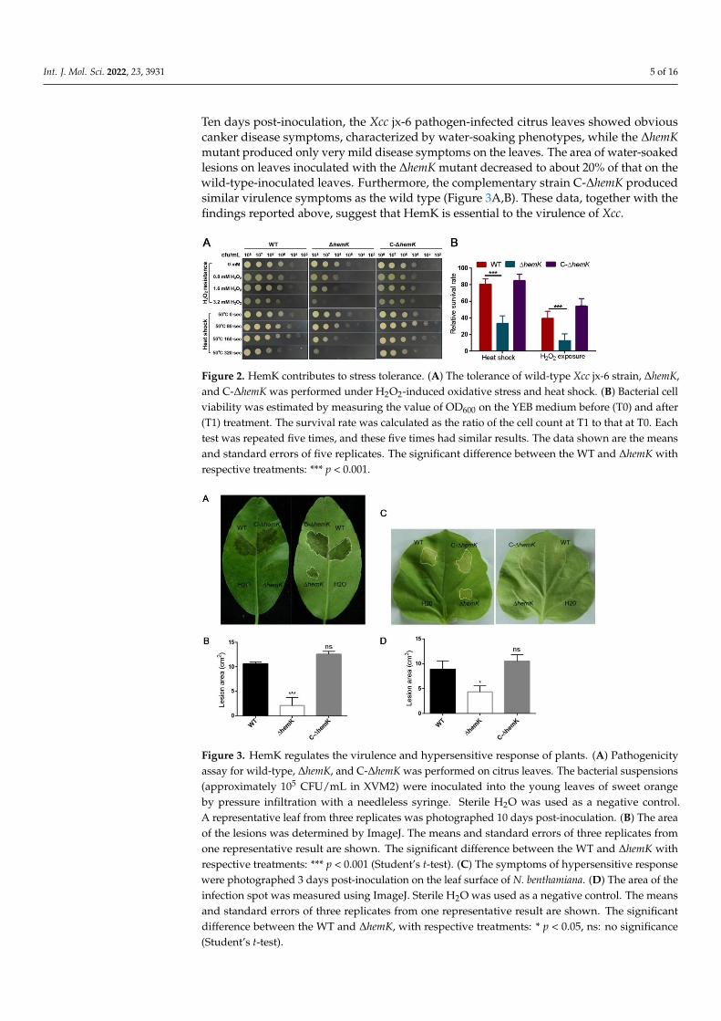
Ten days post-inoculation, the Xcc jx-6 pathogen-infected citrus leaves showed obviouscanker disease symptoms, characterized by water-soaking phenotypes, while the ∆hemKmutant produced only very mild disease symptoms on the leaves. The area of water-soakedlesions on leaves inoculated with the ∆hemK mutant decreased to about 20% of that on thewild-type-inoculated leaves. Furthermore, the complementary strain C-∆hemK producedsimilar virulence symptoms as the wild type (Figure 3A,B). These data, together with thefindings reported above, suggest that HemK is essential to the virulence of Xcc.
Int. J. Mol. Sci. 2022, 23, x FOR PEER REVIEW 5 of 17
showed a 70% lower survival rate than the wild-type strain. Similarly, the survival rate of the high temperature-treated ∆hemK mutant on the YEB agar surface or in liquid YEB media was obviously reduced from that of the wild type (Figure 2B). In all the experiments performed above, the levels of stress tolerance to both H2O2 and the high temperature of C-∆hemK were comparable with those of the wild-type strain (Figure 2). These results indicate that the mutation of hemK reduces bacterial tolerance to oxidative stress and heat shock in Xcc.
Figure 2. HemK contributes to stress tolerance. (A) The tolerance of wild-type Xcc jx-6 strain, ∆hemK, and C-∆hemK was performed under H2O2-induced oxidative stress and heat shock. (B) Bacterial cell viability was estimated by measuring the value of OD600 on the YEB medium before (T0) and after (T1) treatment. The survival rate was calculated as the ratio of the cell count at T1 to that at T0. Each test was repeated five times, and these five times had similar results. The data shown are the means and standard errors of five replicates. The significant difference between the WT and ∆hemK with respective treatments: *** p < 0.001.
2.3. Mutation of hemK Impaired Activation of Virulence on Citrus and Hypersensitive Response to Nicotiana benthamiana
The above finding that hemK deletion resulted in the reduced production of several virulence factors prompted us to further explore whether HemK influences the virulence of Xcc. For this, the pathogenicity of wild-type Xcc jx-6, ΔhemK, and C-ΔhemK strains to the host plant, the Hongjiang sweet orange, was tested as described in the Materials and Methods section. The bacterial cells of these three strains, grown in XVM2 to 105 CFU/mL, were sucked into the needleless syringes and introduced onto the leaves of the sweet orange. Ten days post-inoculation, the Xcc jx-6 pathogen-infected citrus leaves showed obvious canker disease symptoms, characterized by water-soaking phenotypes, while the ΔhemK mutant produced only very mild disease symptoms on the leaves. The area of water-soaked lesions on leaves inoculated with the ΔhemK mutant decreased to about 20% of that on the wild-type-inoculated leaves. Furthermore, the complementary strain C-ΔhemK produced similar virulence symptoms as the wild type (Figure 3A,B). These data, together with the findings reported above, suggest that HemK is essential to the virulence of Xcc.
In addition, we investigated whether hemK deletion has an influence on the ability to induce a hypersensitive response in the non-host plant N. benthamiana. For these experiments, the ΔhemK mutant was infiltrated at a cell concentration of 105 CFU/mL into the leaves of N. benthamiana. The results showed that the hemK mutant elicited a 30% decrease in the hypersensitive response symptoms compared to those of the wild-type strain (Figure 3C,D), suggesting that the HemK trigger compromised the hypersensitive response in its non-host plant N. benthamiana.
Figure 2. HemK contributes to stress tolerance. (A) The tolerance of wild-type Xcc jx-6 strain, ∆hemK,and C-∆hemK was performed under H2O2-induced oxidative stress and heat shock. (B) Bacterial cellviability was estimated by measuring the value of OD600 on the YEB medium before (T0) and after(T1) treatment. The survival rate was calculated as the ratio of the cell count at T1 to that at T0. Eachtest was repeated five times, and these five times had similar results. The data shown are the meansand standard errors of five replicates. The significant difference between the WT and ∆hemK withrespective treatments: *** p < 0.001.
Int. J. Mol. Sci. 2022, 23, x FOR PEER REVIEW 6 of 17
Figure 3. HemK regulates the virulence and hypersensitive response of plants. (A) Pathogenicity assay for wild-type, ∆hemK, and C-∆hemK was performed on citrus leaves. The bacterial suspensions (approximately 105 CFU/mL in XVM2) were inoculated into the young leaves of sweet orange by pressure infiltration with a needleless syringe. Sterile H2O was used as a negative control. A representative leaf from three replicates was photographed 10 days post-inoculation. (B) The area of the lesions was determined by ImageJ. The means and standard errors of three replicates from one representative result are shown. The significant difference between the WT and ∆hemK with respective treatments: *** p < 0.001 (Student’s t-test). (C) The symptoms of hypersensitive response were photographed 3 days post-inoculation on the leaf surface of N. benthamiana. (D) The area of the infection spot was measured using ImageJ. Sterile H2O was used as a negative control. The means and standard errors of three replicates from one representative result are shown. The significant difference between the WT and ∆hemK, with respective treatments: * p < 0.05, ns: no significance (Student’s t-test).
2.4. Transcriptome RNA Sequencing (RNA-Seq) Analysis Reveals Multiple Physiological Processes Regulated by HemK in Xcc
To gain insight into the global regulatory impact of HemK in controlling the virulence of Xcc at a transcriptional level, we performed transcriptomic analysis for wild-type Xcc jx-6 and its isogenic ΔhemK mutant. For this analysis, the Xcc strains were grown to OD600 ≈ 0.8 in the minimal medium XVM2, which is closer to the nutrition environment of the plant intercellular spaces and induces the expression of a series of virulence-related genes [32].
The cells were then collected for total RNA extraction and library construction. The constructed libraries were sequenced using the Illumina Hiseq 2000 platform. Differentially expressed genes (DEGs) between ΔhemK and the Xcc jx-6 strain were determined using DESeq software. Comparative RNA-seq data analysis was performed as described in the Materials and Methods section. Finally, of the 4489 annotated genes from the genome of the Xcc strain 306, a total of 286 genes were determined as significant DEGs between those two strains. Of these, 166 genes were downregulated and 120 genes were upregulated in ∆hemK, with 11 genes being identified as novel genes in this assay (Supplementary Table S1).
To classify the above-identified 286 DEGs into biological function groups, the clusters of orthologous genes (COG) enrichment analysis was employed for those genes according to the genome of the Xcc strain 306. In the COG analysis, based on the conserved domain alignment, a total of 171 DEGs were successfully annotated and grouped into 20 COG functional categories; all the results for each category are presented in Figure 4. Except for those terms with functions unknown (Figure 4, columns R and S), the three most enriched
Figure 3. HemK regulates the virulence and hypersensitive response of plants. (A) Pathogenicityassay for wild-type, ∆hemK, and C-∆hemK was performed on citrus leaves. The bacterial suspensions(approximately 105 CFU/mL in XVM2) were inoculated into the young leaves of sweet orangeby pressure infiltration with a needleless syringe. Sterile H2O was used as a negative control.A representative leaf from three replicates was photographed 10 days post-inoculation. (B) The areaof the lesions was determined by ImageJ. The means and standard errors of three replicates fromone representative result are shown. The significant difference between the WT and ∆hemK withrespective treatments: *** p < 0.001 (Student’s t-test). (C) The symptoms of hypersensitive responsewere photographed 3 days post-inoculation on the leaf surface of N. benthamiana. (D) The area of theinfection spot was measured using ImageJ. Sterile H2O was used as a negative control. The meansand standard errors of three replicates from one representative result are shown. The significantdifference between the WT and ∆hemK, with respective treatments: * p < 0.05, ns: no significance(Student’s t-test).
Int. J. Mol. Sci. 2022, 23, 3931 6 of 16
In addition, we investigated whether hemK deletion has an influence on the abilityto induce a hypersensitive response in the non-host plant N. benthamiana. For these ex-periments, the ∆hemK mutant was infiltrated at a cell concentration of 105 CFU/mL intothe leaves of N. benthamiana. The results showed that the hemK mutant elicited a 30%decrease in the hypersensitive response symptoms compared to those of the wild-typestrain (Figure 3C,D), suggesting that the HemK trigger compromised the hypersensitiveresponse in its non-host plant N. benthamiana.
2.4. Transcriptome RNA Sequencing (RNA-Seq) Analysis Reveals Multiple PhysiologicalProcesses Regulated by HemK in Xcc
To gain insight into the global regulatory impact of HemK in controlling the virulenceof Xcc at a transcriptional level, we performed transcriptomic analysis for wild-type Xcc jx-6and its isogenic ∆hemK mutant. For this analysis, the Xcc strains were grown to OD600 ≈ 0.8in the minimal medium XVM2, which is closer to the nutrition environment of the plantintercellular spaces and induces the expression of a series of virulence-related genes [32].
The cells were then collected for total RNA extraction and library construction. Theconstructed libraries were sequenced using the Illumina Hiseq 2000 platform. Differentiallyexpressed genes (DEGs) between ∆hemK and the Xcc jx-6 strain were determined usingDESeq software. Comparative RNA-seq data analysis was performed as described in theMaterials and Methods section. Finally, of the 4489 annotated genes from the genome of theXcc strain 306, a total of 286 genes were determined as significant DEGs between those twostrains. Of these, 166 genes were downregulated and 120 genes were upregulated in ∆hemK,with 11 genes being identified as novel genes in this assay (Supplementary Table S1).
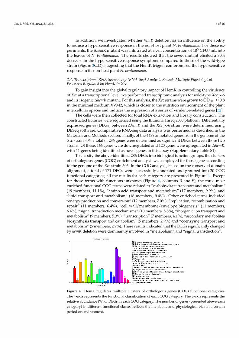
To classify the above-identified 286 DEGs into biological function groups, the clustersof orthologous genes (COG) enrichment analysis was employed for those genes accordingto the genome of the Xcc strain 306. In the COG analysis, based on the conserved domainalignment, a total of 171 DEGs were successfully annotated and grouped into 20 COGfunctional categories; all the results for each category are presented in Figure 4. Exceptfor those terms with functions unknown (Figure 4, columns R and S), the three mostenriched functional COG terms were related to “carbohydrate transport and metabolism”(19 members, 11.1%), “amino acid transport and metabolism” (17 members, 9.9%), and“lipid transport and metabolism” (16 members, 9.4%). Other enriched terms included“energy production and conversion” (12 members, 7.0%), “replication, recombination andrepair” (11 members, 6.4%), “cell wall/membrane/envelope biogenesis” (11 members,6.4%), “signal transduction mechanisms” (10 members, 5.8%), “inorganic ion transport andmetabolism” (9 members, 5.3%), “transcription” (7 members, 4.1%), “secondary metabolitesbiosynthesis transport and catabolism” (5 members, 2.9%) and “coenzyme transport andmetabolism” (5 members, 2.9%). These results indicated that the DEGs significantly changedby hemK deletion were dominantly involved in “metabolism” and “signal transduction”.
Int. J. Mol. Sci. 2022, 23, x FOR PEER REVIEW 7 of 17
functional COG terms were related to “carbohydrate transport and metabolism” (19 members, 11.1%), “amino acid transport and metabolism” (17 members, 9.9%), and “lipid transport and metabolism” (16 members, 9.4%). Other enriched terms included “energy production and conversion” (12 members, 7.0%), “replication, recombination and repair” (11 members, 6.4%), “cell wall/membrane/envelope biogenesis” (11 members, 6.4%), “signal transduction mechanisms” (10 members, 5.8%), “inorganic ion transport and metabolism” (9 members, 5.3%), “transcription” (7 members, 4.1%), “secondary metabolites biosynthesis transport and catabolism” (5 members, 2.9%) and “coenzyme transport and metabolism” (5 members, 2.9%). These results indicated that the DEGs significantly changed by hemK deletion were dominantly involved in “metabolism” and “signal transduction”.
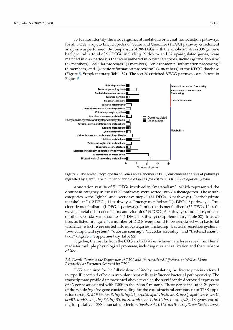
To further identify the most significant metabolic or signal transduction pathways for all DEGs, a Kyoto Encyclopedia of Genes and Genomes (KEGG) pathway enrichment analysis was performed. By comparison of 286 DEGs with the whole Xcc strain 306 genome background, a total of 91 DEGs, including 59 down- and 32 up-regulated genes, were matched into 47 pathways that were gathered into four categories, including “metabolism” (37 members), “cellular processes” (3 members), “environmental information processing” (3 members) and “genetic information processing” (4 members) in the KEGG database (Figure 5, Supplementary Table S2). The top 20 enriched KEGG pathways are shown in Figure 5.
Figure 4. HemK regulates multiple clusters of orthologous genes (COG) functional categories. The x-axis represents the functional classification of each COG category. The y-axis represents the relative abundance (%) of DEGs in each COG category. The number of genes (presented above each category) in different functional classes reflects the metabolic and physiological bias in a certain period or environment.
Annotation results of 51 DEGs involved in “metabolism”, which represented the dominant category in the KEGG pathway, were sorted into 7 subcategories. Those subcategories were “global and overview maps” (33 DEGs, 6 pathways), “carbohydrate metabolism” (12 DEGs, 11 pathways), “energy metabolism” (4 DEGs, 2 pathways), “nucleotide metabolism” (1 DEG, 1 pathway), “amino acids metabolism” (32 DEGs, 10 pathways), “metabolism of cofactors and vitamins” (9 DEGs, 6 pathways), and “biosynthesis of other secondary metabolites” (1 DEG, 1 pathway) (Supplementary Table S2). In addition, as listed in Figure 5, a number of DEGs were found to be associated with bacterial virulence, which were sorted into subcategories, including “bacterial secretion system”, “two-component system”, “quorum sensing”, “flagellar assembly” and “bacterial chemotaxis” (Figure 5, Supplementary Table S2).
Figure 4. HemK regulates multiple clusters of orthologous genes (COG) functional categories.The x-axis represents the functional classification of each COG category. The y-axis represents therelative abundance (%) of DEGs in each COG category. The number of genes (presented above eachcategory) in different functional classes reflects the metabolic and physiological bias in a certainperiod or environment.
Int. J. Mol. Sci. 2022, 23, 3931 7 of 16
To further identify the most significant metabolic or signal transduction pathwaysfor all DEGs, a Kyoto Encyclopedia of Genes and Genomes (KEGG) pathway enrichmentanalysis was performed. By comparison of 286 DEGs with the whole Xcc strain 306 genomebackground, a total of 91 DEGs, including 59 down- and 32 up-regulated genes, werematched into 47 pathways that were gathered into four categories, including “metabolism”(37 members), “cellular processes” (3 members), “environmental information processing”(3 members) and “genetic information processing” (4 members) in the KEGG database(Figure 5, Supplementary Table S2). The top 20 enriched KEGG pathways are shown inFigure 5.
Int. J. Mol. Sci. 2022, 23, x FOR PEER REVIEW 8 of 17
Figure 5. The Kyoto Encyclopedia of Genes and Genomes (KEGG) enrichment analysis of pathways regulated by HemK. The number of annotated genes (x-axis) versus KEGG categories (y-axis).
Together, the results from the COG and KEGG enrichment analyses reveal that HemK mediates multiple physiological processes, including nutrient utilization and the virulence of Xcc.
2.5. HemK Controls the Expression of T3SS and Its Associated Effectors, as Well as Many Extracellular Enzymes Secreted by T2SS
T3SS is required for the full virulence of Xcc by translating the diverse proteins referred to type-III-secreted effectors into plant host cells to influence bacterial pathogenicity. The transcriptome profile data presented above revealed the significantly decreased expression of 43 genes associated with T3SS in the ∆hemK mutant. These genes included 24 genes of the whole hrp/hrc gene cluster coding for the core structural component of T3SS apparatus (hrpF, XAC0395, hpaB, hrpE, hrpD6, hrpD5, hpaA, hrcS, hrcR, hrcQ, hpaP, hrcV, hrcU, hrpB1, hrpB2, hrcJ, hrpB4, hrpB5, hrcN, hrpB7, hrcT, hrcC, hpa1 and hpa2), 18 genes encoding for putative T3SS-associated effectors (hpaF, XAC0419, avrBs2, xopR, avrXacE1, xopX, xopI, xopN/HopAU1, hrpW, xopK, xopL, XAC3230, XAC3666, xopQ/HopQ1, xopAU, xopAV, xopAP, and xopS), and hrpX, encoding a transcriptional regulator of virulence (Figure 6A, Supplementary Table S1) [15,33].
T2SS secretes numerous hydrolases, such as cellulases, proteases, and xylanases as virulence factors to contribute to canker symptom development in the Xanthomonas genus [12,17,18]. RNA-seq data also revealed a decreased expression of 10 genes in ∆hemK, encoding bacterial exoenzymes secreted through T2SS (virK, XAC0346, XAC0612, XAC0795, XAC0933, XAC2831, XAC2833, XAC2853, XAC3490 and XAC3545) (Figure 6A, Supplementary Table S1).
To further confirm whether HemK is required for the regulation of T3SS- and T2SS-associated gene expression, as revealed by the RNA-seq analysis, we conducted quantitative real-time PCR (qRT-PCR) for wild-type Xcc jx-6 and ∆hemK strains to evaluate the relatively endogenous mRNA level of 11 genes in Xcc. These selected genes included hrpB2, hpaA, hrcC, and hrpE (T3SS regulators), avrXacE1, avrBs2 and virK (virulence protein), XAC0612 (cellulase), XAC3545 (protease), XAC3490 (amylosucrase or alpha-amylase) and XAC2853 (cysteine protease). As expected, the qRT-PCR results display a significantly decreased mRNA level of these selected 11 genes in the ∆hemK mutant compared with the wild type (Figure 6B).
Figure 5. The Kyoto Encyclopedia of Genes and Genomes (KEGG) enrichment analysis of pathwaysregulated by HemK. The number of annotated genes (x-axis) versus KEGG categories (y-axis).
Annotation results of 51 DEGs involved in “metabolism”, which represented thedominant category in the KEGG pathway, were sorted into 7 subcategories. Those sub-categories were “global and overview maps” (33 DEGs, 6 pathways), “carbohydratemetabolism” (12 DEGs, 11 pathways), “energy metabolism” (4 DEGs, 2 pathways), “nu-cleotide metabolism” (1 DEG, 1 pathway), “amino acids metabolism” (32 DEGs, 10 path-ways), “metabolism of cofactors and vitamins” (9 DEGs, 6 pathways), and “biosynthesisof other secondary metabolites” (1 DEG, 1 pathway) (Supplementary Table S2). In addi-tion, as listed in Figure 5, a number of DEGs were found to be associated with bacterialvirulence, which were sorted into subcategories, including “bacterial secretion system”,“two-component system”, “quorum sensing”, “flagellar assembly” and “bacterial chemo-taxis” (Figure 5, Supplementary Table S2).
Together, the results from the COG and KEGG enrichment analyses reveal that HemKmediates multiple physiological processes, including nutrient utilization and the virulenceof Xcc.
2.5. HemK Controls the Expression of T3SS and Its Associated Effectors, as Well as ManyExtracellular Enzymes Secreted by T2SS
T3SS is required for the full virulence of Xcc by translating the diverse proteins referredto type-III-secreted effectors into plant host cells to influence bacterial pathogenicity. Thetranscriptome profile data presented above revealed the significantly decreased expressionof 43 genes associated with T3SS in the ∆hemK mutant. These genes included 24 genesof the whole hrp/hrc gene cluster coding for the core structural component of T3SS appa-ratus (hrpF, XAC0395, hpaB, hrpE, hrpD6, hrpD5, hpaA, hrcS, hrcR, hrcQ, hpaP, hrcV, hrcU,hrpB1, hrpB2, hrcJ, hrpB4, hrpB5, hrcN, hrpB7, hrcT, hrcC, hpa1 and hpa2), 18 genes encod-ing for putative T3SS-associated effectors (hpaF, XAC0419, avrBs2, xopR, avrXacE1, xopX,

Int. J. Mol. Sci. 2022, 23, 3931 8 of 16
xopI, xopN/HopAU1, hrpW, xopK, xopL, XAC3230, XAC3666, xopQ/HopQ1, xopAU, xopAV,xopAP, and xopS), and hrpX, encoding a transcriptional regulator of virulence (Figure 6A,Supplementary Table S1) [15,33].
Int. J. Mol. Sci. 2022, 23, x FOR PEER REVIEW 9 of 17
Figure 6. HemK regulates the expression of T3SS- and T2SS-associated genes. (A) Heat map of gene expression of T2SS and T3SS by RNA-seq data. (B) The relative expression of genes associated with the virulence identified by RNA-seq analysis was determined by qRT-PCR analysis. The target genes included hrpB2, hpaA, hrcC and hrpE (T3SS regulators), virK, avrXacE1 and avrBs2 (virulence protein), XAC3545 (protease), XAC0612 (cellulase), XAC3490 (amylosucrase or alpha-amylase) and XAC2853 (cysteine protease).
Given the finding that the deletion of hemK results in reduced exoenzyme secretion (Figure 1F–H) and impaired activity, to trigger the hypersensitive response in its non-host plant, N. benthamiana (Figure 3C,D), it is reasonable to suspect that the HemK-positive regulation of the T3SS- and T2SS-associated genes’ expression at the transcriptional level accounts for the phenotypes that appeared in the ∆hemK mutant.
2.6. HemK Is Implicated in the Regulation of the Expression of Genes Involved in Diffusible Signal Factor (DSF) Mediated Quorum Sensing of Xcc
Previous transcriptome profile and proteomic analyses revealed that DSF-mediated quorum sensing specifically modulates bacterial adaptation, nutrition uptake and metabolism, stress tolerance, virulence, and signal transduction to favor host infection [34,35]. We compared the transcript profiles of the ∆hemK mutant (Supplementary Table S1) and that of the previously reported DSF-deficient (ΔrpfF) mutant during citrus infection in planta [34], and observed 32 genes being regulated by both HemK and RpfF, of which 31 genes were downregulated in ∆hemK(Table 1).
Figure 6. HemK regulates the expression of T3SS- and T2SS-associated genes. (A) Heat map of geneexpression of T2SS and T3SS by RNA-seq data. (B) The relative expression of genes associated withthe virulence identified by RNA-seq analysis was determined by qRT-PCR analysis. The target genesincluded hrpB2, hpaA, hrcC and hrpE (T3SS regulators), virK, avrXacE1 and avrBs2 (virulence protein),XAC3545 (protease), XAC0612 (cellulase), XAC3490 (amylosucrase or alpha-amylase) and XAC2853(cysteine protease).
T2SS secretes numerous hydrolases, such as cellulases, proteases, and xylanasesas virulence factors to contribute to canker symptom development in the Xanthomonasgenus [12,17,18]. RNA-seq data also revealed a decreased expression of 10 genes in ∆hemK,encoding bacterial exoenzymes secreted through T2SS (virK, XAC0346, XAC0612, XAC0795,XAC0933, XAC2831, XAC2833, XAC2853, XAC3490 and XAC3545) (Figure 6A, Supplemen-tary Table S1).
To further confirm whether HemK is required for the regulation of T3SS- and T2SS-associated gene expression, as revealed by the RNA-seq analysis, we conducted quantitativereal-time PCR (qRT-PCR) for wild-type Xcc jx-6 and ∆hemK strains to evaluate the relativelyendogenous mRNA level of 11 genes in Xcc. These selected genes included hrpB2, hpaA,hrcC, and hrpE (T3SS regulators), avrXacE1, avrBs2 and virK (virulence protein), XAC0612(cellulase), XAC3545 (protease), XAC3490 (amylosucrase or alpha-amylase) and XAC2853(cysteine protease). As expected, the qRT-PCR results display a significantly decreasedmRNA level of these selected 11 genes in the ∆hemK mutant compared with the wild type(Figure 6B).
Given the finding that the deletion of hemK results in reduced exoenzyme secretion(Figure 1F–H) and impaired activity, to trigger the hypersensitive response in its non-hostplant, N. benthamiana (Figure 3C,D), it is reasonable to suspect that the HemK-positiveregulation of the T3SS- and T2SS-associated genes’ expression at the transcriptional levelaccounts for the phenotypes that appeared in the ∆hemK mutant.

Int. J. Mol. Sci. 2022, 23, 3931 9 of 16
2.6. HemK Is Implicated in the Regulation of the Expression of Genes Involved in Diffusible SignalFactor (DSF) Mediated Quorum Sensing of Xcc
Previous transcriptome profile and proteomic analyses revealed that DSF-mediated quo-rum sensing specifically modulates bacterial adaptation, nutrition uptake and metabolism,stress tolerance, virulence, and signal transduction to favor host infection [34,35]. We com-pared the transcript profiles of the ∆hemK mutant (Supplementary Table S1) and that ofthe previously reported DSF-deficient (∆rpfF) mutant during citrus infection in planta [34],and observed 32 genes being regulated by both HemK and RpfF, of which 31 genes weredownregulated in ∆hemK (Table 1).
Table 1. List of genes related to quorum sensing in X. citri subsp. citri jx-6, regulated by HemK.
Locus Tag Gene Name Log2Fold Change Protein Function
XAC2992 XAC2992 −1.5 endoproteinase Arg-C, degrading host defense proteinsXAC0612 engXCA −1.3 cellulaseXAC3120 glk 1.3 glucose kinaseXAC3921 ugt −1.2 glucosyltransferaseXAC1556 fucP −1.3 glucose-galactose transporterXAC1557 scrK −1.2 fructokinaseXAC3489 fyuA −2.3 TonB-dependent receptorXAC3490 XAC3490 −1.9 amylosucrase or alpha amylaseXAC0465 XAC0465 −1.0 metalloproteinaseXAC4327 uahA −1.8 urea amidolyaseXAC1820 metL −3.3 aspartokinaseXAC1821 thrB −3.4 homoserine kinaseXAC1823 thrC −3.6 threonine synthaseXAC1828 hisG −2.4 ATP phosphoribosyltransferaseXAC1829 hisD −2.3 histidinol dehydrogenaseXAC1830 hisC −2.4 histidinol-phosphate aminotransferase
XAC1831 hisB −2.5 Imidazole glycerol phosphate dehydratase/histidinol-phosphate phosphatasebifunctional enzyme
XAC1832 hisH −1.9 amidotransferaseXAC1833 hisA −2.2 phosphoribosylformimino-5-aminoimidazole carboxamXAC1834 hisF −2.3 cyclase
XAC1835 hisI −2.4 phosphoribosyl-AMP cyclohydrolase/phosphoribosyl—ATP pyrophosphatasebifunctional enzyme
XAC3451 ilvC −2.7 ketol-acid reductoisomeraseXAC3452 ilvG −2.2 acetolactate synthase isozyme II large subunitXAC3453 ilvM −2.5 acetolactate synthase isozyme II large subunitXAC3454 tdcB −2.1 threonine dehydratase catabolicXAC3455 leuA −1.3 2-isopropylmalate synthaseXAC0999 cirA −1.1 colicin I receptorXAC3546 xadA −1.7 autotransporter adhesion proteinXAC1471 XAC1471 −1.1 glycine zipper 2TM domain containing proteinXAC1827 XAC1827 −3.2 Trp repressor proteinXAC3085 XAC3085 −1.1 putative type III secretion system effector proteinXAC3754 XAC3754 1.4 putative chemotaxis membrane protein
3. Discussion
HemK is evolutionarily conserved from prokaryotes to eukaryotes [22,24,30], but onlya few orthologous genes have been studied so far. The hemK-knockout mutation in micewas lethal [36], while the loss of HemK results in severe growth defects in some bacteria [21]and increased sensitivity to low temperature, salinity, and high calcium concentrationsin addition to growth restriction in yeast [22]. In Arabidopsis, the methyltransferase NRF1knockout line displays imbalances in cellular ion levels, severe growth retardation, anddemonstrates various developmental defects [25]. However, little is known about thephysiological role of HemK in the phytopathogen Xcc. In this study, we demonstrated thatthe ∆hemK mutant showed decreased motility, biofilm formation, and the production ofcellulases, amylases, and proteases, as well as decreased adaptation to oxidative stress andheat shock. Furthermore, the virulence of ∆hemK on host and non-host plants, respectively,was significantly decreased. HemK was further shown by transcriptomic analysis to play arole in the regulation of a variety of genes involved in bacterial virulence, secretion systems,
Int. J. Mol. Sci. 2022, 23, 3931 10 of 16
chemotaxis, and metabolism. Our studies indicated that HemK may play an essential rolein regulating the virulence and environmental adaptation of Xcc.
In this study, RNA-seq analysis showed that the genes regulated by HemK included32 genes related to amino acid uptake and metabolism, 43 genes related to T3SS and itseffectors, 12 genes related to carbohydrate metabolism, 18 genes related to chemotaxisand flagellar assembly, and 32 genes related to the diffusible signal factor (DSF)-mediatedquorum sensing of Xcc. Phenotypical analyses revealed that the deletion of hemK causedreduced swimming and swarming motility, extracellular enzymes, and polysaccharide pro-duction in Xcc. Several reports have shown that the lack of extracellular enzymes reducedthe virulence by affecting the early colonization of the pathogen in Xanthomonas [13,17].The mutation of Bglc 3 in Xcc strain 29-1 lost the ability to produce cellulases, resulting inits weakened pathogenicity on citrus [13]. Bacterial motility and chemotaxis contributed tovirulence at the initial stages of Xcc infection [37,38]. Based on our findings in this report,we speculate that HemK may regulate virulence by affecting the early colonization stepof Xcc infection during the development of symptoms in citrus. The T3SS encoded by thehrp gene cluster is involved in delivering a number of hypersensitive-response elicitors orpathogenic factors into plant cells and is critical for the successful infection and colonizationof Xcc in host and non-host tissues [12,39–41]. HemK and its homologous proteins havebeen reported to be related to T3SS in different pathogens. For example, the virulence-associated protein VagH in Y. pseudotuberculosis shares high homology with the HemKof E. coli, has a methyltransferase activity similar to HemK, and shows a close link withT3SS [29]. In P. aeruginosa PA14, HemK, an S-adenosyl-l-methionine (AdoMet)-dependentmethyltransferase of peptide chain release factors, is essential for the expression of virulencefactors and the type III-secreted toxin ExoT [28]. In this study, the transcriptomic analysisshowed that HemK positively regulated the expression of T3SS genes, including 24 hrpgenes. HrpX is the master regulator of T3SS and its effector genes in Xanthomonas [15,33,42].Our qRT-PCR analysis further confirmed that a number of T3SS effector genes, includinghrpB2, hpaA, hrcC, hrpE, and virulence genes (avrXacE1 and avrBs2), are down-regulated in∆hemK. It is, therefore, likely that HemK may regulate the pathogenicity of Xcc by regulat-ing the expression of hrp genes and T3SS effectors. In summary, our research provides newinsights into the functional roles of HemK in regulating the pathogenicity, virulence, andenvironmental tolerance of Xcc. Further investigations may help to understand whether thecatalytic activity of HemK is critical for bacterial survival under specific stress conditionsand whether HemK is a potential target for antibacterial drug development.
4. Materials and Methods4.1. Bacterial Strains, Culture Media, and Culture Conditions
The bacterial strains and plasmids used in this study are listed in SupplementaryTable S3. The E. coli strains were grown at 37 ◦C in either Luria-Bertani (LB) broth (10 g/Ltryptone, 5 g/L yeast extract, and 10 g/L NaCl, pH 7.0) or on LB with agar. The optimumgrowth temperature for the Xcc jx-6 strain was 28 ◦C, and the strains were grown in NYGmedium (5 g/L peptone, 3 g/L yeast extract, 20 mL/L glycerol, pH 7.0), YEB medium(10 g/L tryptone, 5 g/L yeast extract, 5 g/L NaCl, 0.5 g/L MgSO4·7H2O, 5 g/L sucrose,pH 7.0), or XVM2 medium (20 mM NaCl, 10 mM (NH4)2SO4, 5 mM MgSO4, 1 mM CaCl2,0.16 mM KH2PO4, 0.32 mM K2HPO4, 0.01 mM FeSO4, 10 mM fructose, 10 mM sucrose,0.03% casamino acids (pH 6.7)). NY medium was NYG medium without glycerol [43].YEB medium was used for exoenzyme production assays. Antibiotic gentamicin (SangonBiotech, Shanghai, China) was added at a final concentration of 25 µg·mL−1 when required.
4.2. Plasmid Construction and Primers Used
DNA fragments encoding the full-length or truncated proteins were amplified with apolymerase chain reaction (PCR) from the genomic DNA of the Xcc jx-6 strain. Primers weredesigned based on the published complete genome sequence of Xcc jx-6 (NCBI reference
Int. J. Mol. Sci. 2022, 23, 3931 11 of 16
sequence: NZ_CP011827.2) and are listed in Supplementary Table S4. Primer synthesis andsequencing services were performed by Beijing Qingke Biotechnology (Beijing, China).
4.3. Construction of the ∆hemK Mutant and Its Complementary Strain in Xcc jx-6
The deletion mutant ∆hemK was created from the wild-type Xcc jx-6 strain by homologous-recombination-based procedures, as described previously by using the knockout plasmidpK18-∆hemK. This plasmid was constructed by cloning the fused upstream- and downstream-homologous fragments of the HemK region into the suicide vector pK18mobSacB-carryingGm resistance gene [44]. The DNA fragments of up and down homologous arms of hemKwere amplified by PCR, with primer pairs of hemK-F1/hemK-R2 and hemK-F3/hemK-R4,respectively. The obtained two fragments were fused together by PCR, using the primer pair ofhemK-F1/hemK-R4. The fused DNA fragment was digested with HindIII and BamHI and thenligated into the corresponding sites of the vector pK18mobSacB. Then, the obtained knockoutvector pK18-∆hemK was transformed into wild-type strain Xcc jx-6 by electroporation [45],and positive transformants were selected on LB agar medium, supplemented with gentamicin(25 µg/mL). Colonies resulting from the first crossover events were streaked onto LB agarplates supplemented with 10% sucrose, and sucrose-sensitive colonies were selected as positivecolonies. Colonies with hemK deleted were further confirmed by sequencing the DNA fragmentamplified with the primer pair of hemK-5F and hemK-6R.
To construct the complementary plasmid pBBR-hemK, an 861-bp DNA fragment that cov-ered the entire coding region of HemK protein was cloned into the vector pBBR1MCS-5 [46].The DNA fragment was amplified by PCR using genomic DNA from strain Xcc jx-6 as a tem-plate, with the primer pair of hemK-F/hemK-R; it was then digested with SmaI and XbaI andligated into the corresponding sites of the plasmid pBBR1MCS-5. The ligation mixture wastransformed into E. coli DH5α-competent cells and positive colonies were selected on LB agarwith gentamicin (25 µg/mL). Following PCR-based verification using primers M13/pBAD,the recombinant plasmid was transformed into ∆hemK by electroporation to produce thecomplementary strain that was designated as C-∆hemK.
4.4. Bacterial Growth Curve
We analyzed the growth of the wild type, ∆hemK, and C-∆hemK using the Bioscreen Cautomated microbiology growth curve analysis system (Thermo Lab Systems, Helsinki,Finland) by adapting the method described by Medina et al. [47]. Briefly, a single colonyof each strain was grown overnight at 28 ◦C in LB broth with an antibiotic (25 µg/mLgentamicin). The overnight cultures were adjusted to the same concentration by measuringthe OD600 values and were then diluted 100-fold in fresh YEB medium. An aliquot of200 µL of this diluted solution was added into the wells of a 100-well microplate, with thefresh YEB used as a negative control. The OD at 600 nm was measured every 4 h at 28 ◦Cwith shaking. Four biological replicates were performed for each strain.
4.5. Bacterial Motility Assays
Motility assay was examined as described previously [43,48]. Briefly, bacterial strainswere all grown in LB liquid medium at 28 ◦C with shaking for about 18 h and diluted toan optical density at OD600 of 0.8. For swimming motility, 2 µL aliquots of bacterial cellswere stabbed into the medium in the center of 0.25% Bacto-agar plates containing 0.03%peptone and 0.03% yeast extract with gentamicin and were inoculated for 60h at 28 ◦C; forswarming motility, 2 µL aliquots of bacteria cells were spotted onto the surface of the centerof 0.6% Bacto-agar plates containing NY medium, supplemented with 2% glucose withgentamicin. Following incubation at 28 ◦C for 60 h, all the plates were photographed. Themotility was quantified by measuring the area of the growth circle around the inoculationsite with ImageJ. Each assay was repeated at least three times.

Int. J. Mol. Sci. 2022, 23, 3931 12 of 16
4.6. Bacterial Biofilm Formation Assay
The biofilm formation was tested by measuring the ability of bacterial cells to adhereto the glass tubes as described previously with slight modification [11,49]. Bacterial strainswere grown in YEB medium at 28 ◦C with shaking at 200 rpm. Then, 1 mL of bacterialsuspensions (OD600 = 1.0) were inoculated into a glass tube containing 1 mL fresh YEBmedium supplemented with 0.05% L-arabinose and 25 µg/mL gentamicin, then left tostand for 72 h at 28 ◦C. The culture medium was discarded; the attached bacterial cells werestained with 0.1% (w/v) crystal violet for 45 min, then washed 3 times with distilled waterand dried at 60 ◦C. The crystal violet remaining on the tube wall was dissolved in 33%(v/v) acetic acid, and the absorbance was measured at 590 nm with a microplate reader(BioTek, Winooski, VT, USA). Three repeated quantitative measurements were performed.
4.7. Extracellular Enzymes Activity Assay
Extracellular enzyme activity assays were examined, as described by [50] with somemodifications. Briefly, Xcc strains were grown in YEB media at 28 ◦C for about 18 h, and analiquot of 1.5 µL bacterial culture (adjusted to an OD600 of 0.8) was spotted onto the surfacein the center of YEB agar plates supplemented with 1% (w/v) potato starch (for amylasedetection), or 1% (w/v) sodium carboxymethyl ethyl cellulose (for cellulase detection),together with 0.05% (w/v) L-arabinose and gentamicin. All plates were incubated for 2 daysat 28 ◦C. For protease detection, the bacterial supernatant was collected by centrifuge at13,000 rpm for 5 min, then a 20 µL aliquot of the supernatant was added to the wells ofYEB plates containing 1% (w/v) milk [51], punched with a hole punch, blown dry, andincubated for 48 h at 28 ◦C. Three replicates were used for each assay.
4.8. Extracellular Polysaccharides Production Assays
Extracellular polysaccharides (EPS) in bacterial culture supernatants were determinedquantitatively, as described previously [52]. Bacterial strains were grown in liquid YEBmedium with gentamicin overnight until the OD600 reached 2.5. The supernatant of thecell cultures was obtained after removing the cell pellets by centrifugation (5000 rpm for30 min). Two volumes of ethanol were added to the supernatant and the mixtures wereincubated at −20 ◦C overnight. The precipitated EPS was obtained by centrifugation(5000 rpm for 20 min) and the dry weights of EPS were recorded after drying overnightat 65 ◦C.
EPS production was established by the plate assay; all strains were grown in YEBmedium until the OD600 reached 0.8. An aliquot of 2 µL bacterial culture was spotted ontothe center of a YEB agar plate supplemented with 25 µg/mL gentamicin and 0.05% (m/v)L-arabinose. Following growth at 28 ◦C for 2 days, the YEB agar plates were photographed.These experiments were independently repeated at least three times.
4.9. Stress Tolerance Assay
The stress tolerance assay was performed as previously described, with modifica-tions [53]. Environmental stresses, including ultraviolet (UV) radiation, saline stress, andosmotic challenge, heat shock, and oxidative stress challenges were measured. The bacterialstrains of the mutant ∆hemK, the wild type, and the C-∆hemK strain were cultured in anLB medium containing gentamicin to the early exponential stage (OD600 of 0.7) and werecollected for stress treatment. For oxidative stress treatment, the bacterial cultures wereexposed to three different H2O2 concentrations, namely, 0.8, 1.6, and 3.2 mM for 30 min.For heat shock treatment, the bacterial cultures were heated at 50 ◦C for periods of 0, 80,160, and 320 s. For survival growth tests of stress-treated cells, the treated bacterial cultures(adjusted to a concentration of 108 CFU/mL) were serially diluted by a factor of ten to103 CFU/mL, and a 2 µL aliquot of each ten-fold dilution sample was spotted onto the sur-face of the YEB agar plates and incubated at 28 ◦C for 48 h. By a comparison of the growthof colonies before and after treatment, the levels of stress tolerance were determined.
Int. J. Mol. Sci. 2022, 23, 3931 13 of 16
In the quantitative analysis experiment, the stress-treated bacterial culture was inoc-ulated into fresh YEB medium at a 1:50 ratio, and the diluted solution was cultured viashaking for 28 h, then the optical density at 600 nm (OD600) was measured. The relativesurvival rate was defined as the percentage of viable cell counts from the bacterial culturewith stress treatment (T1, value of OD600) compared with those from the non-treated culture(T0, value of OD600). Each treatment was repeated three times, with three replicates foreach strain.
4.10. Virulence and Hypersensitive Response Assays
Pathogenicity assays were conducted as described previously, with little modifica-tion [32,54]. An assay was performed using the immature leaves of the sweet orange (Citrussinensis) grown outdoors at approximately 18 ◦C to 28 ◦C. The wild-type Xcc jx-6 strain,mutant strain ∆hemK, and complementary strain C-∆hemK were cultured in XVM2 mediumat 28 ◦C with shaking until the value was 0.4 of OD600. First, 1 mL of each cell culture wascentrifuged at 10,000 rpm and resuspended in 1 mL of sterile water. Then, 10 µL of eachbacterial solution was infiltered into the leaves with a needleless syringe. Sterile water wasinfiltered in the same way as the negative control. The disease symptoms of the inoculatedsweet orange leaves were photographed 10 days after inoculation. The hypersensitiveresponse (HR) assay on N.benthamiana was also conducted for these three strains. Thetobacco plants were grown in a quarantine greenhouse facility (parameters: light for 16 hand darkness for 8 h at 28 ◦C). The cells (approximately 105 CFU/mL) were then pelletedby centrifugation and resuspended (1:1) in sterile water, and the disease symptoms of N.benthamiana were photographed at 3 days post-inoculation. Experiments were repeated atleast three times with similar results.
4.11. High-Throughput RNA Sequencing (RNA-Seq) and Data Analysis
A single bacterial colony of the wild-type strain Xcc jx-6 and the ∆hemK mutant wascultured in 10 mL LB broth at 28 ◦C with shaking for 24 h, along with three biologicalrepeats performed for each strain. Overnight cultures were adjusted to the same OD600,and were then diluted (1:100) in XVM2 media and incubated at 28 ◦C. Until OD600 = 0.8,the cells were collected by centrifugation at 12,000 rpm and immediately frozen with liquidnitrogen for total RNA extraction. The total RNA was extracted using the Eastep SuperTotal RNA extraction kit (Shanghai Promega Biological Products Ltd, Shanghai, China).The quantity and quality of the total RNA were determined using a NanoDrop (NanoDrop2000 Technologies, Wilmington, NC, USA), agarose gel electrophoresis, and an Agilent2100 bioanalyzer (Agilent Technologies, Waldbronn, Germany).
Transcriptome library construction and sequencing were performed using Novogene(Beijing, China). Clean read data were obtained following sequencing and data filtering,and the resulting reads were mapped to the Xcc 306 reference genome. Gene expres-sion, as indicated by the expected number of fragments per kilobase of the transcriptsequence, per million base pairs sequenced (FPKM), was calculated using HTSeq. DEGsbetween the strains ∆hemK and Xcc jx-6 were determined using DESeq software, basedon q-values of <0.05 and a minimum |log2 (Fold Change)| > 1, and were exported asa tabular file (Supplementary Table S1). The obtained DEGs between those two strainswere then classified and enriched, based on the Clusters Orthologous Groups database(COG, https://www.ncbi.nlm.nih.gov/COG/, accessed on 29 December 2021) and KEGGpathway analyses.
4.12. Quantitative Real-Time PCR (qRT-PCR) Assay
To verify the RNA-seq results, a qRT-PCR assay was performed using the total RNAextracted from strains grown under the same growth conditions as the RNA-seq. RNAsamples were reverse-transcribed using a HiScript II QRT SuperMix for qPCR (VazymeBiotech, Nanjing, China). The cDNA was subjected to a two-step qRT-PCR assay, using aChamQ Universal SYBR qPCR Master Mix (Vazyme Biotech) on a QuantStudioTM 6 Flex
Int. J. Mol. Sci. 2022, 23, 3931 14 of 16
System. The 16 s rRNA gene was used as an endogenous control. The relative fold changein the target genes’ expression was calculated using the formula 2-∆∆CT [55]. The primersequences used in the qRT-PCR assay are listed in Supplementary Table S4.
Supplementary Materials: The following supporting information can be downloaded at: https://www.mdpi.com/article/10.3390/ijms23073931/s1.
Author Contributions: J.W. and L.X. designed the experiments and analyzed the data with theassistance from other authors. Y.S. constructed the mutant strain, ∆hemK, and its complementationstrain, C-∆hemK. Y.S., X.Y. (Xiaobei Yang), X.Y. (Xiaoxin Ye) and J.F. performed the phenotypicanalysis. Y.S., X.Y. (Xiaobei Yang), T.C. and X.Z. did the RNA-Seq and qRT-PCR analysis. J.W., L.X.,Y.S. and D.X.L. wrote the manuscript. All authors have read and agreed to the published version ofthe manuscript.
Funding: This research was funded by the Science and Technology Planning Project of GuangdongProvince, China (2017A020208053), the Key Area Research and Development Program of GuangdongProvince, China (2018B020205003) and the Natural Science Foundation of Guangdong Province,China (2021A1515010761 and 2022A1515010869).
Institutional Review Board Statement: Not applicable.
Data Availability Statement: Not applicable.
Conflicts of Interest: The authors declare no conflict of interest.
References1. Brunings, A.M.; Gabriel, D.W. Xanthomonas citri: Breaking the surface. Mol. Plant Pathol. 2003, 4, 141–157. [CrossRef]2. Vojnov, A.A.; Do, A.A.; Dow, J.M.; Castagnaro, A.P.; Marano, M.R. Bacteria causing important diseases of citrus utilise distinct
modes of pathogenesis to attack a common host. Appl. Microbiol. Biotechnol. 2010, 87, 467–477. [CrossRef]3. Timilsina, S.; Potnis, N.; Newberry, E.A.; Liyanapathiranage, P.; Iruegas-Bocardo, F.; White, F.F.; Goss, E.M.; Jones, J.B. Xanthomonas
diversity, virulence and plant-pathogen interactions. Nat. Rev. Microbiol 2020, 18, 415–427. [CrossRef]4. An, S.Q.; Potnis, N.; Dow, M.; Vorholter, F.J.; He, Y.Q.; Becker, A.; Teper, D.; Li, Y.; Wang, N.; Bleris, L.; et al. Mechanistic insights
into host adaptation, virulence and epidemiology of the phytopathogen Xanthomonas. FEMS Microbiol. Rev. 2020, 44, 1–32.[CrossRef]
5. Ference, C.M.; Gochez, A.M.; Behlau, F.; Wang, N.; Graham, J.H.; Jones, J.B. Recent advances in the understanding of Xanthomonascitri ssp. citri pathogenesis and citrus canker disease management. Mol. Plant Pathol. 2018, 19, 1302–1318. [CrossRef]
6. Huang, C.J.; Wu, T.L.; Zheng, P.X.; Ou, J.Y.; Ni, H.F.; Lin, Y.C. Comparative Genomic Analysis Uncovered Evolution of Pathogenic-ity Factors, Horizontal Gene Transfer Events, and Heavy Metal Resistance Traits in Citrus Canker Bacterium Xanthomonas citrisubsp. citri. Front. Microbiol. 2021, 12, 731711. [CrossRef]
7. Da, S.A.; Ferro, J.A.; Reinach, F.C.; Farah, C.S.; Furlan, L.R.; Quaggio, R.B.; Monteiro-Vitorello, C.B.; Van Sluys, M.A.; Almeida, N.F.;Alves, L.M.; et al. Comparison of the genomes of two Xanthomonas pathogens with differing host specificities. Nature 2002, 417,459–463. [CrossRef]
8. Zhang, Y.; Teper, D.; Xu, J.; Wang, N. Stringent response regulators (p)ppGpp and DksA positively regulate virulence and hostadaptation of Xanthomonas citri. Mol. Plant Pathol. 2019, 20, 1550–1565. [CrossRef]
9. Braun, S.G.; Meyer, A.; Holst, O.; Puhler, A.; Niehaus, K. Characterization of the Xanthomonas campestris pv. campestris lipopolysac-charide substructures essential for elicitation of an oxidative burst in tobacco cells. Mol. Plant-Microbe Interact. 2005, 18, 674–681.[CrossRef]
10. Li, J.; Wang, N. The wxacO gene of Xanthomonas citri ssp. citri encodes a protein with a role in lipopolysaccharide biosynthesis,biofilm formation, stress tolerance and virulence. Mol. Plant Pathol. 2011, 12, 381–396. [CrossRef]
11. Rigano, L.A.; Siciliano, F.; Enrique, R.; Sendin, L.; Filippone, P.; Torres, P.S.; Questa, J.; Dow, J.M.; Castagnaro, A.P.;Vojnov, A.A.; et al. Biofilm formation, epiphytic fitness, and canker development in Xanthomonas axonopodis pv. citri. Mol.Plant-Microbe Interact. 2007, 20, 1222–1230. [CrossRef]
12. Song, X.; Guo, J.; Ma, W.X.; Ji, Z.Y.; Zou, L.F.; Chen, G.Y.; Zou, H.S. Identification of seven novel virulence genes from Xanthomonascitri subsp. citri by Tn5-based random mutagenesis. J. Microbiol. 2015, 53, 330–336. [CrossRef]
13. Xia, T.; Li, Y.; Sun, D.; Zhuo, T.; Fan, X.; Zou, H. Identification of an Extracellular Endoglucanase That Is Required for FullVirulence in Xanthomonas citri subsp. citri. PLoS ONE 2016, 11, e151017. [CrossRef]
14. Buttner, D.; Bonas, U. Regulation and secretion of Xanthomonas virulence factors. FEMS Microbiol. Rev. 2010, 34, 107–133.[CrossRef]
15. Guo, Y.; Figueiredo, F.; Jones, J.; Wang, N. HrpG and HrpX play global roles in coordinating different virulence traits ofXanthomonas axonopodis pv. citri. Mol. Plant-Microbe Interact. 2011, 24, 649–661. [CrossRef]

Int. J. Mol. Sci. 2022, 23, 3931 15 of 16
16. Zhou, X.; Hu, X.; Li, J.; Wang, N. A Novel Periplasmic Protein, VrpA, Contributes to Efficient Protein Secretion by the Type IIISecretion System in Xanthomonas spp. Mol. Plant-Microbe Interact. 2015, 28, 143–153. [CrossRef]
17. Yan, Q.; Hu, X.; Wang, N. The novel virulence-related gene nlxA in the lipopolysaccharide cluster of Xanthomonas citri ssp. citriis involved in the production of lipopolysaccharide and extracellular polysaccharide, motility, biofilm formation and stressresistance. Mol. Plant Pathol. 2012, 13, 923–934. [CrossRef]
18. Baptista, J.C.; Machado, M.A.; Homem, R.A.; Torres, P.S.; Vojnov, A.A.; Do, A.A. Mutation in the xpsD gene of Xanthomonasaxonopodis pv. citri affects cellulose degradation and virulence. Genet. Mol. Biol. 2010, 33, 146–153. [CrossRef]
19. Li, R.F.; Lu, G.T.; Li, L.; Su, H.Z.; Feng, G.F.; Chen, Y.; He, Y.Q.; Jiang, B.L.; Tang, D.J.; Tang, J.L. Identification of a putative cognatesensor kinase for the two-component response regulator HrpG, a key regulator controlling the expression of the hrp genes inXanthomonas campestris pv. campestris. Environ. Microbiol. 2014, 16, 2053–2071. [CrossRef]
20. Zhou, X.; Yan, Q.; Wang, N. Deciphering the regulon of a GntR family regulator via transcriptome and ChIP-exo analyses and itscontribution to virulence in Xanthomonas citri. Mol. Plant Pathol. 2017, 18, 249–262. [CrossRef]
21. Nakahigashi, K.; Kubo, N.; Narita, S.; Shimaoka, T.; Goto, S.; Oshima, T.; Mori, H.; Maeda, M.; Wada, C.; Inokuchi, H. HemK, aclass of protein methyl transferase with similarity to DNA methyl transferases, methylates polypeptide chain release factors, andhemK knockout induces defects in translational termination. Proc. Natl. Acad. Sci. USA 2002, 99, 1473–1478. [CrossRef]
22. Polevoda, B.; Span, L.; Sherman, F. The yeast translation release factors Mrf1p and Sup45p (eRF1) are methylated, respectively, bythe methyltransferases Mtq1p and Mtq2p. J. Biol. Chem. 2006, 281, 2562–2571. [CrossRef]
23. Heurgue-Hamard, V.; Champ, S.; Mora, L.; Merkulova-Rainon, T.; Kisselev, L.L.; Buckingham, R.H. The glutamine residue of theconserved GGQ motif in Saccharomyces cerevisiae release factor eRF1 is methylated by the product of the YDR140w gene. J. Biol.Chem. 2005, 280, 2439–2445. [CrossRef]
24. Figaro, S.; Scrima, N.; Buckingham, R.H.; Heurgue-Hamard, V. HemK2 protein, encoded on human chromosome 21, methylatestranslation termination factor eRF1. FEBS Lett. 2008, 582, 2352–2356. [CrossRef]
25. Kailasam, S.; Singh, S.; Liu, M.J.; Lin, C.C.; Yeh, K.C. A HemK class glutamine-methyltransferase is involved in the termination oftranslation and essential for iron homeostasis in Arabidopsis. New Phytol. 2020, 226, 1361–1374. [CrossRef]
26. Zhou, X.; Cooke, P.; Li, L. Eukaryotic release factor 1-2 affects Arabidopsis responses to glucose and phytohormones duringgermination and early seedling development. J. Exp. Bot. 2010, 61, 357–367. [CrossRef]
27. Meurer, J.; Lezhneva, L.; Amann, K.; Godel, M.; Bezhani, S.; Sherameti, I.; Oelmuller, R. A peptide chain release factor 2 affects thestability of UGA-containing transcripts in Arabidopsis chloroplasts. Plant. Cell 2002, 14, 3255–3269. [CrossRef]
28. Pustelny, C.; Brouwer, S.; Musken, M.; Bielecka, A.; Dotsch, A.; Nimtz, M.; Haussler, S. The peptide chain release factormethyltransferase PrmC is essential for pathogenicity and environmental adaptation of Pseudomonas aeruginosa PA14. Environ.Microbiol. 2013, 15, 597–609. [CrossRef]
29. Garbom, S.; Olofsson, M.; Bjornfot, A.C.; Srivastava, M.K.; Robinson, V.L.; Oyston, P.; Titball, R.W.; Wolf-Watz, H. Phenotypiccharacterization of a virulence-associated protein, VagH, of Yersinia pseudotuberculosis reveals a tight link between VagH and thetype III secretion system. Microbiology 2007, 153, 1464–1473. [CrossRef]
30. Heurgue-Hamard, V.; Champ, S.; Engstrom, A.; Ehrenberg, M.; Buckingham, R.H. The hemK gene in Escherichia coli encodes theN(5)-glutamine methyltransferase that modifies peptide release factors. EMBO J. 2002, 21, 769–778. [CrossRef]
31. Davey, M.E.; O’Toole, G.A. Microbial biofilms: From ecology to molecular genetics. Microbiol. Mol. Biol. Rev. 2000, 64, 847–867.[CrossRef] [PubMed]
32. Rybak, M.; Minsavage, G.V.; Stall, R.E.; Jones, J.B. Identification of Xanthomonas citri ssp. citri host specificity genes in aheterologous expression host. Mol. Plant Pathol. 2009, 10, 249–262. [CrossRef] [PubMed]
33. Teper, D.; Pandey, S.S.; Wang, N. The HrpG/HrpX Regulon of Xanthomonads-An Insight to the Complexity of Regulation ofVirulence Traits in Phytopathogenic Bacteria. Microorganisms 2021, 9, 187. [CrossRef]
34. Li, L.; Li, J.; Zhang, Y.; Wang, N. Diffusible signal factor (DSF)-mediated quorum sensing modulates expression of diverse traits inXanthomonas citri and responses of citrus plants to promote disease. BMC Genom. 2019, 20, 55. [CrossRef]
35. Samal, B.; Chatterjee, S. New insight into bacterial social communication in natural host: Evidence for interplay of heterogeneousand unison quorum response. PLoS Genet. 2019, 15, e1008395. [CrossRef]
36. Liu, P.; Nie, S.; Li, B.; Yang, Z.Q.; Xu, Z.M.; Fei, J.; Lin, C.; Zeng, R.; Xu, G.L. Deficiency in a glutamine-specific methyltransferasefor release factor causes mouse embryonic lethality. Mol. Cell. Biol. 2010, 30, 4245–4253. [CrossRef]
37. Matilla, M.A.; Krell, T. The effect of bacterial chemotaxis on host infection and pathogenicity. FEMS Microbiol. Rev. 2018, 42,fux052. [CrossRef]
38. Antunez-Lamas, M.; Cabrera-Ordonez, E.; Lopez-Solanilla, E.; Raposo, R.; Trelles-Salazar, O.; Rodriguez-Moreno, A.; Rodriguez-Palenzuela, P. Role of motility and chemotaxis in the pathogenesis of Dickeya dadantii 3937 (ex Erwinia chrysanthemi 3937).Microbiology 2009, 155, 434–442. [CrossRef]
39. Sgro, G.G.; Ficarra, F.A.; Dunger, G.; Scarpeci, T.E.; Valle, E.M.; Cortadi, A.; Orellano, E.G.; Gottig, N.; Ottado, J. Contribution of aharpin protein from Xanthomonas axonopodis pv. citri to pathogen virulence. Mol. Plant Pathol. 2012, 13, 1047–1059. [CrossRef]
40. Galan, J.E.; Collmer, A. Type III secretion machines: Bacterial devices for protein delivery into host cells. Science 1999, 284,1322–1328. [CrossRef]
Int. J. Mol. Sci. 2022, 23, 3931 16 of 16
41. Dunger, G.; Garofalo, C.G.; Gottig, N.; Garavaglia, B.S.; Rosa, M.C.; Farah, C.S.; Orellano, E.G.; Ottado, J. Analysis of threeXanthomonas axonopodis pv. citri effector proteins in pathogenicity and their interactions with host plant proteins. Mol. PlantPathol. 2012, 13, 865–876. [CrossRef] [PubMed]
42. Wengelnik, K.; Van den Ackerveken, G.; Bonas, U. HrpG, a key hrp regulatory protein of Xanthomonas campestris pv. vesicatoria ishomologous to two-component response regulators. Mol. Plant-Microbe Interact. 1996, 9, 704–712. [CrossRef] [PubMed]
43. Li, R.F.; Wang, X.X.; Wu, L.; Huang, L.; Qin, Q.J.; Yao, J.L.; Lu, G.T.; Tang, J.L. Xanthomonas campestris sensor kinase HpaS co-optsthe orphan response regulator VemR to form a branched two-component system that regulates motility. Mol. Plant Pathol. 2020,21, 360–375. [CrossRef] [PubMed]
44. Schafer, A.; Tauch, A.; Jager, W.; Kalinowski, J.; Thierbach, G.; Puhler, A. Small mobilizable multi-purpose cloning vectors derivedfrom the Escherichia coli plasmids pK18 and pK19: Selection of defined deletions in the chromosome of Corynebacterium glutamicum.Gene 1994, 145, 69–73. [CrossRef]
45. Amaral, A.M.D.; Toledo, C.P.; Baptista, J.C.; Machado, M.A. Transformation of Xanthomonas axonopodis pv. citri by electroporation.Fitopatol. Bras. 2005, 30, 292–294. [CrossRef]
46. Malamud, F.; Conforte, V.P.; Rigano, L.A.; Castagnaro, A.P.; Marano, M.R.; Morais, D.A.A.; Vojnov, A.A. HrpM is involved inglucan biosynthesis, biofilm formation and pathogenicity in Xanthomonas citri ssp. citri. Mol. Plant Pathol. 2012, 13, 1010–1018.[CrossRef]
47. Medina, A.; Lambert, R.J.; Magan, N. Rapid throughput analysis of filamentous fungal growth using turbidimetric measurementswith the Bioscreen C: A tool for screening antifungal compounds. Fungal Biol. 2012, 116, 161–169. [CrossRef]
48. Li, R.F.; Cui, P.; Wei, P.Z.; Liu, X.Y.; Tang, J.L.; Lu, G.T. HprKXcc is a serine kinase that regulates virulence in the Gram-negativephytopathogen Xanthomonas campestris. Environ. Microbiol. 2019, 21, 4504–4520. [CrossRef]
49. Yan, Q.; Wang, N. The ColR/ColS two-component system plays multiple roles in the pathogenicity of the citrus canker pathogenXanthomonas citri subsp. citri. J. Bacteriol. 2011, 193, 1590–1599. [CrossRef]
50. Tang, J.L.; Liu, Y.N.; Barber, C.E.; Dow, J.M.; Wootton, J.C.; Daniels, M.J. Genetic and molecular analysis of a cluster of rpf genesinvolved in positive regulation of synthesis of extracellular enzymes and polysaccharide in Xanthomonas campestris pathovarcampestris. Mol. Gen. Genet. 1991, 226, 409–417. [CrossRef]
51. Slater, H.; Alvarez-Morales, A.; Barber, C.E.; Daniels, M.J.; Dow, J.M. A two-component system involving an HD-GYP domainprotein links cell-cell signalling to pathogenicity gene expression in Xanthomonas campestris. Mol. Microbiol. 2000, 38, 986–1003.[CrossRef] [PubMed]
52. Guo, S.; Mao, W.; Han, Y.; Zhang, X.; Yang, C.; Chen, Y.; Chen, Y.; Xu, J.; Li, H.; Qi, X.; et al. Structural characteristics andantioxidant activities of the extracellular polysaccharides produced by marine bacterium Edwardsiella tarda. Bioresour. Technol.2010, 101, 4729–4732. [CrossRef] [PubMed]
53. Li, J.; Wang, N. The gpsX gene encoding a glycosyltransferase is important for polysaccharide production and required for fullvirulence in Xanthomonas citri subsp. citri. BMC Microbiol. 2012, 12, 31. [CrossRef] [PubMed]
54. Wei, C.; Ding, T.; Chang, C.; Yu, C.; Li, X.; Liu, Q. Global Regulator PhoP is Necessary for Motility, Biofilm Formation, ExoenzymeProduction and Virulence of Xanthomonas citri subsp. citri on Citrus Plants. Genes 2019, 10, 340. [CrossRef]
55. Livak, K.J.; Schmittgen, T.D. Analysis of relative gene expression data using real-time quantitative PCR and the 2(-Delta DeltaC(T)) Method. Methods 2001, 25, 402–408. [CrossRef]