Antibiotic Resistance and Virulence Profiles of Gram-Negative ...
12
antibiotics Article Antibiotic Resistance and Virulence Profiles of Gram-Negative Bacteria Isolated from Loggerhead Sea Turtles (Caretta caretta) of the Island of Maio, Cape Verde Matilde Fernandes 1,2 , Miguel L. Grilo 1 , Carla Carneiro 1 , Eva Cunha 1 , Luís Tavares 1 , Juan Patino-Martinez 3 and Manuela Oliveira 1, * Citation: Fernandes, M.; Grilo, M.L.; Carneiro, C.; Cunha, E.; Tavares, L.; Patino-Martinez, J.; Oliveira, M. Antibiotic Resistance and Virulence Profiles of Gram-Negative Bacteria Isolated from Loggerhead Sea Turtles (Caretta caretta) of the Island of Maio, Cape Verde. Antibiotics 2021, 10, 771. https://doi.org/10.3390/ antibiotics10070771 Academic Editor: Maria Lina Mezzatesta Received: 24 May 2021 Accepted: 21 June 2021 Published: 24 June 2021 Publisher’s Note: MDPI stays neutral with regard to jurisdictional claims in published maps and institutional affil- iations. Copyright: © 2021 by the authors. Licensee MDPI, Basel, Switzerland. This article is an open access article distributed under the terms and conditions of the Creative Commons Attribution (CC BY) license (https:// creativecommons.org/licenses/by/ 4.0/). 1 CIISA-Centro de Investigação Interdisciplinar em Sanidade Animal, Faculdade de Medicina Veterinária, Universidade de Lisboa, Av. da Universidade Técnica, 1300-477 Lisboa, Portugal; [email protected] (M.F.); [email protected] (M.L.G.); [email protected] (C.C.); [email protected] (E.C.); [email protected] (L.T.) 2 Veterinários Sem Fronteiras, Av. Da Universidade Técnica, 1300-477 Lisboa, Portugal 3 Maio Biodiversity Foundation (FMB), Cidade Porto Inglês, Ilha do Maio 6110, Cape Verde; [email protected] * Correspondence: [email protected]; Tel.: +351-213602052 Abstract: Previous studies revealed high levels of antimicrobial resistance (AMR) in loggerhead sea turtles (Caretta caretta), describing this species as prime reservoir of antimicrobial-resistant bacteria. This study aimed to characterise, for the first time, the AMR and virulence profiles of Gram-negative bacteria isolated from 33 nesting loggerhead turtles of the island of Maio, Cape Verde. Cloacal, oral, and egg content swab samples (n = 99) were collected and analysed using conventional bacteriological techniques. Shewanella putrefaciens, Morganella morganii, and Vibrio alginolyticus were isolated from the samples under study. The isolates obtained from this loggerhead subpopulation (North-East Atlantic) revealed lower levels of AMR, compared with the results of studies performed in other subpopulations (e.g., Mediterranean). However, the detection of resistance to carbapenems and multiple antimicrobial resistance indices higher than 0.20, raises concern about the potential association of these animals to points of high antimicrobial exposure. Furthermore, virulence phenotypic characterisation revealed that the isolates presented complex virulence profiles, including the ability to produce biofilms. Finally, due to their pathogenic potential, and considering the evidence of illegal consumption of turtle-related products on the island of Maio, the identified bacteria may represent a significant threat to public health. Keywords: Caretta caretta; island of Maio; antimicrobial resistance; bacterial virulence factors; One Health 1. Introduction According to the World Health Organization (WHO), antimicrobial resistance (AMR) is one of the most critical threats to human health, food safety, and communities’ development worldwide [1]. Loggerhead sea turtles (Caretta caretta) have been proposed as prime reservoirs for antimicrobial-resistant bacteria (ARB) due to the species’ unique ecological and physiologi- cal characteristics, including a diverse omnivorous diet, long lifespan, and high site fidelity to coastal nesting habitats [2]. ARB have been mainly studied in live-stranded loggerhead sea turtles from the Mediterranean subpopulation. High levels of resistance were described in Citrobacter spp., Pseudomonas aeruginosa, Morganella morganii, and Proteus vulgaris, namely to penicillins and tetracyclines [2,3]. The evidence of resistant strains in nesting sea turtles is of great concern in the context of One Health, due to the potential transmission of these strains to humans, mainly local populations [4,5]. Furthermore, ARB, including Vibrio spp., Morganella spp., Antibiotics 2021, 10, 771. https://doi.org/10.3390/antibiotics10070771 https://www.mdpi.com/journal/antibiotics
-
Upload
khangminh22 -
Category
Documents
-
view
3 -
download
0
Transcript of Antibiotic Resistance and Virulence Profiles of Gram-Negative ...

antibiotics
Article
Antibiotic Resistance and Virulence Profiles of Gram-NegativeBacteria Isolated from Loggerhead Sea Turtles (Caretta caretta)of the Island of Maio, Cape Verde
Matilde Fernandes 1,2 , Miguel L. Grilo 1, Carla Carneiro 1, Eva Cunha 1 , Luís Tavares 1, Juan Patino-Martinez 3
and Manuela Oliveira 1,*
�����������������
Citation: Fernandes, M.; Grilo, M.L.;
Carneiro, C.; Cunha, E.; Tavares, L.;
Patino-Martinez, J.; Oliveira, M.
Antibiotic Resistance and Virulence
Profiles of Gram-Negative Bacteria
Isolated from Loggerhead Sea Turtles
(Caretta caretta) of the Island of Maio,
Cape Verde. Antibiotics 2021, 10, 771.
https://doi.org/10.3390/
antibiotics10070771
Academic Editor: Maria
Lina Mezzatesta
Received: 24 May 2021
Accepted: 21 June 2021
Published: 24 June 2021
Publisher’s Note: MDPI stays neutral
with regard to jurisdictional claims in
published maps and institutional affil-
iations.
Copyright: © 2021 by the authors.
Licensee MDPI, Basel, Switzerland.
This article is an open access article
distributed under the terms and
conditions of the Creative Commons
Attribution (CC BY) license (https://
creativecommons.org/licenses/by/
4.0/).
1 CIISA-Centro de Investigação Interdisciplinar em Sanidade Animal, Faculdade de Medicina Veterinária,Universidade de Lisboa, Av. da Universidade Técnica, 1300-477 Lisboa, Portugal;[email protected] (M.F.); [email protected] (M.L.G.);[email protected] (C.C.); [email protected] (E.C.); [email protected] (L.T.)
2 Veterinários Sem Fronteiras, Av. Da Universidade Técnica, 1300-477 Lisboa, Portugal3 Maio Biodiversity Foundation (FMB), Cidade Porto Inglês, Ilha do Maio 6110, Cape Verde;
[email protected]* Correspondence: [email protected]; Tel.: +351-213602052
Abstract: Previous studies revealed high levels of antimicrobial resistance (AMR) in loggerheadsea turtles (Caretta caretta), describing this species as prime reservoir of antimicrobial-resistantbacteria. This study aimed to characterise, for the first time, the AMR and virulence profiles ofGram-negative bacteria isolated from 33 nesting loggerhead turtles of the island of Maio, CapeVerde. Cloacal, oral, and egg content swab samples (n = 99) were collected and analysed usingconventional bacteriological techniques. Shewanella putrefaciens, Morganella morganii, and Vibrioalginolyticus were isolated from the samples under study. The isolates obtained from this loggerheadsubpopulation (North-East Atlantic) revealed lower levels of AMR, compared with the results ofstudies performed in other subpopulations (e.g., Mediterranean). However, the detection of resistanceto carbapenems and multiple antimicrobial resistance indices higher than 0.20, raises concern aboutthe potential association of these animals to points of high antimicrobial exposure. Furthermore,virulence phenotypic characterisation revealed that the isolates presented complex virulence profiles,including the ability to produce biofilms. Finally, due to their pathogenic potential, and consideringthe evidence of illegal consumption of turtle-related products on the island of Maio, the identifiedbacteria may represent a significant threat to public health.
Keywords: Caretta caretta; island of Maio; antimicrobial resistance; bacterial virulence factors;One Health
1. Introduction
According to the World Health Organization (WHO), antimicrobial resistance (AMR) isone of the most critical threats to human health, food safety, and communities’ developmentworldwide [1].
Loggerhead sea turtles (Caretta caretta) have been proposed as prime reservoirs forantimicrobial-resistant bacteria (ARB) due to the species’ unique ecological and physiologi-cal characteristics, including a diverse omnivorous diet, long lifespan, and high site fidelityto coastal nesting habitats [2].
ARB have been mainly studied in live-stranded loggerhead sea turtles from theMediterranean subpopulation. High levels of resistance were described in Citrobacter spp.,Pseudomonas aeruginosa, Morganella morganii, and Proteus vulgaris, namely to penicillins andtetracyclines [2,3]. The evidence of resistant strains in nesting sea turtles is of great concernin the context of One Health, due to the potential transmission of these strains to humans,mainly local populations [4,5]. Furthermore, ARB, including Vibrio spp., Morganella spp.,
Antibiotics 2021, 10, 771. https://doi.org/10.3390/antibiotics10070771 https://www.mdpi.com/journal/antibiotics
Antibiotics 2021, 10, 771 2 of 12
Aeromonas hydrophila, Pseudomonas aeruginosa, and Escherichia coli, isolated from loggerheads,were previously associated with several sea turtles’ diseases [6–8].
Caretta caretta is globally categorised by the International Union for the Conservationof Nature (IUCN) Red List of Threatened Species as vulnerable [9]. However, the Carettacaretta subpopulation of Cape Verde (North-East Atlantic subpopulation) is classified asendangered [10]. Moreover, recent data show that this loggerhead subpopulation might bethe largest worldwide, which supports the importance of the archipelago of Cape Verdefor the conservation of the species [11]. Furthermore, after the Island of Boa Vista, theisland of Maio is, together with the Island of Sal, the most important nesting site in thearchipelago [12,13].
Due to the endangered status of the loggerhead colony of the island of Maio, there isan increasing need to study this subpopulation to reinforce conservation and surveillancestrategies. To the best of our knowledge, there are no studies available describing theantimicrobial-resistance profile of loggerhead’s bacteria of the North-East Atlantic subpop-ulation. Therefore, this study aims to evaluate the antimicrobial resistance and virulenceprofiles of Gram-negative bacteria isolated from cloacal, oral, and egg content samples ofloggerhead turtles of the island of Maio. The present study also aims to evaluate the impactof these bacterial species on sea turtles’ health and conservation, as well as the underlyingpublic health risks resulting from interactions with these animals and the consumption ofturtle-derived products.
Our findings revealed a lower level of AMR for this loggerhead subpopulation com-pared with other subpopulations addressed in previous studies [2,3,14]. However, thedetection of multiple antimicrobial resistance indices higher than 0.20, suggests that Maio’snesting loggerheads may contact with points of high antimicrobial exposure. Finally, theidentified isolates revealed the ability to produce several virulence factors, which raisesconcern about their pathogenic potential.
2. Results
Cloacal (oviductal fluid), oral, and egg content swab samples were collected from 33animals, comprising a total of 99 samples. From the 99 samples under study, it was possibleto obtain a total of 49 isolates (49.49%) from 24 samples.
Considering the culture media from which were isolated and the type of sample, 19Gram-negative bacilli were selected for further characterization, including Non Enterobacte-riaceae (n = 12) and Enterobacterales isolates (n = 7). The Gram-negative bacilli were selectedbased on the animal (respective nesting turtle) and on the type of sample from whichthey were collected (cloacal, oral, or egg content) and considering the media from whichthey were obtained, their macro and microscopic characterization, oxidase, and catalasereactions, being representative of all the isolates obtained.
The 19 Gram-negative bacilli were identified using the API 20NE and 20E galleries,allowing to obtain the following results: Shewanella putrefaciens (n = 5), Vibrio alginolyticus(n = 4), Morganella morganii (n = 4), Enterobacter cloacae (n = 2), Aeromonas hydrophila/caviae(n = 1), Brevundimonas vesicularis (n = 1), Burkholderia cepacia (n = 1), and Citrobacter spp.(n = 1).
2.1. Characterisation of Isolates’ Antimicrobial Resistance Profile
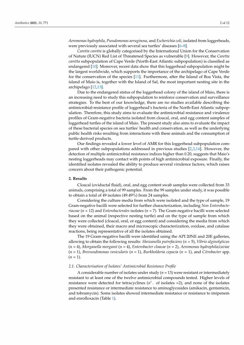
A considerable number of isolates under study (n = 13) were resistant or intermediatelyresistant to at least one of the twelve antimicrobial compounds tested. Higher levels ofresistance were detected for tetracyclines (no . of isolates =2), and none of the isolatespresented resistance or intermediate resistance to aminoglycosides (amikacin, gentamicin,and tobramycin). Some isolates showed intermediate resistance or resistance to imipenemand enrofloxacin (Table 1).
Antibiotics 2021, 10, 771 3 of 12
Table 1. Antimicrobial resistance of bacterial isolates from oral, cloacal, and egg content swab samplesof 33 loggerhead turtles.
AntimicrobialClass
Antimicrobial Compound(Dose)
Number of Isolates Tested
Susceptible Intermediate Resistant
Amikacin (30 µg) 19 0 0Aminoglycosides Gentamicin (120 µg) 19 0 0
Tobramycin (10 µg) 19 0 0
Carbapenems Meropenem (10 µg) 18 1 0Imipenem (10 µg) 13 6 0
Cephalosporins Cefoperazone (75 µg) 17 1 1Ceftazidime (30 µg) 18 1 0
FluoroquinolonesCiprofloxacin (5 µg) 18 1 0Enrofloxacin (5 µg) 16 2 1
Ofloxacin (5 µg) 19 0 0Tetracyclines Tetracycline (30 µg) 17 0 2
Ureidopenicillins Piperacillin (100 µg) 18 0 1
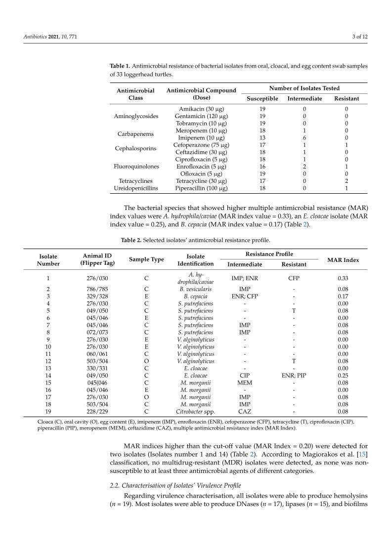
The bacterial species that showed higher multiple antimicrobial resistance (MAR)index values were A. hydrophila/caviae (MAR index value = 0.33), an E. cloacae isolate (MARindex value = 0.25), and B. cepacia (MAR index value = 0.17) (Table 2).
Table 2. Selected isolates’ antimicrobial resistance profile.
IsolateNumber
Animal ID(Flipper Tag) Sample Type Isolate
Identification
Resistance ProfileMAR Index
Intermediate Resistant
1 276/030 C A. hy-drophila/caviae IMP; ENR CFP 0.33
2 786/785 C B. vesicularis IMP - 0.083 329/328 E B. cepacia ENR; CFP - 0.174 276/030 C S. putrefaciens - - 0.005 049/050 C S. putrefaciens - T 0.086 045/046 E S. putrefaciens - - 0.007 045/046 C S. putrefaciens IMP - 0.088 072/073 C S. putrefaciens IMP - 0.089 276/030 E V. alginolyticus - - 0.0010 276/030 E V. alginolyticus - - 0.0011 060/061 C V. alginolyticus - - 0.0012 503/504 O V. alginolyticus - T 0.0813 330/331 C E. cloacae - - 0.0014 049/050 C E. cloacae CIP ENR; PIP 0.2515 045(046 C M. morganii MEM - 0.0816 045/046 E M. morganii - - 0.0017 276/030 O M. morganii IMP - 0.0818 503/504 C M. morganii IMP - 0.0819 228/229 C Citrobacter spp. CAZ - 0.08
Cloaca (C), oral cavity (O), egg content (E), imipenem (IMP), enrofloxacin (ENR), cefoperazone (CFP), tetracycline (T), ciprofloxacin (CIP),piperacillin (PIP), meropenem (MEM), ceftazidime (CAZ), multiple antimicrobial resistance index (MAR Index).
MAR indices higher than the cut-off value (MAR Index = 0.20) were detected fortwo isolates (Isolates number 1 and 14) (Table 2). According to Magiorakos et al. [15]classification, no multidrug-resistant (MDR) isolates were detected, as none was non-susceptible to at least three antimicrobial agents of different categories.
2.2. Characterisation of Isolates’ Virulence Profile
Regarding virulence characterisation, all isolates were able to produce hemolysins(n = 19). Most isolates were able to produce DNases (n = 17), lipases (n = 15), and biofilms
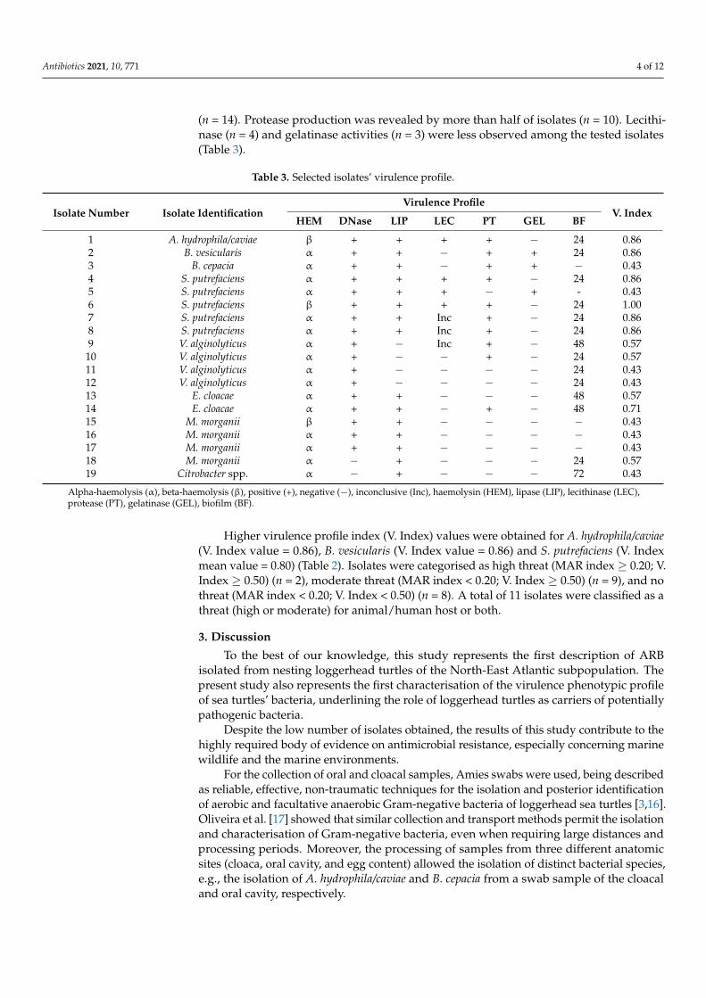
Antibiotics 2021, 10, 771 4 of 12
(n = 14). Protease production was revealed by more than half of isolates (n = 10). Lecithi-nase (n = 4) and gelatinase activities (n = 3) were less observed among the tested isolates(Table 3).
Table 3. Selected isolates’ virulence profile.
Isolate Number Isolate IdentificationVirulence Profile
V. IndexHEM DNase LIP LEC PT GEL BF
1 A. hydrophila/caviae β + + + + − 24 0.862 B. vesicularis α + + − + + 24 0.863 B. cepacia α + + − + + − 0.434 S. putrefaciens α + + + + − 24 0.865 S. putrefaciens α + + + − + - 0.436 S. putrefaciens β + + + + − 24 1.007 S. putrefaciens α + + Inc + − 24 0.868 S. putrefaciens α + + Inc + − 24 0.869 V. alginolyticus α + − Inc + − 48 0.57
10 V. alginolyticus α + − − + − 24 0.5711 V. alginolyticus α + − − − − 24 0.4312 V. alginolyticus α + − − − − 24 0.4313 E. cloacae α + + − − − 48 0.5714 E. cloacae α + + − + − 48 0.7115 M. morganii β + + − − − − 0.4316 M. morganii α + + − − − − 0.4317 M. morganii α + + − − − − 0.4318 M. morganii α − + − − − 24 0.5719 Citrobacter spp. α − + − − − 72 0.43
Alpha-haemolysis (α), beta-haemolysis (β), positive (+), negative (−), inconclusive (Inc), haemolysin (HEM), lipase (LIP), lecithinase (LEC),protease (PT), gelatinase (GEL), biofilm (BF).
Higher virulence profile index (V. Index) values were obtained for A. hydrophila/caviae(V. Index value = 0.86), B. vesicularis (V. Index value = 0.86) and S. putrefaciens (V. Indexmean value = 0.80) (Table 2). Isolates were categorised as high threat (MAR index ≥ 0.20; V.Index ≥ 0.50) (n = 2), moderate threat (MAR index < 0.20; V. Index ≥ 0.50) (n = 9), and nothreat (MAR index < 0.20; V. Index < 0.50) (n = 8). A total of 11 isolates were classified as athreat (high or moderate) for animal/human host or both.
3. Discussion
To the best of our knowledge, this study represents the first description of ARBisolated from nesting loggerhead turtles of the North-East Atlantic subpopulation. Thepresent study also represents the first characterisation of the virulence phenotypic profileof sea turtles’ bacteria, underlining the role of loggerhead turtles as carriers of potentiallypathogenic bacteria.
Despite the low number of isolates obtained, the results of this study contribute to thehighly required body of evidence on antimicrobial resistance, especially concerning marinewildlife and the marine environments.
For the collection of oral and cloacal samples, Amies swabs were used, being describedas reliable, effective, non-traumatic techniques for the isolation and posterior identificationof aerobic and facultative anaerobic Gram-negative bacteria of loggerhead sea turtles [3,16].Oliveira et al. [17] showed that similar collection and transport methods permit the isolationand characterisation of Gram-negative bacteria, even when requiring large distances andprocessing periods. Moreover, the processing of samples from three different anatomicsites (cloaca, oral cavity, and egg content) allowed the isolation of distinct bacterial species,e.g., the isolation of A. hydrophila/caviae and B. cepacia from a swab sample of the cloacaland oral cavity, respectively.

Antibiotics 2021, 10, 771 5 of 12
In the present study, S. putrefaciens was the most prevalent species found in Carettacaretta, as previously reported by Blasi et al. [18]. Unexpectedly, no Pseudomonas spp.isolates were detected, even though P. aeruginosa was one of the most prevalent speciesisolated from sea turtles in previous studies [4,19]. This finding may emphasise theimportance of monitoring AMR in distinct bacterial species regarding the loggerheadsubpopulation under study.
All identified bacterial species have been previously isolated from both injured andstranded sea turtles, as well as healthy wild animals [8,18,20], except for B. vesicularis, whichwas isolated for the first time from the oviductal fluid of sea turtles in the present study.
A. hydrophila/caviae, B. cepacia., V. alginolyticus and Citrobacter spp. isolated in thisstudy were previously associated with diseases of loggerheads and other sea turtle’sspecies, including ulcerative stomatitis, ulcerative esophagitis, granulomatous hepatitis,granulomatous nephritis, bronchopneumonia, conjunctivitis, and septicaemia [6,8].
B. cepacia, S. putrefaciens and V. alginolyticus, isolated from egg samples in this study,were previously associated with unhatched eggs [21–23]. Craven et al. [22] suggestedthat opportunist pathogens found in adult females could infect sea turtle eggs and causeembryonic mortality. Therefore, the presence of these bacterial species in the egg content ofMaio’s loggerheads may represent a potential threat to successful embryonic developmentand the overall reproductive success of this loggerhead colony.
Despite the small number of isolates, the level of AMR detected for the isolates in thisstudy is considerably lower compared with previous studies [2–4,24]. No MDR bacteriawere detected, which is in line with a previous study performed in juvenile hawksbill seaturtles (Eretmochelys imbricata) and green turtles (Chelonia mydas) from potential coincidentfeeding grounds [4], but discordant with previous studies in other loggerhead and greenturtles’ populations [2,3,14,24].
Following previous studies, higher resistance levels were observed for tetracyclinesand lower ones for the aminoglycoside class [2,3,14,17,24]. To the best of our knowledge,no resistance to imipenem was previously described for loggerhead sea turtles’ bacteria.Here, A. hydrophila/caviae, B. vesicularis, S. putrefaciens, and M. morganii presented interme-diate resistance to imipenem, with M. morganii also showing intermediate resistance tomeropenem. Regardless of the low incidence of resistance to carbapenems, this findingshould be further assessed due to the categorisation of these antimicrobial compounds aslast resort options for the treatment of serious Gram-negative infections, being of majorimportance for human medicine [25].
Sea turtles living in ecosystems affected by humans’ activities are at higher risk ofbeing exposed to antimicrobial environmental pressure [14,26]. The island of Maio is mostlycharacterised by a pristine environment, being less affected by anthropogenic impacts,such as the discharge of wastewater carrying high levels of antimicrobials, associated withaquaculture, intensive farms, and medical facilities [27,28].
The lower anthropogenic influence observed in the island of Maio may explain thelower levels of AMR in this loggerhead subpopulation compared with other subpopulationsaddressed in previous studies [2,3,14]. However, the detection of isolates with MAR indicesequal to or higher than 0.20 from animals not directly exposed to antimicrobial compoundssuggests previous contact to points of high antimicrobial exposure [29]. Therefore, althoughtheir nesting sites are less exposed to anthropogenic impacts, Maio’s loggerheads maynot be fully protected from antimicrobial environmental pressure. Moreover, the highlymigratory nature of sea turtles may expose them to a broad range of marine environments,promoting contact with sources of contamination.
Virulence characterisation showed that the isolates from this study could expressvirulence traits that may contribute to the evasion of the host immune system and hosttissue colonisation and damage [30]. Moreover, most isolates showed the ability to producebiofilms. Biofilm synthesis is one of the most important virulence factors in bacteria,playing a prominent role in AMR. In fact, the antimicrobials concentration required toeliminate bacterial biofilms can be up to 1000-fold higher in comparison with their free-
Antibiotics 2021, 10, 771 6 of 12
swimming, planktonic counterparts, which makes these microbial communities extremelydifficult to control [31,32]. The expression of a high number of virulence factors may playan essential role in the pathogenesis of infections [30].
Considering Singh et al. [33] classification, a considerable number of isolates (n =11) in this study were considered a high threat (MAR Index ≥ 0.20; V. Index ≥ 0.50) ormoderate threat (MAR Index < 0.20; V. Index ≥ 0.50) to human/animal host or both. Thesefindings suggest that the isolates obtained from the loggerhead turtles under study canpose a threat as potential pathogens, especially those revealing high virulence indices, suchas A. hydrophila/caviae, B. vesicularis, and S. putrefaciens.
A. hydrophila/caviae, V. alginolyticus, M. morganii, S. putrefaciens, and Brevundimonas spp.,isolated in this study, were previously associated with infections in humans, including skinand soft tissue infections, ear and wound infections, urinary tract infections, gastroenteritis,neonatal sepsis, and septicaemia [34–38].
Despite the existent conservation efforts, the slaughter of nesting loggerheads forconsumption is frequently practised in the island of Maio [11]. The consumption of turtle-related products represents a risk behaviour for public health, which is supported bythe detection of pathogenic and antimicrobial-resistant bacteria isolated from loggerheadturtles in this study.
In conclusion, the lower levels of AMR detected for the Cape Verdean loggerheadsubpopulation, compared with the levels identified for other subpopulations, representpositive and encouraging results regarding the current context of AMR. However, thepresence of potentially pathogenic Gram-negative bacteria expressing several virulencefactors may represent a risk to sea turtles’ health and consequently affect the conservationof this endangered species. Finally, due to their pathogenic potential, the identified bacteriamay represent a significant threat to public health. This threat arises mainly through theunsafe and illegal consumption of turtle-derived products.
Further studies are encouraged to characterise the identified bacterial species at themolecular level, as well as to assess the genetic determinants imparting AMR and virulence.It would be equally relevant to further study the resistance mechanisms involved in theisolates’ antimicrobial resistance profile, especially concerning resistance to carbapenems.
4. Materials and Methods4.1. Area of Study
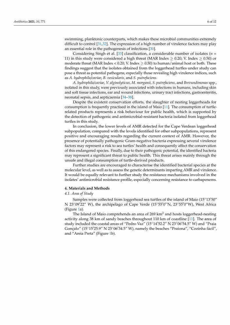
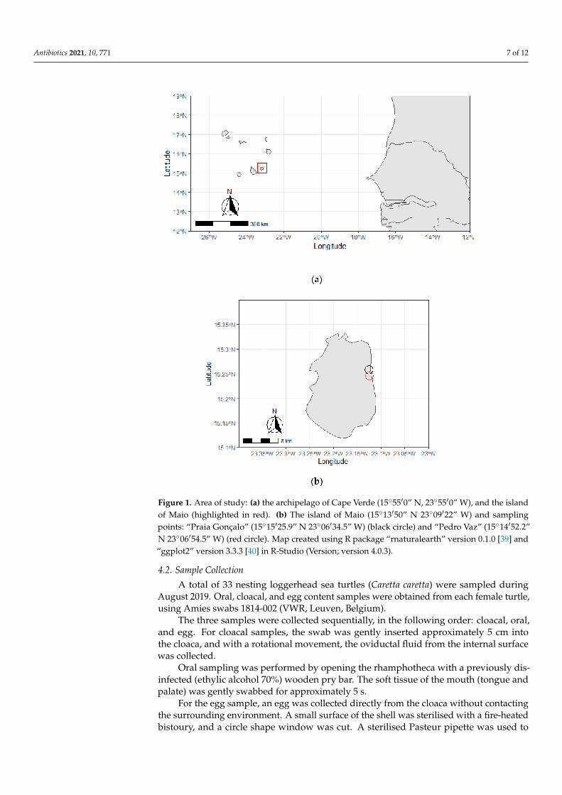
Samples were collected from loggerhead sea turtles of the island of Maio (15◦13′50”N 23◦09′22” W), the archipelago of Cape Verde (15◦55′0′’N, 23◦55′0”W), West Africa(Figure 1a).
The Island of Maio comprehends an area of 269 km2 and hosts loggerhead-nestingactivity along 38 km of sandy beaches throughout 110 km of coastline [11]. The area ofstudy included the coastal areas of “Pedro Vaz” (15◦14′52.2” N 23◦06′54.5” W) and “PraiaGonçalo” (15◦15′25.9” N 23◦06′34.5” W), namely the beaches “Praiona”, “Cozinha fácil”,and “Areia Preta” (Figure 1b).
Antibiotics 2021, 10, 771 7 of 12
Figure 1. Area of study: (a) the archipelago of Cape Verde (15◦55′0” N, 23◦55′0” W), and the islandof Maio (highlighted in red). (b) The island of Maio (15◦13′50” N 23◦09′22” W) and samplingpoints: “Praia Gonçalo” (15◦15′25.9” N 23◦06′34.5” W) (black circle) and “Pedro Vaz” (15◦14′52.2”N 23◦06′54.5” W) (red circle). Map created using R package “rnaturalearth” version 0.1.0 [39] and“ggplot2” version 3.3.3 [40] in R-Studio (Version; version 4.0.3).
4.2. Sample Collection
A total of 33 nesting loggerhead sea turtles (Caretta caretta) were sampled duringAugust 2019. Oral, cloacal, and egg content samples were obtained from each female turtle,using Amies swabs 1814-002 (VWR, Leuven, Belgium).
The three samples were collected sequentially, in the following order: cloacal, oral,and egg. For cloacal samples, the swab was gently inserted approximately 5 cm intothe cloaca, and with a rotational movement, the oviductal fluid from the internal surfacewas collected.
Oral sampling was performed by opening the rhamphotheca with a previously dis-infected (ethylic alcohol 70%) wooden pry bar. The soft tissue of the mouth (tongue andpalate) was gently swabbed for approximately 5 s.
For the egg sample, an egg was collected directly from the cloaca without contactingthe surrounding environment. A small surface of the shell was sterilised with a fire-heatedbistoury, and a circle shape window was cut. A sterilised Pasteur pipette was used to

Antibiotics 2021, 10, 771 8 of 12
collect approximately 1.5 mL of the egg content (yolk and albumen), which was introduceddirectly in the transport medium of the Amies swabs. The samples were identified withdate, time, type of sample, and flipper tag number and then safely placed in a thermic bagat 4 ◦C.
After the sampling period, the collected samples were transported to the Microbiologyand Immunology Laboratory of the Veterinary Faculty, University of Lisbon, Portugal, forfurther processing. The following information was collected regarding each sample fromeach animal: date and time of sampling, local of sample collection, flipper tag identificationnumber, Passive Integrated Transponder (PIT) number (when available), and type ofsample (cloaca, oral cavity, and egg content) (See Supplementary Table S1). The handlingtime did not exceed 5 min, before and after which the animals were observed from a safedistance to ensure that oviposition proceeded normally.
The collection of samples was conducted under Maio Biodiversity Foundation guide-lines and by the permits of the Environmental National Authority DNA (Direção Nacionaldo Ambiente). Research protocols were performed per the IUCN Policy Statement onResearch Involving Species at Risk of Extinction [41] approved by the 27th Meeting ofIUCN Council, Gland Switzerland, 14 June 1989, and the Sea Turtle Research TechniquesManual [42].
4.3. Isolation and Identification of Gram-Negative Bacteria
After pre-enrichment in Buffered Peptone Water (Oxoid, Basingstoke, UK) at 37 ◦Cfor 24 h, aerobic and facultative anaerobic Gram-negative bacteria were isolated fromcollected samples using Glutamate Starch Red Phenol (GSP) Agar plates supplementedwith 100,000 UI penicillin g/L (Merck, Darmstadt, Germany) and MacConkey Agar (Oxoid,Basingstoke, UK), incubated at 37 ◦C for 24–48 h [2]. Positive bacterial colonies wereisolated in Columbia agar supplemented with 5% sheep blood (bioMérieux, Marcy-l’Étoile,France). Isolates were characterised regarding their macro and microscopic morphology,Gram staining, catalase test and oxidase reaction. For further characterisation, 19 Gram-negative bacilli were selected and identified through the biochemical identification galleriesAPI 20E and 20NE (bioMérieux, Marcy-l’Étoile, France), according to the manufacturer’sinstructions.
4.4. Evaluation of Isolates’ Antimicrobial Resistance Profile
Isolates’ susceptibility profiles regarding 12 different antimicrobials commonly andglobally used in veterinary and human medicine belonging to distinct classes were deter-mined using the disk diffusion method according to the Clinical and Laboratory StandardsInstitute (CLSI) guidelines [43]. The tested antibiotics (Oxoid, Basingstoke, Hamp, UK,and Mast Group, Bootle, UK) were: amikacin (AK, 30 µg), cefoperazone (CFP, 75 µg),ceftazidime (CAZ, 30 µg), ciprofloxacin (CIP, 5 µg), enrofloxacin (ENR, 5 µg), gentamicin(GM, 120 µg), imipenem (IMP, 10 µg), meropenem (MEM, 10 µg), ofloxacin (OFX, 5 µg),piperacillin (PIP, 100 µg), tetracycline (T, 30 µg), and tobramycin (TOB, 10 µg), as describedelsewhere [2,44]. The reference strains Escherichia coli ATCC® 25922TM and Pseudomonasaeruginosa ATCC® 27853 TM were used as quality control. The inhibition zones were mea-sured, and isolates were scored as susceptible, intermediate, and resistant, according tothe CLSI guidelines [43]. Intrinsic resistances were taken into consideration, according toMagiorakos et al. [15]. A 10% replica was performed in independent days, by repeating theantimicrobial susceptibility testing of 10% randomly selected isolates [43].
Multiple antimicrobial resistance (MAR) indices were calculated for the selectedisolates as follows: no . antimicrobials to which isolates were resistant/no . antimicrobialstested. A MAR index equal or greater than 0.20 was used as a cut-off to differentiatebetween high and low-risk contamination [29].
Antibiotics 2021, 10, 771 9 of 12
4.5. Evaluation of Isolates’ Virulence Profile
Isolates were characterised regarding their phenotypic virulence profile by assessingthe production of enzymes associated with bacterial pathogenic potential.
Hemolysins production was evaluated using Columbia Agar with 5% sheep blood [30].DNase activity was assessed using DNase Agar supplemented with 0.005% methyl
green (VWR, Leuven, Belgium), using Aeromonas hydrophila ATCC® 7966TM and Escherichiacoli ATCC® 25922TM as positive and negative controls, respectively [45].
Lecithinase activity was determined using Tryptic Soy Agar (VWR, Leuven, Belgium),supplemented with 10% egg yolk emulsion (VWR, Leuven, Belgium). Positive and negativecontrols, Pseudomonas aeruginosa ATCC® 27853TM and Escherichia coli ATCC® 25922TM wereused, respectively [46].
Gelatinase activity was detected using Nutrient Gelatin Agar (Oxoid, Basingstoke,UK), using Pseudomonas aeruginosa ATCC® 27853TM and Escherichia coli ATCC® 25922TM aspositive and negative controls, respectively [30].
Biofilm production ability was assessed resorting to Congo Red Agar plates, composedof Brain Heart Infusion broth (VWR, Leuven, Belgium), Bacteriological Agar (VWR, Leuven,Belgium), and 0.0008% Congo Red indicator (Sigma-Aldrich, St. Louis, USA). Enterococcusfaecium ATCC® 35667TM and Escherichia coli ATCC® 25922TM were, respectively, used aspositive and negative controls [47,48].
Protease activity was analysed, resorting to Skim Milk Agar (Oxoid, Basingstoke, UK),using Pseudomonas aeruginosa ATCC® 27853TM and Staphylococcus aureus ATCC® 29213TM
as positive and negative controls, respectively [49].Lipase activity was tested using Spirit Blue Agar (Difco, Detroit, USA) supplemented
with 0.25% Tween® 80 (AppliChem GmbII, Darmstadt, Germany) and 25% olive oil (com-mercial), using Pseudomonas aeruginosa ATCC® 27853TM and Staphylococcus aureus ATCC®
29213TM as positive and negative controls, respectively [30]. For testing all virulence factors,the plates were incubated at 25 ◦C for 24 h.
The virulence indices were calculated for the tested isolates, as follows: no . positivevirulence factors/no . virulence factors tested [33]. A virulence index equal to or higher than0.50 was used as a cut-off to evaluate the threat levels for the selected isolates, regardingtheir pathogenic potential [33]. According to Singh et al. [33], isolates were categorised as ahigh threat-isolates with virulence and MAR indices greater than or equal to the cut-offvalues (MAR index ≥ 0.20, V. Index ≥ 0.50); moderate threat-isolates having virulenceindex ≥ 0.50 but MAR index < 0.20; and no threat-isolates having virulence and MARindices below the cut-off values.
Supplementary Materials: The following are available online at https://www.mdpi.com/article/10.3390/antibiotics10070771/s1, Table S1: Sampling data.
Author Contributions: Conceptualization, M.F., J.P.-M., and M.O.; methodology, M.F., M.L.G., E.C.,C.C., J.P.-M., M.O.; validation, M.F., L.T., J.P.-M., and M.O.; formal analysis, M.F.; investigation,M.F., M.L.G., J.P.-M., and M.O.; resources, M.F., J.P.-M., and M.O.; data curation, M.F. and M.O.;writing-original draft preparation, M.F.; writing—review and editing, M.F., M.L.G., E.C., L.T., J.P.-M.,and M.O.; visualization, M.F., J.P.-M., and M.O.; supervision, J.P.-M. and M.O.; project administration,M.O.; funding acquisition, M.F. and M.O. All authors have read and agreed to the published versionof the manuscript.
Funding: This research was funded by FCT—Foundation of Science and Technology, project UICB/00276/2020. This work was also supported by CIISA—Centre for Interdisciplinary Research inAnimal Health, Faculty of Veterinary Medicine, University of Lisbon. J.P.M. receives support fromthe MAVA Foundation pour la Nature and NOAA USFWS. The funding bodies were not involved inthe study design, data collection, analysis, and interpretation or in writing the manuscript.
Institutional Review Board Statement: This study was conducted according to the rules given bythe current EU (Directive 2010/63/EC) and national (DL 113/2013) legislation and by the competentauthority (Direção Geral de Alimentação e Veterinária, DGAV, https://www.dgav.pt/, accessedon 23 June 2021) in Portugal. Ethical review and approval were waived for this study, due to the
Antibiotics 2021, 10, 771 10 of 12
collection of only non-invasive samples under Maio Biodiversity Foundation guidelines and by thepermits of the Environmental National Authority DNA (Direção Nacional do Ambiente), of CapeVerde. Trained veterinarians obtained all the samples, following standard monitoring procedures.No animal experiment has been performed in the scope of this research.
Informed Consent Statement: Not applicable.
Data Availability Statement: Data are contained within the article or Supplementary Materials.
Acknowledgments: This research was supported by CIISA—Centro de Investigação Interdisci-plinar em Sanidade Animal, Faculdade de Medicina Veterinária, Universidade de Lisboa, ProjectUIDB/00276/2020 (funded by FCT—Fundação para a Ciência e Tecnologia IP). The authors wouldalso like to recognise the Foundation of Science and Technology for the PhD fellowship SFRH/BD/131384/2017 (Eva Cunha) and to the University of Lisbon for the PhD fellowship C10571K (Miguel L.Grilo). Finally, the authors acknowledge MAVA Foundation pour la Nature and NOAA USFWS forthe financial support of Juan Patino-Martinez.
Conflicts of Interest: The authors declare no conflict of interest.
References1. WHO Antimicrobial Resistance: Fact Sheets. Available online: https://www.who.int/news-room/fact-sheets/detail/
antimicrobial-resistance (accessed on 3 February 2021).2. Foti, M.; Giacopello, C.; Bottari, T.; Fisichella, V.; Rinaldo, D.; Mammina, C. Antibiotic Resistance of Gram Negatives Isolates from
Loggerhead Sea Turtles (Caretta caretta) in the Central Mediterranean Sea. Mar. Pollut. Bull. 2009, 58, 1363–1366. [CrossRef]3. Pace, A.; Dipineto, L.; Fioretti, A.; Hochscheid, S. Loggerhead Sea Turtles as Sentinels in the Western Mediterranean: Antibiotic
Resistance and Environment-Related Modi Fi Cations of Gram-Negative Bacteria. Mar. Pollut. Bull. 2019, 149, 110575. [CrossRef][PubMed]
4. Oliveira, M.; Serrano, I.; Santos, J.P.; Bilocq, F.; Pereira, N.; de Santos Loureiro, N.; Tavares, L.; Pirnay, J.P.; De Vos, D. Pseudomon-ads from Wild Free-Living Sea Turtles in Príncipe Island, Gulf of Guinea. Ecol. Indic. 2017, 81, 260–264. [CrossRef]
5. Mashkour, N.; Jones, K.; Kophamel, S.; Hipolito, T.; Ahasan, S.; Walker, G.; Jakob-Hoff, R.; Whittaker, M.; Hamann, M.;Bell, I.; et al. Disease Risk Analysis in Sea Turtles: A Baseline Study to Inform Conservation Efforts. PLoS ONE 2020, 15, 1–32.[CrossRef] [PubMed]
6. Glazebrook, J.S.; Campbell, R.S.F. A Survey of the Diseases of Marine Turtles in Northern Australia. I. Farmed Turtles. Dis. Aquat.Org. 1990, 9, 83–95. [CrossRef]
7. Pace, A.; Rinaldi, L.; Ianniello, D.; Borrelli, L.; Cringoli, G.; Fioretti, A.; Hochscheid, S.; Dipineto, L. Gastrointestinal Investigationof Parasites and Enterobacteriaceae in Loggerhead Sea Turtles from Italian Coasts. BMC Vet. Res. 2019, 15, 1–9. [CrossRef]
8. Orós, J.; Torrent, A.; Calabuig, P.; Déniz, S. Diseases and Causes of Mortality among Sea Turtles Stranded in the Canary Islands,Spain (1998-2001). Dis. Aquat. Org. 2005, 63, 13–24. [CrossRef]
9. Casale, P.; Marco, A. Caretta caretta (North East Atlantic Subpopulation). In The IUCN Red List of Threatened Species; 2015:e.T83776383A83776554; International Union for Conservation of Nature: Gland, Switzerland, 2015; Volume 8235.
10. Casale, P.; Tucker, A.D. Caretta caretta (Amended Version of 2015 Assessment) Supplementary Material. In IUCN Red ListThreatened Species; International Union for Conservation of Nature: Gland, Switzerland, 2017; p. 7. [CrossRef]
11. Patino-martinez, J.; Passos, L.D.O.S.; Afonso, I.; Teixidor, A.; Maio Biodiversity Foundation, Cidade Porto-Inglês, 6110 Ilha doMaio, Cabo Verde. Globally Important Loggerhead Sea Turtle Conservation Refuge: Maio Island, Cabo Verde. Unpublishedwork. 2021.
12. Martins, S.; Soares, F.; Ribeiro, E.; Abella, E.; Koenen, F.; Marco, A. Importance of the Island of Maio (Cape Verde) for Current andFuture Loggerhead Conservation in the Eastern Atlantic. In Proceedings of the Thirty-Third Annual Symposium on Sea TurtleBiology and Conservation: 2013 International Sea Turtle Symposium, Baltimore, MD, USA, 5–8 February 2013; p. 106.
13. Laloë, J.O.; Cozens, J.; Renom, B.; Taxonera, A.; Hays, G.C. Conservation Importance of Previously Undescribed AbundanceTrends: Increase in Loggerhead Turtle Numbers Nesting on an Atlantic Island. Oryx 2020, 54, 315–322. [CrossRef]
14. Ahasan, M.S.; Picard, J.; Elliott, L.; Kinobe, R.; Owens, L.; Ariel, E. Evidence of Antibiotic Resistance in Enterobacteriales Isolatedfrom Green Sea Turtles, Chelonia Mydas on the Great Barrier Reef. Mar. Pollut. Bull. 2017, 120, 18–27. [CrossRef]
15. Magiorakos, A.P.; Srinivasan, A.; Carey, R.B.; Carmeli, Y.; Falagas, M.E.; Giske, C.G.; Harbarth, S.; Hindler, J.F.; Kahlmeter, G.;Olsson-Liljequist, B.; et al. Multidrug-Resistant, Extensively Drug-Resistant and Pandrug-Resistant Bacteria: An InternationalExpert Proposal for Interim Standard Definitions for Acquired Resistance. Clin. Microbiol. Infect. 2012, 18, 268–281. [CrossRef]
16. Lanci, A.K.J.; Roden, S.E.; Bowman, A.; Lacasella, E.L.; Frey, A.; Dutton, P.H. Evaluating Buccal and Cloacal Swabs for Ease ofCollection and Use in Genetic Analyses of Marine Turtles. Chelonian Conserv. Biol. 2012, 11, 144–148. [CrossRef]
17. Oliveira, M.; Monteiro, J.L.; Rana, S.; Vilela, C.L. Antimicrobial Resistance in Gram-Positive Bacteria from Timorese River Buffalo(Bubalus bubalis) Skin Microbiota. Trop. Anim. Health Prod. 2010, 42, 833–839. [CrossRef]
18. Blasi, M.F.; Migliore, L.; Mattei, D.; Rotini, A.; Thaller, M.C.; Alduina, R. Antibiotic Resistance of Gram-Negative Bacteria fromWild Captured Loggerhead Sea Turtles. Antibiotics 2020, 9, 162. [CrossRef] [PubMed]
Antibiotics 2021, 10, 771 11 of 12
19. Al-Bahry, S.; Mahmoud, I.; Elshafie, A.; Al-Harthy, A.; Al-Ghafri, S.; Al-Amri, I.; Alkindi, A. Bacterial Flora and AntibioticResistance from Eggs of Green Turtles Chelonia Mydas: An Indication of Polluted Effluents. Mar. Pollut. Bull. 2009, 58, 720–725.[CrossRef]
20. Rodgers, M.L.; Toline, C.A.; Rice, C.D. Humoral Immune Responses to Select Marine Bacteria in Loggerhead Sea Turtles Carettacaretta and Kemp’s Ridley Sea Turtles Lepidochelys Kempii from the Southeastern United States. J. Aquat. Anim. Health 2018, 30,20–30. [CrossRef]
21. Wyneken, J.; Burke, T.J.; Salmon, M.; Pedersen, D.K. Egg Failure in Natural and Relocated Sea Turtle Nests. J. Herpetol. 1988, 22,88. [CrossRef]
22. Craven, K.S.; Awong-Taylor, J.; Griffiths, L.; Bass, C.; Muscarella, M. Identification of Bacterial Isolates from UnhatchedLoggerhead (Caretta caretta) Sea Turtle Eggs in Georgia, USA. Mar. Turt. Newsl. 2007, 115, 11, ISBN 2-525-04764-8.
23. Awong-Taylor, J.; Craven, K.S.; Griffiths, L.; Bass, C.; Muscarella, M. Comparison of Biochemical and Molecular Methods for theIdentification of Bacterial Isolates Associated with Failed Loggerhead Sea Turtle Eggs. J. Appl. Microbiol. 2008, 104, 1244–1251.[CrossRef]
24. Al-Bahry, S.N.; Mahmoud, I.Y.; Al-Zadjali, M.; Elshafie, A.; Al-Harthy, A.; Al-Alawi, W. Antibiotic Resistant Bacteria as Bio-Indicator of Polluted Effluent in the Green Turtles, Chelonia Mydas in Oman. Mar. Environ. Res. 2011, 71, 139–144. [CrossRef][PubMed]
25. Raza, A.; Ngieng, S.C.; Sime, F.B.; Cabot, P.J.; Roberts, J.A.; Popat, A.; Kumeria, T.; Falconer, J.R. Oral Meropenem for Superbugs:Challenges and Opportunities. Drug Discov. Today 2020, 26, 551–560. [CrossRef] [PubMed]
26. Arnold, K.E.; Williams, N.J.; Bennett, M. “Disperse Abroad in the Land”: The Role of Wildlife in the Dissemination of Antimicro-bial Resistance. Biol. Lett. 2016, 12. [CrossRef] [PubMed]
27. Shah, S.Q.A.; Cabello, F.C.; L’Abée-Lund, T.M.; Tomova, A.; Godfrey, H.P.; Buschmann, A.H.; Sørum, H. Antimicrobial Resistanceand Antimicrobial Resistance Genes in Marine Bacteria from Salmon Aquaculture and Non-Aquaculture Sites. Environ. Microbiol.2014, 16, 1310–1320. [CrossRef]
28. Le Quesne, W.J.F.; Baker-Austin, C.; Verner-Jeffreys, D.W.; Al-Sarawi, H.A.; Balkhy, H.H.; Lyons, B.P. Antimicrobial Resistance inthe Gulf Cooperation Council Region: A Proposed Framework to Assess Threats, Impacts and Mitigation Measures Associatedwith AMR in the Marine and Aquatic Environment. Environ. Int. 2018, 121, 1003–1010. [CrossRef]
29. Krumperman, P.H. Multiple Antibiotic Resistance Indexing of Escherichia Coli to Identify High-Risk Sources of Faecal Contami-nation of Water. Environ. Sci. Pollut. Res. 1983, 22, 10969–10980. [CrossRef]
30. Seixas, R.; Pissarra, H.; Santos, J.; Bernardino, R.; Fernandes, T.; Correia, J.; Vilela, C.L.; Oliveira, M. Severe FibrinonecroticEnteritis Caused by Pseudomonas aeruginosa in a Captive Monitor Lizard (Varanus niloticus). J. Zoo Wildl. Med. 2014, 45, 410–412.[CrossRef] [PubMed]
31. Mah, T.F.; Pitts, B.; Pellock, B.; Walker, G.C.; Stewart, P.S.; O’Toole, G.A. A Genetic Basis for Pseudomonas aeruginosa BiofilmAntibiotic Resistance. Nature 2003, 426, 306–310. [CrossRef] [PubMed]
32. Costerton, J.W.; Stewart, P.S.; Greenberg, E.P. Bacterial Biofilms: A Common Cause of Persistent Infections. Science 1999, 284,1318–1322. [CrossRef] [PubMed]
33. Singh, S.K.; Ekka, R.; Mishra, M.; Mohapatra, H. Association Study of Multiple Antibiotic Resistance and Virulence: A Strategyto Assess the Extent of Risk Posed by Bacterial Population in Aquatic Environment. Environ. Monit. Assess. 2017, 189, 320.[CrossRef] [PubMed]
34. Holt, H.M.; Gahrn-Hansen, B.; Bruun, B. Shewanella algae and Shewanella putrefaciens: Clinical and Microbiological Characteristics.Clin. Microbiol. Infect. 2005, 11, 347–352. [CrossRef]
35. American Academy of Pediatrics. Other vibrio infections. In Red Book: 2009 Report of the Committee on Infectious Diseases; Pickering,L., Baker, C., Kimberlin, D., Long, S., Eds.; American Academy of Pediatrics: Elk Grove Village, IL, USA, 2009; pp. 729–730.
36. Janda, J.M.; Abbott, S.L. The Genus Aeromonas: Taxonomy, Pathogenicity, and Infection. Clin. Microbiol. Rev. 2010, 23, 35–73.[CrossRef]
37. Chang, H.Y.; Wang, S.M.; Chiu, N.C.; Chung, H.Y.; Wang, H.K. Neonatal Morganella morganii Sepsis: A Case Report and Reviewof the Literature. Pediatrics Int. 2011, 53, 121–123. [CrossRef]
38. Ryan, M.P.; Pembroke, J.T. Brevundimonas spp: Emerging Global Opportunistic Pathogens. Virulence 2018, 9, 480–493. [CrossRef][PubMed]
39. South, A. Rnaturalearth: World Map Data from Natural Earth; R Package Version 0.1.0; The R Foundation: Vienna, Austria, 2017.40. Wickham, H. Ggplot2: Elegant Graphics for Data Analysis; Wiley: Hoboken, NJ, USA, 2016.41. IUCN. Policy Statement on Research Involving Species at Risk of Extinction. In Proceedings of the 27th Meeting of IUCN Council,
Gland, Switzerland, 14 June 1989.42. National Marine Fisheries Service (NMFS). Sea Turtle Research Techniques Manual; NOAA Technical Memorandum NMFS-SEFSC-
579; NMFS: Silver Spring, MD, USA, 2008; p. 92.43. CLSI. Performance Standards for Antimicrobial Disk and Dilution Susceptibility Tests for Bacteria Isolated From Animals, 5th ed.; Clinical
and Laboratory Standards Institute: Wayne, PA, USA, 2020.44. Serrano, I.; Oliveira, M.; Santos, J.P.; Bilocq, F.; Leitão, A.; Tavares, L.; Pirnay, J.P.; De Vos, D. Antimicrobial Resistance and
Genomic Rep-PCR Fingerprints of Pseudomonas aeruginosa Strains from Animals on the Background of the Global PopulationStructure. BMC Vet. Res. 2017, 13, 58. [CrossRef] [PubMed]
Antibiotics 2021, 10, 771 12 of 12
45. Elder, B.L.; Trujillo, I.; Blazevic, D.J. Rapid Deoxyribonuclease Test with Methyl Green. J. Clin. Microbiol. 1977, 6, 312–313.[CrossRef] [PubMed]
46. Chrisope, G.L.; Fox, C.W.; Marshall, R.T. Lecithin Agar for Detection of Microbial. Appl. Environ. Microbiol. 1976, 31, 784–786.[CrossRef]
47. Freeman, D.J.; Falkiner, F.R.; Keane, C.T. New Method for Detecting Slime Production by Coagulase Negative Staphylococci. J.Clin. Pathol. 1989, 42, 872–874. [CrossRef]
48. Rewatkar, A.R.; Wadher, B.J. Staphylococcus aureus and Pseudomonas aeruginosa- Biofilm Formation Methods. IOSR J. Pharm.Biol. Sci. 2013, 8, 36–40. [CrossRef]
49. Sokol, P.A.; Ohman, D.E.; Iglewski, B.H. A More Sensitive Plate Assay for Detection of Protease Production by Pseudomonasaeruginosa. J. Clin. Microbiol. 1979, 9, 538–540. [CrossRef]