Genomic Insights into Drug Resistance and Virulence ...
23
microorganisms Article Genomic Insights into Drug Resistance and Virulence Platforms, CRISPR-Cas Systems and Phylogeny of Commensal E. coli from Wildlife Carla Andrea Alonso 1,2,† , María de Toro 3,† , Fernando de la Cruz 4 and Carmen Torres 1, * Citation: Alonso, C.A.; de Toro, M.; de la Cruz, F.; Torres, C. Genomic Insights into Drug Resistance and Virulence Platforms, CRISPR-Cas Systems and Phylogeny of Commensal E. coli from Wildlife. Microorganisms 2021, 9, 999. https://doi.org/10.3390/ microorganisms9050999 Academic Editor: Vincenzo Scarlato Received: 22 March 2021 Accepted: 30 April 2021 Published: 5 May 2021 Publisher’s Note: MDPI stays neutral with regard to jurisdictional claims in published maps and institutional affil- iations. Copyright: © 2021 by the authors. Licensee MDPI, Basel, Switzerland. This article is an open access article distributed under the terms and conditions of the Creative Commons Attribution (CC BY) license (https:// creativecommons.org/licenses/by/ 4.0/). 1 Área de Bioquímica y Biología Molecular, Universidad de La Rioja, 26006 Logroño, Spain; [email protected] 2 Servicio de Microbiología, Hospital San Pedro, 26006 Logroño, Spain 3 Plataforma de Genómica y Bioinformática, Centro de Investigación Biomédica de La Rioja (CIBIR), 26006 Logroño, Spain; [email protected] 4 Departamento de Biología Molecular, Universidad de Cantabria and Instituto de Biomedicina y Biotecnología de Cantabria (Universidad de Cantabria-CSIC), 39011 Santander, Spain; [email protected] * Correspondence: [email protected]; Tel.: +34-941299750; Fax: +34-941299721 † These authors contributed equally to this work. Abstract: Commensal bacteria act as important reservoirs of virulence and resistance genes. How- ever, existing data are generally only focused on the analysis of human or human-related bacterial populations. There is a lack of genomic studies regarding commensal bacteria from hosts less exposed to antibiotics and other selective forces due to human activities, such as wildlife. In the present study, the genomes of thirty-eight E. coli strains from the gut of various wild animals were sequenced. The analysis of their accessory genome yielded a better understanding of the role of the mobilome on inter-bacterial dissemination of mosaic virulence and resistance plasmids. The study of the presence and composition of the CRISPR/Cas systems in E. coli from wild animals showed some viral and plasmid sequences among the spacers, as well as the relationship between CRISPR/Cas and E. coli phylogeny. Further, we constructed a single nucleotide polymorphisms-based core tree with E. coli strains from different sources (humans, livestock, food and extraintestinal environments). Bacteria from humans or highly human-influenced settings exhibit similar genetic patterns in CRISPR-Cas systems, plasmids or virulence/resistance genes-carrying modules. These observations, together with the absence of significant genetic changes in their core genome, suggest an ongoing flow of both mobile elements and E. coli lineages between human and natural ecosystems. Keywords: E. coli; CRISPR-Cas; antimicrobial resistance; wild animals; WGS; PLACNETw 1. Introduction Escherichia coli is a well-known commensal of the gut of humans and a wide-range of other animals, but it can also reproduce and persist for long periods of time in ex- traintestinal natural environments. Beside these habitats, some strains of E. coli have the potential to cause severe intestinal and extraintestinal illness, such as meningitis, sep- ticemia, pneumonia or urinary tract infections. This diversity in terms of niche distribution and host–pathogen interactions is due to the high plasticity of the E. coli genome, which allows the bacteria to adapt to the varying selective pressures exerted by the different environments. All this, plus the easy and fast growth characteristics of E. coli, makes this species an excellent model to study the evolution and epidemiology of antimicrobial resistance or bacterial virulence [1]. The dynamics of bacterial genomes involve both single mutations in the core genome (microevolution) as well as horizontal gene transfer events (macroevolution) that shape a species pangenome [2]. Both types of evolutionary phenomena have influenced the acquisition of new phenotypes and host/niche adaptation. Horizontal gene transfer (HGT) Microorganisms 2021, 9, 999. https://doi.org/10.3390/microorganisms9050999 https://www.mdpi.com/journal/microorganisms
-
Upload
khangminh22 -
Category
Documents
-
view
1 -
download
0
Transcript of Genomic Insights into Drug Resistance and Virulence ...

microorganisms
Article
Genomic Insights into Drug Resistance and VirulencePlatforms, CRISPR-Cas Systems and Phylogeny ofCommensal E. coli from Wildlife
Carla Andrea Alonso 1,2,† , María de Toro 3,†, Fernando de la Cruz 4 and Carmen Torres 1,*
�����������������
Citation: Alonso, C.A.; de Toro, M.;
de la Cruz, F.; Torres, C. Genomic
Insights into Drug Resistance and
Virulence Platforms, CRISPR-Cas
Systems and Phylogeny of
Commensal E. coli from Wildlife.
Microorganisms 2021, 9, 999.
https://doi.org/10.3390/
microorganisms9050999
Academic Editor: Vincenzo Scarlato
Received: 22 March 2021
Accepted: 30 April 2021
Published: 5 May 2021
Publisher’s Note: MDPI stays neutral
with regard to jurisdictional claims in
published maps and institutional affil-
iations.
Copyright: © 2021 by the authors.
Licensee MDPI, Basel, Switzerland.
This article is an open access article
distributed under the terms and
conditions of the Creative Commons
Attribution (CC BY) license (https://
creativecommons.org/licenses/by/
4.0/).
1 Área de Bioquímica y Biología Molecular, Universidad de La Rioja, 26006 Logroño, Spain;[email protected]
2 Servicio de Microbiología, Hospital San Pedro, 26006 Logroño, Spain3 Plataforma de Genómica y Bioinformática, Centro de Investigación Biomédica de La Rioja (CIBIR),
26006 Logroño, Spain; [email protected] Departamento de Biología Molecular, Universidad de Cantabria and Instituto de Biomedicina y Biotecnología
de Cantabria (Universidad de Cantabria-CSIC), 39011 Santander, Spain; [email protected]* Correspondence: [email protected]; Tel.: +34-941299750; Fax: +34-941299721† These authors contributed equally to this work.
Abstract: Commensal bacteria act as important reservoirs of virulence and resistance genes. How-ever, existing data are generally only focused on the analysis of human or human-related bacterialpopulations. There is a lack of genomic studies regarding commensal bacteria from hosts less exposedto antibiotics and other selective forces due to human activities, such as wildlife. In the present study,the genomes of thirty-eight E. coli strains from the gut of various wild animals were sequenced. Theanalysis of their accessory genome yielded a better understanding of the role of the mobilome oninter-bacterial dissemination of mosaic virulence and resistance plasmids. The study of the presenceand composition of the CRISPR/Cas systems in E. coli from wild animals showed some viral andplasmid sequences among the spacers, as well as the relationship between CRISPR/Cas and E. coliphylogeny. Further, we constructed a single nucleotide polymorphisms-based core tree with E. colistrains from different sources (humans, livestock, food and extraintestinal environments). Bacteriafrom humans or highly human-influenced settings exhibit similar genetic patterns in CRISPR-Cassystems, plasmids or virulence/resistance genes-carrying modules. These observations, togetherwith the absence of significant genetic changes in their core genome, suggest an ongoing flow of bothmobile elements and E. coli lineages between human and natural ecosystems.
Keywords: E. coli; CRISPR-Cas; antimicrobial resistance; wild animals; WGS; PLACNETw
1. Introduction
Escherichia coli is a well-known commensal of the gut of humans and a wide-rangeof other animals, but it can also reproduce and persist for long periods of time in ex-traintestinal natural environments. Beside these habitats, some strains of E. coli have thepotential to cause severe intestinal and extraintestinal illness, such as meningitis, sep-ticemia, pneumonia or urinary tract infections. This diversity in terms of niche distributionand host–pathogen interactions is due to the high plasticity of the E. coli genome, whichallows the bacteria to adapt to the varying selective pressures exerted by the differentenvironments. All this, plus the easy and fast growth characteristics of E. coli, makesthis species an excellent model to study the evolution and epidemiology of antimicrobialresistance or bacterial virulence [1].
The dynamics of bacterial genomes involve both single mutations in the core genome(microevolution) as well as horizontal gene transfer events (macroevolution) that shapea species pangenome [2]. Both types of evolutionary phenomena have influenced theacquisition of new phenotypes and host/niche adaptation. Horizontal gene transfer (HGT)
Microorganisms 2021, 9, 999. https://doi.org/10.3390/microorganisms9050999 https://www.mdpi.com/journal/microorganisms

Microorganisms 2021, 9, 999 2 of 23
is largely mediated by mobile genetic elements (MGEs) such as insertion sequences (ISs),transposons, integrons, prophages, integrative conjugative elements, genomic islands andplasmids. Human activities promote the use of selective agents (antibiotics, disinfectants,heavy metals) that induce a bacterial SOS response and, thus, increase HGT rates [3]. Thisleads to the assembly of mosaic genetic elements and the development of antimicrobialand/or virulence platforms of increasing complexity. Besides the selective pressure exertedby chemicals, the genetic background of bacteria also seems to play a role in the antimicro-bial resistance phenotype. As an example, the strong correlation observed between E. coliphylogroup B2 and antimicrobial susceptibility patterns [4].
Classically, PCR-based genotypic tests and other traditional typing methods (serotyp-ing, multilocus enzyme electrophoresis, pulsed-field gel electrophoresis, multilocus se-quence typing and phylogrouping multiplex PCR) were used to ascertain the geneticrelatedness and transmission dynamics of bacteria [5]. The advent of whole genomesequencing (WGS) technology provides a finer-scale analysis tool that enables the highresolution detection of antimicrobial resistance and virulence determinants, the analysisof genome structural variations and genetic linkages or the identification of potentialadaptations of the bacterial genomes to the host conditions, among others. Since mostgenome sequencing projects focus on the analysis of multi-drug resistant clones, clini-cally relevant pathogens or epidemiological outbreaks, there is a lack of genomic data oncommensal E. coli populations, even more so in those non-directly associated to humans.The study of randomly selected microbial communities, less exposed to antibiotics andother selective forces associated with the human environment, could provide new insightsinto the evolution of E. coli genomes and the flow of clinically relevant genes amongdifferent ecosystems.
In the present work, a collection of 38 E. coli genomes from the gut of various wildanimals were sequenced. Analysis of their accessory genome yielded a better understandingof the role of the mobilome on inter-bacterial dissemination of mosaic virulence and resis-tance platforms. The study of the presence and composition of the CRISPR/Cas systems inE. coli from wild animals showed some phage- and plasmid-related sequences among thespacers, as well as the relationship between CRISPR/Cas and E. coli phylogeny. Further,we constructed a single nucleotide polymorphism (SNP)-based core tree with E. coli strainsfrom different origins (humans, livestock, food and extraintestinal environments) in orderto elucidate whether the commensal population of wild animals is subjected to a parallelindependent microevolution or, on the contrary, an on-going inter-host transmission ismore likely occurring.
2. Materials and Methods2.1. Bacterial Collection, Antibiotic Susceptibility Testing and Phylotyping
In this work, 38 E. coli strains randomly selected among those previously isolated andidentified as part of other studies [6–9] were subjected to sequencing. The basic genomicfeatures of these 38 genomes are included in Supplementary Materials Table S1. Thestrains were isolated from fecal specimens (or intestine segments with fecal content inthe case of wild boars) collected between 2013 and 2015 in various geographic locationsof Spain (Castilla-La Mancha, Cádiz, Aragón) from healthy wild animals as previouslydescribed [6–9]. Eleven strains were isolated from red deer (Cervus elaphus), 11 from wildboars (Sus scrofa), 9 from small rodents ((mice, n = 8 (Apodemus sylvaticus and Mus musculus);rats, n = 1 (Rattus rattus)), and 7 from birds of prey (eagles, n = 2 (Aquila chrysaetos andCircaetus gallicus); vultures, n = 4 (Gyps fulvus); osprey, n = 1 (Gypaetus barbatus)). Intestinesegments with fecal content were collected from wild boars legally hunted in their ownhabitat during the regular hunting season (October to February 2014–2015) approved bythe Consejería de Agricultura of Castilla-La Mancha [8]; no approval was needed from anethical committee since the sacrifice of animals was not performed for research purposes,following the Spanish Policy for Animal Protection RD53/2013 and the European UnionDirective 2010/63.

Microorganisms 2021, 9, 999 3 of 23
All isolates were tested for antimicrobial susceptibility with ampicillin, amoxicillin/clavulanate, ceftazidime, ceftriaxone, cefoxitin, imipenem, nalidixic acid, ciprofloxacin,gentamicin, amikacin, tobramycin, streptomycin, chloramphenicol, sulfonamides, trimetho-prim/sulfamethoxazole and tetracycline, using the CLSI disk-diffusion method. Identifica-tion of the E. coli phylogroups was analyzed by a multiplex PCR-based assay [10], followedby in silico validation.
For comparative phylogenomic analysis, another 242 E. coli full genomes were takenfrom the NCBI database. These belonged to bacteria from different sources: humans,livestock, wild animals and the environment. Information about all these genomes isincluded in Supplementary Materials Table S2.
2.2. Sequencing of the E. coli Genomes
Genomic DNA from E. coli strains was extracted using the QIAmp DNA Mini Kit(Qiagen, Hilden, Germany). Subsequently, libraries were prepared with the TruSeq DNAPCR-free Sample Preparation Kit (Illumina, San Diego, CA, USA) and 100 bp paired-end reads were sequenced in a HiSeq 1500 System (Health in Code Facility). Illuminareads were quality checked with FastQC software (https://www.bioinformatics.babraham.ac.uk/projects/fastqc/) and trimmed to optimize their quality with TrimGalore(https://www.bioinformatics.babraham.ac.uk/projects/trim_galore).
2.3. Genome Analysis
Clean reads were uploaded to the PLACNETw plasmid reconstruction tool [11] inorder to assemble and to elucidate genome components. After manual pruning based onscaffold link and coverage analysis as well as reference sequences comparisons, chromoso-mal and plasmid sequences were identified, clustered and downloaded in separated files.Besides genome reconstruction, PLACNETw enabled the classification of the plasmidsby identification of relaxases, plasmid replication initiator proteins and incompatibilitygroups (Inc).
E. coli MLST profiles were predicted in silico using the Achtman 7 gene schemeat EnteroBase (http://enterobase.warwick.ac.uk (accessed on 18 December 2017)). Thegenomes of six strains showing novel MLST alleles or allele combinations were uploadedto the database for sequence type (ST) assignment. ResFinder 3.1 and VirulenceFinder 2.0(90% identity and 60% minimum length) were used to estimate the antimicrobial resistanceand virulence gene content, respectively [12,13]. To determine the presence of CRISPR/Cassystems and the number and sequences of spacers, the CRISPRfinder program was em-ployed, with manual validation [14]. Spacer homologues were identified by BLASTn searchagainst the nucleotide collection (nr/nt) database (query coverage 100%, identity ≥88%),excluding matches to other clustered, regularly interspaced, short palindromic repeats(CRISPR) sequences.
Whenever necessary, contigs were manually assembled, curated and annotated usingadditional bioinformatic tools as: Artemis and SnapGene softwares, BLAST (www.ncbi.nlm.nih.gov/BLAST), ORFfinder (www.ncbi.nlm.nih.gov/projects/gorf), ISfinder (www-is.biotoul.fr) and UniProt database (http://www.uniprot.org (accessed on 22 December 2017)).To visualize resistance and virulence gene comparisons, EasyFig 2.1 was used [15].
Phylogenetic analysis of the cas set of genes or specific virulence determinants werecarried out using MEGA7 software [16] from alignments generated with CLUSTALW.Concatenated amino acid or nucleotide sequences trees were constructed using the UPGMAmethod, with distances calculated by the Poisson correction or maximum compositelikelihood, respectively, on a pairwise-deletion comparison.
For the phylogenomic study, the core genome was defined as the assembly of genespresent in all the genomes analyzed, with more than 80% similarity and 60% coverage.Genes were clustered with CD-HIT-EST [17], selecting those that were present at least inone copy per genome. This subset was concatenated and aligned with Muscle (v3.8.31)(http://www.drive5.com/muscle). SNPs were extracted and visualized in a SNP-matrix

Microorganisms 2021, 9, 999 4 of 23
with HarvestTools (v1.0) (https://harvest.readthedocs.io/en/latest/content/harvest-tools.html). Finally, RAxML [18] was used to build the core genome phylogenetic tree, by using100 replicates for bootstrap determination.
2.4. Nucleotide Sequence Accession Numbers
Raw sequence reads reported in this paper were deposited under NCBI BioProjectPRJNA699864.
3. Results and Discussion
Our analysis of the 38 genomes of E. coli isolated from wild animals focused on theirCRISPR/Cas systems (Section 3.1), plasmidome and resistome (Section 3.2), virulome(Section 3.3) and phylogenomics (Section 3.4).
3.1. CRISPR/Cas Systems
The discovery of CRISPRs was reported in the late 1980s in Bacteria and, a few yearslater, in Archaea [19,20]. CRISPRs are direct repeats of around 21–47 bp length separatedby other short sequences named spacers, which share homology with phage- and plasmid-related segments. Frequently, CRISPR loci are found associated with genes encodingnucleases, the cas genes. Although different biological roles have been proposed, variousfunctional assays demonstrated that CRISPR/Cas acts as an adaptive and heritable im-munity system able to target and neutralize exogenous DNA through a pathway similarto RNA interference (RNAi) in eukaryotes [21]. Two monophyletic CRISPR/Cas systemshave been reported in E. coli: I-E and I-F1. Subtype I-E consists of two CRISPR arrays(CRISPR-1, CRISPR-2) and a set of eight cas genes, while subtype I-F1 has six cas genesflanked by CRISPR-3 and CRISPR-4 arrays [22]. In contrast to the I-E system, which seemsto be inactive due to H-NS regulation under laboratory conditions [23], the I-F system wasdemonstrated to be constitutively expressed, interfering targeted foreign elements [24,25].
We examined the CRISPR/Cas and spacer repertoire in our collection of 38 E. colifrom wildlife. CRISPR/Cas I-E and I-F1 systems were detected in 28 (73.4%) and fourgenomes (10.5%), respectively. As commonly occurs, none carried both I-E and I-F1subtypes simultaneously.
Supplementary Materials Figure S1 shows the different genetic organizations for I-Eand I-F1 systems regarding the module located between the two CRISPR arrays, com-prising the cas set of genes and adjacent ORFs. The cas genes nomenclature follows therecommendations of Makarova et al., 2020 [26]. As other authors described, we found ahigher diversity of structures in I-E systems, probably due to deletions and rearrangementspromoted by IS elements [27]. In fact, a truncated IS186, which has been suggested toguide the CRISPR/Cas evolution owing to its affinity for GC-rich regions (very frequent indistinct points of the system), was found inserted within the cas3 gene in C7973 and C7974strains. Among the remaining cas genes, only cas2 was present (variant B in SupplementaryMaterials Figure S1). Further, in the strain C7347 (phylogroup B2) carrying the CRISPR/CasI-E a partial deletion of cas7 and cse1 genes and the absence of the complete copy of cse2 wasobserved (variant A * in Supplementary Materials Figure S1). However, CRISPR 1 and 2arrays were present, bearing low number of spacers (four and one, respectively), whichprobably reflect the non-functionality of the system and the progressive loss of spacers(Figure 1).
In addition, the evolutionary relationship of cas genes inferred from the phylogenetictree of the concatenated amino acid sequences of cas-E and cas-F1 genes showed, as pre-viously reported [27], two clearly differentiated clusters in CRISPR/Cas I-E systems (E1and E2) and a minor heterogeneity among CRISPR/Cas I-F1 (Supplementary MaterialsFigure S2). As it was described among ECOR collection [27], the majority of the E. colistrains from wildlife, belonging to a variety of phylogroups (A, B1, D, B2), were includedin cluster E1, whereas cluster E2 comprised few strains of the A group. Although therewas a limited number of strains, the cas-F1 set of genes exhibited lower genetic variability

Microorganisms 2021, 9, 999 5 of 23
and, although the concatenated amino acid sequence differed by 8.6% between clade V andB2 strains (C7969 vs. C7570/C7975/C7963), they shared a common origin, in contrast towhat it is proposed for E1 and E2 variants. Notably, the genetic sequence of cas-F1 clusterand adjacent genes in clade V strain suggests a close relation with Escherichia marmotae, apreviously described species isolated from the feces of wild marmots in China [28].
Figure 1. Scheme of the CRISPR arrays located in CRISPR/Cas I-E and I-F1 systems found in the genome of our E. coli
collection. Spacers are represented as vertical boxes and are oriented with respect to the leader ( ). Boxes with the samecolor represent spacers that appear in ≥2 strains. There were no overlapping spacer sequences between CRISPR 1, 2, 3 and4 arrays, thus colors must be independently interpreted for each array. The different symbols indicate spacers matching atleast 88% with phages (∆), plasmids (•) or IS (~)-like sequences in databases. Origin: deer (D); bird of prey (BP); rodent(R); wild boar (WB). CRISPR/cas I-E (A, A *, B, C) and I-F1 structures (A, B) as well as cas genes clusters (E1, E2, F, F*)correspond to those represented in Supplementary Materials Figures S1 and S2, respectively. ND: non-determined.
Concerning the CRISPR arrays, there were a total of 404, 331, 48 and 58 spacers inCRISPR-1 to -4, respectively; 108, 63, 1 and 4 appeared in more than one isolate, regardlessof the clonal relatedness of bacteria (Figure 1). The analyses of the spacers of CRISPR locihave been used in several bacterial species for strain typing. As shown in Figure 1 forthe studied E. coli collection, a good correlation between CRISPR-1/-2 arrays and ST wasobserved, with some exceptions. We found strains sharing a large CRISPR-1 array that wereascribed to different ST, and even phylogroup (7031 and C7328). The coincidence in spacersegments between non-clonally related strains can be explained by recombination betweenhomologous CRISPR arrays [27], vertical inheritance and differential spacer deletion [29]or a combination of both. Moreover, in agreement with Díez-Villaseñor et al., 2010 [30], weobserved that CRISPR-3/-4 arrays contained a larger proportion of unique spacers thanCRISPR-1/-2 (90.6% vs. 30.7%), which seem to reflect the higher activity of CRISPR/CasI-F1 systems demonstrated in the aforementioned experimental studies.
Regarding the homology of spacer sequences with known genes (proto-spacers), wefound significant differences among CRISPR arrays. Globally, among the 841 spacerspresent in CRISPR 1 to 4, 59 (7.0%) showed high homology (≥88%) to distinct mobileelements available in public databases. Specifically, in decreasing order of prevalence,49 (5.8%), 9 (1.1%) and 1 (0.1%) matched viral, plasmid and IS proto-spacers. How-

Microorganisms 2021, 9, 999 6 of 23
ever, according to previous studies, the distribution of these homologues varies amongCRISPR/Cas I-E and I-F1 [22,30]. Supporting the findings of Díez-Villaseñor et al., 2010 [30],and contrary to Aydin et al., 2017 [22], a significant number of spacers in CRISPR-1 and -2showed homology with different regions of E. coli prophage genomes (percentage amongthe total viral proto-spacers in I-E/reference sequence): E24377A (46.7%/CP000800.1),P1 (15.5%/AF234172), D6 (2.2%/MF356679) and uncharacterized bacteriophages (35.6%).In CRISPR/Cas I-F1 systems, apart from phages, various spacers matched at least 97% withconserved regions of plasmids. We also identified one spacer 100% identical to a specificregion of the ISEc66 element (IS110 family, IS1111 group). The complete nucleotide se-quences of these plasmid- or IS-proto-spacers are shown in Figure 2a. Previous research hasestablished a positive correlation between the presence of CRISPR/Cas I-F1 systems andantimicrobial susceptible phenotypes in E. coli [22]. Authors concluded that the presenceof type I-F1 spacers matching conserved regions within IncI1-, IncFII- and IncFIB-plasmidsinterfered with the acquisition of resistance plasmids. It is worth noting that despite the dif-ferent geographical and host origins of the strains, two out of the five spacers they reportedin E. coli from clinical human specimens were also present in our collection of wildlifecommensal strains. This is most likely due to the existence of common ancestral CRISPRloci which evolve, retaining certain spacers in response to the environmental selectivepressures. In this regard, the detection of two novel plasmid proto-spacers in CRISPR/CasI-F1 systems belonging to E. coli from wildlife may reflect an improvement in the fitness ofbacteria lacking resistance plasmids in environments less exposed to antibiotics.
Figure 2. (a) Complete spacer sequences matching at least 97% with known plasmids or IS regions. (b) Distribution ofCRISPR/Cas I-E and I-F1 systems among E. coli phylogroups.
As previously reported [22,30,31], we observed a clear dependence of the CRISPR/Cassubtype on phylogeny (Figure 2b). On the one hand, concerning CRISPR/Cas I-F1, wemainly found it in B2 group strains (three out of four I-F1-harboring strains). As mentionedbefore, the presence of this I-F1 system has been associated to low levels of antimicrobialresistance, a frequent pattern observed in B2 isolates [4]. However, some members of theB2 phylogroup, such as those belonging to ST131, have contributed in the last decades tothe global dissemination of multi-drug resistance [32]. Notably, in accordance with Aydinet al., 2017 [22], the two strains of the studied collection ascribed to the ST131 complexdid not carry CRISPR/Cas systems, which may favor them in the uptake of resistanceand virulence determinants under selective environmental conditions. In addition, thissuggests a divergent evolution of B2 group members prior to the acquisition of cas genes.The remaining strain of the collection carrying a CRISPR/Cas I-F1 system belonged toEscherichia cryptic clade V (new ST7630). On the other hand, considering CRISPR/Cas I-Esystems, its presence was confirmed in strains ascribed to phylogroups B1, A, D and, morerelevantly, B2. Previous analyses on large collections reported the absence of CRISPR/CasI-E in isolates belonging to B2 group [22,30] or, less frequently, an incomplete I-E systemlacking CRISPR 1 array as a consequence of the deletion/truncation of one or more casgenes [31].

Microorganisms 2021, 9, 999 7 of 23
3.2. Plasmidome and Resistome
PLACNETw reconstructions revealed the presence of plasmids in 29/38 E. coli genomes(76.3%), showing ample diversity in size, replicon families and number/isolate, whichranged from one to up to five.
As discussed by Smilie et al., 2010 [33], plasmid size distributions follow a bimodalcurve, with average sizes of 5 kb for small plasmids and 200 kb for larger ones. Table 1summarizes the sizes and genetic features of small plasmids that were present in 28.9%of the isolates. In most cases, they only contained genes involved in plasmid replicationand/or mobility. However, they showed a high phylogenetic diversity, encoding replicationand relaxase proteins belonging to four different families. Among mobilizable smallplasmids, we observed associations between the MOBP51 relaxase subfamily and RNAI-encoding loci, which characterized ColE1 plasmids (such as C7382-1), but MOBP51 was alsopresent in plasmids with a non-typeable replicon sequence. The same was shown for smallMOBQ plasmids, often but not always carrying a Rep similar to that involved in pColE2replication [col(156)]. The relation of MOBP5 and MOBQ small plasmids with diversereplication systems has been already pointed out [2]. Further, due to its scarcity amongEnterobacteriaceae, it is worth noting the detection of a 3348 bp plasmid (C7382-2) showingthe conserved domains of MOBV2 and a RepL-like replication protein. Besides smallmobilizable plasmids, we found several mini-plasmids carrying barely the information forself-perpetuation (plasmids C6466-2, C7369-1, C7347). They show the smallest length ofthe entire collection (1.5–2.2 kb) and are homologous to the pKST21 family [col(MG828)], amosaic group of plasmids only transferable by vertical inheritance that need two rep genes(repA-repB) for autonomous replication [34].
Regarding adaptive and cargo genes of small plasmids, most appeared to be cryptic,since their detectable ORFs (one to three) encoded unknown hypothetical proteins. Theycould represent adaptive genes to the less studied natural environment. Only three ofthe small plasmids were involved in the production of colicins, carrying specifically thecolE1, colN and colE8 operons. Interestingly, comparing our set of small plasmids withthose deposited in public databases, five of them were identical or highly similar (≥99%) toothers previously reported among Enterobacteriaceae of human and livestock origin (Table 1).Hence, according to previous observations, these small replicons, a priori imposing ametabolic cost but scarce benefit to the cell, are common and circulate among E. coli fromboth clinical and environmental settings [35]. The question about whether these structuresare simply selfish genetic elements or play a role as moldable vectors in the acquisitionof adaptive genes, remains unclear. However, latest evidence points towards the secondscenario [36].
Before focusing on larger plasmids, it is necessary to remark the identification ofextrachromosomal prophages or phage-like elements in two strains. A 5386 bp sequence,assembled in a unique contig, gave a perfect BLASTn match with the genome of the col-iphage phi-X174 (C7347 strain). Further, the reconstruction of the C7136 genome evidenceda large cluster of 13 contigs (~90 kb), well-differentiated from the rest of the genome el-ements but showing a poor coverage. It contains features and large sequence segmentshighly homologues to P1 bacteriophages, known to lysogenize bacteria and replicate asindependent low-copy number plasmid-like elements [37]. The repA protein of P1 andP7 phages belonged to IncY, the incompatibility group associated to C7136 strain. Theseplasmid-like elements have been involved in the spread of relevant antimicrobial resistancedeterminants (blaSHV-12, mcr-1 . . . ) and are distributed both in human as well as naturalenvironments [37,38].
As detailed in Table 2, among the large conjugative plasmids residing in E. coli fromwildlife, members of the MOBF12 subfamily (IncF complex) were predominant, followedby MOBP12 (mainly, IncI1). This was not surprising, since these conjugative replicons arewell-known to have higher prevalence in E. coli, in part due to the lower fitness cost theyimpose based on their capability for self-transfer repression [39]. The most remarkable wasthe heterogeneity of plasmids found among our antimicrobial resistance (AMR) strains.

Microorganisms 2021, 9, 999 8 of 23
Although the resistance rate was low among this collection of E. coli from wildlife (21.0%),AMR gene-carrying plasmids belonged to a variety of MOB/Inc families (MOBF12/IncF,MOBP12/IncI1, MOBF11/IncN; MOBP3/IncX1; -/IncR). In contrast to what happens in wildnatural environments, the overrepresentation of particular AMR-plasmids or clones seemsto be more frequent in human-influenced settings. The high antimicrobial pressures tendto select well-adapted replicons (or even clones) with inherent or newly acquired resistancegenes to proliferate and rapidly spread.
Table 1. Diversity and genetic features of small plasmids found among studied E. coli collection. Plasmids have been namedaccording to the strain in which they are harbored (if ≥1 per isolate, they appear enumerated). Those replicons unable to betyped by using PlasmidFinder database are indicated as “nd”.
SmallPlasmid Size (bp) Relaxasa Replicon Type Accessory Genes (Proteins) ≥99% Identical
Plasmids (Strain Host)
C7369-2 3371 MOBP51 nd orf1 (hypothetical potein) CP012927 (human)
C8124-2 4633 MOBP51 col(RNAI) orf1 (hypothetical potein) -
C7382-1 6493 MOBP51 col(RNAI)cea (colicin E1), cei (colicin E1 immunity),
cel (colicin E1 lysis), exc1 (entryexclusion 1), exc2 (entry exclusion 2)
-
C7962 4203 MOBP51 nd orf1, orf2, orf3 (hypothetical poteins) -
C7971 4182 MOBP51 nd orf1, orf2, orf3 (hypothetical poteins) -
C7969 5428 - col(RNAI)cna (colicin N), cni (colicin N immunity),
cnl (colicin N lysis), exc1 (entry exclusion 1),exc2 (entry exclusion 2)
-
C6466-1 3609 - col(RNAI) orf1 (hypothetical potein) KU166868 (human),CP006052 (chicken)
C6466-2 1506 - col(MG828) orf1 (hypothetical) CP010877 (human)
C7369-1 2255 - col(MG828) orf1, orf2, orf3 (hypothetical) -
C7347 1736 - col(MG828) orf1 (hypothetical) -
C6466-3 5132 MOBQ12 col(156) orf1 (hypothetical) -
C7369-3 3904 MOBQ12 col(156) orf1 (hypothetical) CP012638 (human),CP019896 (beef)
C8124-3 8138 MOBQu col(156)col (colicin E8), cei (3 genes) (colicin
immunity), cel (2 genes) (colicin lysis); orf1,orf2, orf3 (hypothetical)
AP010964 (human)
C8124-1 4110 MOBQu nd - -
C7382-2 3348 MOBV2 nd orf1 (hypothetical) -
C7328 3087 - Col440I orf1, orf2, orf3 (hypothetical) -
C7974 2717 - nd orf1, orf2, orf3 (hypothetical) -
C7973 2717 - nd orf1, orf2, orf3 (hypothetical) -
Figure 3 summarizes the genetic surrounding of the main resistance determinantsfound in the present study. Most of them were included in larger resistance complexesinvolving different hybrid transposons and insertion sequences, but the resolution ofthe complete structure was not possible in all cases due to the limitations of short-readsequence data. We therefore showed those well-characterized gene associations owingto a high coverage and previous experimental assays. It should be noted that resistancegene patterns predicted by in silico analyses were consistent with the phenotypic resistancepatterns obtained by the disk-diffusion method.

Microorganisms 2021, 9, 999 9 of 23
Table 2. Genetic features of commensal E. coli isolates from wildlife based on in silico WGS analysis.
Strain,Origin a
No.Plasm.
Inc/Rep Types,Phages
RelaxaseProtein c
Resistance Genes Virulence Genes CRISPR/Cas SystemsST/PG e
Chromosome Plasmids Chromosome Plasmids Subtype No. Spacers
C6466, BP 5 IncFII, IncX1, col(156),col(MG828) (2)
MOBP3,MOBQ12
- gad - I-E 4, 6 10/A
C6468, BP 1 IncFIA-IncFIB MOBF12
tet(B), blaTEM-1b,catA1, dfrA17, aadA5,
aph(3’)-Iagad, lpfA, iss - I-E 14, 20 1642/B1
C6473, BP 1 IncFIB-IncFIC(FIIA) MOBF12tet(B), blaTEM-1b,
catA1, dfrA17, aadA5 gad, lpfA iss, cva I-E 16, 20 162/B1
C6518, BP 1 IncX1 MOBP3 blaTEM-1b gad, lpfA - I-E 7, ND 3634/A
C6842, R 1 IncI1 MOBP12blaTEM-1a, strA, strB,aph(4)-Ia, aac(3)-IVa gad, iss - I-E 13, 4 10/A
C6847, R 2 IncFIA-IncFII,IncX1-X4 MOBF12 - gad, ast iss, iroN, cva - - 322/Cl. IV
C6894, R - - - - gad, lpfA - I-E 26, ND 7631/B1
C6895, R 1 IncFIA-IncFIB-IncFIC(FII) MOBF12 - gad, lpfA, iss astA I-E 24, 10 295/B1
C6949, R nd IncFII, IncY nd - gad, lpfA - I-E 8, 7 7629/B1
C6950, R 2 IncFIA-IncFII,IncX1-X4 MOBF12 - gad, ast iss, iroN, cva - - 322/Cl. IV
C7030, R - - - - gad, lpfA - I-E 12, 20 58/B1
C7031, R - - - - gad, lpfA, iss - I-E 31, 23 5869/B1
C7032, R 1 IncFIB-IncFII MOBF12 -gad, iss, vat, pic,
mcmA, mchB, mchC,mchF, iroN
cma - - 104/B2
C7136, D 1 IncFII, IncY(phage-like plasmid) - - gad, lpfA, iss - I-E 14, 18 1308/B1
C7143, D - - - gad, lpfA - I-E 25, 23 4511/B1
C7145, D 1 IncB/O/K/Z MOBP12 - gad, lpfA, iss, air, eilA aaiC, astA I-E 17, 9 69/D
C7148, D 1 IncFIA-IncFIB-IncFII MOBF12 - gad, lpfA - I-E 10, 13 906/B1

Microorganisms 2021, 9, 999 10 of 23
Table 2. Cont.
Strain,Origin a
No.Plasm.
Inc/Rep Types,Phages
RelaxaseProtein c
Resistance Genes Virulence Genes CRISPR/Cas SystemsST/PG e
Chromosome Plasmids Chromosome Plasmids Subtype No. Spacers
C7257, D - - - - gad, lpfA - I-E 16, 20 939/B1
C7259, D - - - - gad, lpfA - I-E 16, 20 939/B1
C7277, D 1 IncI1 MOBP12 - gad, lpfA - I-E 9, 22 7624/B1
C7279, D - - - tet(B) gad, iss - I-E 4, 3 1718/A
C7328, D 3 IncN, IncR-IncFIA(HI1), SCP b MOBF11
tet(A), blaPSE-1,blaCTX-M-1, dfrA16,
aadA2, mph(A), sul3gad, lpfA - I-E 31, 15 224/B1
C7347, D 1 col(MG828), phageϕX174 - - gad, iha, ireA, subA,
espI - I-E 4, 1 812/B2
C7349, D 1 IncFIB-IncFII MOBF12 -gad, lpfA, iss, iha,ireA, astA, mchC,
mchB, mchF, subAdehxA (hlyA) I-E 26, 12 26/B1
C7369, BP 4IncFIB-IncFIC(FIIA),col(MG828), col(156),
SCP b
MOBF12,MOBP51,MOBQ12
- gad, lpfA, iss iss, iroN, cva,cma, cba I-E 12, 27 155/B1
C7382, BP 3 IncFIB-IncC(FIIA),colRNAI, SCP b
MOBF12,MOBP51,MOBV2
- gad, lpfA, iss, ast iss, cva, cea I-E 11, 8 767/B1
C7570, BP - - - gad, vat - I-F1 9, 32 135/B2
C7962, WB 2 IncFII, SCP b MOBF12,MOBP51
- gad, lpfA, iss, ast ast, cma, cba I-E 20, 11 388/B1
C7963, WB 1 IncFIB-IncFII MOBF12 - gad, iss, iroN, vat,sfaS
iss, cva, cma,cba, tsh I-F1 6, 9 567/B2
C7968, WB 1 IncFIA-IncFIB nd - gad, lpfA - I-E 7, 12 1423/B1
C7969, WB 2 IncFIB-FII, col(RNAI) nd - gad, astA cna I-F1 21, 12 7630/Cl.V
C7970, WB 1 IncFIB-IncFIC (FII) MOBF12 - gad, iss iroN - - 131/B2
C7971, WB 1 SCP b MOBP51 - gad, astA, vat - - - 1170/B2
C7973, WB 2 IncI1, col(RNAI) MOBP12 tet(B) d - gad, iss, capU - I-E 11, 6 7632/A

Microorganisms 2021, 9, 999 11 of 23
Table 2. Cont.
Strain,Origin a
No.Plasm.
Inc/Rep Types,Phages
RelaxaseProtein c
Resistance Genes Virulence Genes CRISPR/Cas SystemsST/PG e
Chromosome Plasmids Chromosome Plasmids Subtype No. Spacers
C7974, WB nd IncQ1, col(RNAI) MOBP12 tet(B) d tet(A), blaTEM-1b, strA,strB, sul2 gad, iss, capU - I-E 11, 6 7632/A
C7975, WB - - -gad, iss, iroN, vat,
mcmA, mchC, mchB,mchF
- I-F1 13, 5 625/B2
C7979, WB 1 IncFIC(FII) MOBF12 - gad, astA, vat - - - 1317/B2
C8124, WB 4IncFIB-IncFII-IncQ1,col(RNAI), col(156),
SCP b
MOBF12,MOBP51,MOBQu
dfrA5, strA, strB, sul2 gad, lpfA, iss iss, iroN, cva,cea I-E 5, 13 58/B1
a BP: bird of prey; D: deer; WB: wild boar; R: rodent. b SCP: The strain harbors a small cryptic plasmid but its replicase shows not significant homology with known proteins. c nd: “non-determined” (plasmidnumber is unclear due to a complex contig assembly and genome reconstruction) or “not detected” (relaxase and REP domains are not present or could not be found). d Encoded in fragmented contigs of unclearorigin (plasmid or chromosome). e Sequence type/phylogroup (Cl.: cryptic clade).

Microorganisms 2021, 9, 999 12 of 23
Figure 3. Genetic environment and location of antimicrobial resistance determinants identified in E. coli from wildlife.(a) BLAST comparison of the novel resistance complex ∆Tn5393-blaTEM-1a-strA-strB (MDR complex A) with other referencesequences from public databases; (b) BLAST comparison of a hybrid Tn21-1721 transposon carrying an atypical class1 integron and tet(A) in non-conjugative IncR-IncFIA plasmids (MDR complex B). Genes are colored according to theirfunction: purple, resistance to drug or metal; green, recombination/transposition; blue, replication and regulation of geneexpression; grey, known function; white, unknown function.

Microorganisms 2021, 9, 999 13 of 23
Among the five blaTEM-1 genes detected, three different genetic surroundings could beidentified. The variant blaTEM-1b, more prevalent, was located upstream of the Tn2 tnpRgene, as commonly occurs, but was also found flanked by two IS26 elements. The blaTEM-1avariant, different in three nucleotides but encoding the same TEM-1 protein, was found in atruncated Tn3 transposon. An IS26 element was inserted downstream of the Tn3 tnpR gene,likely recruiting this IS26-tnpR (Tn3)-blaTEM-1a gene pool and integrating it in a ∆Tn5393resistance complex by homologous recombination, since no direct target site duplicationswere identified. Thus, the ∆Tn5393-aph(4)-Ia-aac4-IVa-blaTEM-1a-strA-strB complex (MDRcomplex A in Figure 3a), not previously described, emerged from diverse insertion eventswithin the original Tn5393 backbone. Probably, the tnpA transposase gene was disruptedby the inclusion of the genetic unit ISEc59-aph(4)-Ia-aac4-IS26, frequently found in manybacterial genomes, and was followed by the insertion of the IS26-tnpR(Tn3)-blaTEM-1asegment due to a recombinatory event between two directly oriented IS26.
The IS26-∆repA-repC-sul2-strA-strB-IS26 composite transposon, conferring resistancetowards sulfonamides and streptomycin, was present in the multireplicon IncFIA-IncFIB-IncQ1 carried by C8124 strain. This cluster, composed by part of the IncQ1 prototypeplasmid RSF1010 (M28829), also derived from the Tn5393 transposon. In this case, Tn5393harboring strA and strB genes was likely transposed into the resistance gene region ofRSF1010, containing the sul2 gene and a mobile element named CR2 [40]. Secondary eventsremoved parts of Tn5393 and CR2 regions, leading to the conserved ∆repA-repC-sul2-strA-strB configuration [40], which is widely spread as an IS26-bounded transposon. This MGEis present in many bacterial plasmids and integrated chromosomal structures from human,veterinary and environmental settings [40]
Regarding tetracycline efflux pumps encoding genes, tet(A) and tet(B), they werelocated at the well-characterized transposons Tn1721 and Tn10, respectively. In C6468isolate, a copy of IS26 disrupted the IS10-L transposase gene. Furthermore, three class 1integron structures were identified, containing the following gene cassette arrangements:(i) dfrA5; (ii) dfrA17-aadA5 and (iii) dfrA16-blaPSE-1-aadA2-cmlA-aadA1. The integron carryingthe dfrA17-aadA5 array showed the classical 3′-conserved segment (3′-CS), composed by atruncated qac∆E1 and the sulfonamide resistance sul1 genes. The results of a Norwegiansurvey suggested a linkage between this integron and a subpopulation of E. coli adapted toa human host [41]. We found this element in two different E. coli from wild birds (C6468,C6473), belonging to different STs and harboring distinct MOBF12 plasmids. However,although we could not achieve the complete assembly of these plasmids, they showeda very similar resistance gene content. Hence, the spread of dfrA17-aadA5 array seemsto be more associated to a stable plasmid-encoded resistance complex rather than tocertain clonal lineages. The larger dfrA16-blaPSE-1-aadA2-cmlA-aadA1 array, which willbe mentioned latter, was an atypical sul3-associated class 1 integron. Notably, dfrA5gene cassette carrying integron lacked the 3′-CS due to an IS26-mediated deletion. TheintI1-dfrA5-IS26 configuration has been less reported in conventional PCR-based studiestargeting integrons, owing to the absence of the hybridization site of the 3′-CS primer.However, as shown, they are present in both human and animal E. coli populations [42].
Figure 4 shows the PLACNETw reconstruction of the extended-spectrum β-lactamaseproducing C7328 strain. Plasmid 2 showed a MOBF11 relaxase (red node) and an IncNreplicon type (yellow node) and harbored the blaCTX-M-1 in an IS26 composite transposon.An incomplete macrolide resistance cluster, with an entire mphA and a truncated mrx gene,were located downstream of blaCTX-M-1, leading to the gene block IS26-mph(A)-∆mrx-orf477-blaCTX-M-1-∆ISEcp1-IS26. This module, first reported in IncN plasmids of human clinicalisolates, has also been described in IncI1 and IncB/O replicons, which suggest the occur-rence of IS26-mediated transposition events between different plasmid backbones [43,44].In addition, C7328 strain carried a nontransferable IncR-FIA plasmid of around 70 kb,lacking the relaxase gene (plasmid 3 in Figure 4). Its backbone was highly homologous(99% coverage, 100% identity) to plasmid pMRSN346638_67.9, reported in a clinical E. coliisolate [45]. Further, both plasmids carried aadA1/aadA2, cmlA1, blaPSE-1, dfrA16, sul3 and

Microorganisms 2021, 9, 999 14 of 23
tet(A) genes, encoding for resistance against aminoglycosides (streptomycin), phenicols,β-lactams (penicillin, carbenicillin), trimethoprim, sulfonamides and tetracycline, respec-tively. Their resistance regions, almost identical, consisted of a hybrid ∆Tn21/Tn1721transposon containing a large class 1 integron (MDR complex B in Figure 3b). The C7328strain sequenced in this work corresponded to that included in a previous study carriedout using conventional PCR-based approaches [7]. There, we demonstrated the conjugaltransferability of the CTX-M-1 encoding gene, but we could not determine neither itscomplete genetic surrounding nor the genetic basis behind the impossibility of a paralleltransference of the class 1 integron and the tet(A) determinant. WGS data enabled thegenome reconstruction of the strain, which provides a better understanding of the geneticbackground and the mechanisms involved in the antimicrobial resistance spread.
Figure 4. PLACNETw reconstruction of the E. coli C7328 genome. After a manual pruning of the origi-nal network, four components were differentiated: the chromosome, plasmid 1 (small cryptic plasmid),plasmid 2 (IncN replicon) and plasmid 3 (IncR-IncFIA multi-replicon). Contigs are represented asblue nodes of size proportional to their length. Orange nodes indicate reference genomes. Contigsencoding plasmid relaxases and replication proteins are colored in red and yellow, respectively.
3.3. Virulome
It is necessary to specify that, although a web-based program was used to searchfor the virulence traits, the high sequence homology between particular determinants(e.g., iss/bor or cva/mchF) required manual validation. Unlike acquired AMR, preferentiallyplasmid-encoded, virulence genes were found in a variety of MGE, including pathogenicityislands (PAIs), prophages and large virulent plasmids. Among the 25 detected virulence-related genes, the highest frequencies were observed for gad, lpfA and iss. Notably, at leastone of the two isoenzymes of Gad (glutamate descarboxilase A and B), both in most cases,were found encoded in all genomes, invariably located in previously defined chromosomalloci [46]. These stress-related genes, involved in the decarboxylation of glutamate for themaintenance of neutral cytoplasmic pH, seem to be essential in the colonization of thehost gut.

Microorganisms 2021, 9, 999 15 of 23
As shown in Figure 5, the occurrence of some virulence factors (VFs) varied amongE. coli phylogroups. For instance, the fimbrial adhesin gene lpfA was present in all strainsbelonging to phylogenetic group B1, but it was absent in B2 isolates. On the contrary, vac-uolin autotransporter toxin (vat) and, to a lesser extent, enterobactin siderophore receptor(iroN) were more prevalent among B2 phylogroup. These differences in VF distributionseem to be in relation with the propensity of certain E. coli phylogroups to cause particularinfections. In this sense, lpfA carriage and B1 phylogroup have been associated to persistentmastitis in cattle [47], whilst B2 phylogroup is more related with avian pathogenic (APEC)and human extraintestinal pathogenic (ExPEC) E. coli strains.
Figure 5. Distribution of the virulence determinants among E. coli phylogroups.
Focusing on these latter ExPEC/APEC-associated VFs, vat and iroNBCDE cluster,our results suggest that their linkage to B2 phylogroup is in relation to their preferentiallocation in chromosomal PAIs (Figure 6). As it will be described, iroN was also found inB1 and clade IV strains, but in these cases, encoded in large pColV-like MOBF12 plasmids(Figure 6a). Regarding vat, as previously described in human UPEC (CFT073) and avianAPEC (Ec222) strains, it was located on a PAI inserted at the thrW tRNA locus, between theproA and yagU genes. This site seems to be a hotspot for recombination and has been foundto carry different VFs such as iroN, sfaS and microcins in some E. coli isolates harboringPAI-III536 [48]. However, six out of the eight strains belonging to the B2 phylogroup of ourcollection only carried vat at this insertion point (Figure 6b). Most of them also harborediroN, but in a different genomic location. In fact, this siderophore gene was occupying twodifferent PAIs inserted in the serX locus, the so-called PAI-CFT073-serX and the island Vdescribed in IHE3034 strain [49] (Figure 6a). Apart from the iron locus, PAI-CFT073-serXco-harbored the genetic system encoding microcin H47 (mcmA, mchF, mchB, mchC) andthe foc operon, encoding F1C fimbriae. The genetically related SfaS adhesin, also knownto play a role in uropathogenesis, was adjacent to iroNBCDE locus in PAI-V-IHE3034.However, as mentioned before, the plasmid-borne iro cluster was also frequent amongE. coli from wildlife belonging to various phylogroups (B1, B2, clade IV). pColV-plasmids,coding for the production of colicin V (cva gene), were found in 18.4% of the strains. Allbelonged to MOBF12 family and carried different virulence traits such as iroN, iss (increasedserum survival), tsh (temperature-sensitive hemagglutinin), cma (colicin M) and/or cmb(colicin B). These plasmids have been associated to the APEC pathotype and, although their

Microorganisms 2021, 9, 999 16 of 23
background and virulence region is variable, the so-called “conserved” portion (containingamong others iroN cluster) shows high homology with certain chromosomal PAIs [50].Indeed, most of the pColV plasmids identified in E. coli from wildlife contained iss, iroNand cva (C7369, C8124, C6847 and C6950 strains), a set of genes associated to the conservedvirulence region of these plasmids. Taking into consideration the frequent occurrenceof the mentioned ExPEC/APEC-related VFs (e.g., Vat serin-protease, iroNBCDE cluster)among the commensal E. coli population, these traits seem to primarily contribute tobacterial fitness, competitiveness and gut colonization, rather than being directly involvedin infection.
Figure 6. Genetic location and surrounding of iroN (a) and vat (b) virulence genes in some of the E. coli strains from our col-lection. The reference genomes used for BLASTn comparisons are indicated as “Ref. Genetic location-Strain name-(accessionnumber)”. Genes are colored according to their function: orange, virulence genes; green, recombination/transposition; pink,prophage genes; grey, known function; white, unknown function.

Microorganisms 2021, 9, 999 17 of 23
In this regard, bacteriocin synthesis has also been linked to ExPEC strains due to thepositive correlation observed between bacteriocin-encoding genes and other virulencedeterminants [51]. The present study on commensal E. coli corroborated this virulencegene association for particular bacteriocins, such as colicins V, B and M and microcinH47. As detailed in Table 3, colicins V, B and M were located on large MOBF12 plasmids,while microcin H47 was mainly found in chromosomal PAIs. Both platforms usually co-harbored iroN and other aforementioned virulence determinants. However, colE1, E8 andN encoding genes occupied very small plasmids, lacking additional traits. The integrationof bacteriocins–siderophores apparently helps E. coli to adapt to competitive environments,such as the gut, thereby posing an advantage for commensal populations. However, incombination with additional VFs, these strains can also be more prone to invade othertissues causing extraintestinal infections [52].
Table 3. Distribution and genetic location of bacteriocins encoding genes among commensal E. coli from wildlife.
Colicin/Microcin Gene Cluster GeneticLocation
Plasmid or PAI Features Strain(Phylogroup) Host
MOB/Inc Size (kb)
E1 cea, cei, cel Plasmid MOBP51/col(RNAI) 6.6 C7382 (B1) Bird of prey
E8 cea, cei, cel Plasmid MOBQu/col(156) 8.1 C8124 (B1) Wild boar
H47 mchB, mchC,mchF, mcmA Chromosome PAI-CFT073-serX - C7975 (B2) Wild boar
H47 mchB, mchC,mchF, mcmA Chromosome PAI-CFT073-serX - C7032 (B2) Rodent
H47-like mchB, mchC,mchF Chromosome - C7349 (B1) Deer
M, B cma-cmi, cba-cbi Plasmid MOBF12/IncFIB-IncFIC(FIIA) ~120 C7369 (B1) Bird of prey
M, B cma-cmi, cba-cbi Plasmid MOBF12/IncFII ~100 C7962 (B1) Wild boar
M, B cma-cmi, cba-cbi Plasmid MOBF12/IncFIB-IncFII ~130 C7963 (B2) Wild boar
M, B(truncated) cma-cmi, cba-cbi Plasmid MOBF12/IncFIB-IncFII ~120 C7032 (B2) Rodent
N cna, cni, cnl Plasmid -/col(RNAI) 5.4 C7969 (Clade V) Wild boar
V cvaA, cvaB, cvaC Plasmid MOBF12/IncFIB-IncFIC(FIIA) ~150 C6473 (B1) Bird of prey
V cvaA, cvaB, cvaC Plasmid MOBF12/IncFIA-IncFII ~150 C6847 (Clade IV) Rodent
V cvaA, cvaB, cvaC Plasmid MOBF12/IncFIA-IncFII ~150 C6950 (Clade IV) Rodent
V cvaA, cvaB, cvaC Plasmid MOBF12/IncFIB-IncFII ~130 C7963 (B2) Wild boar
V cvaA, cvaB, cvaC Plasmid MOBF12/IncFIB-IncFII-IncQ1 ~150 C8124 (B1) Wild boar
V cvaA, cvaB, cvaC Plasmid MOBF12/IncFIB-IncFIC(FIIA) ~100 C7382 (B1) Bird of prey
V cvaA, cvaB, cvaC Plasmid MOBF12/IncFIB-IncFIC(FIIA) ~120 C7369 (B1) Bird of prey
The Iss factor, frequently associated with colibacillosis in poultry and found onpColV/BM plasmids, was detected in the genome of 55.3% E. coli strains. The highprevalence of this gene, located on both plasmids and/or prophage-like elements on thechromosome, has been previously reported [53]. However, three iss alleles have been dif-ferentiated according to their sequence [54]. The tree of Supplementary Materials Figure S3was constructed based on most of the iss sequences from our collection as well as referencegenes from Johnson et al., 2008 [54] and Xu et al., 2018 [53]. Johnson et al., 2008 [54] reported

Microorganisms 2021, 9, 999 18 of 23
a positive correlation between iss type 3 and human ExPEC as well as iss type 1 and avianAPEC. Xu et al., 2018 [53] demonstrated that differences in the iss nucleotide sequenceand mRNA copy affect its virulence and serum resistance. As shown in SupplementaryMaterials Figure S3, the iss sequence associated with higher serum livability clustered withiss type 1 group, while other representing a lower livability (“type 13”) clustered with isstype 2 group. Although all iss alleles were represented among E. coli from wildlife, most ofthem encoded the chromosomal, and apparently less virulent, iss type 2.
Considering the VFs involved, to a greater or lesser extent, in intestinal colonizationand pathogenicity, we reported astA, aaiC, air, eilA, pic, hlyA, subA, espI, ireA, iha and capU(Figure 4). EAST-1 heat-stable toxin encoding gene (astA) was mainly found embeddedwithin the putative IS1414 transposase, as previously described [55]. The C7145 strainharbored different VFs related to the enteroaggregative E. coli (EAEC) pathotype (Table 2).In addition to astA, it also carried aaiC (type VI secretion protein), air (enteroaggrega-tive immunoglobulin-repeat protein) and eilA (salmonella Hil-A homolog) determinants.Although the type VI secretion system (aaiA-aaiY genes) was first described in a PAI in-serted at pheU in the chromosome of the prototype EAEC 042 strain, in C7145, a partialcluster (aacA-P) was identified on a plasmid. This cluster shared 94% identity with thatdescribed in the pAA700-09 plasmid, which also comprised 16 genes [56]. The air and eilAdeterminants were found to be chromosomally co-located at selC locus. Regarding Picserine protease autotransporter, implicated in intestinal colonization, it was identified inthe chromosome of the C7032 strain showing a configuration highly homologous to thatreported in the CFT073 strain. In C7349, genes encoding α-hemolysin (hlyCABD) wereharbored in a MOBF12 plasmid, as widely occur in enteropathogenic strains [57]. Finally,the hexosiltransferase homolog CapU, which is frequently plasmid-borne in EAEC strains,was located in the chromosome of C7973 and C7974 strains, conserving the configurationreported at the same locus in the prototype EAEC 042 strain.
3.4. Phylogenomics
A phylogenetic tree based on single nucleotide polymorphisms (SNPs) in the coregenome was constructed in order to examine the occurrence of potential adaptive mutationsassociated with the host. In a first approach, with the aim of defining the genetic diversityof our E. coli collection, we performed a phylogenetic analysis considering only the coreSNPs of the 38 genomes from wild animals. The resulting tree was highly concordant withthe population structure inferred by MLST. Thus, additional E. coli strains were selectedfrom Enterobase, based on MLST criteria. Up to 31 E. coli genomes per sequence type,representing various sources and geographic locations, were downloaded from the NCBIBioProjects database. Information about these genomes is detailed in SupplementaryMaterials Table S2. The final dataset for the phylogenomic reconstruction, representedby the tree in Supplementary Materials Figure S4, comprised 280 genomes, includingthose belonging to our collection (n = 38). Of the total gene clusters (108,518), only 0.6%(674) were shared among all genomes, resulting in a core length of 575,173.5 ± 3802.95 bp.This small core genome evidenced the high genetic diversity of the studied collection,particularly highlighted by the remarkable divergence of cryptic Escherichia clades (C6847and C6950 strains-Clade IV/ST322; C7969 strain-Clade V/ST7630). Among the five majorclades defined by Walk et al., 2009 [58] (I to V), members of clades II, III, IV and V appearedoverrepresented in samples from soils, rivers and aquatic sediments. Wild animals seemedto be spillover hosts of these environmentally well-adapted Escherichia lineages. Luo et al.,2009 [59] analyzed nine genomes belonging to these cryptic clades and, by comparingthem with enteric E. coli strains (including clade I), demonstrated that both differed in thecontent of a significant number of genes. Further, they did not find the genetic exchangeof core genes between enteric and environmental clades, contrary to what it was observedwithin members of each group. Thus, they suggested that the evolution of Escherichia genusappeared to be mainly driven by gene acquisition and/or deletion, rather than homologousrecombination, which supports our observations. The other obvious reason that could affect
Microorganisms 2021, 9, 999 19 of 23
the core size is the high number of sequences taken from public databases. When usingsequence data from different research groups, two questions affecting its quality shouldbe taken into consideration: genome annotation and sequence quality [60]. Although weavoided the bias by assembling and predicting CDS from all reference genomes usingVelvet [61] and Prodigal software [62], the quality of the sequences likely varies.
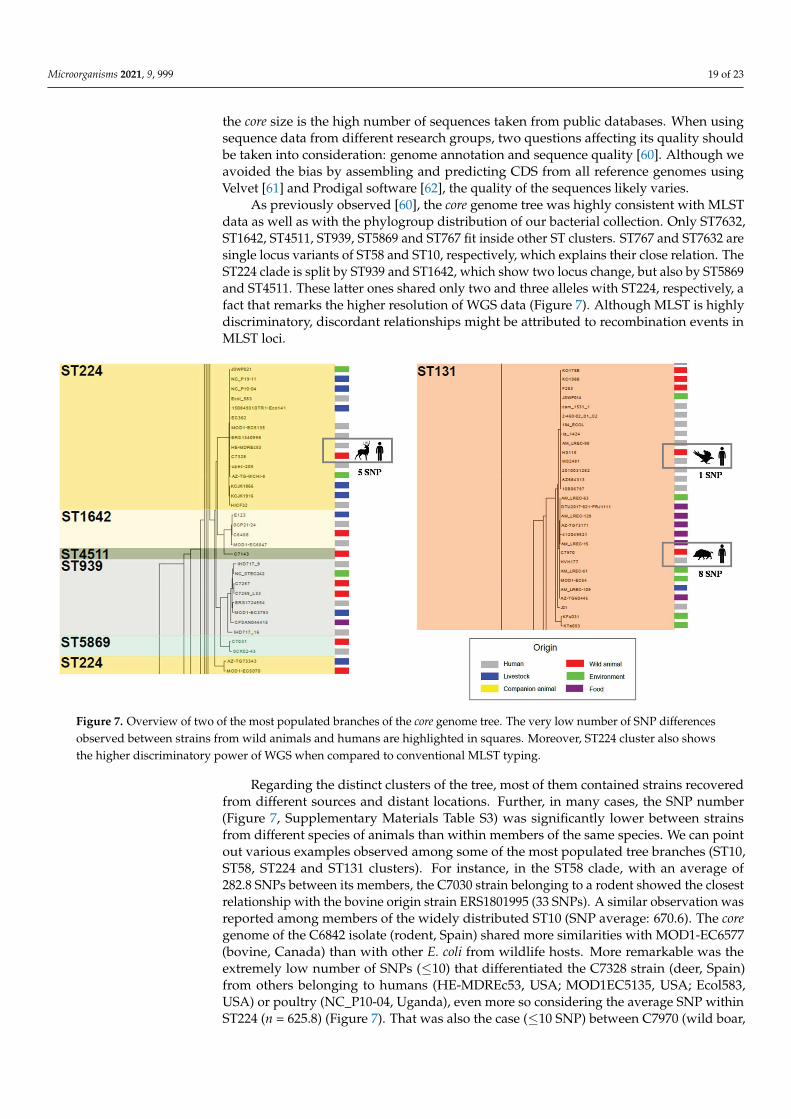
As previously observed [60], the core genome tree was highly consistent with MLSTdata as well as with the phylogroup distribution of our bacterial collection. Only ST7632,ST1642, ST4511, ST939, ST5869 and ST767 fit inside other ST clusters. ST767 and ST7632 aresingle locus variants of ST58 and ST10, respectively, which explains their close relation. TheST224 clade is split by ST939 and ST1642, which show two locus change, but also by ST5869and ST4511. These latter ones shared only two and three alleles with ST224, respectively, afact that remarks the higher resolution of WGS data (Figure 7). Although MLST is highlydiscriminatory, discordant relationships might be attributed to recombination events inMLST loci.
Figure 7. Overview of two of the most populated branches of the core genome tree. The very low number of SNP differencesobserved between strains from wild animals and humans are highlighted in squares. Moreover, ST224 cluster also showsthe higher discriminatory power of WGS when compared to conventional MLST typing.
Regarding the distinct clusters of the tree, most of them contained strains recoveredfrom different sources and distant locations. Further, in many cases, the SNP number(Figure 7, Supplementary Materials Table S3) was significantly lower between strainsfrom different species of animals than within members of the same species. We can pointout various examples observed among some of the most populated tree branches (ST10,ST58, ST224 and ST131 clusters). For instance, in the ST58 clade, with an average of282.8 SNPs between its members, the C7030 strain belonging to a rodent showed the closestrelationship with the bovine origin strain ERS1801995 (33 SNPs). A similar observation wasreported among members of the widely distributed ST10 (SNP average: 670.6). The coregenome of the C6842 isolate (rodent, Spain) shared more similarities with MOD1-EC6577(bovine, Canada) than with other E. coli from wildlife hosts. More remarkable was theextremely low number of SNPs (≤10) that differentiated the C7328 strain (deer, Spain)from others belonging to humans (HE-MDREc53, USA; MOD1EC5135, USA; Ecol583,USA) or poultry (NC_P10-04, Uganda), even more so considering the average SNP withinST224 (n = 625.8) (Figure 7). That was also the case (≤10 SNP) between C7970 (wild boar,

Microorganisms 2021, 9, 999 20 of 23
Spain) and HVH 177 (human, Denmark), two members of the major ST131 cluster (SNPaverage = 259.6) (Figure 7). Thus, no clear evidence of adaptive mutations associated to thecore genome were found in strains from different hosts. Although further studies with amore homogeneous dataset are required to generalize this assumption, our results reflectan on-going inter-host transmission of E. coli strains. Adaptation to the host environmentdoes not seem to leave a genetic mark in the bacterial core genome.
4. Conclusions
This study contributes to better characterize the commensal gut niche from wildlife,which is crucial for understanding the adaptive pathways and genome diversification ofE. coli, the role of different elements in bacterial fitness and the flow of genes and genemodules among interconnected ecosystems. Whole genome sequencing enables a moreconsistent characterization of the composition and diversity of the plasmidome, facilitatingthe study of small replicons, the differentiation of extra-chromosomal phages or phage-likeelements and the detection of non-typeable plasmids. Further, it offers a general viewof different genomic regions related to immunity, phylogeny, pathogenesis or resistance,which favors more robust assumptions than those made on the basis of a few set of genesscreened by conventional methods.
Our analyses seem to rule out the occurrence of a parallel evolution of the E. coligenome in the gut of wild animals. Bacteria from humans or highly human-influenced set-tings exhibit similar genetic patterns in CRISPR-Cas systems, plasmids or virulence/resista-nce gene-carrying modules. These observations, together with the absence of significantgenetic changes in their core genome, suggest an ongoing flow of both mobile elements andE. coli lineages between human and natural ecosystems.
Supplementary Materials: The following are available online at http://www.mdpi.com/xxx/s1.Figure S1: Structural diversity of the CRISPR/Cas I-E and I-F1 systems regarding the modulelocated between the two CRISPR arrays, comprising the cas set of genes and adjacent ORFs. CRISPRarrays depicted in this figure illustrate only its position within the CRISPR/Cas systems (the spacerrepertoire of each isolate is represented in Fig. 1). The cas-E (cas2, cas1, cas6, cas5, cas7, cse2, cse1,and cas3) and cas-F1 (cas1-cas2/3-csy1-csy2-csy3-cas6f ) genes, in pink, are located between CRISPR-1/CRISPR-2 loci in I-E system and CRISPR-3/CRISPR 4 in I-F1 system., Figure S2: Phylogenetictree constructed according to the concatenated amino acid sequences of the cas genes. Colour circlesdenote the different phylogenetic groups (dark blue: B1; red: A; green: D; pink: B2; light blue: CladeV). The concatenated cas-E genes of E. coli K-12 MG1655 are also included as reference., Figure S3:Phylogenetic tree constructed according to the nucleotide sequences of 21 putative iss genes fromour E. coli collection. Some reference sequences used by Jonhson et al. 2008 to define the differentiss alleles are marked with a red circle. Two reference sequences, distinctly associated with high(AF042279) and low (iss type13) serum livability by Xu et al. 2018, appear indicated with a bluecircle., Figure S4: Phylogenetic relationship of the 38 studied E. coli and 242 reference genomes(Table S2). The tree was based on a 575,173.5 ± 3,802.95 bp core genome including 674 orthologouscommon genes, from a total of 108,518 gene clusters, with at least 80% identity and 60% coverageand using 100 bootstrapping replicates. ST types are highlighted in different colors and origin hasbeen indicated in the right part. The pairwise SNP distance matrix used to build the tree is shown inTable S3; Table S1: Genomic features of the 38 E. coli genomes from wildlife. Table S2: Informationabout the genome sequences downloaded from the NCBI for the construction of the phylogenomiccore tree. Table S3: Pairwise SNP distance matrix calculated from the core genome in the set of 242E. coli full genomes from the NCBI database plus 38 E. coli genomes from this study. Genomes areordered according to their Sequence Type (ST).
Author Contributions: C.T. and F.d.l.C. conceived the study; C.A.A. and M.d.T. performed theexperimental work and the genomic analysis. C.T., M.d.T. and F.d.l.C. supervised the study; C.A.A.wrote the first draft of the manuscript. All authors have read and agreed to the published version ofthe manuscript.
Funding: This work was partially supported by project SAF2016-76571-R and PID2019-106158RB-I00from the Agencia Estatal de Investigación (AEI) of Spain and the Fondo Europeo de Desarrollo

Microorganisms 2021, 9, 999 21 of 23
Regional (FEDER) of EU. During the experimental work of this study, Carla Andrea Alonso Alonsohad a predoctoral fellowship FPI from MINECO.
Institutional Review Board Statement: Not applicable.
Informed Consent Statement: Not applicable.
Data Availability Statement: Raw sequence reads reported in this paper were deposited underNCBI BioProject PRJNA699864.
Conflicts of Interest: The authors declare no conflict of interest.
References1. Touchon, M.; Hoede, C.; Tenaillon, O.; Barbe, V.; Baeriswyl, S.; Bidet, P.; Bingen, E.; Bonacorsi, S.; Bouchier, C.; Bouvet, O.; et al.
Organised genome dynamics in the Escherichia coli species results in highly diverse adaptive paths. PLoS Genet. 2009, 5, e1000344.[CrossRef] [PubMed]
2. De Toro, M.; Garcillán-Barcia, M.P.; de la Cruz, F. Plasmid diversity and adaptation analyzed by massive sequencing of Escherichiacoli plasmids. Microbiol. Spectr. 2014, 2. [CrossRef] [PubMed]
3. Gillings, M.R.; Stokes, H.W. Are humans increasing bacterial evolvability? Trends Ecol. Evol. 2012, 27, 346–352. [CrossRef][PubMed]
4. Johnson, J.R.; Kuskowski, M.A.; Gaiewski, A.; Sahm, D.F.; Karlowsky, J.A. Virulence characteristics and phylogenetic backgroundof multidrug-resistant and antimicrobial-susceptible clinical isolates of Escherichia coli from across the United States 2000–2001.J. Infect. Dis. 2004, 190, 1739–1744. [CrossRef]
5. Tenaillon, O.; Skurnik, D.; Picard, B.; Denamur, E. The population genetics of commensal E. coli. Nat. Rev. Microbiol. 2010, 8,207–217. [CrossRef]
6. Alcalá, L.; Alonso, C.A.; Simón, C.; González-Esteban, C.; Orós, J.; Rezusta, A.; Ortega, C.; Torres, C. Wild birds, frequent carriersof extended-spectrum β-lactamase (ESBL) producing Escherichia coli of CTX-M and SHV-12 types. Microb. Ecol. 2016, 72, 861–869.[CrossRef]
7. Alonso, C.A.; González-Barrio, D.; Tenorio, C.; Ruiz-Fons, F.; Torres, C. Antimicrobial resistance in faecal Escherichia coliisolates from farmed reed deer and wild small mammals. Detection of a multiresistant E. coli producing extended-spectrumbeta-lactamase. Comp. Immunol. Microbiol. Infect. Dis. 2016, 45, 34–39. [CrossRef]
8. Alonso, C.A.; González-Barrio, D.; Ruiz-Fons, F.; Ruiz-Ripa, L.; Torres, C. High frequency of B2 phylogroup among non-clonallyrelated fecal Escherichia coli isolates from wild boars, including the lineage ST131. FEMS Microbiol. Ecol. 2017, 93. [CrossRef]
9. Alonso, C.A.; Alcalá, L.; Simón, C.; Torres, C. Novel sequence types of extended-spectrum and acquired AmpC beta-lactamaseproducing Escherichia coli and Escherichia clade V isolated from wild mammals. FEMS Microbiol. Ecol. 2017, 93, fix097. [CrossRef]
10. Clermont, O.; Christenson, J.K.; Denamur, E.; Gordon, D.M. The Clermont Escherichia coli phylo-typing method revisited:Improvement of specificity and detection of new phylo-groups. Environ. Microbiol. Rep. 2013, 5, 58–65. [CrossRef]
11. Vielva, L.; de Toro, M.; Lanza, V.; de la Cruz, F. PLACNETw: A web-based tool for plasmid reconstruction from bacterial genomes.Bioinformatics 2017, 33, 3796–3798. [CrossRef]
12. Zankari, E.; Hasman, H.; Cosentino, S.; Vestegaard, M.; Rasmussen, S.; Lund, O.; Aarestrup, F.M.; Larsen, M.V. Identification ofacquired antimicrobial resistance genes. J. Antimicrob. Chemother. 2012, 67, 2640–2644. [CrossRef]
13. Joensen, K.G.; Scheutz, F.; Lund, O.; Hasman, H.; Kaas, R.S.; Nielsen, E.M.; Aarestrup, F.M. Real-time whole-genome sequencingfor routine typing, surveillance and outbreak detection of verotoxigenic Escherichia coli. J. Clin. Microbiol. 2014, 52, 1501–1510.[CrossRef]
14. Grissa, I.; Vergnaud, G.; Pourcel, C. The CRISPRdb database and tools to display CRISPRs and to generate dictionaries of spacersand repeats. BMC Bioinform. 2007, 8, 172. [CrossRef]
15. Sullivan, M.J.; Petty, N.K.; Beatson, S.A. Easyfig: A genome comparison visualizer. Bioinformatics 2011, 27, 1009–1010. [CrossRef]16. Kumar, S.; Stecher, G.; Tamura, K. MEGA7: Molecular Evolutionary Genetics Analysis Version 7.0 for Bigger Datasets. Mol. Biol.
Evol. 2016, 33, 1870–1874. [CrossRef]17. Fu, L.; Niu, B.; Zhu, Z.; Wu, S.; Li, W. CD-HIT: Accelerated for clustering the next generation sequencing data. Bioinformatics 2012,
28, 3150–3152. [CrossRef]18. Stamatakis, A. RAxML-VI-HPC: Maximum likelihood-based phylogenetic analyses with thousands of taxa and mixed models.
Bioinformatics 2006, 22, 2688–2690. [CrossRef]19. Ishino, Y.; Shinagawa, H.; Makino, K.; Amemura, M.; Nakata, A. Nucleotide sequence of the iap gene, responsable for alkaline
phosphatase isozyme conversion in Escherichia coli, and identification of the gene product. J. Bacteriol. 1987, 169, 5429–5433.[CrossRef]
20. Mojica, F.J.; Juez, G.; Rodríguez-Valera, F. Transcription at different salinities of Haloferax mediterranei sequences adjacent topartially modified PstI sites. Mol. Microbiol. 1993, 9, 613–621. [CrossRef]
21. Makarova, K.S.; Grishin, N.V.; Shabalina, S.A.; Wolf, Y.I.; Koonin, E.V. A putative RNA interference-based immune system inprokaryotes: Computational analysis of the predicted enzymatic machinery, functional analogies with eukaryotic RNAi, andhypothetical mechanisms of action. Biol. Direct. 2006, 1, 7. [CrossRef]

Microorganisms 2021, 9, 999 22 of 23
22. Aydin, S.; Personne, Y.; Newire, E.; Laverick, R.; Russell, O.; Roberts, A.P.; Enne, V.I. Presence of type I-F CRISPR-Cas systems isassociated with antimicrobial susceptibility in Escherichia coli. J. Antimicrob. Chemother. 2017, 72, 2213–2218. [CrossRef]
23. Westra, E.R.; Pul, U.; Heidrich, N.; Heidrich, N.; Jore, M.M.; Lundgren, M.; Stratmann, T.; Wurm, R.; Raine, A.; Mescher, M.; et al.H-NS-mediated repression of CRISPR based immunity in Escherichia coli K12 can be relieved by the transcription activator LeuO.Mol. Microbiol. 2010, 77, 1380–1393. [CrossRef]
24. Cady, K.C.; Bondy-Denomy, J.; Heussler, G.E.; Davidson, A.R.; O’Toole, G.A. The CRISPR/Cas adaptive immune system ofPseudomonas aeruginosa mediates resistance to naturally occurring and engineered phages. J. Bacteriol. 2012, 194, 5728–5738.[CrossRef]
25. Almendros, C.; Guzmán, N.M.; Díez-Villaseñor, C.; García-Martínez, J.; Mojica, F.J.M. Target motifs affecting natural immunity bya constitutive CRISPR-Cas System in Escherichia coli. PLoS ONE 2012, 7, e50797. [CrossRef]
26. Makarova, K.S.; Wolf, Y.I.; Iranzo, J.; Shmakov, S.A.; Alkhnbashi, O.S.; Brouns, S.J.J.; Charpentier, E.; Cheng, D.; Haft, D.H.;Horvath, P.; et al. Evolutionary classification of CRISPR-Cas system: A burst of class 2 and derived variants. Nat. Rev. Microbiol.2020, 18, 67–83. [CrossRef]
27. Almendros, C.; Mojica, F.J.M.; Díez-Villaseñor, C.; Guzmán, N.M.; García-Martínez, J. CRISPR-Cas functional module exchangein Escherichia coli. mBio 2014, 5, e00767-13. [CrossRef]
28. Liu, S.; Jin, D.; Lan, R.; Wang, Y.; Meng, Q.; Dai, H.; Lu, S.; Hu, S.; Xu, J. Escherichia marmotae sp. nov., isolated from faeces ofMarmota himalayana. Int. J. Syst. Evol. Microbiol. 2015, 65, 2130–2134. [CrossRef]
29. Kupczok, A.; Landan, G.; Dagan, T. The contribution of genetic recombination to CRISPR array evolution. Genome Biol. Evol.2015, 7, 1925–1939. [CrossRef]
30. Díez-Villaseñor, C.; Almendros, C.; García-Martínez, J.; Mojica, F.J.M. Diversity of CRISPR loci in Escherichia coli. Microbiology2010, 156, 1351–1361. [CrossRef]
31. Touchon, M.; Charpentier, S.; Clermont, O.; Rocha, E.P.; Denamur, E.; Branger, C. CRISPR distribution within the Escherichia colispecies is not suggestive of immunity-associated diversifying selection. J. Bacteriol. 2011, 193, 2460–2467. [CrossRef] [PubMed]
32. Nicolas-Chanoine, M.H.; Bertrand, X.; Madec, J.Y. Escherichia coli ST131, an Intriguing Clonal Group. Clin. Microbiol. Rev. 2014, 27,543–574. [CrossRef] [PubMed]
33. Smillie, C.; Garcillán-Barcia, M.P.; Francia, M.V.; Rocha, E.P.; de la Cruz, F. Mobility of plasmids. Microbiol. Mol. Biol. Rev. 2010, 74,434–452. [CrossRef] [PubMed]
34. Spaková, T.; Fecskeová, L.K.M.; Javorský, P.; Pristas, P. Two rep genes in small cryptic plasmid pKSTt21 of Escherichia coli. Curr.Microbiol. 2013, 67, 437–441. [CrossRef]
35. Brolund, A.; Franze’n, O.; Melefors, O.; Tegmark-Wisell, K.; Sandegren, L. Plasmidome-analysis of ESBL-producing E. coli usingconventional typing and high-throughput sequencing. PLoS ONE 2013, 8, e65793. [CrossRef]
36. Attéré, S.A.; Vincent, A.T.; Paccaud, M.; Frenette, M.; Charette, S.J. The role for the small cryptic plasmids as moldable vectors forgenetic innovation in Aeromonas salmonicida subsp. salmonicida. Front. Genet. 2017, 8. [CrossRef]
37. Billard-Pomares, T.M.; Fouteau, S.; Jacquet, M.E.; Roche, D.; Barbe, V.; Castellanos, M.; Bouet, J.Y.; Cruveiller, S.; Médique, C.;Blanco, J.; et al. Characterization of a P1-like bacteriophage carrying an SHV-2 extended-spectrum β-lactamase from an Escherichiacoli strain. Antimicrob. Agents Chemother. 2014, 58, 6550–6557. [CrossRef]
38. Zhang, C.; Feng, Y.; Liu, F.; Jiang, H.; Qu, Z.; Lei, M.; Wang, J.; Zhang, B.; Hu, Y.; Ding, J.; et al. A phage-like IncY plasmidcarrying the mcr-1 gene in Escherichia coli from a pig farm in China. Antimicrob. Agents Chemother. 2017, 61, e02035. [CrossRef]
39. Haft, R.J.; Mittler, J.E.; Traxler, B. Competition favours reduced cost of plasmids to host bacteria. ISME J. 2009, 3, 761–769.[CrossRef]
40. Yau, S.; Liu, X.; Djordjevic, S.P.; Hall, R.M. RSF1010-like plasmids in Australian Salmonella enteric serovar Typhimurium and originof their sul2-strA-strB antibiotic resistance gene cluster. Microb. Drug Resist. 2010, 16, 249–252. [CrossRef]
41. Sunde, M.; Simonsen, G.S.; Slettemeas, J.S.; Hockerman, I.; Norstrom, M. Integron, plasmid and host strain characteristics ofEscherichia coli from humans and food included in the Norwegian Antimicrobial Resistance Monitoring Programs. PLoS ONE2015, 10, e0128797. [CrossRef]
42. Dawes, F.E.; Kuzevski, A.; Bettelheim, K.A.; Hornitzky, M.A.; Djordjevic, S.P.; Walker, M.J. Distribution of class 1 integrons wihIS26-mediated deletions in their 3´-CS conserved segments in Escherichia coli of human and animal origin. PLoS ONE 2010, 5,e12754. [CrossRef]
43. Cullik, A.; Pfeifer, Y.; Prager, R.; von Baum, H.; Witte, W. A novel IS26 structure surrounds blaCTX-M genes in different plasmidsfrom German clinical Escherichia coli isolates. J. Med. Microbiol. 2010, 59, 580–587. [CrossRef]
44. Wang, J.; Stephan, R.; Power, K.; Yan, Q.; Hächler, H.; Fanning, S. Nucleotide sequences of 16 transmissible plasmids identifiedin nine multidrug-resistant Escherichia coli isolates expressing an ESBL phenotype isolated from food-producing animals andhealthy humans. J. Antimicrob. Chemother. 2014, 69, 2658–2668. [CrossRef]
45. Snesrud, E.; Ong, A.C.; Corey, B.; Kwak, Y.I.; Clifford, R.; Gleeson, T.; Wood, S.; Whitman, T.J.; Lesho, E.P.; Hinkle, M.; et al.Analysis of serial isolates of mcr-1-positive Escherichia coli reveals a highly active ISApl1 transposon. Antimicrob. Agents Chemother.2017, 61, e00056-17. [CrossRef]
46. Smith, D.K.; Kassam, T.; Singh, B.; Elliott, J.F. Escherichia coli has two homologous glutamate descarboxylase genes that map todistinct loci. J. Bacteriol. 1992, 174, 5820–5826. [CrossRef]

Microorganisms 2021, 9, 999 23 of 23
47. Dogan, B.; Rishniw, M.; Bruant, G.; Harel, J.; Schukken, Y.H.; Simpson, K.W. Phylogroup and lpfA influence epithelial invasion bymastitis associated Escherichia coli. Vet. Microbiol. 2012, 159, 163–170. [CrossRef]
48. Parham, N.J.; Pollard, S.J.; Desvaux, M.; Scott-Tucker, A.; Liu, C.; Fivian, A.; Henderson, I.R. Distribution of the serine proteaseautotransporters of the Enterobacteriaceae among extraintestinal clinical isolates of Escherichia coli. J. Clin. Microbiol. 2005, 43,4076–4082. [CrossRef]
49. Moriel, D.G.; Bertoldi, I.; Spagnuolo, A.; Marchi, S.; Rosini, R.; Nesta, B.; Pastorello, I.; Corea, V.A.; Torricelli, G.; Cartocci, E.; et al.Identification of protective and broadly conserved vaccine antigens from the genome of extraintestinal pathogenic Escherichia coli.Proc. Natl. Acad. Sci. USA 2010, 107, 9072–9077. [CrossRef]
50. Johnson, T.J.; Siek, K.E.; Johnson, S.J.; Nolan, L.K. DNA sequence of a ColV plasmid and prevalence of selected plasmid-encodedvirulence genes among avian Escherichia coli strains. J. Bacteriol. 2006, 188, 745–758. [CrossRef]
51. Micenková, L.; Štaudová, B.; Bosák, J.; Mikalová, L.; Littnerová, S.; Vrba, M.; Ševcíková, A.; Woznicová, V.; Šmajs, D. Bacteriocin-encoding genes and ExPEC virulence determinants are associated in human fecal Escherichia coli strains. BMC Microbiol. 2014, 14,109. [CrossRef]
52. Martin, P.; Tronnet, S.; Garcie, C.; Oswald, E. Interplay between siderophores and colibactin genotoxin in Escherichia coli. IUBMBLife. 2017, 69, 435–441. [CrossRef]
53. Xu, W.Y.; Li, Y.J.; Fan, C. Different loci and mRNA copy number of the increased serum survival gene of Escherichia coli. Can. J.Microbiol. 2018, 64, 147–154. [CrossRef]
54. Johnson, T.J.; Wannemuehler, Y.M.; Nolan, L.K. Evolution of the iss gene in Escherichia coli. Appl. Environ. Microbiol. 2008, 74,2360–2369. [CrossRef]
55. McVeigh, A.; Fasano, A.; Scott, D.A.; Jelacic, S.; Moseley, S.L.; Robertson, D.C.; Savarino, S.J. IS1414, an Escherichia coli insertionsequence with a heat-stable enterotoxin gene embedded in a transposase-like gene. Infect. Immun. 2000, 68, 5710–5715. [CrossRef]
56. Jønsson, R.; Struve, C.; Boll, E.J.; Boisen, N.; Joensen, K.G.; Sørensen, C.A.; Jensen, B.H.; Scheutz, F.; Jenssen, H.; Krogfelt, K.A. ANovel pAA Virulence Plasmid Encoding Toxins and Two Distinct Variants of the Fimbriae of Enteroaggregative Escherichia coli.Front. Microbiol. 2017, 8, 263. [CrossRef]
57. Burgos, Y.; Beutin, L. Common origin of plasmid encoded alpha-hemolysin genes in Escherichia coli. BMC Microbiol. 2010, 10, 193.[CrossRef] [PubMed]
58. Walk, S.T.; Alm, E.W.; Gordon, D.M.; Ram, J.L.; Toranzos, G.A.; Tiedje, J.M.; Whittam, T.S. Cryptic lineages of the genus Escherichia.Appl. Environ. Microbiol. 2009, 75, 6534–6544. [CrossRef] [PubMed]
59. Luo, C.; Walk, S.T.; Gordon, D.M.; Feldgarden, M.; Tiedje, J.M.; Konstantinidis, K.T. Genome sequencing of environmentalEscherichia coli expands understanding of the ecology and speciation of the model bacterial species. Proc. Natl. Acad. Sci. USA2011, 108, 7200–7205. [CrossRef]
60. Kaas, R.S.; Friis, C.; Ussery, D.W.; Aarestrup, F.M. Estimating variation within the genes and inferring the phylogeny of 186sequenced diverse Escherichia coli genomes. BMC Genom. 2012, 13, 577. [CrossRef] [PubMed]
61. Zerbino, D.R. Using the Velvet de novo assembler for short-read sequencing technologies. Curr. Protoc. Bioinform. 2010. [CrossRef]62. Hyatt, D.; Chen, G.L.; LoCascio, P.F.; Land, M.L.; Larimer, F.W.; Hauser, L.J. Prodigal: Prokaryotic gene recognition and translation
initiation site identification. BMC Bioinform. 2010, 11, 119. [CrossRef]