Genomic and proteomic analyses of the coral pathogen Vibrio coralliilyticus reveal a diverse...
13
ORIGINAL ARTICLE Genomic and proteomic analyses of the coral pathogen Vibrio coralliilyticus reveal a diverse virulence repertoire Eidy de O Santos 1 , Nelson Alves Jr 1 , Graciela M Dias 1 , Ana Maria Mazotto 2 , Alane Vermelho 2 , Gary J Vora 3 , Bryan Wilson 4 , Victor H Beltran 4 , David G Bourne 4 , Fre ´de ´rique Le Roux 5,6 and Fabiano L Thompson 1 1 Laboratory of Microbiology, Institute of Biology, Federal University of Rio de Janeiro (UFRJ), Rio de Janeiro, Brazil; 2 Laboratory of Proteases, Institute of Microbiology, UFRJ, Rio de Janeiro, Brazil; 3 Center for Bio/ Molecular Science & Engineering, Naval Research Laboratory, Washington, DC, USA; 4 Centre for Marine Microbiology and Genetics, Australian Institute of Marine Science, Townsville MC, Queensland, Australia; 5 FR2424 CNRS UPMC Station Biologique de Roscoff, France and 6 Ifremer, Laboratoire de Physiologie des Inverte´bre´s, Brest, France Vibrio coralliilyticus has been implicated as an important pathogen of coral species worldwide. In this study, the nearly complete genome of Vibrio coralliilyticus strain P1 (LMG23696) was sequenced and proteases implicated in virulence of the strain were specifically investigated. The genome sequence of P1 (5 513 256 bp in size) consisted of 5222 coding sequences and 58 RNA genes (53 tRNAs and at least 5 rRNAs). Seventeen metalloprotease and effector (vgrG, hlyA and hcp) genes were identified in the genome and expressed proteases were also detected in the secretome of P1. As the VcpA zinc-metalloprotease has been considered an important virulence factor of V. coralliilyticus, a vcpA deletion mutant was constructed to evaluate the effect of this gene in animal pathogenesis. Both wild-type and mutant (DvcpA) strains exhibited similar virulence characteristics that resulted in high mortality in Artemia and Drosophila pathogenicity bioassays and strong photosystem II inactivation of the coral dinoflagellate endosymbiont (Symbiodinium). In contrast, the DvcpA mutant demonstrated higher hemolytic activity and secreted 18 proteins not secreted by the wild type. These proteins included four types of metalloproteases, a chitinase, a hemolysin-related protein RbmC, the Hcp protein and 12 hypothetical proteins. Overall, the results of this study indicate that V. coralliilyticus strain P1 has a diverse virulence repertoire that possibly enables this bacterium to be an efficient animal pathogen. The ISME Journal (2011) 5, 1471–1483; doi:10.1038/ismej.2011.19; published online 31 March 2011 Subject Category: microbe–microbe and microbe–host interactions Keywords: V. coralliilyticus; coral disease; zinc-metalloprotease; genome; secretome Introduction Coral reefs are hotspots of productivity and biodi- versity and therefore represent one of the most important marine biomes (Knowlton and Jackson, 2008; Kiessling et al., 2010). Reefs also sustain significant fisheries worldwide, providing an im- portant economic and food resource for local communities (Hughes et al., 2003). The health of coral reefs are in decline, with recent assessments estimating that Bone-third of global reefs have been effectively destroyed or are under imminent threat of collapse (Wilkinson, 2008). The combined effects of global changes (for example, increase in sea water temperature) and local anthropogenic stressors (for example, water pollution, overfishing) have been implicated as contributing factors to the dramatic decline of coral reefs in recent decades (Rohwer and Youle, 2010). Coral diseases have also been implicated in the loss of reef habitat, especially in the Caribbean where diseases have fundamentally altered reef ecosystems (Sutherland et al., 2004). Bacterial pathogens have been identified as the causative microbial agent of some coral diseases (Bourne et al., 2009). For example, Rosenberg and colleagues demonstrated that the temperature-de- pendent necrosis of Pocillopora damicornis coral tissue and its zooxanthellae was caused by the bacterium Vibrio coralliilyticus (Ben-Haim et al., 2003a). Infection occurred predominantly at high seawater temperatures (27–29 1C) and a single putative metalloprotease was identified as the key Received 4 November 2010; revised 11 January 2011; accepted 25 January 2011; published online 31 March 2011 Correspondence: FL Thompson, Laboratory of Microbiology, Federal University of Rio de Janeiro (UFRJ), Institute of Biology, Rio de Janeiro 21944-970, Brazil. E-mail: [email protected] The ISME Journal (2011) 5, 1471–1483 & 2011 International Society for Microbial Ecology All rights reserved 1751-7362/11 www.nature.com/ismej
Transcript of Genomic and proteomic analyses of the coral pathogen Vibrio coralliilyticus reveal a diverse...

ORIGINAL ARTICLE
Genomic and proteomic analyses of the coralpathogen Vibrio coralliilyticus reveal a diversevirulence repertoire
Eidy de O Santos1, Nelson Alves Jr1, Graciela M Dias1, Ana Maria Mazotto2,Alane Vermelho2, Gary J Vora3, Bryan Wilson4, Victor H Beltran4, David G Bourne4,Frederique Le Roux5,6 and Fabiano L Thompson1
1Laboratory of Microbiology, Institute of Biology, Federal University of Rio de Janeiro (UFRJ), Rio de Janeiro,Brazil; 2Laboratory of Proteases, Institute of Microbiology, UFRJ, Rio de Janeiro, Brazil; 3Center for Bio/Molecular Science & Engineering, Naval Research Laboratory, Washington, DC, USA; 4Centre for MarineMicrobiology and Genetics, Australian Institute of Marine Science, Townsville MC, Queensland, Australia;5FR2424 CNRS UPMC Station Biologique de Roscoff, France and 6Ifremer, Laboratoire de Physiologie desInvertebres, Brest, France
Vibrio coralliilyticus has been implicated as an important pathogen of coral species worldwide. Inthis study, the nearly complete genome of Vibrio coralliilyticus strain P1 (LMG23696) was sequencedand proteases implicated in virulence of the strain were specifically investigated. The genomesequence of P1 (5 513 256 bp in size) consisted of 5222 coding sequences and 58 RNA genes(53 tRNAs and at least 5 rRNAs). Seventeen metalloprotease and effector (vgrG, hlyA and hcp) geneswere identified in the genome and expressed proteases were also detected in the secretome of P1.As the VcpA zinc-metalloprotease has been considered an important virulence factor ofV. coralliilyticus, a vcpA deletion mutant was constructed to evaluate the effect of this gene inanimal pathogenesis. Both wild-type and mutant (DvcpA) strains exhibited similar virulencecharacteristics that resulted in high mortality in Artemia and Drosophila pathogenicity bioassaysand strong photosystem II inactivation of the coral dinoflagellate endosymbiont (Symbiodinium). Incontrast, the DvcpA mutant demonstrated higher hemolytic activity and secreted 18 proteins notsecreted by the wild type. These proteins included four types of metalloproteases, a chitinase, ahemolysin-related protein RbmC, the Hcp protein and 12 hypothetical proteins. Overall, the resultsof this study indicate that V. coralliilyticus strain P1 has a diverse virulence repertoire that possiblyenables this bacterium to be an efficient animal pathogen.The ISME Journal (2011) 5, 1471–1483; doi:10.1038/ismej.2011.19; published online 31 March 2011Subject Category: microbe–microbe and microbe–host interactionsKeywords: V. coralliilyticus; coral disease; zinc-metalloprotease; genome; secretome
Introduction
Coral reefs are hotspots of productivity and biodi-versity and therefore represent one of the mostimportant marine biomes (Knowlton and Jackson,2008; Kiessling et al., 2010). Reefs also sustainsignificant fisheries worldwide, providing an im-portant economic and food resource for localcommunities (Hughes et al., 2003). The health ofcoral reefs are in decline, with recent assessmentsestimating that Bone-third of global reefs have beeneffectively destroyed or are under imminent threatof collapse (Wilkinson, 2008). The combined effects
of global changes (for example, increase in sea watertemperature) and local anthropogenic stressors (forexample, water pollution, overfishing) have beenimplicated as contributing factors to the dramaticdecline of coral reefs in recent decades (Rohwerand Youle, 2010). Coral diseases have also beenimplicated in the loss of reef habitat, especially inthe Caribbean where diseases have fundamentallyaltered reef ecosystems (Sutherland et al., 2004).Bacterial pathogens have been identified as thecausative microbial agent of some coral diseases(Bourne et al., 2009). For example, Rosenberg andcolleagues demonstrated that the temperature-de-pendent necrosis of Pocillopora damicornis coraltissue and its zooxanthellae was caused by thebacterium Vibrio coralliilyticus (Ben-Haim et al.,2003a). Infection occurred predominantly at highseawater temperatures (27–29 1C) and a singleputative metalloprotease was identified as the key
Received 4 November 2010; revised 11 January 2011; accepted25 January 2011; published online 31 March 2011
Correspondence: FL Thompson, Laboratory of Microbiology,Federal University of Rio de Janeiro (UFRJ), Institute of Biology,Rio de Janeiro 21944-970, Brazil.E-mail: [email protected]
The ISME Journal (2011) 5, 1471–1483& 2011 International Society for Microbial Ecology All rights reserved 1751-7362/11
www.nature.com/ismej

toxin in this disease process (Ben-Haim et al.,2003b). At lower temperatures, this bacterium didnot produce several putative toxins, most notably, azinc-metalloprotease (Ben-Haim et al., 2003b).
Investigations of white syndrome coral diseaseoutbreaks in the Indo-Pacific between 2003 and2006 isolated a number V. coralliilyticus strainsfrom different coral species (Pachyseris speciosa,Montipora aequituberculata and Acropora cytherea)and identified them as the potential causative agents(Sussman et al., 2008). The extracellular productsderived from the V. coralliilyticus strain P1(LMG23696) isolated from diseased Montiporaaequituberculata colonies in Nelly Bay (GreatBarrier Reef), caused photoinactivation of theSymbiodiunium (zooxanthellae) endosymbiont(Sussman et al., 2008). Analysis of the extracellularproducts from strain P1 revealed a predominantzinc-metalloprotease that was found to be a homologof the Vibrio cholerae hemagglutinin-protease(Sussman et al., 2008). When purified from theother extracellular products, this zinc-metallopro-tease was demonstrated to cause coral tissue damagewithin 18 h at 27 1C (Sussman et al., 2009). Metallo-proteases have several catalytic activities related tovirulence but little is known about the diversity ofthese potential virulence factors in coral pathogenicbacteria. In the bivalve pathogen V. splendidus, thedeletion of the Vsm metalloprotease resulted inthe expression of other metalloproteases that werefound to compensate for some of the functionsfulfilled by the vsm gene product (Binesse et al.,2008). In the fish pathogen V. anguillarum, mutantslacking a functional EmpA metalloproteaseexpressed two different proteases that were notdetected in the wild-type strain suggesting that theseproteases might compensate for the loss of EmpA(Milton et al., 1992; Varina et al., 2008). Similarly, ametalloprotease mutant of Vibrio vulnificus wasfound to express a serine protease in response thatdemonstrated comparable levels of proteolyticactivity to the wild-type strain (Wang et al., 2008).
Recent work has shown that vibrios, includingV. coralliilyticus, are ubiquitous in coral mucus(Chimetto et al., 2008; Alves et al., 2010) and oftendominate the microbial community of diseasedcorals (Bourne and Munn, 2005; Luna et al., 2007).A similar pattern is observed at the whole reef scaleas vibrios have been demonstrated to have muchhigher loads on unhealthy vs healthy coral reefs(Dinsdale et al., 2008). Vega Thurber et al. (2008)have demonstrated that even when vibrios are atlow-abundance on stressed corals, they were foundto significantly alter the coral microbiome metabo-lism. Such studies suggest that a better under-standing of the pathogenic potential of vibriosisolated from corals is required. In the presentstudy, we sequenced the whole genome of theV. coralliilyticus strain P1 (LMG23696) with aspecific focus on the diversity of metalloproteasesin the genome, which can act as key virulence
factors in the infection of corals. The expression ofthe collection of proteases from strain P1 wasanalyzed by protein and enzyme characterizationassays. A mutant P1 strain (DvcpA) was generated totest if the zinc-metalloprotease VcpA is an essentialvirulence factor for V. coralliilyticus pathogenesis.The pathogenic potential of both the wild-type andmutant strains was evaluated in three modelbioassays (Artemia, Drosophila and Symbiodinium)to test whether a single zinc metalloprotease (VcpA)is essential for the disease process. The comparativeanalyses of both mutant and wild-type strainsenabled us to improve our current understandingof the role of VcpA in V. coralliilyticus animalpathogenesis.
Materials and methods
Bacterial strains, plasmids and mediaThe bacterial strains and plasmids used in thisstudy are described in Supplementary Table S1.Escherichia coli strains were grown in Luria-Bertani(LB) medium at 37 1C. V. coralliilyticus strains weregrown at 28 1C in LB medium (Difco, Sparks, NV,USA), tryptone soy broth (Difco) or marine broth(Difco) supplemented with 0.5 M NaCl for themutagenesis assays. Spectinomycin (100 mg l�1),thymidine (0.3 mM) and diaminopimelic acid(DAP) (0.3 mM) were added as supplements whennecessary. Induction of ccdB expression under thecontrol of the PBAD promoter was achieved by theaddition of 0.2% L-arabinose to the growth medium,and conversely, this activity was repressed by theaddition of 1% D-glucose.
Vibrio coralliilyticus P1 genome sequencing andanalysisThe genome was sequenced by the Roche-454pyrosequencing method according to Margulieset al. (2005) on the Genome Sequencer FLX system(Roche 454, Indianapolis, IN, USA). The highquality DNA extraction was performed as Pitcheret al., 1989. The genome contigs and scaffolds wereassembled with the GS De Novo Assembler, usingthe default setting (http://454.com/products-solu-tions/analysis-tools/gs-de-novo-assembler.asp) andresulted in B12.3� coverage. An automated anno-tation was then performed using RAST (RapidAnnotation using Subsystem Technology) (Azizet al., 2008). The Blast atlas plots were constructedas described previously (Pedersen et al., 2000;Hallin et al., 2008). For building the Atlas, thegenomes were automatically annotated and com-pared against the V. cholerae strain N16961 refer-ence genome. The BLAST matrix perl scriptperformed an all-against-all BLAST comparison ofgenomes from multiple organisms. For every combi-nation, a protein BLAST was carried to identifyall homologous proteins. For our purposes, we usedthe ‘50-50 rule’, which requires the following
Vibrio coralliilyticus genomeE de O Santos et al
1472
The ISME Journal

criteria: (1) at least 50% of the query protein mustoverlap in the alignment, and (2) at least 50% of theresidues within the alignment must be identical.The genome sequence is available in Genbank (ThisWhole Genome Shotgun project is depositedat DDBJ/EMBL/GenBank under the accessionAEQS00000000. The version described in this paperis the first version, AEQS01000000) and RAST.
Vector construction for the vcpA deletionThe DvcpA mutant was constructed by allelicexchange using the suicide vector pSW4426Tdescribed previously (Le Roux et al., 2007). Allelescarrying an internal deletion were generated in vitrousing a two-step PCR construction method (Binesseet al., 2008). Using genomic DNA of the strain P1,two independent PCR amplifications of 500 bpregions encompassing vcpA (GeneBank accessionnumber GQ452012) were performed using primerpairs: C1: 50-gcccgaattcATGAAACAACGTCAAATGCTTTGGC-30 and C2: 50-CAAAACCTTTACGTACATCCCAACGCAACAAAAAAGTCTACCATGTAAACG-30;or C3: 50-CGTTTACATGGTAGACTTTTTTGTTGCGTTGGGATGTACGTAAAGGTTTTG-30 and C4: 50-gcccgaattcTTAGTCTAATCTTAGTGTCACGC-30 (Lowernucleotides are EcoRI enzyme sites). After gelpurification, 100 ng of the two PCR products weremixed and a final PCR amplification was carried outusing the external primer pairs C1 and C4. The PCRproduct was referred to as the DvcpA allele and wasEcoRI digested and ligated in an EcoRI linearizedpSW4426T plasmid. The construct namedpSWDvcpAT was transferred to a chemiocompetentE. coli p3813 strain, and subsequently to E. colib3914 donor cells. The conjugation experimentswith V. coralliilyticus strain P1 were performedaccording to the filter-mating procedure using adonor/recipient ratio of 1/10 as previously described(Le Roux et al., 2007). Selection against the DdapAdonor E. coli b3914 was achieved by plating onto LBNaCl medium devoid of diaminopimelate (DAP),and supplemented with 1% glucose and 100 mg l�1
spectinomycin. Antibiotic-resistant colonies weregrown in LB NaCl medium and spread on platescontaining 0.2% arabinose. Mutants were screenedby PCR amplification using the primerpair C1 and C4, amplifying a 1.8 kb- and a 1 kbbp-fragment in the wild-type and DvcpA strain,respectively. Sequencing through the vcpA genelocus confirmed its deletion in the DvcpA strain.
Pathogenicity testsTwo animal model assays (Artemia and Drosophila)and one whole cell assay consisting of the coralSymbiodinium endosymbiont (zooxanthellae) wereused to test the pathogenicity of the wild-typeand mutant V. coralliilyticus strains. Drosophilaassays were performed as described by Alves et al.(2010). In brief, 10 wild-type Canton-S adult flies
were placed in vials containing a cotton plugsaturated with tryptone soy broth either alone orinoculated with 108 colony-forming units per ml ofV. coralliilyticus strain P1 wild-type or mutant cells(Blow et al., 2005). Each treatment in the Drosophilaexperiment was performed with five replicatesat 24 1C. The Artemia bioassay was carried asdescribed previously (Austin et al., 2005). Briefly,30 Artemia nauplii were placed in a sterile vialwith Vibrio coralliilyticus wild-type or mutant(108 colony-forming units per ml) cells added in20 ml sterile seawater. Each treatment of the Artemiaexperiment was performed in triplicate. Beforeuse, Artemia cysts were pre-treated with sodiumhypochlorite (2%) followed by several washesusing sterile seawater. Cyst hatching was performedby incubation in sterile seawater with filteredaeration and light at 24 1C. Drosophila and Artemiasurvival were determined daily and the Drosophilaand Artemia experiments were repeated threetimes.
Symbiodinium sp. type C1 was isolated fromAcropora tenuis (Nelly Bay, Magnetic Island, GreatBarrier Reef) tissue (15–20 cm in diameter) by airbrushing and sequential washes (5 min at 1600 g) in0.2 mm filtered seawater. Clean symbiont prepara-tions were inoculated into sterile IMK (WakoChemicals USA, Richmond, VA, USA) with anti-biotics (penicillin, neomicin, streptomicin, nystatin,final (100 mg ml�1) each, amphotericin final(2.5 mg ml�1); plus GeO2 final (50 mM)) (Ishikuraet al., 2004). Cultures were maintained at 28 1Cand 35 mmol quanta m�2 s�1, under a 12:12 h light:-dark photoperiod. Cultures were genotyped by SSCPof the ITS1 region (van Oppen et al., 2001). Growthwas monitored periodically by hemocytometry toestimate log phase time. Before the protease chal-lenge assay, cell density was recorded and adjustedto 1� 106 cells per ml�1 in sterile filtered seawater.Symbionts were pipetted into 24-well plates anddark adapted for 20 min before the pulse-amplitude-modulated fluorescence readings.
Vibrio coralliilyticus strain P1 was grown in Luria-Bertani broth with 2% NaCl at 28 1C and 200 r.p.m.for 18 h. Absorbance (600 nm) of culture dilutionswere measured in sterile microtitre 96-well platesusing a Wallac Victor 2 1420 multi label counterspectrophotometer (Perkin Elmer, Waltham, MA,USA) and normalized to 1� 109 cells per ml.Supernatants were obtained by centrifugation(4750� g, 10 min, 4 1C) and serial filtration through0.45 mm and 0.22 mm filters (Millipore, Billerica,MA, USA) and proteolytic activity of the super-natants was measured by azocasein assay (Denkinand Nelson, 1999). Supernatants (1 ml) from thewild-type and mutant (three replicates per treat-ment) were added to Symbiodinium cultures in thewells and mixed by gentle pipetting. Controltreatments comprised of sterile LB broth with 2%NaCl. Samples were maintained in the dark at28 1C for the duration of the experiment. PS II
Vibrio coralliilyticus genomeE de O Santos et al
1473
The ISME Journal

dark-adapted quantum yields (Fv/Fm) were mea-sured by exposing the plates in a Maxi imaging-pulse amplitude-modulation (iPAM) fluorometer(Walz, Effeltrich, Germany) to a saturation lightpulse.
V. coralliilyticus strain P1 protease activityV. coralliilyticus P1 wild-type and mutant strainswere grown in tryptone soy broth NaCl at 28 1C for18 h. Extracellular protease activity was evaluatedvia 10% SDS-polyacrylamide gel electrophoresiscontaining 0.1% gelatin incorporated as substrate(Heussen and Dowdle, 1980). Fifty ml of crudeextracellular products were harvested from theV. coralliilyticus cultures and residual cells wereremoved by filtration (0.2 mm). The extracellularproduct proteins were concentrated 50-fold bydialysis (cut off 9 kDa) against polyethylene glycol6000 for 24 h at 4 1C. Aliquots of 10–20 ml of theconcentrated extracellular product were applied ona gelatin SDS-polyacrylamide gel electrophoresisgel. Inhibition tests were performed by addingprotease inhibitors (EDTA at 20 mM; phenantrolinat 10 mM; phenylmethylsulfonyl fluoride at 1 mM;E-64 at 10 mM and pepstatin at 10 mM) to the samplesbefore electrophoresis. After electrophoresis, the gelwas soaked in 2.5% Triton X-100 for 2 h at roomtemperature, incubated overnight at 37 1C in 50 mM
Tris-HCl (pH 7.5), fixed, stained with 0.4%Coomassie brilliant blue R-250 in methanol–aceticacid (30:10) and destained in the same solvent. Thegels were dried, scanned and digitally processed.
Enzymatic properties of V. coralliilyticus P1Bacterial cultures were tested for caseinase, phos-pholipase and hemolytic enzyme activity asdescribed previously by Austin et al. (2005). Case-inase activity was observed on skimmed milk agarplates, which were prepared with tryptone soya agarmixed with an equal volume of 4% sterile skimmedmilk (Oxoid, Cambridge, UK). A positive responsewas recorded as the presence of clear zones aroundthe bacterial colonies after incubation at 24 1C for upto 5 days. Gelatinase activity was tested as pre-viously described by Loghothetis and Austin (1996).Overnight cultures were spotted onto tryptone soyaagar supplemented with 0.5% gelatin (Oxoid). Afterincubation at 24 1C for up to 7 days, the presence ofzones of clearing around the colonies were recordedas a positive phenotype. Phospholipase activity wasexamined as previously described (Liu et al., 1996).Overnight cultures were inoculated on tryptonesoya agar supplemented with 1% egg yolk emulsion(Oxoid) with incubation at room temperature for upto 7 days. The development of opalescence aroundthe bacterial colonies was recorded. Hemolysis wasrecorded using sheep blood agar (Probac, Sao Paulo,Brazil) and incubation at 24 1C for up to 4 days.Enzymatic activities were tested three times.
Secretome analysesSecreted proteins were extracted from P1 culturesgrown on tryptone soy broth and marine broth.Three replicate P1 cultures (500 ml, OD550 nm 1.1)were used for protein extraction. Secreted proteinswere precipitated using ammonium sulfate at a finalconcentration of 70% on ice. Precipitated proteinswere dialyzed on dialysis tubing cellulose(33� 21 mm; Sigma-Aldrich, St Louis, MO, USA)in 2 mM Tris-HCl solution, pH 8.0, for 16 h. Proteinpurification was carried out by using trichloroaceticacid at 20% followed by acetone washes. Proteinquantification was performed using the Bradfordmethodology (Invitrogen, Carlsbad, CA, USA). Toestablish a 2D proteomic map of the V. coralliilyticuswild-type and mutant secretomes, isoelectric focus-ing was performed in an IPGphor III system(GE Healthcare Biosciences AB, Uppsala, Sweden).To determine the reproducibility of the 2D proteo-mic maps, three replicate analyses were performedbeginning with the extraction from new cultures ofboth the wild-type and mutant. A total of 200 mg ofprotein and 125 ml rehydration solution (8 M urea,2% CHAPS, 0.5% pharmalyte, 18 mM dithiothreitol,0.002% bromo phenol blue) were added to a stripholder containing immobilized pH gradient stripswith 7 cm and a pI range between 4 and 7. Isoelectricfocusing was performed at 60 V for 12 h using anIPGPhor system (GE Healthcare Biosciences AB).Subsequently, proteins were treated with dithio-threitol (64 mM) and IAA (135 mM) for reduction andalkylation.
One-dimensional electrophoresis was performedto separate and identify the main secreted proteinsof the V. coralliilyticus P1 wild-type and mutantstrains. A total of 50 mg of dialyzed proteins wereused in the One-dimensional electrophoresis gels.SDS-polyacrylamide gel electrophoresis was per-formed on a 12% polyacrylamide gel in a RubySE600 electrophoresis system (GE Healthcare Bios-ciences AB). All the bands were excised from threereplicate gels as described previously (Hellmanet al., 1995; Coelho et al., 2004). Sequencing gradeporcine trypsin (Promega, Madison, WI, USA) wasused for protein digestion. The reduction andalkylation were done just before digestion byincubation in 50 mM Tris solution (pH 7.4) plusdithiothreitol (64 mM) and then Tris plus IAA(135 mM) for 30 min each.
Protein identification was performed by liquidchromatography tandem mass spectrometry (LC-MS/MS) using a Q-TOF Ultima API mass spectro-meter (Micromass, Manchester, UK) to analyzetryptic peptides according to Lery et al. (2008). Ananoflow ESI source was used with a lock spraysource for lock mass measurements during all thechromatographic runs. Samples of B0.5 mg ofdigested proteins were desalted using a WatersOpti-Pak C18 trap column (Waters, Milford, MA,USA). The mixture of trapped peptides was thenseparated by elution with a water/ACN 0.1% formic
Vibrio coralliilyticus genomeE de O Santos et al
1474
The ISME Journal

acid gradient through a Nanoease C18 (75-mm id)capillary column. Data were acquired in data-dependent mode, and multiple charged peptidesions (þ 2 and þ 3) were automatically mass selectedand dissociated in MS/MS experiments. Typical LCand ESI conditions were a 200 nl per min flow, a3 kV nanoflow capillary voltage and a 100 V conevoltage. The MS/MS spectra were processed usingthe ProteinLynx 2.0 software (Waters) and theprocess (.pkl) file generated was submitted foridentification against the NCBI database usingMASCOT software with a mass accuracy better than0.1 Da (Matrix Science Home, London, UK). Threeindependent gels were constructed. Identified pro-teins had a Mowse Score higher than 53 andidentifications were confirmed at least three times.
Results
The Vibrio coralliilyticus strain P1 genomePyrosequencing and assembly of the P1 strainresulted in the generation of a draft genomesequence that contains a total of 229 contigs(5 513 256 bp in size and 46% GC). The genomewas annotated using the RAST annotation systemand the quality check using the RAST service wassuccessfully completed. There were 5222 codingDNA sequences and 58 RNA genes (53 tRNAs and atleast 5 rRNAs), representing one of the largest vibriogenomes sequenced to date. Because the genome isnot closed, it is possible that some rRNA operonshave been missed in the assembly. Around 40% ofthe coding DNA sequences (2082 open readingframes) were distributed in 340 different subsystemsaccording to RAST. The remaining 3140 openreading frames (1629 of which were hypotheticalopen reading frames) did not belong to any of theknown RAST subsystems.
Of the subsystem partitioned genes, most be-longed to basic metabolic processes such as themetabolism of carbohydrates, amino acids andderivatives, nucleosides and nucleotides, proteins,vitamins and cofactors. V. coralliilyticus has puta-tive genes for complete glycolysis and Entner–Doudoroff pathways, complete tricarboxylic acid andreductive pentose phosphate cycles and butanoate,propanoate and folate metabolism. Genes-encodingenzymes associated with nitrogen metabolism (forexample, ammonia assimilation (glnA) and denitrifi-cation) were also identified. There were predictedgenes for the synthesis of most essential aminoacids and for the utilization of chitobiose,N-acetylglucosamine, maltose, cellobiose, galactose,fructose and mannose. Complete sec pathways andlarge numbers of ATP-binding cassette transporterswere also found.
The RAST annotation of V. coralliilyticus strain P1also identified a variety of genes belonging to thevirulence subsystem. The genome harbors severalquorum sensing and biofilm production-related
genes, including the hapR and luxT regulators, thelux and las genes for autoinducer-2 (AI-2), thepolysaccharide biosynthesis set (vsp, esp, rbm genesand others) and MSHA type IV pilus biogenesisgenes. The syp genes that are associated withsymbiotic colonization were also found. Genesbelonging to the type IV secretion system (the flppilin and tad genes) and the type VI secretionsystem (virulence-associated secretion (VAS)) weredetected, as well as the tfoX gene and the hcp andvgrG genes encoding the effector hemolysin-corre-gulated proteins. Several auxiliary colonizationfactors (acfABCD) and tagE were also found in thegenome. Genes-encoding proteins involved in ironuptake (transport systems Ton and Tol, Fur repressorgene) and in particular heme receptors and trans-porters were found. In addition, several stressresponse factors related to the oxidative stressresponse, such as glutaredoxins, superoxide dismu-tases, catalase, peroxidase, stress response systemsvia glutathione biosynthesis and the central regula-tors involved in the stress response were alsodetected in the genome. Flagellar components(fla and flg) and methyl-accepting chemotaxisproteins I-IV and other chemotaxis systems (cheand fli genes and cheY regulator) were also present.
Importantly, several virulence genes were notallocated to any known subsystem according toRAST. This is the case for some hemolysins (thepore forming toxin hlyA), proteins of the hemolysinfamily (the hemolysin-related protein RbmC), aputative rtx toxin and Rtx transporters, the toxinco-regulated pilus (tcp) biogenesis genes, the toxTgene, three copies of toxR regulators, the type IIIsecretion system, zona occludens toxin (zot), acces-sory cholera enterotoxin (ace), attachment to hostcells and virulence (attT protein), cytolysin pre-cursor, and the cytolysin secretion protein.
The Blast atlas comparative genomic analysisillustrated the relative similarity in gene contentamong V. coralliilyticus strain P1 and other vibrios(Figure 1). There was a clear conservation of genecontent between P1, the type strain of V. coralliily-ticus (ATCC BAA-450¼LMG20984T), V. splendidusLGP32 and V. vulnificus CMCP6, V. parahaemolyti-cus RIMD, V. campbellii BAA-1116 and the refer-ence strain V. cholerae N16961. There were manyareas corresponding to conserved genes in thegenomes (denoted by strong colors). Four zones(R1-R4), which denoted highly conserved regions,were observed on the Blast Atlas, including genes ofthe replication, transcription and transduction pro-cesses and some flagellar components and transpor-ters (Supplementary Table S2). Some regions (lightcolored gaps) indicated low conservation in genecontent between the reference strain N16961 and allother vibrio strains. There were several differencesparticularly in chromosome II, including the hyper-variable superintegron region (midpoint 375 Kb) thathad high values of global direct repeats andpercentage AT content (Figure 1b). A BLASTP
Vibrio coralliilyticus genomeE de O Santos et al
1475
The ISME Journal

analysis between the Vibrio coralliilyticusLMG20984T type strain (5344 Mb, 5,222 CDS and122 RNAs) and V. coralliilyticus strain P1 revealed4,509 shared predicted coding genes (470% AAidentity) and 462 strain-specific genes on the P1genome (114 functional proteins and 348 hypothe-tical proteins). Among the strain-specific genes ofstrain P1 were several phage genes, membraneproteins (including transporters), regulators, a typeIV restriction endonuclease Mrr and putative effec-tors, the accessory colonization factor AcfD precur-sor, the internalin-like protein, lyzozymes andtoxin-antitoxin system proteins Doc-Phd (Supple-mentary Table S3).
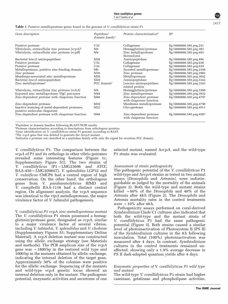
Metalloprotease diversityA search for metalloprotease genes within thederived genome data identified 17 metalloproteases,zinc proteases or putative proteases (Table 1). Theywere dispersed in different contigs and most were
inserted in regions with low conservation among thecompared vibrio genomes. With respect to putativelocalization, some of the proteases were predicted tobe in the membrane, others in the cytoplasm andnine were secreted. Among these genes, there werethree metalloprotease genes from the M4 peptidasefamily. This family comprises metalloproteasesdescribed as virulence factors, including theV. cholerae hemagglutinin/proteases and thermo-lysins, which were orthologous to the vtpA, vtpBand vthA genes from V. tubiashii (V. coralliilyticusnomenclature—vcpA, vcpB and vchA, respectively).Other proteases were found to belong to peptidasefamilies such as the U32 collagenase group, the M6metalloprotease, which is a homolog to the immuneinhibitor A found in V. cholerae, the M50 membranemetalloproteases and the M48 Zn-dependent pro-teases with chaperone function (Table 1). Thisdiverse metalloprotease set and the other viru-lence-related effectors found in the genome indi-cate a high diversity of pathogenic systems in
Figure 1 Blast atlas of the V. coralliilyticus P1 genome. General visualization of differences in gene content in chromosome I (a) andchromosome II (b) using V. cholerae N16961 as the reference genome. The enlarged region (c) depicts the vcpA gene locus (inner redcircle) and its putative homologs. Light colored regions indicate low conservation in gene content, whereas dark colored regions indicatehigh conservation.
Vibrio coralliilyticus genomeE de O Santos et al
1476
The ISME Journal
V. coralliilyticus P1. The comparison between thevcpA of P1 and its orthologs in other vibrio genomesrevealed some interesting features (Figure 1c;Supplementary Figure S1). The two strains ofV. coralliilyticus (P1¼LMG23696 and ATCCBAA-450¼LMG20984T), V. splendidus LGP32 andV. vulnificus CMCP6 had a central region of highconservation. On the other hand, the orthologousgenes from V. parahaemolyticus RIMD andV. campbellii BAA-1116 had a distinct centralregion. On alignment analysis, the vcpA sequencewas identical to the vtpA metalloprotease, the majorvirulence factor of V. tubiashii pathogenesis.
V. coralliilyticus P1 vcpA zinc-metalloprotease mutantThe V. coralliilyticus P1 strain possessed a hemag-glutinin/protease gene, designated as vcpA, similarto a major virulence factor of several vibrios,including V. tubiashii, V. splendidus and V. cholerae(Supplementary Figures S1; Supplementary OnlineMaterial). A vcpA deletion mutant was constructedusing the allelic exchange strategy (see Materialsand methods). The PCR amplicon size of the vcpAgene was B1800 bp in the restored wild type and1000 bp in the mutants obtained by allelic exchange,indicating the internal deletion of the target gene.Approximately 50% of the colonies were positivefor the allelic exchange. Sequencing of the mutantsand wild-type vcpA genetic locus showed aninternal deletion only in the mutant. The pathogenicpotential, enzymatic activities and secretome of one
selected mutant, named DvcpA, and the wild-typeP1 strain was evaluated.
Assessment of strain pathogenicityThe pathogenic potential of the V. coralliilyticus P1wild-type and DvcpA strains as tested in two animalassays (Drosophila and Artemia), were indistin-guishable as judged by the mortality of the animals(Figure 2). Both the wild-type and mutant strainskilled B60% of the Drosophila and 40% of theArtemia after 48 h (Figure 2). The Drosophila andArtemia mortality rates in the control treatmentswere B10% after 48 h.
Pathogenicity assays performed on coral-derivedSymbiodinium Clade C1 cultures also indicated thatboth the wild-type and the mutant strain ofV. coralliilyticus P1 had the same pathogenicpotential (Figure 3). Both strains caused a similarlevel of photoinactivation of Photosystem II (PS II)of the Symbiodinium cultures in the 8 h followinginoculation. Total (100%) photoinactivation wasmeasured after 4 days. In contrast, Symbiodiniumcultures in the control treatments remained un-affected, showing only a 1.9% average decrease inPS II dark-adapted quantum yields after 4 days.
Enzymatic properties of V. coralliilyticus P1 wild typeand mutantThe wild-type V. coralliilyticus P1 strain had highercaseinase, gelatinase and phospholipase activities,
Table 1 Putative metalloprotease genes found in the genome of V. coralliilyticus strain P1
Gene description Peptidase/domain family a
Protein characterizationb IDc
Putative protease U32 Collagenase fig|6666666.585.peg.241Vibriolysin, extracellular zinc protease (vcpA)d M4 Hemagglutinin/protease fig|6666666.585.peg.383Vibriolysin, extracellular zinc protease (vcpB) M4 Zinc metalloprotease,
thermolysinfig|6666666.585.peg.493
Bacterial leucyl aminopeptidase M20 Aminopeptidase fig|6666666.585.peg.494Putative protease U32 Collagenase fig|6666666.585.peg.638Putative protease U32 Collagenase fig|6666666.585.peg.639Metalloprotease, putative zinc-binding domain M6 Secreted metalloprotease fig|6666666.585.peg.2348Zinc protease M16 Zinc protease fig|6666666.585.peg.2982Membrane-associated zinc metalloprotease M50 Metalloprotease fig|6666666.585.peg.3042Bacterial leucyl aminopeptidase M20 Aminopeptidase fig|6666666.585.peg.3342Zinc metalloproteasee PCC domaine Leucine aminopeptidase-
related proteinfig|6666666.585.peg.3343
Vibriolysin, extracellular zinc protease (vchA) M4 Hemagglutinin/protease fig|6666666.585.peg.3360Exported zinc metalloprotease YfgC precursor M48 Zinc metalloprotease fig|6666666.585.peg.3932Zinc-dependent protease with chaperone function M48 Zinc-dependent protease
with chaperone functionfig|6666666.585.peg.4797
Zinc-dependent protease M50 Membrane metalloprotease fig|6666666.585.peg.4798Inactive homolog of metal-dependent proteases,putative molecular chaperone
M22 Glycoprotease fig|6666666.585.peg.4911
Zinc-dependent protease with chaperone function M48 Zinc-dependent proteasewith chaperone function
fig|6666666.585.peg.4997
aPeptidase or domain families following BLAST/NCBI results.bProtease characterization according to descriptions from orthologous proteins.cGene identification on V. coralliilyticus strain P1 genome according to RAST.dThe vcpA gene that was deleted to generate the DvcpA mutant.eIndicates a protease not classified in a peptidase family with only the signal for secretion PCC domain.
Vibrio coralliilyticus genomeE de O Santos et al
1477
The ISME Journal

whereas the DvcpA mutant had higher hemolysinactivity (Table 2). The distinct phenotypes (decreasedproduction of phospholipase and increased pro-duction of hemolysin) point to a change in theenzymatic expression in the mutant in response tothe vcpA deletion.
The proteolytic activity of the secretome of themutant and the wild type were analyzed by gelatinSDS-polyacrylamide gel electrophoresis (Supple-mentary Figure S2). The extracellular proteasefingerprint of the P1 wild type revealed seven major
proteases with a molecular mass range between20 and 105 kDa. These bands were also present inthe mutant, except for one strong zone of clearing (atestimated molecular mass of 66 kDa) (Supplemen-tary Figure S3). This band corresponded to the VcpAzinc-metalloprotease as this protease was not pre-sent in the mutant. Specific enzyme inhibitors wereused to characterize the different bands. EDTAtreatment inhibited all the higher bands (115, 66,51 and 45 kDa). Phenantroline, a metalloproteaseinhibitor, inactivated the 20 kDa band and greatlyreduced the activity of the VcpA zinc-metallopro-tease, a large protease (66 kDa). The serineprotease inhibitor PMSF inactivated an upper band(115 kDa) and one band of 51 kDa (SupplementaryFigure S3). A preparative gel containing concen-trated V. coralliilyticus P1 extracellular productsshowed high levels of serine protease activity. TheE-64, pepstatin, cysteine and aspartic acid peptidaseinhibitors did not inhibit any protease bands (datanot shown).
Secretome of Vibrio coralliilyticus wild-type andmutant strainsThe wild-type and DvcpA mutant strains clearlydisplayed different secretomes as assessed by 2-Delectrophoreses (Supplementary Figure S3). At least11 spots were more intense or only present in themutant secretome, whereas eight spots were specificto the wild type. Further analysis of the secretomesthrough 1-D SDS-polyacrylamide gel electrophoresisanalysis demonstrated fewer protein bands for thewild type when directly compared with the mutantstrain (Supplementary Figure S4). Mass spectro-metry of visible bands excised from the gelsindicated that the wild type and the mutant secreteddistinct groups of proteins (Table 3), possibly inresponse to the vcpA deletion. The analysis of thewild type and the mutant secretomes in two culturemedia (Marine and Tryptone Soy broths) confirmedthe loss of the VcpA metalloprotease expression inthe mutant but not in the wild type. In addition, themutant secreted 18 exclusive proteins in tryptonesoy broth, including four types of metalloproteasesfound previously on the genome (VchA and bacter-ial leucyl aminopeptidase proteins). The mutantalso expressed a chitinase, a hemolysin-relatedprotein RbmC, the Hcp protein and 12 hypotheticalproteins (Table 3). In marine broth, the expression ofthe chitinase and the VthA protein was only seen inthe mutant but the three aminopeptidases, the VcpBthermolysin and an alkaline serine protease weredetected in both strains.
Discussion
Sequencing of the genome of the coral pathogenV. coralliilyticus strain P1 (LMG23696), originallyisolated from diseased corals on the Great Barrier
0
0.1
0.2
0.3
0.4
0.5
0.6
0 10 20 30 40 50 60 70 80 90 100
PS
II d
ark-
adap
ted
quan
tum
yie
ld (
Fv/
Fm
)
Time (h)
P1VN167Control
Figure 3 Photosystem II inactivation of Symbiodinium Clade C1cultures by V. coralliilyticus wild-type and DvcpA strain super-natants. The error bars indicate the mean±s.d.
100a
b
75
50
25
24 48
ControlP1ΔvcpA
00
24Hours
480
Sur
viva
l (%
)
100
75
50
25
0
Sur
viva
l (%
)
Figure 2 Pathogenic potential of the V. coralliilyticus wild-typeand DvcpA strains. Pathogenicity tests using (a) Drosophila and(b) Artemia nauplii. The error bars indicate the mean±s.d. Thecontrol treatments do not contain bacteria.
Vibrio coralliilyticus genomeE de O Santos et al
1478
The ISME Journal

Reef, was shown to possess a diverse repertoireof virulence factors. Most notably, 17 putativemetalloproteases belonging to distinct peptidasefamilies were identified and importantly some ofthese metalloproteases were also detected in thesecretome of P1. Moreover, coding genes thatpotentially improve V. coralliilyticus fitness (suchas stress response, biofilm formation, quorum sen-sing, flagellar proteins and chemotaxis systems)were identified in the genome. The presence of thetfoX regulator and type IV and VI secretion systems,and vgrG and hcp effectors illustrate the genomicversatility and additional factors that are likelyimportant in V. coralliilyticus pathogenicity. Inaddition, several genes responsible for colonizationwere found in the P1 genome. The syp genes, agroup of genes described in V. fischeri that promotesymbiotic colonization and biofilm formation viaSigma54 regulation (Yip et al., 2005), and the flpgenes for biogenesis of type IV Flp pilus described inActinobacillus actinomycetemcomitans (Boyd et al.,2008), may represent important loci for the processof coral colonization. In addition, strain-specificgenes found in the P1 genomes revealed theimportance of phage genes driving intra-speciesdiversification. The P1 genome had several phagegenes that were not present in the LMG 20984Tgenome, indicating that phages may allow genomediversification within the species V. coralliilyticus.
The VcpA zinc-metalloprotease has been identi-fied in previous studies as the key virulence factorlikely important in the bacterial infection of coralsand implicated in coral tissue necrosis (Ben-Haimet al., 2003a, b; Sussman et al., 2008). TheV. coralliilyticus vcpA gene is orthologous to the vtpAgene from the oyster pathogen Vibrio tubiashii andsimilarly identified as central for strain virulence(Hasegawa et al., 2008). A DvcpA V. coralliilyticus P1mutant demonstrated the same pathogenic potentialas the wild-type strain in two animal models and awhole cell Symbiodiunium bioassay evaluated inthis study, highlighting that the pathogenicity ofvibrios in marine animals is a complex interplayof multiple genetic factors and unlikely the result ofone determinant. We suggest that coral infection byV. coralliilyticus depends on several toxins that actin concert to promote bacterial pathogenicity. Themutant has lower caseinase and phospholipaseactivities, but higher hemolysin activity. High levelsof serine protease and metalloprotease activities
were also observed in the wild type. The 66 kDaprotein corresponds to the VcpA zinc-metallopro-tease whose gene was deleted in the DvcpA mutant.Isoforms of the VcpA metalloprotease may alsooccur in the secretome although it is not clear ifisoforms have some proteolytic activity (Miyoshiet al., 1997; Park et al., 2008).
The VcpA loss may activate an alternativemechanism for pathogenesis in DvcpA mutants.The activation of two putative duplicates of thismetalloprotease may be one potential alternativemechanism (Figure 4). We suggest that vcpA, vcpB,and vchA are paralogous genes. VcpB and VchA(both from M4 family) may enable P1 to causedisease as this family is known to be thermolysinswith high proteolytic activity in vibrios (Hasegawaet al., 2008). In addition, the collagenase metallo-peptidases (U32) degrade type I collagens and helpin the invasion of the host. They are homologs of theprtC gene of Bacteroides gingivalis (Kato et al.,1992). The M6 peptidase family comprises theimmune inhibitor A from the insect pathogenicGram-positive Bacillus thuringiensis. This proteincleaves host antibacterial proteins. Homologs of theimmune inhibitor A, named PrtV, have been foundin V. cholerae and V. anguillarum (Vaitkeviciuset al., 2006, 2008). PrtV is required for killingC. elegans and has a cytotoxic effect, leading to celldeath. PrtV can degrade extracellular matrix com-ponents. A Vibrio anguillarum M3 mutant, defectivefor prtV, exhibited decreased growth in turbotintestinal mucus, reduced hemolytic activity onturbot erythrocytes, and increased LD50 in infectionexperiments (Mo et al., 2010). Homologs of theimmune inhibitor A have also been found in othergenomes (Aeromonas hydrophila ATCC 7966 andBacillus thuringiensis BMB171). The presence of aleucine (leucyl) aminopeptidase (LAP) and a (LAP)-related protein in the genome and secretome is animportant finding. LAPs are often viewed as cellmaintenance enzymes with critical roles in turnoverof peptides (Rawlings et al., 2004). Some LAPs maybind DNA enabling them to serve as transcriptionalrepressors that control pyrimidine, alginate andcholera toxin biosynthesis, as well as mediate site-specific recombination events in plasmids andphages (Matsui et al., 2006). LAPs have attractedadditional interest due to their applicability inindustry as whole-cell biocatalysts for the selectivehydrolysis and enzymatic resolution of DL-amino
Table 2 Proteolytic characteristics of the V. coralliilyticus wild type and DvcpA strains
Culture Caseinase Gelatinase Phospholipase against egg yolk Haemolysin (Sheep)
V. coralliilyticus P1 +++ ++ +++ + (b)V. coralliilyticus DvcpA ++ + + +++ (b)
Abbreviations: b, b-haemolysis; +, zone of clearing/opalescence of 1–2 mm; ++, zone of clearing/opalescence of 4–6 mm; +++, zone of clearing/opalescence of X7 mm.
Vibrio coralliilyticus genomeE de O Santos et al
1479
The ISME Journal

Table 3 Secreted proteins identified from the V. coralliilyticus wild type and DvcpA strains
Proteinbanda
IDb Description Proteinmassc
P1 DvcpA
1 fig|6666666.585.peg.2341 3-ketoacyl-CoA thiolase 41 708 X1 fig|6666666.585.peg.1263 ABC transporter, periplasmic spermidine putrescine-binding protein PotD 39 005 X1 fig|6666666.585.peg.33 Amino acid ABC transporter, periplasmic amino acid-binding portion 60 828 X1 fig|6666666.585.peg.4340 Aminoacyl-histidine dipeptidase (peptidase D) 53 915 X1 fig|6666666.585.peg.3512 Arginine deiminase 45 475 X1 fig|6666666.585.peg.1603 Aspartate-semialdehyde dehydrogenase 39 344 X1 fig|6666666.585.peg.3784 D-3-phosphoglycerate dehydrogenase 44 631 X1 fig|6666666.585.peg.3679 Dihydrolipoamide dehydrogenase of pyruvate dehydrogenase complex 51 256 X1 fig|6666666.585.peg.3268 Glutamine synthetase type I 51 623 X1 fig|6666666.585.peg.4475 Heat shock protein 60 family chaperone GroEL 57 590 X1 fig|6666666.585.peg.2448 Maltoporin 43 893 X1 fig|6666666.585.peg.383 Vibriolysin (neutral protease precursor) VcpAd 65 881 X1 fig|6666666.585.peg.3939 Outer membrane protein NlpB, lipoprotein component of the protein
assembly complex38 529 X
1 fig|6666666.585.peg.5024 Oligopeptide ABC transporter, periplasmic oligopeptide-bindingprotein OppA
60 828 X
1 fig|6666666.585.peg.3514 Ornithine carbamoyltransferase 38 107 X1 fig|6666666.585.peg.667 Outer membrane stress sensor protease DegQ (serine protease) 48 032 X1 fig|6666666.585.peg.158 Outer membrane vitamin B12 receptor BtuB 67 400 X1 fig|6666666.585.peg.3681 Pyruvate dehydrogenase E1 component 99 539 X1 fig|6666666.585.peg.3925 UDP-sugar hydrolase 61 372 X3 fig|6666666.585.peg.2503 Hemolysin-related protein RbmC 103 393 X3 fig|6666666.585.peg.796 Hypothetical protein (VIC_004311) 35 219 X3 fig|6666666.585.peg.3372 Hypothetical protein (putative hydroxymethyltransferase) 110 770 X4 fig|6666666.585.peg.2748 Chitinase 91 709 X4 fig|6666666.585.peg.3507 Extracellular deoxyribonuclease Xds 94 471 X4 fig|6666666.585.peg.761 Flagellar hook-associated protein FliD 73 020 X4 fig|6666666.585.peg.3361 Hypothetical protein (VIC_001630) 68 635 X4 fig|6666666.585.peg.203 Hypothetical protein (VIC_002088) 84 685 X4 fig|6666666.585.peg.526 Acyl-CoA dehydrogenase, short-chain specific (peptidase S8 and
S53 subtilisin kexin sedolisin)71 910 X
5 fig|6666666.585.peg.494 Bacterial leucyl aminopeptidase precursor (aminopeptidase)e 56 432 X5 fig|6666666.585.peg.3342 Bacterial leucyl aminopeptidase precursore 54 368 X5 fig|6666666.585.peg.2006 Endonuclease I 59 494 X5 fig|6666666.585.peg.1918 Hypothetical protein (VIC_003980) 56 620 X5 fig|6666666.585.peg.3343 Zinc metalloprotease (leucine aminopeptidase-related protein)e 52 531 X6 fig|6666666.585.peg.3360 Vibriolysin (hemolysin/cytolysin) VchA 53 487 X6 fig|6666666.585.peg.4379 Peptidyl-prolyl cis-trans isomerase SurA 48 390 X7 fig|6666666.585.peg.2756 Hypothetical protein (VIC_002899) 45 749 X8 fig|6666666.585.peg.1029 Flagellar hook protein FlgE 46 945 X8 fig|6666666.585.peg.832 TraF-related protein (hypothetical protein VIC_003016) 40 308 X8 fig|6666666.585.peg.3345 Spindolin-related protein 43 472 X9 fig|6666666.585.peg.55 Hypothetical protein (VIC_000374) 36 006 X9 fig|6666666.585.peg.1802 Hypothetical protein (VIC_003092) 37 704 X9 fig|6666666.585.peg.3320 Putative outer membrane protein (nonspecific porin) 37 176 X1 and 9 fig|6666666.585.peg.5035 Alanine dehydrogenase 40 075 X X1 and 9 fig|6666666.585.peg.759 Flagellin protein FlaA (flagellin B) 39 710 X X1 and 9 fig|6666666.585.peg.3950 Flagellin protein FlaC 40 232 X X1 and 9 fig|6666666.585.peg.3951 Flagellin protein FlaD 39 782 X X1 and 9 fig|6666666.585.peg.2588 Hypothetical protein (VIC_003990) 41 373 X X1 and 9 fig|6666666.585.peg.1548 Hypothetical protein (VIC_004396) 38 409 X X1 and 9 fig|6666666.585.peg.621 Outer membrane protein OmpU 37 027 X X1 and 9 fig|6666666.585.peg.2406 Protease precursor (serine protease/subtilase peptidase) 62 976 X X1 and 9 fig|6666666.585.peg.659 Type I secretion TolC precursor 47 435 X X10 fig|6666666.585.peg.2563 Hypothetical protein (VIC_001560) 29 960 X11 fig|6666666.585.peg.4653 Hcp 19 444 X11 fig|6666666.585.peg.3359 Hypothetical protein (VIC_001628) 31 874 X12 fig|6666666.585.peg.5132 Hypothetical protein (VIC_003927) 18 030 X
Abbreviation: ABC, ATP-binding cassette transporters. Reference numbers denoted in parentheses in protein descriptions refer to the location onthe Vibrio coralliilyticus ATCC BAA-450 genome. X symbol denotes the identification of proteins in each secretome sample.aNumber of the gel bands (Supplementary Figure S4) whose proteins were identified. Proteins were identified from secretomes of the P1 and theDvcpA mutant grown on TSB.bGene identification on V. coralliilyticus P1 genome according to RAST.cTheoretical protein mass (daltons) provided by MASCOT Search with the use of the Expasy Tool Compute software.dSymbol points to the protein corresponding to the vcpA gene, which was deleted to generate the DvcpA mutant.eProteins identified in P1 and the mutant secretome when grown in marine broth.
Vibrio coralliilyticus genomeE de O Santos et al
1480
The ISME Journal

acid amide racemates (Kamphuis et al., 1992;Hermes et al., 1993).
The comparative analysis of the secretome ofwild-type and mutant V. coralliilyticus P1 strainsidentified distinct differences in protein secretion asa result of the deletion of the vcpA gene. Thisobservation indicates that the VcpA zinc-metallo-protease might have a regulatory role on V. corallii-lyticus P1 gene expression. The lack of vcpAexpression in the mutant and the expression ofseveral proteins (chitinase, hemolysin/cytolysinVthA, aminopeptidases (LAPs), leucine aminopep-tidase-related protein, hemolysin-related proteinRbmC, peptidase S8 and S53 subtilisin kexinsedolisin, Hcp protein) only by the mutant supportsthis conclusion. In addition, the presence of thehemolysin VchA in the mutant secretome, corrobo-rated by its increased hemolytic activity in theenzymatic tests may suggest regulation of hemolysinproduction by the deleted zinc-metalloprotease. InV. tubiashii, the zinc-metalloprotease VtpA regulatesVthA hemolysin expression by means of a posttran-scriptional mechanism (Hasegawa and Hase, 2009).The VtpA mutant of V. tubiashii had decreasedpathogenic potential in C. gigas larvae (Hasegawaet al., 2008). The hyper expression of VthA did notincrease the pathogenic potential of V. tubiashii in
C. gigas. Further work is required to determine theexact role of VcpA in gene expression regulation inV. coralliilyticus P1.
Coral disease is a complex multifactorial processinvolving environmental triggers and holobiontresponses. The study highlights that bacterial infec-tion of corals and the virulence (protease) factorsinvolved are highly complex and that coral diseaseis likely the result of an interplay of multiple factorsrather than one determinant. The high diversity andactivity of proteases observed in V. coralliilyticus P1linked with its rapid growth capacity under opti-mum environmental conditions indicates that itmay have a central role in the homeostasis andhealth status of the reef systems.
Acknowledgements
We would like to thank the Luz Sıncroton NationalLaboratory for the use of the nano-LC-MS/MS massspectrometer and Proteomic Network of Rio de Janeirofor equipment and reagent lending. We thank GabrielleV Souza for her excellent technical assistance. This studywas partially funded by grants from the National Councilof Technological and Scientific Development, Brazil(CNPq). BW thanks a Smart state postdoctoral fellowship.FLT and EOS have CNPq fellowships.
Figure 4 Phylogenetic tree based on the 600 AA sequence of VcpA and its homologues. The tree was constructed using the poison andneighbor joining methods. The scale bar indicates 10% sequence divergence. The bootstrap values of 1000 repetitions are indicated at thenodes.
Vibrio coralliilyticus genomeE de O Santos et al
1481
The ISME Journal

References
Alves Jr N, Maia Neto OS, Silva BO, Moura RL, Francini-Filho RB, Barreira e Castro C et al. (2010). Diversityand pathogenic potential of vibrios isolated fromAbrolhos Bank coral. Environ Microbiol Reports 2:90–95.
Austin B, Austin D, Sutherland R, Thompson F, Swings J.(2005). Pathogenicity of vibrios to rainbow trout(Oncorhynchus mykiss, Walbaum) and Artemianauplii. Environ Microbiol 7: 1488–1495.
Aziz RK, Bartels D, Best AA, DeJongh M, Disz T, EdwardsRA et al. (2008). The RAST Server: rapid annotationsusing subsystems technology. BMC Genomics 8: 9–75.
Ben-Haim Y, Thompson FL, Thompson CC, CnockaertMC, Hoste B, Swing J et al. (2003a). Vibrio coralliily-ticus sp. nov., a temperature-dependent pathogen ofthe coral Pocillopora damicornis. Int J Syst EvolMicrobiol 53: 309–315.
Ben-Haim Y, Zicherman-Keren M, Rosenberg E. (2003b).Temperature-regulated bleaching and lysis of thecoral Pocillopora damicornis by the novel pathogenVibrio coralliilyticus. Appl Environ Microbiol 69:4236–4242.
Binesse J, Delsert C, Saulnier D, Champomier-Verge‘s M,Zagorec M, Munier-Lehmann H et al. (2008). Metallo-protease vsm is the major determinant of toxicity forextracellular products of Vibrio splendidus. ApplEnviron Microbiol 74: 7108–7117.
Blow NS, Salomon RN, Garrity K, Reveillaud I, Kopin A,Jackson FR et al. (2005). Vibrio cholerae infection ofDrosophila melanogaster mimics the human diseasecholera. PLoS Pathogens 1: 1–14.
Bourne DG, Garren M, Work TM, Rosenberg E, Smith GW,Harvell CD. (2009). Trends Microbiol 17: 554–562.
Bourne DG, Munn CB. (2005). Diversity of bacteriaassociated with the coral Pocillopora damicornisfrom the Great Barrier Reef. Environ Microbiol 7:1162–1174.
Boyd JM, Dacanay A, Knickle LC, Touhami A, Brown LL,Jericho MH et al. (2008). Contribution of type IV pili tothe virulence of Aeromonas salmonicida subsp.salmonicida in atlantic salmon (Salmo salar L.). InfecImun 76: 1445–1455.
Chimetto LA, Brocchi M, Thompson CC, Martins RC,Ramos HR, Thompson FL. (2008). Vibrios dominate asculturable nitrogen-fixing bacteria of the Braziliancoral Mussismilia hispida. Syst Appl Microbiol 31:312–319.
Coelho A, de Oliveira Santos E, Faria ML, de Carvalho DP,Soares MR, von Kruger WM et al. (2004). A proteomereference map for Vibrio cholerae El Tor. Proteomics 4:1491–1504.
Denkin SM, Nelson DR. (1999). Induction of proteaseactivity in Vibrio anguillarum by gastrointestinalmucus. Appl Environ Microbiol 65: 3555–3560.
Dinsdale EA, Pantos O, Smriga S, Edwards RA, Angly F,Wegley L et al. (2008). Microbial ecology of fourcoral atolls in the Northern Line Islands. PLoS One 3:e1584.
Hallin PF, Binnewies TT, Ussery DW. (2008). The genomeBLAST atlas-a Genewiz extension for visualization ofwhole genome homology. MolBiosyst 4: 363–371.
Hasegawa H, Hase CC. (2009). The extracellularmetalloprotease of Vibrio tubiashii directly inhibitsits extracellular haemolysin. Microbiology 155:2296–2305.
Hasegawa H, Lind EJ, Boin MA, Hase CC. (2008).The extracellular metalloprotease of Vibriotubiashii is a major virulence factor for pacific oyster(Crassostrea gigas) larvae. Appl Environ Microbiol 74:4101–4110.
Hellman U, Wernstedt C, Gonez J, Heldin CH. (1995).Improvement of an ‘In-Gel’ digestion procedure forthe micropreparation of internal protein fragmentsfor amino acid sequencing. Anal Biochem 224:451–455.
Hermes HF, Sonke T, Peters PJ, van Balken JA, Kamphuis J,Dijkhuizen L et al. (1993). Purification and character-ization of an L-aminopeptidase from Pseudomonasputida ATCC 12633. Appl Environ Microbiol 59:4330–4334.
Heussen C, Dowdle EB. (1980). Electrophoretic analysis ofplasminogen activators in polyacrylamide gels con-taining sodium dodecyl sulphate and copolymerizedsubstrates. Anal Biochem 102: 196–202.
Hughes TP, Baird AH, Bellwood DR, Card M, Connolly SR,Folke C et al. (2003). Climate change, humanimpacts, and the resilience of coral reefs. Science301: 929–933.
Ishikura M, Hagiwara K, Takishita K, Haga M, Iwai K,Maruyama T. (2004). Isolation of new Symbiodiniumstrains from Tridacnid giant clam (Tridacna crocea)and sea slug (Pteraeolidia ianthina) using culturemedium containing giant clam tissue homogenate.Mar Biotechnol 6: 378–385.
Kamphuis E, Meijer M, Boesten WH, Sonke T, van denTweel WJ, Schoemaker HE. (1992). New developmentsin the synthesis of natural and unnatural amino acids.Ann N Y Acad Sci 672: 510–527.
Kato T, Takahashi N, Kuramitsu HK. (1992). Sequenceanalysis and characterization of the Porphyromonasgingivalis prtC gene, which expresses a novel collage-nase activity. J Bacteriol 174: 3889–3895.
Kiessling W, Simpson C, Foote M. (2010). Reefs as cradlesof evolution and sources of biodiversity in thePhanerozoic. Science 327: 196–198.
Knowlton N, Jackson JB. (2008). Shifting baselines, localimpacts, and global change on coral reefs. PLoS Biol6: e54.
Le Roux F, Binesse J, Saulnier D, Mazel D. (2007).Construction of a Vibrio splendidus mutant lackingthe metalloprotease gene vsm by use of a novelcounterselectable suicide vector. Appl Environ Micro-biol 73: 777–784.
Lery LM, Coelho A, von Kruger WM, Goncalves MS,Santos MF, Valente RH et al. (2008). A comparativeproteomic analysis of Gluconacetobacter diazotrophi-cus PAL5 at exponential and stationary phases ofcultures in the presence of high and low levels ofinorganic nitrogen compound. Biochim Biophys Acta1784: 1578–1589.
Liu PC, Lee KK, Chen SN. (1996). Pathogenicity ofdifferent isolates of Vibrio harveyi in tigerprawn, Penaeus monodon. Lett Appl Microbiol 22:413–416.
Loghothetis PN, Austin B. (1996). Variations in antigeni-city of Aeromonas hydrophila strains in rainbow trout(Oncorhynchus mykiss, Walbaum): an associationwith surface characteristics. Fish Shellfish Immunol6: 47–55.
Luna GM, Biavasco F, Danovaro R. (2007). Bacteriaassociated with the rapid tissue necrosis of stonycorals. Environ Microbiol 9: 1851–1857.
Vibrio coralliilyticus genomeE de O Santos et al
1482
The ISME Journal

Margulies M, Egholm M, Altman WE et al. (2005). Genomesequencing in microfabricated high-density picolitrereactors. Nature 437: 376–380.
Matsui M, Fowler JH, Walling LL. (2006). Leucineaminopeptidases: diversity in structure and function.Biol Chem 387: 1535–1544.
Milton DL, Norqvist A, Wolf-Watz H. (1992). Cloning of ametalloprotease gene involved in the virulence mechan-ism of Vibrio anguillarum. J Bacteriol 174: 7235–7244.
Miyoshi S-I, Wakae H, Tomochika K-I, Shinoda S.(1997). Functional domains of a zinc metalloproteasefrom Vibrio vulnificus. J Bacteriol 179: 7606–7609.
Mo Z, Guo D, Mao Y, Ye X, Zou Y, Xiao P et al. (2010).Identification and characterization of the Vibrioanguillarum prtV gene encoding a new metallopro-tease. Chinese J Oceanol Limnol 28: 1–14.
Park J, Ryu S-Y, Kim C–M, Shin S-H. (2008). Two forms ofVibrio vulnificus metalloprotease VvpE are secretedvia the type II general secretion system. J Microbiol 46:338–343.
Pedersen AG, Jensen LJ, Brunak S, Staerfeldt HH, UsseryDW. (2000). A DNA structural atlas for Escherichiacoli. J Mol Biol 299: 907–930.
Pitcher DG, Saunders NA, Owen RJ. (1989). Rapidextraction of bacterial genomic DNA with guanidiumthiocyanate. Lett Appl Microbiol 8: 151–156.
Rawlings ND, Tolle DP, Barrett AJ. (2004). Evolutionaryfamilies of peptidase inhibitors. Biochem J 378: 705–716.
Rohwer F, Youle M. (2010). Coral Reefs in the MicrobialSeas. Plaid Press: Basalt, CO, 204 p.
Sussman M, Mieog JC, Doyle J, Victor S, Willis BL, BourneDG. (2009). Vibrio zinc-metalloprotease causes photo-inactivation of coral endosymbionts and coral tissuelesions. PLoS ONE 4: 1–14.
Sussman M, Willis BL, Victor S, Bourne DG. (2008). Coralpathogens identified for white syndrome epizootics inthe Indo-Pacific. PLoS ONE 3: 1–14.
Sutherland KP, Porter JW, Torres DG. (2004). Disease andimmunity in Caribbean and Indo-Pacific zooxanthel-late corals. Mar Ecol Prog Ser 266: 273–302.
Vaitkevicius K, Lindmark B, Ou G, Song T, Toma C,Iwanaga M et al. (2006). A Vibrio cholerae proteaseneeded for killing of Caenorhabditis elegans has a rolein protection from natural predator grazing. Proc NatlAcad Sci USA 103: 9280–9285.
Vaitkevicius K, Rompikuntal PK, Lindmark B, Vaitkevi-cius R, Song T, Wai SN. (2008). The metalloproteasePrtV from Vibrio cholerae. FEBS J 275: 3167–3177.
Van Oppen MJH, Palstra FP, Piquet AMT, Miller DJ.(2001). Patterns of coral-dinoflagellate associations inAcropora: significance of local availability and phy-siology of Symbiodinium strains and host-symbiontselectivity. Proc R Soc Lond [Biol] 268: 1759–1767.
Varina M, Denkin SM, Staroscik AM, Nelson DR. (2008).Identification and characterization of Epp, thesecreted processing protease for the Vibrio anguillarumEmpA metalloprotease. J Bacteriol 190: 6589–6597.
Vega Thurber R, Willner-Hall D, Rodriguez-Mueller B,Desnues C, Edwards RA, Angly F et al. (2008).Metagenomic analysis of stressed coral holobionts.Environ Microbiol 11: 2148–2163.
Wang J, Sasaki T, Maehara Y, Nakao H, Tsuchiya T,Miyoshi S. (2008). Variation of extracellular proteasesproduced by Vibrio vulnificus clinical isolates: Genet-ic diversity of the metalloprotease gene (vvp), andserine protease secretion by vvp-negative strains.Microb Pathogenesis 44: 494–500.
Wilkinson C. (2008). Status of Coral Reefs of the World2008. Australian Institute of Marine Science:Townsville, Australia.
Yip ES, Grublesky BT, Hussa EA, Visick KL. (2005). Anovel, conserved cluster of genes promotes symbioticcolonization and sigma-dependent biofilm formationby Vibrio fischeri. Mol Microbiol 57: 1485–1498.
Supplementary Information accompanies the paper on The ISME Journal website (http://www.nature.com/ismej)
Vibrio coralliilyticus genomeE de O Santos et al
1483
The ISME Journal