Functional interactions between BRCA1 and the checkpoint kinase ATR during genotoxic stress
14
Functional interactions between BRCA1 and the checkpoint kinase ATR during genotoxic stress Randal S. Tibbetts, 1 David Cortez, 2 Kathryn M. Brumbaugh, 1 Ralph Scully, 3 David Livingston, 3 Stephen J. Elledge, 2 and Robert T. Abraham 1,4 1 Department of Pharmacology and Cancer Biology, Duke University Medical Center, Durham, North Carolina 27710, USA; 2 Department of Biochemistry and Molecular Biology and the Howard Hughes Medical Institute, Baylor College of Medicine, Houston, Texas 77030, USA; 3 Dana Farber Cancer Institute, Harvard Medical School, Boston, Massachusetts 02115, USA The BRCA1 gene encodes a tumor suppressor that is mutated in 50% of familial breast cancers. The BRCA1 protein has been implicated in the DNA damage response, as DNA damage induces the phosphorylation of BRCA1 and causes its recruitment into nuclear foci that contain DNA repair proteins. The ataxia-telangiectasia-mutated (ATM) gene product controls overall BRCA1 phosphorylation in response to -irradiation (IR). In this study, we show that BRCA1 phosphorylation is only partially ATM dependent in response to IR and ATM independent in response to treatment with UV light, or the DNA replication inhibitors hydroxyurea (HU) and aphidicolin (APH). We provide evidence that the kinase responsible for this phosphorylation is the ATM-related kinase, ATR. ATR phosphorylates BRCA1 on six Ser/Thr residues, including Ser 1423, in vitro. Increased expression of ATR enhanced the phosphorylation of BRCA1 on Ser 1423 following cellular exposure to HU or UV light, whereas doxycycline-induced expression of a kinase-inactive ATR mutant protein inhibited HU- or UV light-induced Ser 1423 phosphorylation in GM847 fibroblasts, and partially suppressed the phosphorylation of this site in response to IR. Thus, ATR, like ATM, controls BRCA1 phosphorylation in vivo. Although ATR isolated from DNA-damaged cells does not show enhanced kinase activity in vitro, we found that ATR responds to DNA damage and replication blocks by forming distinct nuclear foci at the sites of stalled replication forks. Furthermore, ATR nuclear foci overlap with the nuclear foci formed by BRCA1. The dramatic relocalization of ATR in response to DNA damage points to a possible mechanism for its ability to enhance the phosphorylation of substrates in response to DNA damage. Together, these results demonstrate that ATR and BRCA1 are components of the same genotoxic stress-responsive pathway, and that ATR directly phosphorylates BRCA1 in response to damaged DNA or stalled DNA replication. [Key Words: BRCA1; ATR; checkpoint; DNA damage; phosphorylation; replication] Received September 14, 2000; revised version accepted October 3, 2000. Damage to the genome, which can occur as a conse- quence of exposure to environmental agents or to the byproducts of oxidative metabolism, represents a persis- tent threat to genomic integrity and cellular viability. The deleterious effects of genotoxic stress are countered by surveillance mechanisms that monitor the genome for the presence of genetic lesions, including single- stranded DNA (ssDNA), DNA double-strand breaks (DSBs), and chemically modified nucleotide bases. The DNA damage response pathway that responds to these lesions consists of a cascade of protein kinases that con- trol the phosphorylation of a number of effector proteins involved in DNA repair and cell cycle control. Central among these effectors is the BRCA1 protein. BRCA1 is a tumor suppressor gene mutated in 50% of familial breast and ovarian cancers (Easton et al. 1993). BRCA1 encodes an 1863-amino-acid nuclear phosphoprotein that is es- sential for viability in mice (Chen et al. 1996; Hakem et al. 1996; Bertwistle and Ashworth 1998; Zhang et al. 1998). Although BRCA1 has been reported to act as a transcription factor (Chapman and Verma 1996) and cell growth suppressor (Monteiro et al. 1996; Somasundaram et al. 1997; Aprelikova et al. 1999), the tumor suppressor functions of BRCA1 may be most closely related to its role in DNA repair and recombination (Zhang et al. 1998). BRCA1-deficient cells display spontaneous chro- mosomal abnormalities and defects in both homologous DNA recombination and transcription-coupled repair of oxidative base damage (Gowen et al. 1998; Moynahan et al. 1999; Xu et al. 1999). Cells that express a truncated 4 Corresponding author. E-MAIL [email protected]; FAX (919) 681-8463. Article and publication are at www.genesdev.org/cgi/doi/10.1101/ gad.851000. GENES & DEVELOPMENT 14:2989–3002 © 2000 by Cold Spring Harbor Laboratory Press ISSN 0890-9369/00 $5.00; www.genesdev.org 2989
-
Upload
independent -
Category
Documents
-
view
0 -
download
0
Transcript of Functional interactions between BRCA1 and the checkpoint kinase ATR during genotoxic stress
Functional interactions between BRCA1and the checkpoint kinase ATR duringgenotoxic stressRandal S. Tibbetts,1 David Cortez,2 Kathryn M. Brumbaugh,1 Ralph Scully,3 David Livingston,3
Stephen J. Elledge,2 and Robert T. Abraham1,4
1Department of Pharmacology and Cancer Biology, Duke University Medical Center, Durham, North Carolina 27710, USA;2Department of Biochemistry and Molecular Biology and the Howard Hughes Medical Institute, Baylor College of Medicine,Houston, Texas 77030, USA; 3Dana Farber Cancer Institute, Harvard Medical School, Boston, Massachusetts 02115, USA
The BRCA1 gene encodes a tumor suppressor that is mutated in 50% of familial breast cancers. The BRCA1protein has been implicated in the DNA damage response, as DNA damage induces the phosphorylation ofBRCA1 and causes its recruitment into nuclear foci that contain DNA repair proteins. Theataxia-telangiectasia-mutated (ATM) gene product controls overall BRCA1 phosphorylation in response to�-irradiation (IR). In this study, we show that BRCA1 phosphorylation is only partially ATM dependent inresponse to IR and ATM independent in response to treatment with UV light, or the DNA replicationinhibitors hydroxyurea (HU) and aphidicolin (APH). We provide evidence that the kinase responsible for thisphosphorylation is the ATM-related kinase, ATR. ATR phosphorylates BRCA1 on six Ser/Thr residues,including Ser 1423, in vitro. Increased expression of ATR enhanced the phosphorylation of BRCA1 on Ser1423 following cellular exposure to HU or UV light, whereas doxycycline-induced expression of akinase-inactive ATR mutant protein inhibited HU- or UV light-induced Ser 1423 phosphorylation in GM847fibroblasts, and partially suppressed the phosphorylation of this site in response to IR. Thus, ATR, like ATM,controls BRCA1 phosphorylation in vivo. Although ATR isolated from DNA-damaged cells does not showenhanced kinase activity in vitro, we found that ATR responds to DNA damage and replication blocks byforming distinct nuclear foci at the sites of stalled replication forks. Furthermore, ATR nuclear foci overlapwith the nuclear foci formed by BRCA1. The dramatic relocalization of ATR in response to DNA damagepoints to a possible mechanism for its ability to enhance the phosphorylation of substrates in response toDNA damage. Together, these results demonstrate that ATR and BRCA1 are components of the samegenotoxic stress-responsive pathway, and that ATR directly phosphorylates BRCA1 in response to damagedDNA or stalled DNA replication.
[Key Words: BRCA1; ATR; checkpoint; DNA damage; phosphorylation; replication]
Received September 14, 2000; revised version accepted October 3, 2000.
Damage to the genome, which can occur as a conse-quence of exposure to environmental agents or to thebyproducts of oxidative metabolism, represents a persis-tent threat to genomic integrity and cellular viability.The deleterious effects of genotoxic stress are counteredby surveillance mechanisms that monitor the genomefor the presence of genetic lesions, including single-stranded DNA (ssDNA), DNA double-strand breaks(DSBs), and chemically modified nucleotide bases. TheDNA damage response pathway that responds to theselesions consists of a cascade of protein kinases that con-trol the phosphorylation of a number of effector proteinsinvolved in DNA repair and cell cycle control. Central
among these effectors is the BRCA1 protein. BRCA1 is atumor suppressor gene mutated in 50% of familial breastand ovarian cancers (Easton et al. 1993). BRCA1 encodesan 1863-amino-acid nuclear phosphoprotein that is es-sential for viability in mice (Chen et al. 1996; Hakem etal. 1996; Bertwistle and Ashworth 1998; Zhang et al.1998). Although BRCA1 has been reported to act as atranscription factor (Chapman and Verma 1996) and cellgrowth suppressor (Monteiro et al. 1996; Somasundaramet al. 1997; Aprelikova et al. 1999), the tumor suppressorfunctions of BRCA1 may be most closely related to itsrole in DNA repair and recombination (Zhang et al.1998). BRCA1-deficient cells display spontaneous chro-mosomal abnormalities and defects in both homologousDNA recombination and transcription-coupled repair ofoxidative base damage (Gowen et al. 1998; Moynahan etal. 1999; Xu et al. 1999). Cells that express a truncated
4Corresponding author.E-MAIL [email protected]; FAX (919) 681-8463.Article and publication are at www.genesdev.org/cgi/doi/10.1101/gad.851000.
GENES & DEVELOPMENT 14:2989–3002 © 2000 by Cold Spring Harbor Laboratory Press ISSN 0890-9369/00 $5.00; www.genesdev.org 2989

version of BRCA1 (Tomlinson et al. 1998) are hypersen-sitive to DNA damaging agents and display slowed ki-netics of DSB repair (Cortez et al. 1999; Scully et al.1999; Zhong et al. 1999). BRCA1 physically associateswith proteins implicated in homologous and nonho-mologous DNA recombination, including Rad51 and theRad50–Mre11–p95 DNA repair complex (Scully et al.1997; Zhong et al. 1999; Wang et al. 2000). However, theprecise contributions of BRCA1 to cell cycle checkpointactivation and DNA repair remain unclear.
BRCA1 is maximally expressed during S phase. Expo-sure of S-phase cells to �-radiation (IR), UV light, or theDNA replication inhibitor hydroxyurea (HU) results inthe rapid phosphorylation of BRCA1, indicating thatBRCA1 is a target of the DNA damage response pathway(Scully et al. 1997a). Moreover, these agents induce dra-matic alterations in the nuclear localization pattern ofBRCA1. In the absence of damage, BRCA1 is localized todiscrete nuclear foci during both S and G2 phases of thecell cycle (Scully et al. 1996, 1997a). Exposure of S-phasecells to IR or HU induces the relocalization of BRCA1 tonew foci, some of which are sites of DNA synthesis(Scully et al. 1997a). Recent studies have demonstrated arole for ATM and the ATM-regulated kinase Chk2 in thephosphorylation of BRCA1 in IR-damaged cells (Cortezet al. 1999; Gatei et al. 2000; Lee et al. 2000). WhereasATM controls the overall phosphorylation of BRCA1 inresponse to IR as judged by electrophoretic mobility-shift alterations, the regulated phosphorylation of indi-vidual sites in response to IR has not been examined.Furthermore, the protein kinase(s) responsible forBRCA1 phosphorylation in response to HU or UV lightare unknown.
A candidate kinase in the DNA damage response path-way that may play a role in the ATM-independent regu-lation of BRCA1 is ATR. ATR is a member of a family ofhigh molecular mass protein kinases whose catalytic do-mains bear sequence similarity to those of phosphoi-nositide 3-kinases (PI3-Ks) (Keith and Schreiber 1995;Bentley et al. 1996; Cimprich et al. 1996). Within the PIKfamily, ATR is most closely related to ATM, Rad3 andTel1 (Schizosaccharomyces pombe), Mec1 and Tel1 (Sa-charomyces cerevisiae), and Mei-41 (Drosophila mela-nogaster) (Zhou and Elledge 2000). Mutations in MEC1and rad3 confer hypersensitivity to a variety of agentsincluding IR, methylmethanesulfonate (MMS), UV light,and HU. These mutants are also defective in both theDNA damage-induced G2 checkpoint and the DNA rep-lication checkpoint, which prevents mitotic entry in thepresence of unreplicated DNA. Recent studies suggestthat the HU sensitivity of mec1 mutant yeast strains isnot solely the result of aberrant mitotic entry, but mayalso be a consequence of aborted chromosomal replica-tion (Desany et al. 1998) and defects in suppression oflate origin firing (Santocanale and Diffley 1998). BecauseMec1 and Rad3 are the closest ATR homologs, ATR mayperform similar S-phase surveillance functions in mam-malian cells.
Accumulating evidence suggests that ATM and ATRcarry out distinct as well as partially overlapping genome
surveillance activities in mammalian cells (Keegan et al.1996; Cliby et al. 1998; Tibbetts et al. 1999). Inactivatingmutations in ATM are responsible for the chromosomalinstability syndrome, ataxia-telangiectasia (A-T). Cellsisolated from A-T patients (Kastan et al. 1992; Beamishand Lavin 1994; Canman et al. 1994; Beamish et al.1996;), or from ATM-nullizygous mice (Barlow et al.1996, 1997; Xu and Baltimore 1996; Xu et al. 1996; West-phal et al. 1997), display defective DNA damage check-points, hypersensitivity to IR and radiomimetic drugs,and elevated frequencies of chromosomal abnormalities.The IR-induced phosphorylation of replication protein A(Liu and Weaver 1993), c-Abl (Shafman et al. 1997), p53(Siliciano et al. 1997), Mdm2 (Khosravi et al. 2000),hChk2/Cds1 (Matsuoka et al. 1998; Brown et al. 1999),and BRCA1 (Cortez et al. 1999; Gatei et al. 2000; Zhouand Elledge 2000) is compromised in A-T cell lines, in-dicating that ATM regulates the activities of multipledownstream effectors. In comparison to ATM, the cellu-lar functions of ATR are less understood, due in part tothe lack of ATR-deficient cell lines. Nonetheless, over-expression of a catalytically inactive ATR mutant pro-tein (ATRKI) compromises G2 checkpoint function, anddecreases cell survival in response to IR, alkylatingagents, and HU (Cliby et al. 1998). The observation thatoverexpression of ATR complements the radioresistantDNA synthesis (RDS) defect of A-T cells provided thefirst genetic evidence that ATR and ATM can performoverlapping functions in mammalian cells (Cliby et al.1998).
Despite considerable overlap in terms of substratephosphorylation (Canman et al. 1998; Kim et al. 1999;Lakin et al. 1999; Tibbetts et al. 1999), the relative con-tributions of ATM and ATR vary with the type of geno-toxic stress. Whereas cells that overexpress ATRKI arehypersensitive to HU, UV light, and IR (Cliby et al. 1998;Wright et al. 1998), ATM-deficient cells retain near nor-mal resistance to UV and HU, but are extremely sensi-tive to IR. These findings and others support a model inwhich ATM and ATR respond to distinct, as well aspartially overlapping types of genotoxic stress. Finally,functional distinctions between ATM and ATR are high-lighted by the drastically different phenotypes associatedwith disruption of their respective genes. Whereas ATM-nullizygous mice survive to adulthood (Barlow et al.1996; Xu et al. 1996), ATR deficiency results in earlyembryonic lethality (Brown and Baltimore 2000; de Kleinet al. 2000). ATR−/− cells display accelerated chromo-somal fragmentation, suggesting that functional ATR isessential for chromosome maintenance in the absence ofenvironmentally imposed genotoxic stress (Brown andBaltimore 2000). Thus, ATR appears to carry out genomesurveillance functions during both normal cell cyclesand cellular exposure to DNA-damaging agents.
In this report, we provide evidence that BRCA1 is adirect target of the ATR kinase. Furthermore, we provideevidence supporting the hypothesis that ATM and ATRprovide overlapping as well as distinct regulatory func-tions to control BRCA1 phosphorylation in response togenotoxic stress.
Tibbetts et al.
2990 GENES & DEVELOPMENT

Results
DNA damage-induced phosphorylation of BRCA1at Ser 1423
Recently, we demonstrated that ATM is required for theoptimal phosphorylation of endogenous BRCA1 in �-ir-radiated cells (Cortez et al. 1999). The results obtainedwith Ser�Ala substitution mutants strongly suggestedthat ATM phosphorylates BRCA1 on Ser 1423 in IR-damaged cells; however, ATM-dependent phosphoryla-tion of this site was not determined directly in intactcells. In the present studies, we monitored the phos-phorylation of BRCA1 at Ser 1423 in intact cells by gen-eration of phosphoSer 1423-specific antibodies (�-pS-1423). In asynchronously cycling cells, the �-pS-1423 an-
tibodies detected a 220-kD protein in human embyronickidney (HEK) 293T cell extracts that comigrated withthe BRCA1 protein and was absent in BRCA1-deficientHCC1937 cells (Fig. 1A; data not shown). Treatment ofcell extracts with � phosphatase (Fig. 1A), or mutation ofSer 1423 to Ala (data not shown) eliminated recognitionby �-pS-1423, indicating that the �-pS-1423 antibodyspecifically recognizes BRCA1 phosphorylated on Ser1423. In subsequent experiments, HEK 293T cells weretreated with IR (25 Gy), UV light (50 J/m2), HU (2 mM),or the DNA polymerase inhibitor aphidicolin (APH), andcellular extracts were immunoblotted with �-pS-1423.All four genotoxic agents induced a rapid increase in thephosphorylation of BRCA1 at Ser 1423 (Fig. 1A; data notshown).
Figure 1. DNA damage-induced phosphorylation of BRCA1 on Ser 1423. (A) Endogenous BRCA1 is phosphorylated at Ser 1423 inintact cells. HEK 293T cells were either left untreated or cultured for 1 h following cellular exposure to IR (25 Gy), UV light (50 J/m2)or HU (2 mM). Cellular lysates were prepared and analyzed by immunoblot analysis using �-BRCA1 and �-pS-1423 antibodies. Celllysates were treated with � phosphatase where indicated. The relative levels of Ser 1423 phosphorylation were determined bydensitometric analysis of the �-pS-1423 immunoblot and are presented at bottom. Values were normalized to the total level of BRCA1present in each lane. (B) Phosphorylation of BRCA1 on Ser 1423 in A-T cells. ATM-deficient AT22IJE-T cells that had been stablytransfected with empty expression vector (ATM−) or wild-type ATM (ATM+) were left untreated or exposed to IR (10, 25, or 50 Gy),UV light (50 J/m2), or HU (2 mM) and harvested 1 h later. Cell lysates were analyzed by SDS-PAGE and immunoblot analysis with the�-pS-1423 antibody to detect Ser 1423-phosphorylated BRCA1. Total levels of BRCA1 were then determined by reblotting the strippedmembrane with an �-BRCA1 mAb. Densitometry was performed on the �-pS-1423 and �-BRCA1 immunoblots and the �-pS-1423/�-BRCA1 density ratios were used to calculate normalized levels of Ser 1423 phosphorylation, which are plotted as histograms atbottom. (C) Comparison of the time courses of IR-induced Ser 1423 phosphorylation in ATM− and ATM+ AT22IJE-T cells. Cells wereharvested at the indicated times following exposure to 25 Gy of IR and subjected to immunoblot analysis using �-pS-1423 and�-BRCA1 antibodies as described in B. Levels of normalized Ser 1423 phosphorylation are presented at bottom.
ATR signaling to BRCA1
GENES & DEVELOPMENT 2991

To determine whether functional ATM is required forDNA damage-induced phosphorylation of BRCA1 on Ser1423, we compared the phosphorylation of this site inATM-deficient and ATM-wild-type human fibroblasts.For this analysis, we utilized an A-T cell line (AT22IJE-T) that was stably transfected with plasmids encodingeither wild-type ATM (ATM+) or an empty expressionvector (ATM−) (Ziv et al. 1997). Exposure to IR (10–50Gy), UV light (50 J/m2), or HU (2 mM) caused an increasein the levels of Ser 1423-phosphorylated BRCA1 in boththe ATM− and ATM+ cell lines (Fig. 1B). Densitometricanalyses of the �-pS-1423 immunoblots revealed that,although exposure to UV light or HU triggered Ser 1423phosphorylation at comparable levels in ATM− andATM+ cells, IR-induced phosphorylation at this site wasconsistently 30%–50% higher in ATM+ cells than inATM− cells (Fig. 1B). A time-course analysis also re-vealed that ATM− fibroblasts were defective in IR-in-duced phosphorylation of Ser 1423 relative to theirATM-reconstituted counterparts (Fig. 1C).
Whereas AT fibroblasts including AT22IJE-T andGM05849B cells exposed to 25Gy of IR displayed a two-to threefold increase in Ser 1423 phosphorylation, cellswith wild-type ATM alleles, including GM00637G andHEK 293T cells, consistently showed a seven- to eight-fold increase in the phosphorylation of this site (Fig. 1;data not shown). The ATM-reconstituted AT22IJE-Tcells showed a smaller, four- to sixfold, increase in Ser1423 phosphorylation after IR treatment, suggesting thatthese cells may not be fully complemented for the A-Tdefect. Nevertheless, these results indicate that func-tional ATM is required for optimal Ser 1423 phosphory-lation in response to IR; however, an additional proteinkinase(s) contributes to the phosphorylation of this sitein IR-damaged A-T cells. Furthermore, HU and UV lightinduce the phosphorylation of Ser 1423 through path-ways that are largely independent of functional ATM.
ATR phosphorylates BRCA1 at Ser 1423 in vitro
ATR has been shown previously to exhibit similar sub-strate specificity as ATM (Kim et al. 1999). Therefore, weexamined whether BRCA1 might be a direct substratefor the ATR kinase. Six GST–BRCA1 fusion proteins,which collectively span the entire BRCA1 polypeptide(Scully et al. 1997b), were used as substrates in ATRimmune complex kinase assays (Fig. 2A). These assaysdemonstrated that GST–BRCA1 (1005–1313) and GST–BRCA1 (1314–1863) were highly phosphorylated bywild-type ATR but not a catalytically inactive FLAG–ATRKI mutant (Fig. 2B). The four other GST–BRCA1fragments were not phosphorylated (data not shown).These results suggest that phosphorylation of these frag-ments is catalyzed by ATR rather than by a coprecipi-tating protein kinase.
Examination of the amino acid sequence of BRCA1residues 1005–1863 revealed 14 S/T-Q sites that werecandidates for phosphorylation by ATR (Canman et al.1998; Lakin et al. 1999; Tibbetts et al. 1999). Ten of the
fourteen identified S/T-Q sites were concentrated in a300 amino acid region (amino acids 1250–1550), whichhas been referred to as an SQ cluster domain (SCD)(Cortez et al. 1999). To identify the sites phosphorylatedby ATR in vitro, we mutated each of 14 candidate S/T-Qphosphoacceptor sites to Ala, and examined the phos-phorylation of the resulting panel of fusion proteins inATR immune complex kinase assays. Of the four poten-tial phosphoacceptor sites in the BRCA1 (1005–1313)fragment (Ser 1143, Ser 1239, Ser 1280, Ser 1298), Alasubstitutions at two sites, Ser 1143 and Ser 1280, re-duced the in vitro phosphorylation of GST–BRCA1(1005–1313) by ATR, whereas substitution of Ser 1239 orSer 1298 with Ala had little or no effect (Fig. 2C; data notshown). A Ser 1143/Ser 1280 double mutant was a poorsubstrate for ATR, suggesting that these are the two ma-jor in vitro phosphorylation sites on this BRCA1 frag-ment.
In vitro phosphorylation by ATR of GST–BRCA1(1314–1863) resulted in the appearance of at least fourproducts with different electrophoretic mobilities afterSDS-PAGE. Relative to the wild-type GST–BRCA1(1314–1863) fragment, Ala substitutions at Ser 1387, Thr1394, Ser 1423, or Ser 1457 altered the banding pattern ofthe phosphorylated GST–BRCA1 (1313–1863), and quan-titatively reduced the level of 32Pi incorporation into thesubstrate (Fig. 2D, top). In contrast, Ala substitutions atSer 1330, Ser 1466, Ser 1524, Thr 1720, or Ser 1755 hadlittle or no effect on either the phosphorylation of theGST–BRCA1 (1314–1863) fragment, or the banding pat-tern of the phosphorylated fusion protein (data notshown). Although no single mutation eliminated theGST–BRCA1 (1314–1863) electrophoretic mobility shift,a quadruple mutant (GST–BRCA14A) containing Alasubstitutions at Ser 1387, Thr 1394, Ser 1423, and Ser1457 showed no alteration in electrophoretic mobilityafter phosphorylation by ATR-containing immune com-plexes (Fig. 2D). The total incorporation of 32Pi into theGST–BRCA14A substrate was reduced by 70% relative tothat obtained with wild-type GST–BRCA1 (1314–1863),suggesting that these four residues account for most, butnot all of the phosphorylation sites in this fragment.
ATR regulates BRCA1 phosphorylation in intact cells
Because ATR phosphorylates BRCA1 in vitro within theSQ cluster domain, and BRCA1 phosphorylation in cellstreated with DNA replication inhibitors or UV light isATM independent, we examined whether ATR contrib-uted to the phosphorylation of BRCA1 in intact cells. Weexamined BRCA1 phosphorylation in HEK 293T cellsthat were cotransfected with plasmids encoding full-length BRCA1–HA, and either wild-type ATR (ATRWT)or ATRKI. After transfection, cells were cultured in theabsence or presence of APH, and �-HA immunoprecipi-tates were resolved by SDS-PAGE, followed by �-HA im-munoblotting to detect changes in the electrophoreticmobility of BRCA1–HA. In cells cotransfected with vec-tor alone or ATRWT, APH treatment for 24 h increased
Tibbetts et al.
2992 GENES & DEVELOPMENT

the level of a more slowly migrating species (� form)of BRCA1–HA (Fig. 3A). The electrophoretic mobilityshift was inhibited when the immunoprecipitates weretreated with � phosphatase prior to SDS-PAGE, indicat-ing that the � form represents a hyperphosphorylatedspecies of BRCA1–HA (data not shown). In contrast, co-expression of ATRKI interfered with the appearance ofthe � form of BRCA1–HA in APH-blocked cells, suggest-ing that ATR kinase activity is required for maximalBRCA1 phosphorylation induced by a DNA replicationblock (Fig. 3A). We also noted that in the absence ofAPH treatment, overexpression of ATRKI caused an ac-cumulation of faster-migrating, hypo-phosphorylated,BRCA1–HA relative to ATRWT-transfected cells (Fig.3A). This finding suggests that ATR may regulateBRCA1 phosphorylation during the normal cell cycle, aswell as in response to stalled DNA replication.
As a first step toward determining which BRCA1 resi-dues were phosphorylated by ATR in vivo, we examinedthe impact of specific Ala mutations on BRCA1 phos-phorylation in intact cells. To focus more specifically onthe carboxy-terminal region of BRCA1, which containsthe in vitro phosphorylation sites for ATR, we prepared
an expression construct that encodes an amino-termi-nally truncated fragment of BRCA1 (BRCA1–�N, resi-dues 1317–1863) linked to an AU1 epitope and two tan-dem copies of the SV40 NLS signal. HEK 293T cells weretransiently transfected with the AU1-BRCA1–�N ex-pression construct, and then were cultured for 4 h in theabsence or presence of APH. Immunoblot analysis re-vealed that AU1-BRCA1–�N migrated as a 75-kD, �, �doublet after separation by SDS-PAGE. Cellular expo-sure to APH (Fig. 3B) or HU (not shown) induced theappearance of a third AU1-BRCA1–�N species (� form),which displayed a further reduction in electrophoreticmobility. Treatment of extracts from either control orAPH-treated cells with � phosphatase eliminated boththe � and � forms of BRCA1, which confirmed that theseelectrophoretic variants represented phosphorylatedforms of the BRCA1 fragment (Fig. 3B, top). Thus, likefull-length BRCA1, AU1-BRCA1–�N undergoes multi-site phosphorylation in cells exposed to a DNA replica-tion inhibitor. Next, we generated an AU1-BRCA1–�N4A mutant construct bearing Ala substitutions at Ser1387, Thr 1394, Ser 1423, and Ser 1457. The wild-type ormutant AU1-BRCA1–�N constructs were transfected
Figure 2. ATR phosphorylates BRCA1 in vitro. (A) Schematic depiction of the six GST–BRCA1 fusion proteins tested as ATRsubstrates in immune complex kinase assays. SCD designates the S-Q cluster domain. (B) Phosphorylation of GST–BRCA1 fusionproteins requires a functional ATR kinase domain. HEK 293T cells were transiently transfected with either pcDNA3 (−), or plasmidconstructs encoding FLAG-tagged wild-type ATR (WT), or FLAG-tagged catalytically-inactive ATR (KI). Cellular extracts were im-munoprecipitated with �-FLAG mAb, and immune complex kinase assays were performed with GST–BRCA1 (1005–1313) or GST–BRCA1 (1314–1863) as the substrates. The levels of phosphorylated fusion protein and FLAG–ATR expression are presented at top andbottom, respectively. The other four GST–BRCA1 substrates depicted in A, were not phosphorylated by ATR above background levels(not shown). (C) Identification of ATR phosphorylation sites in GST–BRCA1 (1005–1313). ATR immune complex kinase assays wereperformed using cell lysates prepared from HEK 293T cells that had been transfected with either pcDNA3 (−) or FLAG–ATRWT (+).Wild-type (WT) or mutant GST–BRCA1 (1005–1313) fusion proteins containing either single or combination Ala substitutions at theindicated residues were used as substrates. (D) Identification of ATR phosphorylation sites in GST–BRCA1 (1314–1863). Wild-type(WT) or mutant GST–BRCA1 (1314–1863) fusion proteins containing single Ala substitutions at the indicated residues, or a GST–BRCA1 (1314–1863) mutant protein (4A) containing Ala substitutions at Ser 1387, Thr 1394, Ser 1423, and Ser 1457, were used assubstrates in ATR kinase assays. Each panel represents a single experiment that is representative of the results obtained in independenttrials.
ATR signaling to BRCA1
GENES & DEVELOPMENT 2993

into HEK 293T cells, and the cells were cultured for 4 hin the absence or presence of APH prior to the prepara-tion of cellular extracts. Immunoblot analysis with�-AU1 demonstrated that, in contrast to AU1-BRCA1–�NWT, AU1-BRCA1–�N4A completely failed to shiftinto the hyperphosphorylated � form following APHtreatment (Fig. 3B, middle). In addition, an AU1-BRCA1–�N Ser 1423�Ala single mutant was almost completelydefective with respect to the APH-induced gel mobilityshift. As expected, neither AU1-BRCA-AN4A nor AU1-BRCA1–�N Ser 1423�Ala was recognized by the �-pS-1423 antibody (Fig. 3B, bottom). Thus, phosphorylation
of Ser 1423 appears essential for the generation of elec-trophoretic variant forms of BRCA1 during DNA repli-cational arrest.
To assess the effects of ATR on the site-specific phos-phorylation of BRCA1 on Ser 1423, we cotransfectedHEK 293T cells with AU1-BRCA1–�N and either FLAG–ATRWT or FLAG–ATRKI expression constructs. After 24h, the cells were left untreated or were cultured for 1 h inmedium containing 2 mM HU. Immunoblot analysiswith the �-pS-1423 antibody revealed that HU treatmentcaused an increase in the level of Ser 1423 phosphoryla-tion in AU1-BRCA1–�N (Fig. 4A, lanes 1,2). A similarincrease in phospho-Ser 1423 levels was observed follow-ing overexpression of FLAG–ATRWT in the absence ofHU treatment, indicating that overexpression of ATRenhances the phosphorylation of Ser 1423 in normallycycling cells (Fig. 4A, lanes 1,4). Overexpression of FLAG–ATRKI also induced a variable, smaller increase in Ser1423 phosphorylation in the absence of HU (Fig. 4A, lane3; B, lane 3; C, lane 7). Although the actual mechanismis unknown, it is conceivable that expression of FLAG–ATRKI construct itself triggers a DNA damage response,which leads to the activation of a Ser 1423-directed ki-nase (e.g., ATM). Overexpression of FLAG–ATRWT, butnot FLAG–ATRKI enhanced the phosphorylation of Ser1423 in HU-treated cells (Fig. 4A, cf. lanes 2, 5, and 6).Overexpression of FLAG–ATRWT, but not FLAG–ATRKI,also enhanced Ser 1423 phosphorylation in UV light-treated cells (Fig. 4B). Taken together, the findings indi-cate that the level of Ser 1423-phosphorylated BRCA1induced by HU or UV light exposure is related to thelevel of ATR kinase activity, and suggest that ATR di-rectly phosphorylates BRCA1 at Ser 1423 when cells areexposed to these genotoxic agents.
Previous studies have suggested that ATR may partici-pate in the signaling of IR-induced DNA damage, al-though ATM may be the predominant IR-activated ki-nase (Cliby et al 1998; Banin et al. 1998; Canman et al.1998; Wright et al 1998; Tibbetts et al. 1999). To gainfurther insights into the relative roles of ATM and ATRin BRCA1 phosphorylation in IR-damaged cells, we mea-sured the effects of overexpressing either ATM or ATRon the IR-induced BRCA1 mobility shift. For these ex-periments, we utilized a minimal FLAG–NLS-taggedBRCA1 fragment [FLAG–BRCA1 (1351–1552)] that un-dergoes a dramatic gel mobility shift following exposureof cells to IR (Cortez et al. 1999). When expressed in HEK293T cells in the absence of ATM or ATR expressionplasmids, FLAG–BRCA1 (1351–1552) was strongly phos-phorylated on Ser 1423 at 1 h after cellular exposure to IR(25 Gy) (Fig. 4C, lane 2). Expression of ATMKI or ATRKI
partially inhibited IR-induced Ser 1423 phosphorylation,whereas expression of ATMWT and ATRWT enhancedthis response (Fig. 4C). Compared with ATRWT, ATMWT
consistently showed a greater ability to increase Ser 1423phosphorylation in IR-treated cells, even though ATMexpression levels were 5- to 10-fold lower than the levelof ATR (Fig. 4C, cf. lanes 6 and 10). This finding supportsthe conclusion that ATM is a major mediator of Ser 1423phosphorylation in response to IR exposure. However,
Figure 3. Overexpression of kinase-inactive ATR inhibitsBRCA1 phosphorylation. (A) HEK 293T cells were cotransfectedwith expression constructs encoding BRCA1-HA and eitherpcDNA3, wild-type ATR (WT) or kinase-inactive (KI). Cellswere then cultured in the absence or presence of 5 µg/ml APHfor 24 h. BRCA1-HA was immunoprecipitated from cell lysatesand analyzed by SDS-PAGE and immunoblotting with �-HA.Levels of ATR overexpression were determined by �-ATR im-munoblot. (B) BRCA1 is modified at one or more ATR phos-phorylation sites in intact cells. (top) HEK 293T cells weretransfected with an expression construct encoding an amino-terminally-truncated BRCA1 construct (AU1-BRCA1–�N)spanning BRCA1 amino acids 1317–1863 (see Materials andMethods). Cells were left untreated or cultured for 4 h in thepresence of 5 µg/ml APH. Cell extracts were prepared and ana-lyzed by SDS-PAGE and immunoblotting with �-AU1. Whereindicated, cell lysates were treated with 400 units of � phospha-tase. The positions of �- �-, and �-phosphorylated forms of AU1-BRCA1–�N are marked. (middle, bottom) HEK 293T cells weretransfected with expression constructs encoding wild-typeAU1-BRCA1–�N (WT), AU1-BRCA1–�N containing Ala sub-stitutions at Ser 1387, Thr 1394, Ser 1423, and Ser 1457 (4A), orAU1-BRCA1–�N containing a single Ser 1423�Ala substitu-tion (1423A). Transfected cells were then cultured for 4 h in theabsence or presence of 5 µg/ml APH. Cell extracts were preparedand analyzed by SDS-PAGE and immunoblotting with �-AU1(middle) or �-pS-1423 (bottom).
Tibbetts et al.
2994 GENES & DEVELOPMENT

these results further suggest that ATR also plays a role inIR-induced phosphorylation of BRCA1 on Ser 1423.
Overexpression of catalytically inactive ATR inhibitsthe damage-induced phosphorylation of endogenousBRCA1 on Ser 1423
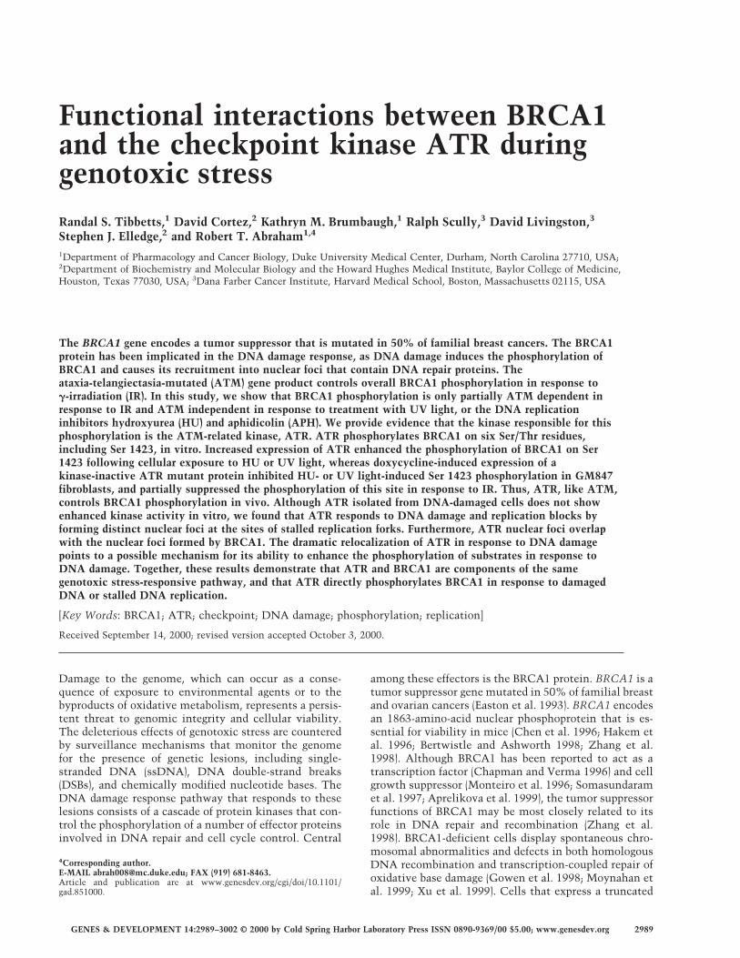
We next sought to determine whether functional ATR isrequired for the phosphorylation of endogenous, full-length BRCA1 on Ser 1423 in DNA-damaged cells. Forthese studies, we examined BRCA1 phosphorylation inGM847 human fibroblasts engineered to overexpressFLAG–ATRKI when cultured in the presence of doxycy-cline (Dox) (Cliby et al. 1998). In the absence of Doxtreatment, exposure of GM847 cells to either HU or UVlight increased the levels of Ser 1423-phosphorylatedBRCA1 by two- to fourfold (Fig. 5A). Dox-induced over-expression of ATRKI inhibited the phosphorylation of Ser1423 in response to both treatments, but had little effecton basal Ser 1423 phosphorylation (Fig. 5A). Overexpres-sion of ATRKI also partially inhibited Ser 1423 phos-phorylation following exposure to IR (Fig. 5B). Thestrongest inhibitory effect of ATRKI overexpression wasobserved at 4 and 6 h post-irradiation, when Ser 1423phosphorylation was reduced by ∼30% relative to theirradiated controls. Considered together, these resultssupport a major role for ATR in the HU- and UV light-induced phosphorylation of BRCA1 on Ser 1423, and sug-
gest that ATR also contributes to the phosphorylation ofthis site in IR-treated cells.
ATR colocalizes with BRCA1 in DNA damage-inducednuclear foci
How ATR enhances BRCA1 phosphorylation in responseto genotoxic stress is unknown. Whereas ATM kinaseactivity increases in response to DNA damage (Banin etal. 1998; Canman et al. 1998), the in vitro kinase activityof ATR is not detectably increased when cells are treatedwith a variety of DNA-damaging agents including IR,alkylating agents, and DNA replication inhibitors (datanot shown). Therefore, we examined whether ATRmight be altered in response to damage in a manner dis-tinct from kinase activation. Immunostaining of K562cells with �-ATR antibodies demonstrated that the sub-cellular localization pattern of ATR changed dramati-cally following cellular exposure to DNA-damagingagents or DNA replication inhibitors. Prior to treatment,most cells exhibited a uniform nuclear ATR localizationpattern. Examination of K562 cells at 4 h after exposureto 10 Gy of IR revealed the presence of intense ATRnuclear foci, which numbered from 10 to more than 100per cell (Fig. 6A). Exposure of K562 cells to APH causedan even more dramatic increase in the appearance ofATR nuclear foci (Fig. 6A). On average, ATR nuclear fociwere observed in 68% of cells that had been cultured in
Figure 4. Overexpression of wild-type ATR potentiates the phosphorylation of BRCA1 at Ser 1423 in intact cells. (A) Overexpressionof FLAG–ATRWT stimulates the HU-induced phosphorylation of Ser 1423 in HEK 293T cells. HEK 293T cells were transfected withan expression vector encoding AU1-BRCA1–�N together with empty expression vector (−) or plasmids encoding FLAG–ATRWT, orFLAG–ATRKI. After 24 h, the transfected cells were left untreated or exposed to 2 mM HU, and harvested 1 h later. Cellular extractswere separated by SDS-PAGE and subjected to immunoblot analysis with the �-pS-1423 antibody to detect Ser 1423-phosphorylatedBRCA1. Immunoblotting with �-FLAG or �-AU1 antibodies was performed to monitor expression levels of the FLAG–ATR andAU1-BRCA1–�N constructs, respectively. �-, �-, and �-phosphorylated forms of AU1-BRCA1–�N are marked. (B) Overexpression ofFLAG–ATRWT stimulates UV light-induced phosphorylation of Ser 1423 in HEK 293T cells. The experiment was performed asdescribed in A, except that the transfected HEK 293T cells were exposed to 50 J/m2 UV light, and then harvested 1 h later. Cell lysateswere subjected to SDS-PAGE and immunoblot analysis with the indicated antibodies. (C) Relative contributions of ATM and ATR tothe IR-induced phosphorylation of BRCA1 on Ser 1423. HEK 293T cells were cotransfected with expression plasmids encodingFLAG-tagged BRCA1 (1351–1552) together with an empty expression vector (−), FLAG–ATR, or FLAG–ATM plasmids. The FLAG–ATR and FLAG–ATM expression constructs encoded either wild-type (WT) or catalytically inactive (KI) proteins. After 24 h, the cellswere either left untreated, or exposed to 25 Gy of IR and harvested 1 h later. Cell lysates were analyzed by SDS-PAGE and immunoblotanalysis with the �-pS-1423 antibody to detect Ser 1423-phosphorylated BRCA1. Expression levels of FLAG–ATR, FLAG–ATM, andFLAG–BRCA1 (1351–1552) were monitored by immunoblotting with �-FLAG, and are presented at top and bottom, respectively.
ATR signaling to BRCA1
GENES & DEVELOPMENT 2995
the presence of APH for 24 h. Similar results were ob-tained with HU (data not shown). The dramatic relocal-ization of ATR provoked by the DNA replication inhibi-tors was not apoptosis related, as ATR nuclear foci werereversible, and cells maintained full viability over thecourse of the experiment. Furthermore, APH induced theformation of ATR nuclear foci in multiple cell lines, in-cluding Jurkat T cells, HEK 293T cells, and Raji B cells(data not shown), indicating that the appearance of ATRnuclear foci is a general cellular response to drug-in-duced S-phase arrest.
To confirm the localization results obtained by �-ATRantibody staining, we transfected HEK 293T cells with aplasmid vector encoding a green fluorescent protein(GFP)–ATR fusion protein, and compared the GFP–ATRlocalization patterns in untreated versus APH-treatedcells. Although the GFP–ATR construct was not local-ized exclusively in the nucleus, the subnuclear pattern ofGFP–ATR fluorescence strongly resembled that of en-dogenous ATR. In the absence of genotoxic stress, themajority of nuclei (∼90% in multiple experiments) dem-
onstrated a diffuse GFP–ATR localization pattern (Fig.6B, left), whereas ∼10% of cells demonstrated one ormore GFP–ATR-containing dots (Fig. 6B, bottom, left,indicated by arrows). When GFP–ATR-expressing HEK293T cells were exposed to APH for 24 h, we again ob-served a dramatic increase in the number of ATR-con-taining nuclear foci in ∼80% of the treated cells (Fig. 6B,middle). IR (right), and HU (data not shown) also inducedthe formation of GFP–ATR nuclear foci. Thus, both ofthe ATR localization assays indicate that cellular expo-sure to genotoxic stress induces the formation of ATRnuclear foci. In contrast, transfected HA-tagged ATMwas not localized to nuclear foci under any of these treat-ment conditions (data not shown).
The staining pattern observed in APH-treated cellssuggested that ATR nuclear foci might be localized nearstalled replication forks, which contain regions of single-stranded and/or damaged DNA. To examine this possi-bility, we compared the pattern of GFP–ATR foci withthat of BrdU-labeled DNA in cells that had been culturedin BrdU-containing medium for 30 min after releasefrom a 3-h APH-induced DNA replication block. In un-treated cells, the patterns of GFP–ATR and �-BrdU anti-body staining were nonoverlapping, and the lack of co-localization was most striking in cells that displayedlarge, BrdU-positive foci, which are indicative of cells inmid-to-late S phase (Bravo and Macdonald 1987). Thenuclear localization pattern of GFP–ATR in these cellswas largely diffuse, with few or no foci apparent (Fig. 6C).In contrast, the GFP–ATR and BrdU staining patternsoverlapped in APH-arrested cells, and the colocalizationwas most pronounced in cells arrested in mid-to-late Sphase, as indicated by the large BrdU-positive foci (Fig.6C, bottom). This finding supports the notion that ATRlocalizes to sites of stalled DNA synthesis in cells ex-posed to the DNA polymerase � inhibitor.
To determine whether ATR nuclear foci containBRCA1, HEK 293T cells were transfected with GFP–ATR, treated with APH for 24 h, and then immuno-stained with an �-BRCAl antibody. In the absence ofAPH treatment, the GFP–ATR and BRCA1 localizationpatterns were largely distinct and, in the majority of cellsthat displayed BRCA1 nuclear foci, GFP–ATR was dis-tributed diffusely throughout the nucleus (Fig. 7A). InAPH-treated cells, the GFP–ATR and BRCA1 localiza-tion patterns overlapped extensively (Fig. 7B). The en-dogenous ATR and BRCA1 proteins were similarly colo-calized in APH-arrested K562 cells, indicating that thepreviously observed colocalization was not a uniqueproperty of the ectopically expressed GFP–ATR (Fig. 7C).Thus, these results suggest that ATR localization isregulated in response to DNA damage or stalled DNAreplication, and that ATR and BRCA1 may function to-gether within nuclear foci induced by these agents. Theincreased phosphorylation of BRCA1 that is observed af-ter treatment with genotoxic agents correlates with anincrease in the colocalization of BRCA1 with ATR, andsuggests that the regulated localization of these proteinsmay play an important role in coordinating their func-tion.
Figure 5. Overexpression of catalytically inactive ATR inhib-its the DNA damage-induced phosphorylation of BRCA1 on Ser1423. (A) Inhibition of HU- and UV-induced Ser 1423 phos-phorylation. GM847/ATRKI cells were cultured either in theabsence (−) or presence (+) of Dox for 96 h to induce ATRKI. Cellswere then either left untreated or exposed to 2 mM HU for 4 h,or harvested 1 h after exposure to 200 J/m2 UV light. Cell lysateswere prepared and immunoblots were performed with the indi-cated antibodies. Autoradiograms were quantitated by densi-tometry and phospho-Ser 1423 levels were normalized to ac-count for differences in total BRCA1 protein levels. (B) Timecourse of Ser 1423 phosphorylation in response to IR. GM847/ATRKI cells were cultured in the presence of Dox for 96 h andthen left untreated or exposed to 20 Gy of IR and harvested atthe indicated times. Analysis of Ser 1423-phosphorylatedBRCA1 were determined as described in A.
Tibbetts et al.
2996 GENES & DEVELOPMENT

Discussion
The results of this study support a role for ATR as anupstream regulator of BRCA1 phosphorylation duringthe cellular response to DNA damage and DNA replica-tional stress. These conclusions are based on the follow-ing data: (1) ATM controls only part of the IR-inducedphosphorylation of BRCA1 and plays no detectable rolein BRCA1 phosphorylation in response to UV, HU, orAPH treatments; (2) ATR phosphorylates BRCA1 on sixsites in vitro including Ser 1423; (3) overexpression ofATR enhances the basal level of BRCA1 phosphorylationon Ser 1423, and potentiates the induced phosphoryla-
tion of this site in response to HU and UV light; (4)interference with ATR function through expression of aninducible kinase-defective ATR mutant reduces endog-enous BRCA1 phosphorylation on Ser 1423 in responseto HU and UV light, and partially inhibits IR-induced Ser1423 phosphorylation. These findings also confirm andextend the recent reports that ATM plays an importantrole in the induction of BRCA1 phosphorylation whencells are exposed to IR (Cortez et al. 1999, Gatei et al.2000). Taken together, the results of these studies sub-stantiate the hypothesis that ATM and ATR serve asproximate BRCA1 kinases in parallel cell cycle check-point pathways activated in response to different types of
Figure 6. DNA damage-induced alter-ations in ATR subcellular localization. (A)Immunolocalization of endogenous ATR.Exponentially growing K562 cells wereleft untreated, treated for 24 h with 5 µg/ml APH, or cultured for 4 h after exposureto 20 Gy IR. Cells were then immuno-stained with �-ATR antibodies and coun-terstained with propidium iodide (PI). (B)Localization of recombinant GFP–ATR.HEK 293T cells were transfected with aGFP–ATR expression vector and culturedfor 24 h. Samples were then either left un-treated, treated for 24 h with 5 µg/ml APH,or cultured for 4 h after exposure to 20 Gyof IR. Arrows highlight typical ATRnuclear foci found in a minority subpopu-lation of untreated HEK 293T cells. (C)ATR migrates to BrdU-positive sites ofDNA replication in APH-blocked cells.The experiment was performed as in A,except that the cells were cultured in thepresence of APH for 3 h, released from theAPH block, pulse-labeled for 30 min with100 µM BrdU, and then immunostainedwith �-BrdU. Foci demonstrating coinci-dent GFP–ATR-and �-BrdU staining ap-pear yellow in the merged image.
ATR signaling to BRCA1
GENES & DEVELOPMENT 2997

genotoxic stress. Moreover, the present findings lend fur-ther support to the idea that ATR and BRCA1 play con-tinuous roles in genome surveillance during the normalprocess of DNA replication.
Functional ATR is necessary for BRCA1phosphorylation in response to multiple formsof genotoxic stress
As noted, the evidence in support of an important rolefor ATR as an upstream effector of BRCA1 phosphoryla-tion comes from two different experimental model sys-tems. In cotransfection assays, we found that overexpres-sion of wild-type ATR augmented the HU- and UV light-induced phosphorylation of Ser 1423 in HEK 293T cells.In the second approach, ATRKI overexpression wasshown to substantially inhibit HU- or UV light-inducedphosphorylation of BRCA1 at Ser 1423 in GM847 fibro-blasts. The combined results strongly suggest that ATRphosphorylates BRCA1 in response to DNA replicationblocks and UV light. In addition, because overexpressionof ATRKI partially inhibited Ser 1423 phosphorylation inIR-treated cells, it is likely that ATR is at least partiallyresponsible for the residual phosphorylation of BRCA1observed in ATM-deficient cells (Fig. 1).
We have provided evidence that ATR phosphorylatesBRCA1 in vitro on six Ser/Thr-Gln sites (Ser 1143, Ser1280, Ser 1387, Thr 1394, Ser 1423, Ser 1457). It is pres-ently unclear how many sites in BRCA1 are phosphory-lated by ATR in vivo following exposure to genotoxicstress. However, the observation that a Ser 1423 �Alasubstitution does not eliminate the APH-induced gelmobility shift of full-length BRCA1 (R. Tibbetts and D.Cortez, unpubl.) suggests that Ser 1423 is not the onlysite of ATR-dependent modification. A more direct as-sessment of the phosphorylation states of individual
ATR phosphorylation sites in vivo awaits the develop-ment of suitable phospho-specific antibodies.
ATR and ATM may regulate BRCA1 phosphorylationin response to distinct and overlapping typesof genotoxic stress
Given the fact that ATM and ATR phosphorylateBRCA1 on at least partially overlapping sites, it may bedifficult to determine the relative contributions of thesekinases to damage-induced BRCA1 phosphorylation incell lines with intact ATM/ATR alleles. The results ofthe present study and two recent studies have indicatedthat functional ATM is required for optimal phosphory-lation of BRCA1 in response to IR (Cortez et al. 1999;Gatei et al. 2000), which is consistent with accumulat-ing evidence that ATM serves as the principal signaltransducer during the response to DNA DSBs (Khannaand Lavin 1993; Siliciano et al. 1997; Matsuoka et al.1998; Brown et al. 1999). In contrast, A-T cells did notdisplay a defect in either the UV- or HU-induced phos-phorylation of BRCA1 on Ser 1423 (Fig. 1), which is con-sistent with these earlier reports that failed to implicateATM in damage responses initiated by these agents. Thefinding that HU- or UV light-induced phosphorylation ofBRCA1 on Ser 1423 is enhanced by overexpression ofwild-type ATR, and inhibited by overexpression ofATRKI, strongly suggests that ATR replaces ATM as themajor Ser 1423 kinase when the genotoxic insult is sup-plied by DNA replication blocks or UV irradiation. Fi-nally, the fact that both ATR and BRCA1 knockout micedisplay chromosomal abnormalities and die early duringembryogenesis (Hakem et al. 1996; Xu et al. 1999; Brownand Baltimore 2000; de Klein et al. 2000) suggests thatATR may additionally regulate BRCA1 during the nor-mal cell cycle. According to this model, cell death re-
Figure 7. Colocalization of ATR andBRCA1 in APH-induced nuclear foci. HEK293T cells were transfected with a GFP–ATR expression vector and then either leftuntreated (A), or cultured in the presenceof APH for 24 h (B). Cells were then fixed,permeabilized, and immunostained with aBRCA1-specific monoclonal antibody.Foci that contain both ATR and BRCA1appear yellow or orange in the merged im-age. (C) Colocalization of endogenousATR and BRCA1 in K562 cells. K562 cellswere treated with APH for 24 h, and thenwere fixed and immunostained with�-ATR and �-BRCA1 antibodies. Typicalfoci that demonstrate colocalization ofATR and BRCA1 are indicated by arrows.
Tibbetts et al.
2998 GENES & DEVELOPMENT
sults from the accumulation of DNA damage, particu-larly as a result of uncorrected errors that occur during Sphase.
It seems clear that the phosphorylation state ofBRCA1 is under complex control by several protein ki-nases. In addition to cyclin-dependent kinases (Ruffneret al. 1999), ATM, and ATR, a recent report has shownthat Chk2 phosphorylates BRCA1 at an independent site(Ser 988) in IR-damaged cells (Lee et al. 2000). Theemerging picture is that BRCA1 lies at the intersectionof multiple damage response pathways, and that specificphosphorylation events on BRCA1 may allow this pro-tein to orchestrate specific responses to different types ofgenotoxic stress.
The carboxy-terminal residues in BRCA1 that arephosphorylated by ATR in vitro are proximal to severalfunctional regions, including the transactivation do-main, bipartite BRCT domains, and binding sites for p53and BRCA2 (Chai et al. 1999; Chapman and Verma 1996;Bork et al. 1997; Chen et al. 1998). These phosphoryla-tion events may also regulate interactions betweenBRCA1 and components of the DNA repair and recom-bination machinery, perhaps through the generation ofbinding sites for FHA domain-containing proteins (Sunet al. 1998; Durocher et al. 1999).
ATR nuclear foci: Implications for ATR functionand regulation
The findings that cellular exposure to DNA-damagingagents or DNA replication inhibitors causes the forma-tion of ATR nuclear foci offers the first evidence thatATR responds to genetic damage in somatic cells andmay have implications for the regulation of ATR-depen-dent signaling pathways. In contrast to ATM, whosecatalytic activity is up-regulated within minutes of cel-lular exposure to IR or radiomimetic drugs (Banin et al.1998; Canman et al. 1998), we have been unable to detectan increase in the protein kinase activity of ATR in cellsexposed to a variety of DNA-damaging agents (R. Tib-betts , unpubl.). Our failure to detect catalytic activationof ATR may reflect a requirement for cofactors that arelost during cellular lysis and immunoprecipitation. Al-ternatively, the phosphorylation of substrates by ATRmay be regulated primarily through induced changes inproximity to these substrates as a consequence of stalledDNA replication forks or overt DNA damage. BecauseATM does not localize to nuclear foci either in responseto DNA damaging agents (Watters et al. 1997), or DNAreplication inhibitors (R. Tibbetts, unpubl.), our findingsraise the interesting possibility that the checkpoint ac-tivities of ATM and ATR are regulated by fundamentallydifferent mechanisms.
The colocalization of ATR and sites of BrdU incorpo-ration in APH-treated cells strongly suggests that ATRcomplexes assemble in the vicinity of stalled DNA rep-lication forks. This conclusion is further supported bythe finding that ATR colocalizes with replication fork-associated proteins, including replication protein A andBloom’s syndrome helicase, in HU-treated cells (R. Tib-
betts and D. Cortez, unpubl.). An obvious, but as yetunproven possibility is that ATR is required to enforcethe DNA replication checkpoint in S-phase-arrestedcells. The localization of ATR to sites of arrested DNAsynthesis may also reflect maintenance and/or stabiliza-tion functions for ATR at stalled DNA replication forks,analogous to those carried out by Mec1p in buddingyeast (Desany et al. 1998; Santocanale and Diffley 1998).In addition, ATR nuclear foci may assemble at eitherpotential or active sites of DNA recombination, whichcan occur as the result of DNA damage or paused DNAreplication in somatic cells (Galli and Schiestl 1996; Es-sers et al. 1997). Consistent with a role in recombina-tion, ATR and BRCA1 are localized to asynapsed regionsof paired chromosomes during meiosis, which might re-flect a role for the complex in monitoring or promotinghomologous recombination (Keegan et al. 1996; Scully etal. 1997b). It is possible that the ATR complex alsomonitors recombinogenic lesions in somatic cells,which occur at low frequency during a typical S phase,and in much higher numbers when cells are exposed togenotoxic stress (Galli and Schiestl 1996; Takata et al.1998). Loss of BRCA1 or ATR function may increasegenetic instability in proliferating cells due to defectiveresponses to replication fork stalling, base misincorpora-tion errors, and other insults during the normal processof DNA replication. This hypothesis is compatible withthe observation that ATR-containing nuclear foci are ob-served in both untreated, cycling cell populations andcells exposed to extrinsic DNA-damaging agents (seeFig. 7).
In summary, this study establishes a regulatory rela-tionship between ATR and BRCA1 and raises the possi-bility that, like mutations in BRCA1 itself, alterations inATR expression or activity may promote genetic insta-bility, and increase susceptibility to the development ofbreast cancer and other tumors.
Materials and methods
Cell culture and antisera
K562 erythroleukemia and HEK 293T cells were maintained inRPMI-1640 and DMEM medium containing 10% FCS, respec-tively. ATM− and ATM+ AT22IJE-T fibroblasts were kindlyprovided by Dr. Yosef Shiloh (Tel Aviv University) and weremaintained as described previously (Ziv et al. 1997). GM847cells that express FLAG epitope-tagged catalytically inactive(ATRKI) ATR under control of the bacterial tet operator havebeen described previously (Cliby et al. 1998). Induction ofATRKI was accomplished by plating out 2 × 10⁵ cells in 100-mmdishes in culture medium containing 1µg/ml Dox for 4 d. Whereindicated, cells were � irradiated with a 137Cs source or UVirradiated with a Stratalinker UV lamp (254 nm wavelength).For the generation of �-pS-1423 antisera, rabbits were immu-nized with a keyhole limpet hemocyanin-conjugated BRCA1phosphopeptide (CVLEQHGpSQPSNS) derived from amino ac-ids 1416–1428 of BRCA1. The resulting antiserum was passedover a column containing the nonphosphorylated peptide, andthe unbound antibodies were subsequently affinity purified bybinding to and elution from a column containing the phospho-peptide used for immunizations. Antibodies specific for ATR
ATR signaling to BRCA1
GENES & DEVELOPMENT 2999

(Ab-1) and BRCA1 (Ab-1, Ab-2) were purchased from OncogeneScience. Other antibodies used in this study included �-BrdU-IU-4 (Caltag), �-AU1 (BAbCo), �-HA-12CA5 (BAbCo), and�-FLAGM2 (Sigma). Aphidicolin (APH, 5 mg/ml) was preparedas a stock solution in dimethylsulfoxide and stored at −80°Cuntil use.
Plasmid constructs and transfections
ATR constructs containing an amino-terminal AU1 (DTYRYI)or FLAG (DYKDDDDK) epitope were generated by replacing a1.0-kb ATR BamHI–SwaI fragment with a corresponding PCR-generated AU1- or FLAG-tagged ATR BamHI–SwaI fragment inthe plasmid pcDNA3.1. GFP–ATR was constructed by subclon-ing a PCR-generated enhanced GFP cDNA (Clontech) into theunique BamHI site of AU1-ATR:pcDNA3.1. The catalyticallyinactive ATR mutant in this study contains a Lys-2327�Argsubstitution in the ATP-binding pocket (Tibbetts et al. 1999).FLAG–NLS-tagged BRCA1 (1351–1552) and FLAG–ATM ex-pression plasmids used in this study have been described(Cortez et al. 1999). The BRCA1 expression plasmids have beendescribed (Scully et al. 1997b; Brown et al. 1999). AU1-BRCA1–�N was created by PCR amplification of full-length BRCA1using the following primers: 5�-GGATCCATGGACACCTACCGCTACATCTTGATTGGTTCTTCCAAAC-3�, 5�-GCGGCCGCGTAGTGGCTGTGGGGGATC-3�. The AU1-BRCA1–�NPCR fragment was subcloned into the BamHI–NotI sites ofpcDNA3.1–2XNLS, which contains a bipartite SV40 nuclear lo-calization sequence immediately downstream of the NotI site.Site-directed mutagenesis was carried out by the QuickChangemethod (Stratagene) according to the manufacturer’s instruc-tions. Transient transfection of HEK 293T cells was performedwith the Fugene 6 lipid transfection reagent (Boehringer-Mann-heim). All constructs were verified by sequencing.
Microscopy
For immunofluorescence microscopy of endogenous ATR,1 × 104 K562 cells were collected, washed in PBS, and cytospunonto poly-L-lysine-coated slides. The cells were fixed in metha-nol at −20°C for 30 min, rehydrated in PBS, and incubated for 30min with blocking solution (3% BSA, 2% goat serum in PBS).Samples were subsequently overlayed with protein G-purified�-ATR at a concentration of 1.0 µg/ml in blocking solution andincubated at 4°C for 1.5 h. Samples were then washed with PBS,0.2% Tween-20, and overlayed for 1 h at room temperature witha fluorescein isothiocyanate (FITC)-conjugated goat anti-rabbitIgG antibody (Caltag) that had been diluted 1:500 in blockingsolution. Samples were then washed and incubated with 0.1µg/ml propidium iodide, 100 µg/ml RNAse A in PBS for 30 min.After extensive washing, specimens were mounted with cover-slips and an aqueous anti-fade mounting reagent (Vectashield,Vector Laboratories). For analysis of GFP–ATR localization,HEK 293T cells were plated onto 22-mm2 glass coverslips andtransfected with 1 µg of GFP–ATR expression plasmid. Forty-eight hours after transfection, cells were fixed in 4% paraform-aldehyde in PBS, and then either mounted onto slides or per-meabilized in PBS containing 0.5% NP-40. The permeabilizedcells were then immunostained with �-BRCA1 at a concentra-tion of 0.5 µg/ml in blocking solution for 1.5 h at room tem-perature. Cells were washed and then overlayed for 1 h with a1:500-diluted cyanin-3 (Cy3)-conjugated goat anti-mouse IgGantibody (Caltag). After staining, cells were washed andmounted as described above. For BrdU colocalization experi-ments, HEK 293T cells were incubated with 100 µM BrdU in
fresh medium for 30 min prior to cell fixation. Cell staining wascarried out by incubating cells with a 1:500 dilution of �-BrdUin blocking solution containing 20 mM MgCl2, 0.5% TritonX-100, and 20 units/ml DNAse I (Boehringer) for 2 h. Cells werethen washed and incubated with Cy3-conjugated secondary an-tibody and mounted as described above. All fluorescence imageswere generated using a Carl Zeiss LSM410 scanning laser con-focal microscope.
Protein analysis and kinase assays
Cell extracts were prepared by resuspending washed cell pelletsin lysis buffer (25 mM HEPES, pH 7.4, 300 mM NaCl, 1.5 mMMgCl2, 1 mM EGTA, 20 nM microcystin, 0.5% NP-40, 50 mM�-glycerophosphate) with protease inhibitors (10 µg/ml leupep-tin, 5 µg/ml pepstatin A, 5 µg/ml aprotinin). Samples were in-cubated on ice for 10 min, and then clarified by centrifugation.For full-length BRCA1 electrophoretic mobility shift assays,6 × 105 HEK 293T cells were plated onto 60-mm dishes and thentransfected 16 h later with 4.5 µg of ATR, and 0.5 µg of BRCA1-HA plasmid DNAs. Twenty-four hours after transfection, APHwas added to a concentration of 5 µg/ml, and the cells werecultured for an additional 24 h. Cells were resuspended in lysisbuffer and immunoprecipitated with 2 µg of �-HA. �-HA im-munoprecipitates were resolved by SDS-PAGE through 7.5%polyacrylamide gels, and BRCA1-HA was detected by immuno-blotting with �-HA. Levels of ATR expression were determinedby �-ATR immunoblotting from whole cell lysates. For AU1-BRCA1–�N mobility shift assays, HEK 293T cells were platedas described above and transfected with 2 µg of expression plas-mid. Twenty-four hours after transfection, cells were culturedin the absence or presence of APH (5 µg/ml) for 4 h. Cell lysateswere prepared and resolved on 10% SDS–polyacrylamide gels.Immunoblots were then carried out by use of �-AU1.
ATR kinase assays were carried out as described (Tibbetts etal. 1999). HEK 293T cells were transiently transfected with 5 µgof either pcDNA3.1, FLAG-tagged wild-type ATR, or catalyti-cally inactive ATR expression constructs. Twenty-four hoursfollowing transfection, cell lysates were prepared and immuno-precipitated with 3 µg of �-FLAGM2. ATR immune complexeswere washed and then incubated with 1 µg of GST–BRCA1fusion protein test substrates. Kinase assays were then per-formed and quantitated as described previously (Tibbetts et al.1999).
Acknowledgments
This work was supported by grants from the National Institutesof Health (CA76193 and CA52995 to R.T.A. and GM44664 toS.J.E.), a Johnson and Johnson Focused Giving Award to R.T.A.,and funding from the Baylor Breast Cancer SPORE to S.J.E.R.S. is supported by a Department of Defense IDEA award(DAMD17–97–1–7180) and a Howard Temin Award (K01CA79576–01) from the National Cancer Institute. R.S.T is aFellow of the Leukemia Society of America. D.C. is a fellow ofthe Jane Coffin Childs Memorial Fund for Medical Research.K.M.B was supported by a postdoctoral fellowship from the Na-tional Cancer Institute (CA83319). S.J.E. is an investigator withthe Howard Hughes Medical Institute. R.T.A. is a Glaxo-Well-come Professor of Molecular-Cancer Biology.
The publication costs of this article were defrayed in part bypayment of page charges. This article must therefore be herebymarked “advertisement” in accordance with 18 USC section1734 solely to indicate this fact.
Tibbetts et al.
3000 GENES & DEVELOPMENT

References
Aprelikova, O.N., Fang, B.S., Meissner, E.G., Cotter, S., Camp-bell, M., Kuthiala, A., Bessho, M., Jensen, R.A., and Liu, E.T.1999. BRCA1-associated growth arrest is RB-dependent.Proc. Natl. Acad. Sci. 96: 11866–11871.
Banin, S., Moyal, L., Shieh, S.Y., Taya, Y., Anderson, C.W.,Chessa, L., Smorodinsky, N.I., Prives, C., Reiss, Y., Shiloh,Y., and Ziv, Y. 1998. Enhanced phosphorylation of p53 byATM in response to DNA damage. Science 281: 1674–1677.
Barlow, C., Hirotsune, S., Paylor, R., Liyanage, M., Eckhaus, M.,Collins, F., Shiloh, Y., Crawley, J.N., Ried, T., Tagle, D., and.Wynshaw-Boris, A 1996. Atm-deficient mice: A paradigm ofataxia telangiectasia. Cell 86: 159–171.
Barlow, C., Brown, K.D., Deng, C.X., Tagle, D.A., and Wyn-shaw-Boris, A. 1997. Atm selectively regulates distinct p53-dependent cell-cycle checkpoint and apoptotic pathways.Nat. Genet. 17: 453–456.
Beamish, H. and Lavin, M.F., 1994. Radiosensitivity in ataxia-telangiectasia: Anomalies in radiation-induced cell cycle de-lay. Int. J. Radiat. Biol. 65: 175–184.
Beamish, H., Williams, R., Chen, P., and Lavin, M.F. 1996. De-fect in multiple cell cycle checkpoints in ataxia-telangiecta-sia postirradiation. J. Biol. Chem. 271: 20486–20493.
Bentley, N.J., Holtzman, D.A., Flaggs, G., Keegan, K.S., DeM-aggio, A., Ford, J.C., Hoekstra, M., and. Carr, A.M 1996. TheSchizosaccharomyces pombe rad3 checkpoint gene. EMBOJ. 15: 6641–6651.
Bertwistle, D. and Ashworth, A. 1998. Functions of the BRCA1and BRCA2 genes. Curr. Opin. Genet. Dev. 8: 14–20.
Bork, P., Hofmann, K., Bucher, P., Neuwald, A.F., Altschul, S.F.,and Koonin, E.V. 1997. A superfamily of conserved domainsin DNA damage-responsive cell cycle checkpoint proteins.FASEB J. 11: 68–76.
Bravo, R. and Macdonald, B. 1987. Existence of two populationsof cyclin/proliferating cell nuclear antigen during the cellcycle: Association with DNA replication sites. J. Cell Biol.105: 1549–1554.
Brown, A.L., Lee, C.H., Schwarz, J.K., Mitiku, N., Piwnica-Worms, H., and Chung, J.H. 1999. A human Cds1-relatedkinase that functions downstream of ATM protein in thecellular response to DNA damage. Proc. Natl. Acad. Sci.96: 3745–3750.
Brown, E. and Baltimore, D. 2000. ATR disruption leads to chro-mosomal fragmentation and early embryonic lethality.Genes & Dev. 14: 397–402.
Canman, C.E., Wolff, A.C., Chen, C.Y., Fornace, A.J., Jr., and.Kastan, M.B. 1994. The p53-dependent G1 cell cycle check-point pathway and ataxia-telangiectasia. Cancer Res.54: 5054–5058.
Canman, C.E., Lim, D.S., Cimprich, K.A., Taya, Y., Tamai, K.,Sakaguchi, K., Appella, E., Kastan, M.B., and Siliciano, J.D.1998. Activation of the ATM kinase by ionizing radiationand phosphorylation of p53. Science 281: 1677–1679.
Chai, Y.L., Cui, J., Shao, N., Shyam, E., Reddy, P., and. Rao, V.N.1999. The second BRCT domain of BRCA1 proteins interactswith p53 and stimulates transcription from the p21WAF1/CIP1 promoter. Oncogene 18: 263–268.
Chapman, M.S. and Verma, I.M. 1996. Transcriptional activa-tion by BRCA1. Nature 382: 678–679.
Chen, J., Silver, D.P., Walpita, D., Cantor, S.B., Gazdar, A.F.,Tomlinson, G., Couch, F.J., Weber, B.L., Ashley, T., Living-ston, D.M., and. Scully, R. 1998. Stable interaction betweenthe products of the BRCA1 and BRCA2 tumor suppressorgenes in mitotic and meiotic cells. Mol. Cell 2: 317–328.
Chen, Y., Farmer, A.A., Chen, C.F., Jones, D.C., Chen, P.L., and
Lee, W.H. 1996. BRCA1 is a 220-kDa nuclear phosphopro-tein that is expressed and phosphorylated in a cell cycle-dependent manner. Cancer Res. 56: 3168–3172.
Cimprich, K.A., Shin, T.B., Keith, C.T., and Schreiber, S.L.1996. cDNA cloning and gene mapping of a candidate hu-man cell cycle checkpoint protein. Proc. Natl. Acad. Sci.93: 2850–2855.
Cliby, W.A., Roberts, C.J., Cimprich, K.A., Stringer, C.M.,Lamb, J.R., Schreiber, S.L., and Friend, S.H. 1998. Overex-pression of a kinase-inactive ATR protein causes sensitivityto DNA-damaging agents and defects in cell cycle check-points. EMBO J. 17: 159–169.
Cortez, D., Wang, Y., Qin, J., and Elledge, S.J. 1999. Require-ment of ATM-dependent phosphorylation of Brca1 in theDNA damage response to double-strand breaks. Science286: 1162–1166.
de Klein, A., Muijtjens, M., van Os, R., Verhoeven, Y., Smit, B.,Carr, A.M., Lehmann, A.R., and Hoeijmakers, J.H. 2000. Tar-geted disruption of the cell-cycle checkpoint gene ATR leadsto early embryonic lethality in mice. Curr. Biol. 10:479–482.
Desany, B.A., Alcasabas, A.A., Bachant, J.B., and Elledge, S.J.1998. Recovery from DNA replicational stress is the essen-tial function of the S-phase checkpoint pathway. Genes &Dev. 12: 2956–2970.
Durocher, D., Henckel, J., Fersht, A.R., and Jackson, S.P. 1999.The FHA domain is a modular phosphopeptide recognitionmotif. Mol. Cell 4: 387–394.
Easton, D.F., Bishop, D.T., Ford, D., and Crockford, G.P. 1993.Genetic linkage analysis in familial breast and ovarian can-cer: results from 214 families. The Breast Cancer LinkageConsortium. Am. J. Hum. Genet. 52: 678–701.
Essers, J., Hendriks, R.W., Swagemakers, S.M., Troelstra, C., deWit, J., Bootsma, D., Hoeijmakers, J.H., and Kanaar, R. 1997.Disruption of mouse RAD54 reduces ionizing radiation re-sistance and homologous recombination. Cell 89: 195–204.
Galli, A. and Schiestl, R.H. 1996. Hydroxyurea induces recom-bination in dividing but not in G1 or G2 cell cycle arrestedyeast cells. Mut. Res. 354: 69–75.
Gatei, M., Scott, S.P., Filippovitch, I, Soronika, N., Lavin, M.F.,Weber, B. and Khanna, K.K. 2000. Role for ATM in DNAdamage-induced phosphorylation of BRCA1. Cancer Res..60: 3299–3304.
Gowen, L.C., Avrutskaya, A.V., Latour, A.M., Koller, B.H., andLeadon, S.A. 1998. BRCA1 required for transcription-coupled repair of oxidative DNA damage. Science 281: 1009–1012.
Hakem, R., de la Pompa, J.L., Sirard, C., Mo, R., Woo, M.,Hakem, A., Wakeham, A., Potter, J., Reitmair, A., Billia, F.,Firpo, E. et al. 1996. The tumor suppressor gene Brca1 isrequired for embryonic cellular proliferation in the mouse.Cell 85: 1009–1023.
Kastan, M.B., Zhan, Q., el-Deiry, W.S., Carrier, F., Jacks, T.,Walsh, W.V., Plunkett, B.S., Vogelstein, B., and Fornace, Jr.,A.J. 1992. A mammalian cell cycle checkpoint pathway uti-lizing p53 and GADD45 is defective in ataxia-telangiectasia.Cell 71: 587–597.
Keegan, K.S., Holtzman, D.A., Plug, A.W., Christenson, E.R.,Brainerd, E.E., Flaggs, G., Bentley, N.J., Taylor, E.M., Meyn,M.S., Moss, S.B. et al. 1996. The Atr and Atm protein kinasesassociate with different sites along meiotically pairing chro-mosomes. Genes & Dev. 10: 2423–2437.
Keith, C.T. and Schreiber, S.L. 1995). PIK-related kinases: DNArepair, recombination, and cell cycle checkpoints. Science270: 50–51.
Khanna, K.K. and Lavin, M.F. 1993. Ionizing radiation and UVinduction of p53 protein by different pathways in ataxia-
ATR signaling to BRCA1
GENES & DEVELOPMENT 3001
telangiectasia cells. Oncogene 8: 3307–3312.Khosravi, R., Maya, R., Gottlieb, T., Oren, M., Shiloh, Y., and
Shkedy, D. 2000). Rapid ATM-dependent phosphorylation ofMDM2 precedes p53 accumulation in response to DNAdamage. Proc. Natl. Acad. Sci. 96: 14973–14977.
Kim, S.T, Lim, D.S., Canman, C.E., and Kastan, M.B. 1999. Sub-strate specificities and identification of putative substratesof ATM kinase family members. J. Biol. Chem. 274: 37538–37543.
Lakin, N.D., Hann, B.C., and Jackson, S.P. 1999. The ataxia-telangiectasia related protein ATR mediates DNA-depen-dent phosphorylation of p53. Oncogene 18: 3989–3995.
Lee, J.S., Collins, K.M., Brown, A.L., Lee, C.H., and. Chung, J.H2000). hCds1-mediated phosphorylation of BRCA1 regulatesthe DNA damage response. Nature 404: 201–204.
Liu, V.F. and Weaver, D.T. 1993. The ionizing radiation-inducedreplication protein A phosphorylation response differs be-tween ataxia telangiectasia and normal human cells. Mol.Cell. Biol. 13: 7222–7231.
Matsuoka, S., Huang, M., and Elledge, S.J. 1998. Linkage ofATM to cell cycle regulation by the Chk2 protein kinase.Science 282: 1893–1897.
Monteiro, A.N., August, A., and Hanafusa, H. 1996. Evidencefor a transcriptional activation function of BRCA1 C-termi-nal region. Proc. Natl. Acad. Sci. 93: 13595–13599.
Moynahan, M.E., Chiu, J.W., Koller, B.H., and Jasin, M. 1999.Brca1 controls homology-directed DNA repair. Mol. Cell4: 511–518.
Ruffner, H., Jiang, W., Craig, A.G., Hunter, T., and Verma, I.M.1999. BRCA1 is phosphorylated at Ser 1497 in vivo at a cy-clin-dependent kinase 2 phosphorylation site. Mol. Cell.Biol. 19: 4843–4854.
Santocanale, C. and. Diffley, J.F 1998. A Mec1- and Rad53-de-pendent checkpoint controls late-firing origins of DNA rep-lication. Nature 395: 615–618.
Scully, R., Ganesan, S., Brown, M., De Caprio, J.A., Cannistra,S.A., Feunteun, J., Schnitt, S., and Livingston, D.M. 1996.Location of BRCA1 in human breast and ovarian cancercells. Science 272: 123–126.
Scully, R., Chen, J., Ochs, R.L., Keegan, K., Hoekstra, M.,Feunteun, J., and. Livingston, D.M 1997a. Dynamic changesof BRCA1 subnuclear location and phosphorylation state areinitiated by DNA damage. Cell 90: 425–435.
Scully, R., Chen, J., Plug, A., Xiao, Y., Weaver, D., Feunteun, J.,Ashley, T., and. Livingston, D.M 1997b. Association ofBRCA1 with Rad51 in mitotic and meiotic cells. Cell88: 265–275.
Scully, R., Ganesan, S., Vlasakova, V., Chen, J.J., Sokolovsky,M., and Livingston, D.M. 1999 Genetic analysis of BRCA1function in a defined tumor cell line. Cell 4: 1093–1099.
Shafman, T., Khana, K.K., Kedar, P., Spring, K., Kozlov, S., Yen,T., Hobson, K., Gatei, M., Zhang, N., Watters, D. et al. 1997.Interaction between ATM protein and c-Abl in response toDNA damage. Nature 387: 520–523.
Siliciano, J.D., Canman, C.E., Taya, Y., Sakaguchi, K., Appella,E., and Kastan, M.B. 1997. DNA damage induces phosphory-lation of the amino terminus of p53. Genes & Dev. 11: 3471–3481.
Somasundaram, K., Zhang, H., Zeng, Y.X., Houvras, Y., Peng,Y., Wu, G.S., Licht, J.D., Weber, B.L., and el-Deiry, W.S.1997. Arrest of the cell cycle by the tumour-suppressorBRCA1 requires the CDK-inhibitor p21WAF1/CiP1. Nature389: 187–190.
Sun, Z., Hsiao, J., Fay, D.S., and Stern, D.F. 1998. Rad53 FHAdomain associated with phosphorylated Rad9 in the DNAdamage checkpoint. Science 281: 272–274.
Takata, M., Sasaki, M.S., Sonoda, E., Morrison, C., Hashimoto,M., Utsumi, H., Yamaguchi-Iwai, Y., Shinohara, A., and Tak-eda, S. 1998. Homologous recombination and non-homolo-gous end-joining pathways of DNA double-strand break re-pair have overlapping roles in the maintenance of chromo-somal integrity in vertebrate cells. EMBO J. 17: 5497–5508.
Tibbetts, R.S., Brumbaugh, K.B., Wiliams, J.W., Sarkaria, J.N.,Cliby, W.A., Shieh, S.Y., Taya, Y., Prives, C., and. Abraham,R.T. 1999. A role for ATR in the DNA damage-induced phos-phorylation of p53. Genes & Dev. 13: 152–157.
Tomlinson, G.E., Chen, T.T., Stastny, V.A., Virmani, A.K.,Spillman, M.A., Tonk, V., Blum, J.L., Schneider, N.R., Wis-tuba, I.I., Shay, J.W. et al. 1998. Characterization of a breastcancer cell line derived from a germ-line BRCA1 mutationcarrier. Cancer Res. 58: 3237–3242.
Wang, Y., Cortez, D., Yazdi, P., Neff, N., Elledge, S.J., and Qin,J. 2000. BASC, a super complex of BRCA1-associated pro-teins involved in the recognition and repair of aberrant DNAstructures. Genes & Dev. 14: 927–939
Watters, D., Khanna, K.K., Beamish, H., Birrell, G., Spring, K.,Kedar, P., Gatei, M., Stenzel, D., Hobson, K., Kozlov, S. et al.1997. Cellular localisation of the ataxia-telangiectasia(ATM) gene product and discrimination between mutatedand normal forms. Oncogene 14: 1911–1921.
Westphal, C.H., Rowan, S., Schmaltz, C., Elson, A., Fisher, D.E.,and Leder, P. 1997. Atm and p53 cooperate in apoptosis andsuppression of tumorigenesis, but not in resistance to acuteradiation toxicity. Nat. Genet. 16: 397–401.
Wright, J.A., Keegan, K.S., Herendeen, D.R., Bentley, N.J., Carr,A.M., Hoekstra, M.F., and Concannon, P. 1998. Protein ki-nase mutants of human ATR increase sensitivity to UV andionizing radiation and abrogate cell cycle checkpoint con-trol. Proc. Natl. Acad. Sci. 95: 7445–7450.
Xu, X., Weaver, Z., Linke, S.P., Li, C., Gotay, J., Wang, X.W.,Harris, C.C., Ried, T., and Deng, C.X. 1999. Centrosomeamplification and a defective G2-M cell cycle checkpointinduce genetic instability in BRCA1 exon 11 isoform-defi-cient cells. Mol. Cell 3: 389–395.
Xu, Y. and Baltimore, D. 1996. Dual roles of ATM in the cellularresponse to radiation and in cell growth control. Genes &Dev. 10: 2401–2410.
Xu, Y., Ashley, T., Brainerd, E.E., Bronson, R.T., Meyn, M.S.,and Baltimore, D. 1996. Targeted disruption of ATM leads togrowth retardation, chromosomal fragmentation duringmeiosis, immune defects, and thymic lymphoma. Genes &Dev. 10: 2411–2422.
Zhang, H., Tombline, G., and Weber, B.L. 1998. BRCA1,BRCA2, and DNA damage response: Collision or collusion?Cell 92: 433–436.
Zhong, Q., Chen, C.F., Li, S., Chen, Y., Wang, C.C., Xiao, J.,Chen, P.L., Sharp, Z.D., and Lee, W.H. 1999. Association ofBRCA1 with the hRad50-hMre11-p95 complex and the DNAdamage response. Science 285: 747–750.
Zhou, Z. and Elledge, S.H. 2000. The DNA damage response:Putting checkpoints in perspective. Nature (in press).
Ziv, Y., Bar-Shira, A., Pecker, I., Russell, P., Jorgensen, T.J.,Tsarfati, I., and Shiloh, Y. 1997. Recombinant ATM proteincomplements the cellular A-T phenotype. Oncogene 15:159–167.
Tibbetts et al.
3002 GENES & DEVELOPMENT


![Anguilla anguilla L. Biochemical and Genotoxic Responses to Benzo[ a]pyrene](https://static.fdokumen.com/doc/165x107/631d4597f26ecf94330a787a/anguilla-anguilla-l-biochemical-and-genotoxic-responses-to-benzo-apyrene.jpg)