Genotoxic effects of some agricultural pesticides in vitro tested with Aspergillus nidulans
Upload
independentCategory
view
0download
0
Genotoxic Effects of Aluminum on the Neotropical FishProchilodus lineatus
Bruno A. Galindo & Gabriel Troilo &
Ilce Mara S. Cólus & Cláudia B. R. Martinez &
Silvia H. Sofia
Received: 14 October 2009 /Accepted: 9 February 2010 /Published online: 2 March 2010# Springer Science+Business Media B.V. 2010
Abstract Applying an integrated approach using theComet, micronucleus (MN), and random amplifiedpolymorphic DNA (RAPD) assays, occurrence oferythrocytic nuclear abnormalities (ENAs) and theliver activity of antioxidants enzymes (catalase andglutathione-S-transferase (GST)) was carried out toevaluate the effects of acute (6, 24, and 96 h) andsubchronic (15 days) exposures to aluminum on fishProchilodus lineatus. The Comet assay showed thatfish erythrocytes exhibited significantly higher DNAdamage after 6 and 96 h of Al exposure. MNfrequencies were very low and did not increasesignificantly after Al exposures, while ENAs frequencyincreased significantly after all exposure periods. RAPDprofiles obtained with DNA from fish fins collectedbefore the toxicity tests were compared to the profileswith DNA from gills and liver of the same fishsampled after Al exposures. Alterations in RAPDprofiles, including appearance and disappearance of
bands, after 6 h, 24 h, and 15 days of Al exposurewere detected. Fish exposed to Al for 6 and 24 h alsoshowed significant increases in GST and catalaseactivities. These results indicated that Al exposurewas genotoxic to P. lineatus, inducing DNA damagein peripheral erythrocytes. The induction of antioxidantenzymes might be an indication that Al causesoxidative damage to DNA, while the very lowfrequency of MN suggests that Al does not produceclastogenic or aneugenic effects. Genotoxic effectsafter 15 days of Al exposure was revealed only byRAPD, showing that this assay represents a sensitivemethod to detect genotoxic damage, occasionally notdetected by other genotoxic tests used in toxicologicalgenetics studies.
Keywords Aquatic environment . Genotoxicity .
Comet assay . RAPD .Micronucleus . Oxidative stress
1 Introduction
Increased concentrations of aqueous aluminum repre-sent a major environmental problem due to therelationship between aluminum toxicity and fish(Alstad et al. 2005). This metal becomes more solubleand, for this reason, potentially more toxic tofreshwater fish as pH decreases below 6.0 and isrecognized as the main toxicant killing fish inacidified waters (Gensemer and Playle 1999). Al-though the genotoxic effects of aluminum for aquatic
Water Air Soil Pollut (2010) 212:419–428DOI 10.1007/s11270-010-0357-5
B. A. Galindo : I. M. S. Cólus : S. H. SofiaDepartamento de Biologia Geral,Centro de Ciências Biológicas,Universidade Estadual de Londrina (UEL),Londrina, Paraná, Brasil
G. Troilo : C. B. R. Martinez (*)Departamento Ciências Fisiológicas,Centro de Ciências Biológicas,Universidade Estadual de Londrina (UEL),Londrina, Paraná, Brasile-mail: [email protected]

invertebrates (Ternjej et al. 2009) as well as forhumans (Crebelli et al. 2002; Lankoff et al. 2006;Lima et al. 2007) have been demonstrated, informa-tion regarding the potential genotoxicity of this metalon fish is still lacking.
In general, metal genotoxicity seems to be linkedto the formation of reactive oxygen species (Soto-Reyes et al. 2005). Oxidative stress is establishedwhenever the rate of reactive oxygen species (ROS)production exceeds the rate of its decomposition byantioxidant defenses and repair systems, leading tothe oxidation of key cell components like proteins,fatty acids, and DNA (Sies 1993). The oxidation ofDNA by ROS can produce strand breaks, whichrepresent a major class of oxidative damage to DNAunder oxidative stress, and a number of differentmodified DNA bases (Cadet et al. 1997). Aluminumis recognized as a pro-oxidant agent promotingbiological oxidation both in vitro and in vivo (Exley2004). The pathology of laboratory animals that havebeen experimentally intoxicated with aluminum inva-riably shows many indices of oxidative stress,including changes in the levels of antioxidantenzymes and the occurrence of oxidative lesions(Zatta et al. 2002).
Different approaches have been developed for theassessment of DNA strand breakage (Mitchelmoreand Chipman 1998). The single-cell gel electropho-resis or Comet assay can detect DNA strand breaks atthe individual cell level under alkaline conditions(Singh et al. 1988). This is a highly sensitive andrapid method that can be performed on almost anyeukaryotic cell, requiring small amounts of tissue (Jha2008). The Comet assay has been extensively used asa non-specific measure of genotoxic damage in fish(Mitchelmore and Chipman 1998; Dhawan et al.2009). According to these authors, several of thesestudies have demonstrated the usefulness of this assayin fish as a model for monitoring genotoxicity ofaquatic habitats using these indicator animals.
Another well-established assay that has beenproven useful in assessing the genotoxic effects of awide range of compounds in fish is the evaluation ofmicronucleus induction (Udroiu 2006). The analysisof erythrocytic nuclear abnormalities (ENAs), avariant of the standard micronucleus test, has alsobeen widely used in fish toxicology (Ergene et al.2007). In this assay, a number of alterations in redblood cell nuclei that may lead to their fragmentation
and/or to micronucleus formation are recorded insteadof counting the micronuclei themselves.
In the last decade, random amplified polymorphicDNA (RAPD) assay, a simple, fast, sensitive, andstraightforward PCR-based method (Williams et al.1990), has been used to detect genotoxic-inducedDNA damage and mutations in different organisms,including fish (Savva 2000; Atienzar and Jha 2006;Swaileh et al. 2008). This method involves theamplification of random segments of genomic DNAusing short and arbitrary primers which interact withthe genomic template at sites where there is somehomology (Williams et al. 1990). Although RAPDassay provides just qualitative results, given that thenature and extent of DNA alterations can only bespeculated (Atienzar et al. 2002), this method comple-ments other well-established techniques in genotoxicity(Atienzar and Jha 2006).
In this context, the present study aimed atevaluating the genotoxicity of aluminum in a nativefish species and the relative sensitivity of RAPDassay compared to other commonly used assays. Inorder to achieve these objectives, an integratedapproach using quantitative (Comet, micronucleus,and ENAs) and qualitative (RAPD) genotoxic assaysassociated with biochemical assays to measure anti-oxidant enzymes activities were used. The fish Prochi-lodus lineatus was chosen because it is representativeof the Neotropical fish fauna, commonly found inrivers of the south and southeast regions of Brazil,and considered bioindicator fish species (Simonatoet al. 2006).
2 Material and Methods
2.1 Animals
Juveniles of P. lineatus (n=89) weighing 13.6±7.3 g(mean ± SD) were obtained from the hatchery stationof State University of Londrina. Animals were accli-mated to laboratory conditions for 7 days in a 600-Ltank with dechlorinated water (T = 21.8±1.1°C, pH7.9±0.3; hardness, 52±2.8 mg L−1 CaCO3), withconstant aeration (DO, 8.1±1.4 mg O2 L−1) and a14/10-h light/dark photoperiod. During this period,fish were fed each 48 h with commercial pellet foodcontaining 36% proteins (Guabi®, BR). Animals werenot fed 24 h before and during the toxicity tests.
420 Water Air Soil Pollut (2010) 212:419–428

2.2 Toxicity Tests and Fish Sampling
Acute (6, 24, and 96 h) static toxicity tests andsubchronic (15 days) semi-static toxicity tests wereperformed in 100-L glass aquaria containing six fisheach, with continuously aerated dechlorinated water.For each experimental interval, one group of fish wasexposed to Al in acid water (pH 5.2) simultaneouslywith a negative control group (NC), exposed only towater with neutral pH. The pH was adjusted with HCl(10%) and was monitored every 6 h. Aluminum wasadded to the water as Al2(SO4)3 reagent grade at anominal concentration of 1 mg Al L−1. All toxicitytests were carried out in duplicate. In the subchronictest, the water was renewed every 5 days and fishwere terminally sampled after 15 days of exposure.An acid only treatment was not included in this studyas it has been already established that increases indissolved Al occur together with decreased pH(Monette et al. 2008). For the RADP and Cometassays, a positive control group, consisting of fishinjected with the clastogenic agent cyclophosphamide(20 mg kg−1; Sigma, CAS no. 64-86-8), wasterminally sampled 24 h after treatment.
During the tests, water was continuously moni-tored for temperature, dissolved oxygen, pH, andconductivity. Samples of water collected immediatelyafter each experimental period were analyzed for Alconcentration using atomic absorption spectropho-tometry. The concentration of total Al was determinedin samples of non-filtered water, and the concentra-tion of dissolved Al was determined in filtered watersamples (0.45 μm); for both analyses, samples wereacidified with HNO3. Immediately after removal fromthe aquaria, fish were anesthetized with benzocaine(0.1 g L−1), and blood samples were taken from thecaudal vein into heparinized plastic syringes. Subse-quently, animals were killed by cervical section andthe gills and liver immediately removed.
2.3 Comet Assay
Immediately after blood sampling, a small amount ofblood (10 μL) was diluted in 700 μL of phosphate-buffered saline (126.6 mM NaCl, 4.8 mM KCl,1.5 mM CaCl2; 3.7 mM NaHCO3; 8.9 mM Na2HPO4;2.9 mM NaH2PO4) and kept in ice until the start ofthe Comet assay. The alkaline Comet assay wasperformed on erythrocyte cells as described by Singh
et al. (1988), with some modifications (Cavalcante etal. 2008). The main steps of the assay were: (a) lysis:1 h, at 4°C, protected from light, in a lysis buffer(2.5 M NaCl, 100 mM EDTA, 10 mM Tris, 10%DMSO, 1 mL Triton X-100, pH 10.0); (b) DNAunwinding: 30 min, in the dark, in an electrophoresisbuffer (0.3 N NaOH, 1 mM EDTA, pH>13); (c)electrophoresis: 20 min, 300 mA, 25 V, 1 V cm−1; and(d) neutralization: three washes for 5 min each inbuffer (0.4 M Tris, pH 7.5). Slides were fixed withabsolute ethanol for 10 min and kept under refriger-ation until cytological analyses.
Slides stained with ethidium bromide (20 μg mL−1)were analyzed under a Nikon fluorescence micro-scope (×1,000 magnification) fitted with a 515- to560-nm excitation filter and a 590-nm barrier filter.All slides were independently coded, and blindanalyses were performed by the same analyzer. Theextent of DNA damage was quantified by the lengthof DNA migration, which was visually determined in100 randomly selected and non-overlapping cells perfish. DNA damage was classified in four classesbased on the Comet tail length (0: no detectabledamage; 1: minimum damage; 2: medium damage; 3:maximum damage) and each Comet assigned a valueof 0 to 3 according to its class; the total score will bebetween 0 and 300 “arbitrary units” (Kumaravel et al.2009). Results for DNA damage in erythrocytes wereexpressed as the mean number of damaged nucleoids(sum of classes 1, 2, and 3) and the mean Comet scorefor each treatment group (NC or Al) for eachexposure period and also for PC.
2.4 Micronucleus Test and ENAs
The micronucleus test was performed with fisherythrocytes according to Hoftman and de Raat(1982) and the analysis of erythrocytic nuclearabnormalities according to Çavas and Ergene-Gözükara (2005). Immediately after sampling, bloodwas smeared on clean glass slides, dried overnight,fixed with methanol for 10 min, and stained withGiemsa (5%). Three thousand erythrocytes per fishwere examined under an Olympus optical microscope(×1,000 magnification). The mean frequencies ofmicronucleus (MN) and the other ENAs found ineach experimental group were calculated andexpressed per 1,000 cells (‰). ENAs were classified,following Pacheco and Santos (1997), into three
Water Air Soil Pollut (2010) 212:419–428 421

categories: segmented nuclei, lobed nuclei, andkidney-shaped nuclei. MN and ENA frequencies wereanalyzed in erythrocytes of fish exposed to the toxicitytests for 24 h or longer, taking into consideration thatthe estimated time for the detection of MN incirculating blood erythrocytes ranges between thesecond and third days after a clastogenic treatment(Udroiu 2006).
2.5 Random Amplified Polymorphic DNA
In order to compare RAPD profiles before and aftertoxicity tests, genomic DNA was isolated from theadipose fin, gills, and liver of each fish. Before thestart of the acclimation period, fish were anesthetizedwith benzocaine and the adipose fin was removed andused as the source of DNA prior to the tests. Anumbered small plastic ring was tight up around thecaudal peduncle for fish identification.
Following each exposure period, animals had theirgills and liver removed for DNA extraction. The choiceof liver and gill tissues for RAPD analysis was basedon the detoxification function of liver, which makesthis organ a frequent target to oxidative damage, andthe direct contact of gills with the environment andconsequently with the xenobiotic present in the water.
DNA from adipose fin, gills, and liver of fish wasextracted according to Almeida et al. (2001). DNAconcentration was determined in a DyNA Quant 200fluorometer (Hoefer) using the dye Hoechst 33258(Sigma). All isolates were used immediately or storedat −20°C. DNA amplifications were performed asdescribed by Sofia et al. (2006), with minor mod-ifications. Final reaction volumes were 15 μL andcontained 15–25 ng of template DNA, 250 μM dNTP(Pharmacia Biotech), 0.3 μM of ten-nucleotide primer(Operon Technologies, Alameda, CA, USA), 3.3 mMof MgCl2, and 1 U of Taq DNA polymerase(Biotools) in the reaction buffer supplied. Thirtydecamer oligonucleotides (from kits OPAC, OPX,and OPW) were used as random primers in RAPDscreening. Control reactions were run with allcomponents except genomic DNA. DNA amplifica-tions were carried out in a thermal cycler (MJResearch PTC-100), and the amplification protocolconsisted of 4 min at 92°C followed by 40 cycles of40 s at 92°C, 1.5 min at 40°C, and 2 min at 72°C. Thelast round of amplification was followed by anadditional extension at 72°C for 5 min.
Samples of 15 μL of amplification products wereassayed by electrophoresis on 1.4% agarose gels withTBE buffer (0.89 mM Tris, 0.89 mM boric acid, 2 mMEDTA, pH 8.3) diluted 1:20 (v/v), run at 3 V cm−1, andstained with ethidium bromide. Agarose gel imageswere documented under UV light using the KodakElectrophoresis Documentation and Analysis System.
Comparative analyses were based on RAPDprofiles obtained from different organs of each fish;fin, gill, and liver RAPD products, from the samefish, were placed side by side on the same gel. Thus,RAPD markers were determined by direct comparisonof the amplified DNA electrophoretic profiles, andeach sample was scored for the presence or absence ofamplification products (binary variable). Aiming toobtain a high reliability of the data, only gain or lossof RAPD amplicons were considered in the analysis.RAPD profiles obtained with DNA extracted fromfish fins collected before toxicity tests were comparedto RAPD profiles with DNA extracted from gills andliver of the same fish collected after experimentalexposures. Thus, the differences in RAPD profilesdetected for each individual can be attributed to thedirect or indirect effects of the contaminants (Atienzarand Jha 2006). The molecular weight of RAPDproducts were based on a 100-bp DNA ladder(Invitrogen) applied to the gel. When some alterationin RAPD profiles was detected, three new amplifica-tions were carried out, in different days, to check theconsistency of results.
2.6 Biochemical Assays
After removal, liver samples were stored frozen at −80°Cuntil biochemical assays. For catalase and glutathione-S-transferase (GST) assays, liver samples were homoge-nized in 10 volumes of ice-cold 0.1 M K phosphatebuffer, pH 7.0, and centrifuged for 20 min at 4°C and15,000×g. The supernatant was used for enzymeassays. Catalase activity was determined by measuringthe rate of decomposition of H2O2 in a spectrophotom-eter at 240 nm (Beutler 1975). GST activity wasdetermined by enzymatic conjugation of reducedglutathione with 1-chloro-2,4-dinitrobenzene in a spec-trophotometer at 340 nm (Habig et al. 1974). Catalase(CAT) activity was expressed as micromoles per minuteper milligram liver per protein and GST activity asnanomoles per minute per milligram liver per protein.The concentrations of protein in liver samples were
422 Water Air Soil Pollut (2010) 212:419–428
determined according to Lowry et al. (1951) usingbovine albumin as standard.
2.7 Statistical Analysis
Differences between the results obtained for thealuminum-exposed group and negative control group,at each exposure time (6 h, 24 h, 96 h, and 15 days),were analyzed by Student’s t test. Values of p<0.05were considered significant. Statistical analyses wereperformed using Sigma Stat 3.5.
3 Results
3.1 Water Chemistry
The overall quality of the water remained stablethroughout the experiments considering all exposureperiods. The mean values (±SD) for NC and Algroups were, respectively, temperature (°C): 22.3±1.7and 22.3±1.6; dissolved oxygen (mg O2 L−1): 6.8±1.9 and 6.7±1.6; and conductivity (μS cm−1): 92.7±15.4 and 121.4±21.3. The mean pH value for NC was7.7±0.5, and in Al-exposed groups, pH values rangedfrom 5.04 to 5.36; the mean value was 5.20±0.13.
The concentrations of aluminum were very similaramong Al groups in different exposure periods. Themean values (±SD, N=5) of total Al concentration inthe water of experimental groups (Al) were 438±36 μg Al L−1, and the concentration of dissolvedaluminum was 196±28.7 μg Al L−1.
3.2 Comet Assay
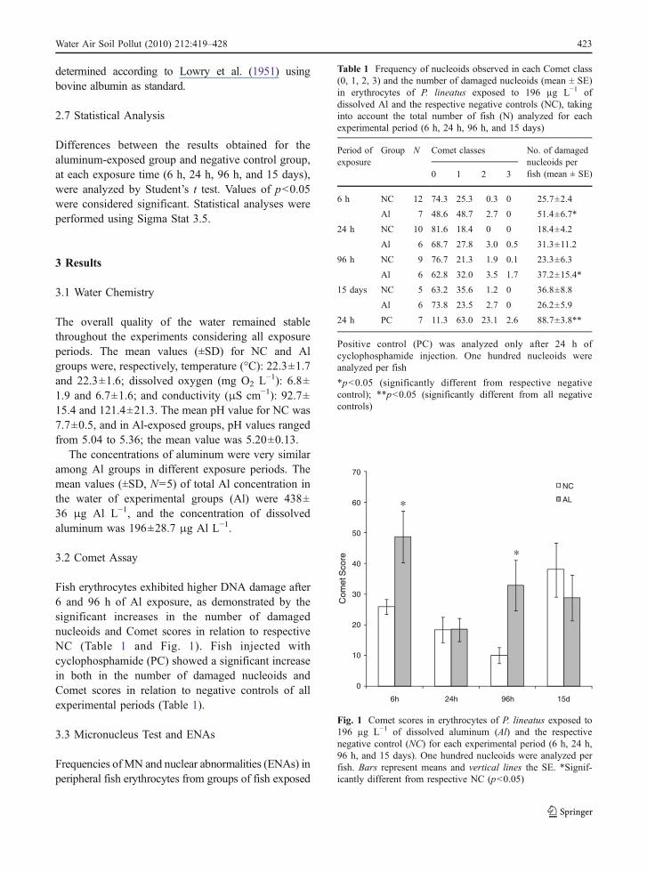
Fish erythrocytes exhibited higher DNA damage after6 and 96 h of Al exposure, as demonstrated by thesignificant increases in the number of damagednucleoids and Comet scores in relation to respectiveNC (Table 1 and Fig. 1). Fish injected withcyclophosphamide (PC) showed a significant increasein both in the number of damaged nucleoids andComet scores in relation to negative controls of allexperimental periods (Table 1).
3.3 Micronucleus Test and ENAs
Frequencies ofMN and nuclear abnormalities (ENAs) inperipheral fish erythrocytes from groups of fish exposed
Table 1 Frequency of nucleoids observed in each Comet class(0, 1, 2, 3) and the number of damaged nucleoids (mean ± SE)in erythrocytes of P. lineatus exposed to 196 µg L−1 ofdissolved Al and the respective negative controls (NC), takinginto account the total number of fish (N) analyzed for eachexperimental period (6 h, 24 h, 96 h, and 15 days)
Period ofexposure
Group N Comet classes No. of damagednucleoids perfish (mean ± SE)0 1 2 3
6 h NC 12 74.3 25.3 0.3 0 25.7±2.4
Al 7 48.6 48.7 2.7 0 51.4±6.7*
24 h NC 10 81.6 18.4 0 0 18.4±4.2
Al 6 68.7 27.8 3.0 0.5 31.3±11.2
96 h NC 9 76.7 21.3 1.9 0.1 23.3±6.3
Al 6 62.8 32.0 3.5 1.7 37.2±15.4*
15 days NC 5 63.2 35.6 1.2 0 36.8±8.8
Al 6 73.8 23.5 2.7 0 26.2±5.9
24 h PC 7 11.3 63.0 23.1 2.6 88.7±3.8**
Positive control (PC) was analyzed only after 24 h ofcyclophosphamide injection. One hundred nucleoids wereanalyzed per fish
*p<0.05 (significantly different from respective negativecontrol); **p<0.05 (significantly different from all negativecontrols)
0
10
20
30
40
50
60
70
6h 24h 96h 15d
Com
et S
core
NC
AL*
*
Fig. 1 Comet scores in erythrocytes of P. lineatus exposed to196 µg L−1 of dissolved aluminum (Al) and the respectivenegative control (NC) for each experimental period (6 h, 24 h,96 h, and 15 days). One hundred nucleoids were analyzed perfish. Bars represent means and vertical lines the SE. *Signif-icantly different from respective NC (p<0.05)
Water Air Soil Pollut (2010) 212:419–428 423
to Al and their respective negative controls groups areshown in Table 2. In contrast to Comet results, MNfrequencies determined in fish erythrocytes after Alacute and subchronic exposures were not significantlydifferent from the respective negative controls.
On the other hand, fish exposed to Al in allexperimental periods showed a significant increase inENAs frequency in comparison to respective NC.Among the three types of nuclear abnormalitiesobserved in P. lineatus erythrocytes, the most com-monly detected, both in NC as in Al groups, werekidney-shaped nuclei, followed by segmented nucleiand lobed nuclei.
3.4 RAPD Analysis
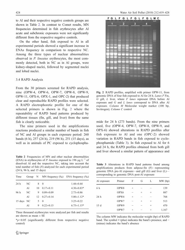
From the 30 primers screened for RAPD analysis,nine (OPW-4, OPW-6, OPW-7, OPW-8, OPW-9,OPW-11, OPX-6, OPX-1, and OPC-2) that producedclear and reproducible RAPD profiles were selected.A RAPD electrophoretic profile for one of theselected primers is shown in Fig. 2 where therepeatability of RAPD band patterns produced bydifferent tissues (fin, gill, and liver) from the samefish is clearly noticeable.
The nine primers used in the amplificationsreactions produced a similar number of bands in fishof NC and Al groups in each exposure period: 260bands (6 h), 257 (24 h), 219 (96 h), 251 (15 days), aswell as in animals of PC exposed to cyclophospha-
mide for 24 h (273 bands). From the nine primersused, five (OPW-4, OPW-7, OPW-8, OPW-9, andOPX-6) showed alterations in RAPD profiles afterfish exposure to Al and one (OPC-2) showedvariation in RAPD bands in fish exposed to cyclo-phosphamide (Table 3). In fish exposed to Al for 6and 24 h, the RAPD profiles obtained from both gilland liver showed a similar pattern of appearance and
Table 2 Frequencies of MN and other nuclear abnormalities(ENA) in erythrocytes of P. lineatus exposed to 196 µg L−1 ofdissolved Al and the respective NC, taking into account thetotal number of fish (N) analyzed for each experimental period(24 h, 96 h, and 15 days)
Time Group N MN frequency (‰) ENA frequency (‰)
24 h NC 8 0 1.44±0.44
Al 10 0.17±0.11 4.30±0.83*
96 h NC 9 0.09±0.09 2.73±0.86
Al 12 0.27±0.14 5.00±1.12*
15 days NC 5 0 3.25±0.22
Al 9 0.22±0.15 6.75±1.15*
Three thousand erythrocytes were analyzed per fish and resultsare shown as mean ± SE
*p<0.05 (significantly different from respective negativecontrol)
Fig. 2 RAPD profiles, amplified with primer OPW-11, fromgenomic DNA of four fish exposed to Al for 24 h. Lanes F Fin,G gill, L liver, where F lanes represent DNA before Alexposure and G and L lanes correspond to DNA after Alexposure. Column M Molecular weight marker (100 bp,Invitrogen); Column C control
Table 3 Alterations in RAPD band patterns found amongamplifications products from adipose-fin (F)—representinggenomic DNA pre-Al exposure—and gill (G) and liver (L)—corresponding to genomic DNA post-Al exposure
Al exposure Primer F G L MW (bp)
6 h OPW8 + − − 139
OPX6 − + + 887
24 h OPW4 + − − 768
OPW7 − + + 513
15 d OPW9 − − + 470
OPW7 + − + 1,167
The column MW indicates the molecular weight (bp) of RAPDband. The symbol + (plus) indicates the band’s presence, and −(minus) indicates the band’s absence
424 Water Air Soil Pollut (2010) 212:419–428
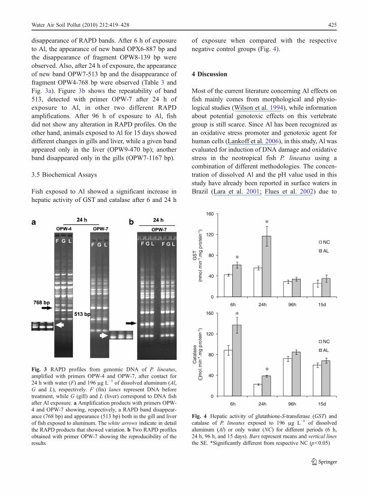
disappearance of RAPD bands. After 6 h of exposureto Al, the appearance of new band OPX6-887 bp andthe disappearance of fragment OPW8-139 bp wereobserved. Also, after 24 h of exposure, the appearanceof new band OPW7-513 bp and the disappearance offragment OPW4-768 bp were observed (Table 3 andFig. 3a). Figure 3b shows the repeatability of band513, detected with primer OPW-7 after 24 h ofexposure to Al, in other two different RAPDamplifications. After 96 h of exposure to Al, fishdid not show any alteration in RAPD profiles. On theother hand, animals exposed to Al for 15 days showeddifferent changes in gills and liver, while a given bandappeared only in the liver (OPW9-470 bp); anotherband disappeared only in the gills (OPW7-1167 bp).
3.5 Biochemical Assays
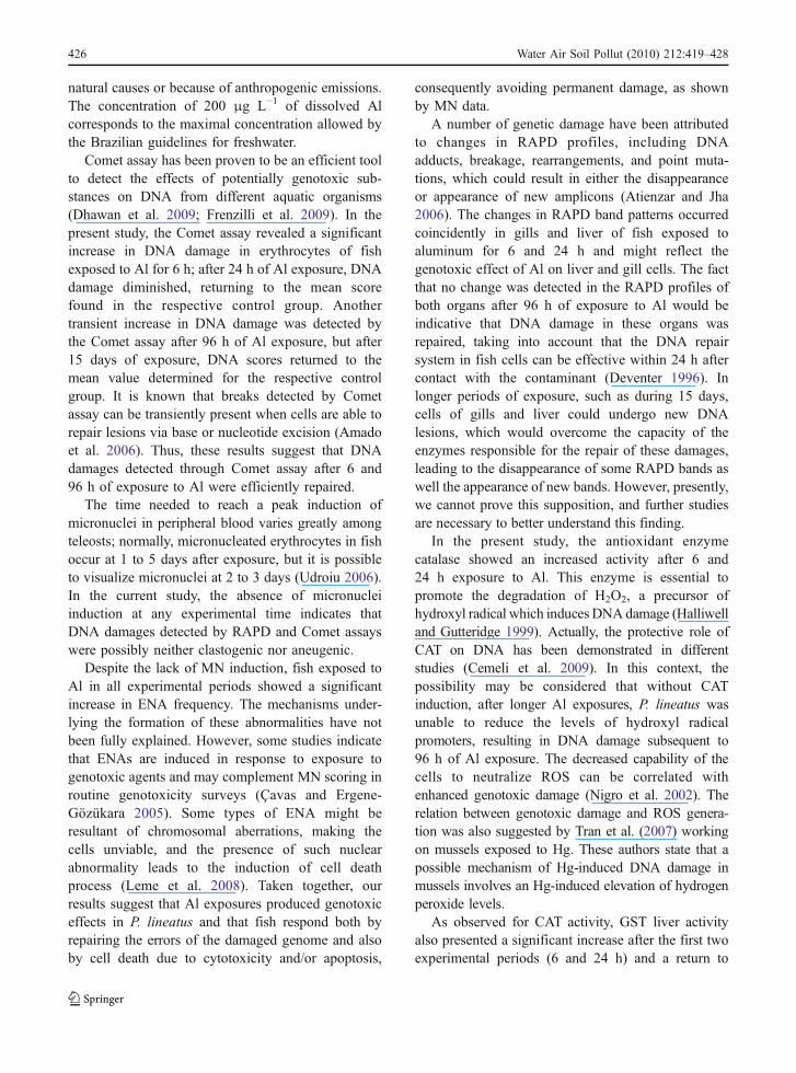
Fish exposed to Al showed a significant increase inhepatic activity of GST and catalase after 6 and 24 h
of exposure when compared with the respectivenegative control groups (Fig. 4).
4 Discussion
Most of the current literature concerning Al effects onfish mainly comes from morphological and physio-logical studies (Wilson et al. 1994), while informationabout potential genotoxic effects on this vertebrategroup is still scarce. Since Al has been recognized asan oxidative stress promoter and genotoxic agent forhuman cells (Lankoff et al. 2006), in this study, Al wasevaluated for induction of DNA damage and oxidativestress in the neotropical fish P. lineatus using acombination of different methodologies. The concen-tration of dissolved Al and the pH value used in thisstudy have already been reported in surface waters inBrazil (Lara et al. 2001; Flues et al. 2002) due to
Fig. 3 RAPD profiles from genomic DNA of P. lineatus,amplified with primers OPW-4 and OPW-7, after contact for24 h with water (F) and 196 µg L−1 of dissolved aluminum (Al,G and L), respectively. F (fin) lanes represent DNA beforetreatment, while G (gill) and L (liver) correspond to DNA fishafter Al exposure. a Amplification products with primers OPW-4 and OPW-7 showing, respectively, a RAPD band disappear-ance (768 bp) and appearance (513 bp) both in the gill and liverof fish exposed to aluminum. The white arrows indicate in detailthe RAPD products that showed variation. b Two RAPD profilesobtained with primer OPW-7 showing the reproducibility of theresults
0
40
80
120
160
6h 24h 96h 15d
GS
T (n
mo
l.min
-1.m
g p
rote
in-1
)
NC
AL
*
*
0
40
80
120
160
6h 24h 96h 15d
Cat
alas
e(µ
mo
l.min
-1.m
g p
rote
in-1
)
NC
AL
*
*
Fig. 4 Hepatic activity of glutathione-S-transferase (GST) andcatalase of P. lineatus exposed to 196 µg L−1 of dissolvedaluminum (Al) or only water (NC) for different periods (6 h,24 h, 96 h, and 15 days). Bars represent means and vertical linesthe SE. *Significantly different from respective NC (p<0.05)
Water Air Soil Pollut (2010) 212:419–428 425
natural causes or because of anthropogenic emissions.The concentration of 200 μg L−1 of dissolved Alcorresponds to the maximal concentration allowed bythe Brazilian guidelines for freshwater.
Comet assay has been proven to be an efficient toolto detect the effects of potentially genotoxic sub-stances on DNA from different aquatic organisms(Dhawan et al. 2009; Frenzilli et al. 2009). In thepresent study, the Comet assay revealed a significantincrease in DNA damage in erythrocytes of fishexposed to Al for 6 h; after 24 h of Al exposure, DNAdamage diminished, returning to the mean scorefound in the respective control group. Anothertransient increase in DNA damage was detected bythe Comet assay after 96 h of Al exposure, but after15 days of exposure, DNA scores returned to themean value determined for the respective controlgroup. It is known that breaks detected by Cometassay can be transiently present when cells are able torepair lesions via base or nucleotide excision (Amadoet al. 2006). Thus, these results suggest that DNAdamages detected through Comet assay after 6 and96 h of exposure to Al were efficiently repaired.
The time needed to reach a peak induction ofmicronuclei in peripheral blood varies greatly amongteleosts; normally, micronucleated erythrocytes in fishoccur at 1 to 5 days after exposure, but it is possibleto visualize micronuclei at 2 to 3 days (Udroiu 2006).In the current study, the absence of micronucleiinduction at any experimental time indicates thatDNA damages detected by RAPD and Comet assayswere possibly neither clastogenic nor aneugenic.
Despite the lack of MN induction, fish exposed toAl in all experimental periods showed a significantincrease in ENA frequency. The mechanisms under-lying the formation of these abnormalities have notbeen fully explained. However, some studies indicatethat ENAs are induced in response to exposure togenotoxic agents and may complement MN scoring inroutine genotoxicity surveys (Çavas and Ergene-Gözükara 2005). Some types of ENA might beresultant of chromosomal aberrations, making thecells unviable, and the presence of such nuclearabnormality leads to the induction of cell deathprocess (Leme et al. 2008). Taken together, ourresults suggest that Al exposures produced genotoxiceffects in P. lineatus and that fish respond both byrepairing the errors of the damaged genome and alsoby cell death due to cytotoxicity and/or apoptosis,
consequently avoiding permanent damage, as shownby MN data.
A number of genetic damage have been attributedto changes in RAPD profiles, including DNAadducts, breakage, rearrangements, and point muta-tions, which could result in either the disappearanceor appearance of new amplicons (Atienzar and Jha2006). The changes in RAPD band patterns occurredcoincidently in gills and liver of fish exposed toaluminum for 6 and 24 h and might reflect thegenotoxic effect of Al on liver and gill cells. The factthat no change was detected in the RAPD profiles ofboth organs after 96 h of exposure to Al would beindicative that DNA damage in these organs wasrepaired, taking into account that the DNA repairsystem in fish cells can be effective within 24 h aftercontact with the contaminant (Deventer 1996). Inlonger periods of exposure, such as during 15 days,cells of gills and liver could undergo new DNAlesions, which would overcome the capacity of theenzymes responsible for the repair of these damages,leading to the disappearance of some RAPD bands aswell the appearance of new bands. However, presently,we cannot prove this supposition, and further studiesare necessary to better understand this finding.
In the present study, the antioxidant enzymecatalase showed an increased activity after 6 and24 h exposure to Al. This enzyme is essential topromote the degradation of H2O2, a precursor ofhydroxyl radical which induces DNAdamage (Halliwelland Gutteridge 1999). Actually, the protective role ofCAT on DNA has been demonstrated in differentstudies (Cemeli et al. 2009). In this context, thepossibility may be considered that without CATinduction, after longer Al exposures, P. lineatus wasunable to reduce the levels of hydroxyl radicalpromoters, resulting in DNA damage subsequent to96 h of Al exposure. The decreased capability of thecells to neutralize ROS can be correlated withenhanced genotoxic damage (Nigro et al. 2002). Therelation between genotoxic damage and ROS genera-tion was also suggested by Tran et al. (2007) workingon mussels exposed to Hg. These authors state that apossible mechanism of Hg-induced DNA damage inmussels involves an Hg-induced elevation of hydrogenperoxide levels.
As observed for CAT activity, GST liver activityalso presented a significant increase after the first twoexperimental periods (6 and 24 h) and a return to
426 Water Air Soil Pollut (2010) 212:419–428

basal levels after 96 h and 15 days of Al exposure.Thus, it seems that the increased catalase and GSTactivity in the first two experimental periods helpedprevent DNA damages caused by Al exposure.However, further studies are necessary to elucidatethe role of oxidative stress on DNA damage in Al-induced toxicity. Comparing the efficacy of RAPDand Comet assays in detecting DNA damages in P.lineatus exposed to aluminum, it was possible tonotice that the RAPD technique indicated DNAdamages in three times of exposure (6 h, 24 h, and15 days), while the Comet test detected damages thatoccurred only after 6 and 96 h of exposure tocontaminant. These results could be attributed to theintrinsic differences between both methods sincechanges in RAPD band patterns reflect a wide rangeof DNA damage (Atienzar and Jha 2006), whileComet assay mainly detects DNA double and singlebreaks, alkali-labile sites, and cross-linking and DNAadducts (Singh et al. 1988; Jha 2008). In addition, theoccurrence of genotoxic effects of Al after 15 daysexposure, as revealed by RAPD technique, indicatesthat DNA damages were not repaired as it couldappear considering only the Comet and MN results.On the contrary, these findings show the importanceof different methodologies to obtain confident resultsin genotoxic studies.
5 Conclusion
The current study points out relevant results of thetoxicity of Al in acid water to a neotropical fishspecies, showing that Al is genotoxic to P. lineatus.This study demonstrates that acute and subchronicexposures to 200 μg L−1 of dissolved Al inducedDNA strand breaks in peripheral erythrocytes. Be-sides, Al exposures promoted nuclear abnormalities infish erythrocytes and the induction of antioxidantenzymes. This study also showed that RAPD assaycan represent a sensitive method to detect genotoxicdamage occasionally not detected by other alternativegenotoxic tests commonly used in toxicologicalgenetics studies.
Acknowledgment The authors thank the Hatchery Station ofUniversidade Estadual de Londrina (EPUEL) for the supply offish and Renata C. Yamashita for micronucleus and ENASanalysis. This work was supported by the Brazilian Council forScientific and Technological Development (CNPq, grant no.
360 477073/2006-9). B. A. Galindo received a masterscholarship from the Brazilian National Higher EducationCoordinating Council (CAPES), and G. Troilo received astudent scholarship from CNPq. C.B.R. Martinez and I.M.S.Cólus are research fellows from CNPq.
References
Almeida, F. S., Fungaro, M. H. P., & Sodré, L. M. K. (2001).RAPD and isoenzyme analysis of genetic variability in threeallied species of catfish (Siluriformes: Pimelodidae) from theTibagi river, Brazil. Journal of Zoology, 253, 113–120.
Alstad, N. E. W., Kjelsberg, B. M. L., Vollestad, A., Lydersen,E., & Poléo, A. B. S. (2005). The significance of waterionic strength on aluminum toxicity in brown trout (Salmotrutta L.). Environmental Pollution, 133, 333–342.
Amado, L. L., da Rosa, C. E., Leite, A.M.,Moraes, L., Pires,W. V.,Pinho, G. L. L., et al. (2006). Biomarkers in croakersMicropogonias furnieri (Teleostei: Sciaenidae) from pollutedand non-polluted areas from the Patos Lagoon estuary(Southern Brazil): Evidences of genotoxic and immunologicaleffects. Marine Pollution Bulletin, 52, 199–206.
Atienzar, F. A., & Jha, A. N. (2006). The random amplifiedpolymorphic DNA (RAPD) assay and related techniquesapplied to genotoxicity and carcinogenesis studies: Acritical review. Mutation Research, 613, 76–102.
Atienzar, F. A., Venier, P., Jha, A. N., & Depledge, M. H.(2002). Evaluation of the random amplified polymorphicDNA (RAPD) assay for the detection of DNA damage andmutations. Mutation Research, 521, 151–163.
Beutler, E. (1975).Red cell metabolism: A manual of biochemicalmethods, 2nd ed.. New York: Grune and Stratton.
Cadet, J., Berger, M., Douki, T., & Ravanat, J. L. (1997).Oxidative damage to DNA: Formation, measurement, andbiological significance. Reviews of Physiology, Biochem-istry and Pharmacology, 131, 1–87.
Cavalcante, D. G. S. M., Martinez, C. B. R., & Sofia, S. H.(2008). Genotoxic effects of Roundup® on the fishProchilodus lineatus. Mutation Research, 655, 41–46.
Çavas, T., & Ergene-Gözükara, S. (2005). Induction of micro-nuclei and nuclear abnormalities in Oreochromis niloticusfollowing exposure to petroleum refinery and chromiumprocessing plant effluents. Aquatic Toxicology, 74, 264–271.
Cemeli, E., Baumgartner, A., & Anderson, D. (2009). Anti-oxidants and the Comet assay. Mutation Research/Reviewsin Mutation Research, 681, 51–67.
Crebelli, R., Carta, P., Andreoli, C., Aru, G., Dobrowolny, G.,Rossi, S., et al. (2002). Biomonitoring of primaryaluminum industry workers: Detection of micronucleiand repairable DNA lesions by alkaline SCGE. MutationResearch, 516, 63–70.
Deventer, K. (1996). Detection of genotoxic effects on cells ofliver and gills of Danio rerio by means of single cell gelelectrophoresis. Bulletin of Environmental ContaminationToxicology, 56, 911–918.
Dhawan, A., Bajpayee, M., & Parmar, D. (2009). Comet assay:a reliable tool for the assessment of DNA damage indifferent models. Cell Biology and Toxicology, 25, 5–32.doi:10.1007/s10565-008-9072-z.
Water Air Soil Pollut (2010) 212:419–428 427

Ergene, S., Çavas, T., Çelik, A., Köleli, N., Kaya, F., & Karah,A. (2007). Monitoring of nuclear abnormalities in periph-eral erythrocytes of three fish species from the GoksuDelta (Turkey): Genotoxic damage in relation to waterpollution. Ecotoxicology, 16, 385–391.
Exley, C. (2004). The pro-oxidant activity of aluminum. FreeRadical Biology and Medicine, 36, 380–387.
Flues, M., Hama, P., Limes, M. J. L., Dantas, E. S. K., &Fornado, A. (2002). Evaluation of the rainwater acidity ofa rural region due to a coal-fired power plant in Brazil.Atmospheric Environment, 36, 2397–2404.
Frenzilli, G., Nigro, M., & Lyons, B. P. (2009). The Cometassay for the evaluation of genotoxic impact in aquaticenvironments. Mutation Research/Reviews in MutationResearch, 681, 80–92.
Gensemer, W., & Playle, R. C. (1999). The bioavailability andtoxicity of aluminum in aquatic environments.Critical Reviewsin Environmental Sciences and Technology, 29, 315–450.
Habig, W. H., Pabst, M. J., & Jakoby, W. B. (1974). Glutathione S-transferases: The first enzymatic step in mercapturic acidformation. Journal of Biological Chemistry, 249, 7130–7139.
Halliwell, B., & Gutteridge, J. M. C. (1999). Free radicals inbiology and medicine, 3rd. ed.. Oxford: Clarendon.
Hoftman, R. N., & de Raat, W. K. (1982). Induction of nuclearanomalies (micronuclei) in the peripheral blood erythro-cytes of the eastern mudminnow Umbra pygmaea by ethylmethanesulfonate. Mutation Research, 104, 147–152.
Jha, A. N. (2008). Ecotoxicological applications and signifi-cance of the Comet assay. Mutagenesis, 23, 207–221.
Kumaravel, T. S., Vilhar, B., Faux, S. P., & Jha, A. N. (2009).Comet assay measurements: A perspective. Cell Biologyand Toxicology, 25, 53–64.
Lankoff, A., Banasik, A., Duma, A., Ochniak, E., Lisowska,H., Kuszewski, T., et al. (2006). A Comet assay studyreveals that aluminum induces DNA damage and inhibitsthe repair of radiation-induced lesions in human peripheralblood lymphocytes. Toxicology Letters, 161, 27–36.
Lara, L. B. L. S., Artaxo, P., Martinelli, L. A., Victoria, R. L.,Camargo, P. B., Krusche, A., et al. (2001). Chemicalcomposition of rainwater and anthropogenic influences inthe Piracicaba River Basin, Southeast Brazil. AtmosphericEnvironment, 35, 4937–4945.
Leme, D.M., de Angelis, D. A. F., &Marin-Morales,M. A. (2008).Action mechanisms of petroleum hydrocarbons present inwaters impacted by an oil spill on the genetic material ofAllium cepa root cells. Aquatic Toxicology, 88, 214–219.
Lima, P. D. L., Leite, D. S., Vasconcellos, M. C., Cavalcanti, B.C., Santos, R. A., Costa-Lotufo, L. V., et al. (2007).Genotoxic effects of aluminum chloride in cultured humanlymphocytes treated in different phases of cell cycle. Foodand Chemical Toxicology, 45, 1154–1159.
Lowry, O. H., Rosenbrough, N. J., Faar, A. L., & Randall, R. J.(1951). Protein measurements with the Folin phenolreagent. Journal of Biological Chemistry, 193, 265–275.
Mitchelmore, J. K., & Chipman, C. L. (1998). DNA strandbreakage in aquatic organisms and the potential value ofthe comet assay in environmental monitoring. MutationResearch, 399, 135–147.
Monette, M. Y., Björnsson, B. T., & McCormick, S. D. (2008).Effects of short-term acid and aluminum exposure on theparr–smolt transformation in Atlantic salmon (Salmo salar):
Disruption of seawater tolerance and endocrine status.General and Comparative Endocrinology, 158, 122–130.
Nigro, M., Frenzilli, G., Scarcelli, V., Gorbi, S., & Regoli, F.(2002). Induction of DNA strand breakage and apoptosis inthe eel Anguilla anguilla. Marine Environmental Research,54, 517–520.
Pacheco, M., & Santos, M. A. (1997). Induction of EROD activityand genotoxic effects by polycyclic aromatic hydrocarbonsand resin acids on the juvenile eel (Anguilla anguilla L.).Ecotoxicology Environmental Safety, 38, 252–259.
Savva, D. (2000). The use of arbitrarily primed PCR (AP-PCR)fingerprinting to detect exposure to genotoxic chemicals.Ecotoxicology, 9, 341–353.
Sies, H. (1993). Strategies of antioxidant defense. EuropeanJournal of Biochemistry, 215(538), 213–219.
Swaileh, K. M., Hussein, R., & Ezzughayyar, A. (2008).Evaluating wastewater-induced plant genotoxicity usingrandomly amplified polymorphic DNA. EnvironmentalToxicology, 23, 117–122.
Simonato, J. D., Albinati, A. C., & Martinez, C. B. R. (2006).Effects of the water soluble fraction of diesel fuel oil onsome functional parameters of neotropical freshwater fishProchilodus lineatus Valenciennes. Bulletin of Environ-omental Contamination Toxicology, 76, 505–511.
Singh, N. P., McCoy, M. T., Tice, R. R., & Schneider, E. L. A.(1988). Single technique for quantification of low levels ofDNA damage in individual cells. Experimental CellResearch, 175, 184–191.
Sofia, S. H., Silva, C. R.M., Galindo, B. A., Almeida, F. S., Sodré,L. M. K., & Martinez, C. B. R. (2006). Population geneticstructure of Astyanax scabripinnis (Teleostei, Characidae)from an urban stream. Hydrobiologia, 553, 245–254.
Soto-Reyes, E., Del Razo, L. M., Valverde, M., & Rojas, E.(2005). Role of the alkalilabile sites, reactive oxygen speciesand antioxidants in DNA damage induced by methylatedmetabolites of inorganic arsenic. BioMetals, 18, 493–506.
Ternjej, I., Stanković, I., Mihaljević, Z., Furač, L., Želježić, D.,& Kopja, N. (2009). Alkaline Comet assay as a potentialtool in the assessment of DNA integrity in freshwaterzooplankton affected by pollutants from water treatmentfacility. Water, Air and Soil Pollution, 204, 299–314.doi:10.1007/s11270-009-0046-4.
Tran, D., Moody, A. J., Fisher, A. S., Foulkes, M. E., & Jha, A.N. (2007). Protective effects of selenium on mercury-induced DNA damage in mussel haemocytes. AquaticToxicology, 84, 11–18.
Udroiu, I. (2006). The micronucleus test in piscine erythro-cytes. Aquatic Toxicology, 79, 201–204.
Williams, J. G. K., Kubelik, A. R., Livak, K. J., Rafalski, J. A.,& Tingey, S. V. (1990). DNA polymorphism amplified byarbitrary primers are useful as genetic markers. NucleicAcids Research, 18, 6531–6535.
Wilson, R. W., Bergman, H. L., & Wood, C. M. (1994).Metabolic costs and physiological consequences of accli-mation to aluminum in juvenile rainbow trout (Oncorhyn-chus mykiss). 2. Gill morphology, swimming performance,and aerobic scope. Canadian Journal of Fisheries andAquatic Sciences, 51, 536–544.
Zatta, P., Kiss, T., Suwalsky, M., & Berthon, G. (2002).Aluminium (III) as a promoter of cellular oxidation.Coordination Chemistry Reviews, 228, 271–284.
428 Water Air Soil Pollut (2010) 212:419–428
Copyright © 2022 FDOKUMEN


![Anguilla anguilla L. Biochemical and Genotoxic Responses to Benzo[ a]pyrene](https://static.fdokumen.com/doc/165x107/631d4597f26ecf94330a787a/anguilla-anguilla-l-biochemical-and-genotoxic-responses-to-benzo-apyrene.jpg)