Estimating Software Reliability Using Ant Colony Optimization ...

ZOOTAXA
ISSN 1175-5326 (print edition)
ISSN 1175-5334 (online edition)Copyright © 2015 Magnolia Press
Zootaxa 4012 (1): 033–056
www.mapress.com/zootaxa/Article
http://dx.doi.org/10.11646/zootaxa.4012.1.2
http://zoobank.org/urn:lsid:zoobank.org:pub:4C19542E-7753-48E2-8462-0684ADDAD72D
Taxonomic revision of the Neotropical Myrmicinae ant genus Blepharidatta
Wheeler
CARLOS ROBERTO F. BRANDÃO1, RODRIGO M. FEITOSA2 & JORGE L. M. DINIZ3
1Museu de Zoologia da Universidade de São Paulo, Av. Nazaré 481, São Paulo, SP, 04263–000, Brazil. E-mail: [email protected] de Zoologia, Universidade Federal do Paraná, C.P. 19020, Curitiba, Paraná, Brazil. E-mail: [email protected] Federal de Goiás – Campus Jataí, Unidade Jatobá, Jataí, GO 75801615. E-mail: [email protected]
Abstract
We revise the taxonomy of the exclusively Neotropical Myrmicinae ant genus Blepharidatta (Attini), redescribing the
known species (B. brasiliensis and B. conops), and describing two new species, B. delabiei sp. n. (Brazil: Bahia) and B.
fernandezi sp. n. (Colombia: Amazonas). We also describe worker sting apparatuses, larvae, males, and ergatoid gynes
of all species, except for B. fernandezi, known only from few worker specimens; we provide a key for identifying workers,
present distribution maps for all species and summarize the knowledge on the biology of Blepharidatta species.
Key words: Taxonomy, Attini, Myrmicinae, ants, Blepharidatta
Introduction
The ant genus Blepharidatta (Myrmicinae) was described by Wheeler (1915) (type species B. brasiliensis, by monotypy) based on workers collected by C. William Beebe in May 15, 1915, in a suburb of the city of Belém, Pará, Brazil, at the mouth of the Amazon river. All specimens Beebe collected came from four square feet of jungle mold at the foot of a single tree. Wheeler (1915) commented that these ants are “evidently” to be placed in the tribe Attini (at the time Attini included exclusively fungus-growing ants), but that they differ so much from other known attine genera in the structure of the head and especially in the 2-segmented club of the antennae, the 4-toothed mandibles, and the regularly arranged setiform hairs on the dorsal surface, that it seemed necessary to him to establish a distinct genus. The name Blepharidatta refers to the inferior border of the scrobe, which is a ridge as long as the frontal carina that runs just above the compound eyes, like an eyelid, and can be loosely translated from Greek as fungus-growing ants with eyelids.
Brown (1953) transferred Blepharidatta to Ochetomyrmecini, calling attention to its close relation to Wasmannia. Kempf (1967) described the second species of the genus, B. conops, based also only on workers collected by Karol Lenko in the Fazenda Retiro das Telhas, Três Lagoas county, Mato Grosso do Sul, Brazil, at daytime “walking on the ground of a xerophilous forest of the savanna type called cerradão”. Brown (1973) listed Blepharidatta as a probable synonym of Ochetomyrmex Mayr in his inventory of ant genera and subgenera of the world, but Kempf (1975) accepted Blepharidatta as valid, provided the first male generic diagnosis and described males of an unnamed species from northeastern Minas Gerais (here described as B. delabiei sp. n.).
George C. Wheeler and Jeanette Wheeler published in 1991 the first description of Blepharidatta larvae, based on specimens of B. brasiliensis. In the same paper, the authors proposed the tribe Blepharidattini, with Blepharidatta as type-genus, including also Wasmannia. When commenting on the larvae of Blepharidattini, Wheeler and Wheeler (1991) characterized them as: “profile attoid; mandibles amblyoponoid, with two acute teeth, one apical and one subapical; body hairs sparse and moderately long; generally distributed; unbranched, smooth and slightly curved”. They presented, however, a diagnosis for Wasmannia that does not match the larval diagnosis of the tribe. For instance they characterize the larval mandibles of Wasmania as pristomyrmecoid, instead of amblyoponoid, and in Fig 3c of the same paper, they depict the left mandible of a B. brasiliensis larva with a single apical tooth instead the two acute teeth mentioned in the larvae description (see our reformed diagnosis for the larva of Blepharidatta).
Accepted by J. Longino: 27 Jul. 2015; published: 2 Sept. 2015 33

Bolton (2003) included Blepharidattini in the Myrmicinae attine tribe group, along with the Attini. Both tribes share the anterior clypeal margin with a broad anteclypeal apron or flange that fits tightly over the basal margin of the mandible and is at an angle to the outline of the clypeus proper; the anteclypeal apron presents different sculpture from the median portion of the clypeus. In the fungus growing ant genera only an isolated median seta arises from the anteclypeus or close to the clypeal-anteclypeal junction (Brandão & Mayhé-Nunes, 2001), while in Blepharidattini the median seta of the anteclypeus is accompanied by a pair of lateral setae. In the two genus groups, the antenna has 11 segments in females, gradually incrassate or with a 2- or weakly 3-segmented club; males have 11 to 13 antennal segments. Bolton (2003) diagnosed Blepharidattini as follows: “With characters of the attine tribe group. Mandible triangular, short [defined as if either the masticatory margin with only 2–5 teeth (rarely 6), or the masticatory margin subequal to or shorter than the basal margin]; total dental count 5 or less. Clypeus broadly inserted between frontal lobes. Propodeum spiracle low on side. Metatibial spur absent. Tergite of abdominal segment IV (first gastral) slightly to broadly overlapping the sternite on ventral surface of gaster. Gastral shoulders absent to weakly present [in ventral view the tergosternal suture curving evenly and shallowly to the base of abdominal sternite IV]. Antenna with a 2-segmented club.” Bolton (op. cit.) commented also that the antennal scrobe and the strongly developed frontal carina are universal in Blepharidattini and the propodeum is always armed.
Different authors considered Blepharidattini as the sister tribe of the traditional Attini, based on larval (Schultz & Meier, 1995) and worker morphology (Diniz et al., 1998; Mayhé-Nunes, 1995). Results of a molecular dating analysis (Schultz & Brady, 2008) supported the notion (see for instance, Müller et al., 2001) that ant agriculture has had a single origin some 50 million years ago in the Neotropics, although this analysis did not afford a clear picture on which group of ants is the closest to non-fungus-growing relatives of the Attini.
Recently, a broad phylogenetic proposal based on molecular data (Ward et al., 2015) rearranged the tribal scheme in Myrmicinae. In this new arrangement, the traditional sister-group relationship between Attini and Blepharidattini has not been confirmed. In fact, the analysis shows a well-supported clade that comprises a diverse selection of predominantly Neotropical taxa, for which the oldest available tribal name is Attini. In the interests of establishing a stable, phylogenetic classification of the Myrmicinae, devoid of paraphyletic groups, Ward et al. subsumed under Attini the genera Allomerus, Anisopheidole, Cephalotes, Diaphoromyrma, Lachnomyrmex, Lenomyrmex, Machomyrma, Ochetomyrmex, Pheidole, Procryptocerus, and Tranopelta, as well as all the genera currently assigned to the tribes Attini (i.e. the fungus-growing ants), Basicerotini, Blepharidattini, Dacetini and Phalacromyrmecini. Thus, Blepharidattini became a junior synonym of Attini.
Diniz (1994), in his doctoral dissertation, revised the taxonomy of Blepharidatta. He defined males of Blepharidatta with the anterior clypeal border convex, resulting in a small anterior denticle when the clypeus is seen laterally, and the median clypeal portion inflated in lateral view; frontal carinae relatively short, separated one from each other; frontal lobes and antennal scrobes lacking; and notauli in general well marked.
Since 1994, fresh material and relevant information on the biology of Blepharidatta species have accumulated, especially regarding the habits of B. conops and B. brasiliensis. More recently we collected and/or received material of two undescribed Blepharidatta species, which we describe below. Also, we describe here for the first time the ergatoid queens of Blepharidatta, very much modified in some species. For instance, heads of B. conops
ergatoid gynes are so strangely modified, flat and rounded that they received the nickname “pizza ants” in a popular magazine (Brandão, 2000a). We describe also males of B. brasiliensis and B. conops, and the larva of the latter.
Methods
We have adopted the morphological terminology used by Bolton (1994). Acronyms for collections follow Brandão (2000b). As the present work represents the first full revision of Blepharidatta, we are designating a lectotype for the type species of the genus, B. brasiliensis, from the available pool of syntypes.
Workers of all known Blepharidatta species, ergatoids of B. brasiliensis and B. conops and males of B.
brasiliensis, B. conops and B. delabiei sp. n., and also larvae of B. brasiliensis and B. conops (two localities) were cleaned in acetone using a Thornton ultra-sound cleaner for 30 minutes (when necessary), dehydrated sequentially through a series of ethanol concentrations to 100% absolute and then critical-point dried in a Balzers CPD-030 using liquid CO2 at the SEM Lab in the MZSP. Once the ethanol was replaced with CO2 the samples were slowly
BRANDÃO ET AL. 34 · Zootaxa 4012 (1) © 2015 Magnolia Press
heated to the critical point, slowly depressurized back to atmospheric pressure, dried, and mounted on aluminum stubs. The specimens were then sputter-coated with 60:40 wt% Gold:Palladium alloy to a thickness of 20–25 nm. The scanning electron micrographs were prepared in a Leo 440 Scanning Electron Microscope. All images were cropped and edited using Photoshop CS5® (Version 12.0) (Adobe Inc.).
We studied and illustrated the venom apparatus of B. brasiliensis, B. conops, and B. delabiei sp. n. workers. The apparatuses were cleared in NaOH for 12 hours, and then in lactophenol at 45–50ºC for 12 more hours (or longer if necessary), rinsed twice in distilled water, and then transferred to Hoyer´s fluid. After the clearing process, the sting apparatuses were dismembered and then soaked in Hoyer´s fluid for observation and illustration under optical light microscopes. The terms for sting apparatus morphology follow Kugler (1978) and Diniz (1997). We did not study the venom apparatus of B. fernandezi sp. n. because it is known from very few specimens.
Measurements were taken using a Leica MZ 9.5 stereomicroscope at 60X magnifications. All measurements in mm are presented as ranges, except for unique specimens. We measured workers from different localities (noted in Material examined sections) in order to describe species ranges.
HL: Head length: Maximum length in full face view, from the median anterior clypeal margin to the posteriormost margin of the vertex.
HW: Head width. Maximum width in full face view (excluding eyes). SL: Scape length: Maximum chord distance from the base (excluding condyle) to the apex, with the head in
frontal view. ML: Mandibular length: maximum distance from base of mandible to its apex.WL Mesosoma length (= Weber’s length): Maximum distance between the inflexion of anterior dorsal margin
of pronotum to the flange of the metapleural gland, in lateral view. PL: Petiole length: From the anteriormost point of the tergo-sternal petiolar suture to the insertion of the
postpetiole, in lateral view.PpL: Postpetiole length: From the anteriormost point of the tergo-sternal postpetiolar suture to the insertion of
the gaster, in lateral view.HfL: Hind femur length: Maximum chord length of the hind femur, in lateral view. GL: Gaster length measured in dorsal view.
Depositories
CPDC Centro de Pesquisas do Cacau, Comissão Executiva do Plano da Lavoura Cacaueira (CEPLAC), Ilhéus, BA, Brazil.
DZUP Coleção Entomológica Pe. Jesus Santiago Moure, Universidade Federal do Paraná, Curitiba, Paraná, Brazil.
EUEC Laboratório de Entomologia, Universidade Estadual do Ceará, Fortaleza, Ceará, BrazilHCJG Coleção de Hymenoptera, Universidade Federal de Goiás, Campus Jataí, Unidade Jatobá, Jataí, GO,
includes JLMD (Jorge L. Machado Diniz collection),ICNC Instituto de Ciencias Naturales de la Universidad Nacional de Colombia, Bogotá, Colombia.INPA Instituto Nacional de Pesquisas da Amazônia, Manaus, AM, Brazil.MCZC Museum of Comparative Zoology, Harvard University, Cambridge, MA, USA.MZSP Museu de Zoologia da Universidade de São Paulo, São Paulo, SP, Brazil.USNM United States National Museum of Natural History, Washington, D.C. USA.
Taxonomic synopsis
Blepharidatta Wheeler, 1913
(Figures 1–9)
Blepharidatta Wheeler, W. M. 1915: 484, workers (type-species: Blepharidatta brasiliensis, by monotypy; Blepharidatta in
Attini); Gallardo, 1916: 318 (misidentification); Forel, 1917: 247 (Blepharidatta in Myrmicinae); Wheeler, W. M. 1922:
66, 376, 668 (habits; Blepharidatta in key to Myrmicinae genera); Emery, 1924: 315 (diagnosis; catalogue), 316 (genus
Zootaxa 4012 (1) © 2015 Magnolia Press · 35TAXONOMIC REVISION OF BLEPHARIDATTA

characters; systematic position in Dacetini and distribution); Donisthorpe, 1943: 628 (Blepharidatta in Dacetini); Brown,
1953: 4: (systematic position in Ochetomyrmecini); Kusnezov, 1964: 59; Kempf, 1967: 358 (description of B. conops
systematic position); Kempf, 1972: 37 (catalogue); Brown, 1973: 179 (Blepharidatta as provisional junior synonym of
Ochetomyrmex Mayr); Kempf, 1975: 369 (Blepharidatta as a valid genus; first description of male); Brandão, 1991
(catalogue); Jaffé, 1993: 12; Wheeler, G. C. & Wheeler, J. 1991: 132 (first description of larvae; Blepharidatta in
Blepharidattini); Bolton, 1995a: 80 (census); Bolton, 1995b: 1048 (catalogue); Diniz et al., 1998 (biology); Rabeling et al.,
2006 (biology); Pereira et al., 2014 (biology); Ward et al. 2015: 17 (Blepharidatta in Attini); Bolton, 2015 (catalogue).
Workers. Total length varying from 2 to 5 mm. Body yellowish to dark castaneous or dark reddish brown. Subopaque integument with more or less bright gaster. Body densely sculptured. Margins of frontal carina with regularly arranged setiform hairs. Head in full-face view subrectangular, longer than broad, lateral margins slightly diverging caudad, posterior margin straight to slightly convex. Masticatory margin of mandible with apical tooth and 3–4 subapical teeth. Labrum bilobed. Anterior clypeal margin convex in frontal view. Frontal area triangular. Frontal lobe short anteriorly, not covering posterior margin of clypeus in frontal view. Compound eye large, located anterior to the longitudinal mid-length of head, bulging conspicuously from lateral margin in full-face view, sometimes conical, always set ventrally to antennal scrobe. Antennal scape apex almost reaching occipital corner when laid back inside scrobe; scrobe closed at vertex; regularly arranged setiform hairs present on scrobe dorsal margins. Antennal club 2-segmented, with apical segments distinctly longer than wide. Post occipital carina always reaching the latero-dorsal corner of head.
Pronotum with ventro-lateral angle always with projecting blunt tooth, sometimes bilobed and foliaceous. Anterior margin of katepisternum projects as triangular, sometimes translucid flange, in some species this structure is duplicated in twin lobes, one anterior to the other. Bulla of metapleural gland indistinct. Propodeum with pair of thin generally diverging spines, which can be extremely large. Petiole elongate, circa three times as long as wide, with an inconspicuous node. Gaster ellipsoidal with compressed sides.
Sting apparatus (Figs 6–8). Spiracular plate sub-quadrate or sub-rectangular; median connection with completely sclerotized margin; spiracle at middle of plate or closer to posterior margin. Dorsal cleft absent. Anterior apodeme narrow but slightly swollen antero-ventrally. Quadrate plate with ventral width approximately equal to dorsal width. Dorsal margin concave medially. Antero-dorsal corner with a well-developed projection. Posterior margin entire. Anal plate with sclerotized anal arch, perimeter well defined. Sensillar insertion at margin. Trichodea sensilla present. Posterior arm of oblong plate lacking ventral margin. Postincision developed. Gonostylus one-segmented. Terminal sensilla with dorso-terminal hair. Basiconic sensilla absent. Triangular plate short, its length less than twice its width. Lancets with a pair of valves and one barbule. Distal half well sclerotized and sharp, probably non-perforating. Dorsal aresta absent. Sting with sharp, well sclerotized shaft, probably perforating. Haemocoele development at sting shaft low or reduced. Sting bulb and valve chamber of size similar to sting shaft. Valve chamber with indistinct dorsum in relation to sting shaft base in profile. Internal apophysis present but not too long and probably does not extend significantly into valve chamber. Base of sting arch slightly curved. Basal ridge present but ill-developed, antero-lateral processes and articular processes present. Basal notches open. Campaniform sensilla of valve chamber present beyond basal half of sting shaft. Sting reduction index 31–41. Furcula dorsal arm present with variable length; dorsal arm subtriangular, with single apex; base of dorsal arm narrow in relation to sting bulb in ventral view. Articulation free, linked to sting base by lateral extremities only.
Ergatoid gynes. Head and body only slightly larger than conspecific worker, with more robust gaster; frons drastically different from conspecific worker, rendering the head phragmotic (as in B. conops). Always wingless; wing buds never fully developed in B. conops, completely absent in B. brasiliensis and in B. delabiei sp. n.
Males (Male unknown for B. fernandezi sp. n.). Total length varying from 2 to 5 mm. Body yellowish to dark castaneous or dark reddish brown. Subopaque integument with gaster brighter than rest of body. Body sculpture denser than in workers. Subrectangular head; falcate subtriangular mandibles with a single apical tooth; anterior border of clypeus convex, resulting in a small anterior denticle when clypeus is seen laterally, and median portion of clypeus inflated in lateral view; frontal carina relatively short and without lobe; frontal lobe obsolete, exposing basal condyle of antenna; antennal scrobes absent; antenna 13 segmented; compound eye ellipsoid, its longitudinal axis perpendicular to median transversal line in lateral view. Notauli well-marked; propodeal spiracle small and distant from declivitous margin; propodeal lobe vestigial. Legs more elongate than in conspecific females. Petiole pedunculate in lateral view.
BRANDÃO ET AL. 36 · Zootaxa 4012 (1) © 2015 Magnolia Press
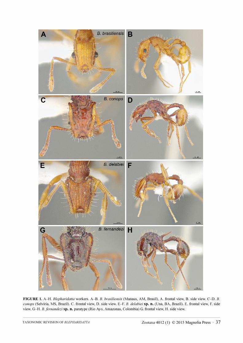
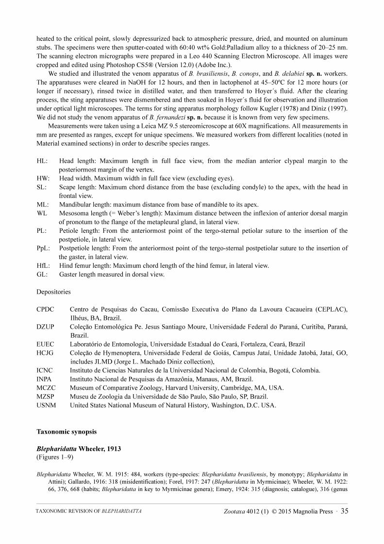
FIGURE 1. A–H. Blepharidatta workers. A–B. B. brasiliensis (Manaus, AM, Brasil), A. frontal view, B. side view. C–D. B.
conops (Selvíria, MS, Brazil). C. frontal view, D. side view. E–F. B. delabiei sp. n. (Una, BA, Brazil). E. frontal view, F. side
view. G–H. B. fernandezi sp. n. paratype (Rio Ayo, Amazonas, Colombia) G. frontal view, H. side view.
Zootaxa 4012 (1) © 2015 Magnolia Press · 37TAXONOMIC REVISION OF BLEPHARIDATTA
FIGURE 2. A–H. Blepharidatta ergatoid gynes. A–B. B. brasiliensis (Manaus, AM, Brasil), A. frontal view, B. side view. C–
F. B. conops. C–D. Selvíria, MS, Brazil. C. frontal view, D. side view. E–F. Crateus, CE, Brazil. E. frontal view, F. side view.
G–H. B. delabiei sp. n. (Una, BA, Brazil) G. frontal view, H. side view.
BRANDÃO ET AL. 38 · Zootaxa 4012 (1) © 2015 Magnolia Press

FIGURE 3. A–F. Blepharidatta males. A–B. B. brasiliensis (Manaus, AM, Brasil), A. frontal view, B. side view. C–D. B.
conops (Selvíria, MS, Brazil). C. frontal view, D. side view. E–F. B. delabiei sp. n. (Una, BA, Brazil) E. frontal view, F. side
view.
Larvae (Larva unknown for B. fernandezi sp. n.). Profile attoid. Segmentation indistinct. No leg or wing vestiges observed under SEM. Integument mostly smooth, spinules minute and concentrated in ventrolateral body region, longitudinal smooth strip present along ventral region of body, and dorsum of posterior somites. Hairs sparse, randomly distributed, unbranched, smooth and slightly curved.
Cranium subcircular (Figs 5 A–D); frons, clypeus, and labrum projecting from cranium; relatively large antenna placed laterad of pronounced area at cranium mid-length with three sensillae, only slightly elevated from head surface; single faceted eye posterior to midlength of cranium. Mandible attoid, broad and short, apical portion abruptly attenuated; with one sharp pointed apical tooth and no subapical teeth. Labrum crescentic, very short and glabrous, with rounded anterior margin. Anterior clypeal margin broadly concave, posterior margin straight. Maxillae with rounded apex, palp shaped as short frustrum, galea relatively elongate and cylindrical paxilliform with three sensilla; labium feebly bilobed, with short rows of minute acute spinules.
Zootaxa 4012 (1) © 2015 Magnolia Press · 39TAXONOMIC REVISION OF BLEPHARIDATTA

FIGURE 4. A–F. Blepharidatta male wings. A–B. B. brasiliensis (Manaus, AM, Brasil), A. fore B. hind. C–D. B. conops
(Selvíria, MS, Brazil). C. fore, D. hind. E–F. B. delabiei sp. n. (Una, BA, Brazil) E. fore, F. hind.
Biology. All four known Blepharidatta species are generalist predators that nest in the ground or in the leaf litter; nests are single, short (up to 20 cm) cylinders excavated in the ground or inside rolled leaves or rotting twigs, with one to 10 ergatoid gynes and up to 450 workers in the biggest colonies. Foragers patrol a roughly circular area around the single nest opening, where they collect live or dead arthropods to feed their larvae.
Distribution. B. brasiliensis and B. fernandezi sp. n. are known, respectively, from the central and western Amazon basin, B. conops from different localities in the Brazilian savannas and caatingas, while B. delabiei sp. n.
is known from different localities in the Atlantic Forest, eastern Brazil (Fig. 9).
BRANDÃO ET AL. 40 · Zootaxa 4012 (1) © 2015 Magnolia Press

FIGURE 5. A–E. Blepharidatta conops mature larvae. A–B. Crateus, CE, Brazil, A. head, frontal view, B. head, diagonal view.
C–E (Selvíria, MS, Brazil). C. head, frontal view. D. ventral side (detail). F. ventral aspect.
Zootaxa 4012 (1) © 2015 Magnolia Press · 41TAXONOMIC REVISION OF BLEPHARIDATTA
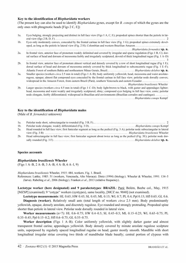
Key to the identification of Blepharidatta workers
(The present key can also be used to identify Blepharidatta gynes, except for B. conops of which the gynes are the only ones with phragmotic heads [Figs 2 C, E])
1a. Eyes bulging, strongly projecting and distinct in full face view (Figs 1 A, C, E); propodeal spines shorter than the petiole in lat-
eral view (figs 2 B, D, F). . . . . . . . . . . . . . . . . . . . . . . . . . . . . . . . . . . . . . . . . . . . . . . . . . . . . . . . . . . . . . . . . . . . . . . . . . . . . . . 2
1b. Eyes only moderately convex, concealed by the frontal carinae in full face view (Fig. 1 G); propodeal spines extremely devel-
oped, as long as the petiole in lateral view (Fig. 2 H); Colombian and western Brazilian Amazon. . . . . . . . . . . . . . . . . . . . . . . .
. . . . . . . . . . . . . . . . . . . . . . . . . . . . . . . . . . . . . . . . . . . . . . . . . . . . . . . . . . . . . . . . . . . . . . . . . . . Blepharidatta fernandezi sp. n.2a. In frontal view, anterior face of pronotum weakly delimited and covered by irregular and sparse rugulation (Figs 1 B, C); dor-
sal surface of head and dorsum of mesosoma feebly and irregularly sculptured, devoid of thick longitudinal rugae (Fig. 1 A–D)
. . . . . . . . . . . . . . . . . . . . . . . . . . . . . . . . . . . . . . . . . . . . . . . . . . . . . . . . . . . . . . . . . . . . . . . . . . . . . . . . . . . . . . . . . . . . . . . . . . . 3
2b. In frontal view, anterior face of pronotum almost vertical and densely covered by a row of short longitudinal rugae (Fig 1 E);
dorsal surface of head and dorsum of mesosoma entirely covered by thick longitudinal to subconcentric rugae (Fig. 1 E–F);
Atlantic Forest of southern Bahia and northeastern Minas Gerais, Brazil.. . . . . . . . . . . . . . . . . . . Blepharidatta delabiei sp. n.3a. Smaller species (workers circa 2.5 mm in total) (Figs 1 A–B); body uniformly yellowish; head, mesosoma and waist areolate-
rugose, opaque; almost flat compound eyes concealed by the frontal carinae in full face view; petiolar node dorsally convex;
widespread in the Amazon Forest, from eastern Brazil (Pará), southern Venezuela and eastern Ecuador. . . . . . . . . . . . . . . . . . .
. . . . . . . . . . . . . . . . . . . . . . . . . . . . . . . . . . . . . . . . . . . . . . . . . . . . . . . . . . . . . . . . . . . . . . . . Blepharidatta brasiliensis Wheeler
3b. Larger species (workers circa 4.5 mm in total) (Figs 1 C–D); body light-brown to black, with gaster and appendages lighter;
head, mesosoma and waist weakly and irregularly sculptured, shiny; compound eyes bulging in full face view, conic; petiolar
node elongate, feebly differentiated; widespread in Brazilian arid environments (Brazilian cerrados and caatingas). . . . . . . . . .
. . . . . . . . . . . . . . . . . . . . . . . . . . . . . . . . . . . . . . . . . . . . . . . . . . . . . . . . . . . . . . . . . . . . . . . . . . . . . Blepharidatta conops Kempf
Key to the identification of Blepharidatta males
(Male of B. fernandezi unknown)
1a. Petiolar node short, subrectangular to rounded (Fig. 3 B, F)... . . . . . . . . . . . . . . . . . . . . . . . . . . . . . . . . . . . . . . . . . . . . . . . . . . 2
1b. Petiolar node elongate, weakly differentiated (Fig. 3 D). . . . . . . . . . . . . . . . . . . . . . . . . . . . . . . . . .Blepharidatta conops Kempf
2a. Head rounded in full face view; first funicular segment as long as the pedicel (Fig. 3 A); petiolar node subrectangular in lateral
view (Fig. 3 B) . . . . . . . . . . . . . . . . . . . . . . . . . . . . . . . . . . . . . . . . . . . . . . . . . . . . . . . . . . . Blepharidatta brasiliensis Wheeler
2b. Head subrectangular in full face view; first funicular segment about twice as long as the pedicel (Fig. 3E); petiolar node dor-
sally rounded (Fig. 3 F) . . . . . . . . . . . . . . . . . . . . . . . . . . . . . . . . . . . . . . . . . . . . . . . . . . . . . . . . . . Blepharidatta delabiei sp. n.
Species accounts
Blepharidatta brasiliensis Wheeler
(Figs 1 A–B, 2 A–B, 3 A–B, 4 A–B, 6 A–I, 9)
Blepharidatta brasiliensis Wheeler, 1915: 484, workers. Fig. 1. Brazil.
References: Lattke, 1985: 51 (workers, Venezuela, Alto Mavaca). Diniz (1994) (biology). Wheeler & Wheeler, 1991: 134–5
(larva). Rabeling et al., 2006 (biology). Franken et al., 2013 (relative frequency).
Lectotype worker (here designated) and 9 paralectotypes: BRAZIL: Pará: Belém, Beebe col., May, 1915 [MZSP] (examined); 9 “cotype” workers (syntypes), same locality, [MCZ no. 9040] (not examined).
Lectotype measurements: HL 0.65; HW 0.45; SL 0.45; ML 0.15; WL 0.7; PL 0.4; Ppl 0.15; Hfl 0.65; GL 0.6.Diagnosis (worker). Relatively small ants (total length of workers circa 2.5 mm). Body predominantly
yellowish, opaque, densely areolate, and discretely rugulose. Eye rounded and strongly protruding. Propodeal spine shorter than petiole in lateral view. Petiolar node dorsally rounded in lateral view.
Worker measurements (n=7): HL 0.6–0.75; HW 0.4–0.5; SL 0.45–0.5; ML 0.15–0.25; WL 0.65–0.75; PL 0.35–0.45; Ppl 0.15–0.2; Hfl 0.6–0.75; GL 0.55–0.75.
Worker description (Figs 1 A–B). Color uniformly yellowish, with slightly darker gaster and almost transparent frontal carina; appendages yellowish. Body densely covered by minute areolate rugulose sculpture units, superposed by regularly spaced longitudinal rugulae on head; gaster mostly smooth. Mandible with short longitudinal irregular striae covering two thirds of mandibular blade basally; central portion of clypeus with
BRANDÃO ET AL. 42 · Zootaxa 4012 (1) © 2015 Magnolia Press

relatively pronounced longitudinal and transversal carinae forming rough reticulation over fine and uniform reticulation. Scrobe divided into four distinct parts, the anterior one is deeper than the rest of the scrobe and accommodates base of antenna; the second area shows 3–4 transverse curved striae over areolate rugae, followed by a deep, almost smooth area, and a posterior area of transversally oriented sculpture units, ending with areolate rugulae. Dorsal surface of head with five prominent longitudinal rugulae at each side. Compound eye set within a net of polygonal cells formed by rugulae, better seen in lateral view, three long and parallel cells anterior to eye, and seven irregular foveae posterior to eye, the first double and others in a row. Mesosoma sculptured throughout entire surface, with irregular, vermiculate, longitudinal rugae over areolate sculpture. Petiole and postpetiole with two longitudinal rugae in lateral view. Areolate sculpture concentrated on the anterior one fifth of gaster, otherwise smooth and shining. Appendages regularly areolate.
In general, body covered by sparse hairs; hairs stiff, long, slightly curved, uniform in width, and truncate; some of them in pairs. Dorsum of mandible with subdecumbent to appressed flexuous short hairs; frontal carina with 6–7 regularly spaced and upwards bent hairs. Dorsum of petiolar node and postpetiole with sparse hairs, mostly in pairs; ventral face of petiole devoid of hairs; ventral face of postpetiole with a single pair of hairs. Anterior face of coxa with a long and erect hair. Legs otherwise covered by appressed small pilosity.
Cephalic occipital corner tuberculate in frontal view. Scape and funiculus partially lodged in scrobe, which is not wide enough to receive whole scape; frontal carina does not cover scrobe, internal area of scrobe visible even with the head in frontal view; convex compound eye with about eight to nine facets along maximum diameter. Ventral face of head slightly convex, head wider anteriorly than rest in lateral view.
Promesonotum slightly convex medially in anterior view, followed by attenuated curve to lateral margin; straight in profile, anterior margin angular; dorsum of promesonotum higher than propodeal dorsum; pronotal humeral angle pointed, slightly bent forwards; anteroventral corner pointed; metanotal groove shallow but clearly visible in lateral view; mesometapleural suture absent or faintly marked, not clearly separating the meso- and metapleuron on the sides of mesosoma; bulla of metapleural gland indistinct; dorsal profile of propodeum straight; propodeal spines relatively long and slightly directed posterodorsally; infraspinal lamella present and fused to propodeal lobe; propodeal lobe subquadrate in side view, length close to one-third of propodeal spine length.
Petiole pedunculate, node moderately elevated and dorsally rounded, posterior face weakly sloped in lateral view; postpetiole feebly convex dorsally and without ventral processes. Gaster suboval, tergum I anterolaterally angular in dorsal view.
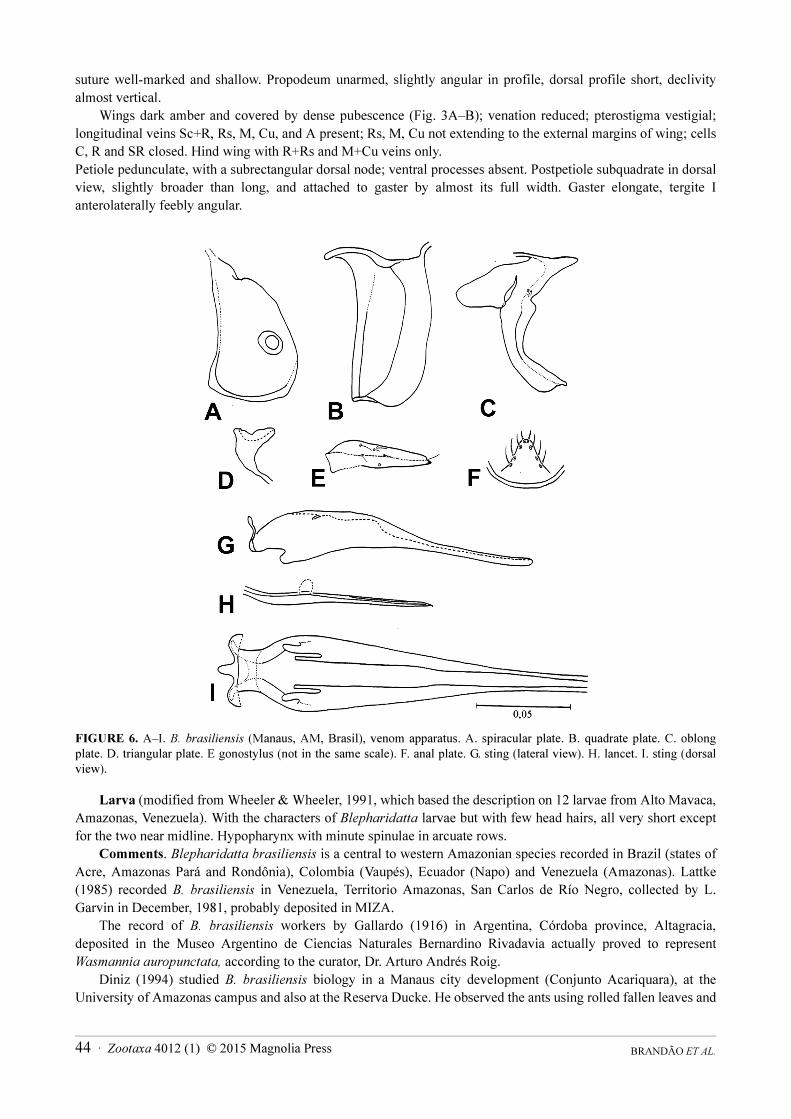
Sting apparatus (Figs. 6 A–I). Spiracular plate sub-rectangular, body extends towards median connection; spiracular external margin separated from ventral plate margin by distance equal to the internal diameter of spiracle; ventral tubercle absent. Quadrate plate apodeme of same size as plate body, anterodorsal corner with distinct projection and rounded apex. Anal plate as in other Blepharidatta species. Oblong plate with short posterior apodeme; subterminal tubercle absent. Gonostylus membranaceous with short terminal band. Triangular plate dorsal tubercle prominent and indistinct; median tubercle absent. Lancets as in other Blepharidatta species. Sting base with distinct antero-lateral processes, slender; campaniform sensillae of valve chamber present beyond basal half of sting shaft; sting reduction index 37. Furcula dorsal arm length smaller than side arm’s length.
Gyne measurements (n=1): HL 0.70, HW 0.50, SL 0.5, ML 0.15, WL 0.70, PL 0.45, Ppl 0.15, HfL 0.65, GL 0,85.
Gyne (ergatoid) description (Figs 2 A–B). Similar to conspecific worker, differing by the slightly larger body size and more robust gaster.
Male measurements (n=1): HL 0.45; HW 0.35; SL 0.15; ML 0.2; WL 0.7; PL 0.3; Ppl 0.1; Hfl 0.45; GL 0.6.Male description (Figs 3 A–B). Body yellowish-brown, with head, scutum, and first third of gaster slightly
darker; appendages yellowish. Body areolate, especially on head; sculpture on lateral portions of mesosoma almost indistinct. Gaster smooth and shiny. Long, subdecumbent, cream-colored hairs sparsely covering head and mesosomal dorsum, denser on apical segments of gaster; appendages with fine and sparse appressed hairs.
Head subrectangular, longer than wide. Mandible short and subfalcate, with vestigial denticles on masticatory margin; scape not reaching posterior margin of eye in frontal view; pedicel enlarged and relatively short; first funicular segment as long as the pedicel. Eye huge and extremely protruding, occupying about three fourths of head in lateral view. Ocelli present and equally developed.
Pronotum reduced in dorsal view, humeral angles discrete and rounded; scutum large, rounded anteriorly and with slightly convex posterior margin; notauli distinct. Prescutellum narrow; scutellum wider than long. Metanotal
Zootaxa 4012 (1) © 2015 Magnolia Press · 43TAXONOMIC REVISION OF BLEPHARIDATTA
suture well-marked and shallow. Propodeum unarmed, slightly angular in profile, dorsal profile short, declivity almost vertical.
Wings dark amber and covered by dense pubescence (Fig. 3A–B); venation reduced; pterostigma vestigial; longitudinal veins Sc+R, Rs, M, Cu, and A present; Rs, M, Cu not extending to the external margins of wing; cells C, R and SR closed. Hind wing with R+Rs and M+Cu veins only.Petiole pedunculate, with a subrectangular dorsal node; ventral processes absent. Postpetiole subquadrate in dorsal view, slightly broader than long, and attached to gaster by almost its full width. Gaster elongate, tergite I anterolaterally feebly angular.
FIGURE 6. A–I. B. brasiliensis (Manaus, AM, Brasil), venom apparatus. A. spiracular plate. B. quadrate plate. C. oblong
plate. D. triangular plate. E gonostylus (not in the same scale). F. anal plate. G. sting (lateral view). H. lancet. I. sting (dorsal
view).
Larva (modified from Wheeler & Wheeler, 1991, which based the description on 12 larvae from Alto Mavaca, Amazonas, Venezuela). With the characters of Blepharidatta larvae but with few head hairs, all very short except for the two near midline. Hypopharynx with minute spinulae in arcuate rows.
Comments. Blepharidatta brasiliensis is a central to western Amazonian species recorded in Brazil (states of Acre, Amazonas Pará and Rondônia), Colombia (Vaupés), Ecuador (Napo) and Venezuela (Amazonas). Lattke (1985) recorded B. brasiliensis in Venezuela, Territorio Amazonas, San Carlos de Río Negro, collected by L. Garvin in December, 1981, probably deposited in MIZA.
The record of B. brasiliensis workers by Gallardo (1916) in Argentina, Córdoba province, Altagracia, deposited in the Museo Argentino de Ciencias Naturales Bernardino Rivadavia actually proved to represent Wasmannia auropunctata, according to the curator, Dr. Arturo Andrés Roig.
Diniz (1994) studied B. brasiliensis biology in a Manaus city development (Conjunto Acariquara), at the University of Amazonas campus and also at the Reserva Ducke. He observed the ants using rolled fallen leaves and
BRANDÃO ET AL. 44 · Zootaxa 4012 (1) © 2015 Magnolia Press

natural cavities in rotten logs as nesting sites; in half out of the eight examined nests, he found insect carcasses around the nest openings. Worker populations of the eight colonies Diniz collected ranged from 49 to 117. One colony had only a single ergatoid gyne, while the others had 3–14 ergatoids; males ranged from 0 (four colonies) to 12. Diniz (op. cit.) listed the items found in pieces or sometimes in whole bodies around collected nests of B.
brasiliensis: spiders, diplopods, orthopteroids, beetles, flies, unidentified Hymenoptera and ants of the genera Camponotus, Cephalotes, Crematogaster, Dolichoderus, Ectatomma, Nesomyrmex, Pachycondyla, Pheidole,
Pseudomyrmex, and Solenopsis. Diniz observed at least once the migration of a colony from one rolled leaf to another, with workers also transferring the carcasses found within and around the nest opening. He also observed that workers keep the larvae in between the mandibles while inside the nest. In two nests, Diniz (op. cit.) recorded isopod inquilines that suffered no hostility from the workers.
Rabeling et al. (2006) described the non-homogeneous nest distribution of B. brasiliensis in a locality near Manaus, AM, Brazil (EMBRAPA campus). The colonies occupied either cavities within rotting branches (more commonly) or natural spaces between leaves (19% out of 26 studied nests), apparently taking advantage of pre-existing cavities, reducing the cavity size, if too large, or sealing it by means of a chamber wall, built from a mixture of soil, vegetable debris, and insect parts. According to them, “B. brasiliensis appears to take advantage of pre-existing cavities, which were only slightly modified by them. The shape of the main chambers was amorphous ellipsoid to round and had an inner dimension of 4 cm length by 1.5 cm width. If pre-existing cavities in branches were too large or the nest was located between leaves, the ants had reduced the nest volume by constructing chamber walls, built from a mixture of soil, vegetable debris, and insect parts in order to seal off the nest chamber.” As in Diniz (op. cit.), they also observed that colonies were polygynous with one to eleven ergatoid gynes and with 132 workers on average (SD = 95.63). Nests contained large numbers of brood (roughly 75% larvae and 25% pupae), which were kept lying on the bottom of the nest chamber. If a colony was disturbed, workers picked up the immatures and held them between their mandibles. The colonies they studied appeared to be mostly active at night and to be omnivorous, scavenging or preying on beetles, bees, cicadas, crickets, termites, spiders, and ants of the genera Cephalotes, Pheidole, Camponotus, and Pachycondyla. The victim’s legs and antennae were cut off before being dragged to the nest. Blepharidatta brasiliensis may also collect nutrient-rich plant structures, such as seeds or elaiosomes, in addition to the arthropod diet.
Franken et al. (2013) recorded B. brasiliensis in the litter trapped around Attalea attaleoides (Barb. Rodr.) Wess. Boer palm bases, which is a refuge for non-dominant ant species because aggressive species in these microhabitats are relatively rare. The trapped litter was occupied mostly by unaggressive predatory and fungus-growing species, which probably find more prey or better conditions to cultivate their food there.
Blepharidatta brasiliensis may be rather frequent in some Amazonian areas; for instance, Oliveira et al. (2009) recorded B. brasiliensis in 8.7% of the 850 pitfall traps and sardine baits set in 30 plots and from litter sifting (Winkler sacks) in 25 plots at Reserva Ducke in Manaus (the 5th most frequent ant species out of the 152 recorded in the area).
Material examined: BRAZIL: Acre: Cruzeiro do Sul, 23–28.xi.1983, F. H. Caetano (“folhiço # 4), 1 worker (MZSP). Amazonas: Manaus, viii.1962, K. Lenko # 4130, 69 workers, 7 gynes [4 workers CPDC, 4 workers, DZUP, 4 workers EUEC, 4 workers HCJG, 4 workers ICNC, 4 workers INPA, 4 workers MCZ, 7 gynes, 37 workers MZSP, 4 workers USNM]; Manaus, Km 24 ZF3, 21.iii.1983, WWF, camp Floresta, E.O. Wilson [MZSP], 1 worker; Manaus, INPA, 02.x.1987, J.L.M. Diniz, 4 workers, 1 gyne [MZSP]; Manaus, Ig. Marianil, Rio Branco, Rod 24, NE Manaus, ix.1962, W.L. Brown Jr. (Kempf collection #4565) 18 workers [MZSP]; Dimona, approx. 80Km N of Manaus, viii.2000, U. Mueller & R. Adams 8 workers, 1 male [MZSP]; Manaus to Itacoatiara Km 50 (M-61), W.L. Brown Jr. 24.viii.1962, 3 workers [MZSP]; Manaus, Conjunto Acariquara, 24.xi–7.xii.1987, 10 workers, 1 male, 1 gyne, J.L.M. Diniz (Diniz collection #2340; 2327) [4 workers HCJC, 6 workers MZSP]. Pará: Belém, 15.v.1915, C.W. Beebe (lectotype) 1 worker [MZSP]; Jari, Corte Seletivo, 0º53'S, 52º36'W, 2011, E.A. Silva, 58 workers [24 DZUP, 4 HCJG, 4 EUEC, 26 MZSP]; Utinga tract, nr. Belém, 12.viii.1962, W.L. Brown 1 worker, 1 male [MZSP]; Porto Trombetas, viii.1992, J.D. Majer (#12), 11 workers [MZSP]. Rondônia: Porto Velho, Área Caiçara, 09º26’13.0”S 64º48’04.1”W, 19.vi–02.vii.2010, R.M. Feitosa & R.R. Silva, 35 workers [20
DZUP, 15 MZSP]. COLOMBIA: Vaupés, Tararaira, Est. Biol. Caparu, 01o04’S, 69o31’W, altitude 85 m, 14–20.v.2001, A. Sabogal [UNAB], 13 workers [7 ICNC, 6 MZSP0. ECUADOR: Napo, Cuyabeno, 12.x–5.xi.1994, J. P. Caldwell (#10342), 1 worker [CPDC]. VENEZUELA: Amazonas, Alto Mavaca, 05.xi.1989, J. E. Lattke (#1255), 10 workers [3 DZUP; 7 MZSP].
Zootaxa 4012 (1) © 2015 Magnolia Press · 45TAXONOMIC REVISION OF BLEPHARIDATTA
Blepharidatta conops Kempf
(Figs 1 C–D, 2 C–F, 3 C–D, 4 C–D, 5 A–E, 7 A–I, 9)
Blepharidatta conops Kempf, 1967: 355, workers. Figs. 4,5. Brazil.
References: Brandão et al. 1998, 2001; Diniz et al., 1998; Silva et al. 2002.
Holotype worker: BRAZIL: Mato Grosso do Sul: Três Lagoas, Faz. Retiro das Telhas, 28.v.1964, Exp. Depto Zoologia (Kempf coll. # 4131) (MZSP, examined).
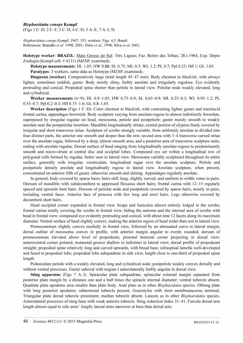
Holotype measurements: HL 1.05; HW 0.88; SL 0.75; ML 0.3; WL 1.2; PL 0.7; Ppl 0.25; Hfl 1; GL 1.05.Paratypes: 3 workers, same data as Holotype (MZSP, examined).Diagnosis (worker). Comparatively large (total length 45–47 mm). Body chestnut to blackish, with always
lighter, sometimes reddish, gaster. Body mostly shiny, feebly areolate and irregularly rugulose. Eye evidently protruding and conical. Propodeal spine shorter than petiole in lateral view. Petiolar node weakly elevated, long and cylindrical.
Worker measurements (n=9): HL 0.9–1.05; HW 0.75–0.9; SL 0.65–0.9; ML 0.25–0.3; WL 0.95–1.2; PL 0.55–0.7; Ppl 0.2–0.3; Hfl 0.75–1.0; GL 0.8–1.05.
Worker description (Figs 1 C–D). Color chestnut to blackish, with contrasting lighter gaster and translucid frontal carina; appendages brownish. Body sculpture varying from areolate-rugose to almost indistinctly foveolate, superposed by irregular rugulae on head, mesosoma, petiole and postpetiole; gaster mostly smooth to weakly areolate near the postpetiolar insertion. Mandible longitudinally striate; central portion of clypeus finely covered by irregular and short transverse striae. Sculpture of scrobe strongly variable, from uniformly areolate to divided into four distinct parts, the anterior one smooth and deeper than the rest; second area with 3–4 transverse curved striae over the areolate rugae, followed by a deep, almost smooth area, and a posterior area of transverse sculpture units, ending with areolate rugulae. Dorsal surface of head ranging from longitudinally areolate-rugose to predominantly smooth and micro-striate at central disc and occipital lobes. Compound eye set within a longitudinal row of polygonal cells formed by rugulae, better seen in lateral view. Mesosoma variably sculptured throughout its entire surface, generally with irregular, vermiculate, longitudinal rugae over the areolate sculpture. Petiole and postpetiole densely areolate and longitudinally rugose in lateral view. Areolate sculpture, when present, concentrated on anterior fifth of gaster, otherwise smooth and shining. Appendages regularly areolate.
In general, body covered by sparse hairs; hairs stiff, long, slightly curved, and uniform in width; some in pairs. Dorsum of mandible with subdecumbent to appressed flexuous short hairs; frontal carina with 12–15 regularly spaced and upwards bent hairs. Dorsum of petiolar node and postpetiole covered by sparse hairs, mostly in pairs, including ventral faces. Anterior face of procoxa with few long and erect hairs. Legs otherwise covered by decumbent short hairs.
Head occipital corner expanded in frontal view. Scape and funiculus almost entirely lodged in the scrobe; frontal carina totally covering the scrobe in frontal view, hiding the antenna and the internal area of scrobe with head in frontal view; compound eye evidently protruding and conical, with about nine 12 facets along its maximum diameter. Ventral surface of head slightly convex, making the anterior region of head wider than rest in lateral view.
Promesonotum slightly convex medially in frontal view, followed by an attenuated curve to lateral margin; dorsal outline of mesosoma convex in profile, with anterior margin angular to evenly rounded; dorsum of promesonotum elevated above level of propodeum; pronotal humeral corner projecting in dorsal view; anteroventral corner pointed; metanotal groove shallow to indistinct in lateral view; dorsal profile of propodeum straight; propodeal spine relatively long and curved upwards, with broad base; infraspinal lamella well-developed and fused to propodeal lobe; propodeal lobe subquadrate in side view, length close to one-third of propodeal spine length.
Pedunculate petiole with a weakly elevated, long and cylindrical node; postpetiole weakly convex dorsally and without ventral processes. Gaster suboval with tergum I anterolaterally feebly angular in dorsal view.
Sting apparatus (Figs 7 A–I). Spiracular plate subquadrate, spiracular external margin separated from posterior plate margin by a distance one and a half times the spiracle internal diameter; ventral tubercle absent. Quadrate plate apodeme area smaller than plate body. Anal plate as in other Blepharidatta species. Oblong plate with long posterior apodeme; subterminal tubercle present. Gonostylus with short membranaceous terminal. Triangular plate dorsal tubercle prominent; median tubercle absent. Lancets as in other Blepharidatta species. Anterolateral processes of sting base with weak anterior tubercle. Sting reduction index 31–41. Furcula dorsal arm length almost equal to side arms´ length; lateral arms narrower at base than dorsal arm.
BRANDÃO ET AL. 46 · Zootaxa 4012 (1) © 2015 Magnolia Press

Gyne measurements (n=2): HL 1.05–1.15; HW 1.35–1.45; SL 0.55–0.65; ML 0.3–0.35; WL 1.25; PL 0.65; Ppl 0.26–0.3; Hfl 0.95; GL 1.35–1.45.
Gyne (ergatoid) description (Figs. 2 C–F). Larger and drastically distinct from conspecific worker. Surface sculpturing extremely variable, especially on head and anterior slope of pronotum, which can be predominantly smooth and minutely foveolate or densely areolate-rugose. Pilosity denser than in the workers, mainly on mesosomal and metasomal dorsum.
Head and anterior face of pronotum phragmotic; dorsal surface of head rounded, with the frontal carina enormously expanded so that the lateral margins of head, eyes, clypeus and mandibles are totally concealed in frontal view; outline of cephalic disc only interrupted anteriorly by a median emargination, which marks the limits between the frontal lobes. Mesosoma subrectangular and robust. Anterior face of pronotum vertical and rounded dorsally in frontal view, forming with the head a large disc; sides of metanotum sometimes with variably developed wing bud; propodeal spines shorter than those of workers. Gaster well-developed.
Male measurements (n=2): HL 0.8–0.95; HW 0.55; SL 0.3; ML 0.1–0.15; WL 1.3–1.45; PL 0.6; Ppl 0.25–0.35; Hfl 1.0–1.11; GL 1.10–1.25.
Male description (Figs 3 C–D). Body uniformly chestnut to dark-brown, with slightly darker gaster; appendages yellowish. Body entirely areolate with vermiculate longitudinal rugulae over the lateral portions of mesosoma and waist. Gaster smooth and shining. Long, subdecumbent, whitish hairs densely covering the entire body; appendages densely covered by subdecumbent hairs.
FIGURE 7. A–I. B. conops (Selvíria, MS, Brazil), venom apparatus. A. spiracular plate. B. oblong plate. C. quadrate plate. D.
triangular plate. E gonostylus (not in the same scale). F. anal plate. G. sting (lateral view). H. lancet. I. sting (dorsal view).
Zootaxa 4012 (1) © 2015 Magnolia Press · 47TAXONOMIC REVISION OF BLEPHARIDATTA

Head rounded in frontal view. Mandible short and subfalcate, with vestigial denticles on the masticatory margin; scape very short, not reaching the posterior margin of eye in frontal view; pedicel enlarged and relatively short; first funicular segment about twice as long as the pedicel. Eye huge and extremely protruding, occupying about three-fourths of head in lateral view. Ocelli present and equally developed.
Pronotum vestigial in dorsal view, with almost inconspicuous humeral angles; scutum large, almost straight anteriorly and with a slightly convex posterior margin; notauli weakly impressed. Prescutellum narrow; scutellum wider than long. Metanotal suture well-marked and shallow. Propodeum unarmed, dorsal and posterior margins continuous and gradually inclined posteriorly.
Wings dark amber in color and covered by a dense pubescence; venation reduced; pterostigma elliptical and well-marked; longitudinal veins Sc+R, Rs, M, Cu, and A present; Rs, M, Cu not extending to the external margins of the wing; cells C, R and SR closed. Hind wing with R+Rs and M+Cu veins only; four sub-median hamuli present.
Petiole pedunculate, with weakly elevated, long and cylindrical node; ventral processes absent. Postpetiole subrectangular in dorsal view, longer than broad, and attached to gaster by almost its full width. Gaster elongate, tergite I anterolaterally feebly angular.
Larva (late instar) description (Fig 5 A–E). Body hairs of two main sizes: short (ca. 50–60 microns) sparsely dispersed throughout body, but commoner in the spinulose areas, and long (more than 200 microns) flexuous tapered hairs in a row around pronotum and some around anus, most broken off at base (Fig. 5 E). Eight stouter hairs present on frons (six anteriorly in a row on a fold at mid-length projection and two dorsoposteriorly); clypeal anterior margin with a row of seven minute hairs (circa 3 microns), the central ones even smaller.
Throphorhinium ventral plate with 6 to 7 transverse spinulose striae. Labium with dorsal face forming two blunt low spinulose projections, spinules covering all surfaces but palps. Paxilliform palp with three sensillae. Opening of sericteries not visible in frontal view.
Comments. Blepharidatta conops has been recorded in different central Brazilian states, mostly those covered by cerrado or caatinga vegetation: western Bahia, southwestern Ceará, Goiás, Maranhão, Minas Gerais, Mato Grosso, Mato Grosso do Sul, Piauí and Tocantins. It is the best studied species in the genus thus far and has been subject of several published studies by the Museu de Zoologia da USP team. In a series of papers, Brandão, 2000, Brandão et al. 1998, 1999, 2000, 2001, 2008; Diniz et al., 1998; Silva, 2003, Silva & Brandão, 2001, and Silva et
al., 2001, 2002, published detailed biological observations on B. conops, briefly summarized below.Jorge L. M. Diniz studied 71 nests of B. conops at Chapada dos Guimarães, Mato Grosso, Três Lagoas and
Selvíria in Mato Grosso do Sul (Diniz et al., 1998). Pereira et al. (2014) studied a population of B. conops with 29 nests in Crateús, Ceará state. All but one of these nests were excavated in the soil; one odd nest also occupied part of a hollow dry branch fallen on the soil. Nest populations varied from a few individuals to a maximum of 248 workers in the savannas and 437 in Ceará, and only one nest contained two ergatoid gynes; all other are monogynous, although in some nests no gynes were recorded, possibly missed during excavation. Blepharidatta
conops nests are easily spotted by the carcass ring the workers carefully arrange around the nest opening. Once one gets used to this search image, it becomes extremely easy to spot the nests. The carcasses around the nest opening are similar in nature to those found in the bottom chamber of the nest; field and laboratory observations suggest the workers collect live arthropods or carcasses, bring them inside the nest for larval feeding, and then put them in the carcass ring around the single nest opening. Populations are rather dense, although separated sometimes by several kilometers, reflecting the low dispersal ability of ergatoid gynes.
Pereira et al. (2014) argue that B. conops populations from Ceará could represent a distinct although closely related species, mostly because of the almost flat anterior cephalic disks of Ceará ergatoids (Fig. 2E) and because this population was collected in a locality within the semi-arid Caatinga, while other B. conops populations come from central Brazil savannas. Brandão et al. (2001) already noticed the extreme variation in the sculpture of the ergatoid gynes frontal disc, but were not able to find a single case in which two forms occur sympatrically. They related this variation to the ergatogyny of the gynes that enhances the genetic viscosity of populations, resulting in local variants. In the case of the Ceará population, the ergatoids show the same sculpturing pattern as in other localities, composed by polygonal units separating punctuated areas, although the sculpture units are not so densely packed in some regions of the disc as in other places, rendering the head and pronotal front more regular (see discussion and figures in Brandão et al. 2001). Moreover, we studied worker larvae of cerrado and caatinga populations, confirming the morphological identity of the immatures (Fig. 5). Pereira et al. (2014) also described
BRANDÃO ET AL. 48 · Zootaxa 4012 (1) © 2015 Magnolia Press
the striking similarities among B. conops populations and that of Crateús, Ceará, in nest architecture, in prey diversity found inside nests and the carcass rings, preferred daily time for foraging activities (with two peaks of activity), and other traits. Additionally, the measurements of the Ceará workers and gynes fall well within the range of other B. conops population measurements. We see no evidence of the existence of a different Blepharidatta
species in Ceará.
Material examined: BRAZIL: Bahia: Barreiras, 15.v.2001 (“Cerrado-solo”), 12o08’59,7”S 45º09’31,5”W, T22P, E.B.A. Koch, 1 worker; same locality and date, T22P, K.S. Carvalho, 4 workers [MZSP]; Ceará: Crateús,
RPPN Serra das Almas, 05o00’S 41o00’W, 21.iv. 2003, Y. Quinet (pitfall) 70 workers; same locality (Grajaú), 03–08.vi.2009, Brandão et al. col., 2 workers, 1 gyne, 1male; same locality (Açude), 2 males, 1 gyne [4 workers CPDC, 4 workers DZUP, 24 workers EUEC, 6 workers HCJG, 4 workers INPA, 4 workers MCZC, 4 workers, USNM, 20 workers, gyne, male MZSP; Goiás: Cascalheira, vi.2009 (“parcela 9, armadilha E”), Valentim et al.
col., 2 workers [MZSP]; Campinaçu, Serra da Mesa, 13o52’S, 48º23’W, 18.ii–02.iii.1998, Silvestre et al. col., 33 workers [6 DZUP, 6 HCJG, 21 MZSP]; Colinas do Sul, Serra da Mesa, 02–15.vii.1995. 14º01’S 48º12’W, Silvestre et al. col., 46 workers, 4 males, 2 gynes [6 CPDC, 6 DZUP, 6 HCJG, 18 workers, 4 males, 2 gynes MZSP]; Mineiros, Parque Nacional das Emas, 18º19’S 52º45’W, 15.i.2004, R. A. Carvalho, #11 (em Cerrado, Campo limpo, sob ninho de Cornitermes cumulans, 6 workers; same data #8, 18.i.2004, 9 workers; same data, # 67, 23.i.2004, 9 workers [MZSP]; Niquelândia, 24.ix–6.x.1995, Silvestre et al. col. 48 workers, 2 males; same locality,
16.xi.2013, T. Carrijo col. 1 gyne, 4 workers [MZSP]; Serranópolis, Pousada das Araras, 18º18’S 51o08’W, 14.v–18.vi.2000, P.R. Silva & C. Prado cols., 13 gynes, 1 male, 1 worker (reared in the lab) [MZSP]; Maranhão: Balsas,
Gerais de Balsas, (“isca solo, lote 31 reserva”) 4–5.xi.1999, 8o34’S 46o42’W, Brandão et al. col., 8 workers, 4
gynes [MZSP]; Estreito, Fazenda Itaueiras, 06o31’54,4”S 47o22’16,0” W, 7–13.i.2005, Silva, R.R. & R.M. Feitosa (Winkler), 21 workers [MZSP]; Mato Grosso: U.H. Manso (“isca chão, manhã”), 13.viii.1988, H.C. Morais col., 14 workers [MZSP]; Chapada dos Guimarães, Faz. Buriti, 14.i.1985, J.L.M. Diniz col., 9 workers [3 DZUP, 3 HCJG, 3 MZSP]; Chapada dos Guimarães, Faz. Chafariz (“isca sardinha sítio III”) 30.ix.2005, F.H.O. Silva, 2 workers [MZSP]; Fátima, 8.iii.1971, W.W. Kempf col. and det., 1 worker [MZSP]; São Lourenço, vi.1974, M. Naves (Kempf coll. 11579), 3 workers [MZSP]. Mato Grosso do Sul: Três Lagoas, Faz. Retiro das Telhas, 28.v.1964, Exp. Depto Zoologia (Kempf coll. #4131), 4 workers (holotype and paratypes) [MZSP]; Selvíria, CECA Reserva, 7.i–18.xi.1985, J.L.M. Diniz (#2221, 2232, 2242), 1 male, 1 gyne, 3 workers; same locality and collector (#2279) 6 workers [3 workers HCJG, gyne and 6 workers MZSP]; Minas Gerais: Grande Sertão Veredas, 12.x.2012 (“coleta manual”), T. Camp col., 1 worker; Paracatu (“cerrado”), 2.iii.1989, Márcio Naves col., 1 worker [MZSP]. Piauí: Bom Jesus, 09.19163 S 44.84255W, 10.xi.2010 (solo), W.T. Frizzo & H. Vasconcelos, 1 worker [MZSP]. Tocantins: Babaçulândia, 07º02’19.0”S 47º52’03,4”W, 14–19.i.2005, R. Silva & R. Silvestre (from several leaf litter samples) 19 workers [3 DZUP, 3 HCJG, 13 MZSP]; Palmeiras do Tocantins, 06º49’12.1”S 47º3’48.6”W, 14–19.i.2005, R. Silva & R. Silvestre, 1 worker [MZSP]; Paranã, Faz. Caldas 12º48’51.6”S 47º53’55.3”W, 12.x.2004, R.R. Silva & B.H. Dietz (“isca solo cerrado sensu stricto”), 2 workers [MZSP]; Paranã 12º56’03.3”S 47º57’42.5”W, R.R. Silva & B.H. Dietz (mata ciliar), 1 worker [MZSP]; Paranã, Rio Ouro Fino, Faz. Contenda, 12º56’03”S 47º57’42”W, R.R. Silva & B.H. Dietz, 1 worker [MZSP]; Aguiarnópolis, 06º36’49.4”S 47º 8’52.2”W, 14–19.i.2005, R. Silva & R. Feitosa (from several leaf litter samples), 8 workers [MZSP].
Blepharidatta delabiei new species
(Figs 1 E–F, 2 G–H, 3 E–F, 4 E–F, 8 A–I, 9)
Blepharidatta sp. Kempf, 1975: 370–371, males. Figs 19–22. Brazil.
Holotype worker: BRAZIL: Bahia: Una (Res. Biol. IBDF), 25.ix.1988, J.L.M. Diniz col. #2373–6 JLMD collection [MZSP].
Holotype measurements: HL 0.8; HW 0.6; SL 0.6; ML 0,25; WL 0.8; PL 0.45; Ppl 0.2; Hfl 0.7; GL 0.8.Paratypes: 20 workers, one gyne and two and males, same data as Holotype (6 workers, one male and one
gyne [MZSP], 3 workers, one male [HCJG], 3 workers [CPDC], 4 workers [DZUP], 3 workers [MCZC], 3 workers [USNM], one worker [INPA]).
Zootaxa 4012 (1) © 2015 Magnolia Press · 49TAXONOMIC REVISION OF BLEPHARIDATTA

Diagnosis (worker). Comparatively small species. Body predominantly chestnut. Head and mesosoma entirely covered by longitudinal to subconcentric thick rugae; in frontal view, anterior face of pronotum almost vertical and densely covered by a row of short longitudinal rugae. Eye rounded and strongly protruding. Propodeal spine shorter than petiole, in lateral view. Petiolar node dorsally rounded in lateral view.
Worker measurements (n=4): HL 0.8–0.85; HW 0.55–0.7; SL 0.55–0.7; ML 0.25; WL 0.8–0.9; PL 0.45–0.6; Ppl 0.2–0.25; Hfl 0.7–0.8; GL 0.8–1.25.
Worker description (Figs 1 E–F). Color predominantly chestnut, with slightly lighter head and gaster; appendages yellowish-brown. Head and mesosoma entirely covered by vermiculate and predominantly longitudinal thick rugae; gaster mostly smooth. Mandible with short fine longitudinal striae covering the anterior third of blade. Clypeus mostly smooth, with a few irregular longitudinal striae on central disc. Antennal scrobe almost entirely smooth and shiny, especially at the central portion, with a few transverse curved striae near the antennal insertion and at the apex. Dorsal surface of head with 10 to 12 prominent longitudinal subparallel rugae, extending from posterior margin of clypeus and becoming gradually divergent and concentric towards the posterior margin of head. In lateral view area between ventral margin of scrobe and ventral margin of head covered by polygonal cells formed by irregular rugulation. Ventral face of head smooth and shiny. Mesosoma entirely sculptured with irregular longitudinal rugae intercalated by transverse thick carinae, which are covered by weak areolae; dorsum covered by four to ten almost perfectly concentric rugae, forming a conspicuous and irregular circular pattern. In full frontal view anterior portion of pronotum elevated, permitting view of 10 short vertical rugae; anterior portion of pronotum separated from the rest of mesosoma by a strongly elevated carina, better seen in lateral view; lateral faces of pronotum weakly and irregularly rugulose. Meso- and metapleuron longitudinally rugose. Petiole with four to six longitudinal rugae in lateral and in dorsal view. Postpetiole weakly sculptured, with faint longitudinal rugulae over an areolate surface. Gaster almost entirely smooth and shining, with feeble isolated areolate sculpture near insertion of postpetiole. Appendages entirely but weakly areolate.
In general, body covered by sparse, cream colored hairs; hairs stiff, long, slightly curved, uniform in width, and truncate. Anterior margin of clypeus with a row of hairs gradually increasing in length from lateral portions to center. Dorsum of mandible with subdecumbent flexuous hairs restricted to the lateral border. Frontal carina with about five regularly spaced and upwards bent hairs. Pilosity on mesosoma sparse, moderately abundant dorsally. Dorsum of petiole and postpetiole covered by sparse paired hairs; ventral face of postpetiole with a single pair of hairs. Gaster entirely covered by regularly spaced hairs. Appendages covered by short, subdecumbent to appressed hairs.
In frontal view, head subtriangular, with lateral margins gently converging towards mandible. Posterior margin of head straight, with strongly expanded, auriculate occipital corners, rounded to acute apically. Anterior margin of clypeus somewhat irregular and slightly convex; central disc feebly concave to flat. Antennal scrobe considerably deep and wide, able to receive the whole scape at rest; frontal carina partially covers the scrobe in frontal view, proximal half of scrobe exposed. Bulging compound eye with about six to seven facets along maximum diameter. Ventral cephalic surface separated from side by strong carina, better seen in lateral view.
Dorsal outline of mesosoma irregularly continuous and weakly convex in lateral view. Promesonotum slightly convex medially in frontal view, gently descending towards humeral corners; anterior face of pronotum vertical in profile, separated from dorsal face by a conspicuous angle; dorsum of promesonotum elevated slightly to well above the level of propodeum; pronotal humeral corner rounded to weakly angulate in dorsal view; anteroventral corner with well-developed anteriorly directed spine. Metanotal groove shallowly impressed to well-marked, clearly visible in lateral view; mesometapleural suture faintly marked to absent, not clearly separating meso- and metapleuron. Dorsal profile of propodeum extremely short and straight; propodeal spines as long as the petiolar peduncle in lateral view and strongly divergent in dorsal view; infraspinal lamella present, fused to the propodeal lobe; propodeal lobe subquadrate in side view, its length half that of propodeal spine.
Petiole strongly pedunculate, node moderately elevated and considerably elongate, anterior face weakly sloped and posterior face almost vertical in lateral view; petiolar peduncle with discrete anteroventral process directed anteriorly. Postpetiole distinctly convex dorsally and about three times shorter than petiole, without ventral processes. Gaster suboval, tergum I anterolaterally discretely angular in dorsal view.
Sting apparatus (Figs 8 A–I). Spiracular plate ventral tubercle absent. Quadrate plate apodeme area smaller than plate body. Anal plate as in other Blepharidatta species. Oblong plate with long posterior apodeme, as long as the triangular plate. Subterminal tubercle present. Gonostylus membranaceous terminal band present and short.
BRANDÃO ET AL. 50 · Zootaxa 4012 (1) © 2015 Magnolia Press

Triangular plate dorsal tubercle prominent; median tubercle absent. Lancets as in other Blepharidatta species. Sting antero-ventral processes absent. Sting reduction index 31–41. Furcula dorsal length equal to lateral arms.
Gyne measurements (n=1): HL 0.85; HW 0.65; SL 0.6; ML 0.25; WL 0.95; PL 0.65; Ppl 0.25; Hfl 0.75; GL 1.25.
Gyne (ergatoid) description (Figs. 2 G–H). Similar to conspecific worker except for the presence of three ocelli triangularly placed on frons and the more robust gaster. No evidence of wing buds on mesosoma.
Male measurements (n=1): HL 0.55; HW 0.4; SL 0.15; ML 0.15; WL 0.95; PL 0.65; Ppl 0.25; Hfl 0.75; GL 1.25.
Male description (Figs 3 E–F). Body dark-brown, with head, pronotum, anterior portion of petiolar peduncle, and first third of gaster slightly lighter; head with a blackish rounded area on cephalic disc; appendages yellowish. Body densely areolate with few irregular striae, especially on head dorsum and sides of mesosoma. Gaster smooth and shiny, without sculpture. Long, subdecumbent, cream-colored hairs sparsely covering head and mesosomal dorsum; metasoma virtually glabrous, with few sparse hairs on the apical segments of gaster; appendages with fine and sparse appressed hairs.
Head subrectangular, longer than wide. Mandible short and subfalcate, with vestigial denticles on masticatory margin; scape not reaching posterior margin of eye in frontal view; pedicel enlarged and relatively short; first funicular segment almost twice longer than the pedicel. Eye huge and extremely protruding, occupying about three fourths of head in lateral view. Ocelli present and equally developed.
FIGURE 8. A–I. B. delabiei (Una, BA, Brazil), venom apparatus. A. spiracular plate. B. oblong plate. C. quadrate plate. D.
triangular plate. E gonostylus (not in the same scale). F. anal plate. G. sting (lateral view). H. lancet. I. sting (dorsal view).
Zootaxa 4012 (1) © 2015 Magnolia Press · 51TAXONOMIC REVISION OF BLEPHARIDATTA

Pronotum reduced in dorsal view, with discrete and rounded humeral angles; scutum large, rounded anteriorly and with a slightly convex posterior margin; notauli weakly distinct. Prescutellum narrow; scutellum wider than long. Metanotal suture well-marked and shallow. Propodeum unarmed, strongly rounded in profile, with a short dorsal profile and almost vertical declivous margin; propodeal spiracle small and distant from the declivous margin; propodeal lobe vestigial. Legs more elongate than in conspecific females.
Wings dark amber and covered by a dense pubescence (Fig. 3E–F); venation reduced and entirely nebular; pterostigma absent; longitudinal veins Sc+R, Rs, M, Cu, and A present; cells C, R and SR closed. Hind wing with R cell only; four sub-median hamuli present.
Petiole pedunculate, node strongly rounded; ventral processes absent. Postpetiole subrectangular in dorsal view, longer than broad, and attached to gaster by almost its full width. Gaster elongate, with rounded gastral shoulder.
Larva. Unknown.Etymology. This species is named for our friend Dr. Jacques H. C. Delabie, in recognition of his many years of
devoted work on the ants of Bahia, Brazil and for his valuable support during the elaboration of this work.Comments. The very particular sculpture pattern readily separates this species from its congeners. All other
species in the genus present sparse and predominantly chaotic rugulation over the body. In B. delabiei the rugae are thicker, denser and more regular. An interesting morphological variation can be observed among different populations of this species: most specimens present the head and mesosomal dorsum covered by longitudinally oriented rugae, while a few individuals display a subconcentric rugulation pattern. Considering the differences observed of the overall sculpturing pattern in other Blepharidatta species, and since intermediate forms can be found, we refrain from separating the material into distinct species.
Workers of this species are known only from the Atlantic Forest of southern Bahia state, frequently collected in leaf litter samples. Kempf (1975) described four males from Pedra Azul, northeastern Minas Gerais state, not accompanied by workers or gynes. Although he believed these specimens probably represented males of B.
brasiliensis, he refrained from identifying them as such and described them as Blepharidatta sp. Diniz and Brandão briefly visited this locality twice but were not able to find more specimens. A fifth male collected in Itambé, Bahia in 2003 solved the matter, as it is identical to those described by Kempf from Pedra Azul.
Material examined: BRAZIL: Bahia: Aurelino Leal, 14o19’52”S 39º21’32”W, 14.iv.1997, J.R.M. Santos, 1 worker (MZSP); Ilhéus, Banco de Pedra 14º40’51”S 39º15’24”W (Mata W-A23) 12.i.1998, J.R.M. Santos & J.C.S. do Carmo, 2 workers (MZSP); Ilhéus, Castelo Novo, 14º39’S 19º11’W, 30.ix–4.x.2002, P.R. Santos & J.R. Santos, 3 workers (CPDC); same data, 24.iv.2000 (Mata – A24), J.R.M. Santos, 3 workers (MZSP); Ilhéus, Águas de
Olivença, 19o00 50’S 39 o01 91’W (sic), J.R.M. Santos, 4 workers (2 CPDC, 2 MZSP); Ilhéus, Pimenteira, 14º32’72”S 39º25’39”W (sic), 06.x.1997, J.R.M. Santos & J.C.S. do Carmo, 6 workers (3 DZUP, 3 MZSP);
Itambé, NENA, 15o23’23”S 40º49’33”W, 20.vii.2003, J.C.S. do Carmo, 4 workers, 1 male (MZSP); L. Encantada, 9.i.1994, J.H.C. Delabie #4756, 6 workers (3 CPDC, 3 MZSP); Serro Grande – Uruçuca, 2.xii.1993, J. Jardim #4731, 3 workers (MZSP); Una, 16.iii.1994, Jomar, #4822, 3 workers (DZUP); Una. Res. Ecológica IBDF, xi.1987, C. Alves #206, 1 worker; Una (A45) 15º11’04”S 39º00’56”W, J.R.M. Santos, 6 workers (CPDC); same data 29.xi.1988, J.L.M. Diniz #2373–2376, 18 workers (holotype and paratypes); Mascote, 153349S 0391834W, 18.vi.1999, J.R.M. Santos col., 2 workers (CPDC). Minas Gerais: Pedra Azul, xii.1972, 800 m, Seabra & Alvarenga (Kempf coll. 8828) 3 males; same data, i.1971, F.M. Oliveira, 1 male (MZSP).
Blepharidatta fernandezi new species
(Figs 1 G–H, 9)
Holotype worker: COLOMBIA: Amazonas, Río Ayo, 1°35’S 69°31’W, Malaise trap, 12.vi.2002, F. Quevedo leg. [ICNC].
Holotype measurements: HL 1.15; HW 1.25; SL 0.8; ML 0.4; WL 1.4; PL 0.85; Ppl 0.3; Hfl 1.45; GL 1.3.Paratypes: 16 workers (1 sputtered with gold), same data as holotype [2 CPDC, 2 DZUP, 2 HCJG, 2 ICNC, 2
INPA, 2 MCZC, 4 MZSP].Diagnosis (worker). Comparatively large species (Total length almost 5.5mm). Body predominantly black.
Body mostly sparsely and irregularly covered by thin rugae. Eye weakly projecting, concealed by the frontal carina
BRANDÃO ET AL. 52 · Zootaxa 4012 (1) © 2015 Magnolia Press
in full face view. Propodeal spine as long as the entire petiole in lateral view. Petiole strongly pedunculate, subcylindrical.
Worker measurements (n=6): HL 1.15–1.3; HW 1.15–1.25; SL 0.7–0.8; ML 0.35–0.4; WL 1.25–1.4; PL 0.7–0.85; Ppl 0.25–0.3; Hfl 1.25–1.45; GL 1.2–1.3.
Worker description (Figs 1 G–H). Dark-brown, with slightly lighter appendages. Body finely vermiculate-rugose, especially on mesosoma; gaster mostly shining, sculpture almost inconspicuous. Mandible longitudinally striate. Clypeus without conspicuous striae or rugae. Antennal scrobe almost entirely smooth and shining, especially at the central portion; scape with a weak reticulation near the antennal insertion and at apex. Central dorsal surface of head and frontal carinae irregularly striate; striae cover a tumuliform and subtriangular central elevation with posterior margin near the vertex and lateral margins converging towards the posterior margin of clypeus. Frontal carina, area between ventral margin of scrobe and ventral margin of head in lateral view covered by polygonal cells formed by irregular striation. Ventral surface of head microreticulate and predominantly shining. Mesosoma coarsely sculptured, with completely irregular striation and fine areolae concentrated at mesopleuron and propodeal sides. Petiole and postpetiole entirely areolate and covered by faint longitudinal irregular rugulae. Gaster almost entirely smooth and shining, with feeble areolae on anterior third of first segment. Appendages entirely areolate.
Body covered by cream to brown colored long hairs; hairs suberect, slightly curved apically, and almost filiform. Anterior margin of clypeus with a row of four equally spaced short hairs. Dorsum of mandible with subdecumbent flexuous hairs. Frontal carina with about 15–17 regularly spaced and upwards bent hairs. Pilosity on mesosoma abundant, including the dorsum of propodeal spines. Dorsum of petiole, postpetiole, and gaster covered by abundant and regularly spaced hairs, except the petiolar peduncle. Appendages covered by short and subdecumbent to appressed hairs.
Head subtriangular in frontal view, with lateral margins gently converging towards mandible. Posterior margin of head slightly convex, with the occipital corners strongly expanded, broad and laterally rounded. Anterior margin of clypeus irregular and slightly convex; central disc feebly concave to flat. Antennal scrobe considerably deep and wide, able to receive the whole scape at rest; frontal carina totally covering the scrobe in frontal view. Compound eye weakly projecting, inconspicuous with head in frontal view, and with about twelve very small facets along maximum diameter. Ventral surface separated from the sides of head by a discrete carina better seen in lateral view.
Dorsal outline of mesosoma continuous and visibly convex in lateral view. Dorsum of promesonotum not interrupted by carinae or angles and elevated well above the level of propodeum. Pronotal humeral corner subquadrate, discretely projecting in dorsal view; anteroventral corner with robust anteriorly directed spine. Posterior portion of mesonotum with two conical projections directed laterally. Metanotal groove obsolete in lateral view; mesometapleural suture well–marked, clearly separating meso– and metapleuron. Dorsal profile of propodeum short and concave; propodeal spiracle directed backwards and connected to the bulla of metapleural gland by a conspicuous carina; propodeal spine as long as the entire petiole in lateral view and strongly divergent in dorsal view, forming a “V”; infraspinal lamella present and fused to the propodeal lobe; propodeal lobe wide basally, with acute apex in side view.
Petiole strongly pedunculate, subcylindrical; petiolar node elongate, with anterior face weakly sloped and posterior face virtually vertical in lateral view; ventral portion of peduncle with discrete anterior process. Postpetiole distinctly convex dorsally and more than three times shorter than petiole, without ventral processes. Gaster somewhat elongate, tergum I anterolaterally rounded in dorsal view.
Gyne, Male, and Larva. Unknown.Etymology. This species is named in honor of the Colombian colleague Dr. Fernando C. Fernández for his
important contributions to Neotropical Myrmecology and for loaning us the material here described for this species.
Comments. This species can be separated from the others in the genus by its larger size, the tumuliform subtriangular projection on the head dorsum, chaotic sculpturation pattern, and the extremely long propodeal spines. Also, this is the only species in the genus in which the eyes are inconspicuous in frontal view. All other species in the genus present bulging eyes.
Blepharidatta fernandezi is known only from two localities in Colombian Amazon and a single record in the Brazilian Amazon. Nothing is known on its biology.
Material examined: BRAZIL: Amazonas: Rio Javari, 2005, 1,979499 N 68,21812 W, J. Vilhena, 1 worker.
Zootaxa 4012 (1) © 2015 Magnolia Press · 53TAXONOMIC REVISION OF BLEPHARIDATTA

COLOMBIA: Amazonas: Rio Ayo 01º36’11”S 69º31’39”W (Malaise trap), vi.2002, F. Quevedo 18 workers (holotype and paratypes); Vaupés, Tararaira, Est. Biol. Caparu, 01º04’S, 67º31’W, 85 m alt. 14–20.v.2001, A. Sabogal (MZSP).
FIGURE 9. Distribution records of Blepharidatta species.
Acknowledgments
Several colleagues collected and kindly sent us Blepharidatta material along with biological notes they took in the field: Rogério Silvestre, Antonio Tavares, Fernando Fernandez, Patricia R. Silva, Yves Quinet and Jacques C. H. Delabie. We thank Yves Quinet for locating the Ceará population of B. conops, for generously taking us there, and for his and Ana’s hospitality in Fortaleza. We thank the curators of all institutions that loaned or gave material to us. Lara M. Guimarães prepared and examined the ants under the MZSP SEM. Lívia Pires do Prado recorded the measurements. Thanks to John Lattke and an anonymous reviewer for the critical reading and for invaluable comments on the manuscript. We acknowledge FAPESP (Fundação de Amparo à Pesquisa do Estado de São Paulo), and CNPq (Conselho Nacional de Desenvolvimento Científico e Tecnológico) for continuous support.
References
Bolton, B. (1995a) A taxonomic and zoogeographical census of the extant ant taxa. Journal of Natural History, 29, 1037–1056.
http://dx.doi.org/10.1080/00222939500770411
Bolton, B. (1995b) A new general catalogue of the ants of the world. Harvard University Press, Cambridge, 505 pp.
Bolton, B. (2003) Synopsis and classification of Formicidae. Memoirs of the American Entomological Institute, 71, 1–370.
BRANDÃO ET AL. 54 · Zootaxa 4012 (1) © 2015 Magnolia Press
Bolton, B. (2015) AntWeb: Bolton World Catalog Ants. Available from: http://www.antweb.org/page.do?name=world
(accessed 26 April 2015)
Brandão, C.R.F. (1991) Adendos ao catálogo abreviado das formigas da região Neotropical (Hymenoptera: Formicidae).
Revista Brasileira de Entomologia, 35, 319–412.
Brandão, C.R.F. (2000a) A formiga-pizza. National Geographic Brasil, 1, 28.
Brandão, C.R.F. (2000b) Major Regional and Type Collections of Ants (Formicidae) of the World and Sources for the
Identification of Ant Species. In: Agosti, D., Majer, J., Alonso, L.E. & Schultz, T. (Eds.), Ants: Standard Methods for
Measuring and Monitoring Biodiversity. Biological Diversity Handbook Series. Smithsonian Institution Press, Washington
D.C., pp. 172–185.
Brandão, C.R.F., Diniz, J.L.M., Silva, P.R., Albuquerque, N.L. & Silvestre, R. (2001) The first case of intranidal phragmosis in
ants: The ergatoid queen of Blepharidatta conops (Formicidae, Myrmicinae) blocks the entrance of the brood chamber.
Insectes Sociaux, 48, 251–258.
http://dx.doi.org/10.1007/PL00001774
Brandão, C.R.F., Diniz, J.L.M., Yamamoto, C.I. & Amarante, S.T.P. (1998) Intranidal phragmosis in Blepharidatta conops ants.
In: Schwarz, M.P. & Hogendoorn, K. (Eds), Social Insects at the turn of the Millennium. Proceedings of the XIII
International Congress of IUSSI, Adelaide, pp. 75.
Brandão, C.R.F., Diniz, J.L.M., Silva, P., Silvestre, R. & Yamamoto, C. (2000) The first case of intranidal phragmosis in ants:
Blepharidatta conops (Myrmicinae) ergatoid queens block the entrance of subsidiary chambers. Insectes Sociaux, 48 (3),
251–258
Brandão, C.R.F. & Nunes, A.M. (2007) A phylogenetic hypothesis for the Trachymyrmex species groups, and the transition
from fungus-growing to leaf-cutting in the Attini. In: Snelling, R.R., Fisher, B.L. & Ward, P.S. (Eds.), Advances in ant
systematics (Hymenoptera: Formicidae): homage to E. O. Wilson 50 years of contributions. Memoirs of the American
Entomological Institute, Gainesville, pp. 72–88.
Brandão, C.R.F., Silva, P.R. & Diniz, J.L.M. (2008) O “mistério” da formiga lenta Blepharidatta. In: Vilela, E.F., Santos, I.A.,
Schoereder, J.H., Serrão, J.E.,Campos, L.A.O. & Lino-Neto, J. (Eds), Insetos Sociais: da Biologia à Aplicação. Editora
UFV, Viçosa, pp. 36–48.
Brandão, C.R.F., Silvestre, R. & Yamamoto, C. (1999) Biologia de formigas Blepharidatta, o grupo irmão de Attini - uma
possível origem da simbiose fungo-formigas. In: Silva Jr., N.J., Brandão, C.R.F. & Santos, H.G.P. (Eds.), Furnas/ Serra da
Mesa Energia. Resumos do Workshop “A fauna do AHE Serra da Mesa”, Goiânia, pp. 19.
Brown, W.L. Jr. (1953) Characters and synonymies among the genera of ants. Breviora, 18, 1–13.
Brown, W.L. Jr. (1973) A comparison of the Hylean and Congo-West rain forest ant faunas. In: Meggers, B.J., Ayensu, E.S. &
Duckworth, W.D. (Eds.), Tropical forest ecosystems in Africa and South America: a comparative review. Smithsonian
Institution Press, Washington, D.C., pp. 350.
Diniz, J.L.M. (1994) Revisão sistemática e biologia de Blepharidatta Wheeler, 1915, com uma discussão sobre a utilização do
aparelho de ferrão na classificação de Formicidae (Hymenoptera). Ph. D. thesis presented to Universidade de São Paulo,
147 pp.
Diniz, J.L.M. (1997) O aparelho de ferrão de formigas. Parte I. Pseudomyrmex gracilis (Fabricius) Neotropical
(Pseudomyrmecinae) e Myrmecia nigriscapa Roger (Myrmeciinae) (Hymenoptera, Formicidae). Papéis Avulsos de
Zoologia, 40 (11), 175–188.
Diniz, J.L.M. & Brandão, C.R.F. (1995) Revisão e biologia de Blepharidatta. In: Diehl-Fleig, E. (Ed.), Annals of the Encontro
de Mirmecologia. Univ. do Vale dos Sinos, São Leopoldo, pp. 74.
Diniz, J.L.M., Brandão, C.R.F. & Yamamoto, C.I. (1998) Biology of Blepharidatta ants, the sister group of the Attini: a
possible origin of fungus-ants symbiosis. Naturwinssenschaften, 85, 270–274.
http://dx.doi.org/10.1007/s001140050497
Donisthorpe, H. (1943) A list of the type-species of the genera and subgenera of Formicidae. Annals and Magazine of Natural
History, 10 (11), 617–688.
http://dx.doi.org/10.1080/03745481.1943.9728055
Emery, C. (1924) ("1922"). Hymenoptera. Fam. Formicidae. Subfam. Myrmicinae. [concl.]. In: Wytsman, P. Genera
Insectorum, 174C, 207–397.
Forel, A. (1917) Cadre synoptique actuel de la faune universelle des fourmis. Bulletin de la Société Vaudoise des Sciences
Naturelles, 51, 229–253.
Franken, E.P., Baccaro, F.B. & Gasnier, T.R. (2013) Is there a refuge for ants in litter accumulated at the base of Attalea
attaleoides (Barb.Rodr.) Wess.Boer (Arecaceae)? Entomotropica, 28 (1), 27–37.
Gallardo, A. (1916) Notes systématiques et éthologiques sur les fourmis attines de la République Argentine. Anales del Museo
Nacional de Historia Natural de Buenos Aires, 28, 317–344.
Jaffé, K. (1993) El mundo de las hormigas. Equinocio: Ediciones de la Universidad Simón Bolívar, Baruta, 188 pp.
Kempf, W.W. (1967) Three new South American ants. Studia entomologica, 10, 353–360.
Kempf, W.W. (1972) Catálogo abreviado das formigas da região Neotropical (Hymenoptera: Formicidae). Studia
Entomologica, 15, 3–344.
Kempf, W.W. (1975) Miscellaneous studies on Neotropical ants VI. (Hymenoptera: Formicidae). Studia entomologica, 18,
341–380.
Zootaxa 4012 (1) © 2015 Magnolia Press · 55TAXONOMIC REVISION OF BLEPHARIDATTA
Kugler, C. (1978) A comparative study of the Myrmicinae sting apparatus. Studia Entomologica, 20, 413–548.
Kusnezov, N. (1964) Zoogeografia de las hormigas em Sudamerica. Acta Zoologica Lilloana, 19, 25–186.
Lattke, J.E. (1985) Hormigas nuevas para Venezuela II. (Hymenoptera: Formicidae). Boletin entomologico venezolano, 4 (6),
51.
Mueller, U.G., Schultz, T.R., Currie, C.R., Adams, R.M.M. & Malloch, D. (2001) The origin of the attine ant-fungus
mutualism. Quarterly Review of Biology, 76, 169–197.
http://dx.doi.org/10.1086/393867
Nunes, A.J.M. (1995) Filogenia de los Attini (Hym., Formicidae): un aporte al conocimiento de las hormigas fungivoras. Ph.
D. thesis presented to Universidad Simón Bolivar, 274 pp.
Oliveira, P.Y., Souza, J.L.P., Baccaro, F.B. & Franklin, E. (2009) Ant species distribution along a topographic gradient in a
"terra-firme" forest reserve in Central Amazonia. Pesquisa Agropecuária Brasileira, 44 (8), 852–860.
http://dx.doi.org/10.1590/S0100-204X2009000800008
Pereira, J.C., Delabie, J.H.C., Zanette, L.R.S. & Quinet, Y. (2014) Studies on an enigmatic Blepharidatta Wheeler population
(Hymenoptera: Formicidae) from the Brazilian Caatinga. Sociobiology, 61 (1), 52–59.
http://dx.doi.org/10.13102/sociobiology.v61i1.52-59
Rabeling, C., Verhaagh, M. & Mueller, U.G. (2006) Behavioral ecology and natural history of Blepharidatta brasiliensis
(Formicidae, Blepharidattini). Insectes Sociaux, 53, 300–306.
http://dx.doi.org/10.1007/s00040-006-0872-y
Schultz, T.R. & Brady, S.G. (2008) Major evolutionary transitions in ant agriculture. PNAS, 105 (14), 5435–5440.
http://dx.doi.org/10.1073/pnas.0711024105
Schultz, T.R. & Meier, R. (1995) A phylogenetic analysis of the fungus-growing ants based on morphological characters of the
larvae. Systematic Entomology, 20, 337–370.
http://dx.doi.org/10.1111/j.1365-3113.1995.tb00100.x
Silva, P.R. (2003) Ergatoginia em Blepharidatta conops (Hymenoptera: Formicidae: Myrmicinae). Ph. D. thesis presented to
Universidade de São Paulo, 228 pp.
Silva, P.R. (2007) Biologia de algumas espécies de Blepharidatta. Biológico, 69, 161–164.
Silva, P.R. & Brandão, C.R.F. (2001) Cuidados aos imaturos e outras interações entre indivíduos de colônias com e sem rainhas
de Blepharidatta conops (Hym., For., Myrmicinae). In: XIX Congresso Brasileiro de Etologia, 2001, Juiz de Fora. Anais
do XIX Congresso Brasileiro de Etologia, Juiz de Fora, pp. 169.
Silva, P.R., Diniz, J.L.M. & Brandão, C.R.F. (2002) The fate of young ergatoids of Blepharidatta conops (Formicidae:
Myrmicinae). Proceedings of the XIV International Congress of IUSSI, 182. [Hokkaido]
Silva, P.R., Wagensberg, J. & Brandão, C.R.F. (2001) Operárias de Blepharidatta conops Kempf, 1967 (Hymenoptera,
Formicidae, Myrmicinae) “disputam” seus imaturos. In: XV Encontro de Mirmecologia, 2001, Londrina. Anais do XV
Encontro de Mirmecologia, Londrina, pp. 225–227.
Ward, P.S., Brady, S.G., Fisher, B.L. & Schultz, T.R. (2015) The evolution of myrmicine ants: phylogeny and biogeography of
a hyperdiverse ant clade (Hymenoptera: Formicidae). Systematic Entomology, 40 (1), 61–81.
http://dx.doi.org/10.1111/syen.12090
Wheeler, G.C. & Wheeler, J. (1991) The larva of Blepharidatta. Journal of the New York Entomological Society, 99, 132–137.
Wheeler, W.M. (1915) Two new genera of myrmicine ants from Brazil. Bulletin of the Museum of Comparative Zoology, 59,
483–491.
Wheeler, W.M. (1922) The ants of the Belgian Congo. Bulletin of the American Museum of Natural History, 45, 1–119.
BRANDÃO ET AL. 56 · Zootaxa 4012 (1) © 2015 Magnolia Press
Copyright © 2022 FDOKUMEN