Functional interactions between BRCA1 and the checkpoint kinase ATR during genotoxic stress
Upload
independentCategory
view
2download
0
P
Pg
CIa
b
c
a
ARRAA
KTSPAT
I
pieto2h(me
faemia
m
h0
Journal of Plant Physiology 171 (2014) 1142–1148
Contents lists available at ScienceDirect
Journal of Plant Physiology
journa l h om epage: www.elsev ier .com/ locate / jp lph
hysiology
hytotoxic and genotoxic effects of silver nanoparticles exposure onerminating wheat seedlings
andida Vanninia,∗, Guido Domingoa, Elisabetta Onelli b, Fabrizio De Mattiac,laria Brunic, Milena Marsonia, Marcella Bracalea
Dipartimento Biotecnologie e Scienze della Vita, Università degli Studi dell’ Insubria, Via J.H. Dunant 3, 21100 Varese, ItalyDipartimento Bioscienze, Università degli Studi di Milano, Via G. Celoria 26, 20133 Milano, ItalyDipartimento Biotecnologie e Bioscienze, Università degli Studi di Milano Bicocca, Piazza della Scienza 2, 20126 Milano, Italy
r t i c l e i n f o
rticle history:eceived 3 February 2014eceived in revised form 7 May 2014ccepted 7 May 2014vailable online 17 May 2014
a b s t r a c t
We investigated the effects of 1 and 10 mg L−1 AgNPs on germinating Triticum aestivum L. seedlings.The exposure to 10 mg L−1 AgNPs adversely affected the seedling growth and induced morphologicalmodifications in root tip cells. TEM analysis suggests that the observed effects were due primarily to therelease of Ag ions from AgNPs.
To gain an increased understanding of the molecular response to AgNP exposure, we analyzed the
eywords:riticum aestivumilver nanoparticlesroteomics
genomic and proteomic changes induced by AgNPs in wheat seedlings. At the DNA level, we applied theAFLP technique and we found that both treatments did not induce any significant DNA polymorphisms.2DE profiling of roots and shoots treated with 10 mg L−1 of AgNPs revealed an altered expression ofseveral proteins mainly involved in primary metabolism and cell defense.
FLPEM
ntroduction
Engineered silver nanoparticles (AgNPs) are widely used inersonal care products, clothing, food, building materials and med-
cal equipment. AgNPs enter natural ecosystems and persist forxtended periods of time (Dobias and Bernier-Latmani, 2013). Theoxicity of AgNPs has been demonstrated in various prokaryoticrganisms and mammalian cell lines (Marambio-Jones and Hoek,010). In higher plants, different and conflicting effects of AgNPsave been reported, depending on the intrinsic AgNP propertiessize and shape, aggregation state, and surface coatings), experi-
ental system, plant species and developmental stage (Remédiost al., 2012).
AgNPs are able to induce DNA damage in plants, causing theormation of chromatin bridges, stickiness, disturbed metaphasend multiple chromosomal breaks (Kumari et al., 2009; Pandat al., 2011; Patlolla et al., 2012). Recently, a whole-genome cDNAicroarray analysis and a proteomic study have provided new
nsights into the response of Arabidopsis thaliana (Kaveh et al., 2013)
nd Eruca sativa (Vannini et al., 2013) to AgNPs and Ag+.In the present study, we combined proteomic, amplified frag-ent length polymorphism (AFLP), light and transmission electron
∗ Corresponding author. Tel.: +39 0332 42 1418; fax: +39 0332 42 1330.E-mail address: [email protected] (C. Vannini).
ttp://dx.doi.org/10.1016/j.jplph.2014.05.002176-1617/© 2014 Elsevier GmbH. All rights reserved.
© 2014 Elsevier GmbH. All rights reserved.
microscopy (LM, TEM) analyses to characterize the molecular andmorphological effects of AgNPs on the early phases of wheat(Triticum aestivum L.) germination, an important and vulnera-ble stage in the angiosperm life cycle. The differences in proteinabundance reflect differences in metabolic activity better than dif-ferences in gene transcript levels because the latter are not alwaysreflected at protein levels. AFLP is a robust, highly informative DNAfingerprinting method (Vos et al., 1995) that has been successfullyemployed to measure genotoxic activity caused by environmentalpollutants (Muller et al., 2004; Labra et al., 2007; Aina et al., 2007).
At the genome level, AFLP did not reveal an appreciable geno-toxic effect of 10 mg L−1 AgNPs on root and shoot cells. However,proteomic and morphologic data indicate that 10 mg L−1 AgNPsaffects the levels of several proteins involved in metabolism, pro-tein synthesis/folding, and stress responses in multiple cellularcompartments. Furthermore, microscopy analysis suggests that theoverall toxicity of AgNPs is driven primarily by the release of Ag ionsfrom these nanoparticles.
Materials and methods
AgNP suspensions and characterization
All experiments were carried out using 10 nm PVP-AgNPs(Biopure AG10, Nanocomposix, San Diego, CA). All experimentalconcentrations were prepared by diluting the AgNP stock solution
nt Phy
(b
a
S
ww4psestpDpwLa
P
st
La
d
dt
D
(1lf
ETafcct(puMflflcc1fiwA
C. Vannini et al. / Journal of Pla
1 g L−1) in deionized water. All dilutions were freshly preparedefore use.
The shape and the size of PVP-AgNPs were determined by TEMs previously described (Vannini et al., 2013).
eed treatment
The seeds (Triticum aestivum L. cv Blasco) were surface sterilizedith 10% sodium hypochlorite solution for 10 min and then rinsedith distilled water. For each treatment, 100 seeds were soaked for
h in 32 mL of 1, 10 mg L−1 of PVP-AgNPs. Two sets of control sam-les were grown: in deionized water only and in deionized waterupplemented with 1 or 10 mg L−1 of PVP. A filter paper moist-ned with 5 mL of test solutions was put into each 100 × 15 mmterilized Petri dish. Fifteen seeds per dish were transferred ontohe filter paper. Each concentration point of the treatments waserformed six times. All treatments were conducted in triplicate.ishes were placed for 5 days in the dark under controlled tem-erature (25 ± 1 ◦C). At the end of the exposure, seedlings wereashed three times with 0.1 M EDTA and then with MilliQ-water.
ength and weight of roots and shoots were measured, separatednd immediately frozen at −80 ◦C.
lant Ag content determination
Ag content was determined by flame atomic absorptionpectroscopy (F-AAS; Thermo-Electron Atomic Absorption Spec-rometer, Vannini et al., 2013).
ight and transmission electron microscopy (LM and TEM)nalysis
Control and treated root samples (2 mm long) were treated asescribed in Vannini et al. (2013).
AgNPs were enhanced with QH silver (Nanoprobes) for 4 min asescribed by the manufacturer. Five plants were analyzed for eachype of treatment. All treatments were conducted in triplicate.
NA extraction and AFLP analysis
DNA was extracted using the Plant Genomic DNA Miniprep KitSigma–Aldrich), starting from single plantlets treated with 1 and0 mg L−1 PVP-AgNPs. For each treatment, 5 individuals were ana-
yzed: root and shoot were separated from each plantlets and usedor DNA extraction.
For each sample a total of 50 ng of DNA was digested (2 h) withcoRI (1 U) and MseI (1 U). The DNA fragments were ligated (with4-DNA ligase) to EcoRI (2.5 pmol) and MseI (25 pmol) adapters in
final volume of 20 �L. Ligation reaction was performed at 22 ◦Cor 2 h. This mixture was used as the template in the preamplifi-ation reaction containing DNA primers E00 and M00 (Table S1)omplementary to the core of the EcoRI and MseI adapters, respec-ively. The amplification was performed as described in Labra et al.2003). The PCR products were amplified in a second round withrimers containing selective bases (Table S1); this was carried outsing primer pairs E32–M38, E32–M40, E38–M38, E38–M40, E38-42. The EcoRI-primer was labeled by using fluorescent 6-carboxy
uorescein (6-FAM) on the 5 nucleotide. The temperature profileor this step was the following: one cycle of 5 min at 94 ◦C, fol-owed by 10 cycles of 30 s at 94 ◦C, 1 min at 65 ◦C (for the firstycle, subsequently reduced each cycle by 1 ◦C for the next nineycles), and 1 min at 72 ◦C, followed by 25 cycles of 30 s at 94 ◦C,
min at 56 ◦C and 1 min at 72 ◦C. The PCR was terminated with anal incubation step of 10 min at 72 ◦C. The amplified fragmentsere fractionated and detected with an ABI PRISM 3130 Geneticnalyzer (Applied Biosystems Inc., Foster City, CA, USA) with the
siology 171 (2014) 1142–1148 1143
data collection software 3.0 (ABI). AFLP fragment analysis was per-formed with GeneScan Analysis Software 4.0 (ABI) and the datawere assembled in binary format. Fragments were resolved usingcapillary electrophoresis. The percent of polymorphisms amonganalyzed samples and control ones was estimated.
Supplementary Table S1 can be found, in the online version, athttp://dx.doi.org/10.1016/j.jplph.2014.05.002.
Protein sample preparation and two-dimensional IEF/SDS-PAGE
Roots and shoots from seedlings exposed to 10 mg L−1 PVP-AgNPs were homogenized by using mortar and pestle in liquidnitrogen with an addition of sand quartz. Total protein extrac-tion and two-dimensional electrophoresis were performed aspreviously described (Marsoni et al., 2008). Three independentextractions and three gel replicas for each experimental conditionwere performed. Protein detection and image analysis were con-ducted according to Vannini et al. (2013). Only the spots with afold change of ±1.5 and ANOVA p-value ≤0.05 were accepted asdifferentially expressed.
Mass spectrometry analysis
Selected spots were excised from the 2-D gels, digestedand tryptic fragments were analyzed by LC–ESI–MS/MS (liquidchromatography–electrospray ionization tandem mass spectrom-etry) (Marsoni et al., 2010). Spectra were compared with in situdatabase by SEQUEST algorithm incorporated in BIOWORKSBROWSER 3.3 software (ThermoFisher Scientific Inc.): search wasperformed against the Triticum subset database (31,852 entries),obtained from NCBI-nr database (http://www.ncbi.nlm.nih.gov/)by FASTA database utilities tool of BIOWORKS, and, in case ofmismatch, against full NCBI-nr database (25,877,237 entries). Thesearches were carried out as described in Marsoni et al. (2008).
If needed the name of unknown proteins was annotated byprotein similarity search performed by alignment analysis againstthe NCBI-nr database using BLAST (http://blast.ncbi.nlm.nih.gov/Blast.cgi). For predicting the subcellular localization we used Euk-mPLoc 2.0 (http://www.csbio.sjtu.edu.cn/bioinf/euk-multi/, Chouand Shen, 2010).
Statistical analysis
All results were presented as mean of the replicates ± standarddeviations (SD). Differences between treatments for the differentmeasured variables were tested by one-way variance (Anova), fol-lowed by Tukey’s HSD post hoc test when significant differenceswere found (p ≤ 0.05).
Results
Effects of AgNPs on wheat seedlings
All experiments were carried out using commercially manufac-tured 10 nm AgNPs coated with PVP to avoid NP aggregation. Themean size of the AgNPs calculated by TEM was 13.2 nm with 70%of the particles ranging from 5 to 15.5 nm (data not shown).
Phytotoxicity was assessed by measuring the effects on germi-nation, shoot and root growth and biomass accumulation relativeto unexposed controls. Germination of control samples was <90%;AgNP treatments did not show any significant effect on the percent-age or rate of germination (data not shown). As shown in Fig. 1A
and B, the root growth was slightly reduced at 1 mg L−1 of AgNPswhereas, the root length of wheat seedlings exposed to 10 mg L−1of AgNPs were 63% of the control (p < 0.05). Root tip light browningwas observed after 10 mg L−1 treatment. The presence of 10 mg L−1
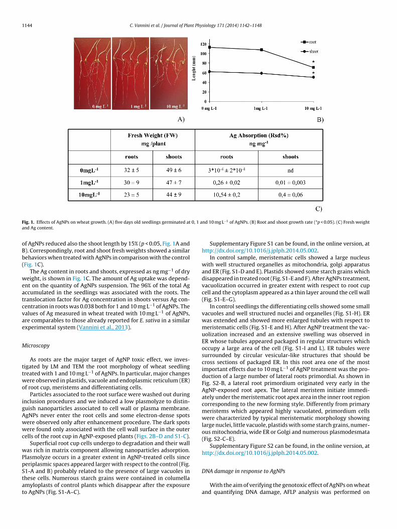
1144 C. Vannini et al. / Journal of Plant Physiology 171 (2014) 1142–1148
F 0, 1 aa
oBb(
weatcvae
M
ttwo
igAwwc
wPpStat
ig. 1. Effects of AgNPs on wheat growth. (A) five days old seedlings germinated atnd Ag content.
f AgNPs reduced also the shoot length by 15% (p < 0.05, Fig. 1A and). Correspondingly, root and shoot fresh weights showed a similarehaviors when treated with AgNPs in comparison with the controlFig. 1C).
The Ag content in roots and shoots, expressed as ng mg−1 of dryeight, is shown in Fig. 1C. The amount of Ag uptake was depend-
nt on the quantity of AgNPs suspension. The 96% of the total Agccumulated in the seedlings was associated with the roots. Theranslocation factor for Ag concentration in shoots versus Ag con-entration in roots was 0.038 both for 1 and 10 mg L−1 of AgNPs. Thealues of Ag measured in wheat treated with 10 mg L−1 of AgNPs,re comparables to those already reported for E. sativa in a similarxperimental system (Vannini et al., 2013).
icroscopy
As roots are the major target of AgNP toxic effect, we inves-igated by LM and TEM the root morphology of wheat seedlingreated with 1 and 10 mg L−1 of AgNPs. In particular, major changesere observed in plastids, vacuole and endoplasmic reticulum (ER)
f root cup, meristems and differentiating cells.Particles associated to the root surface were washed out during
nclusion procedures and we induced a low plasmolyze to distin-uish nanoparticles associated to cell wall or plasma membrane.gNPs never enter the root cells and some electron-dense spotsere observed only after enhancement procedure. The dark spotsere found only associated with the cell wall surface in the outer
ells of the root cup in AgNP-exposed plants (Figs. 2B–D and S1-C).Superficial root cup cells undergo to degradation and their wall
as rich in matrix component allowing nanoparticles adsorption.lasmolyze occurs in a greater extent in AgNP-treated cells sinceeriplasmic spaces appeared larger with respect to the control (Fig.
1-A and B) probably related to the presence of large vacuoles inhese cells. Numerous starch grains were contained in columellamyloplasts of control plants which disappear after the exposureo AgNPs (Fig. S1-A–C).nd 10 mg L−1 of AgNPs. (B) Root and shoot growth rate (*p < 0.05). (C) Fresh weight
Supplementary Figure S1 can be found, in the online version, athttp://dx.doi.org/10.1016/j.jplph.2014.05.002.
In control sample, meristematic cells showed a large nucleuswith well structured organelles as mitochondria, golgi apparatusand ER (Fig. S1-D and E). Plastids showed some starch grains whichdisappeared in treated root (Fig. S1-E and F). After AgNPs treatment,vacuolization occurred in greater extent with respect to root cupcell and the cytoplasm appeared as a thin layer around the cell wall(Fig. S1-E–G).
In control seedlings the differentiating cells showed some smallvacuoles and well structured nuclei and organelles (Fig. S1-H). ERwas extended and showed more enlarged tubules with respect tomeristematic cells (Fig. S1-E and H). After AgNP treatment the vac-uolization increased and an extensive swelling was observed inER whose tubules appeared packaged in regular structures whichoccupy a large area of the cell (Fig. S1-I and L). ER tubules weresurrounded by circular vesicular-like structures that should becross sections of packaged ER. In this root area one of the mostimportant effects due to 10 mg L−1 of AgNP treatment was the pro-duction of a large number of lateral roots primordial. As shown inFig. S2-B, a lateral root primordium originated very early in theAgNP-exposed root apex. The lateral meristem initiate immedi-ately under the meristematic root apex area in the inner root regioncorresponding to the new forming style. Differently from primarymeristems which appeared highly vacuolated, primordium cellswere characterized by typical meristematic morphology showinglarge nuclei, little vacuole, plastids with some starch grains, numer-ous mitochondria, wide ER or Golgi and numerous plasmodesmata(Fig. S2-C–E).
Supplementary Figure S2 can be found, in the online version, athttp://dx.doi.org/10.1016/j.jplph.2014.05.002.
DNA damage in response to AgNPs
With the aim of verifying the genotoxic effect of AgNPs on wheatand quantifying DNA damage, AFLP analysis was performed on

C. Vannini et al. / Journal of Plant Physiology 171 (2014) 1142–1148 1145
F gNP-t
D1loD(a(Ttpcc
h
P
f1tF
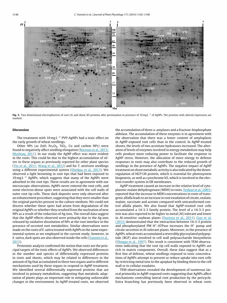
ig. 2. AgNP localization after enhancement procedure. (A) Root apex of 10 mg L−1 Ahe cell wall surface in the outer cells of the root apex. Magnification bar: 1 �m.
NA extracted from controls and from samples treated with 1 and0 mg L−1 AgNPs. Roots and shoots of each individual were ana-
yzed separately to better evaluate the effect on different plantrgans. The AFLP analysis on treated roots revealed a total of 524NA bands by using four primer combinations, however only 24
4.6%) of which were polymorphic. For treated shoots the totalnalyzed DNA bands were 534 and 20 of which were polymorphic3.7%). Details for each primer combination are given in Table S2.he number of polymorphic bands was very low and equally dis-ributed between treated and control samples and root and shootortions. The percent of genetic polymorphism detected by AFLP isompatible with the genetic uniformity characterizing T. aestivumultivars (Salunkhe et al., 2013).
Supplementary Table S2 can be found, in the online version, atttp://dx.doi.org/10.1016/j.jplph.2014.05.002.
roteomic changes induced in root and shoot treated with AgNPs
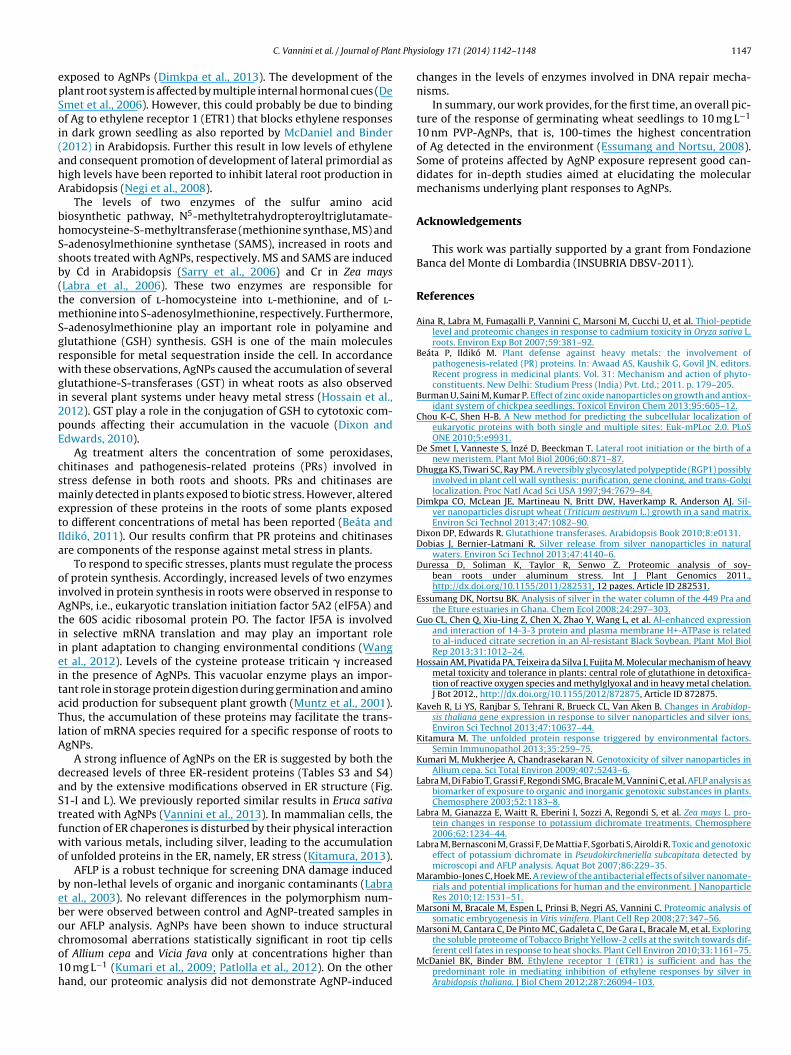
To shed light on the toxic effects of AgNP treatment we per-
ormed a proteomic analysis of roots and shoots treated with0 mg L−1 of AgNPs, concentration that induces a significant reduc-ion in root and shoot elongation in our experimental system.or both analysis, approximately 800 spots were resolved andexposed plants. In B, C, D the arrows indicate the small darks found associated with
detected (Fig. 3). All spots were matched by gel-to-gel compar-isons. Differences in the relative abundance (vol%) of each spotwere evaluated by software-assisted analysis. The ANOVA test(p < 0.05), coupled with a threshold of 1.5-fold change in level,revealed 27 and 12 differentially expressed protein spots in rootsand shoots treated with AgNP, respectively, relative to unexposedcontrols. These data indicate that the roots are the major site ofproteome changes. The differentially expressed protein spots aremarked on the representative 2-DE gels shown in Fig. 3A and B.Among the 27 spots differentially accumulated in AgNP-treatedroots, 22 increased and 5 decreased in abundance. In shoots, AgNPsinduced the up-regulation of 7 spots and the down-regulationof 5 spots. No identified proteins by LC–ESI–MS/MS were foundto be common in the roots and shoots. The identified proteinswere classified into different functional categories according totheir putative physiological functions. The differentially regulatedproteins in AgNP-treated roots mainly belong to the metabolismand cell defense categories. From the predicted localization of dif-ferentially expressed proteins it is apparent that, although the
cytoplamic proteins are the most represented, proteins are presentat other cellular locations also (Tables S3 and S4).Supplementary Tables S3 and S4 can be found, in the onlineversion, at http://dx.doi.org/10.1016/j.jplph.2014.05.002.
1146 C. Vannini et al. / Journal of Plant Physiology 171 (2014) 1142–1148
F erminm
D
t
fMiv(uo1amsoatdoNtrpmiE2
ciiamWitc
ig. 3. Two dimensional electrophoresis of root (A) and shoot (B) proteins after garked.
iscussion
The treatment with 10 mg L−1 PVP-AgNPs had a toxic effect onhe early growth of wheat seedlings.
Other NPs (as ZnO, Fe3O4, TiO2, Cu and carbon NPs) wereound to negatively affect seedling elongation (Burman et al., 2013;
ushtaq, 2011). In our study the AgNP effect was more evidentn the roots. This could be due to the highest accumulation of sil-er in these organs as previously reported for other plant speciesYin et al., 2011; Wang et al., 2012) and for T. aestivum seedlingssing a different experimental system (Dimkpa et al., 2013). Webserved a light browning in root tips that had been exposed to0 mg L−1 AgNPs, which suggests that many of the AgNPs weredsorbed to the root tips. These results are in agreement with ouricroscopic observations. AgNPs never entered the root cells, and
ome electron-dense spots were associated with the cell walls ofuter root tip cells. These dark deposits were only observed aftern enhancement procedure, suggesting that they were smaller thanhe original particles present in the culture medium. We could notiscern whether these spots had arisen from degradation of theriginal AgNPs or whether they resulted from the nucleation of newPs as a result of the reduction of Ag ions. The overall data suggest
hat the AgNP effects observed were primarily due to the Ag ionseleased by oxidative dissolution of NPs at the root interface in theresence of secreted root metabolites. Similar observations wereade on the roots of E. sativa treated with AgNPs in the same exper-
mental system as we employed in the current study; however, in. sativa, dark spots are also observed inside the cells (Vannini et al.,013).
Proteomic analysis confirmed the notion that roots are the prin-ipal targets of the toxic effects of AgNPs. We observed differencesn the number and species of differentially expressed proteinsn roots and shoots, which may be related to differences in themount of Ag that accumulated in these two organs and to differentechanisms used by these organs to respond to AgNP treatment.
e identified several differentially expressed proteins that arenvolved in primary metabolism, suggesting that metabolic adap-ation of plants plays an important role in mitigating unfavorablehanges in the environment. In AgNP-treated roots, we observed
ation in presence of 10 mg L−1 of AgNPs. The proteins with altered expression are
the accumulation of three �-amylases and a fructose-bisphosphatealdolase. The accumulation of these enzymes is in agreement withthe observation that there was a lower content of amyloplastsin AgNP-exposed root cells than in the control. In AgNP-treatedshoots, the levels of two aconitate hydratases increased. The alter-ation of levels of enzymes involved in energy metabolism may helpcells produce more reducing power to facilitate the response toAgNP stress. However, the allocation of more energy to defenseresponses in roots may also contribute to the reduced growth ofseedlings in the presence of AgNPs. The negative impact of AgNPtreatment on shoot metabolic activity is also indicated by the down-regulation of HCF136 protein, which is essential for photosystembiogenesis, as well as cytochrome b5, which is involved in the elec-tron transfer system in ER membranes.
AgNP treatment caused an increase in the relative level of cyto-plasmic malate dehydrogenase (MDH) in roots. Tesfaye et al. (2001)reported that the increase in MDH concentrations in roots of trans-genic alfalfa leads to an increase in root exudation of citrate, oxalate,malate, succinate and acetate compared with untransformed con-trol alfalfa plants. We also found that AgNP-treated root cellsaccumulated a 14-3-3 family protein. The level of a 14-3-3 pro-tein was also reported to be higher in metal (Al) tolerant and lowerin Al-sensitive soybean plants (Duressa et al., 2011). Guo et al.(2013) demonstrated that the interaction between 14-3-3 proteinand phosphorylated PM H+-ATPase increases the H+ pump andcitrate secretion in Al-tolerant plants. Moreover, in the presence ofAgNPs, wheat roots accumulated a reversibly glycosylated polypep-tide (RGP) also involved in cell wall polysaccharide biosynthesis(Dhugga et al., 1997). This result is consistent with TEM observa-tions indicating that the root tip cell walls exposed to AgNPs arerich in matrix components. Overall, these data suggest that as afirst line of defense, wheat seedlings exposed to toxic concentra-tions of AgNPs attempt to prevent or reduce uptake into root cellsby restricting metal ions to the apoplast by binding them to the cellwall or to cellular exudates.
TEM observations revealed the development of numerous lat-eral primordia in AgNP-exposed roots suggesting that AgNPs affectmechanisms controlling lateral root production by the pericycle.Extra branching has previously been observed in wheat roots
nt Phy
epSoi(ahA
bhSsb(tmSgrwgi2pE
csmetIa
oiAtiieitaTlA
daStfwo
beboco1h
C. Vannini et al. / Journal of Pla
xposed to AgNPs (Dimkpa et al., 2013). The development of thelant root system is affected by multiple internal hormonal cues (Demet et al., 2006). However, this could probably be due to bindingf Ag to ethylene receptor 1 (ETR1) that blocks ethylene responsesn dark grown seedling as also reported by McDaniel and Binder2012) in Arabidopsis. Further this result in low levels of ethylenend consequent promotion of development of lateral primordial asigh levels have been reported to inhibit lateral root production inrabidopsis (Negi et al., 2008).
The levels of two enzymes of the sulfur amino acidiosynthetic pathway, N5-methyltetrahydropteroyltriglutamate-omocysteine-S-methyltransferase (methionine synthase, MS) and-adenosylmethionine synthetase (SAMS), increased in roots andhoots treated with AgNPs, respectively. MS and SAMS are inducedy Cd in Arabidopsis (Sarry et al., 2006) and Cr in Zea maysLabra et al., 2006). These two enzymes are responsible forhe conversion of l-homocysteine into l-methionine, and of l-
ethionine into S-adenosylmethionine, respectively. Furthermore,-adenosylmethionine play an important role in polyamine andlutathione (GSH) synthesis. GSH is one of the main moleculesesponsible for metal sequestration inside the cell. In accordanceith these observations, AgNPs caused the accumulation of several
lutathione-S-transferases (GST) in wheat roots as also observedn several plant systems under heavy metal stress (Hossain et al.,012). GST play a role in the conjugation of GSH to cytotoxic com-ounds affecting their accumulation in the vacuole (Dixon anddwards, 2010).
Ag treatment alters the concentration of some peroxidases,hitinases and pathogenesis-related proteins (PRs) involved intress defense in both roots and shoots. PRs and chitinases areainly detected in plants exposed to biotic stress. However, altered
xpression of these proteins in the roots of some plants exposedo different concentrations of metal has been reported (Beáta andldikó, 2011). Our results confirm that PR proteins and chitinasesre components of the response against metal stress in plants.
To respond to specific stresses, plants must regulate the processf protein synthesis. Accordingly, increased levels of two enzymesnvolved in protein synthesis in roots were observed in response togNPs, i.e., eukaryotic translation initiation factor 5A2 (elF5A) and
he 60S acidic ribosomal protein PO. The factor IF5A is involvedn selective mRNA translation and may play an important rolen plant adaptation to changing environmental conditions (Wangt al., 2012). Levels of the cysteine protease triticain � increasedn the presence of AgNPs. This vacuolar enzyme plays an impor-ant role in storage protein digestion during germination and aminocid production for subsequent plant growth (Muntz et al., 2001).hus, the accumulation of these proteins may facilitate the trans-ation of mRNA species required for a specific response of roots togNPs.
A strong influence of AgNPs on the ER is suggested by both theecreased levels of three ER-resident proteins (Tables S3 and S4)nd by the extensive modifications observed in ER structure (Fig.1-I and L). We previously reported similar results in Eruca sativareated with AgNPs (Vannini et al., 2013). In mammalian cells, theunction of ER chaperones is disturbed by their physical interactionith various metals, including silver, leading to the accumulation
f unfolded proteins in the ER, namely, ER stress (Kitamura, 2013).AFLP is a robust technique for screening DNA damage induced
y non-lethal levels of organic and inorganic contaminants (Labrat al., 2003). No relevant differences in the polymorphism num-er were observed between control and AgNP-treated samples inur AFLP analysis. AgNPs have been shown to induce structural
hromosomal aberrations statistically significant in root tip cellsf Allium cepa and Vicia fava only at concentrations higher than0 mg L−1 (Kumari et al., 2009; Patlolla et al., 2012). On the otherand, our proteomic analysis did not demonstrate AgNP-inducedsiology 171 (2014) 1142–1148 1147
changes in the levels of enzymes involved in DNA repair mecha-nisms.
In summary, our work provides, for the first time, an overall pic-ture of the response of germinating wheat seedlings to 10 mg L−1
10 nm PVP-AgNPs, that is, 100-times the highest concentrationof Ag detected in the environment (Essumang and Nortsu, 2008).Some of proteins affected by AgNP exposure represent good can-didates for in-depth studies aimed at elucidating the molecularmechanisms underlying plant responses to AgNPs.
Acknowledgements
This work was partially supported by a grant from FondazioneBanca del Monte di Lombardia (INSUBRIA DBSV-2011).
References
Aina R, Labra M, Fumagalli P, Vannini C, Marsoni M, Cucchi U, et al. Thiol-peptidelevel and proteomic changes in response to cadmium toxicity in Oryza sativa L.roots. Environ Exp Bot 2007;59:381–92.
Beáta P, Ildikó M. Plant defense against heavy metals: the involvement ofpathogenesis-related (PR) proteins. In: Awaad AS, Kaushik G, Govil JN, editors.Recent progress in medicinal plants. Vol. 31: Mechanism and action of phyto-constituents. New Delhi: Studium Press (India) Pvt. Ltd.; 2011. p. 179–205.
Burman U, Saini M, Kumar P. Effect of zinc oxide nanoparticles on growth and antiox-idant system of chickpea seedlings. Toxicol Environ Chem 2013;95:605–12.
Chou K-C, Shen H-B. A New method for predicting the subcellular localization ofeukaryotic proteins with both single and multiple sites: Euk-mPLoc 2.0. PLoSONE 2010;5:e9931.
De Smet I, Vanneste S, Inzé D, Beeckman T. Lateral root initiation or the birth of anew meristem. Plant Mol Biol 2006;60:871–87.
Dhugga KS, Tiwari SC, Ray PM. A reversibly glycosylated polypeptide (RGP1) possiblyinvolved in plant cell wall synthesis: purification, gene cloning, and trans-Golgilocalization. Proc Natl Acad Sci USA 1997;94:7679–84.
Dimkpa CO, McLean JE, Martineau N, Britt DW, Haverkamp R, Anderson AJ. Sil-ver nanoparticles disrupt wheat (Triticum aestivum L.) growth in a sand matrix.Environ Sci Technol 2013;47:1082–90.
Dixon DP, Edwards R. Glutathione transferases. Arabidopsis Book 2010;8:e0131.Dobias J, Bernier-Latmani R. Silver release from silver nanoparticles in natural
waters. Environ Sci Technol 2013;47:4140–6.Duressa D, Soliman K, Taylor R, Senwo Z. Proteomic analysis of soy-
bean roots under aluminum stress. Int J Plant Genomics 2011.,http://dx.doi.org/10.1155/2011/282531, 12 pages. Article ID 282531.
Essumang DK, Nortsu BK. Analysis of silver in the water column of the 449 Pra andthe Eture estuaries in Ghana. Chem Ecol 2008;24:297–303.
Guo CL, Chen Q, Xiu-Ling Z, Chen X, Zhao Y, Wang L, et al. Al-enhanced expressionand interaction of 14-3-3 protein and plasma membrane H+-ATPase is relatedto al-induced citrate secretion in an Al-resistant Black Soybean. Plant Mol BiolRep 2013;31:1012–24.
Hossain AM, Piyatida PA, Teixeira da Silva J, Fujita M. Molecular mechanism of heavymetal toxicity and tolerance in plants: central role of glutathione in detoxifica-tion of reactive oxygen species and methylglyoxal and in heavy metal chelation.J Bot 2012., http://dx.doi.org/10.1155/2012/872875, Article ID 872875.
Kaveh R, Li YS, Ranjbar S, Tehrani R, Brueck CL, Van Aken B. Changes in Arabidop-sis thaliana gene expression in response to silver nanoparticles and silver ions.Environ Sci Technol 2013;47:10637–44.
Kitamura M. The unfolded protein response triggered by environmental factors.Semin Immunopathol 2013;35:259–75.
Kumari M, Mukherjee A, Chandrasekaran N. Genotoxicity of silver nanoparticles inAllium cepa. Sci Total Environ 2009;407:5243–6.
Labra M, Di Fabio T, Grassi F, Regondi SMG, Bracale M, Vannini C, et al. AFLP analysis asbiomarker of exposure to organic and inorganic genotoxic substances in plants.Chemosphere 2003;52:1183–8.
Labra M, Gianazza E, Waitt R, Eberini I, Sozzi A, Regondi S, et al. Zea mays L. pro-tein changes in response to potassium dichromate treatments. Chemosphere2006;62:1234–44.
Labra M, Bernasconi M, Grassi F, De Mattia F, Sgorbati S, Airoldi R. Toxic and genotoxiceffect of potassium dichromate in Pseudokirchneriella subcapitata detected bymicroscopi and AFLP analysis. Aquat Bot 2007;86:229–35.
Marambio-Jones C, Hoek ME. A review of the antibacterial effects of silver nanomate-rials and potential implications for human and the environment. J NanoparticleRes 2010;12:1531–51.
Marsoni M, Bracale M, Espen L, Prinsi B, Negri AS, Vannini C. Proteomic analysis ofsomatic embryogenesis in Vitis vinifera. Plant Cell Rep 2008;27:347–56.
Marsoni M, Cantara C, De Pinto MC, Gadaleta C, De Gara L, Bracale M, et al. Exploring
the soluble proteome of Tobacco Bright Yellow-2 cells at the switch towards dif-ferent cell fates in response to heat shocks. Plant Cell Environ 2010;33:1161–75.McDaniel BK, Binder BM. Ethylene receptor 1 (ETR1) is sufficient and has thepredominant role in mediating inhibition of ethylene responses by silver inArabidopsis thaliana. J Biol Chem 2012;287:26094–103.

1 nt Phy
M
M
M
N
P
P
R
148 C. Vannini et al. / Journal of Pla
uller LAH, Lambaerts M, Vangronsveld J, Colpaert V. AFLP-based assess-ment of the effects of environmental heavy metal pollution on the geneticstructure of pioneer populations of Suillus luteus. New Phytol 2004;164:297–303.
untz K, Belozersky MA, Dunaevsky YE, Schlereth A, Tiedemann J. Stored pro-teinases and the initiation of storage protein mobilization in seeds duringgermination and seedling growth. J Exp Bot 2001;52:1741–52.
ushtaq YK. Effect of nanoscale F3O4, TiO2 and carbon particles on cucumberseedlings germination. J Environ Sci Health 2011;46:1732–5.
egi S, Ivanchenko MG, Muday GK. Ethylene regulates lateral root formation andauxin transport in Arabidopsis thaliana. Plant J 2008;55:175–87.
anda KK, Achary MM, Krishnaveni R, Padhi BK, Sarangi SN, Sahu SN, et al. In vitrobiosynthesis and genotoxicity bioassay of silver nanoparticles using plants. Tox-icol In Vitro 2011;25:1097–105.
atlolla AK, Berry A, May LB, Tchounwou PB. Genotoxicity of silver nanoparticles in
Vicia faba: a pilot study on the environmental monitoring of nanoparticles. Int JEnviron Res Public Health 2012;9:1649–62.emédios C, Rosrio F, Bastos V. Environmental nanoparticlesinteractions withplants: morphological, physiological, and genotoxic aspects. J Bot 2012.,http://dx.doi.org/10.1155/2012/751686.
siology 171 (2014) 1142–1148
Salunkhe A, Tamhankar S, Tetali S, Zaharieva M, Bonnett D, Trethowan R, et al. Molec-ular genetic diversity analysis in emmer wheat (Triticum dicoccon Schrank) fromIndia. Gen Res Crop Evol 2013;60:165–74.
Sarry JE, Kuhn L, Ducruix C, Lafaye A, Junot C, Hugouvieux V, et al. The early responsesof Arabidopsis thaliana cells to cadmium exposure explored by protein andmetabolite profiling analyses. Proteomics 2006;6:2180–98.
Tesfaye M, Temple SJ, Allan DL, Vance CP, Samac DA. Overexpression of malatedehydrogenase in transgenic alfalfa enhances organic acid synthesis and conferstolerance to aluminum. Plant Physiol 2001;127:1836–44.
Vannini C, Domingo G, Onelli E, Prinsi B, Marsoni M, Espen L, et al. Morphologicaland proteomic responses of Eruca sativa exposed to silver nanoparticles or silvernitrate. PLoS ONE 2013;8:1–8.
Vos P, Hogers R, Bleeker M, Reijans M, Lee TV, Hornes M, et al. AFLP: a new techniquefor DNA fingerprinting. Nucleic Acids Res 1995;23:4407–14.
Wang L, Xu C, Wang C, Wang Y. Characterization of a eukaryotic translation initiation
factor 5A homolog from Tamarix androssowii involved in plant abiotic stresstolerance. BMC Plant Biol 2012;12:118–24.Yin L, Cheng Y, Espinasse B, Colman BP, Auffan M, Wiesner M, et al. More thanthe ions: the effects of silver nanoparticles on Lolium multiflorum. Environ SciTechnol 2011;45:2360–7.
Copyright © 2022 FDOKUMEN