A Revision of Sepedonea, a Neotropical Genus of Snail-killing ...
56
A Revision of Sepedonea, a Neotropical Genus of Snail-killing Flies (Diptera: Sciomyzidae) AMNON FREIDBERG, LLOYD KNUTSON, and JAY ABERCROMBIE SMITHSONIAN CONTRIBUTIONS TO ZOOLOGY • NUMBER 506
-
Upload
khangminh22 -
Category
Documents
-
view
0 -
download
0
Transcript of A Revision of Sepedonea, a Neotropical Genus of Snail-killing ...

A Revision of Sepedonea,a Neotropical Genus of
Snail-killing Flies(Diptera: Sciomyzidae)
AMNON FREIDBERG,LLOYD KNUTSON,
andJAY ABERCROMBIE
SMITHSONIAN CONTRIBUTIONS TO ZOOLOGY • NUMBER 506

SERIES PUBLICATIONS OF THE SMITHSONIAN INSTITUTION
Emphasis upon publication as a means of "diffusing knowledge" was expressed by the firstSecretary of the Smithsonian. In his formal plan for the Institution, Joseph Henry outlined aprogram that included the following statement: "It is proposed to publish a series of reports,giving an account of the new discoveries in science, and of the changes made from year to yearin all branches of knowledge." This theme of basic research has been adhered to through theyears by thousands of titles issued in series publications under the Smithsonian imprint,commencing with Smithsonian Contributions to Knowledge in 1848 and continuing with thefollowing active series:
Smithsonian Contributions to Anthropology
Smithsonian Contributions to Astrophysics
Smithsonian Contributions to Botany
Smithsonian Contributions to the Earth Sciences
Smithsonian Contributions to the Marine Sciences
Smithsonian Contributions to Paleobiology
Smithsonian Contributions to Zoology
Smithsonian Folklife Studies
Smithsonian Studies in Air and Space
Smithsonian Studies in History and Technology
In these series, the Institution publishes small papers and full-scale monographs that reportthe research and collections of its various museums and bureaux or of professional colleaguesin the world of science and scholarship. The publications are distributed by mailing lists tolibraries, universities, and similar institutions throughout the world.
Papers or monographs submitted for series publication are received by the SmithsonianInstitution Press, subject to its own review for format and style, only through departments of thevarious Smithsonian museums or bureaux, where the manuscripts are given substantive review.Press requirements for manuscript and art preparation are outlined on the inside back cover.
Robert McC. AdamsSecretarySmithsonian Institution

S M I T H S O N I A N C O N T R I B U T I O N S T O Z O O L O G Y • N U M B E R 5 0 6
A Revision of Sepedonea,a Neotropical Genus of
Snail-killing Flies(Diptera: Sciomyzidae)
Amnon Freidberg,Lloyd Knutson,
andJay Abercrombie
SMITHSONIAN INSTITUTION PRESS
Washington, D.C.
1991

ABSTRACT
Freidberg, Amnon, Lloyd Knutson, and Jay Abercrombie. A Revision of Sepedonea, aNeotropical Genus of Snail-killing Flies (Diptera: Sciomyzidae). Smithsonian Contributions toZoology, number 506, 48 pages, 143 figures, 1991.—The Neotropical genus Sepedonea(Diptera: Sciomyzidae) is revised. Four of the 12 species (S. incipiens, S. neffi, S. trichotypa, andS. veredae) are described as new. Sepedonea vau Mello and Bredt is placed as a nomen nudumunder S. guianica. A key to the adults is included, and the male and female terminalia of allspecies are described and illustrated. The eggs, first-, second-, and third-instar larvae, andpuparia of S. guianica, S. incipiens, S. lagoa, S. lindneri, S. telson, and S. trichotypa aredescribed and figured for the first time. These species, plus 5. guatemalana and S. isthmi, areincluded in keys to the mature third-instar larvae and puparia. The taxonomy, biology, andgeographic distribution of the genus is summarized. The taxonomic placement of the Sepedongroup is discussed. The geographical distributions of 11 species are mapped.
The larvae of all reared species of Sepedonea are predators of non-operculate snails in variousfreshwater situations.
OFFICIAL PUBLICATION DATE is handstamped in a limited number of initial copies and isrecorded in the Institution's annual report, Smithsonian Year. SERIES COVER DESIGN: The coralMontaslrea cavernosa (Linnaeus).
Library of Congress Cataloging-in-Publication DataFreidberg, Amnon.A revision of Sepedonea, a neotropical genus of snail-killing flics (DipteraiSciomyzidae) / Amnon Freidberg, LloydKnutson, Jay Abercrombie.p. cm.—(Smithsonian contributions to zoology ; no. 506)Includes bibliographical references (p. ).Supt. of Docs, no.: SI 1.27:5061. Sepedonea—Classification. I. Knutson, Lloyd V, 1934- II. Abercrombie, Jay. III. Title. IV. Series.QL1.S54 no. 506 [QL537.S365] 591 s-dc20 [595.77F4] 90-10224
@ The paper used in this publication meets the minimum requirements of the AmericanNational Standard for Permanence of Paper for Printed Library Materials Z39.48—1984.

Contents
Page
Introduction 1Historical Review 1Methods and Materials 2Acknowledgments 2
Family SdOMYZIDAE 2Genus Sepedonea Steyskal 2
Description 2Adult 3Egg 6Larva 6Puparium 6
Natural History 6Discussion 6Key to Adults of Sepedonea 9Key to Mature Third-instar Larvae of Sepedonea 10Key to Puparia of Sepedonea 11Sepedonea barbosai Knutson and Bredt 11Sepedonea canabravana Knutson and Bredt 13Sepedonea guatemalana (Steyskal) 13Sepedonea guianica (Steyskal) 16Sepedonea incipiens, new species 19Sepedonea isthmi (Steyskal) 21Sepedonea lagoa (Steyskal) 23Sepedonea lindneri (Hendel) 26Sepedonea neffi, new species 29Sepedonea telson (Steyskal) 29Sepedonea trichotypa, new species 33Sepedonea veredae, new species 36
Literature Cited 38Figures 94-143 39
ui

FIGURE 1.—Sepedonea lindturi, adult male, habitus.

A Revision of Sepedonea,a Neotropical Genus of
Snail-killing Flies(Diptera: Sciomyzidae)
Amnon Freidberg, Lloyd Knutson,and
Jay Abercrombie
Introduction
Among families of higher Diptera, the Sciomyzidae arefairly well established as a phylogenetically unified (Griffiths,1972) and behaviorally diverse family (Berg and Knutson,1978; Ferrar, 1987) that has more or less uniquely exploited afood resource—mollusks. Mollusks are not rigorously ex-ploited by practically any other dipterans, or indeed, by anyother moderately large group of insects. To understand thepredatory or parasitoid behavior of Sciomyzidae on mollusks,it is essential to have a good knowledge of the systematics ofthe taxa involved. This information enables accurate identifica-tion of the species and an understanding of their phylogeneticrelationships.
What little is known on the phylogeny of Sciomyzidae wassummarized by Griffiths (1972) and Berg and Knutson (1978).In the latter paper, the authors also reviewed the biology andsystematics of the family, which now includes about 550species in 61 genera. Data on the natural history and immaturestages are known for about a third of the world's species.Although most sciomyzid larvae are primarily predators orparasitoids of aquatic or terrestrial snails, a few species feed onsnail egg-masses, slugs, or finger-nail clams. Some Scio-myzidae are potential agents for the biological control of snailsthat carry diseases (e.g. fascioliasis and schistosomiasis) (Bergand Knutson, 1978).
Amnon Freidberg, Department of Zoology, The George S. WiseFaculty of Life Sciences, Tel-Aviv University, Tel Aviv 69978, Israel.Lloyd Knutson, Biological Control of Weeds Laboratory—Europe,American Embassy—AGRIC, APO New York 09794. Jay Abercrom-bie, 10th Medical Laboratory, Department of the Army, APO NewYork 09180.
With about 125 known species (at least 20 are undescribed),Sepedon Latreille, 1804, and related genera comprise nearly 25percent of the family's species. The strictly neotropical generaof this group, Sepedonea Steyskal, 1973, and Thecomyia Perty,1833, have never been revised. Our purpose in this paper is torevise the species of Sepedonea.
HISTORICAL REVIEW.—Steyskal (1973) proposed the mod-ern classification of Sepedon and related genera (the Sepedongroup). He included the following genera in this group:Sepedon Latreille, Sepedonella Verbeke 1950, SepedoninusVerbeke 1950, Thecomyia Perty, and his new genera Sepe-donea and Sepedomerus. He further suggested that SepedomyiaVerbeke, 1950, is a subgenus of Sepedon, together with thepreviously established subgenera Mesosepedon Verbeke, 1950,and Parasepedon Verbeke, 1950. In establishing the Sepedongroup, Steyskal did not believe that (p. 143) "this group issufficiently distinct from more typical Tetanocerini, especiallyfrom such genera as Hedria and Dichetophora, to be given therank of tribe or even subtribe." Earlier authors had classifiedthese genera as a tribe, subfamily, or even family (e.g., Bergand Knutson, 1978). The diagnostic characters shared by allmembers of the Sepedon group as defined by Steyskal (1973)are as follows:
1. Lunule well exposed2. Face more or less extended ventrally3. Ocellar setae weak or absent4. One pair of scutellar setae (absent in the oriental
Sepedon lobifera Hendel)5. Subalar (vallar) setae present
In his study of the Western Hemisphere species of Sepedon,Steyskal (1951:272) noted that "the S. lindneri group [=

SMITHSONIAN CONTRIBUTIONS TO ZOOLOGY
Sepedonea] of Central and South America is unique in havingthe posterior forceps fused mesally to form a 'posteriorprocess,' but in view of the diversity of form of the maleterminalia within that group and the conformity otherwise withother forms, I have not thought it wise to distinguish the groupnomenclatorially." Later, however, he reversed his opinion anddescribed Sepedonea (Steyskal, 1973) and designated Sepedonlindneri Hendel as the type species of the genus. Eight specieswere recognized in the genus in the catalog of NeotropicalSciomyzidae (Knutson et al., 1976). These, plus four newspecies described herein, are now included in the genus.
METHODS AND MATERIALS.—During the course of this
study slightly over 1000 adult specimens from most majorNorth and South American collections were examined, includ-ing the primary types of all but one previously describedspecies. Label data for all correctly identified specimens weexamined were recorded, organized according to locality(country, state or province), and are presented under theappropriate species.
The descriptive terminology essentially follows that pub-lished in the recent Manual of Nearctic Diptera (MeAlpine,1981 (adults); Knutson, 1987 (adults and immature stages)).
Because of the paucity of characters available for descrip-tions of adults, and because the key to adults contains most ofthe distinguishing characters, separate diagnoses are not givenfor each species.
ACKNOWLEDGMENTS.—Numerous individuals and institu-tions have contributed to this revision. Without their coopera-tion and assistance, much of the study could not have beencompleted. We are grateful for their time and thoughtfulconsideration.
We thank the following curators and institutions for lendingspecimens:
BMNH British Museum (Natural History), London,England (Mr. B.H. Cogan, Dr. B.R. Pitkin)
CUI Cornell University, Ithaca, New York, USA.(Dr.C.O. Berg)
IOC Instituto Oswaldo Cruz, Rio de Janeiro,Brazil (Dr. L.M. Deane, Dr. D.V. Ferreira,Dr. A.P.A. Lunas Dias)
IZAM Instituto de Zoologia Agricola, Maracay,Venezuela (Dr. Carlos Julio Rosoles)
MNRJ Museu Nacional, Rio de Janeiro, Brazil (Dr.Hugo de Souza Lopes)
MZUSP Museu de Zoologia da Universidade de SaoPaulo, S3o Paulo, Brazil (Dr. Nelson Papav-ero)
OSU Ohio State University, Columbus, Ohio,USA. (Dr. Charles A. Triplehorn)
SMNS Staatliches Museum fur Naturkunde, Lud-wigsberg, Federal Republic of Germany (Dr.B. Herting)
USNM Collections of the United States NationalMuseum in the National Museum of NaturalHistory, Smithsonian Institution, Washing-ton, DC, USA. (Dr. W.N. Mathis)
We thank WN. Mathis, F.C. Thompson, and R.E. Orth forreviewing the manuscript. The advice of the master scio-myzidologist, G.C. Steyskal, was appreciated throughout thestudy. We also thank Mary Lou Cooley, Systematic Entomol-ogy Laboratory, IIBIII, for completion of the illustrationsprepared by AF, and for preparing the plates. We are alsograteful to Linda Heath Lawrence for handling some graphicaspects.
Most specimens of Sepedonea available for this study werecollected by CO. Berg and his students and coworkers whileconducting life-cycle studies in Central and South America.Most of these specimens are in the Department of Entomology,Cornell University. We feel that when systematists conduct ataxonomic study, they should, if at all possible, placespecimens, especially paratypes of new species, in variouscollections in pertinent geographical areas, to serve as referencematerial. Hence, we appreciate the cooperation of CornellUniversity for allowing us to place paratypes and otherspecimens from the Cornell material in several collections.Whenever possible specimens were placed in the followingcollections in addition to those listed above: UniversidadeFederal do Parana', Curitiba, Brazil; Universidad Nacional deColombia, Medellin, Colombia; Universidad de Panama,Panama; Fundaci6n M. Lillo, Tucuman, Argentina.
Family SCIOMYZIDAE
Sciomyzidae are small to moderately large acalyptrateDiptera, often rather slender, ranging from 1.8 to about 12 mmin length. The antenna is usually porrect, and the pedicel isusually elongate. Two orbital setae are usually present, butsometimes only one. The wings are usually longer than theabdomen and are hyaline, slightly spotted, or heavily patterned.The costa (C) is unbroken and the subcosta (Sc) is complete andfree from Rl distally. The abdomen is moderately long andcylindrical. The general color varies from yellowish to shinyblack, but often is grayish or brownish.
Sciomyzidae are best recognized by the above characters andby the close association of most species with aquatic orotherwise damp and shaded habitats. Adults often rest onemergent vegetation.
Genus Sepedonea Steyskal, 1973
Sepedonea Steyskal, 1973:145 [type species: Sepedon lindneri Hendel, byoriginal designation].—Steyskal and Knutson, 1975:276 [generic key].—Knutson et al., 1976:10 [catalog].—Knutson and Bredt, 1976:113[review].—Knutson and Valley, 1978:198 [review].
DESCRIPTION.—The following is a brief characterization of

NUMBER 506
FIGURES 2-6.—Sepedonea spp., various parts: 2, S. lagoa, head in profile; 3-5, left posterior spiracle: 3, S.trichotypa; 4, 5. lindneri; 5, S. guatemalana; 6, S. is thru, wing.
the various life stages of Sepedonea (first- and second-instarlarvae are not included):
Adult: Elongate, generally brown, Sepedon-]ike flies (Fig-ure 1).
Head (Figure 2): One (posterior) pair of orbital setae,occasionally anterior orbital setae weakly developed; postocel-lar setae well developed; frons largely dull yellow with slender,shiny, frontal vitta; lacking round black spots on orbit a short
distance ventral to antenna; face with dark brown spot on eachventral corner, orbit, face, and gena spotted by silverymicrotomentum; antenna with pedicel 3.5-5 times as long aswide, with setulae as long as diameter of pedicel; firstflagellomere with distinct, elongate, sensory pit near base;ventral part of head not forming a tube into which the proboscismay be withdrawn; palpus well developed.
Thorax: Scutum with brownish gray microtomentum;

SMITHSONIAN CONTRIBUTIONS TO ZOOLOGY
10
FIGURES 7-13.—Sepedonea spp., legs: 7-9, right midfemur, posterior view: 7, S. guianica; 8, S. telson; 9, 5.isthmi; 10, S. telson, right hindleg, anterior view; 11-13, right hindcoxa, posterior view: 11, S. lindneri; 12, S.trichotypa; 13, S. canabravana.
pleura and coxae with silvery microtomentum, dense on pleura;pleura with numerous setulae; posterior spiracle (Figures 3-5)with a varying number of setulae of various lengths; postcoxalbridge divided by membranous area; forefemur with at least 1outstanding dorsal seta; midfemur with large anterior seta nearmidlength, usually with a posteroventral row of spines (Figures7-9) extended to varying degrees from apex toward base;hindfemur (Figure 10) often with dorsal and/or lateral,
preapical dark marks (these marks are somewhat variableintraspecifically and the least reliable of the characters used foridentification); hindcoxa posteriorly (Figures 11-13) with avarying number of setulae of varying length; wing (Figure 6)without spots, except sometimes with slight infuscations aboutcrossveins; crossvein dm-cu straight, at right angle withpenultimate section of vein CuAl.
Abdomen (Figures 14-19): with light gray microtomen-

NUMBER 506
CERC
FIGURES 14-16.—Sepedonea isthmi, male abdomen: 14, lateral view; 15, ventral view; 16, dorsal view, (a =anterior plate of sternum 5 (left-hand half); a.SS = anterior surstylus; CERC = cercus; D = distiphallus; EPAN =epandrium; p = posterior plate of sternum 5 (left-hand half); p.SS = posterior surstyli; S 1-5 = sternum 1 through5; T 1-5 = tergum 1 through 5.)

SMITHSONIAN CONTRIBUTIONS TO ZOOLOGY
turn; male posterior surstyli fused to form a median structure(Figures 14-15); female sterna 6-8 fused to form asynsternum (Figures 17-18).
Egg: Eggs of Sepedonea are remarkably uniform fromspecies to species (Figures 94,96), and are very similar to thosedescribed for Sepedon sphegea (Fabricius), S. spinipes (Sco-poli), and Sepedomerus macropus (Walker). Some eggs of S.telson differ from other species in having a dorsal depressionanteriorly in the central area between the dorsal longitudinalridges. It is not present in all individuals, however, so it isunreliable as a distinguishing character. Neff and Berg (1966)found the same structure in eggs of Sepedomerus macropus butnot in others.
The known eggs of eight species of Sepedonea treated herecan be divided roughly into 2 groups on the basis of size, asomewhat arbitrary division that is nonetheless reliable in mostinstances. One group, that includes S. incipiens, S. lindneri, and5. trichotypa, has eggs which range from 0.90 to 1.12 mm long.Sepedonea incipiens is the only species in this group with eggsas large as 1.12 mm; only one specimen of 10 examinedreached this length. Other species in the first group (S. lindneriand S. trichotypa) have eggs with lengths of 0.90-1.06 mm.The second group, which includes S. guatemalana, S. guianica,S. isthmi, S. lagoa, and S. telson, has eggs ranging from 1.12 to1.40 mm in length. Sepedonea lagoa is the only species in thesecond group with eggs as small as 1.12 mm; of 17 individualsexamined, 5 were 1.12 mm long and the remainder wereconsiderably longer. Other species in the second group haveeggs with lengths of 1.16-1.24 mm (S. guianica and S. telson),1.26-1.40 mm (S. guatemalana; Neff and Berg, 1966) and1.28-1.38 mm (5. isthmi; Knutson and Valley, 1978). Themicropyle is subterminal in all eight species.
Larva: The third-instar larva of 5. guianica, S. incipiens, S.isthmi, S. lagoa, S. lindneri, S. telson, and S. trichotypa hassmooth posterior stigmatic tubes that are distinctly scallopedbasally (Figure 131). Neff and Berg (1966) reported that in S.guatemalana (as well as in species of Sepedomerus andSepedon), surfaces of the stigmatic tubes are tuberculate orpapillose; hence, the tubes may appear scalloped basallybecause of these projections.
All third-instar larvae of Sepedonea except S. lagoa have aprominent middorsal stripe and dorsolateral V-shaped marksanteriorly. Larvae of Sepedomerus and Sepedon do not havedorsolateral markings.
Dorsolateral patch of setulae occur on segments 5-11,except in S. guatemalana, S. guianica, and S. isthmi, which lackthem on segment 11. All species of Sepedomerus and some ofSepedon have dorsolateral patches of setulae on segments5-10.
Larvae of Sepedonea, Sepedomerus, and Sepedon all possess5 pairs of lobes on the posterior spiracular disc (Figures127-130,135).
The most reliable character to separate larvae of Sepedoneafrom Sepedomerus and Sepedon is the dorsal most accessorytooth of the cephalopharyngeal skeleton. In all 3 genera, theaccessory teeth are directed mesally (Neff and Berg, 1966;
Knutson and Valley, 1978) (Figures 107, 109-110). However,in Sepedonea the dorsal tooth is larger and darkly sclerotized;the remaining teeth are smaller and only lightly sclerotized. InSepedomerus and Sepedon the accessory teeth are subequal insize and evenly sclerotized, usually lightly.
Puparium: As pointed out by Neff and Berg (1966) forSepedon, most external features of puparia are more useful forspecies recognition than for distinguishing the genus fromother Tetanocerini. The same is true of Sepedonea. Nocharacters of the puparia themselves are as dependablydiagnostic for the genus Sepedonea as the dorsalmost accessorytooth of the third-instar larva, as discussed above. This may beexamined by removing the third-instar larval cephalopharyn-geal skeleton from the inner surface of the ventral cephalic cap.
All puparia of Sepedonea except 5. lagoa have more or lessprominent dorsolateral V-shaped marks anteriorly (Figures 95,97-100). The puparium of S. lagoa lacks dorsolateral V-shapedmarks; it also is considerably different in shape and hasprominent ventral lobes on the posterior spiracular disc (Figure101).
NATURAL HISTORY.—Behaviorally, the reared species ofSepedonea, like almost all species of the Sepedon group, aretypical predators of non-operculate snails in various freshwatersituations (Figures 20-21). There is biological informationavailable on all species except S. neffi and S. veredae.References to the biology and descriptions of immature stagesof the reared species are included in the synonymy of thespecies. The immature stages described in the present paperwere all previously described by Abercrombie (1970) in hisPh.D. dissertation. Mello and Bredt (1978) presented informa-tion on the monthly variation in abundance of four species ofSepedonea (5. barbosai, S. canabravana, S. telson, and "5.vau" (= S. guianica (Steyskal)) and Sepedomerus bipuncticeps(Malloch) in Brazil.
DISCUSSION.—The monophyly of Sepedonea is establishedby the following synapomorphies: (1) face with dark brownspot on each ventral corner; (2) forefemur with at least oneoutstanding dorsal seta; (3) male with posterior surstyli fused toform a median structure; and (4) female sterna six through eightare fused to form a synsternum.
Sepedonea is strictly neotropical and appears to be mostclosely related to Sepedon, the species of which are found in allmajor zoogeographical regions. Sepedon is primarily distin-guished from Sepedonea as follows: the face lacks spots in theventral corner; the forefemur lacks an outstanding dorsal setae;the male surstyli are well separated; and the female stema sixthrough eight are separate (in Sepedonea they form asynsternum). No species of Sepedonea has an hydrostaticsperm pump as found in some Afrotropical and Oriental speciesof Sepedon.
The species of Sepedonea are very similar externally, andidentifications are most reliably based on the terminalia,especially those of the males. Various external characters andthe female terminalia are useful in the identification of certainspecies, and both external and terminalia characters are used inthe key to species.

NUMBER 506
T7
18
19FIGURES 17-19.—Sepedonea veredae, female abdomen: 17, lateral view; 18, ventral view; 19, dorsal view.(CERC = cercus; EP = epiproct; HY = hypoproct; S 2-5 = sternum 2 through 5; SYNST = synstemum; T 1-8= tergum 1 through 8.)

SMITHSONIAN CONTRIBUTIONS TO ZOOLOGY
FIGURES 20-21.—Habitats that are collecting sites for species of Sepedonea. 20, Rio Preto, 70 km northeast ofBrasilia, Goiis, Brazil; November 1974; collecting site for S. canabravana, S. guianica, and S. isthmi. 21, Lagode Pedra, west of Rio Cana Brava, 160 km northeast of Brasilia on Brasflia-Fortaleza Highway (BR-020), Goiis,Brazil; October 1974; collecting site for S. barbosai, S. canabravana, and S. telson.

NUMBER 506
Key to Adults of Sepedonea
1. Midfemur posteroventrally with 8-15 spines extended 2/3 distance to base(frequently, the basad spines becoming weak and hardly distinguishable fromapical setulae in the same row) [Figure 9]; hindfemur without preapical darkmarks; wing usually with crossveins r-m and dm-cu distinctly clouded, especiallyat junction with vein M [Figure 6]. Male terminalia: posterior margin of sternum4 smoothly curved, without protuberances; posterior surstylus with median lobetriangular, large; with lateral lobes curved anterad. Female synstemum: posteriormargin slightly concave (Panama", Trinidad, Venezuela, Colombia, Bolivia,Brazil) S. isthmi
Midfemur posteroventrally usually with less than 8 spines, extended no more than72 distance to base; other characters variable 2
2. Hindfemur with more or less discrete, usually dark, dorsal preapical mark [Figure10] 3
Hindfemur without dark, dorsal preapical mark, although this area may be diffuselyreddened 5
3. Dorsal mark on hindfemur elongate. Male terminalia: posterior margin of sternum4 straight, with 1 pair of approximate posterior tubercles; a subequal ventral pairat ends of interior apodome and just anterior to posterior pair, and a larger ventralpair lateral of these; posterior surstyli with median lobe moderately developed andwith large lateral lobes moderately curved anterad. Female synsternum: posteriormargin distinctly projected in middle, with apical xh distinctly narrowed; in lateralview with apical xh attenuate, pointed, and with posterodorsal shoulder (Brazil).
S. telsonDorsal mark on hindfemur rounded; male and female terminalia d i f f e r e n t . . . . 4
4. Setulae near posterior thoracic spiracle numerous, long and black, obscuringspiracular opening similar to Figure 3. Male terminalia: posterior margin ofsternum 4 sclerotized, straight, with median projection bearing a pair of tubercles,posterior surstyli with median lobe low and rounded; lateral lobe moderatelycurved anterad. Female synsternum: apically with 3 subequal tubercles (Braziland Argentina) S. incipiens, new species
Setulae near posterior thoracic spiracle fewer and shorter, not obscuring spiracularopening [Figure 4]. Male terminalia: posterior margin of sternum 4 deeplyemarginate, with pair of small, posteromesally directed protuberances; posteriorsurstyli with median lobe emarginate ventrally, with lateral lobe slightly curvedanterad. Female synstemum: posterior margin emarginate, with 2 large laterallobes (Brazil, Paraguay, Argentina) S. lindneri
5. Hindfemur with lateral preapical marks 6Hindfemur without preapical marks 8
6. Mesonotum yellowish; hindcoxa posteriorly with row of long setulae, mostlylonger than setulae on dorsum of abdomen [Figure 12]. Male terminalia: posteriormargin of sternum 4 straight, with short, truncate median lobe that is slightly bifidat apex; posterior surstyli with median lobe short, with lateral lobe strongly curvedanterad. Female synsternum: short, about 1.6 times as wide as high, in lateral viewwith distinct ventral convexity, with dorsal margin receding gradually (formingbroad apical point), with more or less distinct shoulder (Brazil and Argentina) .
S. trichotypa, new speciesMesonotum grayish black; hindcoxa with or without long setulae; male and female
terminalia different 77. Hindcoxa posteriorly with dense, long setulae [Figure 13]. Male terminalia:
posterior margin of sternum 4 more or less straight, with median lobe; posteriorsurstyli with median lobe triangular. Female synsternum: posterior margin

10 SMITHSONIAN CONTRIBUTIONS TO ZOOLOGY
apically with rather inconspicuous median convexity and lateral shoulders; inlateral view ventral surface almost straight; posterior margin with apex andshoulder short and rounded (Brazil) S. canabravana
Hindcoxa posteriorly with a few short setulae only as in Figure 11. Male terminalia:posterior margin of sternum 4 gently emarginate, with median lobe; posteriorsurstyli with median lobe large and lateral lobes short and narrow. Femalesynsternum: gradually pointed apically, in lateral view with ventral convexity,posterior margin without ridge (Venezuela, Brazil) . . . . S. neffi, new species
8. Hindcoxa posteriorly with row of setulae that are longer than setulae on dorsum ofabdomen as in Figure 12; crossvein r-m clouded at junction with vein M; setulaenear posterior thoracic spiracle moderately strong. Male terminalia: posteriormargin of sternum 4 straight, with median, hooklike process; posterior surstyliwithout median lobe, lateral lobe acuminate, strongly curved anterad. Femalesynsternum: posterior margin with broad, straight apex; in lateral view ventralsurface gently convex and posterior margin with small ridge (Brazil)
S. barbosaiHindcoxa posteriorly with long setulae restricted mesally as in Figure 11; crossvein
r-m clouded or not; setulae near posterior thoracic spiracle weak; male and femaleterminalia different 9
9. Mesonotum usually yellowish; wing uniformly hyaline. Male terminalia: posteriormargin of sternum 4 gently concave, without protuberances; posterior surstyliwith median lobe undeveloped, with lateral lobe strongly curved anterad. Femalesynsternum: posterior margin very slightly concave; posterodorsal ridge project-ing beyond posterior margin (Brazil) S. veredae, new species
Mesonotum grayish black; wing usually clouded 1010. Setulae near posterior thoracic spiracle moderately strong as in Figure 5. Male
terminalia: posterior margin of sternum 4 deeply emarginate, with pair ofposteriorly directed protuberances; posterior surstyli with median lobe rounded.Female synsternum: posterior margin distinctly projecting in middle; in lateralview with ventral surface more or less straight and with distinct posterior ridge(Surinam, Guiana, Venezuela, Colombia, Brazil, Argentina) S. guianica
Setulae near posterior thoracic spiracle weak as in Figure 4. Male and femaleterminalia different 11
11. Wing distinctly clouded, especially around distal half of vein R2+3; crossvein r-mslightly clouded. Male terminalia: posterior margin of sternum 4 concave;posterior surstyli in form of narrow, elongate, anteriorly directed rod. Femalesynsternum: posterior margin pointed apically; with large light depressioncentrally; in lateral view with distinct ventral and posterior shoulders (Costa Rica,Surinam, Brazil) S. lagoa
Wing not clouded, or clouded only around end of longitudinal veins; crossvein r-mnot clouded. Male terminalia: posterior margin of sternum 4 slightly convex withshort median, triangular lobe and pair of large mammilliform to lobelike, ventrallydirected protuberances; posterior surstyli without median lobe, with lateral lobelong, strongly curved anterad. Female synsternum: posterodorsal margin straight;in lateral view without shoulders, but with posterior ridge projecting slightlybeyond posterior margin (Mexico, Guatemala, Nicaragua, Costa Rica)
S. guatemalana
Key to Mature Third-instar Larvae of Sepedonea
1. Dorsolateral patches of setulae found only on segments 5-10 2Segments 5-11 each bearing a prominent dorsolateral patch of setulae 4
2. Middorsal stripe incomplete; stigmatic tubes tuberculate or papilloseS. guatemalana

NUMBER 506 11
Middorsal stripe complete; stigmatic tubes smooth, scalloped basally [Figures127-130, 135] 3
3. Lateral tubercles on segments 5-10 bearing short setulae and/or a single seta . . .S. isthmi
Lateral tubercles on segments 5-10 with no setulae or setae S. guianica4. Middorsal stripe and dorsolateral V-shaped marks lacking [Figures 106, 107,115,
118, 127, 132] S. lagoaWith middorsal stripe and dorsolateral V-shaped marks anteriorly 5
5. Posterior spiracular plates black as in Figures 129 and 130 6Posterior spiracular plates not black as in Figures 128 and 131 7
6. Epipharyngeal sclerite small, lightly sclerotized as in Figure 117S. incipiens, new species
Epipharyngeal sclerite larger, darkly sclerotized as in Figure 119 . . . . S. telson7. Hypopharyngeal sclerite wide as in Figure 115; ventral arch as in Figure 112 . . .
S. tindneriHypopharyngeal sclerite long and narrow [Figure 114]; ventral arch as in Figure
111 S. trichotypa, new species
Key to Puparia of Sepedonea
1. No dorsolateral marks [Figure 101]; elongate S. lagoaWith at least 2 dorsolateral V-shaped marks anteriorly; roughly barrel-shaped. . 2
2. Found in Mexico, ranging southward to Costa Rica 5. guatemalanaFound in South America and/or Panamd 3
3. Ground color light, with black markings as in Figures 95 and 97 4Ground color dark, with light markings as in Figures 98-100 6
4. Short, less than 5 mm long S. tindneriLonger, ranging from 5.2 to 6.0 mm in length 5
5. Labial sclerite gently arcuate, lying loosely on median projection of hypopharyn-geal sclerite as in Figure 115 S. isthmi
Labial sclerite strongly arcuate, lying wrapped around median projection ofhypopharyngeal sclerite [Figures 97 and 116] S. guianica
6. Anterior segments slightly upturned from longitudinal body axis; small, usuallyunder 4.5 mm [Figure 100] S. incipiens, new species
Anterior segments parallel to longitudinal body axis; usually larger, above 4.5 mm[Figures 98-99] 7
7. Two very prominent, light-colored dorsolateral V-shaped marks anteriorly [Figure99]; hypopharyngeal sclerite long and narrow [Figure 114]
S. trichotypa, new speciesUsually 3 inconspicuous dorsolateral V-shaped marks anteriorly, sometimes almost
completely obscured [Figure 98]; hypopharyngeal sclerite wide, as in Figure 115S. telson
Sepedonea barbosai Knutson and Bredt
FIGURES 22-27,139
Sepedonea barbosai Knutson and Bredt, 1976:114.—Bredt and Mello,1978:767 [biology].—Knutson and Valley, 1978:198 [review].—Mello andBredt, 1978:1459 [phenology].
ADULT.—Head: Lateral facial spot usually small.Thorax: Mesonotum grayish black; postpronotum yel-
lowish; setulae near posterior spiracle moderately dense andstrong. Legs: Midfemur posteroventrally with 6-8 spines,
not extended beyond half distance to base; hindcoxa posteriorlywith row of long setulae that are longer than setulae on dorsumof abdomen; hindfemur lacking dark preapical marks.Wing: Brownish with anteroapical margin and crossvein r-mclouded; length 5.5 mm.
Abdomen: Male terminalia: Posterior margin of sternum4 straight, with median, hooklike process (Figur°. 22); anteriorplate of sternum 5 strongly concave, without fingerlikeprojections (Figure 22); distiphallus (Figure 23) strongly

12 SMITHSONIAN CONTRIBUTIONS TO ZOOLOGY
22
23
2425
FIGURES 22-27.—Sepedonea barbosai: 22, male, sterna 3-5; 23, distiphallus, lateral view; 24, posterior surstyli,anterior view; 25, same, lateral view; 26 female, synsternum, ventral view; 27, same, lateral view.

NUMBER 506 13
curved, with sinuous posteroventral process; anterior surstylusmoderately elongate; posterior surstyli without median lobe(Figure 24), and with lateral lobe acuminate and stronglycurved anterad (Figure 25). Female synsternum: In ventralview posterior margin with broad, straight apex (Figure 26); inlateral view ventral surface gently convex and posterior marginwith small ridge (Figure 27).
TYPE SPECIMENS.—Holotype d": BRAZIL. GoiAs: CanaBrava, 160 km NE Brasilia, 9 January 1974, D. Barbosa,MZUSP.
Allotype: same data, but 30 October 1974, [abdomendissected], MZUSP.
Paratypes: BRAZIL. GoiAs: Cana Brava, 160 km NBrasilia, 23 October 1974, Knutson and Bredt, Id"; 30 October1974, Knutson and Bredt, 4cf, USNM.
OTHER SPECIMENS EXAMINED.—None.
ADDITIONAL RECORDS FROM LITERATURE.—BRAZIL. Dis-
TRITO FEDERAL: Brasilia, Lagoa Paranoa, 5 November 1974,Knutson and Bredt (Knutson and Bredt, 1976). Highway L 2Norte, Brasilia, 5 November 1974, Mello and Bredt. GoiAS:Lagoa do Piripiri, Formosa, 30 October 1974, Mello and Bredt.Lagoa do Golfe, Formosa, 11 February 1974, Mello and Bredt.Lagoa das Pedras, Formosa, 9 January 1974, January-December 1975, January-February, and June-November1976, Mello and Bredt (Bredt and Mello, 1978; Mello andBredt, 1978).
IMMATURE STAGES.—Unknown.
REMARKS.—The similarity between S. barbosai and S.canabravana in both the male and female terminalia suggests aclose relationship between the two species. Both species havea similar distribution, in central Brazil, that is the mostrestricted of any species of Sepedonea (Figures 139,140).
Sepedonea canabravana Knutson and Bredt
FIGURES 13,28-33,140
Sepedonea canabravana Knutson and Bredt, 1976:114.—Bredt and Mello,1978:767 [biology].—Knutson and Valley, 1978:198 [review].—Mello andBredt, 1978:1459 [phenology].
ADULT.—Head: Lateral facial spot rather elongate.Thorax: Mesonotum grayish black, postpronotum yel-
lowish; setulae near posterior spiracle dense and strong.Legs: Midfemur posteroventrally with 5-6 spines, notextended beyond half distance to base; hindcoxa (Figure 13)posteroventrally with 2 irregular rows of long setulae that arelonger than setulae on dorsum of abdomen; hindfemur withdark lateral preapical marks, posterior mark larger and darkerthan anterior mark. Wing: Brownish yellow with anteroapicalmargin, crossveins r-m and dm-cu clouded; length 5-5.5 mm.
Abdomen: Male terminalia: Posterior margin of sternum4 more or less straight, with large, median hooklike lobe(Figure 28); anterior plate of sternum 5 with 2 fingerlikeprojections (Figure 28); distiphallus (Figure 29) moderately
curved, with short posteroventral spur; anterior surstylus small;posterior surstyli with short, triangular, median lobe (Figure30) and with lateral lobe moderately curved anterad (Figure31). Female synsternum: In ventral view posterior marginapically with rather inconspicuous median convexity andlateral shoulders (Figure 32); in lateral view ventral surfacealmost straight; posterior margin with apex and shoulder shortand rounded (Figure 33).
TYPE SPECIMENS.—Holotype d": BRAZIL. GoiAs: RioPreto, 70 km NE Brasilia, 21 October 1974, L. Knutson[abdomen dissected], MZUSP.
Allotype: BRAZIL. GOIAS: Rio Preto, 30 km NE Brasflia, 7November 1974, Bredt and Knutson [abdomen dissected],MZUSP.
Paratypes: BRAZIL. GOIAS: Rio Preto, 30 km NE Brasilia,7 November 1974, Bredt and Knutson, Id". Lagoa de Pedra, WRio Cana Brava, 160 km NE Brasilia, Brasilia-FortalezaHwy.(BR-020), 25 April 1974, D. Barbosa, Id", (This is erroneouslyreported as "Lagoa Preta" in Knutson and Bredt, 1976),USNM.
OTHER SPECIMENS EXAMINED.—None.
ADDITIONAL RECORDS FROM LITERATURE.—BRAZIL.
GOIAS: Rio Preto, DF 06, Formosa, 15 September 1975, Melloand Bredt. Lagoa das Pedras, Formosa, 25 April 1974,January-December 1975, January-December 1976, Mello andBredt (Bredt and Mello, 1978; Mello and Bredt, 1978).
IMMATURE STAGES.—Unknown.
REMARKS.—See remarks under 5. barbosai.
Sepedonea guatemalana (Steyskal)
FIGURES 5,34-39,140
Sepedon guatemalana Steyskal, 1951:293.—Neff and Berg, 1966:41 [biologyand immature stages].
Sepedonea guatemalana.—Steyskal 1973:145 [list].—Knutson et al., 1976:11[catalog].—Knutson and Valley, 1978:198 [review].
ADULT.—Head: Lateral facial spot small to large.Thorax: Mesonotum grayish black; postpronotum yel-
lowish; setulae near posterior spiracle weak and sparse (Figure5). Legs: Midfemur posteroventrally with 3-6 spines; hind-coxa posteriorly with long setulae restricted mesally, with orwithout short setulae laterally; hindfemur without dark preapi-cal marks. Wing: Grayish; crossveins r-m and dm-cu notclouded; length 4.5-6 mm.
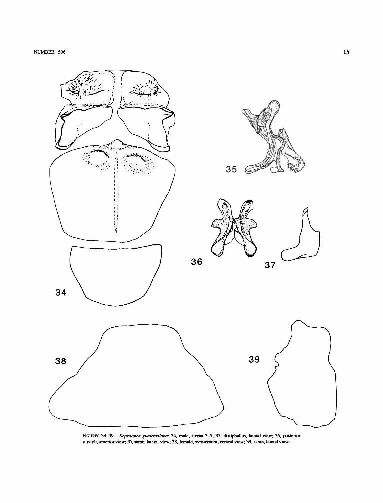
Abdomen: Male terminalia: Posterior margin of sternum4 slightly convex, with short, median lobe and pair of large,mammilliform, ventrally directed protuberances (Figure 34);anterior plate of sternum 5 without fingerlike processes (Figure34); distiphallus (Figure 35) slightly curved, with rather longventral process and hairy epiphallus; anterior surstylus verysmall or indistinct; posterior surstyli without median lobe(Figure 36), and with lateral lobe long, strongly curvedanterad (Figure 37). Female synsternum: In ventral viewposterior margin straight (Figure 38), in lateral view with

14 SMITHSONIAN CONTRIBUTIONS TO ZOOLOGY
29
28 31
32 33
FIGURES 28-33.—Sepedonea canabravana: 28, male, sterna 3-5; 29, distiphallus, lateral view; 30, posteriorsurstyli, anterior view; 31, same, lateral view; 32, female, synslernum, ventral view; 33, same, lateral view.
NUMBER 506 15
34
36
35
37
38 39
FIGURES 34-39.—Sepedonea guatemalana: 34, male, sterna 3-5; 35, distiphallus, lateral view; 36, posteriorsurstyli, anterior view; 37, same, lateral view; 38, female, synstemum, ventral view; 39, same, lateral view.

16 SMITHSONIAN CONTRIBUTIONS TO ZOOLOGY
shallow ventral depression, and with posterior ridge projectingslightly beyond posteroventral margin (Figure 39).
TYPE SPECIMEN.—Holotype d": GUATEMALA. LosAmates, 18-28 February 1905, Ja[me]s. S. Hine, [red label],OSU.
OTHER SPECIMENS EXAMINED.—HONDURAS. FRANCISCO
MORAZAN: Zamorano, nr. Tegucigalpa, 19, 20 November1987, L. Knutson, 37c?, I9. MEXICO. CHIAPAS: Las Cruces,16 July 1958, SB. Neff, 12d", 59 (field collected), 7d \ 4$(laboratory reared). NICARAGUA. Managua, April 1943, lcT,D. Gilail, USNM.
ADDITIONAL RECORDS FROM LITERATURE.—NICARA-
GUA. Chinandega, Baker, Id1 (Steyskal, 1951). GUATE-MALA. SANTA ROSA: 2.1 km E Barbarena (1,341 m), 23 July1958, 2 d \ 1$. COSTA RICA. SAN JOSE: San Antonio-Desamparados (1,160 m), 16 June 1964,4d", 19 (both Neff andBerg, 1966).
IMMATURE STAGES.—See Neff and Berg, 1966.REMARKS.—This species is found from Chiapas (Mexico) to
Costa Rica (Figure 140) and thus is the northernmostrepresentative of the genus and the only species not occurringin South America. With a lack of accepted phylogeny for thegenus, this distribution may either suggest that the species hasevolved relatively late or, conversely, that it is an early offshootof the genus and a sister group to all remaining species. Theshape of the posterior surstyli, which are only weakly fused,supports the latter possibility. Neff and Berg (1966) studied theimmature stages.
Sepedonea guianica (Steyskal)
FIGURES 7.40-45,97,110,113,116.126.135,138.141
Septdon guianica Steyskal, 1951:295.Sepedonea guianica.—Steyskal, 1973:145 [list].—Knutson et al., 1976:11
[catalog].—Knutson and Valley, 1978:198 [review].Sepedonea vau.—Mello and Bredt, 1978:1459 [nomen nudum; phenology].
ADULT.—Head: Lateral facial spot large.Thorax: Mesonotum grayish black; postpronotum brow-
nish; setulae near posterior spiracle moderately strong anddense. Legs: Midfemur posteroventrally usually withoutspines but with setulae only, sometimes setulae near apex offemur distinctly stronger than basal setulae (Figure 7), and insome Brazilian specimens there are 3-6 spines, not extendingbeyond half distance to base; hindcoxa posteriorly with shortsetulae, mainly restricted mesally; hindfemur usually withoutdark preapical marks. Wing: Grayish, with crossveins r-mand dm-cu not clouded, or brownish and clouded anteroapicallyand over the crossveins; length 5-6 mm.
Abdomen: Male terminalia: Posterior margin of sternum4 deeply emarginate, with pair of posteriorly directed protuber-ances (Figure 40); anterior plate of sternum 5 with wide,indented flange at posteromesal margin (Figure 40); distiphal-lus (Figure 41) sinuous, with posteroventral angle covered bylarge flat, hairy epiphallus; anterior surstylus indistinct;
posterior surstyli with (Figure 42a) or without (Figure 42b)median lobe; median lobe, when present, rounded; posteriorsurstyli with lateral lobe moderately strongly curved anterad(Figure 43a, b). Female synsternum: In ventral view posteriormargin distinctly projecting in middle (Figure 44); in lateralview with ventral surface more or less straight, and withposterior ridge distinctly projecting (Figure 45).
TYPE SPECIMEN.—Holotype d": Comoro Is., Maroni,Guyane, Purch. E. Le Moult, B.M. 1933-189, Type H. T.[round label with red margin], [red label], BMNH. This localityrefers to an island in the Maroni River (= Marowijne River),which is the border between Surinam and Guiana. The river isabout 150 km in length, and the exact location is not known.
OTHER SPECIMENS EXAMINED.—COLOMBIA. VALLE DEL
CAUCA: Morga, 20 km SE Univ., 22-25 June 1964, CO. Berg,16d\ 129. 6.5 km SE Cali (Navarro), 11 June 1969, Karl R.Valley, Id" (field collected), Id", 19 (laboratory reared). 14June 1969, Karl R. Valley, 2d", 29,1 puparium. 5 km SE Cali,near Navarro, 11 June 1969, Karl R. Valley, 19. VENE-ZUELA. COJEDES: L. Taguanes, near Tinaquillo, 13 April1972, L. Knutson, Id". BRAZIL. MINAS GERAIS: Lagoa Santa,near Belo Horizonte, 18 January 1977, L. Knutson, l id", 69.17 km N Belo Horizonte, 18-23 July 1964, CO. Berg, 49(field collected), 1 puparium (laboratory reared). Jockey Club,23 August 1966, CO. Berg, Id1, 15 September 1966, CO.Berg, 29. Serra Verde, 30 km E Belo Horizonte, 22 August1974, A. Bredt, Id", 10 October 1974, A. Bredt, Id". PARANA:Praia do Leste, 4 May 1967, Berg and Abercrombie, 3d1, 17May 1967, Berg and Abercrombie, Id", 1 puparium. 4,17 May1967, Berg and Abercrombie, 3d" (field collected), 4d", IO9(laboratory reared). 61 km S Curitiba, Rio Varzea, 16 May1967, Berg and Abercrombie, 2d". SAo PAULO: Sao Vicente,Parque Bitaru, 29 May 1967, Berg and Abercrombie, Id", 19(field collected), 8 eggs (laboratory reared). Rio Claro, 14January 1977, L. Knutson, Id". S3o Jose" do Rio Preto, 27 July1966, N. Papavero, 9d". GoiAs: Rio Preto, 70 km NE Brasilia,7 November 1974, Bredt and Knutson, Id". ESPIRTTO SANTO:Baixo Guandu, October 1970, P C Elias, Id". ARGENTINA.TUCUMAN: Monteros, 7 February 1967, Berg and Abercrom-bie, Id", (all USNM).
ADDITIONAL RECORDS FROM LITERATURE.—BRAZIL. PA-
RANA: Campina Grande, 2 August 1975, Mello and Bredt.DISTRTTO FEDERAL: Nucleo Bandeirantes, 11 November 74,Mello and Bredt. Highway L 2 Norte, Brasilia, 9 January 74,Mello and Bredt. Riberao Extrema, DF 21, 6 February 1974,Mello and Bredt. MINAS GERAIS: Hip6dromo Serra Verde,Santa Luzia, 24 August 1974, Mello and Bredt. GoiAs: RioPreto, DF 06, Formosa, 19 June 1974, Mello and Bredt. Lagoadas Pedras, Formosa, 9 January 1974, January-May, July-October, December 1975, January-February, April, June, July,October-December 1976, Mello and Bredt (all from Mello andBredt, 1978).
IMMATURE STAGES.—Egg: White. Length 1.20-1.24 mm(average = 1.24); greatest width 0.32-0.44 mm (average =

NUMBER 506 17
44
FIGURES 40-45.—Sepedonea guianica: 40, male, sterna 3-5; 41, distiphallus, lateral view; 42, posterior surstyli,anterior view, two variations; 43, same, lateral view, two variations; 44, female, synstemum, ventral view; 45,same, lateral view.

18 SMITHSONIAN CONTRIBUTIONS TO ZOOLOGY
0.40). Larger than S. incipiens, but otherwise very similar inshape, ridging, reticulation, and subglobular ends. (Based on 9specimens: Praia do Leste, Paran£, Brazil).
First-instar Larva: White. Integument transparent. Length1.2-2.9 mm (average = 1.9); greatest width 0.3-0.7 mm(average = 0.4). Cephalopharyngeal skeleton 0.28-0.31 mmlong; mandible 0.05-0.06 mm long, with 3 component parts;very similar to that of other Sepedonea. Indentation index29-38. Segment 1 bilobed anteriorly, each lobe bearing asensory papilla. Segments 2-4 each with 1 seta dorsolaterally,1 laterally, and 1 ventrally, all quite small. Segments 5-10 eachwith dorsolateral patch of setulae; segment 11 with single setadorsolaterally; segments 5-11 each with lateral tubercle grouparrangement as in other Sepedonea; none with setae; mainventral tubercle group with 4 tubercles in transverse row, eachbearing short, stout setae. Posterior spiracular disc with 5 pairsof lobes: ventral pair subconical, quite wide basally; ventro-lateral pair two-segmented, basal section truncate, distal sectiondigitiform; lateral, dorsolateral, and dorsal lobes low, rounded,each bearing 1 long seta distally; annulation of ventral lobesand distal section of ventrolateral lobes mostly obscured byvery long setulae; center of disc glabrous. Two stigmatic tubes,each bearing a spiracular plate with a B-shaped spiracular slitand 4 transparent, irregularly branched float setulae, larger inproportion to size of disc than in second- or third-instar larvae.Anal proleg small, inconspicuous, hookless. (Based on 15specimens: 4 from Praia do Leste, Parana1, and 11 from ParqueBitaru, Sao Vicente, Sao Paulo, both in Brazil).
Second-instar Larva (Figure 126): Light brown. Integu-ment diaphanous. Length 2.8-5.3 mm (average = 3.9); greatestwidth 0.6-1.1 mm (average = 0.8). Cephalopharyngealskeleton 0.49-0.52 mm long, with paired mandibles 0.09-0.10mm in length, each with 3 mesally directed accessory teeth;very similar to others in Sepedonea. Ventral arch connectedposterolaterally on each side to mandibles. Parastomal bars ofepipharyngeal sclerite connected to fused hypopharyngeal-pharyngeal sclerite; the latter light to dark brown with smallclear areas near dorsal margin of dorsal cornu; ventral windowindistinct in light ventral cornu. Indentation index 31-35.Segment 1 as in first-instar larva. Seta arrangement of segments2-4 as in first-instar larva. Segments 5-10 each withdorsolateral patch of setulae; segment 11 with single setadorsolaterally. Segments 5-11 each with lateral tubercle groupof 3 contiguous tubercles in a vertical row, the middle 1 slightlyanterior to other 2; dorsal and ventral tubercles of lateraltubercle group of segment 11 with single seta each and middletubercle with group of small, stout setae; main ventral tuberclegroup of 4 tubercles in a transverse row. Posterior spiraculardisc with 5 pairs of lobes, very similar to that of S. telson;ventral pair subconical; ventrolateral pair two-segmented;lateral pair low, rounded; dorsolateral pair even less conspicu-ous; dorsal pair somewhat larger, broadly rounded. Twostigmatic tubes arising from center of disc, each bearing aspiracular plate (Figure 126) with a stigmatic scar, 3 rather
small spiracular slits, and 4 large, prominent, irregularlybranched, semitransparent float setulae each with glandularpore at base. Anal proleg scarcely larger than a main ventraltubercle when viewed laterally; without hooks. (Based on 12specimens: Praia do Leste, Parana\ Brazil).
Third-instar Larva (Figures 110, 113, 116, 135,138): Length 8.2-9.3 mm (average = 8.7); greatest width1.8-2.3 mm (average = 2.0). Light to dark brown, with darkbrown middorsal stripe and dorsolateral V-shaped marksprominent anteriorly; laterally, a less prominent, broken,irregular lateral stripe; quite similar to S. trichotypa. Integu-ment opaque. Cephalopharyngeal skeleton length 0.74-0.82mm, with paired mandibles (Figure 110), each with 4-6 large,sharply pointed accessory teeth, directed mesally; hookstrongly decurved; entire sclerite 0.08-0.16 mm long; ventralarch (Figure 113) with 20-25 denticles directed anteriorly;connected posterolaterally with both mouth-hooks; hypophar-yngeal and labial sclerites (Figure 116) uniquely shaped amongSepedonea; former free from tentoropharyngeal sclerite;epipharyngeal sclerite very similar to that of S. telson and 5.trichotypa, with parastomal bars fused to tentoropharyngealsclerite; latter light to dark brown, lacking small clear areas indorsal cornu and with ventral window very indistinct.Indentation index 31-33. Segment 1 as in first-instar larva.Seta arrangement of segments 2-4 as in first-instar larva.Segment 2 with 2 anterior spiracles (Figure 138), each0.13-0.14 mm long and bearing 6 or 7 papillae distally.Segments 5-10 each with dorsolateral patch of setulae withshort setulae; segment 11 with single dorsolateral seta; lateraltubercle group on segment 11 with dorsal and ventral onesbearing single seta each; other lateral tubercles setaless;segments 5-11 each with main ventral tubercle group of 4tubercles in a transverse row, followed posteriorly by 2 rows ofmuch smaller intrasegmental tubercles arranged in transverserows. Segment 8 with posterior spiracular disc (Figure 135) of5 lobes: ventral pair quite wide at base, tapered sharply distally;ventrolateral pair basically similar to those of other species ofSepedonea; lateral pair rather acute; dorsolateral pair very lowand inconspicuous; dorsal pair each with tuft of setulae distally,lobes broadly rounded. Entire disc moderately setulose, withglabrous center, with 2 separate stigmatic tubes, each scallopedbasally, and each with a stigmatic scar, 3 spiracular slits, and 4irregularly branched float setulae on a spiracular plate muchlighter in color than 5. incipiens and S. telson. Anal proleglarger than in other species of Sepedonea but still relativelyinconspicuous and hookless. (Based on 8 specimens: Praia doLeste, Parana^ Brazil).
Puparium (Figure 97): Light brown with dark brownstripes along middorsal line and dorsolaterally on anteriorsegments, strongest on segments 6 and 7. Integument opaque.Length 5.2-5.7 mm (average = 5.4); greatest width 2.2-2.3mm (average = 2.2). Barrel-shaped, with ends of cephalic capsprojecting anteriorly slightly dorsal to the longitudinal bodyaxis and slightly upturned. Anterior spiracles protruding from

NUMBER 506 19
anterolateral corners of dorsal cephalic cap. Lateral tuberclegroup of segments 5-11 visible as strongly contrasting darkareas; main tubercles of ventral tubercle group persisting as 4less noticeable light spots in a transverse row. Dorsolateralpatches of setulae of segments 5-10 with setulae appearingappressed to puparium surface. Posterior end sharply upturned,stigmatic tubes forming an angle of 100-110 degrees withlongitudinal body axis. Lobes of posterior spiracular discshrunken. Anal plate invaginated; anal proleg lacking. Quitesimilar to S. telson and S. trichotypa, but much lighter, colorpattern being dark-on-light rather than light-on-dark as in 5.telson and S. trichotypa. (Based on 5 specimens; Praia doLeste, Parana", Brazil).
REMARKS.—This species, which occurs widely from north-ern to central South America, is especially well represented ineastern Brazil, but is practically lacking in the entire Amazonbasin (Figure 141). Most specimens from Brazil differ from thespecimens originating farther in the north, including theholotype, in having the midfemur distinctly spinose, in usuallyhaving dark lateral preapical marks on the hindfemur, in thedarker and clouded wing, and in the posterior surstyli, whichlack a median lobe. However, the overall similarity between thetwo groups of specimens, the male and female terminalia inparticular, suggests that these differences merely representintraspecific variation.
Sepedonea incipiens, new species
FIGURES 46-51,94, 100.104, 108-109,117,123,129. 134.139
ADULT.—Head: Lateral facial spot moderately large.Thorax: Mesonotum usually grayish black; postpronotum
brownish yellow; setulae near posterior spiracle very strongand dense. Legs: Midfemur posteroventrally with 3-5 spines,not extended beyond half distance to base; hindcoxa posteriorlywith short setulae, restricted to mesal area; hindfemur with darkposteroventral and anteroventral preapical marks and with darkor light dorsal preapical marks. Wing: Yellowish, withanteroapical margin and crossveins r-m and dm-cu more or lessdistinctly clouded; length 4-5 mm.
Abdomen: Male terminalia: Posterior margin of sternum4 straight, with median projection bearing a pair of tubercles(Figure 46); anterior plate of sternum 5 with triangular,anterolaterally directed flange (Figure 46); distiphallus (Figure47) slightly curved, with short, slightly curved posteroventralspur; anterior surstylus indistinct; posterior surstyli withmedian lobe low and rounded (Figure 48) and with lateral lobemoderately curved anterad (Figure 49). Female synster-num: In ventral view posterior margin with 3 subequaltubercles (Figure 50); in lateral view ventral surface straight,posterior margin with apex and shoulders short and rounded(Figure 51).
TYPE SPECIMENS.—Holotype d": ARGENTINA. BUENOS
AIRES: 28 km SW Buenos Aires, 15-16 April 1967, J.
Abercrombie, USNM. [Label: J. Abercrombie field trip #6729].Allotype: BRAZIL. GUANABARA: InsL Oswaldo Cruz, 6
June 1967, Berg and Souza-Lopes. [Label: J. AbercrombieBiol. Note #67118].
Paratypesr. ARGENTINA. BUENOS AIRES: 28 km SWBuenos Aires, 15-16 April 1967, J. Abercrombie, Id", USNM.BRAZIL. GUANABARA: InsL Oswaldo Cruz, 6 June 1967, Bergand Souza-Lopes, Id", 19 (field collected), 14d", 149(laboratory reared), USNM; 6 April 1967, Berg and Souza-Lopes, 29, USNM; 7 April 1967, Berg and Souza-Lopes, lcT,USNM. SAo PAULO: S3o Paulo, InsL de Botanica, July 1964,Berg and Papavero, Id", MZUSP. 15 July 1964, N. Papavero,lcf, MZUSP. September 1964, N. Papavero, lcf, MZUSP;Sao Paulo, InsL Bot. Serv. Agric, 11-13 July 1964, Berg andPapavero, \<f, USNM. Sao Paulo, Parque D. Pedro II, 11 July1964, Berg and Papavero, \<f, MZUSP. Sao Paulo, 17 April1967, C O . Berg, Id1, USNM; 26 May 1967, J. Abercrombie,l d \ USNM. 7 June 1967, J. Abercrombie, l d \ USNM. RioGRANDE DO SUL: Sao Leopoldo, 3, 11 May 1967, Berg andAbercrombie, 3d1, USNM.
OTHER SPECIMENS EXAMINED.—BRAZIL. SAo PAULO:S3o Vicente, 12 larvae (laboratory reared).
ADDITIONAL RECORDS FROM LITERATURE.—None.
ETYMOLOGY.—The specific epithet, incipiens, was a manu-script name given by George Steyskal, but nobody, includinghim, can say why.
IMMATURE STAGES.—Egg (Figure 94): White. Length0.90-1.12 mm (average = 1.00); greatest width 0.28-0.38 mm(average = 0.34). Elongate-ovoid, tapered at both ends. Twoprominent, subparallel dorsal ridges flanked laterally byanother salient ridge on each side; all 4 visible dorsally. Areabetween ridges and lateral and ventral surfaces reticulate inirregular hexagonal pattern, more variable laterally andventrally. Hexagons elongated at both ends in areas betweenridges. Micropyle shielded dorsally by subglobose tuberclewith minute punctations appearing hexagonal under highmagnification (x400). Posterior end rounded, subglobose, withpunctations as on anterior end. (Based on 10 specimens:Instituto Oswaldo Cruz, Rio de Janeiro, Guanabara, Brazil).
First-instar Larva: White. Integument transparent. Length1.7-2.8 mm (average = 2.2); greatest width 0.3-0.7 mm(average = 0.5). Cephalopharyngeal skeleton length 0.29-0.30mm. Indentation index 24-29. Very similar to S. lindneri insize, general appearance, seta and tubercle arrangement,mouthparts, and posterior spiracular disc. (Based on 5specimens: S3o Vicente, Sao Paulo, Brazil).
Second-instar Larva (Figures 104, 123): Light brown;integument diaphanous. Length 3.2-4.4 mm (average = 3.8);greatest width 0.5-1.0 mm (average = 0.7). Cephalopharyngealskeleton (Figure 104) 0.44-0.49 mm long, with pairedmandibles 0.07-0.10 mm in length, each with 4 mesallydirected accessory teeth and 2 holes, 1 near center and the othernear dorsal margin. Mandibles fused posteroventrally withlateral edges of ventral arch; latter with 17-19 small, rounded

20 SMITHSONIAN CONTRIBUTIONS TO ZOOLOGY
47
46
48
50
FIGURES 46-51.—Sepedonea incipiens: 46, male, sterna 3-5; 47, distiphallus, lateral view; 48, posterior surstyli,anterior view; 49, same, lateral view; 50, female, synstemum, ventral view; 51, same, lateral view.

NUMBER 506 21
denticles directed anteriorly. Epistomal and hypopharyngealsclerites fused with paired tentoropharyngeal sclerite; latterlight brown, with ventral cornu very light, thus obscuringventral window; dorsal cornu with small hyaline areas neardorsal margin. Indentation index 27-30. Segment 1 bilobedanteriorly, each lobe with a sensory papilla. Segment 2 bearing1 dorsolateral seta and an anterior spiracle on each side.Segment 3 with 1 dorsolateral seta and 1 on each side. Segment4 with 1 seta dorsolaterally, 1 ventrally, and 1 on each side.Segments 5-11 each with prominent dorsolateral patch ofsetulae (sometimes only 1 large seta on segment 11); lateraltubercle group of 3 contiguous tubercles more or less in avertical row, dorsal and ventral tubercles each bearing a seta;main ventral group of 4 tubercles (some with short, stout setae)in a transverse row, followed posteriorly by 2 transverse rowsof much smaller intrasegmental tubercles. Posterior spiraculardisc (Figure 123) with 5 pairs of lobes: ventral pair short,subconical; ventrolateral pair two-segmented, basal portiontruncate and distal portion digitiform; lateral lobes rather large,smoothly rounded; dorsolateral lobes very small, inconspicu-ous; dorsal lobes low, rounded. Two stigmatic tubes each withspiracular plate, stigmatic scar, 3 spiracular slits, and 4semi-transparent, irregularly branched float setulae. Analproleg very inconspicuous, same size as a main ventral tuberclewhen viewed laterally; no hooks. (Based on 8 specimens: 3from Instituto Oswaldo Cruz, Rio de Janeiro, Guanabara, and 5from S3o Vicente, S3o Paulo, both in Brazil).
Third-instar Larva (Figures 108, 109, 117, 129,134): Light brown, with dark brown stripes dorsally anddorsolaterally; integument diaphanous. Length 3.8-7.5 mm(average = 6.1); greatest width 0.8-1.5 mm (average = 1.2).Cephalopharyngeal skeleton (Figure 108) length 0.66-0.74mm, with paired mouth-hooks, each with 3 or 4 accessory teethdirected mesally; mandible (Figure 109) 0.13-0.15 mm long;ventral arch with 21-23 small, rounded denticles on anterioredge, connected posterolaterally with both mandibles; hypo-pharyngeal sclerite free from tentoropharyngeal sclerite; paras-tomal bars of epipharyngeal sclerite (Figure 117) connected topaired tentoropharyngeal sclerite and continued posteriorly inlatter structure as salient black lines; tentoropharyngeal scleritelight brown, with ventral window obscured in light ventralcornu. Indentation index 28-31. Segment 1 as that insecond-instar larva. Segment 2 with 1 seta dorsolaterally, 1ventrally, and an anterior spiracle (Figure 134) on each side;latter 0.12-0.13 mm long with 4-6 papillae and bulbousposteriorly at junction with trachea. Segment 3 with 1 setadorsolaterally, 1 ventrally, and 1 on each side. Segment 4 with1 seta dorsolaterally, 1 ventrally, and 2 on each side; lateral andventral setae borne on small tubercles. Segments 5-11 eachwith conspicuous dorsolateral patch of setulae; tuberclearrangement as in second-instar larva, except lateral tuberclesnot always with setae. Posterior spiracular disc (Figure 129)with 5 pairs of lobes: ventral pair fairly short, tapered;ventrolateral lobes truncate basally with digitiform distal
section; lateral lobes acute; dorsolateral lobes very low,inconspicuous; dorsal lobes broadly rounded, low. Discmoderately setulose. Two stigmatic tubes brown when viewedlaterally, each with darkly sclerotized spiracular plate, stig-matic scar, 3 spiracular slits, and 4 irregularly branched,subopaque float setulae. Anal proleg as in second instar larva.(Based on 5 specimens: 3 from Instituto Oswaldo Cruz, Rio deJaneiro, Guanabara, and 2 from S3o Vicente, SSo Paulo, both inBrazil).
Puparium (Figure 100): Light to dark brown, with greatestcontrast dorsally and dorsolaterally; integument opaque.Length 3.8-4.5 mm (average = 4.2); greatest width 1.8-2.0mm (average = 1.95). Barrel-shaped, with ends of cephalic capsprojecting anteriorly dorsal to the longitudinal body axis andslightly upturned from it. Anterior spiracles protruding fromanterolateral corners of dorsal cephalic cap. Lateral tuberclesand main ventral tubercles of segments 5-11 persisting asconspicuous light areas, giving puparium a blotched appear-ance. Posterior end sharply upturned, with posterior surface atright angle to longitudinal body axis. Stigmatic tubes formingan angle of 110-120 degrees with longitudinal body axis.Lobes of posterior spiracular disc shrunken. Anal plate alight-colored invagination on posterior wall. Anal proleglacking. Very similar to S. lindneri, but much darker, colorpattern being light-on-dark rather than dark-on-light as in S.lindneri. (Based on 5 specimens: Instituto Oswaldo Cruz, Riode Janeiro, Guanabara, Brazil).
REMARKS.—This species has a distribution very similar tothat of S. trichotypa (Figures 139, 143). The two species alsoshare many morphological characters, including very similarterminalia. They can be reliably distinguished, however, by theshape of the male's sterna 3-5 and the female's synsternum.
Sepedonea isthmi (Steyskal)
FIGURES 6.9,14-16,52-57
Sepedon isthmi Steyskal, 1951:291.
Sepedonea isthmi.—Steyskal, 1973:145 [list].—Knutson et al., 1976:11[catalog].—Knutson and Valley, 1978:197 [biology and immature stages].
ADULT.—Head: Lateral facial spot large.Thorax: Mesonotum grayish black; postpronotum dorsally
concolorous with mesonotum, ventrally more yellowish;setulae near posterior spiracle moderately dense and strong.Legs: Midfemur posteroventrally with 8-15 spines extendedV2-2/3 distance to base (Figure 9); hindcoxa posteriorly withrow of setulae, long setulae restricted mesally; hindfemurlacking dark preapical marks. Wing (Figure 6): Distinctlyclouded anteroapically and over crossveins r-m and dm-cu;length 5-6 mm.
Abdomen: Male abdomen as in Figures 14-16. Maleterminalia: Posterior margin of sternum 4 slightly convex,without protuberances (Figure 52); anterior plate of sternum 5without fingerlike processes (Figure 52); distiphallus (Figure

22 SMITHSONIAN CONTRIBUTIONS TO ZOOLOGY
52
56
FIGURES 52-57.—Sepedonea isthmi: 52, male, sterna 3-5; 53, distiphallus, lateral view; 54, posterior surstyli,anterior view; 55, same, lateral view; 56, female, synstemum, ventral view; 57, same, lateral view.

NUMBER 506 23
53) elongate, gently curved and somewhat flattened, withbroad, recurved epiphallus posteroventrally; anterior surstyluslarge, elongate; posterior surstyli with large, triangular medianlobe (Figure 54), and with lateral lobe rather pointed, curvedanterad (Figure 55). Female synsternum: In ventral viewposterior margin gently concave (Figure 56); in lateral viewwith distinct ventral convexity, broadly rounded posteroventralmargin and small posterior ridge (Figure 57).
TYPE SPECIMENS.—Holotype <?: PANAMA. "Canal Zone":Corazal, 1 March 1912, Aug. Busck, no. 60905, USNM.
Allotype: same data as holotype.Paratype: "Canal Zone": Juan Mina, 2 September 1923,
R.C. Shannon, Id1, USNM.OTHER SPECIMENS EXAMINED.—PANAMA. La Jagua Hunt
Club, 32 mi. ENE Balboa, 1 July 1969, Karl R. Valley, lcf.TRINIDAD. Curepe, nr. Port-of-Spain, 4-5 May 1972, L.Knutson, 15b", I69, 25 eggs, 5 puparia. Caroni River, 12October 1916, Harold Morrison, Id". VENEZUELA. ARAGUA:Cata, W. Maracay, 17 April 1972, L. Knutson, lcf. Pto. deCata, N Maracay, 17 April 1972, L. Knutson, 7d", I9.Ocumare, 28 km NW Maracay, 14 March 1971, CO. Berg,3d". CARABOBO: Valencia, 16 March 1971, CO. Berg, I9.Embalse de Guataparo, W Valencia, 13 April 1972, L.Knutson, Id". Valle Seco, January 1940, P. Anduze, lcf.COJEDES: L. Taguanes, near Tinaquillo, 13 April 1972, L.Knutson, 39. COLOMBIA. VALLE DEL CAUCA: Morga, 20 kmSE of Univ., 22-25 June 1964, CO. Berg, 4c?, 39- 1.7 km WCali Puerto, 14 June 1969, Karl R. Valley, 3d1, 29, 1 larva. 5km SE Cali near Navarro, 11 June 1969, Karl R. Valley, 2cf.6.5 km SE Cali (Navarro), 11 June 1969, Karl R. Valley, 13d1,149, 3 puparia (field collected), 7d", 19 (laboratory reared).BRAZIL. AMAZONAS: Parana" da Cigana, Parintins, November1959, Exp. Perm. Amaz., Id1 ,19. GOIAS: Rio Preto, 70 km NEBrasilia, 7 November 1974, Bredt and Knutson, Id1. GUANAB-ARA: Inst. Oswaldo Cruz, 6 April 1967, Berg and Souza-Lopes,19. SAo PAULO: Sao Vicente, Parque Bitaru, 29 May 1967,Berg and Abercrombie, Id1 (all USNM).
ADDITIONAL RECORDS FROM LITERATURE.—BOLIVIA.
BENI: Rurrenabaque (175 m), 10-23 October 1956, L.E. Pena,Id1. BRAZIL. PARA: Breves, Ilha do Marajo, September 1969,Exp. Perm. Amaz., 19. EspiRrro SANTO: Itaguacu, October1970, P.C Elias, 1 adult. VENEZUELA. COJEDES: La Piedrita,16 February 1911, S. Brown, I9. TRINIDAD. PrincessMargaret Highway, 9 km W Port-of-Spain, 4-5 May 1972,Bennett, Yaseen, and Knutson, 8d", 29 (Knutson and Valley,1978).
IMMATURE STAGES.—See Knutson and Valley, 1978.REMARKS.—This species is widespread over most of South
America (as far south as the Tropic of Capricorn) and PanamlBecause the material we studied is essentially the same as thatstudied by Knutson and Valley, the reader is refered to theirmap (Knutson and Valley, 1978, Fig. 1). The species is easilydistinguished from all other congeners by the extensivelyspinose midfemur.
Sepedonea lagoa (Steyskal)
FIGURES 2.58-63,101-103,106-107,115,118,120,127,132.140
Sepedon lagoa Steyskal, 1951:293.Sepedonea lagoa.—Steyskal, 1973:145 [list].—Knutson et al., 1976:11
[catalog].—Knutson and Valley, 1978:198 [review].
ADULT.—Head: Lateral facial spot small.Thorax: Mesonotum grayish black; postpronotum brow-
nish or yellowish; setulae near posterior spiracle weak andsparse. Legs: Midfemur posteroventrally with 5-8 spines,not extended beyond half distance to base; hindcoxa withsetulae mainly restricted posterodorsally; hindfemur lackingdark preapical marks. Wing: Brownish, clouded anteroapi-cally and slightly over crossveins r-m and dm-cu; length5.5-6.5 mm.
Abdomen: Male terminalia: Posterior margin of sternum4 concave, without protuberances (Figure 58); anterior plate ofsternum 5 reduced to small sclerite (Figure 58); distiphallus(Figure 59) straight, short and robust, with anterodorsalspur; anterior surstylus indistinct; posterior surstyli (Figures60, 61) in form of an elongate, slightly broadened apically,anteriorly directed rod. Female synsternum: In ventral viewgradually narrowing posteriorly, with large, rounded, light(weakly sclerotized) depression at posterior half (Figure 62); inlateral view with distinct ventral depression and posteriorshoulder (Figure 63).
TYPE SPECIMEN.—Holotype <f: BRAZIL. Minas [Gerais],Lagoa Santa, 19 January [19]39, Martins, Lopes, Mangabeira,IOC The holotype was kindly loaned to us by Dr. Orlando V.Ferreira. The specimen was damaged enroute: we glued thehead, both wings and several leg parts to a small piece of paperbeneath the pinned specimen. The postabdomen is missing. Weprepared the remaining part of the abdomen. The characteristicsternum 5 allows us to identify the specimens listed below asconspecific with the holotype.
OTHER SPECIMENS EXAMINED.—BRAZIL. Rio GRANDE DO
SUL: Sao Leopoldo, 3, 11 May 1967, Berg and Abercrombie,lcf. SANTA CATARINA: 4 km E Corupa, 3 May 1967, Berg andAbercrombie, 4d", 19. PARANA: Praia do Leste, 4 May 1967,Berg and Abercrombie, 29. 4, 17 May 1967, Berg andAbercrombie, 2d". Rio Iguassu at Araucaria, Id1 (laboratoryreared). Rio Iguassu, S Laranjeiras, 29 April 1967, Berg andAbercrombie, 3d1 ,19. Sapitandura, nr. Morretes, 7 November1986, Carvalho, Knutson, and Marinon, lcf. 6 km S Morretes,4, 17 May 1967, Berg and Abercrombie, 2Uf, 49. 19 km WGuarapuava, Rio Coutinho, 28 April 1967, Berg and Aber-crombie, 1 Id1 ,69 (field collected), 4d", 89 (laboratory reared),5 eggs. MINAS GERAIS: Sena Verde, 30 km E Belo Horizonte,lcf (laboratory reared). 35 km N Belo Horizonte, Lagoa Santa,18-23 July 1964, CO. Berg, Id". 17 km N Belo Horizonte,18-23 July 1964, CO. Berg, 3d1. Belo Horizonte, July 1964,CO. Berg, Id1 ,19. Juiz de Fora, 10 June 1967, CO. Berg, 29.MATO GROSSO: Pucone, Rod. Transpantaneira, km. 17, 16February 1986, N. Papavero, Id1. SAo PAULO: S3o Paulo, Inst.

24 SMITHSONIAN CONTRIBUTIONS TO ZOOLOGY
58
62
61
63
FIGURES 58-63.—Sepedonea lagoa: 58, male, sterna 3-5; 59, disliphallus, lateral view; 60, posterior surstyli,anterior view; 61, same, lateral view; 62, female, synstemum, ventral view; 63, same, lateral view.

NUMBER 506 25
Bot. Seer. Agric, 13 July 1964, Berg and Papavero, 2 d \11-13 July 1964, Berg and Papavero, 29. 18 August 1964, N.Papavero, Id". September 1964, N. Papavero, 2o", I9. Mogidas Crazes, 11-13 July 1964, Berg and Papavero, 7 d \ 15August 1964, N. Papavero, 2<?, 19- Rio Claro, 14 January1977, L. Knutson, 4<?, 29. Barueri, 18 July 1955, K. Lenko,lef. 20 July 1955, K. Lenko, Id". AMAZONAS: Parana" daCigana, Parintins, November 1969, Exp. Perm. Amaz., 4<f.SURINAM. SURINAME: Paramaribo, 4 September 1943, DavidG. Hall, l o \ PERU. HUANUCO: 1 km S Tingo Maria, 6February 1984, W.N. Mathis, 29. COSTA RICA. San Jose",Farm La Caja, 20 January, H. Schmidt, lef. All USNM.
ADDITIONAL RECORDS FROM LITERATURE.—BRAZIL.
MINAS GERAIS: Hipodromo Serra Dourada, 6 October 1974,
Mello and Bredt (Mello and Bredt, 1978).IMMATURE STAGES.—Egg: White. Length 1.12-1.24 mm
(average = 1.17); greatest width 0.20-0.36 mm (average =0.28). Very similar to that of S. incipiens, but longer. Differingalso in its subglobular anterior end, in dorsal view, with smalldepression or notch on anterior border. (Based on 17specimens: Rio Coutinho, 19 km west of Guarapuava, Parana",Brazil).
First-instar Larva (Figures 102,103,120): White; integu-ment transparent. Length 1.6-2.4 mm (average = 1.9); greatestwidth 0.3-0.4 mm (average = 0.35). Cephalopharyngealskeleton (Figure 102) length 0.31-0.33 mm; mandible (Figure103) 0.04-0.06 mm long; epistomal and hypopharyngealsclerites fused to paired tentoropharyngeal sclerite; latter lightbrown, with very light ventral cornu, making rim of ventralwindow indistinct; indentation index 37-39. Segment 1bilobed anteriorly, each lobe with a sensory papilla. Segment 2with 1 dorsolateral and 1 lateral seta. Segments 3 and 4 eachwith a lateral seta, often bifid on segment 4. Segments 5-11each with a prominent dorsolateral patch of setulae, very dense,with setulae reaching 0.2 mm in length; transverse row ofshorter, less dense setulae extended across middorsal line,joining dorsolateral patches of setulae; lateral tubercle group of3 contiguous tubercles, the middle one slightly anterior to theother 2, each with a seta; main ventral tubercle group of 4 smalltubercles in a transverse row. Posterior spiracular disc (Figure120) with 4 pairs of lobes: ventral pair conical, annulate;ventrolateral pair two-segmented, basal section broad, truncate,distal section digitiform, annulate; lateral lobes rather acute,each with a single long seta at distal end; dorsolateral lobeslacking; dorsal lobes very low, rounded; entire disc coveredwith long setulae. Two spiracular plates each borne on astigmatic tube and each with a B-shaped spiracular slit and 4irregularly branched float setulae, proportionately longer thanin later instars and covering the face of the entire disc. Analproleg small, bearing tiny spinules, no hooks. (Based on 19specimens: Rio Coutinho, 19 km west of Guarapuava, Parana",Brazil).
Second-instar Larva: Light brown; integument diapha-nous. Length 3.3-3.8 mm (average = 3.6); greatest width
0.5-0.8 mm (average = 0.6). Cephalopharyngeal skeleton0.53-0.55 mm long, with paired mandibles 0.09-0.11 mm inlength, each with 3 mesally directed accessory teeth, dorsaltooth strongest and heaviest and others lightly sclerotized; eachalso with a small hole dorsad of teeth and a lobelike projectionon the dorsal margin. Epistomal and hypopharyngeal scleritesfused to paired tentoropharyngeal sclerite; latter with darkcenter line, extended posteriorly from point of juncture withparastomal bars, with obfuscation anterodorsally; ventral cornuvery light, obscuring ventral window; dorsal comu with smallhyaline areas near dorsal margin. Indentation index 31-33.Segment 1 bilobed anteriorly, each lobe with sensory papilla.Segment 2 bearing 1 anterior spiracle on each side, 1 setadorsolaterally, 1 ventrally, and 1 laterally. Segment 3 with sameseta pattern. Segment 4 with 1 seta dorsolaterally, 1 ventrally,and 2 on each side. Segments 5-11 as in first-instar larva.Posterior spiracular disc with 5 pairs of lobes: ventral pairconical, tapered; ventrolateral pair as in first-instar larva, exceptlarger and more hirsute; dorsolateral, lateral, and dorsal lobeslow, rounded protuberances; entire disc very setulose; 2stigmatic tubes each bearing a spiracular plate, each with 3oblong-ovoid spiracular slits, outer 2 somewhat arcuate, 1stigmatic scar, and 4 irregularly branched, semitransparent floatsetulae. Anal proleg small, about size of main ventral tuberclewhen viewed laterally; hookless. (Based on 5 specimens: RioCoutinho, 19 km west of Guarapuava, Parana", Brazil).
Third-instar Larva (Figures 106, 107, 115, 118, 127,132): Light brown; integument opaque. Length 7.3-10.7 mm(average = 9.0); greatest width 1.5-2.3 mm (average = 1.8); thelargest larva of Sepedonea seen in this study. Cephalopharyn-geal skeleton (Figure 106) 0.91-0.95 mm long. Mandible with3 or 4 accessory teeth directed mesad; dorsal tooth darker andmore heavily sclerotized but approximately equal in size toothers; mandible (Figure 107) 0.17-0.18 mm in length, withsmall hole dorsal to teeth and with dorsal projection. Ventralarch connected posterolaterally on each side with mandibles,bearing 17-21 rounded denticles anteriorly. Hypopharyngealsclerite (Figure 115) free from tentoropharyngeal sclerite butparastomal bars of epipharyngeal sclerite (Figure 118) con-nected to paired tentoropharyngeal sclerite and continuedposteriorly as dark lines. Tentoropharyngeal sclerite as insecond-instar larva. Indentation index 33-35. Segments 1-4 asin second-instar larva, except segment 4 with only 1 ventralseta in center. Segments 5-11 each with prominent dorsolateralpatch of setulae, flanked closely by smaller, lateral patch ofsetulae; lateral tubercle group of same arrangement as insecond-instar larva; dorsal and ventral tubercle often without aseta; middle tubercle usually bearing patch of tiny setulae; mainventral tubercle group as in second-instar larva, exceptoutermost tubercles each bearing small, stout seta; 2 transverserows of small, low, intrasegmental tubercles between mainventral tubercle groups. Posterior spiracular disc (Figure 127)with 5 pairs of lobes: ventral pair subconical near base, buttapered sharply distally; ventrolateral lobes as in second-instar

26 SMITHSONIAN CONTRIBUTIONS TO ZOOLOGY
larva except larger and more hirsute, retaining hint ofannulation on distal segment despite shagginess; lateral lobessomewhat acute; dorsolateral and dorsal lobes low, broadlyrounded. Entire disc thickly clothed with setulae. Twostigmatic tubes scalloped basally, each bearing spiracular platedistally. Latter each with 3 spiracular slits, 1 stigmatic scar, and4 irregularly branching, dark, subopaque float setulae. Analproleg small, hookless. Anterior spiracles (Figure 132) large,conspicuous, protruding from each side of segment 2 at rightangles to trachea! trunk, and bearing 8 papillae. Length0.15-0.17 mm. (Based on 6 specimens: Rio Coutinho, 19 kmwest of Guarapuava, Parana", Brazil).
Puparium (Figure 101): Black; opaque. Length 5.2-6.2mm (average = 5.8); greatest width 1.7-2.7 mm (average =2.1); the largest puparium of any Sepedonea reared in thisstudy. Subcylindrical, elongate, with ends of cephalic capsprojecting anteriorly on a line almost on and parallel with thelongitudinal body axis. Anterior spiracles protruding fromanterolateral corners of dorsal cephalic cap. Lateral and ventraltubercles indistinct, existing chiefly as discrepancies in thesmoothness of the integument; dorsolateral patches of setulaeon segments 5-11 persisting as elongate-oval black spotsvisible dorsally—no actual setulae on surface. Posterior endupturned 140-150 degrees from longitudinal body axis,bearing 2 separate, subshiny stigmatic tubes. Lobes ofspiracular disc shrunken but ventral lobes remaining ratherprominent. Anal plate marked by imagination; anal proleglacking. (Based on 5 specimens: Rio Coutinho, 19 km west ofGuarapuava, Parana", Brazil).
REMARKS.—This is a widespread South American speciesthat extends as far north as Costa Rica (Figure 140). It is uniqueamong all congeners in having sternum 5 with anterior platesreduced, a straight distiphallus and an elongate median lobe ofthe posterior surstyli. Knutson collected S. guianica at LagoaSanta, the type locality for S. lagoa, but did not find S. lagoa atthat locality.
Sepedonea Hndneri (Hendel)
FIGURES 1,4,11,64-69,95,105,112,122,128,133,142
Sepedon Hndneri Hendel, 1932:99.—Steyskal, 1951:275 [key], 291 [review].Sepedonea Hndneri.—Steyskal, 1973:145 [list; designation as type species].—
Knutson et aL, 1976:11 [catalog].—Knutson and Valley, 1978:198 [review].
ADULT.—Head: Lateral facial spot small or large.Thorax: Mesonotum usually grayish black, sometimes
yellowish; postpronotum yellowish; setulae near posteriorspiracle weak and sparse (Figure 4). Legs: Midfemurposteroventrally with 4-7 spines; hindcoxa with setulaerestricted to mesal !/3 to 1/2 (Figure 11); hindfemur with darklateral and rounded dorsal preapical spots. Wing: Grayishhyaline, with indistinct clouds on crossveins r-m and dm-cu;length 4-5 mm.
Abdomen: Male terminalia: Posterior margin of sternum4 deeply emarginate, with membrane "covering" emargination,
and with pair of small, posteromesally directed protuberances(Figure 64); anterior plate of sternum 5 with fingerlikeprocess (Figure 64); distiphallus (Figure 65) bent at rightangle, with long, apically recurved ventral process; anteriorsurstylus short; posterior surstyli with median lobe emarginateventrally (Figure 66), and with lateral lobe slightly curvedanterad (Figure 67). Female synsternum: In ventral viewemarginate posteriorly, with 2 large lateral lobes (Figure 68); inlateral view with ventral surface rather straight, posteriormargin rather broadly rounded, with narrow ridge (Figure 69).
TYPE SPECIMENS.—We have studied Hendel's type material,which consists of three specimens collected during the ChacoExpedition of 1925-1926 (San Josd, Argentina, October 1925,Lindner, 9; Tapikiole, Argentina, December 1925-January1926, Lindner, and Trinidad bei Asuncion, Paraguay, August1925, cf ), all in SMNS. The male from Trinidad bei Asuncion,which bears a label in E. Lindner's handwriting, is designatedas the lectotype and the other two specimens are designated asparalectotypes.
OTHER SPECIMENS EXAMINED.—ARGENTINA. BUENOS
AIRES: 28 km SW Buenos Aires, 7-8 July 1964, CO. Berg,5cf, 59 (field collected), 16cf, I I 9 (laboratory reared), 46eggs. 7-8 December 1966, CO. Berg, l d \ 29. 15-16 April1967, J. Abercrombie, 2cf. Tigre, 8 February 1920, CornellUniv. Expedition, 19. CORRIENTES: Corrientes, 23 February1927, R.C Shannon, 19. MisiONES: Iguazu, 4-10 October1947, R.C and E.M. Shannon, lcf. CHACO: Mahalte, June1964, 19. Presidencia Roque Sa6nz Pena, 15 September-15November 1974, M. Graciela Arias, 5cf, 59. TUCUMAN.Cuidad Universitaria, 5 February 1967, Berg and Abercrombie,2cf. PARAGUAY. LA CORDILLERA: San Bernardino, K.Fiebrig, Id1. BRAZIL. Rio GRANDE DO SUL: 87 km S PortoAlegre, 10 May 1967, Berg and Abercrombie, lcf. SaoLeopoldo, 3, 11 May 1967, Berg and Abercrombie, 2cf, 19.SANTA CATARINA: 5 km W Lajes, 6 May 1967, Berg andAbercrombie, lcf, I9.
ADDITIONAL RECORDS FROM LITERATURE.—ARGENTINA.
LA RIOJA: "Patquila", January 1932, KJ. Hayward. "San Jos6C. Paz", Ogloblin, 19 (Steyskal, 1951). Patquia, 25 February1944, KJ. Hayward, lcf; 25 March 1944, KJ. Hayward, lcf.FORMOSA: Clorinda, November 1947, J. Morel, lcf (Steyskal,in litL). BRAZIL. Rio GRANDE DO SUL: Morretes, Canoas, 19December 1974, Mello and Bredt (Mello and Bredt, 1978a).
IMMATURE STAGES.—Egg: White. Length 0.90-1.04 mm(average = 0.97); greatest width 0.16-0.34 mm (average =0.25). Very similar to S. indpiens but slightly smaller. (Basedon 19 specimens: 28 km southwest of Buenos Aires, BuenosAires, Argentina).
First-instar Larva (Figure 122): White; integument trans-parent. Length 1.8-2.9 mm (average = 2.2); greatest width0.4-0.8 mm (average = 0.5). Metapneustic. Cephalopharyngealskeleton 0.29-0.30 mm long; mandible 0.04-0.06 mm inlength, with 3 component parts; epistomal and hypopharyngealsclerites fused to paired tentoropharyngeal sclerite; latter lightbrown, with L-shaped obfuscation anterodorsally, and very

NUMBER 506 27
65
67
68 69
FIGURES 64-69.—Sepedonea lindneri: 64, male, sterna 3-5; 65, distiphallus, lateral view; 66, posterior surstyli,anterior view; 67, same, lateral view; 68, female, synstemum, ventral view; 69, same, lateral view.

28 SMITHSONIAN CONTRIBUTIONS TO ZOOLOGY
light ventral cornu, thus obscuring ventral window. Indentationindex 24-29. Segment 1 as in 5. lagoa. Segments 2-4 eachwith 1 seta dorsolaterally, 1 ventrally and 1 on each side.Segments 5-11 each like those of S. lagoa in dorsolateralpatches of setulae and ventral and lateral tubercle groups,except that only dorsal and ventral tubercles of lateral tuberclegroup with setae. Posterior spiracular disc (Figure 122) with 5pairs of lobes: ventral pair long, annulate, regularly tapered;ventrolateral pair two-segmented with truncate basal portionand digitiform distal portion; lateral pair short, small, annulate,bearing seta at tip; dorsolateral pair each also with end seta andvery low, rounded; dorsal pair inconspicuous, low. Setulaearranged in concentric circles on disc, considerably lesssetulose than disc of S. lagoa. Two stigmatic tubes, each withspiracular plate bearing a B-shaped spiracular slit and 4 ratherlarge, irregularly branched, transparent float setulae. Anal platesomewhat darker in color than surrounding integument. Analproleg anterior to anal plate; small, inconspicuous, hookless.(Based on 7 specimens: 28 km southwest of Buenos Aires,Buenos Aires, Argentina).
Second-instar Larva (Figure 105): Light brown; integu-ment diaphanous. Length 2.7-5.8 mm (average = 3.9); greatestwidth 0.5-1.0 mm (average = 0.7). Cephalopharyngealskeleton 0.43-0.49 mm long, with paired mandibles 0.08-0.11mm in length, each with 3 or 4 mesally directed accessory teethwith the dorsal tooth strongest and heaviest; each also withsmall hole dorsad of teeth and with lobelike projection ondorsal margin (Figure 105). Ventral arch fused to mandiblesposterolaterally on both sides. Epistomal and hypopharyngealsclerites fused to paired tentoropharyngeal sclerite; latter lightbrown with darker raylike pattern extended dorsally andventrally from black line extended posteriorly the length of thesclerite from point of juncture with parastomal bars; ventralwindow obscured in very light ventral cornu; dorsal cornu withsmall hyaline areas near dorsal margin. Indentation index29-31. Segment 1 as that in S. lagoa. Segments 2 and 3 eachwith 1 seta dorsolaterally and 1 on each side. Segment 4 with 1seta dorsolaterally and 2 on each side. Segments 5-11 each asin first-instar larva except that tubercles of main ventraltubercle group bearing either a single, short, stout seta or agroup of such setae. Posterior spiracular disc with 5 pairs oflobes, very similar to that of S. incipiens; ventral pair elongate,tapered, subconical, annulate; ventrolateral pair two-segmentedas in 5. incipiens, distal portion appearing annulate; lateral,dorsolateral, and dorsal lobes very low, rounded protuberances.Entire disc moderately hirsute. Two stigmatic tubes, each witha spiracular plate bearing 3 oblong-ovoid spiracular slits, 1stigmatic scar, and 4 darkly transparent, irregularly branchedfloat setulae. Anal proleg inconspicuous, somewhat darker incolor than surrounding integument; hookless. (Based on 15specimens: 28 km southwest of Buenos Aires, Buenos Aires,Argentina).
Third-instar Larva (Figures 112, 128, 133): Light brown,with dark brown middorsal stripe and 2 irregular, broken
dorsolateral stripes. Integument subopaque. Length 5.0-8.3mm (average = 6.5); greatest width 1.0-1.8 mm (average =1.3). Cephalopharyngeal skeleton 0.67-0.75 mm in length,with paired mandibles, each with 3 or 4 accessory teeth directedmesally; very similar to that of S. incipiens; mandible0.13-0.15 mm long. Ventral arch (Figure 112) connectedposterolaterally on each side with mandibles; bearing 19-25small, rounded denticles. Hypopharyngeal sclerite free fromtentoropharyngeal sclerite but parastomal bars of epipharyn-geal sclerite connected to paired tentoropharyngeal sclerite andcontinued posteriorly as dark lines throughout its length.Tentoropharyngeal sclerite as in second-instar larva. Indenta-tion index 29-30. Segments 1-3 as in S. lagoa, except smaller(length: 0.10-1.12 mm) and narrower anterior spiracles withonly 4 or 5 papillae. Segments 4-11 each with prominentlateral tubercle group of 3 continguous tubercles, middle onelying slightly anterior to dorsal and ventral ones; latter 2 eachalmost always having a seta, the middle one rarely; conspicu-ous main ventral tubercle group of 4 tubercles lying in atransverse row, followed posteriorly by 2 rows of much smallerintrasegmental tubercles; prominent dorsolateral patch ofsetulae on each side flanked mesally by much smaller patch ofsetulae on each side of middorsal line; segment 4 with 1dorsolateral seta. Posterior spiracular disc (Figure 128) with 5pairs of lobes: ventral pair long, tapered; ventrolateral pairtwo-segmented with truncate basal portion and digitiform distalportion; lateral lobes somewhat acute; dorsolateral lobes verylow, inconspicuous; dorsal lobes higher, broadly rounded.Center of disc glabrous; outer parts of disc and lobes coveredwith fine setulae. Two stigmatic tubes arising near center ofdisc, each with a spiracular plate bearing a stigmatic scar, 3spiracular slits with openings as those in S. lagoa, and 4irregularly branched, dark, subopaque float setulae. Analproleg small, inconspicuous, hookless. (Based on 19 speci-mens: 28 km southwest of Buenos Aires, Buenos Aires,Argentina).
Puparium (Figure 95): Light brown, with dark brownmarkings. Subopaque. Length 4.5-4.7 mm (average = 4.6);greatest width 1.9-2.1 mm (average = 2.0). Barrel-shaped, with3-5 dark brown, V-shaped markings visible dorsally andlaterally on anterior half. Ends of cephalic caps slightlyupturned and projecting considerably dorsal to the longitudinalbody axis. Anterior spiracles protruding upward from antero-lateral corners of dorsal cephalic cap. Lateral tubercles ofsegments 5-11 persisting as light-colored protuberances; mainventral tubercles persisting as spots of roughened integument,lighter in color than most of puparium. Posterior end sharplyupturned with posterior wall practically forming a right anglewith longitudinal body axis. Two stigmatic tubes projectedslightly backward, forming an angle of 150-160 degrees withlongitudinal body axis. Lobes of spiracular disc shrunken. Analplate marked by slight invagination; anal proleg lacking. Verysimilar in size and shape to S. incipiens, but markedly lighter incolor; S. lindneri has dark brown, V-shaped markings on a light

NUMBER 506 29
brown background and 5. incipiens has light brown, V-shapedmarkings on a dark-brown-to-black background. (Based on 5specimens: 28 km southwest of Buenos Aires, Buenos Aires,Argentina).
REMARKS.—This species has the southernmost distributionof all congeners (Figure 142). It is easily distinguished fromother congeners by the T-shaped distiphallus and the distinctlyemarginate female synsternum. The only other species ap-proaching these conditions is S. veredae, which is allopatric.Although the terminalia of the type specimens were notdissected, the species is demonstrably distinct on the basis ofexternal characteristics, particularly the two protuberances onsternum five of the male. Similar protuberances are known onlyin 5. guianica, which is distinct from S. lindneri by all otherexternal characteristics.
Sepedonea neffi, new species
FIGURES 70-75,140
ADULT.—Head: Lateral facial spot rather large.Thorax: Mesonotum grayish or greenish black; post-
pronotum brownish; setulae near posterior spiracle moderatelydense and strong. Legs: Midfemur posteroventrally with 3-6spines, not extended beyond half distance to base; hindcoxaposteriorly with row of setulae, decreasing in length laterally;hindfemur with dark lateral preapical marks; darker and largeron mesal surface. Wing: Usually distinctly clouded an-teroapically and over crossveins r-m and dm-cu; length 5-6mm.
Abdomen: Male terminalia: Posterior margin of sternum4 broadly emarginate, with median lobe (Figure 70); anteriorplate of sternum 5 mesally with short posterior flange and moreanterior projection (Figure 70); distiphallus (Figure 71)strongly curved, with elongate, slightly curved, posteroventralprocess; anterior surstylus indistinct; posterior surstyli withlarge, triangular median lobe (Figure 72) and with small laterallobe, gently curved anterad (Figure 73). Female synster-num: Shape in ventral view almost an equilateral triangle(Figure 74); in lateral view ventral surface sinuous;posterior margin with small ridge (Figure 75).
TYPE SPECIMENS.—Holotype <?: VENEZUELA. ARAGUA:Maracay, 13 March 1971, CO. Berg, USNM.
Allotype: BRAZIL. ESPIRITO SANTO: Baixo Guandu, Octo-ber 1970, P.C. Elias, USNM.
Paratypes: same data as allotype, lcf, USNM; 5cf, 49,MZUSP.
OTHER SPECIMENS EXAMINED.—None.
ADDITIONAL RECORDS FROM LITERATURE.—None.
IMMATURE STAGES.—Unknown.
ETYMOLOGY.—This species is named after S.E. Neff inrecognition of his contributions to the study of Sciomyzidae.
REMARKS.—This species is known from only two localitiesthat are about 4000 kilometers apart (Figure 140). The
terminalia of specimens, especially males, from the twodisjunct localities are virtually identical, however, assuring ouridentification of this species. We expect this species willeventually be found to occur across northern South America,between the two known localities.
Sepedonea telson (Steyskal)
FIGURES 8,10,76-81,96,98,119,121,124-125,130,136,142
Sepedon telson Steyskal, 1951:291.Sepedonea telson.—Steyskal, 1973:145 [list].—Knutson et aL, 1976:11
[catalog].—Knutson and Valley, 1978:189 [review].—Mello and Bredt,1978:1459 [phenology].
ADULT.—Head: Lateral facial spot large.Thorax: Mesonotum, including postpronotum, grayish
black; setulae near posterior spiracle moderately strong anddense. Legs: Midfemur posteroventrally with 4-8 spines, notextended beyond half distance to base (Figure 8); hindcoxaposteriorly with setulae decreasing in size laterally; hindfemurwith dark lateral and elongate dorsal preapical marks (Figure10). Wing: With rather distinct clouds anteroapically andover crossveins r-m and dm-cu; length 4.5-6 mm.
Abdomen: Male terminalia: Posterior margin of sternum4 straight, with 1 pair of approximate posterior tubercles,subequal ventral pair at ends of median apodeme and justanterior to posterior pair, and larger ventral pair laterad of these(Figure 76); anterior plate of sternum 5 with 1 anterolaterallydirected fingerlike process (Figure 76); distiphallus (Figure 77)moderately curved, with short, straight posterior process;anterior surstylus indistinct; posterior surstyli with median lobemoderately developed (Figure 78) and with large lateral lobesmoderately curved anterad (Figure 79). Female synster-num: In ventral view posterior margin distinctly projecting inmiddle with apical lh distinctly narrowed (Figure 80); in lateralview with ventral surface straight, posteroventral xh attenuate,pointed, and with posterior shoulder (Figure 81).
TYPE SPECIMENS.—Holotype cf: BRAZIL. MINAS GERAIS:
Juiz de Fora, January 1945, H. de Souza Lopes, IOC (notexamined).
Allotype: same data, (not examined).Paratypes: same data, 3cf (not examined). One male
paratype, same data according to Steyskal (1951), but actuallylabeled "Juiz De Fora, Salvatera, Lopes, Jan. 945", USNM(terminalia examined).
OTHER SPECIMENS EXAMINED.—BRAZIL. SAo PAULO:
S3o Paulo, 17 April 1967, CO. Berg, lcf, 29 (field collected),lcf, 19 (laboratory reared). Sao Paulo, Inst. Bot. Seer. Agric,11-13 July 1964, Berg and Papavero, 19, all in USNM. SaoPaulo, Inst. de Botanica, 15 August 1964, N. Papavero, lcf,19, September 1964, N. Papavero, lcf, 29, all in MZUSP. SaoVicente, Parque Bitaru, 29 May 1967, Berg and Abercrombie,I9 (field collected), I9 (laboratory reared). 10 km N Rio Preto,13 January 1977, L. Knutson, 2cf, 19- Onda Verde, Faz S3oJoao, January 1946, F. Lane, 19. PARANA: Rio Iguacu at

30 SMITHSONIAN CONTRIBUTIONS TO ZOOLOGY
74
FIGURES 70-75.—Sepedonea neffi: 70, male, sterna 3-5; 71, distiphallus, lateral view; 72, posterior surstyli,anterior view; 73, same, lateral view; 74, female, synstemum, ventral view; 75, same, lateral view.

NUMBER 506 31
80
FIGURES 76-81.—Sepedonea telson: 76, male, stema 3-5; 77, distiphallus, lateral view; 78, posterior surstyli,anterior view; 79, same, lateral view; 80, female, synstemum, ventral view; 81, same, lateral view.

32 SMITHSONIAN CONTRIBUTIONS TO ZOOLOGY
Araucaria, 1 May 1967, Berg and Abercrombie, Id" (rearedfrom field-collected larva). 6 km E Morretes, 4, 17 May 1967,Berg and Abercrombie, 2$. SANTA CATARINA: 4 km E Corupa,
3 May 1967, Berg and Abercrombie, 16a", 79 (field collected),2c?, 5? (reared from field-collected larvae), 29 (reared fromfield-collected pupae), lie?1, 99 (laboratory reared). 5 km WLajes, 6 May 1967, Berg and Abercrombie, 9c?1, 89 (fieldcollected), 29 (reared from field-collected larvae), 19 (rearedfrom field-collected pupa). 30 km S Lajes, 12 May 1967, Bergand Abercrombie, lOcf, 49, all in USNM. 192 km S ofCuritaba, 16 May 1967, Berg and Abercrombie, 2 pupae, CU.Rio GRANDE DO SUL: Sao Leopoldo, 3,11 May 1967, Berg and
Abercrombie, 17c?, 99, USNM.ADDITIONAL RECORDS FROM LITERATURE.—BRAZIL. Dis-
TRITO FEDERAL: Niicleo Bandeirantes, 11 November 1974,Mello and Bredt. Rio S. Bartolomeu-DF 13, 8 May 1974,Mello and Bredt. L 2 Norte, Brasilia, 5 November 1974, Melloand Bredt. Lago do Paranoa, Brasilia, 5 November 1974, Melloand Bredt. GOIAS: Lagoa das Pedras, Formosa, 15 January1975, Mello and Bredt. GOIAS: Formosa, March 1975, 19,September 1975, lef, January 1976, 29, April 1976,19, June1976, 2<f, 29, Mello and Bredt (all from Mello and Bredt,1978).
IMMATURE STAGES.—Egg (Figure 96): White. Length:
1.16-1.24 mm (average = 1.9); greatest width 0.32-0.44 mm(average = 0.38). Very similar to 5. incipiens, except larger.Central area between dorsal ridges marked anteriorly withelongate depression in some specimens. Appearing in cross-section as in Figure 96. (Based on 11 specimens: 4 km east ofCorupa, Santa Catarina, Brazil).
First-instar Larva (Figures 121, 124): White. Integumenttransparent. Length: 1.8-3.1 mm (average = 2.5); greatestwidth 0.3-0.6 mm (average = 0.4). Cephalopharyngealskeleton 0.32-0.33 mm long; mandible 0.05-0.07 mm inlength, with 3 component parts. Epistomal and hypopharyngealsclerites fused to paired tentoropharyngeal sclerite; latter lightbrown, with very light ventral cornu, thus obscuring ventralwindow; obfuscation anterodorsally. Indentation index 26-33.Segment 1 bilobed anteriorly, each lobe with a sensory papilla.Segment 2 without spiracles or setae. Segment 3 with 1 verysmall, white seta dorsolaterally and 1 ventrally. Segment 4 with1 seta dorsolaterally, 1 ventrally, and 2 on each side. Segments5-11 each with a transverse dorsal row of setulae, forming longand thick patches dorsolaterally except segment 11 with short,inconspious setulae; lateral tubercle group of 3 contiguoustubercles, more or less in a vertical row, with middle andventral ones bearing short, stout setulae; main ventral tuberclegroup with 4 small tubercles in a transverse row, each withshort, stout setulae or longer setae. Posterior spiracular disc(Figure 124) with 5 pairs of lobes: ventral lobes conical,annulate; ventrolateral lobes two-segmented with short, trun-cate base and rather long, subconical, annulate distal section;lateral lobes very small, conical, annulate, each bearing longseta at tip; dorsolateral lobes very low, inconspicuous; dorsal
lobes low, broadly rounded. Entire disc rather hirsute, withsetulae arranged in concentric rows. Two stigmatic tubes, eachwith a spiracular plate (Figure 121) bearing 1 B-shapedspiracular slit and 4 darkly transparent, irregularly branchedfloat setulae. Anal proleg small, the size of a main ventraltubercle when viewed laterally; hookless. (Based on 12specimens: 8 from 4 km east of Corupa, Santa Catarina, and 4from S3o Paulo, Sao Paulo, both in Brazil).
Second-instar Larva (Figure 125): Light brown, notstriped. Integument diaphanous. Length 2.3-5.3 mm (average= 3.4); greatest width 0.4-1.2 mm (average = 0.7). Cephalo-pharyngeal skeleton 0.47-0.49 mm long, with paired mandi-bles 0.10-0.11 mm in length, each with 3 or 4 accessory teeth,and each with a hole in the low dorsal projection. Ventral archfused posterolaterally on each side with mandibles; with 17-19small, rounded, closely situated denticles; very similar inappearance to that of the third-instar larva. Epipharyngealsclerite with parastomal bars fused to tentoropharyngealsclerite. Hypopharyngeal sclerite fused to tentoropharyngealsclerite; latter light brown with dark line extended posteriorlyfrom juncture with parastomal bars; obfuscation anterodor-sally; ventral window inconspicuous and indistinct; dorsalcornu with several very small hyaline areas near dorsal margin.Indentation index 28-35. Segment 1 as in first-instar larva.Segments 2-4 each with 1 seta dorsolaterally, 1 ventrally, and1 on each side; ventral setae of segment 3 very small anddifficult to see; in addition, segment 2 with an anterior spiracleon each side. Segments 5-11 each with dorsolateral patch ofsetulae (reduced on segment 11); lateral tubercle group of 3conterminous tubercles, middle 1 slightly anterior to other 2;dorsal and ventral ones each bearing a seta (but dorsal onesometimes setaless); main ventral tubercle group of 4 tuberclesin a transverse row, followed posteriorly by 2 rows of muchsmaller, less concpicuous intrasegmental tubercles. Posteriorspiracular disc (Figure 125) with 5 pairs of lobes: ventral pan-conical; ventrolateral pair two-segmented, with truncate basalportion and digitiform distal portion; lateral lobes rather acute;dorsolateral lobes very inconspicuous; dorsal lobes (in maturesecond-instar larvae) developed, broadly rounded. Disc hirsute,except for center section. Two stigmatic tubes, each with aspiracular plate with 3 small spiracular slits, 1 stigmatic scar,and 4 irregularly branched, darkly transparent float setulae.Anal proleg small, inconspicuous, hookless. (Based on 16specimens: 4 km east of Corupa, Santa Catarina, Brazil).
Third-instar Larva (Figures 119, 130,136): Light to darkbrown, with very broad (up to 0.72 mm wide) dark browndorsal stripe, flanked on both sides by broken, irregular lightbrown area and continuous, dark brown dorsolateral stripe.Integument opaque. Length 4.3-7.3 mm (average = 6.1);greatest width 1.0-2.0 mm (average = 1.5). Cephalopharyngealskeleton 0.77-0.80 mm in length, with paired mandibles, eachwith 3 or 4 accessory teeth. Mandible 0.15-0.16 mm long; verysimilar to that of S. lagoa in appearance, but somewhat smallerand narrower. Ventral arch fused to mandibles as in second-

NUMBER 506 33
instar larva; with 23-25 denticles; very similar to that of S.lindneri except more deeply emarginate in median posteriorly.Epipharyngeal sclerite (Figure 119) distinct, with parastomalbars fused to tentoropharyngeal sclerite and continuingposteriorly as conspicuous dark lines throughout length ofsclerite. Hypopharyngeal and labial sclerites free of othersclerites. Paired tentoropharyngeal sclerite light brown, withvery light ventral comu, thus obscuring ventral window;obfuscation anterodorsally; no hyaline areas in dorsal comu.Indentation index 30-36. Segment 1 as in second-instar larva.Segment 2 with 1 seta dorsolaterally, 1 ventrally, and 2 on eachside; bearing anterior spiracle (Figure 136) on each side0.15-0.17 mm in length, with 5 or 6 papillae anteriorly; verysimilar to that of S. lindneri but larger. Segments 3 and 4 as insecond-instar larva. Segments 5-11 each with salient dorso-lateral patch of setulae; lateral tubercle group of 3 contiguoustubercles, middle 1 slightly anterior to other 2 and bearingsmall patch of setulae; dorsal and ventral tubercles each withsingle seta; ventral tubercles as in second-instar larva. Posteriorspiracular disc (Figure 130) with 5 pairs of lobes: ventral pairrather long, tapered; ventrolateral pair two-segmented; basalsection short, truncate; distal section digitiform; lateral lobesrather acute; dorsolateral and dorsal lobes small, broadlyrounded. Center of disc glabrous; outer parts of disc and lobescovered with setulae. Stigmatic tubes dark, scalloped basally asin other Sepedonea. Two spiracular plates, 1 on each stigmatictube, darkly sclerotized, each bearing 3 spiracular slits, 1stigmatic scar with radial pattern, and 4 dark, semitransparent,irregularly branched float setulae. Anal proleg small, hookless.(Based on 6 specimens: 4 km east of Corupa, Santa Catarina,Brazil).
Puparium (Figure 98): Dark brown; opaque. Length4.9-6.2 mm (average = 5.4); greatest width 2.1-2.7 mm(average = 2.3). Barrel-shaped, with ends of cephalic capsprojecting anteriorly slightly dorsal to the longitudinal bodyaxis and parallel to it; anterior spiracles protruding at rightangles (when viewed dorsally) from anterolateral corners ofdorsal cephalic cap. Lateral and ventral tubercles persisting aslight areas. Dorsolateral patches of setulae discemable as finewhite lines when viewed dorsally. From 3 to 4 very faintV-shaped markings visible dorsally and dorsolaterally onanterior segments; more prominent in alcoholic specimens.Posterior end upturned, forming angle of 120-130 degreeswith the longitudinal body axis. Lobes of spiracular discshrunken. Two stigmatic tubes separate, each bearing spiracu-lar plates with 3 spiracular slits and 4 float setulae. Anal plateinvaginated. Anal proleg lacking. Distinguished from S.guianica and S. trichotypa chiefly by its very dark color andlight, very faint markings. (Based on 7 specimens: 6 from 4 kmeast of Corupa and 1 from 192 km south of Curitiba, both inSanta Catarina, Brazil).
REMARKS.—This species is widely found in the southernhalf of Brazil (Figure 142). The terminalia are generally similarto those of S. guianica but differ in details. The male sternum
4 and some other external characters, such as leg coloration andsetation, are also different
Sepedonea trichotypa, new species
FIOURES 3.12. 82-87,99. I l l , 114,131,137,143
ADULT.—Head: Lateral facial spot black.Thorax: Mesonotum, including postpronotum, yellowish;
setulae near posterior spiracle dense and strong (Figure 3).Legs: Midfemur posteroventrally with 3-8 spines not ex-tended beyond half distance to base; hindcoxa posteriorly withrow of setulae, mostly longer than setulae on dorsum ofabdomen, most ventrad setulae longest (Figure 12); hindfemurusually with red dorsal and lateral preapical marks.Wing: Grayish yellow, with crossveins r-m and dm-cuslightly clouded; length 4-6 mm.
Abdomen: Male terminalia: Posterior margin of sternum4 straight, with short, truncate median lobe that is slightly bifidat apex (Figure 82); anterior plate of sternum 5 with anteriorlydirected, triangular flange (Figure 82); distiphallus (Figure 83)strongly curved, with ventral spur, anterior surstylus indistinct;posterior surstyli with lateral lobe larger than median lobe(Figure 84), strongly curved anterad (Figure 85). Femalesynsternum: In ventral view relatively short and broad,strongly tapered posteriorly (Figure 86); in lateral view ventralsurface sinuous; and posterior margin with ridge (Figure 87).
TYPE SPECIMENS.—Holotype <?: BRAZIL. PARANA: Rio
Iguassu at Araucaria, 1 May 1967, Berg and Abercrombie,USNM.
Allotype: same data as holotype, 9, USNM.Paratypesr. same data as holotype, 83c?, 32$ (field col-
lected), 2<f, 19 (laboratory reared), 17 eggs. 61 km S Curitiba,Rio Varzea, 16 May 1967, Berg and Abercrombie, 13cf, 39. 8km E Imbituva, 30 April 1967, Berg and Abercrombie, Ad1.19km W Guarapuava, Rio Coutinho, 28 April 1967, Berg andAbercrombie, \<f. 6 km W Guarapuava, 28 April 1967, Bergand Abercrombie, \<f. Morretes (Canoas)?, 22 December1974, V.P. Daniel, lc?. MlNAS GERAIS: 17 km N BeloHorizonte, 18-23 July 1964, CO. Berg, 19. SAO PAULO: SaoPaulo, 26 May 1967, J. Abercrombie, 19 (reared fromfield-collected larva). 7 June 1967, J. Abercrombie, lcf, 19(reared from field-collected pupae). S3o Paulo, Inst deBotanica, September 1964, N. Papavero, 8<f, 89. Sao Paulo,Parque D. Pedro II, 11 July 1964, Berg and Papavero, 3<f.Mogi das Crazes, 15 August 1964, N. Papavero, 19. SANTACATARINA: 5 km W Lajes, 6 May 1967, Berg and Abercrom-bie, 2<f. 30 km S Lajes, 12 May 1967, Berg and Abercrombie,6c?1. 71 km N Lajes, 6 May 1967, J. Abercrombie, 1<?. 50 kmSW Concordia, 14 May 1967, Berg and Abercrombie, 19. 192km S Curitiba, 16 May 1967, Berg and Abercrombie, 4<f, 19,and lcf, 19 (reared from field-collected pupae). Rio GRANDEDO SUL: Rio Pelotas Valley, 10 May 1967, Berg andAbercrombie, 2c?. 87 km S Porto Alegre, 10 May 1967, Berg

34 SMITHSONIAN CONTRIBUTIONS TO ZOOLOGY
83
84
85
FIGURES 82-87.—Sepedonea trichotypa: 82, male, sterna 3-5; 83, distiphallus, lateral view; 84, posteriorsurstyli, anterior view; 85, same, lateral view; 86, female, synsternum, ventral view; 87, same, lateral view.

NUMBER 506 35
and Abercrombie, 13d" ,5$, 11 eggs. 19 km S Camaqua, 9 May1967, Berg and Abercrombie, 8cT, 9? (field collected), 4c?, 29(laboratory reared), 29 (reared from field-collected larvae),7c?, 29 (reared from field-collected pupae), 23 eggs. S3oLeopoldo, 3, 11 May 1967, Berg and Abercrombie, 49.ARGENTINA. BUENOS AIRES: 28 km SW Buenos Aires,
15-16 April 1967, J. Abercrombie, 15c?, 99 (field collected),lcf, 39 (laboratory reared), Id", 19 (reared from field-collected larvae), 2c? (reared from field-collected pupae),. Ao.Carnaval, Villa Elisa, 26 November 1979, CM. and O.S. Flint,Jr., lc?.
OTHER SPECIMENS EXAMINED.—None.
ADDITIONAL RECORDS FROM LITERATURE.—None.
ETYMOLOGY.—This species is named after its setuloseposterior spiracle.
IMMATURE STAGES.—Egg: White. Length 0.90-1.06 mm
(average = 1.01); greatest width 0.22-0.34 mm (average =0.27). Elongate-ovoid, tapered at both ends. Similar to 5.incipiens in ridging, reticulation, and globose ends. (Based on12 specimens: 19 km south of Camagua, Rio Grande do Sul,Brazil).
First-instar Larva: White. Integument transparent. Length1.2-2.3 mm (average = 1.7); greatest width 0.3-0.4 mm(average = 0.35). Cephalopharyngeal skeleton length 0.30-0.32 mm. Mandible composed of 3 separate sclerites; 0.05-0.07 mm long. Epistomal and hypopharyngeal sclerites fused topaired tentoropharyngeal sclerite; latter light brown in colorwith vague L-shaped obfuscation anterodorsally. Indentationindex 38-40. Segment 1 bilobed anteriorly, each lobe withsensory papilla. Segments 2 and 4 each with 2 lateral setae.Segment 3 as in S. lagoa. Segments 5-11 each with prominentdorsolateral patch of setulae, with setulae somewhat shorterthan in other species of Sepedonea: lateral tubercle group of 3contiguous tubercles, middle one slightly anterior to dorsal andventral ones; latter 2 each usually bearing a seta; main ventraltubercle group of 4 quite small tubercles in transverse row;intrasegmental tubercles even smaller. Posterior spiracular discwith 5 pairs of lobes: ventral pair subconical, annulate;ventrolateral pair two-segmented as in other Sepedonea, withdistal part annulate; lateral pair small, subtriangular, eachbearing solitary seta at tip; dorsolateral and dorsal pairs verylow, inconspicuous, broadly rounded. Entire disc quite hirsuteand very similar to those of S. lindneri and S. telson. Twostigmatic tubes separate, each with spiracular plate bearing aB-shaped spiracular slit and 4 irregularly branched, transparentfloat setulae each with glandular pore at its base. Anal prolegsmall, inconspicuous, hookless. (Based on 15 specimens:Araucaria, Parana", Brazil).
Second-instar Larva: Light brown; integument diapha-nous. Length 3.4-5.0 mm (average = 4.2); greatest width0.8-1.3 mm (average = 1.0). Cephalopharyngeal skeleton0.52-0.54 mm long, with paired mandibles 0.09-0.11 mm inlength, each with 3 accessory teeth directed mesally; verysimilar to that of S. lindneri. Indentation index 39-41. Segment
1 bilobed anteriorly, each lobe with sensory papilla. Segments2-4 with setae as in 5. lagoa. Segment 2 bearing 2 anteriorspiracles 0.06-0.08 mm in length, each with 3 small papillae atanterior end. Segments 5-11 each with dorsolateral patch ofsetulae composed of fine, light-colored setulae; lateral tuberclegroup as in other species of Sepedonea except each bearing 1seta; main ventral tubercle group of 4 tubercles in transverserow, many with short, stout setae, frequently borne in groups;intrasegmental tubercles smaller, also with setae. Posteriorspiracular disc with 5 pairs of lobes; very similar to that of S.telson. Stigmatic tubes, spiracular plates, spiracular slits, andfloat setulae very similar to those of S. telson. Anal proleg as infirst-instar larva. (Based on 5 specimens: 4 from 87 km southof Porto Alegre, Rio Grande do Sul, and 1 from Araucaria,Parana*, both in Brazil).
Third-instar Larva (Figures 111, 114, 131, 137): Lightbrown, with dark brown middorsal stripe, narrower than in S.telson; also, dark brown dorsolateral V-shaped markingsanteriorly but not posteriorly; irregular dark brown lateral stripeextended length of body; integument opaque. Length 6.2-8.3mm (average = 7.1); greatest width 1.5-2.3 mm (average =1.9). Cephalopharyngeal skeleton 0.83-0.84 mm long, withpaired mandibles, each with 4 accessory teeth; mandible0.15-0.16 mm in length, very similar to that of otherSepedonea. Ventral arch (Figure 111) fused posterolaterally tomandibles, with 21-23 rounded denticles anteriorly. Epiphar-yngeal sclerite similar to S. telson, with parastomal barsconnected to paired tentoropharyngeal sclerite. Hypopharyn-geal sclerite (Figure 114) roughly H-shaped, narrow, free fromtentoropharyngeal sclerite. Labial sclerite (Figure 114) heavilysclerotized, resting between anterior arms of hypopharyngealsclerite. tentoropharyngeal sclerite with small clear areas neardorsal margin of dorsal cornu; ventral window indistinct inlight, ventral cornu. Indentation index 30-32. Segment 1 as insecond-instar larva. Segment 2 bearing on each side an anteriorspiracle (Figure 137) 0.07-0.09 mm long with 5-8 papillae.Segment 3 bearing 1 dorsolateral seta and 1 ventral seta,differing from other Sepedonea in lacking lateral seta. Segment4 with setae as in S. lindneri. Segments 5-11 each withdorsolateral patch of setulae, less prominent than in otherspecies of Sepedonea; lateral tubercle group of 3 contiguoustubercles, more or less in vertical row but with middle oneslightly anterior to other 2 and bearing small patch of shortsetulae; other 2 each bearing a seta; ventral tubercles as insecond-instar larva. Posterior spiracular disc with 5 pairs oflobes: ventral pair subconical; ventrolateral pair two-segmented, basal section truncate and distal section digitiform;lateral, dorsolateral, and dorsal lobes low, broadly rounded.Disc moderately hirsute, with glabrous area in center. Stigmatictubes (Figure 131) separate, scalloped at base as in otherSepedonea. Spiracular plate (Figure 131) with 3 spiracularslits, a stigmatic scar, and 4 large, semitransparent, irregularlybranched float setulae, each with glandular pore at base. Anal

36 SMITHSONIAN CONTRIBUTIONS TO ZOOLOGY
proleg as in second-instar larva. (Based on 5 specimens:Araucaria, Parana^ Brazil).
Puparium (Figure 99): Light to dark brown. Variable incolor pattern, but always marked by 2 prominent dorsolateralV-shaped marks near anterior end; light areas usually wellseparated but sometimes with borders merging; these marksmuch more prominent than in other species of Sepedonea withdorsolateral markings; integument opaque. Length 4.5-5.7 mm(average = 5.1); greatest width 2.2-2.6 mm (average = 2.3).Barrel-shaped, with ends of cephalic caps projecting anteriorlydorsal to the longitudinal body axis and parallel to it. Anteriorspiracles protruding from anterolateral corners of dorsalcephalic cap. Lateral and main ventral tubercles usuallyappearing as very distinct, conspicuous light brown spots, withall 3 lateral and all 4 ventral tubercles clearly apparent; spotssometimes obscured by large, light brown splotches laterallyand ventrally. Color pattern dull or absent on dried specimens.Posterior segments sharply upturned; stigmatic tubes formingan angle of 130-140 degrees with longitudinal body axis.Lobes of posterior disc shrunken. Float setulae large andprominent. Anal plate invaginated. Anal proleg lacking. (Basedon 5 specimens: 3 from Araucaria, Parana^ 1 from 87 km southof Porto Alegre, Rio Grande do Sul; and 1 from 19 km south ofCamaqua, Rio Grande do Sul; all in Brazil).
REMARKS.—This species and S. veredae are the only speciesof Sepedonea that consistently have a yellowish mesonotum (inS. lindneri it is occasionally yellowish). The two species caneasily be separated by details of the male and female terminalia.Distinguishing between them is facilitated by their allopatricdistribution (Figure 143).
Sepedonea veredae, new species
FIGURES 17-19,88-93,143
ADULT.—Head: Lateral facial spot small.Thorax: Mesonotum yellowish to grayish-brownish yel-
low; setulae near posterior spiracle weak, moderately dense.Legs: Midfemur posteroventrally with 7-10 spines, notextended beyond half distance to base; hindcoxa posteriorlywith row of setulae, longest setulae restricted mesally;hindfemur without dark preapical marks. Wing: Grayishhyaline, not clouded; length 5-6.5 mm.
Abdomen: Male terminalia: Posterior margin of sternum4 gently concave without protuberances (Figure 88); anteriorplate of sternum 5 with anteriorly curved, narrow projection(Figure 88); distiphallus (Figure 89) slightly curved, withposteroventral projection; anterior surstylus indistinct; poste-rior surstyli lacking median lobe, straight mesoventrally(Figure 90), and with lateral lobe strongly curved anterad(Figure 91). Female abdomen as in Figures 17-19. Femalesynsternum: In ventral view gently concave posteriorly(Figure 92); in lateral view with ventral surface broadlyconvex; posterior margin with ridge (Figure 93).
TYPE SPECIMENS.—Holotype cT: BRAZIL. BAHIA: between
Capim Grosso and Graviao, August 1974, N. Papavero,MZUSP.
Allotype: same data as holotype, 9, USNM.Paratypes: ESPIRTTO SANTO: Baixo Guandu, October 1970,
PC. Elias, 29, USNM.OTHER SPECIMENS EXAMINED.—BRAZIL. ESPIRITO
SANTO: Baixo Guandu, October 1970, PC. Elias, 21d\ 219.PERNAMBUCO: Faz Boi Hanso, Sanharo, July 1974, N.Papavero, l d \ 19. SERGIPE: Sao Cristovao, A. Bredt, I9.Aracaju, A. Bredt, 2d". MATO GROSSO: S. Luiz de Caceres,
November 1955, M. Alvarenga, Id". Two specimens reared inlaboratory: Id" emerged 16 September 1975, 19 emerged 6October 1975, no further data available.
ADDITIONAL RECORDS FROM LITERATURE.—None.
IMMATURE STAGES.—Unknown.
ETYMOLOGY.—This species is named after Vered Freidberg,the elder daughter of the senior author.
REMARKS.—See remarks under S. trichotypa.

NUMBER 506 37
8890 91
92
FIGURES 88-93.—Sepedonea veredae: 88, male, sterna 3-5; 89, distiphallus, lateral view; 90, posterior surstyli,anterior view; 91, same, lateral view; 92, female, synstemum, ventral view; 93, same, lateral view.

Literature Cited
Abercrombie, J.1970. Natural History of Snail-killing Flies of South America (Diptera:
Sciomyzidae: Tetanocerini). 335 pages. Ph.D. Thesis, Cornell Univ.(L.C. Card No. Mic 70-23. 095), Univ. Microfilms, Arm Arbor,Mich. (Dissertation Abstracts International (B) 31:3456-3457).
Berg, CO., and L. Knutson1978. Biology and Systematics of the Sciomyzidae. Annual Review of
Entomology, 23:239-258,1 table.Bredt, A., and D.A. Mello
1978. Nota sobre o ciclo bioldgico de duas especies de dipteros da famfliaSciomyzidae. Revista Brasileira de Biologia, 38:767-770.
Ferrar, P.1987. A Guide to the Breeding Habits and Immature Stages of Diptera
Cyclorrhapha. In, L. Lyneborg, editor, Entomonograph, 8(l):l-478.Leiden: E J. Brill.
Griffiths, G.C.D.1972. The Phylogenetic Classification of Diptera Cyclorrhapha with
Species Reference to the Structure of the Male Postabdomen. In E.Schimitschek, editor, Series Entomologica, 8:1-340, 154 figures.The Hague: Dr. W. Junk N.V.
Hendel, F.1932. Die Ausbeute der deutschen Chaco-Expedition 1925/26—Diptera,
XXX-XXXVI: Sciomyzidae, Lauxaniidae, Tanypezidae, Lon-chaeidae, Tylidae, Drosophilidae, Milichiidae. Konowia, 11:98-145.
Knutson, L.1987. Sciomyzidae. In J.F. McAlpine et al., editors, Manual of Nearctic
Diptera, Volume 2. Agriculture Canada, Research Branch, Mono-graph, 28:927-940. Ottawa.
Knutson, L., and A. Bredt1976. Two New Species of Snail-killing Flies from West-central Brazil
(Diptera: Sciomyzidae). Papeis Avulsos de Zoologia, 30:113-118.
Knutson, L., G.C. Steyskal, J. Zuska, and J. Abercrombie1976. Family Sciomyzidae. In A Catalogue of the Diptera of the Americas
South of the United States. Fascicle 64, pages 1-24. Museu deZoologia, Universidade de Sao Paulo.
Knutson, L., and K. Valley
1978. Biology of a Neotropical Snail-killing Fly, Sepedonea isthmi(Diptera; Sciomyzidae). Proceeding of the Entomological Society ofWashington, 80:197-209.
McAlpine, J.F.1981. Morphology and Terminology—Adults. In J.F. McAlpine et al.,
editors. Manual of Nearctic Diptera, Volume 1. Agriculture Canada,Research Branch, Monograph, 27:9-63. Ottawa.
Mello, D.A., and A. Bredt1978. Estudos populacionais de cinco especies de Sciomyzidae (Diptera-
Lisecta) no none de Formosa, Goiis. Ciincia e Cultura, 30:1459-1464.
Neff, S.E., and CO. Berg
1966. Biology and Immature Stages of Malacophagous Diptera of theGenus Sepedon (Sciomyzidae). Bulletin of the Virginia AgriculturalExperiment Station, 566:1-113.
Steyskal, G.C.1951. The Genus Sepedon Latreille in the Americas (Diptera: Scio-
myzidae). Wasmann Journal of Biology, 8:271-297.1973. A New Classification of the Sepedon Group of the Family
Sciomyzidae (Diptera) with Two New Genera. Entomological News,84: 143-146.
Steyskal, G.C, and L. Knutson
1975. Key to the Genera of Sciomyzidae (Diptera) from the AmericasSouth of the United States, with Descriptions of Two New Genera.Proceedings of the Entomological Society of Washington, 77:274-277.
38

NUMBER 506 39
9495
96
98 99
100 101
FIGURES 94-101.—Sepedonea spp., immature stages: 94, S. incipiens, egg, dorsal view; 95, 5. lindneri,puparium, lateral view; 96, S. telson, egg, diagrammatic cross-section; 97, S. guianica, puparium, lateral view; 98,5. telson, puparium, lateral view; 99, S. trichotypa, puparium, lateral view; 100, S. incipiens, puparium, lateralview; 101,5. lagoa, puparium, lateral view.

40 SMITHSONIAN CONTRIBUTIONS TO ZOOLOGY
102 103 104 105
114
117
FIGURES 102-119.—Sepedonea spp., immature stages: 102-103, 5. lagoa, first-instar larva: 102, cephalopharyn-gcal skeleton, lateral view; 103, mandible and ventral arch, lateral view; 104, 5. incipiens, second-instar larva,cephalopharyngeal skeleton, lateral view; 105, 5. lindneri, second-instar larva, mandible, mesal view; 106-107:5. lagoa, third-instar larva: 106, cephalopharyngeal skeleton, lateral view; 107, mandible, mesal view; 108-109:5. incipiens, third-instar larva, 108, cephalopharyngeal skeleton, lateral view; 109, mandible, mesal view; 110, 5.guianica, third-instar larva, mandible, mesal view; 111, 5. trichotypa, third-instar larva, ventral arch, ventralview; 112, 5. lindneri, third-instar larva, ventral arch, ventral view; 113,5. guianica, third-instar larva, ventralarch, ventral view; 114, S. trichotypa, third-instar larva hypopharyngeal and labial sclerites, ventral view; 115, 5.lagoa, third-instar larva, hypopharyngeal and labial sclerites, ventral view; 116, 5. guianica, third-instar larva,hypopharyngeal and labial sclerites, ventral view; 117, 5. incipiens, third-instar larva, epipharyngeal sclerite,dorsal view; 118,5. lagoa, third-instar larva, epipharyngeal sclerite, dorsal view; 119,5. telson, third-instar larva,epipharyngeal sclerite, dorsal view.

NUMBER 506 41
120
122
123
124
FIGURES 120-125.—Sepedonea spp., immature stages: 120, S. lagoa, first-insUr larva, posterior spiracular disc,caudal view; 121, S. telson, first-instar larva, left posterior spiracular plate, caudal view; 122, S. lindneri,first-instar larva, posterior spiracular disc, caudal view; 123,5. incipiens, second-instar larva, posterior spiraculardisc, caudal view; 124, S. telson, first-instar larva, posterior spiracular disc, caudal view; 125, S. telson,second-instar larva, posterior spiracular disc, caudal view.

42 SMITHSONIAN CONTRIBUTIONS TO ZOOLOGY
129 I
j
FIGURES 126-130.—Sepedonea spp., immature stages: 126, S. guianica, second instar larva, left posteriorspiracular plate, caudal view; 127, 5. lagoa, third-instar larva, posterior spiracular disc, caudal view; 128, S.Hndneri, third-instar larva, posterior spiracular disc, caudal view; 129, 5. incip'uns, third-instar larva, posteriorspiracular disc, caudal view; 130, 5. telson, third-instar larva, posterior spiracular disc, caudal view.

NUMBER 506 43
131
132
133
134
135 136
137
138
FIGURES 131-138.—Sepedonea spp., immature stages: 131, S. trichotypa, third-instar larva, left posteriorspiracular plate, caudal view; 132, S. lagoa, third-instar larva, anterior spiracle, lateral view; 133, S. lindntri, thirdinstar larva, anterior spiracle, lateral view; 134, S. incipiens, third-instar larva, anterior spiracle, lateral view; 135,S. guianica, third-instar larva, posterior spiracular disc, caudal view; 136, S. telson, third-instar larva, anteriorspiracle, lateral view; 137, S. trichotypa, third-instar larva, anterior spiracle, lateral view; 138, S. guianica,third-instar larva, anterior spiracle, lateral view.

44 SMITHSONIAN CONTRIBUTIONS TO ZOOLOGY
• S. barbosal* S. incipiens
FIGURE 139.—Distribution map of Sepedonea barbosai and 5. incipiens.

NUMBER 506 45
• S. canabravana
m S. guatemalana
• S. lagoa
A S. neffi
FIGURE 140.—Distribution map of Sepedonea canabravana, S. guatemalana, S. lagoa, and S. neffi.

46 SMITHSONIAN CONTRIBUTIONS TO ZOOLOGY
FIGURE 141.—Distribution map of Sepedonea guianica.

NUMBER 506 47
FIGURE 142.—Distribution map of Sepedonea lindneri and S. telson.

48 SMITHSONIAN CONTRIBUTIONS TO ZOOLOGY
• S. trlchotypa
* S. veredae
FIGURE 143.—Distribution map of Sepedonea trichotypa and S. veredae.

REQUIREMENTS FOR SMITHSONIAN SERIES PUBLICATION
Manuscripts intended for series publication receive substantivereview (conducted by their originating Smithsonian museums oroffices) and are submitted to the Smithsonian Institution Presswith Form SI-36, which must show the approval of the appropriateauthority designated by the sponsoring organizational unit. Re-quests for special treatment—use of color, foldouts, case-boundcovers, etc.—require, on the same form, the added approval ofthe sponsoring authority.
Review of manuscripts and art by the Press for requirementsof series format and style, completeness and clarity of copy, andarrangement of all material, as outlined below, will govern, withinthe judgment of the Press, acceptance or rejection of manuscriptsand art.
Copy must be prepared on typewriter or word processor,double-spaced, on one side of standard white bond paper (noterasable), with 1 VA" margins, submitted as ribbon copy (notcarbon or xerox), in loose sheets (not stapled or bound), andaccompanied by original art. Minimum acceptable length is 30pages.
Front matter (preceding the text) should include: title pagewith only title and author and no other information, abstract pagewith author, title, series, etc., following the established format;table of contents with indents reflecting the hierarchy of heads inthe paper; also, foreword and/or preface, if appropriate.
First page of text should carry the title and author at the topof the page; second page should have only the author's nameand professional mailing address, to be used as an unnumberedfootnote on the first page of printed text.
Center heads of whatever level should be typed with initialcaps of major words, with extra space above and below the head,but no other preparation (such as all caps or underline, except forthe underline necessary for generic and specific epithets). Run-inparagraph heads should use period/dashes or colons as neces-sary.
Tabulations within text (lists of data, often in parallel columns)can be typed on the text page where they occur, but they shouldnot contain rules or numbered table captions.
Formal tables (numbered, with captions, boxheads, stubs,rules) should be submitted as carefully typed, double-spaced copyseparate from the text; they will be typeset unless otherwiserequested. If camera-copy use is anticipated, do not draw ruleson manuscript copy.
Taxonomic keys in natural history papers should use thealigned-couplet form for zoology and may use the multi-levelindent form for botany. If cross referencing is required betweenkey and text, do not include page references within the key, butnumber the keyed-out taxa, using the same numbers with theircorresponding heads in the text.
Synonymy in zoology must use the short form (taxon, author,year: page), with full reference at the end of the paper under"Literature Cited." For botany, the long form (taxon, author,abbreviated journal or book title, volume, page, year, with noreference in "Literature Cited") is optional.
Text-reference system (author, year:page used within the text,with full citation in "Literature Cited" at the end of the text) mustbe used in place of bibliographic footnotes in all ContributionsSeries and is strongly recommended in the Studies Series"(Jones. 1910:122)" or "...Jones (1910:122)." If bibliographic
footnotes are required, use the short form (author, brief title, page)with the full citation in the bibliography.
Footnotes, when few in number, whether annotative or biblio-graphic, should be typed on separate sheets and inserted imme-diately after the text pages on which the references occur. Exten-sive notes must be gathered together and placed at the end ofthe text in a notes section.
Bibliography, depending upon use, is termed "Literature Cited,""References," or "Bibliography." Spell out titles of books, articles,journals, and monographic series. For book and article titles usesentence-style capitalization according to the rules of the lan-guage employed (exception: capitalize all major words in English).For journal and series titles, capitalize the initial word and allsubsequent words except articles, conjunctions, and prepositions.Transliterate languages that use a non-Roman alphabet accordingto the Library of Congress system. Underline (for italics) titles ofjournals and series and titles of books that are not part of a series.Use the parentheses/colon system for volume (number):pagination: "10(2):5-9." For alignment and arrangement of ele-ments, follow the format of recent publications in the series forwhich the manuscript is intended. Guidelines for preparing bibli-ography may be secured from Series Section, SI Press.
Legends for illustrations must be submitted at the end of themanuscript, with as many legends typed, double-spaced, to apage as convenient.
Illustrations must be submitted as original art (not copies)accompanying, but separate from, the manuscript. Guidelines forpreparing art may be secured from Series Section, SI Press. Alltypes of illustrations (photographs, line drawings, maps, etc.) maybe intermixed throughout the printed text. They should be termedFigures and should be numbered consecutively as they willappear in the monograph. If several illustrations are treated ascomponents of a single composite figure, they should be desig-nated by lowercase italic letters on the illustration; also, in thelegend and in text references the italic letters (underlined in copy)should be used: "Figure 9b." Illustrations that are intended tofollow the printed text may be termed Plates, and any componentsshould be similarly lettered and referenced: "Plate 9b." Keys toany symbols within an illustration should appear on the art ratherthan in the legend.
Some points of style: Do not use periods after such abbrevi-ations as "mm, ft. USNM, NNE." Spell out numbers "one' throughnine" in expository text, but use digits in all other cases if possible.
Use of the metric system of measurement is preferable; whereuse of the English system is unavoidable, supply metric equiva-lents in parentheses. Use the decimal system for precise meas-urements and relationships, common fractions for approximations.Use day/month/year sequence for dates: "9 April 1976." Formonths in tabular listings or data sections, use three-letter abbre-viations with no periods: "Jan, Mar, Jun," etc. Omit space betweeninitials of a personal name: "J.B Jones."
Arrange and paginate sequentially every sheet of manu-script in the following order: (1) title page. (2) abstract, (3) con-tents, (4) foreword and/or preface, (5) text, (6) appendixes, (7)notes section, (8) glossary, (9) bibliography (10) legends, (11)tables. Index copy may be submitted at page proof stage, butplans for an index should be indicated when manuscript is sub-mitted.

«•
,111/.,