Cortical dendritic activity correlates with spindle-rich ... - Nature
Upload
utsouthwesternCategory
view
0download
0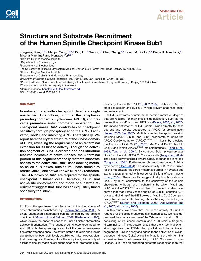
Molecular Cell
Article
Structure and Substrate Recruitmentof the Human Spindle Checkpoint Kinase Bub1Jungseog Kang,1,2,7 Maojun Yang,1,2,6,7 Bing Li,1,2 Wei Qi,1,2 Chao Zhang,4,5 Kevan M. Shokat,4,5 Diana R. Tomchick,3
Mischa Machius,3 and Hongtao Yu1,2,*1Howard Hughes Medical Institute2Department of Pharmacology3Department of Biochemistry
The University of Texas Southwestern Medical Center, 6001 Forest Park Road, Dallas, TX 75390, USA4Howard Hughes Medical Institute5Department of Cellular and Molecular PharmacologyUniversity of California at San Francisco, 600 16th Street, San Francisco, CA 94158, USA6Present address: Center for Structural Biology, Institute of Biomedicine, Tsinghua University, Beijing 100084, China7These authors contributed equally to this work
*Correspondence: [email protected] 10.1016/j.molcel.2008.09.017
SUMMARY
In mitosis, the spindle checkpoint detects a singleunattached kinetochore, inhibits the anaphase-promoting complex or cyclosome (APC/C), and pre-vents premature sister chromatid separation. Thecheckpoint kinase Bub1 contributes to checkpointsensitivity through phosphorylating the APC/C acti-vator, Cdc20, and inhibiting APC/C catalytically. Wereport here the crystal structure of the kinase domainof Bub1, revealing the requirement of an N-terminalextension for its kinase activity. Though the activa-tion segment of Bub1 is ordered and has structuralfeatures indicative of active kinases, the C-terminalportion of this segment sterically restricts substrateaccess to the active site. Bub1 uses docking motifs,so-called KEN boxes, outside its kinase domain torecruit Cdc20, one of two known KEN box receptors.The KEN boxes of Bub1 are required for the spindlecheckpoint in human cells. Therefore, its unusualactive-site conformation and mode of substrate re-cruitment suggest that Bub1 has an exquisitely tunedspecificity for Cdc20.
INTRODUCTION
In mitosis, the spindle microtubules attach to the kinetochores of
sister chromatids asynchronously (Tanaka and Desai, 2008). A
single unattached kinetochore can be sensed by the spindle
checkpoint (Musacchio and Salmon, 2007; Rieder et al., 1995),
which delays the onset of anaphase until all sister chromatids
achieve biorientation. The unattached kinetochores need to
emit diffusible checkpoint signals to block the premature separa-
tion of the attached ones. The nature of the diffusible checkpoint
signals has not been definitively established. It is, however, clear
that these signals ultimately block the ubiquitin ligase activity of
a large molecular machine called the anaphase-promoting com-
394 Molecular Cell 32, 394–405, November 7, 2008 ª2008 Elsevier
plex or cyclosome (APC/C) (Yu, 2002, 2007). Inhibition of APC/C
stabilizes securin and cyclin B, which prevent anaphase onset
and mitotic exit.
APC/C substrates contain small peptide motifs or degrons
that are required for their efficient ubiquitination, such as the
destruction box (D box) and KEN box (Peters, 2006; Yu, 2007).
The mitotic activator of APC/C, Cdc20, binds directly to these
degrons and recruits substrates to APC/C for ubiquitination
(Peters, 2006; Yu, 2007). Multiple spindle checkpoint proteins,
including Mad2, BubR1, and Bub1, collaborate to inhibit the
APC/C-Cdc20 complex (APC/CCdc20) in mitosis by blocking
the function of Cdc20 (Yu, 2007). Mad2 and BubR1 bind to
Cdc20 and inhibit APC/CCdc20 stoichiometrically (Fang et al.,
1998; Tang et al., 2001). By contrast, Bub1 phosphorylates
Cdc20 and inhibits APC/CCdc20 catalytically (Tang et al., 2004).
The kinase activity of Bub1 toward Cdc20 is enhanced in mitosis
(Tang et al., 2004). Furthermore, chromosome-bound Bub1 is
hyperactive (Chen, 2004). The kinase activity of Bub1 is required
for the nocodazole-triggered metaphase arrest in Xenopus egg
extracts supplemented with low concentrations of sperm nuclei
(Chen, 2004). These results suggest that phosphorylation of
Cdc20 by Bub1 contributes to the sensitivity of the spindle
checkpoint. Although the mechanisms by which Mad2 and
Bub1 inhibit APC/CCdc20 are unclear, two recent studies have
shown that Mad3 (the yeast ortholog of BubR1) contains KEN
boxes and binding of the KEN boxes of Mad3 to Cdc20 compet-
itively blocks substrate binding, thus inhibiting the activity of
APC/CCdc20 (Burton and Solomon, 2007; Diaz-Martinez and
Yu, 2007; King et al., 2007).
In this study, we show that the kinase activity of Bub1 is
required for the spindle checkpoint in human cells. We have de-
termined the crystal structure of the C-terminal domain of Bub1,
consisting of its kinase domain and a 60 residue fragment
N-terminal to it. The structure reveals that the N-terminal exten-
sion organizes the ATP-binding pocket and the activation
segment of Bub1 in a way analogous to the activation of cyclin-
dependent kinases (Cdks) by cyclins. Mutations of the N-terminal
extension disrupt the kinase activity of Bub1. Compared to other
kinases, Bub1 has an extended substrate recognition loop that
Inc.

Molecular Cell
Structure and Substrate Binding of the Kinase Bub1
Figure 1. Structure and Inhibitor of the
Extended Kinase Domain of Bub1
(A) Domain architecture of human Bub1. TPR,
tetratricopeptide repeat; GLEBS, Gle2-binding
sequence; KEN, lysine-glutamate-asparagine.
(B) Ribbon drawing of the crystal structure of the
extended kinase domain of Bub1. The N-terminal
extension is colored yellow. The substrate-binding
P+1 loop is shown in green, while the rest of the
activation segment is in blue. The catalytic loop
is shown in red. ATP is shown as sticks. The
Mg2+ ion is shown as a gray sphere. The N and
C termini are indicated. All structure figures were
generated with PyMOL (http://pymol.sourceforge.
net/).
(C) The ATP-binding site of Bub1. ATP is shown as
sticks. The extra pocket is indicated.
(D) The chemical structure of 2OH-BNPP1.
(E) Determination of the IC50 values of
2OH-BNPP1 against Aurora B, p38, Bub1C, and
Bub1.
blocks the active site of Bub1 and limits the access of nonspecific
substrates. Bub1 contains two KEN boxes in its central region,
which mediates its degradation by APC/CCdh1 in the G1 phase
of the cell cycle (Qi and Yu, 2007). We show here that the KEN
boxes of Bub1 are also required for Cdc20 binding, efficient
phosphorylation of Cdc20 by Bub1, and the spindle checkpoint.
Because Cdc20 is one of only two known KEN box receptors,
Bub1 has a well-tuned specificity toward Cdc20. These findings
provide insight into how multiple APC/C-inhibitory mechanisms
are coordinated during checkpoint signaling.
RESULTS AND DISCUSSION
Structure Determination of the Kinase Domain of Bub1Bub1 contains an N-terminal tetratricopeptide repeat (TPR)
domain that is required for its kinetochore localization and a
C-terminal serine/threonine (S/T) kinase domain (Figure 1A)
(Bharadwaj and Yu, 2004; Kiyomitsu et al., 2007). Phosphoryla-
tion of Cdc20 by Bub1 inhibits APC/CCdc20 (Tang et al., 2004).
Ectopic expression of a nonphosphorylatable mutant of Cdc20
compromises the spindle checkpoint in human cells (Tang
et al., 2004). These findings implicate a role for the kinase activity
of Bub1 in the spindle checkpoint. We thus formally tested
whether the Bub1 kinase activity was required for the spindle
checkpoint in human cells. We found that HeLa cells depleted
of Bub1 by RNA interference (RNAi) failed to undergo mitotic
arrest in the presence of nocodazole, as indicated by their lower
mitotic index and decreased levels of securin and phosphohi-
stone H3 (see Figure S1 available online). Ectopic expression
of RNAi-resistant wild-type (WT) Myc-Bub1 in Bub1 RNAi cells
restored the Bub1 protein to its endogenous levels and rescued
the mitotic arrest deficiency of these cells. By contrast, expres-
sion of a kinase-dead (KD) mutant of Myc-Bub1 was less effec-
tive in rescuing the defect of Bub1 RNAi cells. These results
Mol
indicate that the kinase activity of Bub1 is required for the spindle
checkpoint in human cells.
We next crystallized a C-terminal fragment of human Bub1
(referred to as Bub1C) containing residues 724–1085 in the pres-
ence of ATP and determined its crystal structure with the single-
wavelength anomalous diffraction (SAD) method using data to
a resolution of 2.5 A (Figure 1B and Table 1). Bub1C consists
of the kinase domain and an N-terminal extension (Figures 1A
and 1B). The kinase domain contains residues 784–1085 and
adopts a canonical kinase fold with two lobes. ATP and the
active site are located at the interface of the two lobes. The
N-terminal extension contains three b strands and an a helix
and wraps around the N lobe of the kinase domain.
Identification of a Chemical Inhibitor of Bub1Chemical inhibitors of mitotic regulators, such as monastrol and
blebbistatin, are powerful tools for studying rapid processes in
mitosis (Mayer et al., 1999; Straight et al., 2003). Highly specific in-
hibitorsofsinglemembers of large protein families, suchasprotein
kinases, are difficult to obtain, however. Mutations of large resi-
dues (so-called ‘‘gatekeeper’’ residues) in the ATP-binding pocket
of kinases toglycinesoralanines enlarge the active sites of kinases
and enable their inhibition by adenine analogs (Alaimo et al., 2001;
Bishop et al., 2000). Introduction of these mutant kinases into cells
that lack the corresponding WT kinases then sensitizes these cells
to the inhibition by these orthogonal inhibitors, allowing the dissec-
tion of the cellular functions of kinases (Alaimo et al., 2001). The
vertebrate Bub1 proteins contain a glycine (G866 in human
Bub1) at the C-terminal end of strand b5 (Figure S2), a position oc-
cupied by a large hydrophobic gatekeeper residue in most other
kinases. As a result, the active site of Bub1 has an unusual addi-
tional pocket that is not fully occupied by ATP, suggesting that
inhibitors designed to inhibit engineered kinaseswith glycine gate-
keepers might be good inhibitors of WT Bub1 (Figure 1C). We thus
ecular Cell 32, 394–405, November 7, 2008 ª2008 Elsevier Inc. 395
Molecular Cell
Structure and Substrate Binding of the Kinase Bub1
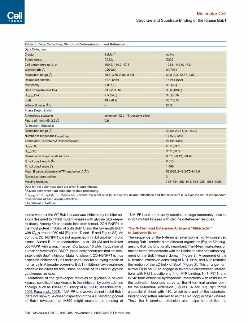
Table 1. Data Collection, Structure Determination, and Refinement
Data Collection
Crystal SeMeta native
Space group C2221 C2221
Cell parameters (a, b, c) 109.2, 150.2, 47.5 108.8, 147.6, 47.2
Wavelength (A) 0.97922 0.97954
Resolution range (A) 45.5–3.50 (3.56–3.50) 32.5–2.32 (2.37–2.32)
Unique reflections 5122 (276) 16,251 (848)
Multiplicity 7.0 (7.1) 3.0 (3.0)
Data completeness (%) 99.9 (100.0) 95.8 (100.0)
Rmerge (%)b 9.5 (34.5) 3.3 (53.3)
I/s(I) 19.4 (6.2) 50.7 (3.2)
Wilson B value (A2) – 55.3
Phase Determination
Anomalous scatterer selenium (12 of 15 possible sites)
Figure of merit (45–3.5 A) 0.6
Refinement Statistics
Resolution range (A) 32.50–2.32 (2.37–2.32)
Number of reflections Rwork/Rfree 14,976/1258
Atoms (non-H protein/ATP/ions/solvent) 2713/31/3/20
Rwork (%) 23.2 (29.1)
Rfree (%) 30.2 (38.8)
Overall anisotropic scale factorsc 0.57, �0.12, �0.46
Rmsd bond length (A) 0.013
Rmsd bond angle (�) 1.466
Mean B value (all/protein/ATP/ions/solvent) (A2) 55.0/55.2/41.3/79.4/45.0
Ramachandran outliers 3
Missing residues 726–734, 807–813, 933–938, 1081–1085
Data for the outermost shell are given in parentheses.a Bijvoet pairs were kept separate for data processing.b Rmerge = 100 ShSijIh, i � hIhij/ShSiIh, i, where the outer sum (h) is over the unique reflections and the inner sum (i) is over the set of independent
observations of each unique reflection.c As defined in Refmac.
tested whether the WT Bub1 kinase was inhibited by inhibitor an-
alogs designed to inhibit mutant kinases with glycine gatekeeper
residues. Among 49 candidate inhibitors tested, 2OH-BNPP1 is
the most potent inhibitor of both Bub1C and the full-length Bub1
with IC50s around 250 nM (Figures 1D and 1E and Figure S3). As
controls, 2OH-BNPP1 did not appreciably inhibit another mitotic
kinase, Aurora B, at concentrations up to 100 mM and inhibited
p38MAPK with a much larger IC50 (about 10 mM). Incubation of
human cells with 2OH-BNPP1 produces phenotypes that are con-
sistent with Bub1 inhibition (data not shown). 2OH-BNPP1 is thus
a specific inhibitor of Bub1 and a useful tool for studying mitosis of
human cells. A broader screen for Bub1 inhibitors may reveal more
selective inhibitors for this kinase because of its unusual glycine
gatekeeper residue.
Mutations of the gatekeeper residues to glycines in several
kinases sensitize these kinases to the inhibition by bulky adenine
analogs, such as 1NM-PP1 (Bishop et al., 2000; Jaeschke et al.,
2006; Papa et al., 2003). 1NM-PP1, however, did not inhibit Bub1
(data not shown). A closer inspection of the ATP-binding pocket
of Bub1 revealed that M850 might occlude the binding of
396 Molecular Cell 32, 394–405, November 7, 2008 ª2008 Elsevier I
1NM-PP1 and other bulky adenine analogs commonly used to
inhibit mutant kinases with glycine gatekeeper residues.
The N-Terminal Extension Acts as a ‘‘Minicyclin’’to Activate Bub1The sequence of the N-terminal extension is highly conserved
among Bub1 proteins from different organisms (Figure S2), sug-
gesting that it is functionally important. The N-terminal extension
makes extensive contacts with the N lobe and the activation seg-
ment of the Bub1 kinase domain (Figure 2). A segment of the
N-terminal extension consisting of Nb1, NaA, and Nb2 restrains
the motion of the aC helix of Bub1 (Figure 2). This arrangement
allows E830 on aC to engage in favorable electrostatic interac-
tions with K821, positioning it for ATP binding. Nb1, P741, and
W742 form extensive hydrophobic interactions with residues of
the activation loop and serve as the N-terminal anchor point
for the N-terminal extension (Figures 3A and 3B). Nb1 forms
a parallel b sheet with b10, which is a part of the substrate-
binding loop (often referred to as the P+1 loop) in other kinases.
Thus, the N-terminal extension also helps to stabilize the
nc.

Molecular Cell
Structure and Substrate Binding of the Kinase Bub1
Figure 2. The N-Terminal Extension Activates Bub1 with a Cyclin-like Mechanism
Ribbon diagrams of Bub1C, Cdk2, and Cdk2-Cyclin A. The N and C termini are indicated. The color schemes are as described in Figure 1B except that aC is
colored magenta. The ATP-binding lysine residues and the conserved glutamate residues in aC are shown as sticks. Cyclin A is shown in yellow.
conformation of the activation segment of Bub1. These functions
of the N-terminal extension of Bub1 are reminiscent of the ways
in which cyclins activate Cdks (Figure 2) (Pavletich, 1999). First,
cyclins restrain the movement of aC and enable ATP binding by
facilitating the interaction between a glutamate on aC and the
conserved ATP-binding lysine. Second, cyclins contact the
activation loop of Cdks and stabilize its active conformation.
Both activating mechanisms of cyclins are likely shared by the
N-terminal extension of Bub1 in promoting the kinase activity
of Bub1.
We next used site-directed mutagenesis to study the function
of the N-terminal extension of Bub1. As discussed above, W742
and residues in Nb1 form extensive hydrophobic interactions
with the activation segment of Bub1. Consistently, Bub1C
W742A was inactive in phosphorylating an N-terminal fragment
of Cdc20 (Cdc20N) (data not shown). We then tested whether
the same mutation also affected the activity of full-length
Bub1. Myc-Bub1 WT, KD, and W742A were immunoprecipitated
from lysates of mitotic HeLa cells transfected with plasmids
encoding these proteins and subjected to kinase assays. Bub1
W742A was inactive in phosphorylating Cdc20N (Figure 3C).
Mutations that are designed to disrupt Nb1, such as I737P/
V738P, also diminished the kinase activity of Bub1. Our results
indicate that the N-terminal extension is required for the kinase
activity of full-length Bub1.
Finally, several conserved C-terminal basic residues of Bub1
are near the ATP-binding pocket (Figure S2). Similar basic resi-
dues in TAO2 are required for its kinase activity (Zhou et al.,
2004). Deletion of the C-terminal five residues of Bub1 (Bub1
DCt5) only slightly reduced its kinase activity toward Cdc20N
(Figure 3C), indicating that these residues do not play a major
role in ATP binding of Bub1.
The Unusual Conformation of the ActivationSegment of Bub1Kinases in their inactive states typically have disordered activa-
tion segments, whereas active kinases generally have ordered
ones (Nolen et al., 2004). In many kinases, the transition be-
Mo
tween disordered and ordered conformations of the activation
segment is regulated by phosphorylation of the activation
loop, binding of cofactors, or both. Our crystal structure of the
kinase domain of Bub1 has features characteristic of active
kinases. First, the activation segment of Bub1 is ordered in our
crystal structure (Figure 3A). The N-terminal end of the relatively
short activation loop of Bub1 folds into a 310 helix. Several
conserved residues in the activation loop engage in extensive
hydrophobic interactions with residues in the N-terminal exten-
sion. These elaborate hydrophobic interactions maintain the
integrity of the active site, as disruption of these interactions
by the W742A mutation inactivates Bub1 (Figure 3C). Second,
the pairing between b6 preceding the catalytic loop and b9 at
the start of the activation segment is a hallmark of active kinases
(Figure 4A) (Nolen et al., 2004). This pairing is observed in Bub1
(Figure 4B).
On the other hand, in our structure, Bub1 has an extended P+1
loop that adopts a conformation not found in active kinases. A
characteristic GT dipeptide motif marks the start of the P+1
loop in many S/T kinases, including Bub1 and PKA (Figure S2)
(Nolen et al., 2004). In the active state of PKA and other S/T
kinases, the hydroxyl group of the threonine in this GT motif
(T201 in PKA) forms a hydrogen bond with the catalytic aspartate
(D166 in PKA). The C-terminal part of the P+1 loop forms a 310
helix. These two structural features allow much of the P+1 loop
to lie parallel to the catalytic loop and to form the foundation
for substrate binding (Figures 4A and 4C). In our structure of
Bub1, T960 of the GT motif is located at the start of b10, which
pairs with Nb1 (Figure 4B). The pairing between b10 and Nb1 pla-
ces T960 about 19 A away from the catalytic aspartate residue,
D917. The C-terminal half of the P+1 loop in Bub1 adopts a
hairpin-like structure that covers the catalytic loop and restricts
substrate access to ATP (Figure 4D). Therefore, the P+1 loop
of Bub1 adopts a conformation that is suboptimal for substrate
binding. Significant structural rearrangement of the C-terminal
half of the P+1 loop (residues 965–972) has to occur during
catalysis to allow efficient phosphorylation of substrates by
Bub1.
lecular Cell 32, 394–405, November 7, 2008 ª2008 Elsevier Inc. 397

Molecular Cell
Structure and Substrate Binding of the Kinase Bub1
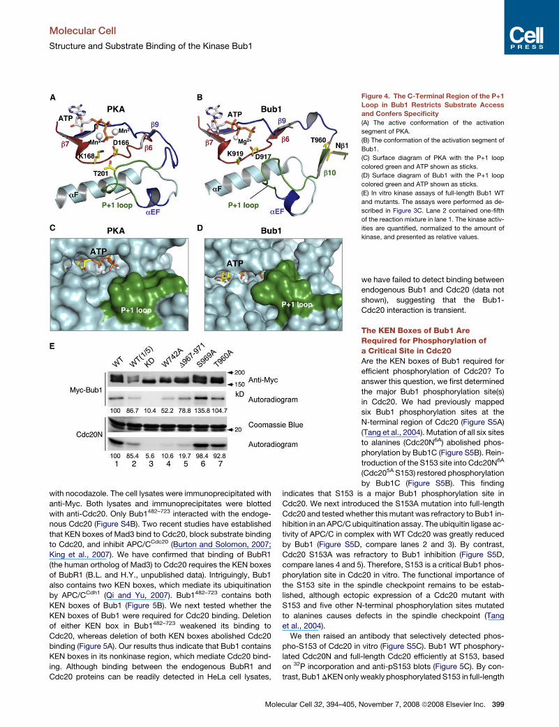
We next tested whether this part of the P+1 loop in Bub1 was
required for efficient phosphorylation of Cdc20N. Deletion of
residues 967–971 (Bub1 D967–971) reduced the kinase activity
of Bub1 toward Cdc20N about 5-fold (Figure 4E, compare lanes
1 and 5), whereas the same mutant retained about 80% of its au-
tokinase activity. Similar results were obtained with mutations of
V974, which is located in helix aEF and just C-terminal to the P+1
loop (data not shown). Therefore, mutations in the C-terminal
portion of the P+1 loop in Bub1 affect its kinase activity toward
different substrates to varying degrees, suggesting that this re-
gion might be directly involved in substrate binding. We cannot,
however, rule out the possibility that perturbation of the P+1 loop
disrupts the structural integrity of the active site of Bub1, thus
indirectly affecting substrate binding.
Taken together, Bub1 in our crystal structure is likely in the
active state, but it is not optimized for substrate binding. How
can the P+1 loop of Bub1 adopt a conformation suitable for sub-
strate binding? We envision two possible mechanisms: (1) preor-
ganization and (2) induced fit. In the preorganization mechanism,
posttranslational modifications of Bub1 or binding of other
proteins preorganize the P+1 loop into an optimal conformation
for substrate binding. In the induced-fit mechanism, binding of
substrate induces a conformational change in the P+1 loop.
The vertebrate Bub1 proteins contain only three conserved
phosphorylatable residues in the activation segment: T960,
T968, and S969 (Figure S2). Mutations of T960 and S969 to ala-
nines did not affect the kinase activity of Bub1 toward Cdc20N
Figure 3. The N-Terminal Extension Is
Required for the Kinase Activity of Bub1
(A) The N-terminal anchor of the N-terminal exten-
sion. The color schemes are as described in
Figure 2. W742 and its neighboring residues are
shown in sticks and labeled.
(B) Ribbon diagram of the N-terminal extension
superposed with the surface of the rest of the
protein. W742 is shown in sticks.
(C) In vitro kinase assays of full-length Bub1 WT
and mutants. Myc-Bub1 WT and the indicated
mutants were expressed in mitotic HeLa cells,
immunoprecipitated with anti-Myc, and assayed
for their autokinase activity and kinase activity
using the N-terminal fragment of Cdc20 (Cdc20N)
as a substrate. Anti-Myc blot of the Myc-Bub1 pro-
teins and Coomassie blue staining of the Cdc20N
protein are also shown. The kinase activities are
quantified, normalized to the amount of kinase,
and presented as relative values directly below
each lane.
(Figure 4E). By contrast, the T968A muta-
tion greatly diminished the kinase activity
of Bub1 toward Cdc20N (data not
shown). Using mass spectrometry analy-
sis, we have mapped multiple phosphor-
ylation sites on the endogenous Bub1
protein isolated from mitotic HeLa cell ly-
sates (J.K. and H.Y., unpublished data).
T968 was not one of the phosphorylation
acceptor residues identified in Bub1. In fact, none of the phos-
phorylation sites are located in the kinase domain. It is thus un-
clear which mechanism Bub1 uses to organize its P+1 loop.
Regardless of the mechanism, the unusual conformation of the
P+1 loop in Bub1 is expected to restrict the access of nonspe-
cific substrates to its active site and enhance the substrate
specificity of Bub1.
The KEN Boxes of Bub1 Bind to Cdc20The suboptimal conformation of the P+1 loop in Bub1 prompted
us to search for additional mechanisms that might promote
substrate binding to Bub1. Cdc20 is the only known physiologi-
cally relevant substrate of Bub1 (Tang et al., 2004). To examine
whether the N-terminal nonkinase region of Bub1 enhanced
phosphorylation of Cdc20, we performed kinase assays with
Bub1C or the full-length Bub1 as the kinase and Cdc20N or
full-length Cdc20 as the substrate. Full-length recombinant Bub1
phosphorylated both full-length Cdc20 and Cdc20N efficiently
(Figure 5A). In contrast, Bub1C only phosphorylated Cdc20N,
but not full-length Cdc20, suggesting that the N-terminal region
of Bub1 was required for the efficient phosphorylation of
full-length Cdc20. This finding further suggests that the Bub1-
phosphorylation site(s) is masked in full-length Cdc20 but is
accessible in Cdc20N.
We next constructed plasmids encoding three Myc-tagged
Bub1 fragments that spanned its nonkinase region (Figure S4A).
HeLa cells were transfected with these plasmids and treated
398 Molecular Cell 32, 394–405, November 7, 2008 ª2008 Elsevier Inc.
Molecular Cell
Structure and Substrate Binding of the Kinase Bub1
with nocodazole. The cell lysates were immunoprecipitated with
anti-Myc. Both lysates and immunoprecipitates were blotted
with anti-Cdc20. Only Bub1482–723 interacted with the endoge-
nous Cdc20 (Figure S4B). Two recent studies have established
that KEN boxes of Mad3 bind to Cdc20, block substrate binding
to Cdc20, and inhibit APC/CCdc20 (Burton and Solomon, 2007;
King et al., 2007). We have confirmed that binding of BubR1
(the human ortholog of Mad3) to Cdc20 requires the KEN boxes
of BubR1 (B.L. and H.Y., unpublished data). Intriguingly, Bub1
also contains two KEN boxes, which mediate its ubiquitination
by APC/CCdh1 (Qi and Yu, 2007). Bub1482–723 contains both
KEN boxes of Bub1 (Figure 5B). We next tested whether the
KEN boxes of Bub1 were required for Cdc20 binding. Deletion
of either KEN box in Bub1482–723 weakened its binding to
Cdc20, whereas deletion of both KEN boxes abolished Cdc20
binding (Figure 5A). Our results thus indicate that Bub1 contains
KEN boxes in its nonkinase region, which mediate Cdc20 bind-
ing. Although binding between the endogenous BubR1 and
Cdc20 proteins can be readily detected in HeLa cell lysates,
Figure 4. The C-Terminal Region of the P+1
Loop in Bub1 Restricts Substrate Access
and Confers Specificity
(A) The active conformation of the activation
segment of PKA.
(B) The conformation of the activation segment of
Bub1.
(C) Surface diagram of PKA with the P+1 loop
colored green and ATP shown as sticks.
(D) Surface diagram of Bub1 with the P+1 loop
colored green and ATP shown as sticks.
(E) In vitro kinase assays of full-length Bub1 WT
and mutants. The assays were performed as de-
scribed in Figure 3C. Lane 2 contained one-fifth
of the reaction mixture in lane 1. The kinase activ-
ities are quantified, normalized to the amount of
kinase, and presented as relative values.
we have failed to detect binding between
endogenous Bub1 and Cdc20 (data not
shown), suggesting that the Bub1-
Cdc20 interaction is transient.
The KEN Boxes of Bub1 AreRequired for Phosphorylation ofa Critical Site in Cdc20Are the KEN boxes of Bub1 required for
efficient phosphorylation of Cdc20? To
answer this question, we first determined
the major Bub1 phosphorylation site(s)
in Cdc20. We had previously mapped
six Bub1 phosphorylation sites at the
N-terminal region of Cdc20 (Figure S5A)
(Tang et al., 2004). Mutation of all six sites
to alanines (Cdc20N6A) abolished phos-
phorylation by Bub1C (Figure S5B). Rein-
troduction of the S153 site into Cdc20N6A
(Cdc205A S153) restored phosphorylation
by Bub1C (Figure S5B). This finding
indicates that S153 is a major Bub1 phosphorylation site in
Cdc20. We next introduced the S153A mutation into full-length
Cdc20 and tested whether this mutant was refractory to Bub1 in-
hibition in an APC/C ubiquitination assay. The ubiquitin ligase ac-
tivity of APC/C in complex with WT Cdc20 was greatly reduced
by Bub1 (Figure S5D, compare lanes 2 and 3). By contrast,
Cdc20 S153A was refractory to Bub1 inhibition (Figure S5D,
compare lanes 4 and 5). Therefore, S153 is a critical Bub1 phos-
phorylation site in Cdc20 in vitro. The functional importance of
the S153 site in the spindle checkpoint remains to be estab-
lished, although ectopic expression of a Cdc20 mutant with
S153 and five other N-terminal phosphorylation sites mutated
to alanines causes defects in the spindle checkpoint (Tang
et al., 2004).
We then raised an antibody that selectively detected phos-
pho-S153 of Cdc20 in vitro (Figure S5C). Bub1 WT phosphory-
lated Cdc20N and full-length Cdc20 efficiently at S153, based
on 32P incorporation and anti-pS153 blots (Figure 5C). By con-
trast, Bub1 DKEN only weakly phosphorylated S153 in full-length
Molecular Cell 32, 394–405, November 7, 2008 ª2008 Elsevier Inc. 399
Molecular Cell
Structure and Substrate Binding of the Kinase Bub1
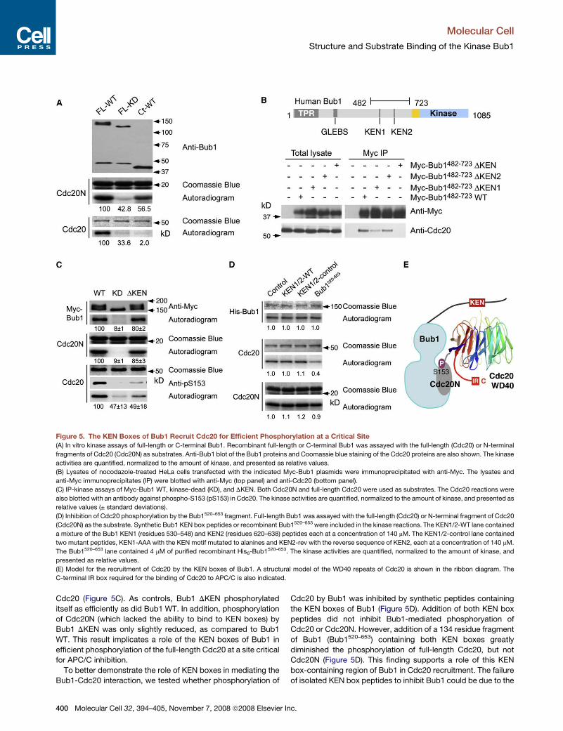
Figure 5. The KEN Boxes of Bub1 Recruit Cdc20 for Efficient Phosphorylation at a Critical Site
(A) In vitro kinase assays of full-length or C-terminal Bub1. Recombinant full-length or C-terminal Bub1 was assayed with the full-length (Cdc20) or N-terminal
fragments of Cdc20 (Cdc20N) as substrates. Anti-Bub1 blot of the Bub1 proteins and Coomassie blue staining of the Cdc20 proteins are also shown. The kinase
activities are quantified, normalized to the amount of kinase, and presented as relative values.
(B) Lysates of nocodazole-treated HeLa cells transfected with the indicated Myc-Bub1 plasmids were immunoprecipitated with anti-Myc. The lysates and
anti-Myc immunoprecipitates (IP) were blotted with anti-Myc (top panel) and anti-Cdc20 (bottom panel).
(C) IP-kinase assays of Myc-Bub1 WT, kinase-dead (KD), and DKEN. Both Cdc20N and full-length Cdc20 were used as substrates. The Cdc20 reactions were
also blotted with an antibody against phospho-S153 (pS153) in Cdc20. The kinase activities are quantified, normalized to the amount of kinase, and presented as
relative values (± standard deviations).
(D) Inhibition of Cdc20 phosphorylation by the Bub1520–653 fragment. Full-length Bub1 was assayed with the full-length (Cdc20) or N-terminal fragment of Cdc20
(Cdc20N) as the substrate. Synthetic Bub1 KEN box peptides or recombinant Bub1520–653 were included in the kinase reactions. The KEN1/2-WT lane contained
a mixture of the Bub1 KEN1 (residues 530–548) and KEN2 (residues 620–638) peptides each at a concentration of 140 mM. The KEN1/2-control lane contained
two mutant peptides, KEN1-AAA with the KEN motif mutated to alanines and KEN2-rev with the reverse sequence of KEN2, each at a concentration of 140 mM.
The Bub1520–653 lane contained 4 mM of purified recombinant His6-Bub1520–653. The kinase activities are quantified, normalized to the amount of kinase, and
presented as relative values.
(E) Model for the recruitment of Cdc20 by the KEN boxes of Bub1. A structural model of the WD40 repeats of Cdc20 is shown in the ribbon diagram. The
C-terminal IR box required for the binding of Cdc20 to APC/C is also indicated.
Cdc20 (Figure 5C). As controls, Bub1 DKEN phosphorylated
itself as efficiently as did Bub1 WT. In addition, phosphorylation
of Cdc20N (which lacked the ability to bind to KEN boxes) by
Bub1 DKEN was only slightly reduced, as compared to Bub1
WT. This result implicates a role of the KEN boxes of Bub1 in
efficient phosphorylation of the full-length Cdc20 at a site critical
for APC/C inhibition.
To better demonstrate the role of KEN boxes in mediating the
Bub1-Cdc20 interaction, we tested whether phosphorylation of
400 Molecular Cell 32, 394–405, November 7, 2008 ª2008 Elsevier
Cdc20 by Bub1 was inhibited by synthetic peptides containing
the KEN boxes of Bub1 (Figure 5D). Addition of both KEN box
peptides did not inhibit Bub1-mediated phosphoryation of
Cdc20 or Cdc20N. However, addition of a 134 residue fragment
of Bub1 (Bub1520–653) containing both KEN boxes greatly
diminished the phosphorylation of full-length Cdc20, but not
Cdc20N (Figure 5D). This finding supports a role of this KEN
box-containing region of Bub1 in Cdc20 recruitment. The failure
of isolated KEN box peptides to inhibit Bub1 could be due to the
Inc.
Molecular Cell
Structure and Substrate Binding of the Kinase Bub1
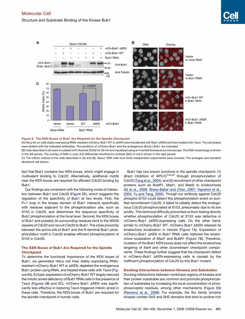
Figure 6. The KEN Boxes of Bub1 Are Required for the Spindle Checkpoint(A) HeLa tet-on cells stably expressing RNAi-resistant mCherry-Bub1 WT or DKEN were transfected with Bub1 siRNA and then treated with Taxol. The cell lysates
were blotted with the indicated antibodies. The positions of mCherry-Bub1 and the endogenous (Endo.) Bub1 are indicated.
(B) Cells described in (A) were incubated with Hoechst 33342 for 30 min and visualized using an inverted fluorescence microscope. The DNA morphology is shown
in the left panels. The overlay of DNA in cyan and differential interference contrast (DIC) in red is shown in the right panels.
(C) The mitotic indices of the cells described in (A) and (B). About 1000 cells from three independent experiments were counted. The averages and standard
deviations are shown.
fact that Bub1 contains two KEN boxes, which might engage in
multivalent binding to Cdc20. Alternatively, additional motifs
near the KEN boxes are required for efficient Cdc20 binding by
Bub1.
Our findings are consistent with the following mode of interac-
tion between Bub1 and Cdc20 (Figure 5E), which suggests the
regulation of the specificity of Bub1 at two levels. First, the
P+1 loop in the kinase domain of Bub1 interacts specifically
with residues adjacent to the phosphorylation site, such as
S153 in Cdc20, and determines the sequence specificity of
Bub1 phosphorylation at the local level. Second, the KEN boxes
of Bub1 and possibly its surrounding residues bind to the WD40
repeats of Cdc20 and recruit Cdc20 to Bub1. The close proximity
between the active site of Bub1 and the N-terminal Bub1 phos-
phorylation motif in Cdc20 enables efficient phosphorylation of
S153 in Cdc20.
The KEN Boxes of Bub1 Are Required for the SpindleCheckpointTo determine the functional importance of the KEN boxes of
Bub1, we generated HeLa cell lines stably expressing RNAi-
resistant mCherry-Bub1 WT or DKEN, depleted the endogenous
Bub1 protein using RNAi, and treated these cells with Taxol (Fig-
ure 6A). Ectopic expression of mCherry-Bub1 WT largely rescued
the mitotic arrest deficiency of Bub1 RNAi cells in the presence of
Taxol (Figures 6B and 6C). mCherry-Bub1 DKEN was signifi-
cantly less effective in restoring Taxol-triggered mitotic arrest in
these cells. Therefore, the KEN boxes of Bub1 are required for
the spindle checkpoint in human cells.
Mol
Bub1 has two known functions in the spindle checkpoint: (1)
direct inhibition of APC/CCdc20 through phosphorylation of
Cdc20 (Tang et al., 2004); and (2) recruitment of other checkpoint
proteins such as BubR1, Mad1, and Mad2 to kinetochores
(Qi et al., 2006; Sharp-Baker and Chen, 2001; Vigneron et al.,
2004; Yu and Tang, 2005). Though our antibody against Cdc20
phospho-S153 could detect this phosphorylation event on puri-
fied recombinant Cdc20, it failed to reliably detect the endoge-
nous Cdc20 phosphorylated at S153, presumably due to its low
avidity. This technical difficulty prevented us from testing directly
whether phosphorylation of Cdc20 at S153 was defective in
mCherry-Bub1 DKEN-expressing cells. On the other hand,
similar to mCherry-Bub1 WT, mCherry-Bub1 DKEN retained its
kinetochore localization in mitosis (Figure 7A). Expression of
mCherry-Bub1 DKEN in Bub1 RNAi cells restored the kineto-
chore localization of Mad1 and BubR1 (Figure 7B). Therefore,
mutation of the Bub1 KEN boxes does not affect the kinetochore
targeting of itself and other downstream checkpoint compo-
nents. These findings further suggest that the checkpoint defect
in mCherry-Bub1 DKEN-expressing cells is caused by the
inefficient phosphorylation of Cdc20 by this Bub1 mutant.
Docking Interactions between Kinases and SubstratesDocking interactions between nonkinase regions of kinases and
their protein substrates are common and promote phosphoryla-
tion of substrates by increasing the local concentration of phos-
phoacceptor residues, among other mechanisms (Figure S6)
(Remenyi et al., 2006). For example, the Src family tyrosine
kinases contain SH3 and SH2 domains that bind to proline-rich
ecular Cell 32, 394–405, November 7, 2008 ª2008 Elsevier Inc. 401

Molecular Cell
Structure and Substrate Binding of the Kinase Bub1
Figure 7. The KEN Boxes of Bub1 Are Not Required for the Kinetochore Localization of BubR1 and Mad1
(A) HeLa tet-on cells stably expressing mCherry-Bub1 WT or DKEN were fixed and stained with CREST (green in overlay) and DAPI (blue in overlay). The scale bars
indicate 5 mm.
(B) HeLa tet-on cells stably expressing RNAi-resistant mCherry-Bub1 WT or DKEN were transfected with Bub1 siRNA, fixed, and stained with anti-Mad1 (three left
panels, red in overlay) or anti-BubR1 (three right panels, red in overlay). The cells were costained with CREST (green in overlay) and DAPI (blue in overlay). The
scale bars indicate 5 mm.
and phosphotyrosine-containing docking motifs on certain
substrates, respectively (Lim, 2002). The polo-like kinase (Plk1)
uses its polo box domain (PBD) to recognize phospho-S/T motifs
that are created by priming kinases (Lowery et al., 2005). This in-
teraction promotes efficient phosphorylation of Plk1 substrates
at other sites. We have now shown that efficient phosphorylation
of Cdc20 by Bub1 requires a docking interaction between Cdc20
and the KEN boxes of Bub1. Therefore, the Cdc20-KEN box
interaction likely promotes Cdc20 phosphorylation by Bub1, in
ways analogous to the binding between SH3 and proline-rich
peptides.
There is an important difference between the docking interac-
tion involving Bub1 and Cdc20 and the docking interactions of
other kinases (Figure S6). In Src, Plk1, and numerous other
cases, the kinases contain the receptors for the docking motifs,
which are located in the substrates. In the case of Bub1-Cdc20
interaction, the kinase Bub1 contains the docking motifs (KEN
402 Molecular Cell 32, 394–405, November 7, 2008 ª2008 Elsevier I
boxes), whereas the substrate Cdc20 is the receptor for KEN
boxes. Obviously, both types of arrangements are functionally
equivalent in promoting substrate phosphorylation. The unusual
configuration of the Bub1-Cdc20 docking event does, however,
provide important clues about the substrate specificity of Bub1.
To engage the KEN boxes of Bub1, protein substrates of Bub1
need to contain domains that can serve as KEN box receptors.
While many proteins may contain short peptide motifs that can
bind to SH3, PBD, and other docking motif receptors, very few
proteins are expected to contain receptors for KEN boxes. In
fact, only Cdc20 and Cdh1 are known KEN box receptors (Yu,
2007). Cdh1 is not efficiently phosphorylated by Bub1 (Tang
et al., 2004), presumably due to the lack of an optimal phos-
phoacceptor residue in Cdh1 that can interact with the P+1
loop of the kinase domain of Bub1. Although we cannot rule
out the possibility that Bub1 uses different mechanisms to pro-
mote phosphorylation of other substrates, it is clear that Bub1
nc.

Molecular Cell
Structure and Substrate Binding of the Kinase Bub1
is exquisitely tuned to phosphorylate Cdc20 through the docking
interaction between Cdc20 and the KEN boxes in Bub1.
Implications for Spindle Checkpoint SignalingMultiple checkpoint mechanisms inhibit APC/CCdc20, including
binding of Mad2 to Cdc20, binding of Mad3/BubR1 to Cdc20, and
phosphorylation of Cdc20 by Bub1 (Yu, 2007). Although each of
the three proteins on its own is able to inhibit APC/CCdc20 in vitro,
Mad2, BubR1, and Bub1 must cooperate with each other in
human cells because depletion of each protein alone by RNAi
causes checkpoint defects (Meraldi et al., 2004; Tang et al.,
2004). The spatiotemporal coordination of these APC/C-inhibi-
tory checkpoint mechanisms in vivo remains unclear.
Recently, it has been shown that Mad3 contains KEN boxes,
which bind to Cdc20 and inhibit APC/CCdc20 through competing
with substrate binding (Burton and Solomon, 2007; King et al.,
2007). We have shown herein that Bub1 uses its KEN boxes to
recruit Cdc20 for efficient phosphorylation. This finding suggests
that binding of BubR1 to Cdc20 and phosphorylation of Cdc20
by Bub1 are competing events and need to be coordinated in
cells. Two possible ways of coordination can be envisioned. In
one way, phosphorylation of Cdc20 by Bub1 may need to occur
prior to BubR1 binding to Cdc20, because BubR1 likely has
a higher affinity toward Cdc20. Bub1-mediated phosphorylation
of Cdc20 may then promote binding of BubR1 and Mad2 to
Cdc20. Alternatively, because Bub1 can more efficiently phos-
phorylate Cdc20 that is not bound to BubR1, Bub1 and BubR1
may inhibit different pools of Cdc20. Experiments aimed at
testing these hypotheses will advance our understanding of
the spindle checkpoint.
ConclusionIn this study, we have determined the structure of the extended
kinase domain of Bub1, identified a chemical inhibitor of Bub1,
and uncovered a role of the KEN boxes of Bub1 as docking
motifs for Cdc20. Our combined structural, biochemical, and
functional analyses indicate that Bub1 is a specific kinase opti-
mized for Cdc20 phosphoryation. The expected competition
between Bub1 and BubR1 in Cdc20 recognition further suggests
testable models for the spatiotemporal coordination of multiple
APC/C-inhibitory mechanisms during spindle checkpoint signal-
ing in living cells.
EXPERIMENTAL PROCEDURES
Protein Expression and Purification
Recombinant baculoviruses encoding an N-terminal His6-tagged C-terminal
fragment of human Bub1 (Bub1C) containing residues 724–1085 were
constructed using the Bac-to-Bac system (Invitrogen) according to manufac-
turer’s protocols. Sf9 cells were infected with the Bub1C baculovirus and
harvested at about 50 hr postinfection. Cells were resuspended in lysis buffer
(25 mM phosphate [pH 7.4], 150 mM KCl, 20 mM imidazole, 0.1% [v/v] Triton
X-100, 1 mM PMSF, 5 mM DTT, and a protease inhibitor cocktail). After soni-
cation and centrifugation, the supernatant was filtered through 0.45 mm filters
and passed through a 5 ml HisTrap Chelating HP column (GE Healthcare).
After washing with the wash buffer (25 mM phosphate [pH 7.4], 150 mM
KCl, 20 mM imidazole, 1 mM PMSF, and 5 mM DTT), the bound proteins
were eluted with a linear gradient (0%–70%) of the elution buffer (25 mM phos-
phate [pH 7.4], 300 mM KCl, 300 mM imidazole, 1 mM PMSF, 5 mM DTT). The
His6-Bub1C-containing fractions were pooled, desalted, and purified using ion
Mole
exchange chromatography (resource S 5/5 and resource Q 5/5, GE Health-
care). His6-Bub1C was incubated with tobacco etch virus (TEV) protease
and passed through a HisTrap Chelating HP column (GE Healthcare). Bub1C
was further purified by a gel filtration column (Superdex 200, GE Healthcare),
concentrated to 15 mg/ml in the purification buffer (50 mM Tris [pH 7.7],
100 mM KCl, 5 mM MgCl2, 5 mM DTT, and 1 mM PMSF), flash frozen in liquid
nitrogen, and stored at �80�C for crystallization or kinase assays.
The selenomethionine variant of Bub1C protein was expressed in Sf9 cells
as described (Zhou et al., 2004). Briefly, Sf9 cells cultured in the EX-420
medium (JRH Bioscience) were infected with the Bub1C baculovirus. The cells
were harvested at 20 hr postinfection, washed twice with the EX-420 medium
without L-methionine, and resuspended into this medium. The cells were incu-
bated for 6 hr to deplete the remaining L-methionine, harvested again, and
resuspended into the EX-420 medium supplied with 100 mg/l selenomethio-
nine. After an additional 24–30 hr, the cells were harvested, and the selenome-
thionine Bub1C protein was purified as described above.
Crystallization, Data Collection, and Structure Determination
The Bub1C protein was thawed in ice-cold water, and ATP was added to a final
concentration of 10 mM. Crystals were grown at 20�C by the vapor diffusion
method in sitting drop mode by mixing 1.5 ml protein with 1.5 ml crystallization
solution (20% [w/v] polyethylene glycol 3350, 0.1 M sodium formate [pH 6.29],
and 25 mM DTT) and equilibrating against 200 ml of reservoir solution. Large
single crystals were obtained by repeated seeding. The crystals were cryopro-
tected in reservoir solution supplemented with 18% (v/v) glycerol and then
flash cooled in liquid propane. The crystals exhibit the symmetry of space
group C2221 with cell dimensions of a = 109 A, b = 148 A, and c = 47 A and
contained one molecule of Bub1C in the asymmetric unit with a solvent con-
tent of 46%. The selenomethionine-derivatized Bub1C crystals were obtained
in the same way and had similar cell parameters.
Diffraction data were collected at beamline 19-ID (SBC-CAT) of the Advanced
Photon Source (Argonne National Laboratory, Argonne, IL, USA) and processed
with HKL2000 (Otwinowski and Minor, 1997). Native and selenomethionine-
derivatized crystals diffracted to a minimum Bragg spacing of about 2.3 A
and about 3.5 A, respectively (Table 1). All crystals showed significant aniso-
tropic diffraction. SAD data on the selenomethionine Bub1C crystals were
collected at 3.2 A with weak anomalous signal present to about 3.5 A.
Phases for the selenomethionine variant were obtained from a single anom-
alous dispersion (SAD) experiment. Using data to 3.5 A, 12 of the 15 expected
selenomethionine sites were identified using SHELX (Schneider and Sheldrick,
2002; Sheldrick, 2002) and refined using SHARP (Bricogne et al., 2003). The re-
sulting electron density map clearly showed several a helices, which allowed
placing the similar structures of the kinase domains of c-Jun, CK2, and twitchin
(PDB ID codes 1jnk, 1 daw, and 1kob, respectively). Using these structures, the
phases from the selenium-SAD experiment, and the known positions of 12 se-
lenium atoms as guides, a model was constructed manually in an iterative fash-
ion using the program XtalView and automatically rebuilt and refined with the
programs ARP/wARP and Refmac5 of the CCP4 package (Consortium, 1994;
Murshudov et al., 1997; Perrakis et al., 1999). Completion of the model and final
refinement were then carried out using the native data to 2.32 A. The final model
contains residues 735–806, 814–931, and 939–1080 of Bub1C; one molecule
Mg2+-ATP; two chloride ions; and 20 water molecules. After final refinement,
the model had an Rwork of 23.2% and an Rfree of 30.2%. The model has good
geometry, except for three residues that are outliers in a Ramachandran plot
as defined by MolProbity (Davis et al., 2007). One of them, D946, is a direct
ligand of the bound ATP. Its electron density is well defined. The other two res-
idues, P776 and T778, are located in a surface loop with weak electron density.
Cell Culture and Transfection
HeLa tet-on cells were cultured in DMEM (Invitrogen) supplemented with fetal
bovine serum. The Bub1 DKEN1 (with K535, E536, and N537 mutated to
alanines), Bub1 DKEN2 (with K625, E626, and N627 mutated to alanines),
and Bub1 DKEN (with both KEN motifs mutated to alanines) mutants were
constructed using the QuikChange mutagenesis kit (QIAGEN). Plasmid trans-
fections were performed using the Effectene reagent (QIAGEN). To establish
stable cell lines, HeLa tet-on cells were transfected with the pIRES-
mCherry-Bub1 WT or DKEN plasmids and incubated with 0.5 mg/ml
cular Cell 32, 394–405, November 7, 2008 ª2008 Elsevier Inc. 403

Molecular Cell
Structure and Substrate Binding of the Kinase Bub1
puromycin. Surviving clones that expressed mCherry-Bub1 WT or DKEN at
similar levels were transfected with Bub1 siRNA for 24 hr and treated with
100 nM Taxol for 18 hr. After a 30 min incubation with Hoechst 33342, the cells
were analyzed using an inverted fluorescence microscope. Round cells with
condensed chromosomes were counted as mitotic cells.
Kinase Assays
Todetermine the IC50 valuesof2OH-BNPP1forBub1, Bub1C,AuroraB,andp38,
about 0.1 mg of various kinases were incubated with 2 mg of substrates for 30 min
atRT in25ml ofkinasebuffer I (50mMTris [pH 7.5], 0.2M NaCl,1mMDTT, 0.1mM
ATP, 0.1 mCi/ml g-32P-ATP) containing various concentrations of 2OH-BNPP1.
Reaction mixtures were quenched with SDS sample buffer, separated on
SDS-PAGE, and analyzed using a phosphoimager. The immunoprecipitation
(IP) kinase assays were performed as described previously (Kang et al., 2007).
Immunofluorescence
For Bub1 localization, HeLa tet-on cells stably expressing mCherry-Bub1 WT
or DKEN were grown in 6-well plates, treated with Taxol for 18 hr, and
sedimented on slides using Shandon cytospin 4 (Thermo electron). The cells
were fixed in 4% (w/v) paraformaldehyde for 10 min and incubated with the
appropriate primary antibodies (1 mg/ml) in immunofluorescence (IF) buffer I
(PBS with 0.1% [v/v] Triton X-100) containing 3% (w/v) BSA for 2 hr. The cells
were further washed with IF buffer I; incubated with the appropriate FITC-,
Cy3-, or Cy5-conjugated secondary antibodies (4 mg/ml, Molecular Probes)
in IF buffer I containing 3% (w/v) BSA for 1 hr; washed again with IF buffer I con-
taining 1 mg/ml DAPI; and analyzed using a 633 objective on a Zeiss Axiovert
200M inverted fluorescence microscope. The images were acquired with the
Intelligent Imaging software and processed with Photoshop.
For Mad1 and BubR1 localization, HeLa tet-on cells were grown in 4-well
chamber slides, permeabilized with IF buffer II (60 mM PIPES [pH 7.0],
20 mM HEPES [pH 7.5], 5 mM EGTA [pH 8.0], 2 mM MgCl2, and 4 M glycerol)
containing 0.2% (v/v) Triton, fixed in IF buffer II containing 4% (w/v) paraformal-
dehyde and 0.05% (v/v) glutaraldehyde for 10 min, and incubated with the
appropriate primary antibodies (1 mg/ml) in IF buffer I containing 3% (w/v)
BSA for 2 hr. The cells were then processed and analyzed as described above.
ACCESSION NUMBERS
The atomic coordinates of Bub1C have been deposited in the Protein Data
Bank with the ID code 3E7E.
SUPPLEMENTAL DATA
The Supplemental Data include six figures and can be found with this article
online at http://www.molecule.org/supplemental/S1097-2765(08)00694-1.
ACKNOWLEDGMENTS
We thank Dr. Sheng Ye for assistance with structure determination. Results
shown in this report are derived from work performed at Argonne National Lab-
oratory, Structural Biology Center at the Advanced Photon Source. Argonne is
operated by University of Chicago Argonne, LLC, for the U.S. Department of
Energy, Office of Biological and Environmental Research. This work is sup-
ported by the National Institutes of Health (GM61542 and GM76481 to H.Y.
and AI44009 to K.M.S.), the W.M. Keck Foundation, the March of Dimes Foun-
dation, the Welch Foundation, and the Leukemia and Lymphoma Society.
Received: May 7, 2008
Revised: August 5, 2008
Accepted: September 26, 2008
Published: November 6, 2008
REFERENCES
Alaimo, P.J., Shogren-Knaak, M.A., and Shokat, K.M. (2001). Chemical
genetic approaches for the elucidation of signaling pathways. Curr. Opin.
Chem. Biol. 5, 360–367.
404 Molecular Cell 32, 394–405, November 7, 2008 ª2008 Elsevier I
Bharadwaj, R., and Yu, H. (2004). The spindle checkpoint, aneuploidy, and
cancer. Oncogene 23, 2016–2027.
Bishop, A.C., Ubersax, J.A., Petsch, D.T., Matheos, D.P., Gray, N.S., Blethrow,
J., Shimizu, E., Tsien, J.Z., Schultz, P.G., Rose, M.D., et al. (2000). A chemical
switch for inhibitor-sensitive alleles of any protein kinase. Nature 407,
395–401.
Bricogne, G., Vonrhein, C., Flensburg, C., Schiltz, M., and Paciorek, W. (2003).
Generation, representation and flow of phase information in structure determi-
nation: recent developments in and around SHARP 2.0. Acta Crystallogr.
D Biol. Crystallogr. 59, 2023–2030.
Burton, J.L., and Solomon, M.J. (2007). Mad3p, a pseudosubstrate inhibitor of
APCCdc20 in the spindle assembly checkpoint. Genes Dev. 21, 655–667.
Chen, R.H. (2004). Phosphorylation and activation of Bub1 on unattached
chromosomes facilitate the spindle checkpoint. EMBO J. 23, 3113–3121.
Consortium (1994). The CCP4 suite: programs for protein crystallography.
Acta Crystallogr. D Biol. Crystallogr. 50, 760–763.
Davis, I.W., Leaver-Fay, A., Chen, V.B., Block, J.N., Kapral, G.J., Wang, X.,
Murray, L.W., Arendall, W.B., 3rd, Snoeyink, J., Richardson, J.S., and Richard-
son, D.C. (2007). MolProbity: all-atom contacts and structure validation for
proteins and nucleic acids. Nucleic Acids Res. 35, W375–W383.
Diaz-Martinez, L.A., and Yu, H. (2007). Running on a treadmill: dynamic inhibi-
tion of APC/C by the spindle checkpoint. Cell Div. 2, 23.
Fang, G., Yu, H., and Kirschner, M.W. (1998). The checkpoint protein MAD2
and the mitotic regulator CDC20 form a ternary complex with the anaphase-
promoting complex to control anaphase initiation. Genes Dev. 12, 1871–1883.
Jaeschke, A., Karasarides, M., Ventura, J.J., Ehrhardt, A., Zhang, C., Flavell,
R.A., Shokat, K.M., and Davis, R.J. (2006). JNK2 is a positive regulator of
the cJun transcription factor. Mol. Cell 23, 899–911.
Kang, J., Chen, Y., Zhao, Y., and Yu, H. (2007). Autophosphorylation-depen-
dent activation of human Mps1 is required for the spindle checkpoint. Proc.
Natl. Acad. Sci. USA 104, 20232–20237.
King, E.M., van der Sar, S.J., and Hardwick, K.G. (2007). Mad3 KEN boxes
mediate both Cdc20 and Mad3 turnover, and are critical for the spindle check-
point. PLoS ONE 2, e342. 10.1371/journal.pone.0000342.
Kiyomitsu, T., Obuse, C., and Yanagida, M. (2007). Human Blinkin/AF15q14 is
required for chromosome alignment and the mitotic checkpoint through direct
interaction with Bub1 and BubR1. Dev. Cell 13, 663–676.
Lim, W.A. (2002). The modular logic of signaling proteins: building allosteric
switches from simple binding domains. Curr. Opin. Struct. Biol. 12, 61–68.
Lowery, D.M., Lim, D., and Yaffe, M.B. (2005). Structure and function of
Polo-like kinases. Oncogene 24, 248–259.
Mayer, T.U., Kapoor, T.M., Haggarty, S.J., King, R.W., Schreiber, S.L., and
Mitchison, T.J. (1999). Small molecule inhibitor of mitotic spindle bipolarity
identified in a phenotype-based screen. Science 286, 971–974.
Meraldi, P., Draviam, V.M., and Sorger, P.K. (2004). Timing and checkpoints in
the regulation of mitotic progression. Dev. Cell 7, 45–60.
Murshudov, G.N., Vagin, A.A., and Dodson, E.J. (1997). Refinement of macro-
molecular structures by the maximum-likelihood method. Acta Crystallogr.
D Biol. Crystallogr. 53, 240–255.
Musacchio, A., and Salmon, E.D. (2007). The spindle-assembly checkpoint in
space and time. Nat. Rev. Mol. Cell Biol. 8, 379–393.
Nolen, B., Taylor, S., and Ghosh, G. (2004). Regulation of protein kinases;
controlling activity through activation segment conformation. Mol. Cell 15,
661–675.
Otwinowski, Z., and Minor, W. (1997). Processing X-ray diffraction data
collected in oscillation mode. Methods Enzymol. 276, 307–326.
Papa, F.R., Zhang, C., Shokat, K., and Walter, P. (2003). Bypassing a kinase
activity with an ATP-competitive drug. Science 302, 1533–1537.
Pavletich, N.P. (1999). Mechanisms of cyclin-dependent kinase regulation:
structures of Cdks, their cyclin activators, and Cip and INK4 inhibitors. J.
Mol. Biol. 287, 821–828.
nc.
Molecular Cell
Structure and Substrate Binding of the Kinase Bub1
Perrakis, A., Morris, R., and Lamzin, V.S. (1999). Automated protein model
building combined with iterative structure refinement. Nat. Struct. Biol. 6,
458–463.
Peters, J.M. (2006). The anaphase promoting complex/cyclosome: a machine
designed to destroy. Nat. Rev. Mol. Cell Biol. 7, 644–656.
Qi, W., and Yu, H. (2007). KEN-box-dependent degradation of the Bub1
spindle checkpoint kinase by the anaphase-promoting complex/cyclosome.
J. Biol. Chem. 282, 3672–3679.
Qi, W., Tang, Z., and Yu, H. (2006). Phosphorylation- and polo-box-dependent
binding of Plk1 to Bub1 is required for the kinetochore localization of Plk1. Mol.
Biol. Cell 17, 3705–3716.
Remenyi, A., Good, M.C., and Lim, W.A. (2006). Docking interactions in protein
kinase and phosphatase networks. Curr. Opin. Struct. Biol. 16, 676–685.
Rieder, C.L., Cole, R.W., Khodjakov, A., and Sluder, G. (1995). The checkpoint
delaying anaphase in response to chromosome monoorientation is mediated
by an inhibitory signal produced by unattached kinetochores. J. Cell Biol.
130, 941–948.
Schneider, T.R., and Sheldrick, G.M. (2002). Substructure solution with
SHELXD. Acta Crystallogr. D Biol. Crystallogr. 58, 1772–1779.
Sharp-Baker, H., and Chen, R.H. (2001). Spindle checkpoint protein Bub1 is
required for kinetochore localization of Mad1, Mad2, Bub3, and CENP-E,
independently of its kinase activity. J. Cell Biol. 153, 1239–1250.
Sheldrick, G.M. (2002). Macromolecular phasing with SHELXE. Zeitschrift Fur
Kristallographie 217, 644–650.
Mol
Straight, A.F., Cheung, A., Limouze, J., Chen, I., Westwood, N.J., Sellers, J.R.,
and Mitchison, T.J. (2003). Dissecting temporal and spatial control of cytokine-
sis with a myosin II Inhibitor. Science 299, 1743–1747.
Tanaka, T.U., and Desai, A. (2008). Kinetochore-microtubule interactions: the
means to the end. Curr. Opin. Cell Biol. 20, 53–63.
Tang, Z., Bharadwaj, R., Li, B., and Yu, H. (2001). Mad2-independent inhibition
of APCCdc20 by the mitotic checkpoint protein BubR1. Dev. Cell 1, 227–237.
Tang, Z., Shu, H., Oncel, D., Chen, S., and Yu, H. (2004). Phosphorylation of
Cdc20 by Bub1 provides a catalytic mechanism for APC/C inhibition by the
spindle checkpoint. Mol. Cell 16, 387–397.
Vigneron, S., Prieto, S., Bernis, C., Labbe, J.C., Castro, A., and Lorca, T.
(2004). Kinetochore localization of spindle checkpoint proteins: who controls
whom? Mol. Biol. Cell 15, 4584–4596.
Yu, H. (2002). Regulation of APC-Cdc20 by the spindle checkpoint. Curr. Opin.
Cell Biol. 14, 706–714.
Yu, H. (2007). Cdc20: a WD40 activator for a cell cycle degradation machine.
Mol. Cell 27, 3–16.
Yu, H., and Tang, Z. (2005). Bub1 multitasking in mitosis. Cell Cycle 4,
262–265.
Zhou, T., Raman, M., Gao, Y., Earnest, S., Chen, Z., Machius, M., Cobb, M.H.,
and Goldsmith, E.J. (2004). Crystal structure of the TAO2 kinase domain:
activation and specificity of a Ste20p MAP3K. Structure 12, 1891–1900.
ecular Cell 32, 394–405, November 7, 2008 ª2008 Elsevier Inc. 405
Copyright © 2022 FDOKUMEN