CEP41 is mutated in Joubert syndrome and is required for tubulin glutamylation at the cilium
19
CEP41 is mutated in Joubert syndrome and is required for tubulin glutamylation at the cilium Ji Eun Lee 1 , Jennifer L. Silhavy 1 , Maha S. Zaki 2 , Jana Schroth 1 , Stephanie L. Bielas 1 , Sarah E. Marsh 1 , Jesus Olvera 1 , Francesco Brancati 3,4 , Miriam Iannicelli 3 , Koji Ikegami 12 , Andrew M. Schlossman 1 , Barry Merriman 5 , Tania Attié-Bitach 6 , Clare V. Logan 7 , Ian A. Glass 8 , Andrew Cluckey 9 , Carrie M. Louie 1 , Jeong Ho Lee 1 , Hilary R. Raynes 10 , Isabelle Rapin 10 , Ignacio P. Castroviejo 11 , Mitsutoshi Setou 12 , Clara Barbot 13 , Eugen Boltshauser 14 , Stanley F. Nelson 5 , Friedhelm Hildebrandt 9 , Colin A. Johnson 7 , Daniel A. Doherty 8 , Enza Maria Valente 3,15 , and Joseph G. Gleeson 1 1 Neurogenetics Laboratory, Institute for Genomic Medicine, Department of Neurosciences and Pediatrics, Howard Hughes Medical Institute, University of California, San Diego, CA 92093, USA 2 Clinical Genetics Department, Human Genetics and Genome Research Division, National Research Centre, El-Tahrir St., Dokki, Giza, 12311 Cairo, Egypt 3 Casa Sollievo della Sofferenza Hospital, CSS-Mendel Laboratory, 71013 San Giovanni Rotondo (FG), Italy 4 Medical Genetics Unit, Department of Biopathology and Diagnostic Imaging, Tor Vergata University, 00133 Rome, Italy 5 Department of Human Genetics, Pathology and Laboratory Medicine, David Geffen School of Medicine, University of California, Los Angeles CA 90095, USA 6 Département de Génétique, INSERM U781, Hôpital Necker-Enfants Malades, Université Paris Descartes, Paris, France 7 Section of Ophthalmology and Neurosciences, Wellcome Trust Brenner Building, Leeds Institute of Molecular Medicine, St James’s University Hospital, Leeds, LS9 7TF, UK 8 Divisions of Developmental Medicine and Genetic Medicine, Department of Pediatrics, University of Washington Seattle Children’s Hospital, 4800 Sand Point Way NE, Seattle, WA 98105, USA 9 Department of Pediatrics and Communicable Diseases, Howard Hughes Medical Institute, University of Michigan, Ann Arbor, Michigan, USA 10 Saul R. Korey Department of Neurology, Department of Pediatrics, and Rose F. Kennedy Intellectual and Developmental Disabilities Research Center, Albert Einstein College of Medicine, Bronx, N. Y. 10461, USA 11 Pediatric Neurology Service, University Hospital La Paz, Paseo de la Castellana 261, Madrid 28046, Spain 12 Department of Cell Biology and Anatomy, Hamamatsu University School of Medicine, 1-20-1 Handayama, Hamamatsu 431-3192, Japan 13 Serviço de Neuropediatria, Hospital de Crianças Maria Pia, Rua da Boavista, 827, 4050-111 Porto, Portugal 14 Deparmtent of Pediatric Neurology, University Children’s Hospital of Zürich, 75, CH 8032, Switzerland 15 Department of Medical and Surgical Pediatric Sciences, University of Messina, 98125 Messina, Italy Abstract Tubulin glutamylation is a post-translational modification (PTM) occurring predominantly on ciliary axonemal tubulin and has been suggested to be important for ciliary function 1,2 . However, Correspondence should be addressed to J.G.G. ([email protected]). AUTHOR CONTRIBUTIONS J.E.L. M.S.Z. and J.G.G. designed the study and experiments with substantial contributions from B.M. and S.F.N. helped fine mapping, J.L.S., S.L.B., J.O., F.B., M.I., A.M.S., T.A.-B., C.V.L., I.A.G., A.C., F.H., C.A.J., D.A.D., and E.M.V. performed genetic screening, and J.E.L., J.L.S., J.S., J.O., F.B., M.I., T.A.-B., I.A.G., D.A.D., C.M.L., and J.H.L. performed mutation analysis. M.S.Z., S.E.M., H.R.R., I.R., I.P.C., E.B. and E.M.V. identified and recruited patients, K.I. and M.S. shared critical reagents, and J.S. helped genotyping of mutant mice. J.E.L. performed microscopy, biochemical assays, zebrafish and mouse experiments. J.E.L. and J.G.G. interpreted the data and wrote the manuscript. NIH Public Access Author Manuscript Nat Genet. Author manuscript; available in PMC 2012 August 01. Published in final edited form as: Nat Genet. ; 44(2): 193–199. doi:10.1038/ng.1078. NIH-PA Author Manuscript NIH-PA Author Manuscript NIH-PA Author Manuscript
-
Upload
independent -
Category
Documents
-
view
0 -
download
0
Transcript of CEP41 is mutated in Joubert syndrome and is required for tubulin glutamylation at the cilium

CEP41 is mutated in Joubert syndrome and is required fortubulin glutamylation at the cilium
Ji Eun Lee1, Jennifer L. Silhavy1, Maha S. Zaki2, Jana Schroth1, Stephanie L. Bielas1, SarahE. Marsh1, Jesus Olvera1, Francesco Brancati3,4, Miriam Iannicelli3, Koji Ikegami12, AndrewM. Schlossman1, Barry Merriman5, Tania Attié-Bitach6, Clare V. Logan7, Ian A. Glass8,Andrew Cluckey9, Carrie M. Louie1, Jeong Ho Lee1, Hilary R. Raynes10, Isabelle Rapin10,Ignacio P. Castroviejo11, Mitsutoshi Setou12, Clara Barbot13, Eugen Boltshauser14, StanleyF. Nelson5, Friedhelm Hildebrandt9, Colin A. Johnson7, Daniel A. Doherty8, Enza MariaValente3,15, and Joseph G. Gleeson1
1Neurogenetics Laboratory, Institute for Genomic Medicine, Department of Neurosciences andPediatrics, Howard Hughes Medical Institute, University of California, San Diego, CA 92093, USA2Clinical Genetics Department, Human Genetics and Genome Research Division, NationalResearch Centre, El-Tahrir St., Dokki, Giza, 12311 Cairo, Egypt 3Casa Sollievo della SofferenzaHospital, CSS-Mendel Laboratory, 71013 San Giovanni Rotondo (FG), Italy 4Medical GeneticsUnit, Department of Biopathology and Diagnostic Imaging, Tor Vergata University, 00133 Rome,Italy 5Department of Human Genetics, Pathology and Laboratory Medicine, David Geffen Schoolof Medicine, University of California, Los Angeles CA 90095, USA 6Département de Génétique,INSERM U781, Hôpital Necker-Enfants Malades, Université Paris Descartes, Paris, France7Section of Ophthalmology and Neurosciences, Wellcome Trust Brenner Building, Leeds Instituteof Molecular Medicine, St James’s University Hospital, Leeds, LS9 7TF, UK 8Divisions ofDevelopmental Medicine and Genetic Medicine, Department of Pediatrics, University ofWashington Seattle Children’s Hospital, 4800 Sand Point Way NE, Seattle, WA 98105, USA9Department of Pediatrics and Communicable Diseases, Howard Hughes Medical Institute,University of Michigan, Ann Arbor, Michigan, USA 10Saul R. Korey Department of Neurology,Department of Pediatrics, and Rose F. Kennedy Intellectual and Developmental DisabilitiesResearch Center, Albert Einstein College of Medicine, Bronx, N. Y. 10461, USA 11PediatricNeurology Service, University Hospital La Paz, Paseo de la Castellana 261, Madrid 28046, Spain12Department of Cell Biology and Anatomy, Hamamatsu University School of Medicine, 1-20-1Handayama, Hamamatsu 431-3192, Japan 13Serviço de Neuropediatria, Hospital de CriançasMaria Pia, Rua da Boavista, 827, 4050-111 Porto, Portugal 14Deparmtent of Pediatric Neurology,University Children’s Hospital of Zürich, 75, CH 8032, Switzerland 15Department of Medical andSurgical Pediatric Sciences, University of Messina, 98125 Messina, Italy
AbstractTubulin glutamylation is a post-translational modification (PTM) occurring predominantly onciliary axonemal tubulin and has been suggested to be important for ciliary function 1,2. However,
Correspondence should be addressed to J.G.G. ([email protected]).
AUTHOR CONTRIBUTIONSJ.E.L. M.S.Z. and J.G.G. designed the study and experiments with substantial contributions from B.M. and S.F.N. helped finemapping, J.L.S., S.L.B., J.O., F.B., M.I., A.M.S., T.A.-B., C.V.L., I.A.G., A.C., F.H., C.A.J., D.A.D., and E.M.V. performed geneticscreening, and J.E.L., J.L.S., J.S., J.O., F.B., M.I., T.A.-B., I.A.G., D.A.D., C.M.L., and J.H.L. performed mutation analysis. M.S.Z.,S.E.M., H.R.R., I.R., I.P.C., E.B. and E.M.V. identified and recruited patients, K.I. and M.S. shared critical reagents, and J.S. helpedgenotyping of mutant mice. J.E.L. performed microscopy, biochemical assays, zebrafish and mouse experiments. J.E.L. and J.G.G.interpreted the data and wrote the manuscript.
NIH Public AccessAuthor ManuscriptNat Genet. Author manuscript; available in PMC 2012 August 01.
Published in final edited form as:Nat Genet. ; 44(2): 193–199. doi:10.1038/ng.1078.
NIH
-PA Author Manuscript
NIH
-PA Author Manuscript
NIH
-PA Author Manuscript
its relationship to disorders of the primary cilium, termed ‘ciliopathies’, has not been explored.Here, in Joubert syndrome (JBTS) 3, we identify the JBTS15 locus and the responsible gene asCEP41, encoding a centrosomal protein of 41 KDa 4. We show that CEP41 is localized to thebasal body/primary cilium, and regulates the ciliary entry of TTLL6, an evolutionarily conservedpolyglutamylase enzyme 5. Depletion of CEP41 causes ciliopathy-related phenotypes in zebrafishand mouse, and induces cilia axonemal glutamylation defects. Our data identify loss of CEP41 asa cause of JBTS ciliopathy and highlight involvement of tubulin PTM in pathogenesis of theciliopathy spectrum.
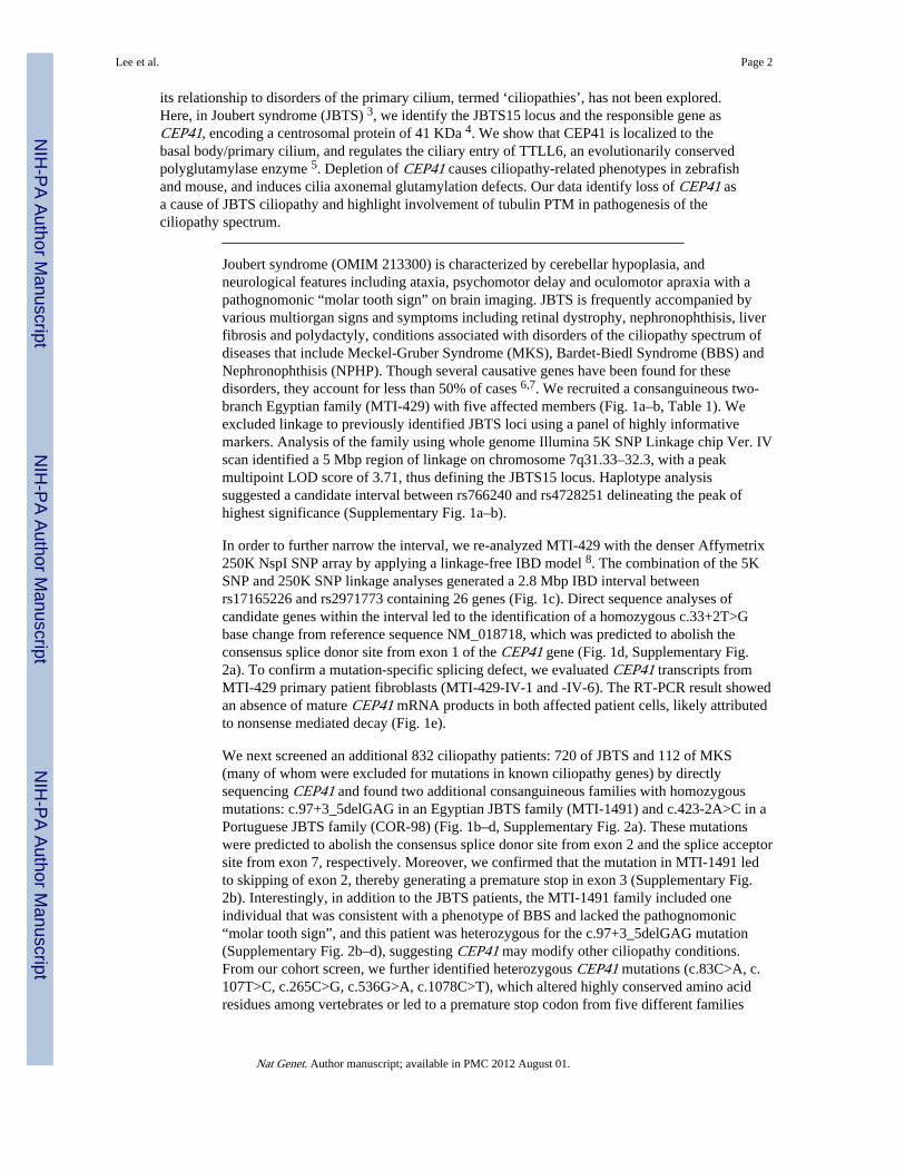
Joubert syndrome (OMIM 213300) is characterized by cerebellar hypoplasia, andneurological features including ataxia, psychomotor delay and oculomotor apraxia with apathognomonic “molar tooth sign” on brain imaging. JBTS is frequently accompanied byvarious multiorgan signs and symptoms including retinal dystrophy, nephronophthisis, liverfibrosis and polydactyly, conditions associated with disorders of the ciliopathy spectrum ofdiseases that include Meckel-Gruber Syndrome (MKS), Bardet-Biedl Syndrome (BBS) andNephronophthisis (NPHP). Though several causative genes have been found for thesedisorders, they account for less than 50% of cases 6,7. We recruited a consanguineous two-branch Egyptian family (MTI-429) with five affected members (Fig. 1a–b, Table 1). Weexcluded linkage to previously identified JBTS loci using a panel of highly informativemarkers. Analysis of the family using whole genome Illumina 5K SNP Linkage chip Ver. IVscan identified a 5 Mbp region of linkage on chromosome 7q31.33–32.3, with a peakmultipoint LOD score of 3.71, thus defining the JBTS15 locus. Haplotype analysissuggested a candidate interval between rs766240 and rs4728251 delineating the peak ofhighest significance (Supplementary Fig. 1a–b).
In order to further narrow the interval, we re-analyzed MTI-429 with the denser Affymetrix250K NspI SNP array by applying a linkage-free IBD model 8. The combination of the 5KSNP and 250K SNP linkage analyses generated a 2.8 Mbp IBD interval betweenrs17165226 and rs2971773 containing 26 genes (Fig. 1c). Direct sequence analyses ofcandidate genes within the interval led to the identification of a homozygous c.33+2T>Gbase change from reference sequence NM_018718, which was predicted to abolish theconsensus splice donor site from exon 1 of the CEP41 gene (Fig. 1d, Supplementary Fig.2a). To confirm a mutation-specific splicing defect, we evaluated CEP41 transcripts fromMTI-429 primary patient fibroblasts (MTI-429-IV-1 and -IV-6). The RT-PCR result showedan absence of mature CEP41 mRNA products in both affected patient cells, likely attributedto nonsense mediated decay (Fig. 1e).
We next screened an additional 832 ciliopathy patients: 720 of JBTS and 112 of MKS(many of whom were excluded for mutations in known ciliopathy genes) by directlysequencing CEP41 and found two additional consanguineous families with homozygousmutations: c.97+3_5delGAG in an Egyptian JBTS family (MTI-1491) and c.423-2A>C in aPortuguese JBTS family (COR-98) (Fig. 1b–d, Supplementary Fig. 2a). These mutationswere predicted to abolish the consensus splice donor site from exon 2 and the splice acceptorsite from exon 7, respectively. Moreover, we confirmed that the mutation in MTI-1491 ledto skipping of exon 2, thereby generating a premature stop in exon 3 (Supplementary Fig.2b). Interestingly, in addition to the JBTS patients, the MTI-1491 family included oneindividual that was consistent with a phenotype of BBS and lacked the pathognomonic“molar tooth sign”, and this patient was heterozygous for the c.97+3_5delGAG mutation(Supplementary Fig. 2b–d), suggesting CEP41 may modify other ciliopathy conditions.From our cohort screen, we further identified heterozygous CEP41 mutations (c.83C>A, c.107T>C, c.265C>G, c.536G>A, c.1078C>T), which altered highly conserved amino acidresidues among vertebrates or led to a premature stop codon from five different families
Lee et al. Page 2
Nat Genet. Author manuscript; available in PMC 2012 August 01.
NIH
-PA Author Manuscript
NIH
-PA Author Manuscript
NIH
-PA Author Manuscript
(Fig. 1d, Supplementary Fig. 2e, Supplementary Table 1). Each of these CEP41-mutatedpatients was additionally sequenced at the known JBTS genes, and in four there was anadditional heterozygous potentially deleterious variant. It was notable that all homozygousmutations in CEP41 were splice site mutations and identified only in JBTS patients whileheterozygous variants were present in several ciliopathies including BBS and MKS. Ourfindings suggest that constitutive disruptions of CEP41 result in JBTS, but that it may alsoserve as a modifier in the broader class of ciliopathies.
The CEP41 gene has been poorly characterized except for its expression analysis in humanorgans including brain, testis and kidney 9. CEP41 encodes for Centrosomal Protein 41KDa, predicted to contain two coiled-coils and a rhodanese-like domain (RHOD), which isstructurally related to the catalytic subunit of the Cdc25 class of phosphatases 10. However,we found that it lacks phosphatase activity in the in vitro para-Nitrophenylphosphate (pNPP)phosphatase assay (not shown). The RHOD domain therefore may be an enzymaticallyinactive version like some described RHOD domains in other proteins 10 functioning inprotein interactions.
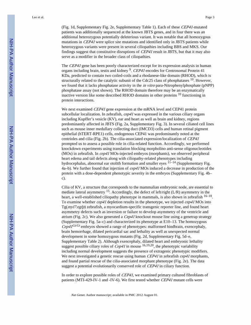
We next examined CEP41 gene expression at the mRNA level and CEP41 proteinsubcellular localization. In zebrafish, cep41 was expressed in the various ciliary organsincluding Kupffer’s vesicle (KV), ear and heart as well as brain and kidney, regionspredominantly affected in JBTS (Fig. 2a, Supplementary Fig. 3). In several ciliated cell linessuch as mouse inner medullary collecting duct (IMCD3) cells and human retinal pigmentepithelial (hTERT-RPE1) cells, endogenous CEP41 was predominantly noted at thecentrioles and cilia (Fig. 2b). The cilia-associated expression/localization of CEP41prompted us to assess a possible role in cilia-related function. Accordingly, we performedknockdown experiments using translation blocking morpholino anti-sense oligonucleotides(MOs) in zebrafish. In cep41 MOs-injected embryos (morphants), we observed peripheralheart edema and tail defects along with ciliopathy-related phenotypes includinghydrocephalus, abnormal ear otolith formation and smaller eyes 11–14 (Supplementary Fig.4a–b). We further found that injection of cep41 MOs induced a decrease in production of theprotein with a dose-dependent phenotypic severity in the embryos (Supplementary Fig. 4b–c).
Cilia of KV, a structure that corresponds to the mammalian embryonic node, are essential tomediate lateral asymmetry 15. Accordingly, the defect of left/right (L/R) asymmetry in theheart, a well-established ciliopathy phenotype in mammals, is also shown in zebrafish 16–18.To examine whether cep41 depletion results in the phenotype, we injected cep41 MOs intoTg(myl7:egfp) zebrafish, a myocardium-specific transgenic reporter line, and found heartasymmetry defects such as inversion or failure to develop asymmetry of the ventricle andatrium (Fig. 2c). We also generated a Cep41 knockout mouse line using a genetrap strategy(Supplementary Fig. 5a–c) and characterized its phenotype at E10–13. The homozygousCep41Gt/Gt embryos showed a range of phenotypes: malformed hindbrain, exencephaly,brain hemorrhage, dilated pericardial sac and lethality as well as unexpected normaldevelopment in some homozygous mutants (Fig. 2d, Supplementary Fig. 5d–e,Supplementary Table 2). Although exencephaly, dilated heart and embryonic lethalitysuggest possible ciliary roles of Cep41 in mouse 16,19,20, the phenotypic variabilityincluding normal development suggests the presence of extragenic phenotypic modifiers.We next investigated a genetic rescue using human CEP41 in zebrafish cep41 morphants,and found partial rescue of the cilia-associated morphant phenotype (Fig. 2e). The datasuggest a potential evolutionarily conserved role of CEP41 in ciliary function.
In order to explore possible roles of CEP41, we examined primary cultured fibroblasts ofpatients (MTI-429-IV-1 and -IV-6). We first tested whether CEP41 mutant cells were
Lee et al. Page 3
Nat Genet. Author manuscript; available in PMC 2012 August 01.
NIH
-PA Author Manuscript
NIH
-PA Author Manuscript
NIH
-PA Author Manuscript
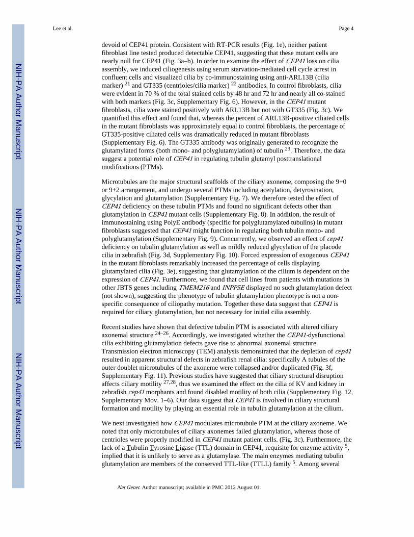
devoid of CEP41 protein. Consistent with RT-PCR results (Fig. 1e), neither patientfibroblast line tested produced detectable CEP41, suggesting that these mutant cells arenearly null for CEP41 (Fig. 3a–b). In order to examine the effect of CEP41 loss on ciliaassembly, we induced ciliogenesis using serum starvation-mediated cell cycle arrest inconfluent cells and visualized cilia by co-immunostaining using anti-ARL13B (ciliamarker) 21 and GT335 (centrioles/cilia marker) 22 antibodies. In control fibroblasts, ciliawere evident in 70 % of the total stained cells by 48 hr and 72 hr and nearly all co-stainedwith both markers (Fig. 3c, Supplementary Fig. 6). However, in the CEP41 mutantfibroblasts, cilia were stained positively with ARL13B but not with GT335 (Fig. 3c). Wequantified this effect and found that, whereas the percent of ARL13B-positive ciliated cellsin the mutant fibroblasts was approximately equal to control fibroblasts, the percentage ofGT335-positive ciliated cells was dramatically reduced in mutant fibroblasts(Supplementary Fig. 6). The GT335 antibody was originally generated to recognize theglutamylated forms (both mono- and polyglutamylation) of tubulin 23. Therefore, the datasuggest a potential role of CEP41 in regulating tubulin glutamyl posttranslationalmodifications (PTMs).
Microtubules are the major structural scaffolds of the ciliary axoneme, composing the 9+0or 9+2 arrangement, and undergo several PTMs including acetylation, detyrosination,glycylation and glutamylation (Supplementary Fig. 7). We therefore tested the effect ofCEP41 deficiency on these tubulin PTMs and found no significant defects other thanglutamylation in CEP41 mutant cells (Supplementary Fig. 8). In addition, the result ofimmunostaining using PolyE antibody (specific for polyglutamylated tubulins) in mutantfibroblasts suggested that CEP41 might function in regulating both tubulin mono- andpolyglutamylation (Supplementary Fig. 9). Concurrently, we observed an effect of cep41deficiency on tubulin glutamylation as well as mildly reduced glycylation of the placodecilia in zebrafish (Fig. 3d, Supplementary Fig. 10). Forced expression of exogenous CEP41in the mutant fibroblasts remarkably increased the percentage of cells displayingglutamylated cilia (Fig. 3e), suggesting that glutamylation of the cilium is dependent on theexpression of CEP41. Furthermore, we found that cell lines from patients with mutations inother JBTS genes including TMEM216 and INPP5E displayed no such glutamylation defect(not shown), suggesting the phenotype of tubulin glutamylation phenotype is not a non-specific consequence of ciliopathy mutation. Together these data suggest that CEP41 isrequired for ciliary glutamylation, but not necessary for initial cilia assembly.
Recent studies have shown that defective tubulin PTM is associated with altered ciliaryaxonemal structure 24–26. Accordingly, we investigated whether the CEP41-dysfunctionalcilia exhibiting glutamylation defects gave rise to abnormal axonemal structure.Transmission electron microscopy (TEM) analysis demonstrated that the depletion of cep41resulted in apparent structural defects in zebrafish renal cilia: specifically A tubules of theouter doublet microtubules of the axoneme were collapsed and/or duplicated (Fig. 3f,Supplementary Fig. 11). Previous studies have suggested that ciliary structural disruptionaffects ciliary motility 27,28, thus we examined the effect on the cilia of KV and kidney inzebrafish cep41 morphants and found disabled motility of both cilia (Supplementary Fig. 12,Supplementary Mov. 1–6). Our data suggest that CEP41 is involved in ciliary structuralformation and motility by playing an essential role in tubulin glutamylation at the cilium.
We next investigated how CEP41 modulates microtubule PTM at the ciliary axoneme. Wenoted that only microtubules of ciliary axonemes failed glutamylation, whereas those ofcentrioles were properly modified in CEP41 mutant patient cells. (Fig. 3c). Furthermore, thelack of a Tubulin Tyrosine Ligase (TTL) domain in CEP41, requisite for enzyme activity 5,implied that it is unlikely to serve as a glutamylase. The main enzymes mediating tubulinglutamylation are members of the conserved TTL-like (TTLL) family 5. Among several
Lee et al. Page 4
Nat Genet. Author manuscript; available in PMC 2012 August 01.
NIH
-PA Author Manuscript
NIH
-PA Author Manuscript
NIH
-PA Author Manuscript
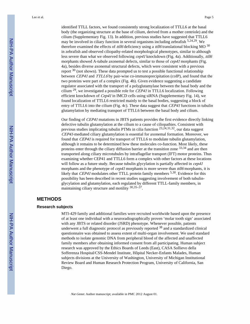
identified TTLL factors, we found consistently strong localization of TTLL6 at the basalbody (the organizing structure at the base of cilium, derived from a mother centriole) and thecilium (Supplementary Fig. 13). In addition, previous studies have suggested that TTLL6may be involved in ciliary function in several organisms including zebrafish 5,24,29. Wetherefore examined the effects of ttll6 deficiency using a ttll6 translational blocking MO 30
in zebrafish and observed ciliopathy-related morphological phenotypes, similar to althoughless severe than what we observed following cep41 knockdown (Fig. 4a). Additionally, ttll6morphants showed A-tubule axonemal defects, similar to those of cep41 morphants (Fig.4a), besides diverse axonemal structural defects, which were consistent with a previousreport 30 (not shown). These data prompted us to test a possible functional relationshipbetween CEP41 and TTLL6 by pair-wise co-immunoprecipitation (coIP), and found that thetwo proteins were part of a complex (Fig. 4b). Given evidence suggesting a candidateregulator associated with the transport of a polyglutamylase between the basal body and thecilium 29, we investigated a possible role for CEP41 in TTLL6 localization. Followingefficient knockdown of Cep41 in IMCD cells using siRNA (Supplementary Fig. 14), wefound localization of TTLL6 restricted mainly to the basal bodies, suggesting a block ofentry of TTLL6 into the cilium (Fig. 4c). These data suggest that CEP41 functions in tubulinglutamylation by mediating transport of TTLL6 between the basal body and cilium.
Our finding of CEP41 mutations in JBTS patients provides the first evidence directly linkingdefective tubulin glutamylation at the cilium to a cause of ciliopathies. Consistent withprevious studies implicating tubulin PTMs in cilia function 23,24,31,32, our data suggestCEP41-mediated ciliary glutamylation is essential for axonemal formation. Moreover, wefound that CEP41 is required for transport of TTLL6 to modulate tubulin glutamylation,although it remains to be determined how these molecules co-function. Most likely, theseproteins enter through the ciliary diffusion barrier at the transition zone 33,34 and are thentransported along ciliary microtubules by intraflagellar transport (IFT) motor proteins. Thusexamining whether CEP41 and TTLL6 form a complex with other factors at these locationswill follow as a future study. Because tubulin glycylation is partially affected in cep41morphants and the phenotype of cep41 morphants is more severe than ttll6 morphants, it islikely that CEP41 modulates other TTLL protein family members 5,30. Evidence for thispossibility has been described in recent studies suggesting involvement of both tubulin-glycylation and glutamylation, each regulated by different TTLL-family members, inmaintaining ciliary structure and motility 30,35–37.
METHODSResearch subjects
MTI-429 family and additional families were recruited worldwide based upon the presenceof at least one individual with a neuroradiographically proven ‘molar tooth sign’ associatedwith any JBTS or related disorder (JSRD) phenotype. Whenever possible, patientsunderwent a full diagnostic protocol as previously reported 38 and a standardized clinicalquestionnaire was obtained to assess extent of multi-organ involvement. We used standardmethods to isolate genomic DNA from peripheral blood of the affected and unaffectedfamily members after obtaining informed consent from all participating. Human subjectresearch was approved by the Ethics Boards of Leeds (East), CASA Sollievo dellaSofferenza Hospital/CSS-Mendel Institute, Hôpital Necker-Enfants Malades, Humansubjects divisions at the University of Washington, University of Michigan InstitutionalReview Board and Human Research Protection Program, University of California, SanDiego.
Lee et al. Page 5
Nat Genet. Author manuscript; available in PMC 2012 August 01.
NIH
-PA Author Manuscript
NIH
-PA Author Manuscript
NIH
-PA Author Manuscript
Genome-wide screen and fine mappingA 5K whole genome linkage SNP-scan was performed with family MTI-429 using theIllumina Linkage IVb mapping panel 39, and analyzed with easyLinkage-Plus software 40,which runs Allegro version 1.2c in a PC Windows interface to calculate multipoint LODscores. Parameters were set to autosomal recessive with full penetrance, and disease allelefrequency of 0.001. Fine mapping on the pedigree was performed with the Affymetrix 250KNsp1 SNP array, and data were searched for common shared homozygous intervals from allaffected family members using a custom script implemented in Mathematica (B. Merriman,unpublished). This script identifies all homozygous intervals longer than 2 Mbp for whichthere are no more than 1 % heterozygous calls (to permit potential genotyping errors).
Mutation screeningMutational screening of CEP41 was performed by direct sequencing of the 11 coding exonsand the adjacent intronic junctions in patients. PCR products obtained were treated withExonuclease I (EXO) (Fermentas) and shrimp alkaline phosphatase (SAP) (Promega), andboth strands were sequenced using a BigDye terminator cycle sequencing kit with anABI3100 automated sequencer (Applied Biosystems). The primers and optimized PCRconditions used are depicted (Supplementary Table 3). Segregation of the identifiedmutations was investigated in all available family members. All identified mutations inCEP41 were not encountered in 96 ethnically matched controls (188 chromosomes) upondirect sequencing.
BioinformaticsGenetic location is according to the March 2006 Human Genome Browser build hg18. Theciliary proteome was searched using web-based tools 41,42. Protein sequence conservationwas determined using ClustalW multiple amino acid sequence alignment (see URL section).
CloningFull-length human CEP41 was cloned into the TOPO blunt vector, and then shuttled intoEGFP- and HA- containing vectors. Human and zebrafish cep41 open reading frame wereamplified by RT-PCR and cloned into the pCS2+ vector in order to synthesize RNA forinjection into zebrafish embryos. Mouse Cep41 open reading frame was amplified andcloned into the GST-, EGFP-, and FLAG-conjugated vectors for biochemical assays.
Generation of Cep41 mutant miceSIGTR ES cell line (AW0157), derived from 129P2/OlaHsd mice carrying a gene trapinsertion 43 in the intron 1 of Cep41, was obtained from the European Conditional MouseMutagenesis Program (EUCOMM) and injected into C57BL/6 recipient blastocysts at theMouse Biology Program, UC Davis. High-percentage chimeras (≥ 70 %) were bred toC57BL/6 for germline transmission. Gene-trapped mice were isolated by PCR for β-galactosidase-neomycin fusion gene and genotyped by Southern blot analysis hybridizing a~10 kb (wild-type allele) and a ~8 kb (gene-trapped allele) product (Supplementary Fig. 5).
Zebrafish experimentsAB wild type zebrafish strains were used for in situ hybridization carried out by standardprotocols using a DIG-labeled sense and anti-sense RNA probe for cep41. To knockdownzebrafish cep41, a translational blocking morpholino antisense oligonucleotide (MO, GeneTools Inc) was used: 5′-CATCTTCCAGCAGCAGAGCTTCGGC-3′, diluted to appropriateconcentrations in deionized sterile water, and injected into one-two cell stage embryos,obtained from natural spawning of zebrafish lines. To rescue the phenotypes in MO-injectedembryos (morphants), RNA transcribed in vitro with the SP6 mMessage mMachine kit
Lee et al. Page 6
Nat Genet. Author manuscript; available in PMC 2012 August 01.
NIH
-PA Author Manuscript
NIH
-PA Author Manuscript
NIH
-PA Author Manuscript
(Ambion) was co-injected. For characterization of ciliary defects in zebrafish embryos, themorphological phenotype of either cep41 or ttll6 morphants were observed until 5 days post-fertilization (dpf) and quantified under bright-field microscopy based upon previouslyestablished criteria 44. For western blot analysis at 1–2 dpf zebrafish control embryos andcep41 morphants, about 50 embryos with each genotype were deyolked and the embryolysates were extracted with RIPA buffer. For immunostaining with GT335, Ac-Tub andPolyG Abs in whole-mount zebrafish embryos, 3 dpf control embryos and cep41 morphantswere fixed in Dent’s fixative (80 % MeOH: 20 % DMSO) at 4 °C overnight and incubatedwith GT335 Ab (1:400), Ac-Tub (1:400) and PolyG (1:300) as primary and with anti-goat-mouse 594 (1:1000) as secondary in diluted blocking solution (10 % normal goat serum: 0.5% Tween 20 in PBS).
Cell culture and transfectionIMCD3, hTERT-RPE1, human fibroblast, and human embryonic kidney (HEK293) cellswere grown in appropriate DMEM or MEM media supplemented with 10–20 % fetal bovineserum (FBS) at 37 °C in 5 % CO2. Healthy human female and male control fibroblast cellswere obtained from ATCC, and patient fibroblasts from skin biopsies were propagated inculture (≤ 5 passage number). Human fibroblast cells were transfected using the BasicNucleofector Kit for Primary Mammalian Fibroblasts (Lonza). Other cells were transfectedat 60–80 % confluency with plasmids or siRNAs using Lipofectamine 2000 (Invitrogen).The transfected cells were incubated for 24–72 hr with FBS or without FBS, dependent onthe experimental purpose.
Fluorescence microscopy and transmission electron microscopy (TEM)Images of immunofluorescent stained cells were obtained on a Deltavision RTDeconvolution microscope (Olympus IX70), under the same parameters for eachexperiment. Taken images were edited and analyzed using Adobe Photoshop CS. Forelectron microscopy, a standard protocol 45 was used except for one modification that tannicacid was included in the fixative to enhance the final contrast of the images. Formvar-coatedslot grids (Electron microscopy Sciences) were used for sections (60–70 nm) to maximizevisibility of the tissue and cross section performed to observe cilia axonemal structure.
Live imaging of zebrafish embryosZebrafish embryos (12 hpf for cilia in the KV and 2.5 dpf for the renal cilia) weretransferred with embryo media to glass bottom culture dishes (MatTek), and 2.5 dpfembryos were anesthetized in tricaine solution (~0.016 mg/ml). Images were acquired for 30sec – 2 min using a Perkin Elmer UltraView Vox Spinning Disk Confocal with EMCCDHamamatsu 14 bit 1K × 1K camera, and edited with Volocity imaging software (PerkinElmer).
Immunofluorescence and biochemical assayFor immunofluorescence, cells were fixed in 100 % methanol at −20 °C for 10 min. Primaryantibodies used for immunofluorescence are: rabbit anti-CEP41, raised in rabbit to a purifiedbacterially expressed protein of GST-fused CEP41, (PRF&L, PA), mouse anti-acetylated-tubulin (Sigma), mouse GT335 Ab (gift from C. Janke), rabbit PolyE (gift from M.Gorovsky) and rabbit anti-ARL13B (gift from T. Caspary). We used Alexa 488- or Alexa594-conjugated secondary antibodies (Molecular Probes) and Hoechst 33342 nuclear dye.All antibodies were diluted in 4 % normal donkey serum in PBS, primary antibodies wereincubated for overnight at 4 °C and secondary antibodies were applied for 1 hr at roomtemperature. For immunoblotting, cells were extracted with RIPA lysis buffer 3 days aftertransfection and boiled with SDS sample buffer. Same concentrations of lysates were loaded
Lee et al. Page 7
Nat Genet. Author manuscript; available in PMC 2012 August 01.
NIH
-PA Author Manuscript
NIH
-PA Author Manuscript
NIH
-PA Author Manuscript
for paired experiments. Primary antibodies used for western blotting were: rabbit anti-GFP(Roche), mouse anti-Flag (Sigma), and mouse anti-alpha-Tubulin (Sigma). Boundantibodies were detected using horseradish peroxidase-conjugated secondary antibodies(Pierce). For co-immunoprecipitation (co-IP), whole cell extracts (WCE) were preparedfrom confluent HEK293 cells transiently transfected with 14 μg plasmid DNA in 10 cmtissue culture dishes. WCE supernatants were processed for IP experiments by using 2 μgprimary antibodies and protein A/G mixed agarose beads (Pierce).
Statistical analysisThe χ2 staticstic was computed manually with P value assigned for 1 degree of freedom inthe characterization of mouse embryonic phenotype (Supplementary Table 2). For otherstudies, Student’s two-tailed non-paired t-tests were carried out to determine the statisticalsignificance of differences between samples. P < 0.05 was considered statistically significantfor all tests.
URLsHuman Genome Browser, http://www.genome.ucsc.edu; ClustalW,http://www.ebi.ac.uk/Tools/msa/clustalw2/.
Supplementary MaterialRefer to Web version on PubMed Central for supplementary material.
AcknowledgmentsWe thank the Marshfield Clinic Research Foundation, Center for Inherited Disease Research (supported by the USNational Institutes of Health and National Heart, Lung, and Blood Institute) for genotyping support. We thank theInternational JSRD Study Group, E. Bertini, and the French Society of Foetal Pathology for patient referrals, J.Meerloo at the UCSD Microscopy Core (P30NS047101), T. Meerloo, Y. Jones and M. Farquhar and the CMMElectron Microscopy Core Facility at UCSD, S. Wirth and B. Willis for mutant mouse generation, C. Janke at theInstitute Curie Research Center for GT335 antibody, TTLLs plasmids and technical advice, I. Drummond and N.Pathak at the Massachusetts General Hospital for ttll6 MO, M. Gorovsky at the Univ. of Rochester for polyE andpolyG antibodies, S. Audollent at the Hôpital Necker-Enfants Malades for technical help, B. Sotak, N. Akizu, A.Crawford, V. Cantagrel, and E-J. Choi for stimulating scientific discussion and comments. This work wassupported by the US National Institutes of Health R01NS048453 and R01NS052455 (J.G.G.), R01DK068306(F.H.), R01NS064077 (D.A.D.) the American Heart Association 09POST2250641 (J.E.L.), the Italian Ministry ofHealth (Ricerca Finalizzata Malattie Rare and Ricerca Corrente 2011), the Telethon Foundation Italy GGP08145,and the Pierfranco and Luisa Mariani Foundation (E.M.V.), Grants-in-Aid from MEXT and JSPS, #23117517 and#23570209 (K.I.), the BDF Newlife, the Medical Research Council (G0700073) and the Sir Jules Thorn CharitableTrust 09/JTA (C.A.J.), l’Agence National pour la Recherche ANR 07-MRAR-Fetalciliopathies (T.A.-B.), SimonsFoundation Autism Research Initiative (J.G.G.) and the Howard Hughes Medical Institute (F.H. and J.G.G.).
References1. Ikegami K, Sato S, Nakamura K, Ostrowski LE, Setou M. Tubulin polyglutamylation is essential for
airway ciliary function through the regulation of beating asymmetry. Proc Natl Acad Sci U S A.2010; 107:10490–5. [PubMed: 20498047]
2. Kubo T, Yanagisawa HA, Yagi T, Hirono M, Kamiya R. Tubulin polyglutamylation regulatesaxonemal motility by modulating activities of inner-arm dyneins. Curr Biol. 2010; 20:441–5.[PubMed: 20188560]
3. Parisi MA. Clinical and molecular features of Joubert syndrome and related disorders. Am J MedGenet C Semin Med Genet. 2009; 151C:326–40. [PubMed: 19876931]
4. Andersen JS, et al. Proteomic characterization of the human centrosome by protein correlationprofiling. Nature. 2003; 426:570–4. [PubMed: 14654843]
5. van Dijk J, et al. A targeted multienzyme mechanism for selective microtubule polyglutamylation.Mol Cell. 2007; 26:437–48. [PubMed: 17499049]
Lee et al. Page 8
Nat Genet. Author manuscript; available in PMC 2012 August 01.
NIH
-PA Author Manuscript
NIH
-PA Author Manuscript
NIH
-PA Author Manuscript

6. Valente EM, Brancati F, Dallapiccola B. Genotypes and phenotypes of Joubert syndrome andrelated disorders. Eur J Med Genet. 2008; 51:1–23. [PubMed: 18164675]
7. Hildebrandt F, Zhou W. Nephronophthisis-associated ciliopathies. J Am Soc Nephrol. 2007;18:1855–71. [PubMed: 17513324]
8. Lander ES, Botstein D. Homozygosity mapping: a way to map human recessive traits with the DNAof inbred children. Science. 1987; 236:1567–70. [PubMed: 2884728]
9. Yamada T, et al. The gene TSGA14, adjacent to the imprinted gene MEST, escapes genomicimprinting. Gene. 2002; 288:57–63. [PubMed: 12034494]
10. Bordo D, Bork P. The rhodanese/Cdc25 phosphatase superfamily. Sequence-structure-functionrelations. EMBO Rep. 2002; 3:741–6. [PubMed: 12151332]
11. Beales PL, et al. IFT80, which encodes a conserved intraflagellar transport protein, is mutated inJeune asphyxiating thoracic dystrophy. Nat Genet. 2007; 39:727–9. [PubMed: 17468754]
12. Gorden NT, et al. CC2D2A is mutated in Joubert syndrome and interacts with the ciliopathy-associated basal body protein CEP290. Am J Hum Genet. 2008; 83:559–71. [PubMed: 18950740]
13. Colantonio JR, et al. The dynein regulatory complex is required for ciliary motility and otolithbiogenesis in the inner ear. Nature. 2009; 457:205–9. [PubMed: 19043402]
14. Khanna H, et al. A common allele in RPGRIP1L is a modifier of retinal degeneration inciliopathies. Nat Genet. 2009; 41:739–45. [PubMed: 19430481]
15. Essner JJ, Amack JD, Nyholm MK, Harris EB, Yost HJ. Kupffer’s vesicle is a ciliated organ ofasymmetry in the zebrafish embryo that initiates left-right development of the brain, heart and gut.Development. 2005; 132:1247–60. [PubMed: 15716348]
16. Nonaka S, et al. Randomization of left-right asymmetry due to loss of nodal cilia generatingleftward flow of extraembryonic fluid in mice lacking KIF3B motor protein. Cell. 1998; 95:829–37. [PubMed: 9865700]
17. McGrath J, Brueckner M. Cilia are at the heart of vertebrate left-right asymmetry. Curr Opin GenetDev. 2003; 13:385–92. [PubMed: 12888012]
18. Amack JD, Yost HJ. The T box transcription factor no tail in ciliated cells controls zebrafish left-right asymmetry. Curr Biol. 2004; 14:685–90. [PubMed: 15084283]
19. Ross AJ, et al. Disruption of Bardet-Biedl syndrome ciliary proteins perturbs planar cell polarity invertebrates. Nat Genet. 2005; 37:1135–40. [PubMed: 16170314]
20. Hoover AN, et al. C2cd3 is required for cilia formation and Hedgehog signaling in mouse.Development. 2008; 135:4049–58. [PubMed: 19004860]
21. Caspary T, Larkins CE, Anderson KV. The graded response to Sonic Hedgehog depends on ciliaarchitecture. Dev Cell. 2007; 12:767–78. [PubMed: 17488627]
22. Jurczyk A, et al. Pericentrin forms a complex with intraflagellar transport proteins and polycystin-2and is required for primary cilia assembly. J Cell Biol. 2004; 166:637–43. [PubMed: 15337773]
23. Million K, et al. Polyglutamylation and polyglycylation of alpha- and beta-tubulins during in vitrociliated cell differentiation of human respiratory epithelial cells. J Cell Sci. 1999; 112 (Pt 23):4357–66. [PubMed: 10564653]
24. Suryavanshi S, et al. Tubulin glutamylation regulates ciliary motility by altering inner dynein armactivity. Curr Biol. 2010; 20:435–40. [PubMed: 20189389]
25. Vogel P, Hansen G, Fontenot G, Read R. Tubulin tyrosine ligase-like 1 deficiency results inchronic rhinosinusitis and abnormal development of spermatid flagella in mice. Vet Pathol. 2010;47:703–12. [PubMed: 20442420]
26. Wloga D, et al. Hyperglutamylation of tubulin can either stabilize or destabilize microtubules inthe same cell. Eukaryot Cell. 2010; 9:184–93. [PubMed: 19700636]
27. Becker-Heck A, et al. The coiled-coil domain containing protein CCDC40 is essential for motilecilia function and left-right axis formation. Nat Genet. 2011; 43:79–84. [PubMed: 21131974]
28. Zhao C, Malicki J. Genetic defects of pronephric cilia in zebrafish. Mech Dev. 2007; 124:605–16.[PubMed: 17576052]
29. Pathak N, Obara T, Mangos S, Liu Y, Drummond IA. The zebrafish fleer gene encodes anessential regulator of cilia tubulin polyglutamylation. Mol Biol Cell. 2007; 18:4353–64. [PubMed:17761526]
Lee et al. Page 9
Nat Genet. Author manuscript; available in PMC 2012 August 01.
NIH
-PA Author Manuscript
NIH
-PA Author Manuscript
NIH
-PA Author Manuscript

30. Pathak N, Austin CA, Drummond IA. Tubulin tyrosine ligase-like genes ttll3 and ttll6 maintainzebrafish cilia structure and motility. J Biol Chem. 2011; 286:11685–95. [PubMed: 21262966]
31. Gaertig J, Wloga D. Ciliary tubulin and its post-translational modifications. Curr Top Dev Biol.2008; 85:83–113. [PubMed: 19147003]
32. Shida T, Cueva JG, Xu Z, Goodman MB, Nachury MV. The major alpha-tubulin K40acetyltransferase alphaTAT1 promotes rapid ciliogenesis and efficient mechanosensation. ProcNatl Acad Sci U S A. 2010; 107:21517–22. [PubMed: 21068373]
33. Kim SK, et al. Planar cell polarity acts through septins to control collective cell movement andciliogenesis. Science. 2010; 329:1337–40. [PubMed: 20671153]
34. Williams CL, et al. MKS and NPHP modules cooperate to establish basal body/transition zonemembrane associations and ciliary gate function during ciliogenesis. J Cell Biol. 2011; 192:1023–41. [PubMed: 21422230]
35. Rogowski K, et al. Evolutionary divergence of enzymatic mechanisms for posttranslationalpolyglycylation. Cell. 2009; 137:1076–87. [PubMed: 19524510]
36. Wloga D, et al. TTLL3 Is a tubulin glycine ligase that regulates the assembly of cilia. Dev Cell.2009; 16:867–76. [PubMed: 19531357]
37. Bulinski JC. Tubulin posttranslational modifications: a Pushmi-Pullyu at work? Dev Cell. 2009;16:773–4. [PubMed: 19531345]
38. Valente EM, et al. Distinguishing the four genetic causes of Jouberts syndrome-related disorders.Ann Neurol. 2005; 57:513–9. [PubMed: 15786477]
39. Murray SS, et al. A highly informative SNP linkage panel for human genetic studies. Nat Methods.2004; 1:113–7. [PubMed: 15782173]
40. Hoffmann K, Lindner TH. easyLINKAGE-Plus--automated linkage analyses using large-scaleSNP data. Bioinformatics. 2005; 21:3565–7. [PubMed: 16014370]
41. Inglis PN, Boroevich KA, Leroux MR. Piecing together a ciliome. Trends Genet. 2006; 22:491–500. [PubMed: 16860433]
42. Gherman A, Davis EE, Katsanis N. The ciliary proteome database: an integrated communityresource for the genetic and functional dissection of cilia. Nat Genet. 2006; 38:961–2. [PubMed:16940995]
43. Schnutgen F, et al. Genomewide production of multipurpose alleles for the functional analysis ofthe mouse genome. Proc Natl Acad Sci U S A. 2005; 102:7221–6. [PubMed: 15870191]
44. Valente EM, et al. Mutations in TMEM216 perturb ciliogenesis and cause Joubert, Meckel andrelated syndromes. Nat Genet. 2010; 42:619–25. [PubMed: 20512146]
45. Majumdar A, Drummond IA. Podocyte differentiation in the absence of endothelial cells asrevealed in the zebrafish avascular mutant, cloche. Dev Genet. 1999; 24:220–9. [PubMed:10322630]
Lee et al. Page 10
Nat Genet. Author manuscript; available in PMC 2012 August 01.
NIH
-PA Author Manuscript
NIH
-PA Author Manuscript
NIH
-PA Author Manuscript
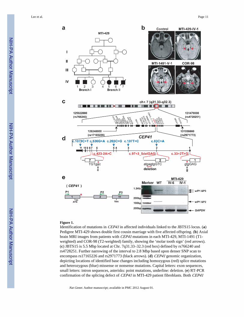
Figure 1.Identification of mutations in CEP41 in affected individuals linked to the JBTS15 locus. (a)Pedigree MTI-429 shows double first cousin marriage with five affected offspring. (b) Axialbrain MRI images from patients with CEP41 mutations in each MTI-429, MTI-1491 (T1-weighted) and COR-98 (T2-weighted) family, showing the ‘molar tooth sign’ (red arrows).(c) JBTS15 is 5.5 Mbp located at Chr. 7q31.33–32.3 (red box) defined by rs766240 andrs4728251. Further narrowing of the interval to 2.8 Mbp based upon denser SNP scan toencompass rs17165226 and rs2971773 (black arrows). (d) CEP41 genomic organization,depicting locations of identified base changes including homozygous (red) splice mutationsand heterozygous (blue) missense or nonsense mutations. Capital letters: exon sequences,small letters: intron sequences, asterisks: point mutations, underline: deletion. (e) RT-PCRconfirmation of the splicing defect of CEP41 in MTI-429 patient fibroblasts. Both CEP41
Lee et al. Page 11
Nat Genet. Author manuscript; available in PMC 2012 August 01.
NIH
-PA Author Manuscript
NIH
-PA Author Manuscript
NIH
-PA Author Manuscript
mutant cells failed to produce CEP41 mRNA, compared with WT. GAPDH is control.Green arrows: positions of primers, asterisk: region of splice mutation in MTI-429.
Lee et al. Page 12
Nat Genet. Author manuscript; available in PMC 2012 August 01.
NIH
-PA Author Manuscript
NIH
-PA Author Manuscript
NIH
-PA Author Manuscript

Figure 2.CEP41 is expressed in ciliated tissues and its loss recapitulates ciliopathy-related phenotypesin zebrafish and mouse. (a) Zebrafish cep41 mRNA is expressed ubiquitously at gastrulationstages (6 hpf), but at later stages, it is specifically expressed in ciliated organs: Kupffer’svesicle (red box), inner ear (white boxes), brain (brackets), eyes (arrows), pronephric duct(black box), and heart (asterisks). A, anterior; D, dorsal; P, posterior; V, ventral. (b) CEP41is predominantly localized to the basal body (arrowheads) and primary cilium (arrows) inciliated IMCD3 and hTERT-RPE1 cells. Insets: co-localization of CEP41 with GT335, amarker of basal bodies and cilia. Scale bar 5 μm. (c) Knockdown of cep41 by injection ofmorpholino oligonucleotide (MO) causes heart asymmetry defects in Tg (myl7:egfp)zebrafish embryos. The cep41 morphants show either loss of asymmetry (midline) orinversion of V/A asymmetry (reversed) at 72hpf. *P < 0.01, **P < 0.001. Error bars: s.e.m.
Lee et al. Page 13
Nat Genet. Author manuscript; available in PMC 2012 August 01.
NIH
-PA Author Manuscript
NIH
-PA Author Manuscript
NIH
-PA Author Manuscript

A, atrium; L, left; R, right; V, ventricle. (d) Murine Cep41 gene-trap shows alteredembryonic morphogenesis. Range of phenotypes of Cep41Gt/Gt embryos includes mildmalformed hindbrain (arrowheads), exencephaly (brackets), hemorrhage in the head(asterisk), dilated pericardial sac (arrows), failure to rotate and lethality at E10–12. (e)Injection of human CEP41 RNA into cep41 morphants rescued ciliary phenotypes ofpericardial edema (arrowhead), hydrocephalus (asterisk) and curved tail (arrow), completelyor partially.
Lee et al. Page 14
Nat Genet. Author manuscript; available in PMC 2012 August 01.
NIH
-PA Author Manuscript
NIH
-PA Author Manuscript
NIH
-PA Author Manuscript

Figure 3.CEP41 is required for tubulin glutamylation at the ciliary axoneme. (a) Absent CEP41protein in CEP41 mutant patient cells. (b) Ciliary localization of endogenous CEP41 inhuman fibroblasts and loss of the protein in CEP41 mutant cilia. Scale bar 5 μm. (c) WTcells have both GT335-positive basal bodies (arrowheads) and cilia (arrows, marked byARL13B), while mutant cells show staining of GT335 only at the basal bodies. Scale bar 5μm. (d) Depletion of cep41 causes glutamylation defects in zebrafish olfactory placode cilia(red arrows). Images of GT335-stainined WT and cep41 morphant embryos were taken inanterior view (head is up and tail is down, D, dorsal; V, ventral), quantified below. (e)Exogenous CEP41 expression restores ciliary axoneme glutamylation in CEP41 mutantcells. Arrows: primary cilia stained for CEP41 and GT335. Insets: merged images at higherpower, quantified below. Scale bar 5 μm. **P < 0.001; error bars = s.e.m. (f) Ultrastructuralanalysis of the pronephric ciliary axoneme at 72 hpf zebrafish embryos. Compared to WT,cep41 morphants have A-tubule specific defects in the outer doublet microtubules. Arrows:
Lee et al. Page 15
Nat Genet. Author manuscript; available in PMC 2012 August 01.
NIH
-PA Author Manuscript
NIH
-PA Author Manuscript
NIH
-PA Author Manuscript
A-tubules and one of nine outer doublet microtubules is magnified in the red box. Thenumbers of cilia, categorized in normal and abnormal A-tubules according to the schematic,were counted in both WT embryos and cep41 morphants (n = 3 embryos, >20 cilia peranimal), quantified below. Scale bar 100 nm. **P < 0.01; error bars = s.e.m.
Lee et al. Page 16
Nat Genet. Author manuscript; available in PMC 2012 August 01.
NIH
-PA Author Manuscript
NIH
-PA Author Manuscript
NIH
-PA Author Manuscript

Figure 4.CEP41 interacts with TTLL6 and is required for localizing TTLL6 to the cilium. (a)Morpholino knockdown of zebrafish ttll6 associates with ciliary phenotypes such as curvedtail (arrows), abnormal number/orientation of ear otolith (boxes), cystic kidney (arrowheads)and peripheral cardiac edema (asterisks) at different dosages indicated and show A-tubulespecific defects in the outer doublet microtubules. The numbers of defective cilia werecounted in both WT embryos and ttll6 morphants (n = 3 embryos, >20 cilia per animal),quantified below. (b) GFP-CEP41 was immunoprecipitated with anti-Flag antibodyrecognizing Flag-tagged TTLL6 from whole-cell extract (WCE), compared with GFP-emptyvector. In the reciprocal co-IP experiment with anti-GFP antibody, the interaction betweenCEP41 and TTLL6 was confirmed. (c) Disturbed localization of TTLL6 to the ciliumfollowing Cep41 siRNA co-transfection with GFP-TTLL6 into IMCD3 cells andimmunostained with either GT335 or ARL13B antibody. Arrows: cilia; Arrowheads: basalbodies. Cells expressing ciliary localized TTLL6 were counted only in siRNA-transfectedcells quantified in graph. Scale bar 5 μm. *P< 0.01, **P < 0.001. Error bars = s.e.m.
Lee et al. Page 17
Nat Genet. Author manuscript; available in PMC 2012 August 01.
NIH
-PA Author Manuscript
NIH
-PA Author Manuscript
NIH
-PA Author Manuscript

NIH
-PA Author Manuscript
NIH
-PA Author Manuscript
NIH
-PA Author Manuscript
Lee et al. Page 18
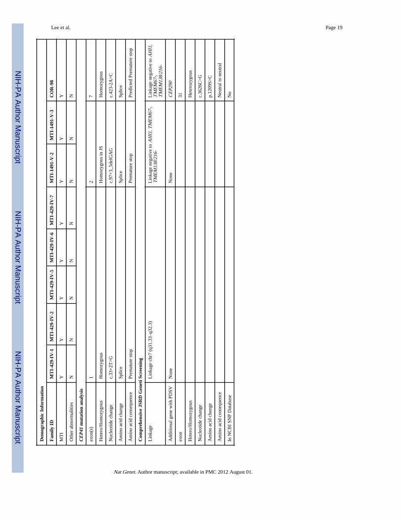
Tabl
e 1
Abb
revi
atio
ns. A
G: A
mbi
guou
s ge
nita
lia, B
: Bila
tera
l, F:
Fem
ale,
GH
D: G
row
th h
orm
one
defi
cien
cy, M
: Mal
e, M
P: M
icro
peni
s, N
: No,
N/A
: Not
avai
labl
e/N
ot a
pplic
able
, NPH
P: N
ephr
onop
hthi
sis,
OM
A: o
culo
mot
or a
rpax
ia, P
DSV
: pot
entia
lly d
elet
erio
us s
eque
nce
vari
ant,
UC
D: U
rina
ryco
ncen
trat
ion
defe
ct, U
: Uni
late
ral,
Y: Y
es
Dem
ogra
phic
Inf
orm
atio
n
Fam
ily I
DM
TI-
429-
IV-1
MT
I-42
9-IV
-2M
TI-
429-
IV-5
MT
I-42
9-IV
-6M
TI-
429-
IV-7
MT
I-14
91-V
-2M
TI-
1491
-V-3
CO
R-9
8
Cou
ntry
of
orgi
nE
gypt
Egy
ptE
gypt
Egy
ptE
gypt
Egy
ptE
gypt
Port
ugal
Patie
nt (
sex)
MM
FM
MF
FM
Dea
thN
NN
Ndi
ed a
t 7 d
ays
NN
N
Doc
umen
ted
Con
sang
uini
tyY
YY
YY
YY
Y
Neu
rolo
gica
l sig
ns
Hyp
oton
ia/A
taxi
aY
YY
YY
YY
Y
Psyc
hom
otor
Del
ayY
YY
YN
/AY
YY
Men
tal R
etar
datio
nm
ildbo
rder
line
bord
erlin
ebo
rder
line
N/A
YY
Y
OM
AY
YN
NN
/AY
YN
Bre
athi
ng A
bnor
mal
ities
NN
NN
YY
YN
Hea
d C
ircu
mfe
renc
e50
%ile
50%
ile50
%ile
50%
ile75
%ile
50%
ile50
%ile
N/A
Ocu
lar
Sign
s
Ret
inop
athy
NN
NN
UN
NY
Oth
er a
bnor
mal
ities
B p
tosi
sU
pto
sis
and
leuk
oma
U p
tosi
sN
NU
pto
sis,
squ
int,
leuk
oma
B s
quin
t and
leuk
oma
N
Col
obom
aN
NN
NU
NN
N
Ren
al s
igns
NPH
P/U
CD
NN
NN
UN
NN
Kid
ney
ultr
asou
ndN
NN
NN
NN
N
Oth
er o
rgan
s
Liv
er a
bnor
mal
ities
NN
NN
MN
NN
Poly
dact
yly
NN
U p
osta
xial
U p
osta
xial
B p
osta
xial
NU
pos
taxi
alY
Oth
er a
bnor
mal
ities
GH
D, M
PG
HD
, MP
NM
PA
G, M
P,hy
popl
astic
scro
tum
NN
N
MR
I re
adin
g
Nat Genet. Author manuscript; available in PMC 2012 August 01.
NIH
-PA Author Manuscript
NIH
-PA Author Manuscript
NIH
-PA Author Manuscript
Lee et al. Page 19
Dem
ogra
phic
Inf
orm
atio
n
Fam
ily I
DM
TI-
429-
IV-1
MT
I-42
9-IV
-2M
TI-
429-
IV-5
MT
I-42
9-IV
-6M
TI-
429-
IV-7
MT
I-14
91-V
-2M
TI-
1491
-V-3
CO
R-9
8
MT
IY
YY
YY
YY
Y
Oth
er a
bnor
mal
ities
NN
NN
NN
NN
CE
P41
mut
atio
n an
alys
is
exon
(s)
12
7
Het
ero/
Hom
ozyg
ous
Hom
ozyg
ous
Hom
ozyg
ous
in J
SH
omoz
ygou
s
Nuc
leot
ide
chan
gec.
33+
2T>
Gc.
97+
3_5d
elG
AG
c.42
3-2A
>C
Am
ino
acid
cha
nge
Splic
eSp
lice
Splic
e
Am
ino
acid
con
sequ
ence
Prem
atur
e st
opPr
emat
ure
stop
Pred
icte
d Pr
emat
ure
stop
Com
preh
ensi
ve J
SRD
Gen
eti S
cree
ning
Lin
kage
Lin
kage
chr
7 (q
31.3
3–q3
2.3)
Lin
kage
neg
ativ
e to
AH
I1, T
ME
M67
-,T
ME
M13
8/21
6-L
inka
ge n
egat
ive
to A
HI1
,T
ME
M67
-,T
ME
M13
8/21
6-
Add
ition
al g
ene
with
PD
SVN
one
Non
eC
EP2
90
exon
31
Het
ero/
Hom
ozyg
ous
Het
eroz
ygou
s
Nuc
leot
ide
chan
gec.
3626
C>
G
Am
ino
acid
cha
nge
p.12
09S>
C
Am
ino
acid
con
sequ
ence
Neu
tral
to n
eutr
al
In N
CB
I SN
P D
atab
ase
No
Nat Genet. Author manuscript; available in PMC 2012 August 01.