Temporary desynchronization among circadian rhythms with lateral fornix ablation
Biochemical Pharmacology 85 (2013) 1405–1416
Commentary
The circadian clock circuitry and the AHR signaling pathway inphysiology and pathology
George Anderson a, Timothy V. Beischlag b,Manlio Vinciguerra c,1, Gianluigi Mazzoccoli d,1,*a Clinical Research Centre/Communications, Glasgow, United Kingdomb The Faculty of Health Sciences, Simon Fraser University, Burnaby, Canadac University College London, Institute of Liver and Digestive Health, Division of Medicine, Royal Free Campus, London, United Kingdomd Department of Medical Sciences, Division of Internal Medicine and Chronobiology Unit, IRCCS Scientific Institute and Regional General Hospital ‘‘Casa
Sollievo della Sofferenza’’, Opera di Padre Pio da Pietrelcina, San Giovanni Rotondo (FG), Italy
A R T I C L E I N F O
Article history:
Received 18 January 2013
Accepted 14 February 2013
Available online 21 February 2013
Keywords:
AHR
ARNT
Clock gene
Circadian rhythm
A B S T R A C T
Life forms populating the Earth must face environmental challenges to assure individual and species
survival. The strategies predisposed to maintain organismal homeostasis and grant selective advantage
rely on anticipatory phenomena facing periodic modifications, and compensatory phenomena facing
unpredictable changes. Biological processes bringing about these responses are respectively driven by
the circadian timing system, a complex of biological oscillators entrained to the environmental light/
dark cycle, and by regulatory and metabolic networks that precisely direct the body’s adjustments to
variations of external conditions and internal milieu. A critical role in organismal homeostatic functions
is played by the aryl hydrocarbon receptor (AHR) complex, which senses environmental and endogenous
compounds, influences metabolic responses controlling phase I/II gene expression, and modulates vital
phenomena such as development, inflammation and adaptive immunity. A physiological cross-talk
between circadian and AHR signaling pathways has been evidenced. The alteration of AHR signaling
pathway deriving from genetic damage with polymorphisms or mutations, or produced by exogenous or
endogenous AHR activation, and chronodisruption caused by mismatch between the body’s internal
clock and geophysical time/social schedules, are capable of triggering pathological mechanisms involved
in metabolic, immune-related and neoplastic diseases. On the other hand, the molecular components of
the circadian clock circuitry and AHR signaling pathway may represent useful tools for preventive
interventions and valuable targets of therapeutic approaches.
� 2013 Elsevier Inc. All rights reserved.
Contents lists available at SciVerse ScienceDirect
Biochemical Pharmacology
jo u rn al h om epag e: ww w.els evier .c o m/lo cat e/b io c hem p har m
1. Introduction
Ever-changing environmental conditions challenge life forms,which must face periodic changes with anticipatory modifications,and episodic events with adaptive reactions. The anticipatoryresponses are driven by a circadian timing system composed ofbiological clocks ticking through molecular oscillators, operated inevery cell by transcriptional/translational feedback loops, andcoordinated by a central pacemaker in the suprachiasmatic nuclei(SCN) of the hypothalamus [1]. The adaptive responses rely onmetabolic and regulatory networks that compensate for changes inenergy and redox balance through homeostatic processes, face
* Corresponding author. Tel.: +39 0882410255; fax: +39 0882410255.
E-mail address: [email protected] (G. Mazzoccoli).1 These authors contributed equally to this article.
0006-2952/$ – see front matter � 2013 Elsevier Inc. All rights reserved.
http://dx.doi.org/10.1016/j.bcp.2013.02.022
biological hazards such as micro-organisms or their by-productsthrough immune response, and counteract physical hazardsthrough reparative processes [1]. Another challenge is representedby chemical compounds consisting of environmental xenobioticsand endogenous reactive by-products of normal metabolism thatare mediated through a specific receptor system, the arylhydrocarbon receptor (AHR) complex [2]. Similarly to thegenetically encoded circadian oscillators, AHR is conserved invertebrates and invertebrates, suggesting an essential functionthrough evolution. Exogenous AHR activation causes harmfuleffects on the living organism, whereas unrelenting activation byendogenous ligands plays a key role for basic cellular processes andfunctions [2]. The AHR complex transcriptionally regulates drugmetabolizing phase I enzymes (cytochrome P450 enzymes of the Aand B subfamily, including CYP1A1, CYP1A2, and CYP1B1) andphase II enzymes (UDP-glucuronosyltransferase 1A1 and glutathi-one S-aryltransferase A1). Phase I of drug metabolism in the liver is

G. Anderson et al. / Biochemical Pharmacology 85 (2013) 1405–14161406
characterized by oxidative, reductive, and hydrolytic pathwaysadding a functional group (e.g. OH, SH, or NH2) to the substrate,whereas in phase II the newly introduced functional group ismodified to O- and N-glucuronides, sulfate esters, various a-carboxyamides, and glutathionyl adducts, all with increasedpolarity relative to the unconjugated molecules. In particular,cytochrome P450 enzymes catalyze oxidation of xenobiotics toreactive metabolites susceptible for conjugation by phase IIdetoxifying enzymes, and for transformation into more hydrophil-ic, easily disposable and less reactive molecules. Anyway, manypromutagens are inactive as mutagens prior to this biotransfor-mation, and oxidative metabolites can form DNA adducts,triggering mutagenesis and carcinogenesis [2]. The AHR complexcross-talks with the biological clock playing a key role inhomeostatic processes. Disruption of the circadian clockworkinduces host hyper-responsiveness to environmental toxicants,and AHR signaling and circadian pathways are closely engaged inthe pathophysiological basis of metabolic, immune-related, andneoplastic diseases. The connection between circadian and AHRsignaling pathways is getting an ever-growing relevance, consid-ering that both systems affect numerous physiological processes[2]. In particular, the AHR/ARNT heterodimer, apart fromtranscriptionally regulating the expression of genes encodingenzymes that detoxify but also bioactivate carcinogens, amazinglymodulates endocrine, reproductive, cardiovascular, metabolic,developmental, central nervous system and immunological func-tions by mechanisms that remain not completely explored up untilnow.
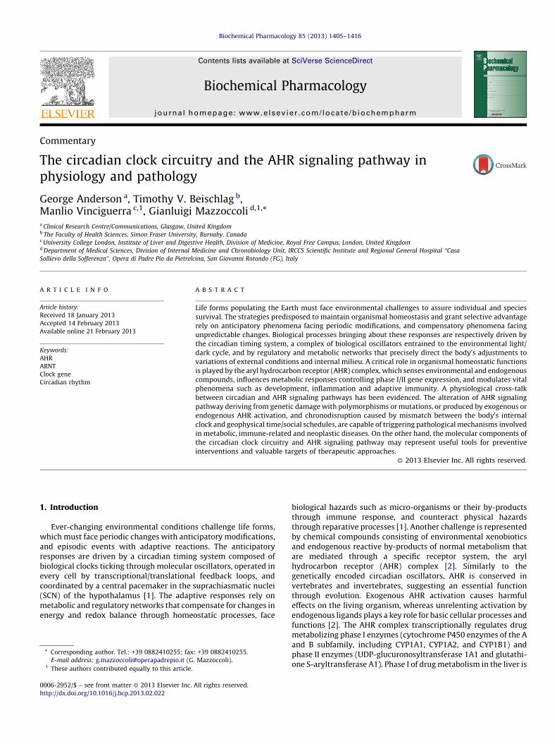
Fig. 1. The biological clock and AHR signaling pathway in pathological conditions. C
inflammation and diet, as well as genetic and epigenetic factors are shown to act on the
circadian gene interactions. Dysregulation of AHR/circadian/cAMP interactions can dri
Concurrent decreases in serotonin, melatonin, coupled to a decrease in alpha7 nicoti
depression, somatization and cognitive deficits. Alterations in immune regulation contr
tumor susceptibility. PDE2 inhibitors and a combination of valproate, melatonin and
processes. a7nAChr, alpha 7 nicotinic acetylcholine receptor; ADAM, A disintegrin a
monophosphate; IDO, indoleamine 2,3-dioxygenase; O&NS, oxidative and nitrosative stre
histocompatibility complex class I chain-related gene A/B; MMP, metalloproteinase; N
dioxygenase; Th, T helper.
2. The circadian clock circuitry
The various life forms on Earth are organized in hierarchicalmulti-component systems that contribute to maintenance ofhomeostasis. The functioning of the single components of organsystems show different time related variations, and these oscilla-tions realize a multi-frequency and multi-phase array of rhythms,coordinated by a circadian timing system. The circadian timingsystem drives body rhythmicity with a period of approximately 24 h(circadian), entrained principally by the light/dark cycle resultingfrom the rotation of our planet around its axis, through photic inputsperceived by melanopsin-containing retinal ganglion cells, andconveyed by the retino-hypothalamic tract [1]. Organ systemsfunction is driven by the central oscillators located in the SCN bymeans of humoral (cortisol, melatonin) and neural (autonomicnerve fibers) outputs, and peripheral cells are timed by self-sustained molecular clockworks directly influenced by circadiansystemic cues. The circadian clock circuitry provides synchroniza-tion among the several components of the organ systems, drivingnycthemeral oscillation of function in the neuro-endocrine system,the innate and adaptive arms of the immune system, the digestivesystem, the cardiovascular and urogenital apparatus. This affords ananticipatory spatio/temporal organization of circadian, metabolicand regulatory networks and allowing homeostatic processes towork at the right time and at their best [1].
Central and peripheral oscillators tick via an intracellularmolecular clockwork operated by a transcriptional/translationalfeedback loop relying on a set a genes (so called clock genes).
hronodisruption, toxins, endogenous AHR ligands, substance P, leptin resistance,
evolutionarily-driven role of the cAMP pathways in the co-ordination of AHR and
ve increased IDO, O&NS and inflammation, leading to altered immune regulation.
nic receptor activity drive changes in neuronal and immune activity, leading to
ibute to a tumor microenvironment, driving inflammatory disease association with
vitamin D are treatments that act on different stages of the inflammatory/tumor
nd metalloproteinase; AHR, aryl hydrocarbon receptor; cAMP, cyclic adenosine
ss; IFNg, interferon-gamma; kyn, kynurenine; KYNA, kynurenic acid; MICA/B, major
K, natural killer cells; Per, Period; TReg, regulatory T cell; TDO, tryptophan 2,3-

G. Anderson et al. / Biochemical Pharmacology 85 (2013) 1405–1416 1407
At the center of the core positive feedback loop, the basic helix-loop-helix/Per-Arnt-Sim (period circadian protein-aryl hydrocar-bon receptor nuclear translocator protein-single-minded protein,bHLH/PAS) transcription factors CLOCK/NPAS2 and BMAL1/2(ARNTL/ARNTL2) heterodimerize and bind to E-box enhancerelements in the promoters of the Period genes (Per1-3) andCryptochrome genes (Cry1 and Cry2), activating their transcription.The Per and Cry mRNAs give rise to PER and CRY proteins forming arepression complex that translocates back into the nucleus whereit hinders CLOCK:BMAL1 transcriptional activity [1].
The transcriptional heterodimers also drive the expression ofgenes encoding the nuclear receptors REV-ERBa/b and RORa/g,which in turn play negative and positive roles respectively in thetranscriptional limb of the clock gene machinery. The circadianproteins are post-translationally modified by processes of phos-phorylation (mediated by Casein kinase I d/e, AMP kinase, glycogensinthase kinase b), sumoylation, and ubiquitylation. Acetylation ofBMAL1 is mediated by CLOCK. This process is counteracted bySIRT1, a histone/protein deacetylase whose activity is dependenton the metabolic oscillator nicotinamide adenine dinucleotide(NAD+), synthesized from tryptophan via the catalytic activity ofnicotinamide phosphoribosyltransferase (NAMPT/visfatin), con-trolled by the biological clock [3]. The core clock genes drive theexpression of clock controlled genes encoding transcription factors(Dbp, Hlf, Tef, E4bp4, Dec1-2, Ppara, Pparg) and tissue specific outputgenes that control regulatory networks and homeostatic processes,as well as cell, tissue and organ systems functions [4,5].
3. The clock gene machinery and the AHR signaling pathway inmetabolism
A bidirectional cross-talk between circadian and metabolicnetworks, neural hard-wiring and hormones, cyto/chemokines,monoamines, receptors and post-receptor signaling pathwaysbrings about an intertwined modulation of working and phasing inthe circadian timing system. The alteration of this set ofconnections may be involved in the pathophysiological mecha-nisms triggered by mis-matched phasing of biological processesand behavioral cycles (rest/activity, sleep/wake, fasting/feeding) inrelation to environmental cues and social schedules (chronodis-ruption), leading to chronic sleep restriction and internaldesynchronization [4]. Challenges represented by environmentalchemical xenobiotics and endogenous reactive by-products ofnormal metabolism may heavily impinge on the array ofhomeostatic processes, and may play a key role in the mechanismsimplicated in the pathophysiology of metabolic, immune-relatedand neoplastic diseases. Several structurally diverse exogenousand endogenous chemical compounds activate AHR, also called thedioxin receptor, a ligand-activated transcriptional factor of thebHLH/PAS superfamily [6]. The PAS domain is a multifunctionalprotein motif governing ligand and DNA binding as well asinteractions between PAS and non-PAS proteins. Generally PASproteins function as heterodimers consisting of a sensor proteinassociated with a general binding partner. Members of the PASfamily of transcriptional regulators are involved in the control ofvaried biological processes such as morphogenesis, circadianrhythmicity, and responses to hypoxia and toxins, and play acritical role in development, sensing and adapting to environmen-tal conditions.
AHR is a 96 kDa protein encoded by a gene about 50 kb long andcontaining 11 exons, assigned to chromosome 7p15 [6], capable ofbinding a great variety of chemical substances. Exogenous ligandsare represented by halogenated dioxins, polychlorinated biphe-nyls, polycyclic aromatic hydrocarbons (PAHs), and related orunrelated compounds, among which the prototypic is representedby 2,3,7,8-tetrachlorodibenzo-p-dioxin (TCDD), environmentally
dispersed all over the world particularly at high trophic levelsalong the food chain. Natural compounds capable of binding AHRare principally represented by dietary chemicals, such as poly-phenols and in particular flavonoids (catechins, flavones, flavonols,flavanones, and isoflavones such as resveratrol), carotenoids,berberine. Endogenous ligands are represented by endogenouslysynthesized chemicals, such as indigoids, equilenin, arachidonicacid metabolites, heme metabolites and tryptophan metabolites(Table 1).
Concerning molecular functions, in its unliganded state AHR issequestered in the cytosol in a multiprotein heterotetrameric 9Score complex consisting of 90-kDa heat shock protein (HSP90),p23, c-Src and an immunophilin-like protein XAP2/ARA9 (alsoknown as the AHR-binding protein [AIP]) [7]. Upon bindinghydrophobic ligands entering through the cell membrane, AHRtranslocates in the nucleus and forms a heterodimer with ARNT [8].AHR/ARNT complex triggers the expression of genes encodingdrug-metabolizing and detoxifying enzymes, binding to specificDNA consensus sequence (50-T/GNGCGTGA/CG/CA-30; core se-quence 50-GCGTG-30) called xenobiotic responsive element (XRE)or dioxin responsive element (DRE) in the promoter. As a negativefeedback, AHR is exported from the nucleus to the cytosol anddegraded by proteasome–ubiquitin system [6]. The activation ofthe AHR also induces its own repressor, the AHRRepressor (AHRR),which although decreased in cancer cells is under-investigated [9].It is unknown if a circadian regulation of the AHRR occurs, andwhether this influences the levels of AHR circadian activation. Onthe other hand, variations in expression of many regulators andenzymes involved in drug metabolism and disposition have beenput in evidence: they are driven by the circadian clock andcharacterized by circadian rhythmicity, inducing nychthemeralchanges in the body detoxification capability [10–13], and in theresponse to drug treatment [14].
An intricate relationship has been shown between the clockgene machinery and AHR signaling pathway. AHR/ARNT and thecircadian protein BMAL1/ARNTL share structural similarities, andAHR has been proposed to influence the rhythmicity of biologicalclocks, also through its interaction with BMAL1, the activity ofwhich is altered in AHR�/� mice [15]. AHR expression and DNAbinding ability is characterized by 24-h oscillation, and CLOCKprotein modulates AHR activity affecting the toxin-induced anddosing time-dependent expression of detoxification enzymes,which is dampened in Clock mutant (Clk/Clk) mice [16,17]. AHR hasno effects on the biological clock in the absence of exogenousagonist, whereas AHR activation alters circadian rhythmicity andclock gene expression [18,19]. Xenobiotics hinder proper workingof the clock gene machinery via activation of the AHR pathway[20], and AHR interaction with BMAL1 in turn attenuates thetranscriptional activity of CLOCK:BMAL1 heterodimer, decreasingCLOCK binding at Ebox1 and Ebox3 in the Per1 promoter andcausing suppression of Per1 expression [21]. AHR activationinhibits the responsiveness of the biological clock to changes inenvironmental lighting, and dampens the phase response of thecircadian timekeeping system to light pulses by affecting the clockgene machinery [19]. However, studies performed with tissuesexplanted from PER2::LUCIFERASE mice show that activation ofthe AHR with TCDD does not alter the mouse biologicalclockwork[22]. Besides, in the liver PER1 and PER2 may induce oppositeeffects (negative and positive respectively), acting on AHR-mediated and ligand binding dependent AHR activation signaling,and playing an important role in the regulation of xenobioticresponses [23]. PER1 and PER2 modulate AHR mRNA levels [24]and disruption of Per1 and Per2 expression alters the AHR signalingpathway increasing the activation of cytochromes P450 familygenes [24,25]. In effect, TCDD activation of the AHR complexsignificantly increases the amplitude of Cyp1A1 during the night

Table 1Endogenous and exogenous ligands of aryl hydrocarbon receptor.
Exogenous
ligands
Synthetic
compounds
Polycyclic aromatic
hydrocarbons (PAHsa)
Naphthalene, anthracene, fluorene, phenanthrene,
benzo[a]pyrene, benzo[a]anthracene,
benzo[c]phenanthrene, pyrene, 3-methylcholanthrene,
chrysene.
Polychlorinated biphenyls 2,3,7,8-Tetrachlorodibenzo-p-dioxin (TCDD)
2,3,6,7-Tetrachloronaphthalene
1,2,3,6,7,8-Hexachlorodibenzo[b,e][1,4]dioxin
3,30 ,4,40-Tetrachloroazoxybenzene
3,30 ,4,40 ,5-Pentachlorobiphenyl
1,2,3,7,8-Pentachlorodibenzo[b,e][1,4]dioxin
2,3,7,8-Tetrachlorodibenzofuran
1,2,3,6,7,8-Hexachlorodibenzo[b,d]furan
1,2,3,4,6,7,8-Heptachlorodibenzo[b,d]furan
Benzimidazole derivates Omeprazole lansoprazole
8-Aminoquinoline Primaquine
Kinase inhibitors 1,4-Diamino-2,3-dicyano-1,4-bis[2-aminophenylthio]butadiene
Anthra[1,9-cd]pyrazol-6(2H)-one
2-(29-Amino-39-methoxyphenyl)-oxanaphthalen-4-one
(Z)-3-[(2,4-Dimethylpyrrol-5-yl)methylidenyl]-2-indolinone
Synthetic flavonoid 6,20 ,40-Trimethoxyflavone
30-Methoxy-40-nitroflavone
5,6-Benzoflavone (b-naphthoflavone)
7,8-Benzoflavone (a-naphthoflavone
Synthetic compounds 4-(3-Chloro-phenyl)-pyrimidin-2-yl]-(4-trifluoromethyl-phenyl)-amine,
ethyl 3-hydroxy-3-[2-(2-phenylethyl), diindolylmethane, benzoimidazol–
4-yl]propanoate, 4-hydroxy-tamoxifen,
6-Methyl-1,3,8-trichlorodibenzofuran, pesticides (acifluorfen-methyl,
bifenox, chlorpyrifos, isoxanthion, quinalphos, chlorpropham,
diethofencarb, propanil, diuron, linuron, prochloraz, carbaryl),
printing ink, ozone
Natural
compounds
Polyphenols Flavones (baicalein, apigenin)
Flavonoids (diosmin)
Flavonols (quercetin)
Flavanones (naringenin)
Isoflavones (daidzein, genistein, resveratrol)
Catechins (S)-epigallocatechin gallate, (S)-epigallocatechin,(S)-epicatechin gallate, (S)-epicatechinChlorophylls, lutein (carotenoid)
Antraquinones (emodin)
PropolisCurcumin (diferuloylmethane)
Alkaloids (harman, berberine)
Endogenous
ligands
Indigoids Indigo
Indirubin
Heme metabolites Bilirubin, biliverdin, hemin
Eicosanoids Lipoxin A4, prostagandins (B3, D3, F3a, G2, H1 and H2)
Tryptophan derivatives Tryptamin, indole acetic acid, 6-formylindolo[3,2-b]carbazole,
6,12-diformylindolo[3,2-b]carbazole, 2-(1-H-indole-3-carbonyl)-
thiazole-4-carboxylic acid methyl ester, kynurenic acid, xanthurenic acid
Inhibitory effect on the in vitro activation of AhR induced by TCDD given in bold.a PAHs contain aromatic hydrocarbons with two or more condensed benzene rings formed during thermal decomposition of organic compounds and their subsequent
recombination. PAHs can be of both natural (forest and rangeland fires, oil seeps, volcanic eruptions and exudates from trees) or anthropogenic origin (burning of fossil fuel,
wood, coal tar, garbage, used lubricating oil and oil filters, municipal solid waste incineration and petroleum spills, charbroiled food, and cigarette smoke).
G. Anderson et al. / Biochemical Pharmacology 85 (2013) 1405–14161408
compared to daytime in the mouse mammary gland and liver,driven by the circadian genes Per1 and Per2 [25], being anothermeans by which circadian genes interact with the AHR. Further-more, in vitro studies performed with serum-shocked HepG2 cellshave shown that CYP3A4 mRNA levels and metabolic activityoscillate rhythmically with approximately 24-h periodicity. At amolecular level, DBP and E4BP4 drive a reciprocating mechanismin which DBP activates the transcription of the CYP3A4 gene duringthe time of day when DBP is abundant, and E4BP4 suppresses thetranscription at other times of day. In this way the transactivationof the CYP3A4 gene by DBP is repressed by E4BP4, a negativecomponent of the circadian clock, providing a further molecularlink between the circadian clock and the xenobiotic metabolism[26].
Although endogenous and exogenous ligands of the AHR arecontinually being discovered, ligand independent regulation of theAHR is also important. AHR activation by cyclic adenosine 3050
monophosphate (cAMP) leads to AHR translocation from thecytosol to the nucleus [27], driving signaling significantly differentfrom that induced by exogenous dioxin ligands. In the SCN as wellas in peripheral mammalian tissues and cell lines cytoplasmiccAMP signaling is a core component of the circadian pacemaker.cAMP signaling constitutes an additional element of the oscillatorynetwork and impacts on amplitude, phase and period, as well asinducing Per1 and Per2 [28,29]. This suggests that the cAMPmodulation of AHR transcriptional activity will be co-ordinatedwith regulation of circadian genes. cAMP-mediated activation maybe the evolutionarily-derived primary endogenous regulator oftheAHR, with chronic dioxin binding to the AHR for a long span oftime, ranging from days to months, disrupting the cAMP-mediatedactivation, playing a crucial role in the mechanism of TCDD toxicity[30]. The compartmentalization of cAMP signaling in microdo-mains confers specificity to the role of cAMP, with exogenous AHRactivators then interacting with cAMP inducers [31]. The light

G. Anderson et al. / Biochemical Pharmacology 85 (2013) 1405–1416 1409
induced AHR endogenous ligand 6-formylindolo[3,2-b]carbazole(FICZ) is capable of influencing the circadian expression of clockgenes, the light entrainment and light-dependent regulation ofcircadian rhythm through AHR signaling activation [32], andrequires the cAMP pathway to increase ROS and IL-6 in mast cells[33]. Interestingly, AHR activation by PAHs such as benzo(a)pyrene(B(a)P) concurrently induces cAMP via b2-adrenoreceptor agon-ism, increasing intracellular concentration of ionized Ca2+ ([Ca2+]i),which is crucial to the specific effects of B(a)P on the AHR signalingpathway [34]. TCDD and PAHs increase intracellular concentrationof ([Ca2+]i), and the activity of the Ca2+/calmodulin (CaM)-dependent protein kinase (CaMK) Ia pathway plays an importantrole in the AHR-mediated genomic response [35]. Intriguingly,melatonin, one of the principal humoral outputs of the circadiantiming system, modulates the Ca2+/CaM signaling pathway eitherby changing intracellular calcium concentration via activation ofits G-protein coupled membrane receptors, or through a directinteraction with CaM [36].
Furthermore, a link between AHR signaling pathway, clockgenes, and glucose metabolism has been put in evidence,underlying the involvement of AHR activation, PPARa signaling,and alteration of the clock gene machinery in the development inhumans of type 2 diabetes related to xenobiotic exposure: AHRworks on the liver to control PPARa and modulates the clock genemachinery, PPARa influences AHR levels, BMAL1 and PPARa showinterdependent circadian regulation, and PPARa levels alterglucose metabolism and insulin sensitivity [15].
In summary, the multiple components and patterns offunctioning of the circadian and AHR signaling pathways showdaily variations, with oscillation of molecular levels and variationsof activity according to the time of day and the light–dark cycle.The circadian clock circuitry and the AHR complex influencesphysiological processes crucial for homeostasis maintenance, andthe derangement of these pathways is involved in pathophysio-logical mechanisms of disease.
4. The AHR signaling pathway, circadian genes andinflammatory and neoplastic diseases
The etiologies of most human inflammatory disorders andcancers remain elusive or continue to pose puzzling questions forbasic and clinical scientists, alike. The role of the AHR complex inthe promotion and genesis of several inflammatory disorders andcancers is indisputable and the epidemiological evidence for a roleinvolving wider circadian dysregulation in many such disorders isfairly convincing. As such, the similarities between the molecularmachineries regulating both systems present us with a provocativequestion: do the two pathways converge in such a way thatalterations in one put individuals at an increased risk of developingcertain inflammatory disorders or cancers? To put this topic inperspective there are several questions one might consider. First,how do circadian rhythms affect AHR activity? Moreover, whatimpact if any, will bHLH-PAS proteins regulating circadian function(hereafter referred to as circadian factors), as well as alterations inthe circadian rhythm or circadian genes have on chronic exposureto AHR activators such as TCDD, polychlorinated biphenyls (PCBs)and benzo[a]pyrene (B[a]P)? Next, does activation of AHR byenvironmental contaminants affect circadian rhythms or circadiangenes, and does this place an individual at increased risk ofdeveloping certain inflammatory disorders or cancers that can beassociated with disruption of circadian rhythm? Also it remains tobe clarified as to what is the normal role of the AHR in cellularphysiology? Finally, if there is an association with activated AHRand circadian-sensitive cancers, is this a causative or casualrelationship? We look at the data pertaining to these questions in
Sections 5 and 6, which deal with inflammatory disorders andcancers respectively.
5. The AHR complex, the clock gene machinery andinflammatory disorders
Many diseases are associated with changes in circadianregulation and AHR complex function, including Alzheimer’sdisease (AD) [37], bipolar disorder [38] and inflammatory boweldisease (IBD) [39].
Here we focus on the wider implications of changes in circadiangenes and AHR signaling pathway regulation in the context ofinflammatory disorders, focusing on IBD, including Crohn’s disease(CD) and ulcerative colitis (UC).
5.1. IDO, TDO and leptin
The intimate association of the cAMP, AHR complex andcircadian genes interacts with other processes relevant ininflammatory disorders and associated cancer susceptibility.Increased pro-inflammatory cytokines, including interleukin(IL)-1b, tumor necrosis factor (TNF)-a, IL-18 and especiallyinterferon (IFN)-g will increase levels of indoleamine 2,3-dioxygenase (IDO), contributing to immuno-suppression byincreasing regulatory T (TReg) cells. IDO takes tryptophan downthe kynurenine pathway, producing among other productskynurenine (kyn) and kynurenic acid (KYNA), which are AHRactivators. AHR activation by kyn or KYNA increases IDO,suggesting the emergence of a positive feedback loop. Increasedkyn/tryptophan ratio is commonly found in inflammatorydisorders, contributing to comorbid depression [39]. Over 60% ofcentral kyn is peripherally derived and readily crosses the blood–brain-barrier (BBB), where it is rapidly converted into KYNA. KYNAinhibits the alpha 7 nicotinic acetylcholine receptor (a7nAChr),decreasing cortex glutamate, acetylcholine and dopamine, in turndecreasing optimized cortex arousal and driving changes incognition [39]. Peripherally derived inflammatory processesinvolving AHR activation therefore significantly contribute tocentral changes in mood and cognition, common in inflammatorydisorders.
cAMP increases KYNA in astrocytes, indicative of tryptophan2,3-dioxygenase (TDO) induction [40]. TDO driven tryptophancatabolism is restricted to kyn and KYNA. TDO is very highlyexpressed in the liver, but is also evident in other tissues includingin most cancers [41]. This suggests that the cAMP regulation of theAHR and circadian genes will be co-ordinated with the regulationof tryptophan catabolites, producing kyn and KYNA that are knownactivators of the AHR, contributing to changes in neuronal activity,immune regulation, mood and cognition.
On the other hand, XAP2, one of AHR cytosolic partners, bindsthe cyclic nucleotide phosphodiesterases PDE2A and PDE4A5.PDE2A binds the AHR and inhibits its nuclear translocation. PDE2Adegrades cAMP and cGMP, suggesting that increased degradationof cyclic nucleotides concurrently inhibits the cAMP pathway andAHR activity [42]. PDE2A inhibitors are under extensive research inthe treatment of inflammatory processes, including in AD [43].
Furthermore, obesity and metabolic syndrome increase thesusceptibility to most other inflammatory disorders, including AD,IBD and many cancer types. Raised leptin levels are evident inobesity, resulting in leptin resistance mediated by increased cAMP[44]. It requires testing as to whether elevated cAMP in leptinresistance will modulate the AHR and its interactions withcircadian genes and the tryptophan catabolite (TRYCAT) pathways.The AHR signaling pathway is proposed to play a crucial role in theinteraction with western-type diet in the etiology and course ofobesity and metabolic syndrome [45], as well as linking diet and

G. Anderson et al. / Biochemical Pharmacology 85 (2013) 1405–14161410
immunity at the intestinal mucosal surfaces [46]. An increase insubstance P (SubP), a cAMP inducer, is common in manyinflammatory disorders and contributes to high levels of comorbiddepression [47], suggesting a role for neuropeptides in themodulation of AHR interactions with circadian genes duringinflammation. Interestingly, SubP shows a circadian oscillation indorsal root ganglion, under the transcriptional control of BMAL1:-CLOCK heterodimers [48] (Fig. 1).
5.2. IBD
CD and UC are chronic inflammatory diseases related to geneticpredisposition, with candidate genes including those that regulateinnate immunity and epithelial barrier function, driving adysregulated immune response to an environmental trigger [4].The AHR is a significant modulator of inflammatory responses inIBD, with AHR+/� mice being afforded protection versus wild typeand AHR�/� animals, suggesting therapeutic efficacy of AHRantagonists [49]. AHR agonists influence T lymphocytes differen-tiation and function [50], enhancing the T helper (h)2/Th1 ratio,resulting in a Th1 bias. AHR agonists also stimulate thedifferentiation of IL17 secreting Th17 cells, generating a potentiallyproinflammatory autoimmune environment. However, AHR acti-vation by TCDD is protective in a CD model decreasing IL-6, IL-12,TNF-a and IFN-g, by epigenetically increasing TReg cells,preferentially over the AHR induction of Th17 cells [51,52]. TheAHR also downregulates natural killer (NK) cell inflammatorycytokine production [53]. This evidence highlights the complex,and sometimes opposing, effects of AHR agonism in specific celltypes in inflammatory disorders in vivo, and suggests a powerfulrole for the AHR in determining the crucial differentiation of Th17versus TReg cells induction. It requires investigation as to therelationship to the type of ligand and its affinity to the AHR in thebalance of such opposing immune responses.
The circadian and cAMP regulation of the AHR drives anincrease in TDO/IDO induction of KYNA, in turn inhibiting thea7nAChr. This contributes not only to mood and cognitive changes[54], but is important in the regulation of UC itself. Nicotine, via theactivation of the a7nAChr on immune cells, provides anti-inflammatory effects in both obesity and UC [55].
Circadian disruption is common in IBD and in rodentsexacerbates induced colitis [4,56]. Circadian disruption is alsolikely to alter the dramatic AHR circadian rhythm, in turnimpacting on IDO regulation. IDO is increased around IBD lesions,correlating with disease severity [57]. Likewise an increase in thekyn/tryptophan ratio is found in CD, which positively correlateswith disease severity and lipid peroxidation [58]. Increased IDOwill induce immuno-suppression via tryptophan depletion andTReg cell induction, which is beneficial in IBD but may contributeto set up a microenvironment promoting tumorigenesis.
Leptin is upregulated in the colonic mucosa of IBD and hasdiverse roles in the gastrointestinal tract, including increasingproliferation and decreasing apoptosis [59]. Investigation isrequired as to whether local leptin resistance is induced, increasingcAMP, and in turn regulating the AHR, circadian genes and theirinteraction. The leptin receptor can be cleaved by a disintegrin andmetalloprotease (ADAM)10, which along with matrix metallopro-tease (MMP)-9 and ADAM17 is increased in tumors and IBD [60].How leptin resistance and altered cAMP/AHR/circadian geneinteractions modulate these proteases is important to investigateas it significantly regulates the local immune response and courseof IBD, and several MMPs are targets of the AHR pathway [61].
ADAM10 and MMP-9 cleave NKG2D ligands, leading to therelease of soluble major histocompatibility complex class I chain-related gene A (MICA) and MICB, which then act to inhibit NK cells,gdTCR bearing cells and some other T lymphocyte subsets,
contributing to immuno-suppression. Increased ADAM17 isevident in CD, contributing to the initiation and course via thecleaving of TNF-a [62]. Epithelial MICA and MICB mRNA areincreased in UC patients, as is the NKG2D on NK cells [63],suggesting an increased activation and induction of IFN-g by NKcells. However, such activation is modulated by palmitoylation andthe presence of MICA/B in caveolae, which increases their shedding[64]. Protease regulation of receptor shedding is important toinvestigate in inflammatory disorders and in the transition totumorigenic microenvironment. Preliminary data show an associ-ation of MICA single nucleotide polymorphisms (SNP) withsusceptibility to UC [65]. The cAMP and circadian gene modulationof the AHR signaling pathway will potentially influence AHRdependent regulation of immune responses, including modulationof dendritic cell TGF-B1 [66], which is known to decreaselymphocyte NKG2D levels [67], leading to an inhibition of immuneresponses during inflammation. The AHR is constitutivelyexpressed in human mast cells, increasing degranulation andcytokines in a cAMP dependent manner [33]. Mast cells have asignificant role in the etiology and course of IBD [68]. PER2 drivesmast cell circadian regulation, suggesting that cAMP driven AHRinteractions with circadian genes may also be evident in mast cells[69]. Increased SubP will contribute to mast cell degranulation,increasing VEGF induced membrane permeability as well asleukocyte chemoattraction and activation [70]. An increase in SubPis evident in CD and UC, correlating with symptom severity andcontributing to cAMP and depression [47].
As such, we could consider that the interaction of the AHRcomplex and circadian genes, including via their modulation bycAMP, is intimately involved in many aspects of inflammatorydisorders, exemplified here in IBD. The concurrent effects onimmune regulation via alterations in TDO and IDO are seeminglyimportant to the treatment of inflammatory disorders and their co-morbidities, as well as in the transition to microenvironment-induced cancer. The causal interactions of the AHR signalingpathway and circadian genes, including modulation by cAMP assuggested above, requires investigation and clarification by usingin vitro and in vivo experimental models
5.3. Some treatment implications: melatonin, valproate, vitamin D
Given the melatonin’s anti-inflammatory and anti-oxidanteffects, as well as its induction of mitochondrial oxidativephosphorylation, its expected benefits have been shown in anumber of inflammatory disorders, including IBD [71,72]. Adecrease in melatonin is commonly found in inflammatorydisorders, including in UC [73]. This is due to the driving oftryptophan down the kynurenine pathway and away fromserotonin and melatonin production, but also by increased TNF-a, which decreases pineal melatonin production. However,melatonin is also synthesized in the lower gut, which containsup to 400 times the melatonin levels of the pineal gland [74]. Assuch local changes in IDO and TDO may be of particular relevanceto gut melatonin. As well as being an integral circadian entrainer,melatonin also increases SIRT1 [75] and will therefore modulateSIRT1 interaction with CLOCK and circadian genes. Melatoninreceptors are negatively coupled to adenylyl cyclase, inhibitingcAMP as well as inhibiting AHR induction of CYP1 family genes[76]. This suggests that melatonin will impact on the cAMP/AHR/circadian gene interactions, including in the regulation of IDO andTDO, with its anti-oxidant effects inhibiting the ROS drivenincrease in proinflammatory cytokines.
Interestingly histone deacetylase (HDAC) inhibitors, includingvalproate, have shown efficacy in ameliorating different models ofexperimental colitis [77]. In cancer cells valproate can increase theplasma membrane expression of MICA/B, leading to enhance NK

G. Anderson et al. / Biochemical Pharmacology 85 (2013) 1405–1416 1411
cytotoxicity [78]. The possibility that this occurs in colonicepithelial cells requires investigation, including as to whetherthe adjunctive use of melatonin and/or vitamin D would modulateany impact on MICA/B levels/shedding or on ADAM10 and MMP-9cleavage of NKG2D ligands. Decreased vitamin D is common inmany inflammatory disorders, including IBD [79,80], wherevitamin D receptor SNPs increase IBD susceptibility [81]. Increasedvitamin D intake reduces IBD symptoms [80]. In the CNS, valproateincreases bcl-2 associated anthanogene-1 (BAG-1) and HSP70,which via their chaperone activity increase the transport ofvitamin D3 to the nuclear vitamin D receptor, increasing vitamin Deffects. Valproate also dramatically increases melatonin receptorlevels, either directly and/or via the induction of melatonin, whichinduces its own receptors [82]. This suggests that the utility ofvalproate in IBD, as well as cancers, may include its potentiation ofthe known efficacy of both melatonin and vitamin D. Many of theeffects of melatonin and valproate in inflammatory disorders andcancer are likely to be mediated by altering the interactions of theAHR, cAMP and circadian genes. How this occurs in specific celltypes over the course of inflammation and its transition todysplasia and cancer, including via alterations in TDO, IDO, leptinand neuropeptides awaits further investigation.
6. The biological clock and the AHR signaling pathway inneoplastic diseases
Human epidemiological studies and animal experimentalstudies have put in evidence that derangement of the circadianclock circuitry and circadian disruption play a role in carcinogene-sis. The alteration of proper alignment of physiological functions,behavioral cycles and environmental synchronizers (principallythe geophysical light/dark alternance) impedes the correct phasingof internal biological systems and external cues, leading tochronodisruption, which hinders appropriate maintenance ofinternal temporal order and alters the circadian organization oforgan systems function. On its side, AHR signaling pathway isinvolved in onset and progression of many different cancer types,and sensing external and internal chemical compounds mayrepresent an important transducer of environmental and interiormilieu, which might modulate the biological clock and interferewith the circadian pathways.
6.1. Circadian rhythmicity, AHR complex and cancer
It has been nearly three decades since the association betweenirregularities in diurnally sensitive melatonin levels and cancerwas first reported [83]. Twelve years later, Haldorsen and co-workers while investigating a possible link between radio wavesand breast cancer reported that chronological parameters may beassociated with higher breast cancer incidence in women [84].Since then, the association between exposure to light at night
(LAN), the disruption of the normal circadian clock and thedevelopment of certain types of cancer has drawn considerableattention, as discussed in the last section of the review.
Several case-controlled and cohort studies have found anassociation [85,86] or have suggested [87,88] a link between shiftwork and breast cancer. Indeed, NPAS2, the dimerization partner ofBMAL1/ARNTL, is a biomarker for triple negative breast cancer (+vesurvival) [89]. Similarly, epidemiological studies have found linksbetween shift work and other forms of cancer – particularly thosethat have strong links to AHR-mediated toxicities includingprostate [90], colo-rectal [91], lung cancer [92], and non-Hodgkinlymphoma [93]. In addition, associations have been found betweencircadian clock genes and lymphoma [94,95] and differentleukemias [96–98], respectively.
Associations between exposures to activators of the AHR andcancer have been fairly well established. The evidence for a linkbetween B[a]P exposure and other PAHs (particularly inhalational)is overwhelming and the mechanisms by which many of thesechemicals mediate oncogenesis is well understood [99]. Inaddition, case-controlled studies involving retired servicemenexposed directly or indirectly to chemical defoliant and TCDDprecursor, Agent Orange, found a significantly increased risk ofprostate and other cancers, particularly leukemias [100,101], whilean earlier study found the incidence of prostate cancer in theircohort was 41%, though they reported that this was not significant[102]. Finally, a ten-year mortality study of the Seveso populationstrongly suggested a link between TCDD exposure and leukemia[103].
Thus, the epidemiological evidence indicating associationsbetween circadian factors, AHR complex and several cancers israther provocative, but do AHR–circadian factors interactionsmediate the development or progression of human disease states,particularly cancer? In order to explore this further, we would liketo consider these potential associations in the context of twospecific examples; cancers of the blood (lymphomas andleukemias) and cancers of the breast.
6.2. Biochemical links between AHR complex and circadian factors
Circadian factors function and activation of AHR have bothbeen independently implicated in several cancers, but they alsoappear to converge on several cellular biochemical processes.Ovariectomized mice lacking AHR had a significantly shortercircadian period than wild-type counterparts [19]. Additionally,dioxin significantly altered the expression of Per1 and Bmal1 inthe SCN and liver of these animals. These observations maysuggest that; (i) AHR’s intrinsic activity is intimately linked tocircadian rhythmicity; (ii) exposure to endogenous activators ofAHR will disrupt this function, and; (iii) if this system ishormonally regulated, this might explain why certain tissues(i.e. breast and prostate) are more sensitive to disruption ofcircadian factors and, therefore at increased risk of cancer. Thelast point is supported by the observation that women harboringselective single nucleotide polymorphisms in the Cry2 gene hadsignificantly higher risk of estrogen receptor (ER)/progesteronereceptor (PR)-negative breast cancer [104]. This same studyindicated that the Cry2 promoter becomes more methylated inthese patients. Thus, it may be worthy to consider whetheractivators of AHR influence the tumor suppressor capabilities ofcircadian factors by virtue of their disruption of endocrinesignaling. If true, this may suggest that AHR intrinsicallymediates circadian factors-dependent regulation of certainendocrine systems.
While no direct evidence exists for AHR mediating cancersdirectly associated to bHLH-PAS proteins regulating circadianrhythms there are strong mechanistic links between AHR functionand Period gene expression. Per2 knock-out mice die prematurelyand are sensitive to gamma irradiation with a large percentagesuffering from lymphoma [105]. Indeed, PER2 down-regulationhas been implicated in human B-cell lymphoma [106] and myeloidleukemia [97,107]. Conversely, activation or deregulation of AHRhas been associated with the development of lymphomas andleukemia in rodents and man [108–112]. TCDD interferes with thecircadian-dependent production of myeloid progenitor cells inC57BL/6J mice with an associated and significant circadian-dependent decrease in Per2 mRNA accumulation [18]. Further-more, the AHR promoter was found to be hyper-methylated in thecancerous cells of a significant percentage of patients sufferingfrom mantle cell lymphoma and methylation negatively correlatedwith survival [113].

G. Anderson et al. / Biochemical Pharmacology 85 (2013) 1405–14161412
While the mechanism(s) by which AHR regulates PER1/2expression is unknown, we do know that activation of AHR resultsin an increase in CYP1A2 in the liver which in turn is capable ofmetabolizing melatonin [114]. Melatonin has been shown toinduce the circadian-dependent expression of PER1 and PER2 inhuman MCF10A and MCF7 breast cancer cells [115]. Thus, thenormal physiological role of AHR may include mediating thenormal activity of PER proteins and downstream circadian factors.Prolonged activation by chronic exposure to exogenous environ-mental toxins or epigenetic silencing could disrupt these functions.This link is strengthened if we consider that disruption of Per1 orPer2 in mouse liver allows for increased dioxin-inducible CYP1A1and CYP1B1 mRNA accumulation [23]. Furthermore, the diurnalrhythmicity of these enzymes is lost in mice lacking Per1/2 [25].
6.3. Transcriptional interference
We have touched on the subject of AHR activation andmelatonin metabolism. Certainly, cellular metabolism of otherendogenous biochemicals and the impact on cellular signaltransduction pathways by activated AHR may play a role incircadian rhythms and the etiologies of cancer. However, giventhat the functional nature of bHLH-PAS proteins includes mediat-ing gene activation, it is worthwhile to exam the ability of thesefactors to interact at the transcriptional level. AHR’s intrinsicactivity impacts several signaling pathways that mediate hormon-al and inflammatory responses among others and likely contributeto the etiology of certain cancers. Where these pathways intersectwith circadian pathways, there exists the potential for interactions.
Early dimerization studies indicated that AHR does not interactdirectly with ARNTL/BMAL1 [116]. Likewise, AHR’s primarydimerization partner ARNT, was unable to interact with NPAS2[117] or CLOCK [116]. Thus, simple competition for dimerizationpartners is unlikely to account for transcriptional interferencebetween signaling pathways. AHR and circadian factors pathwaysdo share common pools of coactivators including cAMP, ashighlighted above, cAMP response element binding (CREB)-binding protein (p300/CBP) [118–121], p300/CBP-associatedfactor (PCAF) [122,123] and p/CIP (ACTR, SRC3) [123–125], andgiven the promiscuity of most co-activators, likely many others.We have never observed co-activator pools to be rate limiting (T.Beischlag, personal observation). However, shared co-activatorsprovide multiple interaction platforms allowing for the possibilityof additional factors (i.e. AHR, ARNT, etc.) to incorporate intotranscriptional machinery [126,127].
Indeed, non-DNA binding roles for AHR and ARNT have beendescribed for both estrogen receptor function and NFkB [128–132]. While we reported that TCDD-activated AHR and estrogen-activated ER could mutually repress the transcription of the other’starget genes [128], Matthews et al. have reported that activated ERenhances dioxin-mediated transcription [131] in several humancancer cell lines. It is likely that these effects are context specific,but highlights the reality that different factors must come into playin different cell types.
A notable example of circadian factors and AHR convergence ata hormonal signaling pathway is the case of CLOCK and ERa.Sumoylated CLOCK has been shown to directly interact with ER toup-regulate its activity as well as enhance estradiol (E2)-mediatedproliferation of MCF7 breast cancer cells [133]. Whether CLOCK ispresent at estrogen-responsive promoters is unknown, but thenature of this phenomenon warrants further investigation. IfCLOCK is capable of interacting with the ER transcriptionalmachinery, it would be intriguing to know how sumoylationstatus affects recruitment of CLOCK and other factors such as AHRand ARNT to ER responsive gene promoters. The ability of AHR toimpact E2 signaling both positively and negatively has been well
established and much of this evidence is reviewed in detailelsewhere [126,134,135]. In addition, ARNT has pleiotropic effectson ER function [130,136] and both activation of AHR and circadianfluctuations could have profound impact on these activities.
We should also consider interactions that have not been fullyexplored and the heterogeneous nature of transcription machin-ery. Data collected in our lab suggests that ARNTL can be recordedover the CYP1A1 and PS2 promoters during dioxin and estrogen-activated transcription by chromatin immune-precipitation (Beis-chlag et al., unpublished data). Whether this is through a directinteraction with AHR/ER is unclear and issues regarding specificityof the Ab need to be addressed. Nevertheless, this interactionrequires closer scrutiny and it appears that many factors commonto both the AHR and circadian factors transcriptional machinerieswill likely converge at genes regulated by multiple classes oftranscription factors. The pleiotropic nature of the bHLH-PASproteins makes plausible the hypotheses that these factors mightinteract. As such, the deregulation of any factor could contribute tothe etiology of a disease associated or mediated by another familymember.
AHR and circadian factors also interact with NF-kB, a mediatorof both inflammatory signaling and neoplastic cell transformation.The NF-kB family of transcription factors consists of five members:RelA (p65), c-Rel, RelB, p50/p105 and p52/p100. Dimers of theseproteins are kept inactive by interacting with inhibitory proteins ofthe IkB family, while the IkB kinase (IKK) complex can target theIkB proteins for proteosome-mediated degradation [49].
Increased phosphorylation of p65 at S276 residue and activa-tion of NF-kB signaling with constitutive elevation of proinflam-matory cytokines in a cell-autonomous manner has been put inevidence by studies performed in Cry1�/�Cry2�/� cells. Absence ofthe circadian protein CRY releases its inhibition on cAMPproduction, elevates cAMP and increases protein kinase A (PKA)signaling, suggesting a patho-physiological connection betweendisruption of the circadian clock circuitry and increased suscepti-bility to chronic inflammatory diseases [137].
AHRs ability to interact with NFkB is well documented[138,139]. Indeed, not only did p50/RelA and AHR co-immunopre-cipitate in the absence of an activator of AHR but they were alsoshown to regulate the expression of the c-myc proto-oncogene innon-malignant and neoplastic human breast cell lines [140]. Inaddition, RelB can directly interact with ARNTL/BMAL1 [141] as itheterodimerizes with CLOCK repressing the expression of Per andCry. Conversely, this same study demonstrated that LPS-stimulated(NFkB-dependent) expression of NFkB target genes including IL-6,was severely impaired in CLOCK mutant embryonic fibroblasts.TCDD enhances PMA-inducible IL-6 expression in endometrialcarcinoma ECC-1 cells and AHR can be recorded over the IL-6
promoter under these conditions [129]. These factors alsoconverge to regulate IL-8 gene expression in human breast cancercells [142]. These interactions may not occur in trans or cis, in factsome of the interactions between these transcription factors maynot occur in the nucleus. In the absence of an exogenous activator,most if not all of AHR is cytosolic in many cell lines [126]. However,if these factors are directly interacting with the NFkB transcrip-tional machinery, it is likely that they are present on NFkB targetgene promoters at the same time. Nevertheless, while these casualassociations may not be overly compelling, they are provocative.
6.4. The biological clock, melatonin, and the AHR complex
The pineal neurohormone, melatonin, is a downstreamtranscriptional target of the clock gene machinery and circadiansignaling pathway in the SCN. Thus, expression of melatonin issensitive to circadian fluctuations and can be inhibited by light.Indeed, disruption of the biological clock leads to dysregulation of

G. Anderson et al. / Biochemical Pharmacology 85 (2013) 1405–1416 1413
circulating melatonin levels. In particular, the transcriptionalactivator CLOCK and/or CLOCK analogs serve to positively regulatemelatonin production, but they also induce the transcription ofPer1, Cry1 and Cry2 genes, which encode circadian proteins that inturn suppress melatonin expression [143,144]. Similarly, there is agrowing body of information concerning the role of melatonin inthe regulation of circadian factors [144].
Melatonin helps synchronize the biological clock, regulatessleep and has anti-oxidant properties as well as playing a role inimmune function [143]. Disruption of melatonin signaling hasbeen linked to several pathologies discussed above as well ascancer [145,146]. Low melatonin level has been linked to ER/PRpositive breast cancers [147]. Furthermore, melatonin promotesgrowth inhibition in cancer cells by a variety of mechanisms[148,149].
Activation of AHR by many exogenous environmental con-taminants results in the induction of CYP1A2 [8], and importantly,CYP1A2 can account for the majority of the biotransformation ofmelatonin entering the hepatic circulation [150]. Thus, activationof AHR could synergize with LAN to reduce circulating melatoninlevels. However, the potential for further interactions betweenAHR and circadian signaling pathways exist.
In spite of a clear mechanistic link between circadian factorsand tumorigenesis, and a dearth of evidence linking AHR tocircadian factors-linked cancers, the circumstantial evidencesupporting a role for AHR in circadian factors-associated cancersis appealing. Future studies that will help elucidate the linksbetween circadian factors and AHR will likely need to employmutant mice using crosses of knock-out and transgenic hyper-morphs matched to the appropriate cancer model. These studiesshould focus on how loss or over-expression and activation of AHRaffect the development of LAN-sensitive cancers.
7. Conclusion and perspectives
A temporal dimension characterizes most biological phenome-na commonly with a 24-h periodicity driven by environmentalcues, in particular the light/dark cycle dictated by Earth’s rotationaround its axis. The array of rhythms is generated by sub-cellularand genetically encoded biological oscillators orchestrated by thecircadian timing system that also drives behavioral cycles of sleep/wake, rest/activity, and fasting/feeding. Circadian rhytmicitycharacterizes functioning of the AHR complex, involved in sensingand biotransformation of environmental xenobiotics and endoge-nous metabolic byproducts. The correct functioning of circadianand AHR signaling pathways is crucial for the maintenance of bodyhomeostasis and for the preservation of body health. The alterationof nychthemeral rhythmicity causes circadian disruption andinternal desynchronization, leading in due course to degenerative,inflammatory and neoplastic diseases. Similarly, the derangementand disorganization of time related homeostatic mechanismsinvolving circadian and AHR signaling pathways play a key role inseveral pathophysiological mechanisms. On the other hand, AHRcan be activated by several, structurally different chemicals andmay represent a promising target for therapeutic intervention.Addressment of the circadian clock circuitry by lifestyle modifica-tion and proper timing of behavioral cycles, alongside chronomo-dulated drug administration, as well as modulation of AHRcomplex activity through administration of agonist/antagonistligands as appropriate, could represent a new frontier inpreventive medicine and pharmacotherapy, opening new waysin the treatment of a great number of diseases.
Conflict of interest statement
The authors declare that they have no competing interests.
Acknowledgements
We apologize to all those researchers whose work could not becited and for not comment on all of the relevant studies due tospace limitation. For in depth explanations of chronobiologic termsused in the review we suggest to the reader to refer to the web siteof the American Association of Medical Chronobiology andChronotherapeutics at http://www.aamcc.net/glossary.htm. Wethank Ms. Sandra Rose Gunn for proofreading the manuscript. Thiswork was supported by ‘‘Italian Ministry of Health’’ grantRC1201ME04 through Department of Medical Sciences, Divisionof Internal Medicine and Chronobiology Unit, IRCCS ScientificInstitute and Regional General Hospital ‘‘Casa Sollievo dellaSofferenza’’, Opera di Padre Pio da Pietrelcina, San GiovanniRotondo (FG), Italy.
References
[1] Mazzoccoli G, Pazienza V, Vinciguerra M. Clock genes and clock-controlledgenes in the regulation of metabolic rhythms. Chronobiol Int 2012;29:227–51.
[2] Shimba S, Watabe Y. Crosstalk between the AHR signaling pathway andcircadian rhythm. Biochem Pharmacol 2009;77:560–5.
[3] Vinciguerra M, Santini MP, Martinez C, Pazienza V, Claycomb WC, Giuliani A,et al. mIGF-1/JNK1/SirT1 signaling confers protection against oxidative stressin the heart. Aging Cell 2012;11:139–49.
[4] Mazzoccoli G, Palmieri O, Corritore G, Latiano T, Bossa F, Scimeca D, et al.Association study of a polymorphism in clock gene PERIOD3 and risk ofinflammatory bowel disease. Chronobiol Int 2012;29:994–1003.
[5] Bozek K, Relogio A, Kielbasa SM, Heine M, Dame C, Kramer A, et al. Regulationof clock-controlled genes in mammals. PLoS ONE 2009;4:e4882.
[6] Stejskalova L, Dvorak Z, Pavek P. Endogenous and exogenous ligands of arylhydrocarbon receptor: current state of art. Curr Drug Metab 2011;12:198–212.
[7] Meyer BK, Pray-Grant MG, Vanden Heuvel JP, Perdew GH. Hepatitis B virus X-associated protein 2 is a subunit of the unliganded aryl hydrocarbon receptorcore complex and exhibits transcriptional enhancer activity. Mol Cell Biol1998;18:978–88.
[8] Hankinson O. The aryl hydrocarbon receptor complex. Annu Rev PharmacolToxicol 1995;35:307–40.
[9] Zudaire E, Cuesta N, Murty V, Woodson K, Adams L, Gonzalez N, et al. The arylhydrocarbon receptor repressor is a putative tumor suppressor gene inmultiple human cancers. J Clin Invest 2008;118:640–50.
[10] Kanno Y, Otsuka S, Hiromasa T, Nakahama T, Inouye Y. Diurnal difference inCAR mRNA expression. Nucl Recept 2004;2:6.
[11] Gachon F, Olela FF, Schaad O, Descombes P, Schibler U. The circadian PAR-domain basic leucine zipper transcription factors DBP, TEF, and HLF modulatebasal and inducible xenobiotic detoxification. Cell Metab 2006;4:25–36.
[12] Claudel T, Cretenet G, Saumet A, Gachon F. Crosstalk between xenobioticsmetabolism and circadian clock. FEBS Lett 2007;581:3626–33.
[13] Zmrzljak UP, Rozman D. Circadian regulation of the hepatic endobiotic andxenobitoic detoxification pathways: the time matters. Chem Res Toxicol2012;25:811–24.
[14] Levi F, Schibler U. Circadian rhythms: mechanisms and therapeutic implica-tions. Annu Rev Pharmacol Toxicol 2007;47:593–628.
[15] Wang C, Xu CX, Krager SL, Bottum KM, Liao DF, Tischkau SA. Aryl hydrocarbonreceptor deficiency enhances insulin sensitivity and reduces PPAR-alphapathway activity in mice. Environ Health Perspect 2011;119:1739–44.
[16] Huang P, Ceccatelli S, Rannug A. A study on diurnal mRNA expression ofCYP1A1, AHR, ARNT, and PER2 in rat pituitary and liver. Environ ToxicolPharmacol 2002;11:119–26.
[17] Tanimura N, Kusunose N, Matsunaga N, Koyanagi S, Ohdo S. Aryl hydrocarbonreceptor-mediated Cyp1a1 expression is modulated in a CLOCK-dependentcircadian manner. Toxicology 2011;290:203–7.
[18] Garrett RW, Gasiewicz TA. The aryl hydrocarbon receptor agonist 2,3,7,8-tetrachlorodibenzo-p-dioxin alters the circadian rhythms, quiescence, andexpression of clock genes in murine hematopoietic stem and progenitor cells.Mol Pharmacol 2006;69:2076–83.
[19] Mukai M, Lin TM, Peterson RE, Cooke PS, Tischkau SA. Behavioral rhythmicityof mice lacking AhR and attenuation of light-induced phase shift by 2,3,7,8-tetrachlorodibenzo-p-dioxin. J Biol Rhythms 2008;23:200–10.
[20] Tischkau SA, Jaeger CD, Krager SL. Circadian clock disruption in the mouseovary in response to 2,3,7,8-tetrachlorodibenzo-p-dioxin. Toxicol Lett2011;201:116–22.
[21] Xu CX, Krager SL, Liao DF, Tischkau SA. Disruption of CLOCK-BMAL1 tran-scriptional activity is responsible for aryl hydrocarbon receptor-mediatedregulation of Period1 gene. Toxicol Sci 2010;115:98–108.
[22] Pendergast JS, Yamazaki S. The mammalian circadian system is resistant todioxin. J Biol Rhythms 2012;27:156–63.

G. Anderson et al. / Biochemical Pharmacology 85 (2013) 1405–14161414
[23] Qu X, Metz RP, Porter WW, Cassone VM, Earnest DJ. Disruption of period geneexpression alters the inductive effects of dioxin on the AhR signaling pathwayin the mouse liver. Toxicol Appl Pharmacol 2009;234:370–7.
[24] Qu X, Metz RP, Porter WW, Cassone VM, Earnest DJ. Disruption of clock geneexpression alters responses of the aryl hydrocarbon receptor signaling path-way in the mouse mammary gland. Mol Pharmacol 2007;72:1349–58.
[25] Qu X, Metz RP, Porter WW, Neuendorff N, Earnest BJ, Earnest DJ. The clockgenes period 1 and period 2 mediate diurnal rhythms in dioxin-inducedCyp1A1 expression in the mouse mammary gland and liver. Toxicol Lett2010;196:28–32.
[26] Takiguchi T, Tomita M, Matsunaga N, Nakagawa H, Koyanagi S, Ohdo S.Molecular basis for rhythmic expression of CYP3A4 in serum-shocked HepG2cells. Pharmacogenet Genomics 2007;17:1047–56.
[27] Oesch-Bartlomowicz B, Oesch F. Role of cAMP in mediating AHR signaling.Biochem Pharmacol 2009;77:627–41.
[28] O‘Neill JS, Maywood ES, Chesham JE, Takahashi JS, Hastings MH. cAMP-dependent signaling as a core component of the mammalian circadianpacemaker. Science 2008;320:949–53.
[29] Beaule C, Swanstrom A, Leone MJ, Herzog ED. Circadian modulation of geneexpression, but not glutamate uptake, in mouse and rat cortical astrocytes.PLoS ONE 2009;4:e7476.
[30] Oesch-Bartlomowicz B, Huelster A, Wiss O, Antoniou-Lipfert P, Dietrich C,Arand M, et al. Aryl hydrocarbon receptor activation by cAMP vs dioxin:divergent signaling pathways. Proc Natl Acad Sci USA 2005;102:9218–23.
[31] Oesch-Bartlomowicz B, Oesch F. Phosphorylation of cytochromes P450: firstdiscovery of a posttranslational modification of a drug-metabolizing enzyme.Biochem Biophys Res Commun 2005;338:446–9.
[32] Mukai M, Tischkau SA. Effects of tryptophan photoproducts in the circadiantiming system: searching for a physiological role for aryl hydrocarbonreceptor. Toxicol Sci 2007;95:172–81.
[33] Sibilano R, Frossi B, Calvaruso M, Danelli L, Betto E, Dall’Agnese A, et al. Thearyl hydrocarbon receptor modulates acute and late mast cell responses. JImmunol 2012;189:120–7.
[34] Mayati A, Levoin N, Paris H, N’Diaye M, Courtois A, Uriac P, et al. Induction ofintracellular calcium concentration by environmental benzo(a)pyrene involvesa beta2-adrenergic receptor/adenylyl cyclase/Epac-1/inositol 1,4,5-trispho-sphate pathway in endothelial cells. J Biol Chem 2012;287:4041–52.
[35] Monteiro P, Gilot D, Le Ferrec E, Rauch C, Lagadic-Gossmann D, Fardel O.Dioxin-mediated up-regulation of aryl hydrocarbon receptor target genes isdependent on the calcium/calmodulin/CaMKIalpha pathway. Mol Pharmacol2008;73:769–77.
[36] Dai J, Inscho EW, Yuan L, Hill SM. Modulation of intracellular calcium andcalmodulin by melatonin in MCF-7 human breast cancer cells. J Pineal Res2002;32:112–9.
[37] Cermakian N, Lamont EW, Boudreau P, Boivin DB. Circadian clock geneexpression in brain regions of Alzheimer’s disease patients and controlsubjects. J Biol Rhythms 2011;26:160–70.
[38] Osland TM, Ferno J, Havik B, Heuch I, Ruoff P, Laerum OD, et al. Lithiumdifferentially affects clock gene expression in serum-shocked NIH-3T3 cells. JPsychopharmacol 2011;25(7):924–33. http://dx.doi.org/10.1177/0269881110379508.
[39] Anderson G, Maes M, Berk M. Inflammation-related disorders in the trypto-phan catabolite pathway in depression and somatization. Adv Protein ChemStruct Biol 2012;88:27–48.
[40] Luchowska E, Kloc R, Olajossy B, Wnuk S, Wielosz M, Owe-Larsson B, et al.beta-adrenergic enhancement of brain kynurenic acid production mediatedvia cAMP-related protein kinase A signaling. Prog NeuropsychopharmacolBiol Psychiatry 2009;33:519–29.
[41] Opitz CA, Litzenburger UM, Sahm F, Ott M, Tritschler I, Trump S, et al. Anendogenous tumour-promoting ligand of the human aryl hydrocarbon re-ceptor. Nature 2011;478:197–203.
[42] de Oliveira SK, Hoffmeister M, Gambaryan S, Muller-Esterl W, Guimaraes JA,Smolenski AP. Phosphodiesterase 2A forms a complex with the co-chaperoneXAP2 and regulates nuclear translocation of the aryl hydrocarbon receptor. JBiol Chem 2007;282:13656–63.
[43] Sierksma AS, Rutten K, Sydlik S, Rostamian S, Steinbusch HW, van den HoveDL, et al. Chronic phosphodiesterase type 2 inhibition improves memory inthe APPswe/PS1dE9 mouse model of Alzheimer’s disease. Neuropharmacol-ogy 2013;64:124–36.
[44] Fukuda M, Williams KW, Gautron L, Elmquist JK. Induction of leptin resis-tance by activation of cAMP-Epac signaling. Cell Metab 2011;13:331–9.
[45] Kerley-Hamilton JS, Trask HW, Ridley CJ, Dufour E, Ringelberg CS, Nurinova N,et al. Obesity is mediated by differential aryl hydrocarbon receptor signalingin mice fed a Western diet. Environ Health Perspect 2012;120:1252–9.
[46] Kiss EA, Vonarbourg C. Aryl hydrocarbon receptor: a molecular link betweenpostnatal lymphoid follicle formation and diet. Gut Microbes 2012;3:577–82.
[47] Tavano F, di Mola FF, Latiano A, Palmieri O, Bossa F, Valvano MR, et al.Neuroimmune interactions in patients with inflammatory bowel diseases:disease activity and clinical behavior based on substance P serum levels. JCrohns Colitis 2012;6:563–70.
[48] Zhang J, Li H, Teng H, Zhang T, Luo Y, Zhao M, et al. Regulation of peripheralclock to oscillation of substance P contributes to circadian inflammatory pain.Anesthesiology 2012;117:149–60.
[49] Arsenescu R, Arsenescu V, Zhong J, Nasser M, Melinte R, Dingle RW, et al. Roleof the xenobiotic receptor in inflammatory bowel disease. Inflamm Bowel Dis2012;17:1149–62.
[50] Rohlman D, Pham D, Yu Z, Steppan LB, Kerkvliet NI. Aryl hydrocarbonreceptor-mediated perturbations in gene expression during early stages ofCD4(+) T-cell differentiation. Front Immunol 2012;3:223.
[51] Benson JM, Shepherd DM. Aryl hydrocarbon receptor activation by TCDDreduces inflammation associated with Crohn’s disease. Toxicol Sci2011;120:68–78.
[52] Singh NP, Singh UP, Singh B, Price RL, Nagarkatti M, Nagarkatti PS. Activationof aryl hydrocarbon receptor (AhR) leads to reciprocal epigenetic regulationof FoxP3 and IL-17 expression and amelioration of experimental colitis. PLoSONE 2011;6:e23522.
[53] Monteleone I, MacDonald TT, Pallone F, Monteleone G. The aryl hydrocarbonreceptor in inflammatory bowel disease: linking the environment to diseasepathogenesis. Curr Opin Gastroenterol 2012;28:310–3.
[54] Wells CW, Lewis S, Barton JR, Corbett S. Effects of changes in hemoglobin levelon quality of life and cognitive function in inflammatory bowel diseasepatients. Inflamm Bowel Dis 2006;12:123–30.
[55] Lakhan SE, Kirchgessner A. Anti-inflammatory effects of nicotine in obesityand ulcerative colitis. J Transl Med 2011;9:129.
[56] Swanson GR, Burgess HJ, Keshavarzian A. Sleep disturbances and inflamma-tory bowel disease: a potential trigger for disease flare. Expert Rev ClinImmunol 2011;7:29–36.
[57] Zhou L, Chen H, Wen Q, Zhang Y. Indoleamine 2,3-dioxygenase expression inhuman inflammatory bowel disease. Eur J Gastroenterol Hepatol2012;24:695–701.
[58] Gupta NK, Thaker AI, Kanuri N, Riehl TE, Rowley CW, Stenson WF, et al. Serumanalysis of tryptophan catabolism pathway: correlation with Crohn’s diseaseactivity. Inflamm Bowel Dis 2012;18:1214–20.
[59] Yarandi SS, Hebbar G, Sauer CG, Cole CR, Ziegler TR. Diverse roles of leptin inthe gastrointestinal tract: modulation of motility, absorption, growth, andinflammation. Nutrition 2011;27:269–75.
[60] Lakatos G, Hritz I, Varga MZ, Juhasz M, Miheller P, Cierny G, et al. The impactof matrix metalloproteinases and their tissue inhibitors in inflammatorybowel diseases. Dig Dis 2012;30:289–95.
[61] Kung T, Murphy KA, White LA. The aryl hydrocarbon receptor (AhR) pathwayas a regulatory pathway for cell adhesion and matrix metabolism. BiochemPharmacol 2009;77:536–46.
[62] Cesaro A, Abakar-Mahamat A, Brest P, Lassalle S, Selva E, Filippi J, et al.Differential expression and regulation of ADAM17 and TIMP3 in acuteinflamed intestinal epithelia. Am J Physiol Gastrointest Liver Physiol2009;296:G1332–43.
[63] Ge LQ, Jiang T, Zhao J, Chen ZT, Zhou F, Xia B. Upregulated mRNA expression ofmajor histocompatibility complex class I chain-related gene A in colon andactivated natural killer cells of Chinese patients with ulcerative colitis. J DigDis 2011;12:82–9.
[64] Aguera-Gonzalez S, Gross CC, Fernandez-Messina L, Ashiru O, Esteso G, HangHC, et al. Palmitoylation of MICA, a ligand for NKG2D, mediates its recruit-ment to membrane microdomains and promotes its shedding. Eur J Immunol2011;41:3667–76.
[65] Lopez-Hernandez R, Valdes M, Lucas D, Campillo JA, Martinez-Garcia P,Salama H, et al. Association analysis of MICA gene polymorphism andMICA-129 dimorphism with inflammatory bowel disease susceptibility ina Spanish population. Hum Immunol 2010;71:512–4.
[66] Simones T, Shepherd DM. Consequences of AhR activation in steady-statedendritic cells. Toxicol Sci 2011;119:293–307.
[67] Zocchi MR, Catellani S, Canevali P, Tavella S, Garuti A, Villaggio B, et al. HighERp5/ADAM10 expression in lymph node microenvironment and impairedNKG2D ligands recognition in Hodgkin lymphomas. Blood 2012;119:1479–89.
[68] Kurashima Y, Amiya T, Nochi T, Fujisawa K, Haraguchi T, Iba H, et al.Extracellular ATP mediates mast cell-dependent intestinal inflammationthrough P2X7 purinoceptors. Nat Commun 2012;3:1034.
[69] Nakamura Y, Harama D, Shimokawa N, Hara M, Suzuki R, Tahara Y, et al.Circadian clock gene Period2 regulates a time-of-day-dependent variation incutaneous anaphylactic reaction. J Allergy Clin Immunol 2011;127:1038–45.e1–3.
[70] Theoharides TC, Zhang B, Kempuraj D, Tagen M, Vasiadi M, Angelidou A, et al.IL-33 augments substance P-induced VEGF secretion from human mast cellsand is increased in psoriatic skin. Proc Natl Acad Sci USA 2010;107:4448–53.
[71] Motilva V, Garcia-Maurino S, Talero E, Illanes M. New paradigms in chronicintestinal inflammation and colon cancer: role of melatonin. J Pineal Res2011;51:44–60.
[72] Chojnacki C, Wisniewska-Jarosinska M, Walecka-Kapica E, Klupinska G,Jaworek J, Chojnacki J. Evaluation of melatonin effectiveness in the adjuvanttreatment of ulcerative colitis. J Physiol Pharmacol 2011;62:327–34.
[73] Chen M, Mei Q, Xu J, Lu C, Fang H, Liu X. Detection of melatonin andhomocysteine simultaneously in ulcerative colitis. Clin Chim Acta2012;413:30–3.
[74] Chen CQ, Fichna J, Bashashati M, Li YY, Storr M. Distribution, function andphysiological role of melatonin in the lower gut. World J Gastroenterol2011;17:3888–98.
[75] Tajes M, Gutierrez-Cuesta J, Ortuno-Sahagun D, Camins A, Pallas M. Anti-aging properties of melatonin in an in vitro murine senescence model:involvement of the sirtuin 1 pathway. J Pineal Res 2009;47:228–37.
[76] Chang TK, Chen J, Yang G, Yeung EY. Inhibition of procarcinogen-bioactivat-ing human CYP1A1, CYP1A2 and CYP1B1 enzymes by melatonin. J Pineal Res2010;48:55–64.

G. Anderson et al. / Biochemical Pharmacology 85 (2013) 1405–1416 1415
[77] Glauben R, Batra A, Fedke I, Zeitz M, Lehr HA, Leoni F, et al. Histonehyperacetylation is associated with amelioration of experimental colitis inmice. J Immunol 2006;176:5015–22.
[78] Yamanegi K, Yamane J, Kobayashi K, Ohyama H, Nakasho K, Yamada N, et al.Downregulation of matrix metalloproteinase-9 mRNA by valproic acid playsa role in inhibiting the shedding of MHC class I-related molecules A and B onthe surface of human osteosarcoma cells. Oncol Rep 2012;28:1585–90.
[79] Jussila A, Virta LJ, Salomaa V, Maki J, Jula A, Farkkila MA. High and increasingprevalence of inflammatory bowel disease in Finland with a clear North-Southdifference. J Crohns Colitis 2012. http://dx.doi.org/10.1016/j.crohns.2012.10.007.
[80] Zhao H, Zhang H, Wu H, Li H, Liu L, Guo J, et al. Protective role of 1,25(OH)2vitamin D3 in the mucosal injury and epithelial barrier disruption in DSS-induced acute colitis in mice. BMC Gastroenterol 2012;12:57.
[81] Xue LN, Xu KQ, Zhang W, Wang Q, Wu J, Wang XY. Associations betweenvitamin D receptor polymorphisms and susceptibility to ulcerative Colitisand Crohn’s disease: a meta-analysis. Inflamm Bowel Dis 2012;19:54–60.
[82] Castro LM, Gallant M, Niles LP. Novel targets for valproic acid: up-regulationof melatonin receptors and neurotrophic factors in C6 glioma cells. J Neu-rochem 2005;95:1227–36.
[83] Dempsey RJ, Chandler WF. Abnormal serum melatonin levels in patients withintrasellar tumors. Neurosurgery 1984;15:815–9.
[84] Tynes T, Hannevik M, Andersen A, Vistnes AI, Haldorsen T. Incidence of breastcancer in Norwegian female radio and telegraph operators. Cancer CausesControl 1996;7:197–204.
[85] Hansen J, Lassen CF. Nested case–control study of night shift work and breastcancer risk among women in the Danish military. Occup Environ Med2012;69:551–6.
[86] Lie JA, Kjuus H, Zienolddiny S, Haugen A, Stevens RG, Kjaerheim K. Night workand breast cancer risk among Norwegian nurses: assessment by differentexposure metrics. Am J Epidemiol 2011;173:1272–9.
[87] Menegaux F, Truong T, Anger A, Cordina-Duverger E, Lamkarkach F, Arveux P,et al. Night work and breast cancer: a population-based case-control study inFrance (the CECILE study). Int J Cancer 2012;132(4):924–31. http://dx.doi.org/10.1002/ijc.27669.
[88] Pesch B, Harth V, Rabstein S, Baisch C, Schiffermann M, Pallapies D, et al. Nightwork and breast cancer – results from the German GENICA study. Scand JWork Environ Health 2010;36:134–41.
[89] Yi C, Mu L, de la Longrais IA, Sochirca O, Arisio R, Yu H, et al. The circadian geneNPAS2 is a novel prognostic biomarker for breast cancer. Breast Cancer ResTreat 2010;120:663–9.
[90] Kubo T, Ozasa K, Mikami K, Wakai K, Fujino Y, Watanabe Y, et al. Prospectivecohort study of the risk of prostate cancer among rotating-shift workers:findings from the Japan collaborative cohort study. Am J Epidemiol2006;164:549–55.
[91] Schernhammer ES, Laden F, Speizer FE, Willett WC, Hunter DJ, Kawachi I, et al.Night-shift work and risk of colorectal cancer in the nurses’ health study. JNatl Cancer Inst 2003;95:825–8.
[92] Wang XS, Armstrong ME, Cairns BJ, Key TJ, Travis RC. Shift work and chronicdisease: the epidemiological evidence. Occup Med (Lond) 2011;61:78–89.
[93] Lahti TA, Partonen T, Kyyronen P, Kauppinen T, Pukkala E. Night-time workpredisposes to non-Hodgkin lymphoma. Int J Cancer 2008;123:2148–51.
[94] Hoffman AE, Zheng T, Stevens RG, Ba Y, Zhang Y, Leaderer D, et al. Clock-cancer connection in non-Hodgkin’s lymphoma: a genetic association studyand pathway analysis of the circadian gene cryptochrome 2. Cancer Res2009;69:3605–13.
[95] Zhu Y, Leaderer D, Guss C, Brown HN, Zhang Y, Boyle P, et al. Ala394Thrpolymorphism in the clock gene NPAS2: a circadian modifier for the risk ofnon-Hodgkin’s lymphoma. Int J Cancer 2007;120:432–5.
[96] Eisele L, Prinz R, Klein-Hitpass L, Nuckel H, Lowinski K, Thomale J, et al.Combined PER2 and CRY1 expression predicts outcome in chronic lympho-cytic leukemia. Eur J Haematol 2009;83:320–7.
[97] Gery S, Koeffler HP. Per2 is a C/EBP target gene implicated in myeloidleukemia. Integr Cancer Ther 2009;8:317–20.
[98] Taniguchi H, Fernandez AF, Setien F, Ropero S, Ballestar E, Villanueva A, et al.Epigenetic inactivation of the circadian clock gene BMAL1 in hematologicmalignancies. Cancer Res 2009;69:8447–54.
[99] Krawczak M, Cooper DN. p53 mutations, benzo[a]pyrene and lung cancer.Mutagenesis 1998;13:319–20.
[100] Giri VN, Cassidy AE, Beebe-Dimmer J, Ellis LR, Smith DC, Bock CH, et al.Association between Agent Orange and prostate cancer: a pilot case–controlstudy. Urology 2004;63:757–60 [discussion 60–1].
[101] Pavuk M, Michalek JE, Schecter A, Ketchum NS, Akhtar FZ, Fox KA. Did TCDDexposure or service in Southeast Asia increase the risk of cancer in air forceVietnam veterans who did not spray agent orange. J Occup Environ Med2005;47:335–42.
[102] Zafar MB, Terris MK. Prostate cancer detection in veterans with a history ofAgent Orange exposure. J Urol 2001;166:100–3.
[103] Bertazzi PA, Zocchetti C, Pesatori AC, Guercilena S, Sanarico M, Radice L. Ten-year mortality study of the population involved in the Seveso incident in1976. Am J Epidemiol 1989;129:1187–200.
[104] Hoffman AE, Zheng T, Yi CH, Stevens RG, Ba Y, Zhang Y, et al. The corecircadian gene Cryptochrome 2 influences breast cancer risk, possibly bymediating hormone signaling. Cancer Prev Res (Phila) 2010;3:539–48.
[105] Fu L, Pelicano H, Liu J, Huang P, Lee C. The circadian gene Period2 plays animportant role in tumor suppression and DNA damage response in vivo. Cell2002;111:41–50.
[106] Thoennissen NH, Thoennissen GB, Abbassi S, Nabavi-Nouis S, Sauer T, DoanNB, et al. Transcription factor CCAAT/enhancer-binding protein alpha andcritical circadian clock downstream target gene PER2 are highly deregulatedin diffuse large B-cell lymphoma. Leuk Lymphoma 2012;53:1577–85.
[107] Gery S, Gombart AF, Yi WS, Koeffler C, Hofmann WK, Koeffler HP. Transcrip-tion profiling of C/EBP targets identifies Per2 as a gene implicated in myeloidleukemia. Blood 2005;106:2827–36.
[108] Yu Z, Loehr CV, Fischer KA, Louderback MA, Krueger SK, Dashwood RH, et al.In utero exposure of mice to dibenzo[a,l]pyrene produces lymphoma in theoffspring: role of the aryl hydrocarbon receptor. Cancer Res 2006;66:755–62.
[109] Ng CH, Janoo-Gilani R, Sipahimalani P, Gallagher RP, Gascoyne RD, ConnorsJM, et al. Interaction between organochlorines and the AHR gene, and risk ofnon-Hodgkin lymphoma. Cancer Causes Control 2010;21:11–22.
[110] Vogel CF, Li W, Sciullo E, Newman J, Hammock B, Reader JR, et al. Pathogenesis ofaryl hydrocarbon receptor-mediated development of lymphoma is associatedwith increased cyclooxygenase-2 expression. Am J Pathol 2007;171:1538–48.
[111] Mulero-Navarro S, Carvajal-Gonzalez JM, Herranz M, Ballestar E, Fraga MF,Ropero S, et al. The dioxin receptor is silenced by promoter hypermethylationin human acute lymphoblastic leukemia through inhibition of Sp1 binding.Carcinogenesis 2006;27:1099–104.
[112] Rager JE, Fry RC. The aryl hydrocarbon receptor pathway: a key component ofthe microRNA-mediated AML signalisome. Int J Environ Res Public Health2012;9:1939–53.
[113] Enjuanes A, Fernandez V, Hernandez L, Navarro A, Bea S, Pinyol M, et al.Identification of methylated genes associated with aggressive clinicopatho-logical features in mantle cell lymphoma. PLoS ONE 2011;6:e19736.
[114] Wang B, Zhou SF. Synthetic and natural compounds that interact with humancytochrome P450 1A2 and implications in drug development. Curr MedChem 2009;16:4066–218.
[115] Xiang S, Mao L, Duplessis T, Yuan L, Dauchy R, Dauchy E, et al. Oscillation ofclock and clock controlled genes induced by serum shock in human breastepithelial and breast cancer cells: regulation by melatonin. Breast Cancer2012;6:137–50.
[116] Takahata S, Sogawa K, Kobayashi A, Ema M, Mimura J, Ozaki N, et al.Transcriptionally active heterodimer formation of an Arnt-like PAS protein,Arnt3, with HIF-1a, HLF, and clock. Biochem Biophys Res Commun1998;248:789–94.
[117] Hogenesch JB, Gu YZ, Jain S, Bradfield CA. The basic-helix-loop-helix-PASorphan MOP3 forms transcriptionally active complexes with circadian andhypoxia factors. Proc Natl Acad Sci USA 1998;95:5474–9.
[118] Hosoda H, Kato K, Asano H, Ito M, Kato H, Iwamoto T, et al. CBP/p300 is a celltype-specific modulator of CLOCK/BMAL1-mediated transcription. Mol Brain2009;2:34.
[119] Hung HC, Maurer C, Kay SA, Weber F. Circadian transcription depends onlimiting amounts of the transcription co-activator nejire/CBP. J Biol Chem2007;282:31349–57.
[120] Kobayashi A, Numayama-Tsuruta K, Sogawa K, Fujii-Kuriyama Y. CBP/p300functions as a possible transcriptional coactivator of Ah receptor nucleartranslocator (Arnt). J Biochem 1997;122:703–10.
[121] Takahata S, Ozaki T, Mimura J, Kikuchi Y, Sogawa K, Fujii-Kuriyama Y.Transactivation mechanisms of mouse clock transcription factors, mClockand mArnt3. Genes Cells 2000;5:739–47.
[122] Beedanagari SR, Taylor RT, Bui P, Wang F, Nickerson DW, Hankinson O. Role ofepigenetic mechanisms in differential regulation of the dioxin-induciblehuman CYP1A1 and CYP1B1 genes. Mol Pharmacol 2010;78:608–16.
[123] Curtis AM, Seo SB, Westgate EJ, Rudic RD, Smyth EM, Chakravarti D, et al.Histone acetyltransferase-dependent chromatin remodeling and the vascu-lar clock. J Biol Chem 2004;279:7091–7.
[124] Beischlag TV, Wang S, Rose DW, Torchia J, Reisz-Porszasz S, Muhammad K,et al. Recruitment of the NCoA/SRC-1/p160 family of transcriptional coacti-vators by the aryl hydrocarbon receptor/aryl hydrocarbon receptor nucleartranslocator complex. Mol Cell Biol 2002;22:4319–33.
[125] Kumar MB, Perdew GH. Nuclear receptor coactivator SRC-1 interacts with theQ-rich subdomain of the AhR and modulates its transactivation potential.Gene Expr 1999;8:273–86.
[126] Beischlag TV, Luis Morales J, Hollingshead BD, Perdew GH. The aryl hydro-carbon receptor complex and the control of gene expression. Crit RevEukaryot Gene Expr 2008;18:207–50.
[127] Labrecque MP, Prefontaine GG, Beischlag TV. The aryl hydrocarbon receptornuclear translocator (ARNT) family of proteins: transcriptional modifierswith multi-functional protein interfaces. Curr Mol Med 2012.PMID:23116263 [Epub ahead of print].
[128] Beischlag TV, Perdew GH. ER alpha-AHR-ARNT protein–protein interactionsmediate estradiol-dependent transrepression of dioxin-inducible gene tran-scription. J Biol Chem 2005;280:21607–11.
[129] Hollingshead BD, Beischlag TV, Dinatale BC, Ramadoss P, Perdew GH. In-flammatory signaling and aryl hydrocarbon receptor mediate synergisticinduction of interleukin 6 in MCF-7 cells. Cancer Res 2008;68:3609–17.
[130] Labrecque MP, Takhar MK, Hollingshead BD, Prefontaine GG, Perdew GH,Beischlag TV. Distinct roles for aryl hydrocarbon receptor nuclear translo-cator and ah receptor in estrogen-mediated signaling in human cancer celllines. PLoS ONE 2012;7:e29545.
[131] Matthews J, Wihlen B, Thomsen J, Gustafsson JA. Aryl hydrocarbon receptor-mediated transcription: ligand-dependent recruitment of estrogen receptor{alpha} to 2,3,7,8-tetrachlorodibenzo-p-dioxin-responsive promoters. MolCell Biol 2005;25:5317–28.

G. Anderson et al. / Biochemical Pharmacology 85 (2013) 1405–14161416
[132] Ohtake F, Takeyama K, Matsumoto T, Kitagawa H, Yamamoto Y, Nohara K,et al. Modulation of oestrogen receptor signalling by association with theactivated dioxin receptor. Nature 2003;423:545–50.
[133] Li S, Wang M, Ao X, Chang AK, Yang C, Zhao F, et al. CLOCK is a substrate ofSUMO and sumoylation of CLOCK upregulates the transcriptional activity ofestrogen receptor-alpha. Oncogene 2012. http://dx.doi.org/10.1038/onc.2012.518 [Epub ahead of print].
[134] Ohtake F, Fujii-Kuriyama Y, Kawajiri K, Kato S. Cross-talk of dioxin andestrogen receptor signals through the ubiquitin system. J Steroid BiochemMol Biol 2011;127:102–7.
[135] Swedenborg E, Pongratz I. AhR and ARNT modulate ER signaling. Toxicology2010;268:132–8.
[136] Ruegg J, Swedenborg E, Wahlstrom D, Escande A, Balaguer P, Pettersson K,et al. The transcription factor aryl hydrocarbon receptor nuclear translocatorfunctions as an estrogen receptor beta-selective coactivator, and its recruit-ment to alternative pathways mediates antiestrogenic effects of dioxin. MolEndocrinol 2008;22:304–16.
[137] Narasimamurthy R, Hatori M, Nayak SK, Liu F, Panda S, Verma IM. Circadianclock protein cryptochrome regulates the expression of proinflammatorycytokines. Proc Natl Acad Sci USA 2012;109:12662–67.
[138] Tian Y, Ke S, Denison MS, Rabson AB, Gallo MA. Ah receptor and NF-kappaBinteractions, a potential mechanism for dioxin toxicity. J Biol Chem1999;274:510–5.
[139] Tian Y, Rabson AB, Gallo MA. Ah receptor and NF-kappaB interactions: mecha-nisms and physiological implications. Chem Biol Interact 2002;141:97–115.
[140] Kim DW, Gazourian L, Quadri SA, Romieu-Mourez R, Sherr DH, SonensheinGE. The RelA NF-kappaB subunit and the aryl hydrocarbon receptor (AhR)cooperate to transactivate the c-myc promoter in mammary cells. Oncogene2000;19:5498–506.
[141] Bellet MM, Zocchi L, Sassone-Corsi P. The RelB subunit of NFkappaB acts as anegative regulator of circadian gene expression. Cell cycle 2012;11:3304–11.
[142] Vogel CF, Li W, Wu D, Miller JK, Sweeney C, Lazennec G, et al. Interaction ofaryl hydrocarbon receptor and NF-kappaB subunit RelB in breast cancer isassociated with interleukin-8 overexpression. Arch Biochem Biophys2011;512:78–86.
[143] Borjigin J, Zhang LS, Calinescu AA. Circadian regulation of pineal glandrhythmicity. Mol Cell Endocrinol 2012;349(1):13–9.
[144] Hardeland R, Madrid JA, Tan DX, Reiter RJ. Melatonin, the circadian multi-oscillator system and health: the need for detailed analyses of peripheralmelatonin signaling. J Pineal Res 2012;52(March (2)):139–66.
[145] Bartsch C, Bartsch H, Schmidt A, Ilg S, Bichler KH, Fluchter SH. Melatonin and6-sulfatoxymelatonin circadian rhythms in serum and urine of primaryprostate cancer patients: evidence for reduced pineal activity and relevanceof urinary determinations. Clin Chim Acta 1992;209(3):153–67.
[146] Schernhammer ES, Hankinson SE. Urinary melatonin levels and postmeno-pausal breast cancer risk in the Nurses’ Health Study cohort. Cancer Epide-miol Biomarkers Prev 2009;18(1):74–9.
[147] Danforth Jr DN, Tamarkin L, Mulvihill JJ, Bagley CS, Lippman ME. Plasmamelatonin and the hormone-dependency of human breast cancer. J ClinOncol 1985;3(7):941–8.
[148] Proietti S, Cucina A, D’Anselmi F, Dinicola S, Pasqualato A, Lisi E, et al.Melatonin and vitamin D3 synergistically down-regulate Akt and MDM2leading to TGFb-1-dependent growth inhibition of breast cancer cells. JPineal Res 2011;50(2):150–8.
[149] Jung-Hynes B, Schmit TL, Reagan-Shaw SR, Siddiqui IA, Mukhtar H, Ahmad N.Melatonin, a novel Sirt1 inhibitor, imparts antiproliferative effects againstprostate cancer in vitro in culture and in vivo in TRAMP model. J Pineal Res2011;50(March (2)):140–9.
[150] Skene DJ, Papagiannidou E, Hashemi E, Snelling J, Lewis DF, Fernandez M,et al. Contribution of CYP1A2 in the hepatic metabolism of melatonin: studieswith isolated microsomal preparations and liver slices. J Pineal Res2001;31(4):333–42.
Copyright © 2022 FDOKUMEN