Phosphorylation by Cdk2 is required for Myc to repress Ras-induced senescence in cotransformation
Upload
independentCategory
view
0download
0
MOLECULAR AND CELLULAR BIOLOGY, July 2009, p. 3465–3477 Vol. 29, No. 130270-7306/09/$08.00�0 doi:10.1128/MCB.00206-09Copyright © 2009, American Society for Microbiology. All Rights Reserved.
The Active Form of Human Aryl Hydrocarbon Receptor (AHR)Repressor Lacks Exon 8, and Its Pro185 and Ala185 Variants Repress
both AHR and Hypoxia-Inducible Factor�†Sibel I. Karchner,1 Matthew J. Jenny,1 Ann M. Tarrant,1 Brad R. Evans,1,2 Hyo Jin Kang,3
Insoo Bae,3 David H. Sherr,4 and Mark E. Hahn1*Biology Department, Woods Hole Oceanographic Institution, Woods Hole, Massachusetts 025431; Biology Department,
Boston University, Boston, Massachusetts 022152; Department of Oncology, Georgetown University Medical Center,Washington, DC 200073; and Department of Environmental Health, Boston University School of
Public Health, Boston, Massachusetts 021184
Received 16 February 2009/Accepted 10 April 2009
The aryl hydrocarbon receptor (AHR) repressor (AHRR) inhibits AHR-mediated transcription and has beenassociated with reproductive dysfunction and tumorigenesis in humans. Previous studies have characterizedthe repressor function of AHRRs from mice and fish, but the human AHRR ortholog (AHRR715) appeared tobe nonfunctional in vitro. Here, we report a novel human AHRR cDNA (AHRR�8) that lacks exon 8 ofAHRR715. AHRR�8 was the predominant AHRR form expressed in human tissues and cell lines. AHRR�8effectively repressed AHR-dependent transactivation, whereas AHRR715 was much less active. Similarly,AHRR�8, but not AHRR715, formed a complex with AHR nuclear translocator (ARNT). Repression of AHR byAHRR�8 was not relieved by overexpression of ARNT or AHR coactivators, suggesting that competition forthese cofactors is not the mechanism of repression. AHRR�8 interacted weakly with AHR but did not inhibitits nuclear translocation. In a survey of transcription factor specificity, AHRR�8 did not repress the nuclearreceptor pregnane X receptor or estrogen receptor � but did repress hypoxia-inducible factor (HIF)-dependentsignaling. AHRR�8-Pro185 and -Ala185 variants, which have been linked to human reproductive disorders, bothwere capable of repressing AHR or HIF. Together, these results identify AHRR�8 as the active form of humanAHRR and reveal novel aspects of its function and specificity as a repressor.
The aryl hydrocarbon receptor (AHR) repressor (AHRR) isa basic-helix-loop-helix/Per-AHR nuclear translocator (ARNT)-Sim (bHLH-PAS) protein discovered because of its similarity tothe AHR, a ligand-activated transcription factor involved in theresponse to synthetic aromatic hydrocarbons (48). The AHR andAHRR form a negative regulatory loop that is evolutionarilyconserved in vertebrates (32); expression of AHRR is regulatedby the AHR, and AHRR acts as a transcriptional repressor ofAHR function (1, 32, 48). Like the AHR, AHRR can dimerizewith the ARNT, and the AHRR-ARNT complex can bind toAHR-responsive enhancer elements (AHREs). Repression oc-curs through competition between AHR and AHRR for bindingto AHREs (14, 48) as well as through additional mechanisms thatdo not involve competition for ARNT and are independent ofAHRE binding by AHRR (14).
The biological and toxicological functions of AHRR are notwell understood, but recent findings suggest that AHRR isinvolved in human reproductive physiology and in the regula-tion of cell growth (reviewed in references 20 and 22). Ahuman AHRR (hAHRR) Ala185Pro polymorphism has beenassociated with altered reproductive development and infertil-
ity in men (16, 46, 59, 64) and endometriosis in women (19, 35,62, 65), but the functional properties of the polymorphic vari-ants have never been assessed. AHRR overexpression inhibitsthe growth of human tumor cells in culture (30, 56, 68). Con-versely, knockdown of AHRR expression enhances cell growthand confers resistance to apoptosis; consistent with this, theAHRR gene has been found to be silenced by hypermethylationin a variety of human cancers (71). Based on these and otherfindings, the AHRR has been proposed to function as a tumorsuppressor gene (22, 71).
In order to assess the functions of AHRR and its polymor-phic variants and their relationship to human disease, it isimportant to understand the nature of the transcripts andproteins encoded by the AHRR gene, as well as their expres-sion in human tissues and cell lines. An hAHRR cDNA iden-tified in a large-scale screen of cDNAs from brain (50) encodesa protein of 715 amino acids (aa) (referred to here asAHRR715). The human AHRR gene encoding this protein hasbeen reported to contain 12 exons, the first of which is non-coding (8, 16). Our initial functional analysis of this proteinsuggested that, unlike AHRRs from mouse, frog, and fish (15,32, 48, 70), human AHRR715 was not an effective repressor ofAHR function in vitro. In phylogenetic analyses involvingamino acid sequence alignments of multiple vertebrateAHRRs, we identified an 18-aa segment of AHRR715 that wasabsent from all other AHRRs. We therefore hypothesized theexistence of an alternative hAHRR form lacking this segmentand also hypothesized that this alternative form might exhibitcharacteristic repressor function.
* Corresponding author. Mailing address: Biology Department,MS#32, Woods Hole Oceanographic Institution Woods Hole, MA02543. Phone: (508) 289-3242. Fax: (508) 457-2134. E-mail: [email protected].
† Supplemental material for this article may be found at http://mcb.asm.org/.
� Published ahead of print on 20 April 2009.
3465
on January 20, 2015 by guesthttp://m
cb.asm.org/
Dow
nloaded from

Here, we report the identification and cloning of a novelhAHRR cDNA that lacks exon 8 of the original AHRR clone.This new AHRR (AHRR�8) is the predominant form ex-pressed in multiple human tissues and human tumor cell lines.We compare the functions of the two AHRR splice variantsand provide the first functional mechanistic assessment of thehAHRR Pro185Ala polymorphic variants that have been as-sociated with increased susceptibility to reproductive dysfunc-tion in human populations. We also show that competition forARNT or AHR coactivators is not involved in the mechanismof AHR repression and that human AHRR�8 (hAHRR�8) iscapable of repressing hypoxia-inducible factor (HIF)-depen-dent signaling.
MATERIALS AND METHODS
Chemicals and cell lines. 2,3,7,8-Tetrachlorodibenzo-p-dioxin (TCDD) wasobtained from Ultra Scientific (Hope, RI). Dimethyl sulfoxide (DMSO), clo-trimazole, cobalt chloride, and 17�-estradiol were obtained from Sigma-Aldrich(St. Louis, MO). COS-7 cells and the human cell lines HepG2, HeLa, MCF-7,Hs578T, and MDA231 were obtained from the American Type Culture Collec-tion (ATCC; Manassas, VA) and grown according to standard procedures. BP-1cells were generously provided by J. Russo (Fox Chase Cancer Center, Phila-delphia, PA).
Human cDNA panel screening. The expression of AHRR exon 8 in varioushuman tissues was determined by amplifying a partial fragment of AHRR withprimers flanking the exon 8 region. Primers hRR-F635 (5�-AGTACTCGGCCTTCCTGACC-3�) and hRR-R816 (5�-CGCCTTCTTCTTCTGTCCAA-3�) wereused with 5 �l of cDNAs from adult and fetal human cDNA panels (BDBiosciences, Mountain View, CA) in a 25-�l amplification reaction mixture usingAmpliTaqGold DNA polymerase (Applied Biosystems, Foster City, CA). ThePCR conditions were as follows: 94°C for 10 min, 94°C for 15 s and 60°C for 30 sfor 35 cycles, and 72°C for 7 min. The PCR products were resolved on 15%Tris-borate EDTA gels.
Screening of human cell lines for the Pro/Ala polymorphism and the presenceof exon 8. Total RNA was isolated from human cell lines MCF-7, Hs578T,MDA231, and BP-1 as described earlier (68). Briefly, cells were frozen andpulverized into a fine powder. Total cellular RNA was isolated using RNAzol asdescribed by the manufacturer (Leedo Medical Laboratories, Houston, TX).RNA was quantified with a spectrophotometer at optical densities of 260 nm and280 nm. cDNA was synthesized from 2 �g of total RNA using Omniscript reversetranscriptase (Qiagen, Valencia, CA). A partial fragment of AHRR was ampli-fied from cDNA derived from each cell line using primers hRR-F494 (5�-AGGACTTCTGCCGGCAGCTCC-3�) and RRex8R (5�-CAGCTGCCAAGCCTGTGACC-3�) flanking the region containing the Pro185Ala polymorphism and exon8. The PCR products were cloned into the pGEM-T vector (Promega), andmultiple clones were sequenced for each cell line.
Generation of AHRR plasmid constructs. The pcDNAhAHRR (AHRR715)construct was prepared by subcloning the KIAA1234 cDNA (clone fH08618; agift from Takahiro Nagase, Kazusa DNA Research Institute, Chiba, Japan [50])into pcDNA3.1, as we described earlier (32). Full-length hAHRR�8 was ampli-fied from testes cDNA (human cDNA panel; BD Biosciences, Mountain View,CA) using primers hRR-F39 (5�-GATCATATGCCGAGGACGAT-3�) andhRR-R2227 (5�-GAGCTTGGATGGTGGTCACT-3�) and Advantage DNApolymerase (BD Biosciences). The PCR conditions were as follows: 94°C for 1min; 94°C for 5 s, 64°C for 10 s, and 68°C for 2.5 min for five cycles; 94°C for 5 s,62°C for 10 s, and 68°C for 2.5 min for five cycles; 94°C for 5 s, 60°C for 10 s, and68°C for 2.5 min for 25 cycles; 72°C for 10 min. The amplified product was clonedinto the pGEM-T vector (Promega, Madison, WI) and sequenced. The insert wascut out of EcoRI and SpeI sites and transferred to the EcoRI and XbaI sites ofthe pcDNA3.1 vector (Invitrogen, Carlsbad, CA).
Other plasmid constructs. The pEF-hAHR and mouse AHR-yellow fluores-cent protein (YFP) fusion constructs were provided by Gary Perdew (Pennsyl-vania State University, University Park, PA). The plasmid pGudLuc 6.1, whichcontains the firefly luciferase reporter under the control of a mouse mammarytumor virus promoter regulated by four AHREs from the murine CYP1A1promoter, was a gift from M. Denison (University of California, Davis, CA). RatCyp1a1-Luc and human XRE.1A1-Luc were obtained from R. Barouki (Univer-sity of Rene Descartes, France) and S. K. Kim (Seoul National University, SouthKorea), respectively. Expression constructs for human ARNT and the hypoxia-
responsive luciferase reporter, HRE-luc (PL949 [25]), were obtained from C.Bradfield (University of Wisconsin, Madison, WI). The human pregnane Xreceptor (PXR) expression construct was provided by S. Kliewer (University ofNorth Carolina at Chapel Hill), and the PXR reporter construct (XREM-tk-luc)was obtained from J. Moore (Molecular Discovery Research, GlaxoSmithKline,Research Triangle Park, NC). The human estrogen receptor � (ER�) constructand an estrogen-responsive luciferase reporter (3xERE-TATA-luc) were ob-tained from D. McDonnell (Duke University Medical Center, Durham, NC).Expression constructs for the receptor coactivators GRIP, CoCoA, and GAC63were provided by M. Stallcup (University of Southern California, Los Angeles,CA). Src-1a and Src-1e constructs were obtained from E. Kalkhoven (UniversityMedical Center Utrecht, The Netherlands) and M. Parker (Imperial CollegeLondon, United Kingdom). The p300 expression construct was from UpstateBiotechnologies (Lake Placid, NY).
Transient transfections and luciferase assays. Transient transfections wereperformed as described earlier (14, 32). Briefly, transfections of DNA withLipofectamine 2000 reagent (Invitrogen, Carlsbad, CA) were carried out intriplicate wells 24 h after plating. Approximately 300 ng of DNA was complexedwith 1 �l of Lipofectamine 2000 and then added to cells; the amount of DNAused for each expression construct is listed in the figure legends. The totalamount of DNA was kept constant by adding in empty vector. Five hours aftertransfection, cells were exposed to DMSO (0.5%), TCDD (10 nM final concen-tration), clotrimazole (10 �M final concentration), CoCl (150 �M final concen-tration), or 17�-estradiol (10 nM final concentration). (For transfections involv-ing 17�-estradiol and ER�, cells were grown in phenol red-free medium withcharcoal-stripped serum.) Renilla luciferase (pRL-TK or pGL4.74; Promega,Madison, WI) was used as the transfection control. Cells were lysed 19 h afterdosing, and luminescence was measured using the dual luciferase assay kit(Promega) in a TD 20/20 luminometer (Turner Designs, Sunnyvale, CA). Thefinal values are expressed as a ratio of the firefly luciferase units to the Renillaluciferase units. Experiments were repeated multiple times.
AHRR antibody production and Western blots. Polyclonal antibodies tohAHRR (designed to recognize both forms) were raised in two rabbits (21stCentury Biochemicals, Marlboro, MA) by coimmunizing the animals with twopeptides corresponding to amino acid residues 18 to 31 (LQKQRPAVGAEKSN) and 80 to 101 (FQVVQEQSSRQPAAGAPSPGDS). To avoid cross-re-activity with AHR, the peptides were in regions of the AHRR protein exhibitinglow sequence identity with the AHR (see Fig. S1 in the supplemental material).Preimmune serum and serum from six bleeds were collected over the course ofseveral weeks, and antibody titer was tested by Western blotting using hAHRR-transfected COS-7 cell lysates; lysates from COS-7 cells transfected with emptyvector served as a control for specificity. COS-7 cells were plated and transfectedas described above. Twenty-four hours after transfection, cells were rinsed withphosphate-buffered saline and resuspended in 2� sample treatment buffer. Celllysates were subjected to sodium dodecyl sulfate-polyacrylamide gel electro-phoresis, and the gels were blotted onto nitrocellulose. The serum antibody titerfor each bleed was tested by blotting with two dilutions (1:250 and 1:1,000). Twoaffinity-purified polyclonal antibodies were isolated from serum (bleeds 3 thru 6)from a single rabbit by separate affinity purification procedures using the twoindividual peptides. The affinity-purified antibodies are designated PAb-RR-18-1(against residues 18 to 31) and PAb-RR-80-2 (against residues 80 to 101). Thespecificity of the affinity-purified polyclonal antibodies was assessed by blottingagainst lysates from COS-7 cells transfected with plasmids for hAHRR, humanAHR, mouse AHRR, and killifish AHRR. All results reported here (Westernblots and immunoprecipitations) were performed using PAb-RR-80-2.
Expression of hAHRR protein in the transient-transfection assays was mea-sured by Western blotting with PAb-RR-80-2 (3 �g/ml), followed by a goatanti-rabbit immunoglobulin G (IgG)�horseradish peroxidase (Upstate/Milli-pore, Billerica, MA) secondary antibody (1:5,000). The AHRR proteins werethen visualized by enhanced chemiluminescence (ECL-Plus; GE Healthcare,Piscataway, NJ).
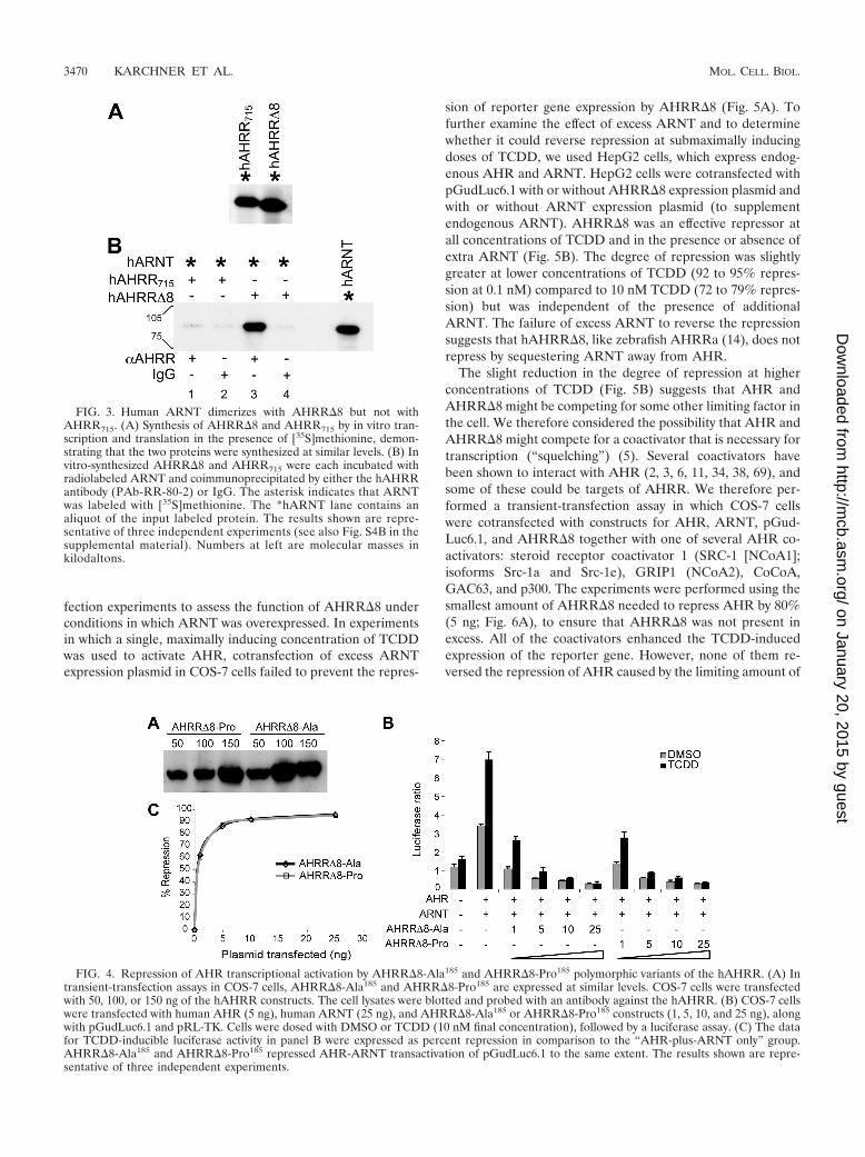
Coimmunoprecipitation assay. The full-length AHRR715, AHRR�8, AHR,and ARNT proteins were synthesized by in vitro transcription and translation(TnT; Promega, Madison, WI) in the presence or absence of [35S]methionine(MP Biomedicals, Solon, OH). Five microliters of unlabeled protein was mixedwith 15 �l of radiolabeled protein and incubated at room temperature for 2 h.For mixtures containing AHR, TCDD (10 nM) was added. The mixtures wereadjusted to 25 mM HEPES, 200 mM NaCl, 1.2 mM EDTA, 10% glycerol, and0.1% Nonidet P-40, pH 7.4, with protease inhibitors (immunoprecipitationbuffer). After two rounds of preclearing with normal mouse IgG and proteinG-agarose, 5 �g of specific antibody or IgG was added and incubated for 2 h,followed by precipitation with protein G-agarose overnight. The beads werewashed two times with IP buffer, boiled in sample treatment buffer, and subjected
3466 KARCHNER ET AL. MOL. CELL. BIOL.
on January 20, 2015 by guesthttp://m
cb.asm.org/
Dow
nloaded from
to sodium dodecyl sulfate-polyacrylamide gel electrophoresis on an 8% poly-acrylamide gel. The gels were dried and visualized by fluorography. hAHRRantibody (PAb-RR-80-2) or normal rabbit IgG was used to precipitate AHRRcomplexes and nonspecific complexes, respectively. For ARNT complexes,monoclonal ARNT antibody (MA1-515; Affinity BioReagents, Golden, CO) andnormal mouse IgG were used.
Subcellular localization of mouse AHR-YFP. COS-7 cells were grown oncoverslips in six-well plates. Cells were cotransfected with 350 ng of mouseAHR-YFP and 350 ng of human ARNT expression constructs, with or without350 ng of hAHRR�8 construct, using Lipofectamine 2000 reagent (Invitrogen).Luciferase reporter pGudLuc6.1 (300 ng) and the transfection control pRL-TK(40 ng) were also transfected. Cells were dosed with DMSO or TCDD (10 nMfinal concentration) 5 h after transfection. Twenty-four hours after transfection,cells were washed with 1� phosphate-buffered saline and fixed in 4% formalde-hyde. The coverslips were inverted onto slides and mounted with Vectashieldhard-setting mounting medium (Vector Laboratories, Burlingame, CA). Cellswere visualized using a Zeiss Axio Imager.Z1 fluorescence microscope, andAxiovision software was used to collect the images. To confirm the effectivenessof AHRR�8 as a repressor under the conditions of the assay, luciferase wasmeasured in a plate of cells run in parallel.
Nucleotide sequence accession numbers. The AHRR�8 sequences have beendeposited in GenBank, with accession numbers EU293605 (mRNA) andABX89616 (protein).
RESULTS
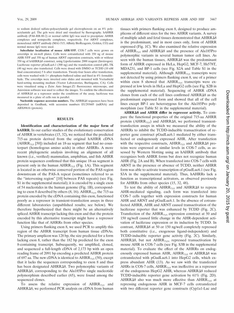
Identification and characterization of the major form ofhAHRR. In our earlier studies of the evolutionary conservationof AHRR in vertebrates (15, 32), we noticed that the predicted715-aa protein derived from the original hAHRR cDNA(AHRR715 [50]) included an 18-aa segment that had no coun-terpart (homologous amino acids) in other AHRRs. A morerecent phylogenetic analysis involving an alignment of allknown (i.e., verified) mammalian, amphibian, and fish AHRRprotein sequences confirmed that this unique 18-aa segment ispresent only in the human AHRR715 (Fig. 1A). This segmentis located in an otherwise conserved portion of the PAS regiondownstream of the PAS-A repeat (sometimes referred to asthe “intervening region” [10] between PAS repeats) (see Fig.S1 in the supplemental material); it is encoded by a single exonof 54 nucleotides in the human genome (Fig. 1B), correspond-ing to exon 8 described by others (8, 16). AHRR715, the 715-aaprotein encoded by the cDNA containing this exon, functionedpoorly as a repressor in transient-transfection assays in threedifferent laboratories (unpublished results; see below). Wetherefore hypothesized that there might be an alternativelyspliced AHRR transcript lacking this exon and that the proteinencoded by this alternative transcript might have a repressorfunction like that of AHRRs from other species.
Using primers flanking exon 8, we used PCR to amplify thisregion of the AHRR transcript from human tissue cDNAs.The primary amplicon was 128 bp, the size predicted for a formlacking exon 8, rather than the 182 bp predicted for the exon8-containing transcript. Subsequently, we amplified, cloned,and sequenced a full-length cDNA of 2,173 bp with an openreading frame of 2091 bp encoding a predicted AHRR proteinof 697 aa. The new cDNA is identical to AHRR715 (50), exceptthat it lacks the sequences corresponding to exon 8 and thushas been designated AHRR�8. Two polymorphic variants ofAHRR�8, corresponding to the Ala185Pro single nucleotidepolymorphism described earlier (65), were found among thesequenced clones.
To assess the relative expression of AHRR715 andAHRR�8, we performed PCR analysis on cDNA from human
tissues with primers flanking exon 8, designed to produce am-plicons of different sizes for the two AHRR variants. A surveyof multiple adult and fetal tissues demonstrated that AHRR�8is the predominant, and in most cases only, form of AHRRexpressed (Fig. 1C). We also examined the relative expressionof AHRR715 and AHRR�8 and the presence of Ala185Propolymorphic variants in several human tumor cell lines. Asseen with the human tissues, AHRR�8 was the predominantform of AHRR expressed in HeLa, HepG2, MCF-7, Hs578T,MDA231, and BP-1 cells (see Fig. S2A and Table S1 in thesupplemental material). Although AHRR715 transcripts werenot detected by using primers flanking exon 8, use of a primerwithin exon 8 showed that AHRR715 transcripts were ex-pressed at low levels in HeLa and HepG2 cells (see Fig. S2B inthe supplemental material). Sequencing of AHRR cDNAclones from each of the cell lines confirmed AHRR�8 as thepredominant expressed form and showed that all of the celllines except BP-1 are heterozygous for the Ala185Pro poly-morphism (see Table S1 in the supplemental material).
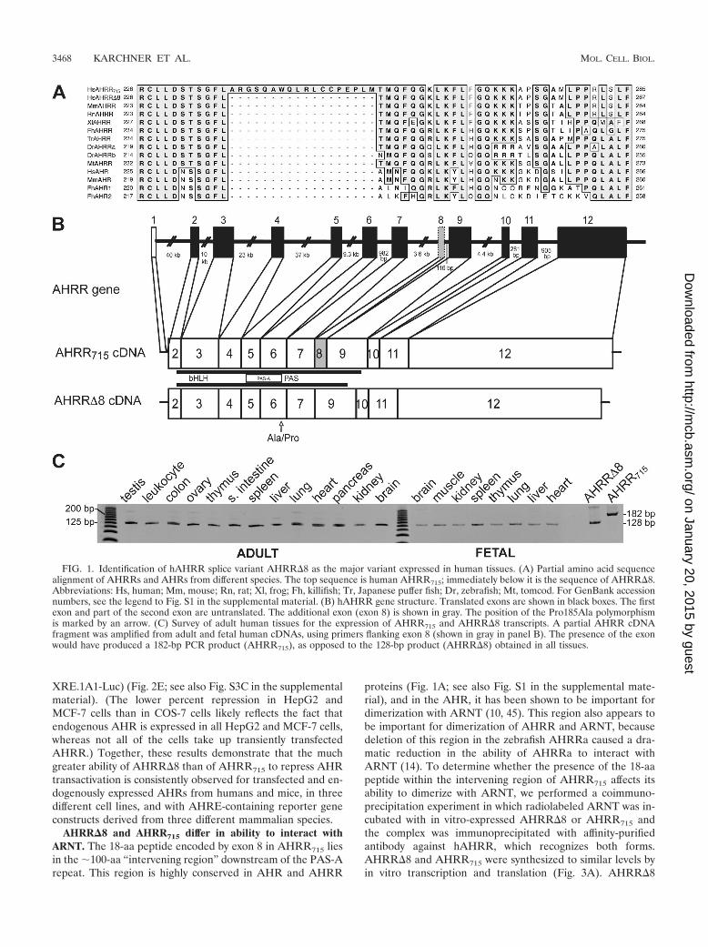
AHRR�8 and AHRR differ in repressor activity. To com-pare the functional properties of the original 715-aa AHRRprotein (AHRR715) and AHRR�8, we performed transient-transfection assays in which we measured the ability of theAHRRs to inhibit the TCDD-inducible transactivation of re-porter gene construct pGudLuc6.1 mediated by either trans-fected or endogenously expressed AHR. After transfectionwith the respective constructs, AHRR715 and AHRR�8 pro-teins were expressed at similar levels in COS-7 cells, as as-sessed by Western blotting using an hAHRR antibody thatrecognizes both AHRR forms but does not recognize humanAHR (Fig. 2A and B). When transfected into COS-7 cells withARNT in the presence or absence of TCDD, neither AHRRform was able to activate transcription of pGudLuc6.1 (see Fig.S3A in the supplemental material). Thus, hAHRRs lack afunction as transcriptional activators, as observed previouslyfor AHRRs from other species (15, 32, 48).
To test the ability of AHRR715 and AHRR�8 to repressAHR-mediated signaling, each form was transfected intoCOS-7 cells together with expression constructs for humanAHR and ARNT and pGudLuc6.1. In the absence of cotrans-fected AHRR, AHR and ARNT caused transactivation of theluciferase reporter that was enhanced by TCDD (Fig. 2C).Transfection of the AHRR715 expression construct at 50 and150 ng/well caused little change in the AHR-dependent acti-vation of luciferase expression or its induction by TCDD. Incontrast, AHRR�8 at 50 or 150 ng/well completely repressedboth constitutive (i.e., exogenous ligand-independent) andTCDD-inducible reporter gene activity (Fig. 2C). Similarly,AHRR�8, but not AHRR715, repressed transactivation bymouse AHR in COS-7 cells (see Fig. S3B in the supplementalmaterial). To evaluate the effect of the AHRRs on endog-enously expressed human AHR, AHRR715 or AHRR�8 wascotransfected with pGudLuc6.1 into HepG2 cells, which ex-press abundant AHR (13). As we saw with the transfectedAHRs in COS-7 cells, AHRR715 was ineffective as a repressorof the endogenous HepG2 AHR, whereas AHRR�8 reducedTCDD-inducible reporter gene activation by 61% (Fig. 2D).AHRR�8 also was much more effective than AHRR715 atrepressing endogenous AHR in MCF-7 cells cotransfectedwith two different reporter gene constructs (Cyp1a1-Luc and
VOL. 29, 2009 HUMAN AHRR�8 AND VARIANTS REPRESS AHR AND HIF 3467
on January 20, 2015 by guesthttp://m
cb.asm.org/
Dow
nloaded from
XRE.1A1-Luc) (Fig. 2E; see also Fig. S3C in the supplementalmaterial). (The lower percent repression in HepG2 andMCF-7 cells than in COS-7 cells likely reflects the fact thatendogenous AHR is expressed in all HepG2 and MCF-7 cells,whereas not all of the cells take up transiently transfectedAHRR.) Together, these results demonstrate that the muchgreater ability of AHRR�8 than of AHRR715 to repress AHRtransactivation is consistently observed for transfected and en-dogenously expressed AHRs from humans and mice, in threedifferent cell lines, and with AHRE-containing reporter geneconstructs derived from three different mammalian species.
AHRR�8 and AHRR715 differ in ability to interact withARNT. The 18-aa peptide encoded by exon 8 in AHRR715 liesin the 100-aa “intervening region” downstream of the PAS-Arepeat. This region is highly conserved in AHR and AHRR
proteins (Fig. 1A; see also Fig. S1 in the supplemental mate-rial), and in the AHR, it has been shown to be important fordimerization with ARNT (10, 45). This region also appears tobe important for dimerization of AHRR and ARNT, becausedeletion of this region in the zebrafish AHRRa caused a dra-matic reduction in the ability of AHRRa to interact withARNT (14). To determine whether the presence of the 18-aapeptide within the intervening region of AHRR715 affects itsability to dimerize with ARNT, we performed a coimmuno-precipitation experiment in which radiolabeled ARNT was in-cubated with in vitro-expressed AHRR�8 or AHRR715 andthe complex was immunoprecipitated with affinity-purifiedantibody against hAHRR, which recognizes both forms.AHRR�8 and AHRR715 were synthesized to similar levels byin vitro transcription and translation (Fig. 3A). AHRR�8
FIG. 1. Identification of hAHRR splice variant AHRR�8 as the major variant expressed in human tissues. (A) Partial amino acid sequencealignment of AHRRs and AHRs from different species. The top sequence is human AHRR715; immediately below it is the sequence of AHRR�8.Abbreviations: Hs, human; Mm, mouse; Rn, rat; Xl, frog; Fh, killifish; Tr, Japanese puffer fish; Dr, zebrafish; Mt, tomcod. For GenBank accessionnumbers, see the legend to Fig. S1 in the supplemental material. (B) hAHRR gene structure. Translated exons are shown in black boxes. The firstexon and part of the second exon are untranslated. The additional exon (exon 8) is shown in gray. The position of the Pro185Ala polymorphismis marked by an arrow. (C) Survey of adult human tissues for the expression of AHRR715 and AHRR�8 transcripts. A partial AHRR cDNAfragment was amplified from adult and fetal human cDNAs, using primers flanking exon 8 (shown in gray in panel B). The presence of the exonwould have produced a 182-bp PCR product (AHRR715), as opposed to the 128-bp product (AHRR�8) obtained in all tissues.
3468 KARCHNER ET AL. MOL. CELL. BIOL.
on January 20, 2015 by guesthttp://m
cb.asm.org/
Dow
nloaded from
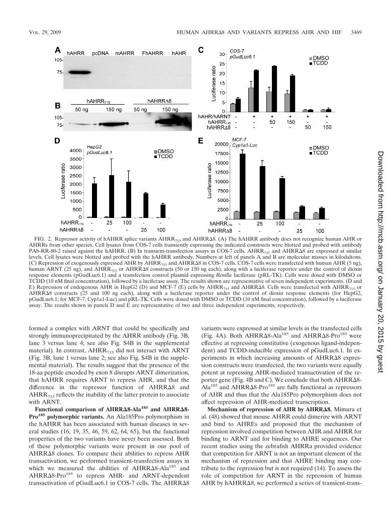
formed a complex with ARNT that could be specifically andstrongly immunoprecipitated by the AHRR antibody (Fig. 3B,lane 3 versus lane 4; see also Fig. S4B in the supplementalmaterial). In contrast, AHRR715 did not interact with ARNT(Fig. 3B, lane 1 versus lane 2; see also Fig. S4B in the supple-mental material). The results suggest that the presence of the18-aa peptide encoded by exon 8 disrupts ARNT dimerization,that hAHRR requires ARNT to repress AHR, and that thedifference in the repressor function of AHRR�8 andAHRR715 reflects the inability of the latter protein to associatewith ARNT.
Functional comparison of AHRR�8-Ala185 and AHRR�8-Pro185 polymorphic variants. An Ala185Pro polymorphism inthe hAHRR has been associated with human diseases in sev-eral studies (16, 19, 35, 46, 59, 62, 64, 65), but the functionalproperties of the two variants have never been assessed. Bothof these polymorphic variants were present in our pool ofAHRR�8 clones. To compare their abilities to repress AHRtransactivation, we performed transient-transfection assays inwhich we measured the abilities of AHRR�8-Ala185 andAHRR�8-Pro185 to repress AHR- and ARNT-dependenttransactivation of pGudLuc6.1 in COS-7 cells. The AHRR�8
variants were expressed at similar levels in the transfected cells(Fig. 4A). Both AHRR�8-Ala185 and AHRR�8-Pro185 wereeffective at repressing constitutive (exogenous ligand-indepen-dent) and TCDD-inducible expression of pGudLuc6.1. In ex-periments in which increasing amounts of AHRR�8 expres-sion constructs were transfected, the two variants were equallypotent at repressing AHR-mediated transactivation of the re-porter gene (Fig. 4B and C). We conclude that both AHRR�8-Ala185 and AHRR�8-Pro185 are fully functional as repressorsof AHR and thus that the Ala185Pro polymorphism does notaffect repression of AHR-mediated transcription.
Mechanism of repression of AHR by AHRR�8. Mimura etal. (48) showed that mouse AHRR could dimerize with ARNTand bind to AHREs and proposed that the mechanism ofrepression involved competition between AHR and AHRR forbinding to ARNT and for binding to AHRE sequences. Ourrecent studies using the zebrafish AHRRa provided evidencethat competition for ARNT is not an important element of themechanism of repression and that AHRE binding may con-tribute to the repression but is not required (14). To assess therole of competition for ARNT in the repression of humanAHR by hAHRR�8, we performed a series of transient-trans-
FIG. 2. Repressor activity of hAHRR splice variants AHRR715 and AHRR�8. (A) The hAHRR antibody does not recognize human AHR orAHRRs from other species. Cell lysates from COS-7 cells transiently expressing the indicated constructs were blotted and probed with antibodyPAb-RR-80-2 raised against the hAHRR. (B) In transient-transfection assays in COS-7 cells, AHRR715 and AHRR�8 are expressed at similarlevels. Cell lysates were blotted and probed with the hAHRR antibody. Numbers at left of panels A and B are molecular masses in kilodaltons.(C) Repression of exogenously expressed AHR by AHRR715 and AHRR�8 in COS-7 cells. COS-7 cells were transfected with human AHR (5 ng),human ARNT (25 ng), and AHRR715 or AHRR�8 constructs (50 or 150 ng each), along with a luciferase reporter under the control of dioxinresponse elements (pGudLuc6.1) and a transfection control plasmid expressing Renilla luciferase (pRL-TK). Cells were dosed with DMSO orTCDD (10 nM final concentration), followed by a luciferase assay. The results shown are representative of seven independent experiments. (D andE) Repression of endogenous AHR in HepG2 (D) and MCF-7 (E) cells by AHRR715 and AHRR�8. Cells were transfected with AHRR715 orAHRR�8 constructs (25 and 100 ng each), along with a luciferase reporter under the control of dioxin response elements (for HepG2,pGudLuc6.1; for MCF-7, Cyp1a1-Luc) and pRL-TK. Cells were dosed with DMSO or TCDD (10 nM final concentration), followed by a luciferaseassay. The results shown in panels D and E are representative of two and three independent experiments, respectively.
VOL. 29, 2009 HUMAN AHRR�8 AND VARIANTS REPRESS AHR AND HIF 3469
on January 20, 2015 by guesthttp://m
cb.asm.org/
Dow
nloaded from
fection experiments to assess the function of AHRR�8 underconditions in which ARNT was overexpressed. In experimentsin which a single, maximally inducing concentration of TCDDwas used to activate AHR, cotransfection of excess ARNTexpression plasmid in COS-7 cells failed to prevent the repres-
sion of reporter gene expression by AHRR�8 (Fig. 5A). Tofurther examine the effect of excess ARNT and to determinewhether it could reverse repression at submaximally inducingdoses of TCDD, we used HepG2 cells, which express endog-enous AHR and ARNT. HepG2 cells were cotransfected withpGudLuc6.1 with or without AHRR�8 expression plasmid andwith or without ARNT expression plasmid (to supplementendogenous ARNT). AHRR�8 was an effective repressor atall concentrations of TCDD and in the presence or absence ofextra ARNT (Fig. 5B). The degree of repression was slightlygreater at lower concentrations of TCDD (92 to 95% repres-sion at 0.1 nM) compared to 10 nM TCDD (72 to 79% repres-sion) but was independent of the presence of additionalARNT. The failure of excess ARNT to reverse the repressionsuggests that hAHRR�8, like zebrafish AHRRa (14), does notrepress by sequestering ARNT away from AHR.
The slight reduction in the degree of repression at higherconcentrations of TCDD (Fig. 5B) suggests that AHR andAHRR�8 might be competing for some other limiting factor inthe cell. We therefore considered the possibility that AHR andAHRR�8 might compete for a coactivator that is necessary fortranscription (“squelching”) (5). Several coactivators havebeen shown to interact with AHR (2, 3, 6, 11, 34, 38, 69), andsome of these could be targets of AHRR. We therefore per-formed a transient-transfection assay in which COS-7 cellswere cotransfected with constructs for AHR, ARNT, pGud-Luc6.1, and AHRR�8 together with one of several AHR co-activators: steroid receptor coactivator 1 (SRC-1 [NCoA1];isoforms Src-1a and Src-1e), GRIP1 (NCoA2), CoCoA,GAC63, and p300. The experiments were performed using thesmallest amount of AHRR�8 needed to repress AHR by 80%(5 ng; Fig. 6A), to ensure that AHRR�8 was not present inexcess. All of the coactivators enhanced the TCDD-inducedexpression of the reporter gene. However, none of them re-versed the repression of AHR caused by the limiting amount of
FIG. 3. Human ARNT dimerizes with AHRR�8 but not withAHRR715. (A) Synthesis of AHRR�8 and AHRR715 by in vitro tran-scription and translation in the presence of [35S]methionine, demon-strating that the two proteins were synthesized at similar levels. (B) Invitro-synthesized AHRR�8 and AHRR715 were each incubated withradiolabeled ARNT and coimmunoprecipitated by either the hAHRRantibody (PAb-RR-80-2) or IgG. The asterisk indicates that ARNTwas labeled with [35S]methionine. The *hARNT lane contains analiquot of the input labeled protein. The results shown are repre-sentative of three independent experiments (see also Fig. S4B in thesupplemental material). Numbers at left are molecular masses inkilodaltons.
FIG. 4. Repression of AHR transcriptional activation by AHRR�8-Ala185 and AHRR�8-Pro185 polymorphic variants of the hAHRR. (A) Intransient-transfection assays in COS-7 cells, AHRR�8-Ala185 and AHRR�8-Pro185 are expressed at similar levels. COS-7 cells were transfectedwith 50, 100, or 150 ng of the hAHRR constructs. The cell lysates were blotted and probed with an antibody against the hAHRR. (B) COS-7 cellswere transfected with human AHR (5 ng), human ARNT (25 ng), and AHRR�8-Ala185 or AHRR�8-Pro185 constructs (1, 5, 10, and 25 ng), alongwith pGudLuc6.1 and pRL-TK. Cells were dosed with DMSO or TCDD (10 nM final concentration), followed by a luciferase assay. (C) The datafor TCDD-inducible luciferase activity in panel B were expressed as percent repression in comparison to the “AHR-plus-ARNT only” group.AHRR�8-Ala185 and AHRR�8-Pro185 repressed AHR-ARNT transactivation of pGudLuc6.1 to the same extent. The results shown are repre-sentative of three independent experiments.
3470 KARCHNER ET AL. MOL. CELL. BIOL.
on January 20, 2015 by guesthttp://m
cb.asm.org/
Dow
nloaded from

AHRR�8 (Fig. 6B). We conclude that AHRR�8 does not actby competing with AHR for these specific coactivators.
Another possible mechanism of repression is the direct in-teraction of AHRR�8 with the AHR, which might block nu-
clear translocation of the AHR or prevent it from interactingwith essential coregulatory proteins. There are a number ofrepressors, including the bHLH-PAS protein IPAS (inhibitoryPAS protein, an HIF-3� splice variant), that inhibit transcrip-
FIG. 5. Effect of ARNT overexpression and increasing TCDD concentrations on the repression of AHR by AHRR�8. (A) COS-7 cells weretransfected with human AHR (5 ng) and human ARNT (25 and 200 ng) with or without AHRR�8 (5 ng). The presence of excess ARNT did notrescue the repression of AHR by AHRR�8. The results shown are representative of three independent experiments. (B) HepG2 cells weretransfected with the luciferase reporter pGudLuc6.1 with or without additional ARNT (50 ng). Cells were dosed with DMSO or increasingconcentrations of TCDD, followed by luciferase assays. Cotransfection of AHRR�8 (100 ng) caused repression of the endogenous AHR. Theextent of repression was unaffected by the presence of additional ARNT.
FIG. 6. Overexpression of AHR coactivators does not rescue repression by AHRR. (A) Determination of the minimal amount of AHRR�8needed to repress AHR by 80%. COS-7 cells were transfected with 5 ng each of human AHR and ARNT constructs and increasing amounts (1,5, 10, 25, and 50 ng) of the hAHRR�8 construct, along with the pGudLuc6.1 luciferase reporter and pRL-TK. Cells were dosed with DMSO orTCDD (1 nM), followed by luciferase assays. (B) COS-7 cells were transfected with 5 ng of human AHR and 25 ng of human ARNT, with andwithout 5 ng of hAHRR�8 construct and 200 ng each of the different receptor coactivators. The results shown are representative of threeindependent experiments.
VOL. 29, 2009 HUMAN AHRR�8 AND VARIANTS REPRESS AHR AND HIF 3471
on January 20, 2015 by guesthttp://m
cb.asm.org/
Dow
nloaded from
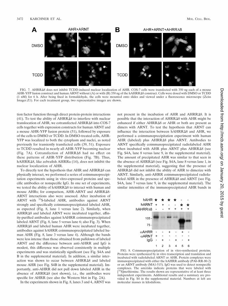
tion factor function through direct protein-protein interactions(41). To test the ability of AHRR�8 to interfere with nucleartranslocation of AHR, we cotransfected AHRR�8 into COS-7cells together with expression constructs for human ARNT anda mouse AHR-YFP fusion protein (51), followed by exposureof the cells to DMSO or TCDD. In DMSO-treated cells, AHR-YFP was localized to both the cytoplasm and nuclei, as notedpreviously for transiently transfected cells (39, 51). Exposureto TCDD resulted in nearly all AHR-YFP becoming nuclear(Fig. 7A). Cotransfection of AHRR�8 had no effect onthese patterns of AHR-YFP distribution (Fig. 7B). Thus,hAHRR�8, like zebrafish AHRRa (14), does not inhibit thenuclear localization of AHR.
To directly test the hypothesis that AHR and AHRR�8 canphysically interact, we performed a series of coimmunoprecipi-tation experiments using in vitro-expressed proteins and spe-cific antibodies or nonspecific IgG. In one set of experiments,we tested the ability of hAHRR�8 to interact with human andmouse AHRs; for comparison, AHR-ARNT and AHRR�8-ARNT interactions also were assessed. After incubation ofARNT with 35S-labeled AHR, antibodies against ARNTstrongly and specifically coimmunoprecipitated labeled AHR,as expected (Fig. 8, lane 1 versus lane 2). Similarly, whenAHRR�8 and labeled ARNT were incubated together, affin-ity-purified antibodies against hAHRR coimmunoprecipitatedlabeled ARNT (Fig. 8, lane 5 versus lane 6; also Fig. 3). WhenAHRR�8 and labeled human AHR were incubated together,antibodies against hAHRR coimmunoprecipitated labeled hu-man AHR (Fig. 8, lane 3 versus lane 4). Although the bandswere less intense than those obtained from pulldown of labeledARNT and the difference between anti-AHRR and IgG ismodest, this difference was observed consistently in multipleexperiments and was statistically significant (see Fig. S4A andB in the supplemental material). In addition, a similar inter-action was shown to occur between AHRR�8 and labeledmouse AHR (see Fig. S4B in the supplemental material). Im-portantly, anti-AHRR did not pull down labeled AHR in theabsence of AHRR�8 (not shown), i.e., the antibodies werespecific for AHRR (see also the Western blot in Fig. 2A).
In the experiments shown in Fig. 8, lanes 3 and 4, ARNT was
not present in the incubation of AHR and AHRR�8. It ispossible that the interaction of AHRR�8 with AHR might beenhanced if either AHRR�8 or AHR or both are present asdimers with ARNT. To test the hypothesis that ARNT caninfluence the interaction between hAHRR�8 and AHR, weperformed a coimmunoprecipitation experiment with humanAHR (labeled) plus AHRR�8 plus ARNT. Antibodies toARNT specifically coimmunoprecipitated radiolabeled AHRwhen incubated with AHR plus ARNT plus AHRR�8 (seeFig. S4A, lane 8 versus lane 9, in the supplemental material).The amount of precipitated AHR was similar to that seen inthe absence of AHRR�8 (see Fig. S4A, lane 8 versus lane 1, inthe supplemental material), suggesting that the presence ofAHRR�8 did not inhibit the ability of AHR to dimerize withARNT. Similarly, anti-AHRR coimmunoprecipitated radiola-beled AHR in the presence of AHRR�8 and ARNT (see Fig.S4A, lane 7 versus lane 9, in the supplemental material). Thesimilar intensities of the immunoprecipitated AHR bands in
FIG. 7. AHRR�8 does not inhibit TCDD-induced nuclear localization of AHR. COS-7 cells were transfected with 350 ng each of a mouseAHR-YFP fusion construct and human ARNT without (A) or with (B) 350 ng of the hAHRR�8 construct. Cells were dosed with DMSO or TCDD(1 nM) for 6 h. After being fixed in formaldehyde, the cells were mounted onto slides and viewed under a fluorescence microscope (ZeissImager.Z1). For each treatment group, two representative images are shown.
FIG. 8. Coimmunoprecipitation of in vitro-synthesized proteins.Proteins were synthesized by in vitro transcription and translation andincubated with radiolabeled ARNT or AHR. Protein complexes wereimmunoprecipitated with either the hAHRR antibody (PAb-RR-80-2)or an ARNT antibody (MA1-515). IgG was used to detect nonspecificcomplexes. The asterisks indicate proteins that were labeled with[35S]methionine. The results shown are representative of at least threeindependent experiments. Additional results and a summary are pro-vided in Fig. S4 in the supplemental material. Numbers at left aremolecular masses in kilodaltons.
3472 KARCHNER ET AL. MOL. CELL. BIOL.
on January 20, 2015 by guesthttp://m
cb.asm.org/
Dow
nloaded from
lanes 3 (AHR plus AHRR�8) and 7 (AHR plus AHRR�8 plusARNT) suggest that the interaction between AHR andAHRR�8 is ARNT independent.
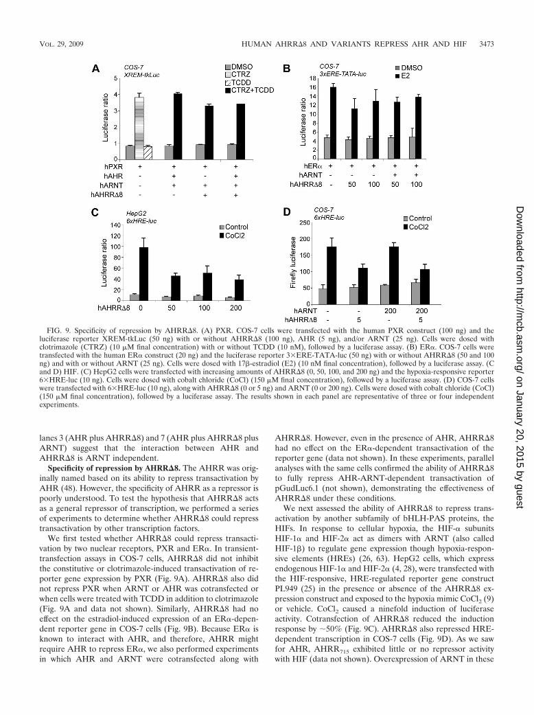
Specificity of repression by AHRR�8. The AHRR was orig-inally named based on its ability to repress transactivation byAHR (48). However, the specificity of AHRR as a repressor ispoorly understood. To test the hypothesis that AHRR�8 actsas a general repressor of transcription, we performed a seriesof experiments to determine whether AHRR�8 could represstransactivation by other transcription factors.
We first tested whether AHRR�8 could repress transacti-vation by two nuclear receptors, PXR and ER�. In transient-transfection assays in COS-7 cells, AHRR�8 did not inhibitthe constitutive or clotrimazole-induced transactivation of re-porter gene expression by PXR (Fig. 9A). AHRR�8 also didnot repress PXR when ARNT or AHR was cotransfected orwhen cells were treated with TCDD in addition to clotrimazole(Fig. 9A and data not shown). Similarly, AHRR�8 had noeffect on the estradiol-induced expression of an ER�-depen-dent reporter gene in COS-7 cells (Fig. 9B). Because ER� isknown to interact with AHR, and therefore, AHRR mightrequire AHR to repress ER�, we also performed experimentsin which AHR and ARNT were cotransfected along with
AHRR�8. However, even in the presence of AHR, AHRR�8had no effect on the ER�-dependent transactivation of thereporter gene (data not shown). In these experiments, parallelanalyses with the same cells confirmed the ability of AHRR�8to fully repress AHR-ARNT-dependent transactivation ofpGudLuc6.1 (not shown), demonstrating the effectiveness ofAHRR�8 under these conditions.
We next assessed the ability of AHRR�8 to repress trans-activation by another subfamily of bHLH-PAS proteins, theHIFs. In response to cellular hypoxia, the HIF-� subunitsHIF-1� and HIF-2� act as dimers with ARNT (also calledHIF-1�) to regulate gene expression though hypoxia-respon-sive elements (HREs) (26, 63). HepG2 cells, which expressendogenous HIF-1� and HIF-2� (4, 28), were transfected withthe HIF-responsive, HRE-regulated reporter gene constructPL949 (25) in the presence or absence of the AHRR�8 ex-pression construct and exposed to the hypoxia mimic CoCl2 (9)or vehicle. CoCl2 caused a ninefold induction of luciferaseactivity. Cotransfection of AHRR�8 reduced the inductionresponse by 50% (Fig. 9C). AHRR�8 also repressed HRE-dependent transcription in COS-7 cells (Fig. 9D). As we sawfor AHR, AHRR715 exhibited little or no repressor activitywith HIF (data not shown). Overexpression of ARNT in these
FIG. 9. Specificity of repression by AHRR�8. (A) PXR. COS-7 cells were transfected with the human PXR construct (100 ng) and theluciferase reporter XREM-tkLuc (50 ng) with or without AHRR�8 (100 ng), AHR (5 ng), and/or ARNT (25 ng). Cells were dosed withclotrimazole (CTRZ) (10 �M final concentration) with or without TCDD (10 nM), followed by a luciferase assay. (B) ER�. COS-7 cells weretransfected with the human ER� construct (20 ng) and the luciferase reporter 3�ERE-TATA-luc (50 ng) with or without AHRR�8 (50 and 100ng) and with or without ARNT (25 ng). Cells were dosed with 17�-estradiol (E2) (10 nM final concentration), followed by a luciferase assay. (Cand D) HIF. (C) HepG2 cells were transfected with increasing amounts of AHRR�8 (0, 50, 100, and 200 ng) and the hypoxia-responsive reporter6�HRE-luc (10 ng). Cells were dosed with cobalt chloride (CoCl) (150 �M final concentration), followed by a luciferase assay. (D) COS-7 cellswere transfected with 6�HRE-luc (10 ng), along with AHRR�8 (0 or 5 ng) and ARNT (0 or 200 ng). Cells were dosed with cobalt chloride (CoCl)(150 �M final concentration), followed by a luciferase assay. The results shown in each panel are representative of three or four independentexperiments.
VOL. 29, 2009 HUMAN AHRR�8 AND VARIANTS REPRESS AHR AND HIF 3473
on January 20, 2015 by guesthttp://m
cb.asm.org/
Dow
nloaded from
cells did not relieve the repressive effect of AHRR�8 (Fig.9D), suggesting that AHRR�8 was not acting simply to se-quester ARNT away from HIFs. The two polymorphic variantsof AHRR�8 (Pro185 and Ala185) repressed HIF to the sameextent (see Fig. S5 in the supplemental material). Together,these results with PXR, ER�, and HIF demonstrate thatAHRR�8 is not a general repressor of transcription but thatthe targets of AHRR�8 repression are not limited to AHR.
DISCUSSION
The AHRR is an evolutionarily conserved repressor of AHRsignaling that has been studied in rodents, amphibians, andfish. However, despite reports linking this protein to tumor cellgrowth and reproductive dysfunction in human populations,the structural and functional properties of the hAHRR havenot been widely investigated and thus are not well understood.We report here the identification and functional characteriza-tion of the major, active isoform of the hAHRR, as well as analternatively spliced form that lacks the ability to bind ARNTand is much less active as a repressor of AHR. We provide thefirst functional assessment of the Pro185Ala polymorphismand present new information concerning the molecular mech-anism of repression. We also demonstrate that the specificityof AHRR-mediated repression is broader than just AHR, ex-tending at least to another set of bHLH-PAS proteins, theHIFs.
AHRR�8 is the predominant, and functionally active,AHRR isoform in human tissues and cells. The identificationof AHRR�8, which lacks one exon compared to the originallydescribed hAHRR cDNA (50), indicates that the AHRR tran-script undergoes alternative splicing to generate at least twomRNAs. Our analyses show that AHRR�8, encoding a 697-aaAHRR isoform, is the major AHRR form expressed in a va-riety of human tissues and cell lines. The original AHRRtranscript, which encodes a 715-aa protein, was identified in abrain cDNA library (50). The brain and developing nervoussystem are known to be enriched in alternatively spliced geneproducts (12, 40). Consistent with the possibility that thelarger, exon 8-containing AHRR form is alternatively spliced,exon 8 possesses several features typical of conditional exons(33). For example, the splice recognition sequences surround-ing exon 8 are relatively weak compared to those of the otherAHRR exons (see Fig. S6 and Table S2 in the supplementalmaterial). In addition, exon 8 is relatively short, is preceded bya long intron, and is symmetrical (preserving reading frame)(33).
Analysis by comparative genomics sheds light on the evolu-tionary history of exon 8. A VISTA global alignment showsthat the genomic sequences corresponding to exon 8 occur inprimates but not in other mammals or other vertebrate groups(birds, amphibians, and fish) (see Fig. S7A in the supplementalmaterial), as suggested by our earlier alignment of amino acidsequences. Closer examination of the primate sequences showsa nonsense mutation in the macaque and an altered spliceacceptor sequence in the orangutan, suggesting that exon 8 isnot expressed in these other primates (see Fig. S7B in thesupplemental material). Thus, this exon may have been utilizedin an ancestral primate species and has subsequently been
inactivated in some primates but retained (but poorly ex-pressed) in humans.
Most previous reports of AHRR gene structure in humanshave included exon 8 (see references 8 and 16 but also refer-ence 65). However, in the few studies to examine the functionof the hAHRR protein, it has not always been clear which formwas being investigated (21, 29, 30, 71). The substantial differ-ence in repressor activity between the two forms describedhere (Fig. 2) highlights the importance of knowing whichAHRR protein is being expressed in any experimental system.AHRR�8 was the major form present in the several humancell lines (representing three different tissues) that we studied,but there could be other cell lines in which the longer formpredominates.
Alternative splicing has been reported for several bHLH-PAS proteins, including AHR (7), BMAL (27, 58), HIF-3� (18,42, 44), SIM2 (47), and ARNT (36). In some cases, the alter-natively spliced products have been shown to possess distinctfunctional properties (42, 44, 47, 52, 53). Similarly, we showhere that the alternatively spliced forms of hAHRR differ intheir ability to act as repressors. In the original (longer)AHRR715 protein, the presence of an 18-aa insertion (com-pared to AHRR�8 and AHRR orthologs in other species)causes a dramatic loss in the ability to repress both AHR andHIF. The insertion occurs in a region of the AHRR proteinthat is highly conserved among vertebrate species and thatcorresponds to the conserved, 100-aa “intervening region” ofthe AHR between the PAS-A and PAS-B repeats. In the AHR,this intervening region is important for dimerization withARNT (10, 45). Similarly, deletion of this region of thezebrafish AHRRa causes a dramatic loss in ARNT dimerization(14). Our coimmunoprecipitation data demonstrate that dis-ruption of this region with the 18-aa insertion eliminates theability of AHRR to dimerize with ARNT. The fact that re-pressor activity is lost concomitantly with loss of ARNT bind-ing suggests that it is the AHRR-ARNT complex, rather thanAHRR alone, that is the active repressor. However, it is alsopossible that the loss of repression activity in the presence ofthe additional 18 aa is the result of other structural changesand is unrelated to the ability to bind ARNT. Additional stud-ies will be needed to establish conclusively whether repressionby AHRR�8 requires dimerization with ARNT.
AHRR�8 Pro185 and -Ala185 variants are functionally indis-tinguishable. Recent epidemiological studies (reviewed in ref-erence 22) have linked a Pro185Ala polymorphism in thehAHRR (65) to human reproductive disorders, including en-dometriosis (35, 62) and male reproductive abnormalities suchas micropenis and oligospermia (16, 46, 59, 61, 64). Fujita et al.(16) suggested that the Pro185 allele might be a hypomorphicallele with a weaker inhibitory effect on AHR. However, thereare no previous reports in which the functions of the AHRR-Pro185 and AHRR-Ala185 variants have been compared. Here,we found that these two variants are qualitatively and quanti-tatively indistinguishable in their abilities to repress AHR andHIF (Fig. 4; see also Fig. S5 in the supplemental material). Itis possible that in other contexts these two variants functiondifferently. However, we note that the residue of the AHRRprotein corresponding to this Pro185Ala polymorphism is notwell conserved, even among mammals, and it occurs in a highlyvariable region of the protein (see Fig. S1 in the supplemental
3474 KARCHNER ET AL. MOL. CELL. BIOL.
on January 20, 2015 by guesthttp://m
cb.asm.org/
Dow
nloaded from
material). Thus, it seems more likely that this single nucleotidepolymorphism (SNP) is not of functional significance. Otherexplanations for the apparent association between this SNPand reproductive disorders should be considered. For example,the Pro185Ala SNP may be in linkage disequilibrium eitherwith noncoding SNPs that affect AHRR expression or withSNPs at a nearby locus that plays a role in reproductive pro-cesses (22).
Mechanism of repression. Although bHLH-PAS proteinsare generally thought of as transcriptional activators, severalare known to act as repressors: SIM2 (49, 66), NPAS1 (60),HIF-3� (41, 44, 67), and (in some circumstances) AHR (43, 55,68). The mechanisms of repression are varied and includecompetition for ARNT (49, 66, 67), formation of abortivecomplexes with the target transcription factor (41, 44), dis-placement of DNA-bound transcription factor complexes (17,37, 49), and displacement of coregulatory proteins (24, 43).
The mechanism by which AHRR represses AHR signalinghas been assumed to involve sequestration of ARNT awayfrom AHR combined with competition between AHR-ARNTand AHRR-ARNT complexes for binding to AHRE se-quences, as originally proposed (48). However, recent studieswith two zebrafish AHRR paralogs (AHRRa and AHRRb)have indicated that (i) sequestration of ARNT complexes isunlikely to be involved in the mechanism of repression and (ii)competition for AHRE binding may contribute to repressionbut is not the sole mechanism involved (14). The results pre-sented here for hAHRR�8 provide additional evidence thatsequestration of ARNT does not play a role in the mechanismof repression. Overexpression of ARNT had no effect on theability of AHRR�8 to repress AHR (Fig. 5), as we showedearlier for the zebrafish AHRRs (14). Thus, AHRR�8 differsfrom SIM2 and SIM2s, which repress AHR by competing forARNT (47, 66).
Another possible mechanism of repression, squelching(competition for coregulatory proteins such as coactivators), iswell known to occur with nuclear receptors (5). Squelching hasalso been demonstrated in interactions involving bHLH-PASproteins. For example, repression of E2F by the AHR (43) andrepression of HIF signaling by p53 (57) both were rescued byoverexpression of p300. In contrast, we found that overexpres-sion of several coregulatory proteins known to interact withAHR (CoCoA, GRIP1, SRC1, p300, and GAC63) had noeffect on the ability of AHRR�8 to repress AHR signaling,suggesting that competition for these proteins is not involvedin the mechanism of repression. Conceivably, other AHR co-activators (2, 23) could be targets for AHRR�8, or repressionmight involve simultaneous competition for multiple coactiva-tors.
We also considered the possibility that AHRR�8 could in-teract directly with AHR, similar to the way in which somealternatively spliced products of the HIF-3� locus (18) are ableto repress HIF-1� by forming an abortive complex (41, 44).hAHRR�8 did not interfere with the TCDD-dependent nu-clear translocation of AHR, as we showed earlier for zebrafishAHRRa (14). Interestingly, in coimmunoprecipitation assayswe found a modest but reproducible interaction betweenAHRR�8 and AHR. This interaction was neither enhancednor inhibited by inclusion of ARNT in the incubation, suggest-ing that AHRR�8 can interact with both the AHR monomer
and the AHR-ARNT dimer. AHRR�8 does not requireARNT in order to interact with AHR, because we saw suchinteractions in the absence of ARNT (Fig. 8, lane 3); in addi-tion, both AHRR�8 and AHRR715 (which does not dimerizewith ARNT [Fig. 3]) were able to associate with AHR (see Fig.S4B in the supplemental material). Conceivably, both ARNTdimerization and association with AHR are required for re-pression to occur. Nevertheless, while our results may be sug-gestive, it is clear that the consequences of the AHRR-AHRinteraction and its role in the mechanism of repression willrequire further investigation.
AHRR�8 specificity extends beyond AHR. The name “AHRrepressor” implies a specificity of AHRR for AHR, but this hasnot yet been established. We therefore sought to determinewhether the hAHRR is capable of repressing other transcrip-tion factors in addition to AHR. AHRR�8 inhibited theCoCl2-stimulated transcription of the hypoxia-responsive re-porter PL949 in two different cell lines (Fig. 9C and D). Thisinhibition of HIF signaling provides a possible explanation forthe results of Zudaire et al. (71), who found that silencing ofAHRR enhanced the angiogenic activity of A549 cells in adirected in vivo angiogenesis assay. Thus, one role ofAHRR�8 may be to modulate HIF signaling and its down-stream consequences such as angiogenesis. The AHRR�8-HIFinteraction provides a mechanism for cross talk between AHRand hypoxia signaling; for example, AHR ligands may regulateHIF-dependent responses through AHR-dependent inductionof AHRR�8 and subsequent inhibition of HIF signaling. Theability of AHRR�8 to repress both AHR and HIF also pro-vides a possible mechanism for the tumor suppressor activity ofAHRR (71), in which it may simultaneously limit the activity ofthese two transcription factors, both of which are overex-pressed in a variety of human cancers (54, 56).
In contrast to the AHRR�8 inhibition of HIF-dependentsignaling, we found that neither PXR nor ER� was repressedby AHRR. The result with ER� is in contrast to data obtainedby Kanno et al. (31), who reported recently that hAHRRbound to ER� and repressed ER�-mediated transactivation ofa reporter gene. We cannot yet explain the differences in re-sults, but one possibility is that AHRR-ER� interactions arecell specific; our data were obtained in COS-7 cells, whereasKanno et al. (31) performed their assays in HepG2 and MCF-7cells.
Regardless of the explanation for these differences, both theresults of Kanno et al. (31) and our data on AHRR�8-HIFinteractions suggest that AHRR is more than simply a repres-sor of AHR but rather has interactions that reach beyond theAHR pathway to affect other signaling pathways. Such broadinteractions are consistent with the emerging view of AHRR asan important regulatory protein with pleiotropic effects on cellgrowth and differentiation, including a possible role as a tumorsuppressor (22, 30, 56, 68, 71). Understanding these interac-tions of the hAHRR and their biological significance will be animportant goal of future research. The results presented hereprovide a foundation for that research by identifying AHRR�8as the major active form of hAHRR, providing the first func-tional assessment of the polymorphic AHRR variants thathave been linked to human reproductive disease, and provid-ing new insight into AHRR’s function as a repressor of mul-tiple cellular signaling pathways.
VOL. 29, 2009 HUMAN AHRR�8 AND VARIANTS REPRESS AHR AND HIF 3475
on January 20, 2015 by guesthttp://m
cb.asm.org/
Dow
nloaded from
ACKNOWLEDGMENTS
We are grateful for plasmids provided by M. Denison, G. Perdew, C.Bradfield, S. Kliewer, J. Moore, D. McDonnell, M. Stallcup, E. Kalk-hoven, M. Parker, S. K. Kim, and R. Barouki. We thank Diana Franksand Supraja Narasimhan for expert technical assistance.
This work was supported in part by National Institutes of Healthgrants R01ES006272 (M.E.H.) and P01ES11624 (D.H.S.) andthe Superfund Basic Research Program at Boston University(P42ES007381). I. Bae was supported by Susan G. Komen for theCure (FAS0703858).
REFERENCES
1. Baba, T., J. Mimura, K. Gradin, A. Kuroiwa, T. Watanabe, Y. Matsuda,J. Inazawa, K. Sogawa, and Y. Fujii-Kuriyama. 2001. Structure and expres-sion of the Ah receptor repressor gene. J. Biol. Chem. 276:33101–33110.
2. Beischlag, T. V., J. L. Morales, B. D. Hollingshead, and G. H. Perdew. 2008.The aryl hydrocarbon receptor complex and the control of gene expression.Crit. Rev. Eukaryot. Gene Expr. 18:207–250.
3. Beischlag, T. V., S. Wang, D. W. Rose, J. Torchia, S. Reisz-Porszasz, K.Muhammad, W. E. Nelson, M. R. Probst, M. G. Rosenfeld, and O. Hankin-son. 2002. Recruitment of the NCoA/SRC-1/p160 family of transcriptionalcoactivators by the aryl hydrocarbon receptor/aryl hydrocarbon receptornuclear translocator complex. Mol. Cell. Biol. 22:4319–4333.
4. Bracken, C. P., A. O. Fedele, S. Linke, W. Balrak, K. Lisy, M. L. Whitelaw,and D. J. Peet. 2006. Cell-specific regulation of hypoxia-inducible factor(HIF)-1alpha and HIF-2alpha stabilization and transactivation in a gradedoxygen environment. J. Biol. Chem. 281:22575–22585.
5. Cahill, M. A., W. H. Ernst, R. Janknecht, and A. Nordheim. 1994. Regula-tory squelching. FEBS Lett. 344:105–108.
6. Carlson, D. B., and G. H. Perdew. 2002. A dynamic role for the Ah receptorin cell signaling? Insights from a diverse group of Ah receptor interactingproteins. J. Biochem. Mol. Toxicol. 16:317–325.
7. Carver, L. A., J. B. Hogenesch, and C. A. Bradfield. 1994. Tissue specificexpression of the rat Ah-receptor and ARNT mRNAs. Nucleic Acids Res.22:3038–3044.
8. Cauchi, S., I. Stucker, S. Cenee, P. Kremers, P. Beaune, and L. Massaad-Massade. 2003. Structure and polymorphisms of human aryl hydrocarbonreceptor repressor (AhRR) gene in a French population: relationship withCYP1A1 inducibility and lung cancer. Pharmacogenetics 13:339–347.
9. Chandel, N. S., E. Maltepe, E. Goldwasser, C. E. Mathieu, M. C. Simon, andP. T. Schumacker. 1998. Mitochondrial reactive oxygen species trigger hyp-oxia induced transcription. Proc. Natl. Acad. Sci. USA 95:11715–11720.
10. Chapman-Smith, A., J. K. Lutwyche, and M. L. Whitelaw. 2004. Contribu-tion of the Per/Arnt/Sim (PAS) domains to DNA binding by the basichelix-loop-helix PAS transcriptional regulators. J. Biol. Chem. 279:5353–5362.
11. Chen, Y. H., T. V. Beischlag, J. H. Kim, G. H. Perdew, and M. R. Stallcup.2006. Role of GAC63 in transcriptional activation mediated by the arylhydrocarbon receptor. J. Biol. Chem. 281:12242–12247.
12. Clark, T. A., A. C. Schweitzer, T. X. Chen, M. K. Staples, G. Lu, H. Wang,A. Williams, and J. E. Blume. 2007. Discovery of tissue-specific exons usingcomprehensive human exon microarrays. Genome Biol. 8:R64.
13. Davarinos, N. A., and R. S. Pollenz. 1999. Aryl hydrocarbon receptor im-ported into the nucleus following ligand binding is rapidly degraded via thecytoplasmic proteasome following nuclear export. J. Biol. Chem. 274:28708–28715.
14. Evans, B. R., S. I. Karchner, L. L. Allan, R. S. Pollenz, R. L. Tanguay, M. J.Jenny, D. H. Sherr, and M. E. Hahn. 2008. Repression of aryl hydrocarbonreceptor (AHR) signaling by AHR repressor: role of DNA binding andcompetition for AHR nuclear translocator. Mol. Pharmacol. 73:387–398.
15. Evans, B. R., S. I. Karchner, D. G. Franks, and M. E. Hahn. 2005. Duplicatearyl hydrocarbon receptor repressor genes (ahrr1 and ahrr2) in the zebrafishDanio rerio: structure, function, evolution, and AHR-dependent regulationin vivo. Arch. Biochem. Biophys. 441:151–167.
16. Fujita, H., R. Kosaki, H. Yoshihashi, T. Ogata, M. Tomita, T. Hasegawa, T.Takahashi, N. Matsuo, and K. Kosaki. 2002. Characterization of the arylhydrocarbon receptor repressor gene and association of its Pro185Ala poly-morphism with micropenis. Teratology 65:10–18.
17. Gillesby, B. E., M. Stanostefano, W. Porter, S. Safe, Z. F. Wu, and T. R.Zacharewski. 1997. Identification of a motif within the 5� regulatory regionof pS2 which is responsible for AP-1 binding and TCDD-mediated suppres-sion. Biochemistry 36:6080–6089.
18. Gu, Y.-Z., S. M. Moran, J. B. Hogenesch, L. Wartman, and C. A. Bradfield.1998. Molecular characterization and chromosomal localization of a thirda-class hypoxia inducible factor subunit, HIF3a. Gene Expr. 7:205–213.
19. Guo, S. W. 2006. The association of endometriosis risk and genetic polymor-phisms involving dioxin detoxification enzymes: a systematic review. Eur. J.Obstet. Gynecol. Reprod. Biol. 124:134–143.
20. Haarmann-Stemmann, T., and J. Abel. 2006. The arylhydrocarbon receptor
repressor (AhRR): structure, expression, and function. Biol. Chem. 387:1195–1199.
21. Haarmann-Stemmann, T., H. Bothe, A. Kohli, U. Sydlik, J. Abel, and E.Fritsche. 2007. Analysis of the transcriptional regulation and molecularfunction of the aryl hydrocarbon receptor repressor in human cell lines. DrugMetab. Dispos. 35:2262–2269.
22. Hahn, M. E., L. L. Allan, and D. H. Sherr. 2009. Regulation of constitutiveand inducible AHR signaling: complex interactions involving the AHR re-pressor. Biochem. Pharmacol. 77:485–497.
23. Hankinson, O. 2005. Role of coactivators in transcriptional activation by thearyl hydrocarbon receptor. Arch. Biochem. Biophys. 433:379–386.
24. Hockings, J. K., P. A. Thorne, M. Q. Kemp, S. S. Morgan, O. Selmin, andD. F. Romagnolo. 2006. The ligand status of the aromatic hydrocarbonreceptor modulates transcriptional activation of BRCA-1 promoter by es-trogen. Cancer Res. 66:2224–2232.
25. Hogenesch, J. B., Y. Z. Gu, S. J. Jain, and C. A. Bradfield. 1998. Thebasic-helix-loop-helix-PAS orphan MOP3 forms transcriptionally activecomplexes with circadian and hypoxia factors. Proc. Natl. Acad. Sci. USA95:5474–5479.
26. Hu, C. J., L. Y. Wang, L. A. Chodosh, B. Keith, and M. C. Simon. 2003.Differential roles of hypoxia-inducible factor 1� (HIF-1�) and HIF-2� inhypoxic gene regulation. Mol. Cell. Biol. 23:9361–9374.
27. Ikeda, M., and M. Nomura. 1997. cDNA cloning and tissue-specific expres-sion of a novel basic helix-loop-helix/PAS protein (BMAL1) and identifica-tion of alternatively spliced variants with alternative translation initiation siteusage. Biochem. Biophys. Res. Commun. 233:258–264.
28. Kallio, P. J., I. Pongratz, K. Gradin, J. McGuire, and L. Poellinger. 1997.Activation of hypoxia-inducible factor 1alpha: posttranscriptional regulationand conformational change by recruitment of the Arnt transcription factor.Proc. Natl. Acad. Sci. USA 94:5667–5672.
29. Kanno, Y., Y. Miyama, Y. Takane, T. Nakahama, and Y. Inouye. 2007.Identification of intracellular localization signals and of mechanisms under-lining the nucleocytoplasmic shuttling of human aryl hydrocarbon receptorrepressor. Biochem. Biophys. Res. Commun. 364:1026–1031.
30. Kanno, Y., Y. Takane, T. Izawa, T. Nakahama, and Y. Inouye. 2006. Theinhibitory effect of aryl hydrocarbon receptor repressor (AhRR) on thegrowth of human breast cancer MCF-7 cells. Biol. Pharm. Bull. 29:1254–1257.
31. Kanno, Y., Y. Takane, Y. Takizawa, and Y. Inouye. 2008. Suppressive effectof aryl hydrocarbon receptor repressor on transcriptional activity of estrogenreceptor alpha by protein-protein interaction in stably and transiently ex-pressing cell lines. Mol. Cell. Endocrinol. 291:87–94.
32. Karchner, S. I., D. G. Franks, W. H. Powell, and M. E. Hahn. 2002. Regu-latory interactions among three members of the vertebrate aryl hydrocarbonreceptor family: AHR repressor, AHR1, and AHR2. J. Biol. Chem. 277:6949–6959.
33. Kim, E., A. Goren, and G. Ast. 2008. Alternative splicing: current perspec-tives. Bioessays 30:38–47.
34. Kim, J. H., and M. R. Stallcup. 2004. Role of the coiled-coil coactivator(CoCoA) in aryl hydrocarbon receptor-mediated transcription. J. Biol.Chem. 279:49842–49848.
35. Kim, S. H., Y. M. Choi, G. H. Lee, M. A. Hong, K. S. Lee, B. S. Lee, J. G. Kim,and S. Y. Moon. 2007. Association between susceptibility to advanced stageendometriosis and the genetic polymorphisms of aryl hydrocarbon receptorrepressor and glutathione-S-transferase T1 genes. Hum. Reprod. 22:1866–1870.
36. Korkalainen, M., J. Tuomisto, and R. Pohjanvirta. 2003. Identification ofnovel splice variants of ARNT and ARNT2 in the rat. Biochem. Biophys.Res. Commun. 303:1095–1100.
37. Krishnan, V., W. Porter, M. Santostefano, X. H. Wang, and S. Safe. 1995.Molecular mechanism of inhibition of estrogen-induced cathepsin D geneexpression by 2,3,7,8-tetrachlorodibenzo-p-dioxin (TCDD) in MCF-7 cells.Mol. Cell. Biol. 15:6710–6719.
38. Kumar, M. B., and G. H. Perdew. 1999. Nuclear receptor coactivator SRC-1interacts with the Q.-rich subdomain of the AhR and modulates its transac-tivation potential. Gene Expr. 8:273–286.
39. LaPres, J. J., E. Glover, E. E. Dunham, M. K. Bunger, and C. A. Bradfield.2000. ARA9 modifies agonist signaling through an increase in cytosolic arylhydrocarbon receptor. J. Biol. Chem. 275:6153–6159.
40. Li, Q., J. A. Lee, and D. L. Black. 2007. Neuronal regulation of alternativepre-mRNA splicing. Nat. Rev. Neurosci. 8:819–831.
41. Makino, Y., R. Cao, K. Svensson, G. Bertilsson, M. Asman, H. Tanaka, Y.Cao, A. Berkenstam, and L. Poellinger. 2001. Inhibitory PAS domain proteinis a negative regulator of hypoxia-inducible gene expression. Nature 414:550–554.
42. Makino, Y., A. Kanopka, W. J. Wilson, H. Tanaka, and L. Poellinger. 2002.Inhibitory PAS domain protein (IPAS) is a hypoxia-inducible splicing variantof the hypoxia-inducible factor-3alpha locus. J. Biol. Chem. 277:32405–32408.
43. Marlowe, J. L., E. S. Knudsen, S. Schwemberger, and A. Puga. 2004. The arylhydrocarbon receptor displaces p300 from E2F-dependent promoters andrepresses S phase-specific gene expression. J. Biol. Chem. 279:29013–29022.
3476 KARCHNER ET AL. MOL. CELL. BIOL.
on January 20, 2015 by guesthttp://m
cb.asm.org/
Dow
nloaded from
44. Maynard, M. A., A. J. Evans, T. Hosomi, S. Hara, M. A. Jewett, and M. Ohh.2005. Human HIF-3alpha4 is a dominant-negative regulator of HIF-1 and isdown-regulated in renal cell carcinoma. FASEB J. 19:1396–1406.
45. McGuire, J., K. Okamoto, M. L. Whitelaw, H. Tanaka, and L. Poellinger.2001. Definition of a dioxin receptor mutant that is a constitutive activator oftranscription: delineation of overlapping repression and ligand binding func-tions within the PAS domain. J. Biol. Chem. 276:41841–41849.
46. Merisalu, A., M. Punab, S. Altmae, K. Haller, T. Tiido, M. Peters, and A.Salumets. 2007. The contribution of genetic variations of aryl hydrocarbonreceptor pathway genes to male factor infertility. Fertil. Steril. 88:854–859.
47. Metz, R. P., H. I. Kwak, T. Gustafson, B. Laffin, and W. W. Porter. 2006.Differential transcriptional regulation by mouse single-minded 2s. J. Biol.Chem. 281:10839–10848.
48. Mimura, J., M. Ema, K. Sogawa, and Y. Fujii-Kuriyama. 1999. Identificationof a novel mechanism of regulation of Ah (dioxin) receptor function. GenesDev. 13:20–25.
49. Moffett, P., M. Reece, and J. Pelletier. 1997. The murine Sim-2 gene productinhibits transcription by active repression and functional interference. Mol.Cell. Biol. 17:4933–4947.
50. Nagase, T., K. Ishikawa, R. Kikuno, M. Hirosawa, N. Nomura, and O.Ohara. 1999. Prediction of the coding sequences of unidentified humangenes. XV. The complete sequences of 100 new cDNA clones from brainwhich code for large proteins in vitro. DNA Res. 6:337–345.
51. Petrulis, J. R., N. G. Hord, and G. H. Perdew. 2000. Subcellular localizationof the aryl hydrocarbon receptor is modulated by the immunophilin homologhepatitis B virus X-associated protein 2. J. Biol. Chem. 275:37448–37453.
52. Pollenz, R. S., H. R. Sullivan, J. Holmes, B. Necela, and R. E. Peterson. 1996.Isolation and expression of cDNAs from rainbow trout (Oncorhynchusmykiss) that encode two novel basic helix-loop-helix/PER-ARNT-SIM(bHLH/PAS) proteins with distinct functions in the presence of the arylhydrocarbon receptor. Evidence for alternative mRNA splicing and domi-nant negative activity in the bHLH/PAS family. J. Biol. Chem. 271:30886–30896.
53. Prasch, A. L., R. L. Tanguay, V. Mehta, W. Heideman, and R. E. Peterson.2006. Identification of zebrafish ARNT1 homologs: 2,3,7,8-tetrachlorod-ibenzo-p-dioxin toxicity in the developing zebrafish requires ARNT1. Mol.Pharmacol. 69:776–787.
54. Qing, G., and M. C. Simon. 2009. Hypoxia inducible factor-2alpha: a criticalmediator of aggressive tumor phenotypes. Curr. Opin. Genet. Dev. 19:60–66.
55. Safe, S., and M. Wormke. 2003. Inhibitory aryl hydrocarbon receptor-estro-gen receptor-a cross-talk and mechanisms of action. Chem. Res. Toxicol.16:807–816.
56. Schlezinger, J. J., D. Liu, M. Farago, D. C. Seldin, K. Belguise, G. E.Sonenshein, and D. H. Sherr. 2006. A role for the aryl hydrocarbon receptorin mammary gland tumorigenesis. Biol. Chem. 387:1175–1187.
57. Schmid, T., J. Zhou, R. Kohl, and B. Brune. 2004. p300 relieves p53-evokedtranscriptional repression of hypoxia-inducible factor-1 (HIF-1). Biochem. J.380:289–295.
58. Schoenhard, J. A., M. Eren, C. H. Johnson, and D. E. Vaughan. 2002.Alternative splicing yields novel BMAL2 variants: tissue distribution andfunctional characterization. Am. J. Physiol. Cell Physiol. 283:C103–C114.
59. Soneda, S., M. Fukami, M. Fujimoto, T. Hasegawa, Y. Koitabashi, and T.
Ogata. 2005. Association of micropenis with Pro185Ala polymorphism of thegene for aryl hydrocarbon receptor repressor involved in dioxin signaling.Endocr. J. 52:83–88.
60. Teh, C. H., K. K. Lam, C. C. Loh, J. M. Loo, T. Yan, and T. M. Lim. 2006.Neuronal PAS domain protein 1 is a transcriptional repressor and requiresarylhydrocarbon nuclear translocator for its nuclear localization. J. Biol.Chem. 281:34617–34629.
61. Tiido, T., A. Rignell-Hydbom, B. A. Jonsson, L. Rylander, A. Giwercman,and Y. L. Giwercman. 2007. Modifying effect of the AR gene trinucleotiderepeats and SNPs in the AHR and AHRR genes on the association betweenpersistent organohalogen pollutant exposure and human sperm Y:X ratio.Mol. Hum. Reprod. 13:223–229.
62. Tsuchiya, M., T. Katoh, H. Motoyama, H. Sasaki, S. Tsugane, and T. Ike-noue. 2005. Analysis of the AhR, ARNT, and AhRR gene polymorphisms:genetic contribution to endometriosis susceptibility and severity. Fertil.Steril. 84:454–458.
63. Wang, G. L., B.-H. Jiang, E. A. Rue, and G. L. Semenza. 1995. Hypoxia-inducible factor 1 is a basic-helix-loop-helix-PAS heterodimer regulated bycellular O2 tension. Proc. Natl. Acad. Sci. USA 92:5510–5514.
64. Watanabe, M., K. Sueoka, I. Sasagawa, A. Nakabayashi, Y. Yoshimura, andT. Ogata. 2004. Association of male infertility with Pro185Ala polymorphismin the aryl hydrocarbon receptor repressor gene: implication for the suscep-tibility to dioxins. Fertil. Steril. 82(Suppl. 3):1067–1071.
65. Watanabe, T., I. Imoto, Y. Kosugi, Y. Fukuda, J. Mimura, Y. Fujii, K. Isaka,M. Takayama, A. Sato, and J. Inazawa. 2001. Human arylhydrocarbon re-ceptor repressor (AHRR) gene: genomic structure and analysis of polymor-phism in endometriosis. J. Hum. Genet. 46:342–346.
66. Woods, S. L., and M. L. Whitelaw. 2002. Differential activities of murinesingle minded 1 (SIM1) and SIM2 on a hypoxic response element. Cross-talkbetween basic helix-loop-helix/per-Arnt-Sim homology transcription factors.J. Biol. Chem. 277:10236–10243.
67. Yamashita, T., O. Ohneda, M. Nagano, M. Iemitsu, Y. Makino, H. Tanaka,T. Miyauchi, K. Goto, K. Ohneda, Y. Fujii-Kuriyama, L. Poellinger, and M.Yamamoto. 2008. Abnormal heart development and lung remodeling in micelacking the hypoxia-inducible factor-related basic helix-loop-helix PAS pro-tein NEPAS. Mol. Cell. Biol. 28:1285–1297.
68. Yang, X., D. Liu, T. J. Murray, G. C. Mitchell, E. V. Hesterman, S. I.Karchner, R. R. Merson, M. E. Hahn, and D. H. Sherr. 2005. The arylhydrocarbon receptor constitutively represses c-myc transcription in humanmammary tumor cells. Oncogene 24:7869–7881.
69. Zhang, S., C. Rowlands, and S. Safe. 2008. Ligand-dependent interactions ofthe Ah receptor with coactivators in a mammalian two-hybrid assay. Toxicol.Appl. Pharmacol. 227:196–206.
70. Zimmermann, A. L., E. A. King, E. Dengler, S. R. Scogin, and W. H. Powell.2008. An aryl hydrocarbon receptor repressor from Xenopus laevis: function,expression, and role in dioxin responsiveness during frog development. Toxi-col. Sci. 104:124–134.
71. Zudaire, E., N. Cuesta, V. Murty, K. Woodson, L. Adams, N. Gonzalez, A.Martinez, G. Narayan, I. Kirsch, W. Franklin, F. Hirsch, M. Birrer, and F.Cuttitta. 2008. The aryl hydrocarbon receptor repressor is a putative tumorsuppressor gene in multiple human cancers. J. Clin. Investig. 118:640–650.
VOL. 29, 2009 HUMAN AHRR�8 AND VARIANTS REPRESS AHR AND HIF 3477
on January 20, 2015 by guesthttp://m
cb.asm.org/
Dow
nloaded from
Copyright © 2022 FDOKUMEN