Practical Enzymatic Resolution of Racemic Alcohols and Amines in Organic Solvents
Upload
independentCategory
view
1download
0
Toxicology and Applied Pharmacology 249 (2010) 91–100
Contents lists available at ScienceDirect
Toxicology and Applied Pharmacology
j ourna l homepage: www.e lsev ie r.com/ locate /ytaap
Preferential induction of the AhR gene battery in HepaRG cells after a single orrepeated exposure to heterocyclic aromatic amines
Julie Dumont 1, Rozenn Jossé, Carine Lambert, Sébastien Anthérieu, Véronique Laurent, Pascal Loyer,Marie-Anne Robin, André Guillouzo ⁎Inserm UMR 991, F-35043 Rennes cedex, FranceUniversité de Rennes 1, Faculté des Sciences Pharmaceutiques et Biologiques, F-35043 Rennes cedex, France
Abbreviations: 3-MC, 3-methylcholanthrene; AhR, Aα-naphthoflavone; CYP, Cytochrome P450; DMSO, DimeFCS, Fetal calf serum; HAA, Heterocyclic aromatic amAnalysis; MeIQx, 2-amino-3,8-dimethylimidazo[4,5-f]qNrf2, NF-E2-related factor 2; PhIP, 2-amino-1-methyl-6-S.E.M., Standard error of the mean; XRE, xenobiotic-res⁎ Corresponding author. Inserm UMR 991, Faculté de
Biologiques, F-35043 Rennes Cedex, France. Fax: +33 2E-mail addresses: [email protected] (J. D
[email protected] (R. Jossé), [email protected] (S. Anthérieu),[email protected] (V. Laurent), Pascal(P. Loyer), [email protected] (M.-A. Robin),[email protected] (A. Guillouzo).
1 Present address: Inserm UMR 744 - Université Lille Nde Lille, BP 245, 1, rue du professeur Calmette, F-59019
0041-008X/$ – see front matter © 2010 Elsevier Inc. Adoi:10.1016/j.taap.2010.08.027
a b s t r a c t
a r t i c l e i n f oArticle history:Received 8 August 2010Revised 26 August 2010Accepted 27 August 2010Available online 15 September 2010
Keywords:Heterocyclic aromatic aminesToxicogenomicsArylhydrocarbon receptorHepaRG cellsRepeated exposure
2-Amino-1-methyl-6-phenylimidazo[4,5-b]pyridine (PhIP) and 2-amino-3,8-dimethylimidazo[4,5-f]qui-noxaline (MeIQx) are two of the most common heterocyclic aromatic amines (HAA) produced duringcooking of meat, fish and poultry. Both HAA produce different tumor profiles in rodents and are suspected tobe carcinogenic in humans. In order to better understand the molecular basis of HAA toxicity, we haveanalyzed gene expression profiles in the metabolically competent human HepaRG cells using pangenomicoligonucleotide microarrays, after either a single (24-h) or a repeated (28-day) exposure to 10 μM PhIP orMeIQx. The most responsive genes to both HAA were downstream targets of the arylhydrocarbon receptor(AhR): CYP1A1 and CYP1A2 after both time points and CYP1B1 and ALDH3A1 after 28 days. Accordingly,CYP1A1/1A2 induction in HAA-treated HepaRG cells was prevented by chemical inhibition or smallinterference RNA-mediated down-regulation of the AhR. Consistently, HAA induced activity of the CYP1A1promoter, which contains a consensus AhR-related xenobiotic-responsive element (XRE). In addition, severalother genes exhibited both time-dependent and compound-specific expression changes with, however, asmaller magnitude than previously reported for the prototypical AhR target genes. These changes concernedgenes mainly related to cell growth and proliferation, apoptosis, and cancer. In conclusion, these resultsidentify the AhR gene battery as the preferential target of PhIP andMeIQx in HepaRG cells and further supportthe hypothesis that intake of HAA in diet might increase human cancer risk.
rylhydrocarbon receptor; ANF,thylsulfoxide; FC, Fold change;ines; IPA, Ingenuity Pathwayuinoxaline; NR, Neutral red;phenylimidazo[4,5-b]pyridine;ponsive element.s Sciences Pharmaceutiques et23235385.umont),[email protected] (C. Lambert),
ord de France - Institut PasteurLille cedex, France.
ll rights reserved.
© 2010 Elsevier Inc. All rights reserved.
Introduction
Risk assessment for human health of long-term exposure toenvironmental and food contaminants is currently a major chal-lenge. While a number of pollutants induce cancers in experimentalanimals after chronic exposure, they are only suspected to becarcinogenic in humans based on the weight of evidence from
epidemiological studies. Accordingly, 2-amino-1-methyl-6-pheny-limidazo[4,5-b]pyridine (PhIP) and 2-amino-3,8-dimethylimidazo[4,5-f]quinoxaline (MeIQx), which are among the most abundantheterocyclic aromatic amines (HAA) formed in fried or grilled fishand meat (Felton et al., 1986), are genotoxic and carcinogenic atmultiple tissue sites and in multiple species after long-term feeding(Sugimura et al., 2004). However, although chemically similar, thesetwo HAA produce, in general, different tumor profiles in rodents andonly MeIQx induces liver tumors (Sugimura et al., 2004). Someepidemiological studies have also suggested positive associationsbetween the intake of well-done or grilled meat and human cancerrisk (Alaejos et al., 2008) but they are insufficient to support thatincreased cancer risk is due specifically to PhIP and/or MeIQx and/or another HAA.
PhIP and MeIQx are mainly metabolized in the liver of bothrodents and humans in a N-hydroxy-derivative, which is mutagenic(Turesky et al., 2002). However, interspecies differences have beenshown in both catalytic activity and regioselectivity of cytochromesP450 (CYP) and other liver enzymes involved in their metabolism(Langouet et al., 2002), potentially affecting their biological activitiesand making questionable the use of animal models for estimatingrisks of HAA for human health.
92 J. Dumont et al. / Toxicology and Applied Pharmacology 249 (2010) 91–100
Toxicogenomics technologies have emerged as promisingapproaches for a better identification and discrimination of cellularresponses to chemicals. Recent studies have suggested that geneexpression profiles following short-term in vivo or in vitro treatmentscould permit predicting carcinogenic and other toxic properties ofenvironmental chemicals (Fielden et al., 2008; Ellinger-Ziegelbaueret al., 2009; Tsujimura et al., 2006; van Delft et al., 2005). In thepresent study, we used metabolically competent human hepatomaHepaRG cells that remain functionally stable during several weeks atconfluence (Josse et al., 2008) to analyze changes in gene expressionin response to PhIP and MeIQx after a single or repeated exposureduring 4 weeks. Although both time-dependent and compound-specific alterations of gene expression were identified after exposureto PhIP and MeIQx, target genes of the arylhydrocarbon receptor(AhR) were the most induced in all experimental conditions.
Materials and methods
Chemicals
Dimethylsulfoxide (DMSO), 3-methylcholanthrene (3-MC), α-naphthoflavone (ANF) and neutral red (NR) were purchased fromSigma-Aldrich (St. Quentin-Fallavier, France). Williams'E mediumwas supplied by Eurobio (Les Ulis, France) and fetal calf serum (FCS)by Perbio (Brebières, France). PhIP and MeIQx, were obtained fromToronto Research Chemicals (Toronto, Canada). All other chemicalswere of the highest quality available.
Cell culture and HAA treatments
HepaRG cells were seeded at a density of 2.6×10⁴ cells/cm2 inthe Williams'E medium supplemented with 10% FCS, 100 units/mLpenicillin, 100 μg/mL streptomycin, 5 μg/mL insulin, 2 mM gluta-mine and 5×10⁻⁵ M hydrocortisone hemisuccinate (Gripon et al.,2002). After 2 weeks they were shifted to the same culture mediumsupplemented with 2% DMSO for 2 more weeks. At that timeHepaRG cells were well differentiated and used for HAA testing. PhIPand MeIQx were dissolved in DMSO; both control and treatedcultures received the same final concentration of vehicle. Whateverthe time of exposure, HAA-treatment was performed in a culturemedium containing both 10% FCS and 2% DMSO, which maintainsmaximum metabolic capacity of HepaRG cells without any cytotoxi-city (Aninat et al., 2006; Josse et al., 2008). For 28-day exposure, HAAwere added every 2 days with each medium renewal. All samples foranalysis of HAA-induced effects at 24 h and 28 days were collectedsimultaneously.
Cytotoxicity measurements
ATP assay. After treatment, cultures were observed under phase-contrast microscopy using an Olympus 1x70 microscope andintracellular ATP content was measured using the CellTiter-GloLuminescent Cell Viability Assay kit (Promega, Charbonnieres,France) according to manufacturer's instructions.
Neutral Red (NR) incorporation assay. At the end of treatments,cells were stained with 0.4% NR solution for 3 h, then rapidly fixedwith a 4% formaldehyde/1% calcium chloride solution and destainedwith 50% ethanol/1% acetic acid. Dye uptake was determined asoptical density (540 nm).
RNA isolation
Total RNA was extracted with the SV total RNA isolation system(Promega, Madison, WI), which includes a direct DNase treatmentstep. RNA quantity and purity were assessed with a Nanodrop ND-
1000 spectrophotometer (Nyxor Biotech, Paris, France) and RNAintegrity was checked with a Bioanalyzer 2100 (Agilent Technologies,Massy, France).
Retrotranscription and real-time quantitative PCR (RT-qPCR) analysis
Five hundred nanograms of total RNA were reversed-transcribedinto cDNA using the High-Capacity cDNA Archive kit (AppliedBiosystems, Foster City, CA). RT-qPCR was performed by thefluorescent dye SYBR Green methodology using the SYBR Green PCRMaster Mix and the ABI Prism 7000 (Applied Biosystems). Primerspairs for each transcript are described in Supplemental table S1.Amplification curves were read with the ABI Prism 7000 SDS Softwareusing the comparative cycle threshold method and the relativequantification of the steady-state mRNA levels was normalizedagainst 18S RNA.
Microarray analysis
Experimental conditions were previously described (Lambertet al., 2009). Briefly, 500 ng of total RNA from each cell culture werereverse-transcribed into double-strand cDNAs using the Low RNAInput Linear Amplification Labeling Kit (Agilent). cDNAs weretranscribed into antisense cRNA and labeled with either CTP-Cy3 orCTP-Cy5 fluorescent dyes following the manufacturer's protocol.Cyanine-labeled cRNAs were purified using RNeasy minikit (Qiagen).Then, control Cy3-RNAs and HAA-treated Cy5-RNAs were pooledtogether and hybridized using the Hybridisation Kit Plus (Agilent)onto 4×44 K Whole Human Genome Microarrays (G4112F, Agilent),satisfying Minimum Information About a Microarray Experiment(MIAME) requirements. Two independent hybridizations with Fluorreversal corresponding to two technical replicates were performed foreach experimental condition. After incubation and washing, micro-arrays were scanned with the Agilent microarray G2566AA scanner.Data were extracted and normalized from the resulting images usingAgilent Feature Extraction software (v.9.5). Data analyses wereperformed using GeneSpring GX v.10.0 software for databasemanagement, quality control and analysis. Microarray quality wasassessed by examining the values in the principal component analysisand the quality control metrics. Hierarchical clustering usingPearson's correlation and the Ward's min variance link heuristiccriteria was performed on all arrays to determine if replicates behavesimilarly in order to exclude outliers. Significantly modulated geneswere analyzed from combined replicates with p≤0.01 and 1.5-foldchange (FC) as filters (FCN1.5). Biological functions and pathwayswere generated and analyzed using Ingenuity Pathway Analysisv.4.0™ (IPA, Ingenuity System, CA) from combined data.
Transfection and luciferase assay
The pCYP1A1-FL(−1566) construct containing a 1639-base-pairregion (from−1566 to +73) of the human CYP1A1 gene upstream ofthe firefly luciferase reporter gene (a gift from Pr. R. Barouki, Paris,France) and the pGL3-XRE3 construct, containing only threeXRE sequences from CYP1A1 gene, have been described previously(Le Ferrec et al., 2002). HepaRG cells were transiently electroporatedas previously described (Laurent et al., 2010) using a MP-100Microporator (LabTech France, now available as the Neon systemfrom Invitrogen). Briefly, 105 HepaRG cells were resuspended in 10 μlof Microporation Buffer with 500 ng of pCYP1A1-FL(−1566) or pGL3-XRE3 constructs and 50 ng of pRL-SV40 vectors. HepaRG cells wereelectroporated at 1500 V for 20 ms and then plated at a density of2×105 cells/cm2. After a 24-h period, cells were exposed to HAA for24 h. Firefly (pGL3-vector) and Renilla (pRL-vector) luciferaseactivities were measured using the Dual-luciferase Reporter AssaySystem (Promega). Firefly luciferase activity was normalized to

93J. Dumont et al. / Toxicology and Applied Pharmacology 249 (2010) 91–100
Renilla luciferase activity and data were expressed in arbitrary unit(a.u.), relative to the value of luciferase activity levels measured inuntreated cells, arbitrarily set at 1.
siRNA knockdown of the AhR
A SMART-pool siRNA directed against the human AhR (si-AhR, Cat#L-004990) was purchased from Dharmacon RNA technologies(Boulder, CO). One million HepaRG cells was transiently electro-porated with either 100 pmol of si-AhR or 100 pmol of negativecontrol siRNA (Dharmacon, Cat #D-001210-01) as described above.Two days after si-RNA transfection, HepaRG cells were treated with orwithout HAA and 24 h later, total HepaRG cell RNA was extracted andsubjected to reverse transcription as described above. The knockdownof AhR in the Si-RNA transfected cells was validated by westernblotting.
Preparation of total cell lysates
Total cellular protein extracts were obtained by lysis of HepaRGcells in buffer containing 50 mM Tris–HCl, pH 8, 150 mM NaCl, 5 mMEDTA, 2.5 mM EGTA, 0.1% Tween-20, 10% glycerol, 0.1 mM sodiumorthovanadate, 1 mM sodium fluoride, 10 mM beta-glyceropho-sphate, and 0.1 mM phenylmethanesulfonyl fluoride, supplementedwith an EDTA-free cocktail protease inhibitor (Roche Diagnostics,Meylan, France).
Western blotting
Fifty μg of total cellular protein extracts were resolved on 7.5%SDS–PAGE, transferred onto nitrocellulose membranes (Millipore,Guyancourt, France) and analysed using chemiluminescence detec-tion. The following antibodies were used: rabbit anti-human arylhydrocarbon receptor (AhR) (SA-210, Biomol Research Labs, Ply-mouth, PA) and mouse anti-human Heat Shock Cognate 70 (HSC70)(B-6, sc-7298, Santa Cruz Biotechnology, Tebu, France).
Statistical analysis
Data are presented asmeans±standard error of themean (S.E.M.).Comparisons between groups were performed using the Kruskal–Wallis test. In case of significant difference between groups, theMann–Whitney U test was used to test whether any condition wassignificantly different from control. pb0.05 was interpreted assignificant.
Results
Toxicity of PhIP and MeIQx as a function of time of exposure
Differentiated HepaRG cells were exposed to PhIP or MeIQx atconcentrations ranging from 0.1 to 10 μM for either 24 h or 28 days.Regardless of the time of exposure, neither morphological alterationsnor significant changes in cell viability using the ATP content assaywere observed (Supplemental Figure 1). Similarly, the NR assayshowed no obvious cytotoxicity (data not shown). Moreover, sinceHepaRG cell cultures were composed of both hepatocyte- and biliary-like cells (in a 50/50 ratio) (Cerec et al., 2007), we verified thathepatocyte functions were not preferentially affected during pro-longed exposure to HAA at the highest concentration tested (10 μM).No significant changes in either CYP3A4 activity or hepatocyte-specific transcripts encoding aldolase B and albumin were found overa 28-day period following treatment with either HAA (SupplementalFigure 2).
Global gene expression patterns in response to PhIP and MeIQx
At 10 μM or higher concentrations, PhIP and MeIQx werepreviously shown to induce CYP1A1, CYP1A2, and CYP1B1 expressionafter a 24-h exposure (Dumont et al., 2010). We hypothesized thatadditional changes in gene expression could occur after repeatedexposure to 10 μM PhIP and MeIQx for 28 days in the absence ofcytotoxic effects. Pangenomic Agilent microarrays were used andsignificantly modulated genes were extracted with p≤0.01 and 1.5-fold change as filters.
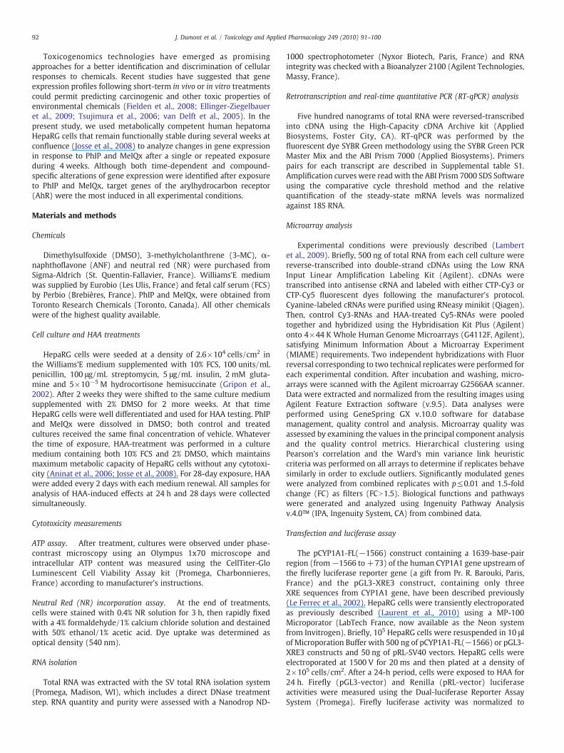
The total number of genes significantly regulated by PhIP andMeIQx was 108 and 69 at 24 h, and 89 and 112 at 28 days,respectively. Up-regulated genes represented 16 and 55% with PhIPand 52 and 63% with MeIQx after 24 h and 28 days of exposure,respectively. The Venn diagrams in Figs. 1A and B show the number ofgenes that were common or unique to the different treatmentconditions. Only 8 and 15 genes were modulated by both PhIP andMeIQx after 24 h and 28 days of exposure, respectively (Fig. 1A).Likewise, only 5 and 8 genes were modulated by PhIP and MeIQxrespectively, at the two time points (Fig. 1B).
Time-dependent effects of PhIP and MeIQx on gene expression profiling
Only the well-annotated genes modulated by each HAA aftereither 24 h or 28 days of exposure (Tables 1–3) were considered forsubsequent functional analyses using IPA. Regardless of the HAA andthe time of exposure, most genes had relatively small fold changes(below 2). In contrast, a 2.4- to 5.0-fold induction of CYP1A1 andCYP1A2, two key genes of the AhR gene battery (Nebert et al., 2000);was observed in all treatment conditions (Table 1). Only one othergene was modulated by both PhIP and MeIQx at 24 h, i.e. themethionine adenosyltransferase II alpha (MAT2A), which was down-regulated by PhIP while up-regulated by MeIQx. The 10 other genesmodulated by both HAA after 28 days included two additional targetgenes of the AhR (CYP1B1 and ALDH3A1), two genes related toapoptosis (CASP8 andMT-CYB) and two genes related to cell cycle andcancer (DHRS2 and DIAPH3, respectively).
Otherwise, most deregulated genes were restricted to onecondition. Among the 36 genes specifically modulated after 24 h ofexposure to PhIP (Table 2), 15 were related to “Cell death”, “Cellulargrowth and proliferation” and “Cancer”. EGR1, F2R, and FOS, which arerelated to the three functions, were either up-regulated (EGR1 andFOS) or down-regulated (F2R). Genes related to cell death (apoptosis)included NTRK1 and S100A1, which were up-regulated and PDCD1,RAD23B, RAPGEF1, and RNF34, which were down-regulated. Inaddition to EGR1, F2R, and FOS, 8 other genes were related to cancer:NTRK1 and S100A1 were induced whereas CDK9, HNF1B, RAPGEF1,RIMS4, VPRBP and TBC1D2B were repressed. Some of these genes(CDK9, NTRK1, and VPRBP) as well as CYP27B1 and PDCD1were relatedto cell proliferation.
After 28 days of repeated exposure to PhIP, the biological functions“Cell death,” “Cellular growth and proliferation” and “Cancer” wereenriched but included genes different from those modulated after24 h. Nineteen out of the 52 genes specifically modulated at 28 dayswere related to these three cellular processes (Table 3). Six geneswere related to cell proliferation and apoptosis: CXCR3, GAB1, andOSM were up-regulated while APAF1, CASP8, and EGF were down-regulated. ALOX5 and WTAP which were repressed and CD27, whichwas induced were other PhIP-modulated genes related to cell death.In addition to ALOX5, CASP8, CD27, CXCR3 and EGF, 10 othermodulatedgenes were cancer-related: AKAP12, CYP1B1, DIAPH3, and TTC37 wereup-regulated while MT-CYB, GNA13, HCCS, N4BP1, SNF1LK, and SRRM2were down-regulated. In addition, 14 genes specifically modulated at28 days were related to “Immunological disorder.” This gene setincluded CHAT, DIAPH3, NAV2, OSM, PRKAR2B, and TDRD10, which

61 104869 112
103 845108 89
A
B
100 618
PhIP 24 h
PhIP 24 h
MeIQx 24 h
MeIQx 24 h
108 69
74 9715
PhIP 28 days
PhIP 28 days
MeIQx 28 days
MeIQx 28 days
89 112
Fig. 1. Identification of differentially expressed genes in HepaRG cells treated by PhIP or MeIQx, for 24 h and 28 days. (A) Venn diagrams depicting the differentially expressed genesbetween PhIP and MeIQx, either after 24 h or 28 days of exposure. (B) Venn diagrams depicting the differentially expressed genes between 24 h and 28 days of exposure to PhIP orMeIQx.
94 J. Dumont et al. / Toxicology and Applied Pharmacology 249 (2010) 91–100
were up-regulated and ALOX5, APAF1, BNC2, CASP8, CDH26, EGF,GNA13 and SNF1LK, which were down-regulated.
After 24 h of exposure to MeIQx, the functional analysis uncoveredthat few functions were significantly affected. Among the 29 genesthat were specifically modulated (Table 2), only 11 could be furthercategorized according to their known function using IPA. Fourmodulated genes were involved in “cellular growth and prolifera-tion”: TNFSF9was induced while LIF, YAP1, and RBBP6were repressed.In addition, 3 of the modulated genes (LIF, ABCD3 and KIFC1) could berelated to “cellular assembly and organization.”
A total of 71 well-annotated genes were found to be specificallymodulated after 28 days of repeated exposure to MeIQx (Table 3).Among the 13 MeIQx-modulated genes related to cell divisionprocess, 10 were induced (ALDH3A1, ANLN, CDC2, CDC25C, CDK13,ERCC6L, INCENP, NEK2, RAD54L, and TOP2A) whereas 3 were repressed(DHRS2, FOS, and KLF6). Interestingly, 34% were found to be involvedin cancer; in addition to ALDH3A1, CDC2, FOS, KLF6, NEK2, and TOP2A,other cancer-related genes included: ADORA2B, CEP55, CYP1B1,DIAPH3, KIF1B, MKI67, ORC1L, RRM2, S100A2, SLC7A11, and UHRF1which were up-regulated and EGR1, FAM153A, MT-CYB, NR0B2,SLC2A14, VAMP1 and VIL1 which were down-regulated. Moreover,IPA identified 20 MeIQx-modulated genes as involved in inflamma-tory disorders. This gene set comprised some genes previouslydescribed as involved in cancer, i.e. ADORA2B, DIAPH3, EGR1,FAM153A, FOS, RAD54L, RRM2, SLC7A11, TOP2A, and VAMP1. It alsoincluded COL9A2, GPATCH1, INPP5D, LAMA1, LRRC58, and RAD51AP1which were induced and ARG1, C1QTNF7, CSGALNACT1, and CLYBLwhich were repressed.
Table 1Well-annotated genes modulated by both PhIP and MeIQx after either 24 h or 28 days of e
Time of exposure Gene symbol Description
24 h CYP1A1 cytochrome P450, family 1, subfamCYP1A2 cytochrome P450, family 1, subfamMAT2A methionine adenosyltransferase II,
28 days ALDH3A1 aldehyde dehydrogenase 3 family, mCASP8 caspase 8, apoptosis-related cysteinCDK13 cyclin-dependent kinase 13CFHR4 complement factor H-related 4CLDN14 claudin 14CYP1A1 cytochrome P450, family 1, subfamCYP1A2 cytochrome P450, family 1, subfamCYP1B1 cytochrome P450, family 1, subfamDHRS2 dehydrogenase/reductase (SDR famDIAPH3 diaphanous homolog 3 (DrosophilaKCTD2 potassium channel tetramerisationMT-CYB mitochondrially encoded cytochrom
*UniProtKB/Swiss-Prot accession number. Values are fold changes relative to the vehicle co
Validation of HAA target genes by RT-qPCR
Ten HAA target genes from our microarray data were nextvalidated by RT-qPCR (Supplemental Table S2). These genes wereselected on the basis of the following criteria: (i) genes displaying thehighest fold changes in the same functional pathway (CYP1A1,CYP1A2, CYP1B1 and ALDH3A1), (ii) genes previously reported in theliterature to be regulated by HAA (CDC2, TOP2A, RAPGEF1 andRAD23B) and (iii) genes modulated by both PhIP and MeIQx after a28 days exposure (CASP8 and MT-CYB). Except for RAD23B andRAPGEF1, induction or repression found by microarray analysis wasconfirmed by RT-qPCR. Consistent with microarray analysis, thehighest fold changes were observed for members of the AhR genebattery, i.e. ALDH3A1, CYP1A1, CYP1A2 and CYP1B1. The fold inductionof these genes remained relatively similar between 24 h and 28 daysof exposure, and was higher with MeIQx than with PhIP (Supple-mental Table S2). This greater responsiveness to MeIQx wasconfirmed by a dose-response qPCR analysis showing a significantinduction of CYP1A1 and CYP1A2 by a 1 μM concentration (data notshown). Otherwise, RT-qPCR analysis confirmed that most of theother investigated transcripts displayed small fold changes(1.3≤FC≤1.5, except for MT-CYB).
Involvement of AhR in CYP1A induction by PhIP and MeIQx
Previous analyses identified some AhR target genes, includingCYP1A1 and CYP1A2, as the most strongly regulated genes in responseto PhIP and MeIQx. To test the possible involvement of the AhR in
xposure.
Gene ID PhIP MeIQx
ily A, polypeptide 1 NM_000499 3.74 2.80ily A, polypeptide 2 NM_000761 2.39 2.38alpha NM_005911 −1.79 1.51emberA1 NM_000691 1.62 1.61
e peptidase NM_033356 −1.54 −1.57NM_031267 1.89 1.60NM_006684 −1.61 −1.54NM_144492 −1.52 −2.06
ily A, polypeptide 1 NM_000499 4.27 4.97ily A, polypeptide 2 NM_000761 2.56 2.57ily B, polypeptide 1 NM_000104 1.63 2.20ily) member 2 NM_182908 −1.65 −1.57) NM_001042517 1.61 2.18domain containing 2 NM_015353 1.56 1.63e b P00156* −1.98 −1.93
ntrol at each time point.

Table 2Well-annotated genes specifically modulated after 24 h of exposure.
HAA Gene symbol Description Gene ID FC
PhIP ANKRD21 ankyrin repeat domain 21 NM_174981 −1.59C1QTNF3 C1q and tumor necrosis factor related protein 3 NM_181435 −1.53CDK9 Cyclin- dependent kinase 9 NM_001261 −1.51CYP27B1 cytochrome P450, family 27, subfamily B, polypeptide 1 NM_000785 −1.57DNAJB8 DnaJ (Hsp40) homolog, subfamily B, member 8 NM_153330 1.56EGR1 early growth response 1 NM_001964 1.65F2R coagulation factor II (thrombin) receptor NM_001992 −1.54FOS FBJ murine osteosarcoma viral oncogene homolog NM_005252 1.70GPR135 G protein-coupled receptor 135 NM_022571 −1.51GRM3 glutamate receptor, metabotropic 3 NM_000840 −1.53GRM4 glutamate receptor, metabotropic 4 NM_000841 1.60GUSBL2 glucuronidase, beta-like 2 NM_206908 −1.55HNF1B HNF1 homeobox B NM_000458 −1.78HSFX1 heat shock transcription factor family, X linked 1 NM_016153 −1.51LRRIQ3 leucine-rich repeats and IQ motif containing 3 NM_001105659 −1.52MAT2A methionine adenosyltransferase II, alpha NM_005911 −1.79NTRK1 neurotrophic tyrosine kinase, receptor, type 1 NM_002529 1.53ODF4 outer dense fiber of sperm tails 4 NM_153007 −1.56PAQR7 progestin and adipoQ receptor family member VII NM_178422 −1.53PDCD1 programmed cell death 1 NM_005018 −1.59RAD23B RAD23 homolog B (S. cerevisiae) NM_002874 −1.51RAPGEF1 Rap guanine nucleotide exchange factor (GEF) 1 NM_198679 −1.53RBM14 RNA binding motif protein 14 NM_006328 −2.06RIMS4 regulating synaptic membrane exocytosis 4 NM_182970 −1.68RNF34 ring finger protein 34 NM_194271 −1.67RPGR retinitis pigmentosa GTPase regulator NM_000328 −1.61S100A1 S100 calcium binding protein A1 NM_006271 1.60SLAIN2 SLAIN motif family, member 2 NM_020846 −1.57SPG3A spastic paraplegia 3A (autosomal dominant) NM_015915 1.60SYNGR3 synaptogyrin 3 NM_004209 −1.66TBC1D2B TBC1 domain family, member 2B NM_015079 −1.62SPDYE3 speedy homolog E3 (Xenopus laevis) NM_001004351 −1.71VPRBP Vpr (HIV-1) binding protein NM_014703 −1.57YTHDC1 YTH domain containing 1 NM_001031732 −1.58ZNF624 zinc finger protein 624 NM_020787 −1.69ZNF654 zinc finger protein 654 NM_018293 −1.54
MeIQx ABCD3 ATP-binding cassette, sub-family D (ALD), member 3 NM_002858 1.64ADSS adenylosuccinate synthase NM_001126 −1.71APAF1 apoptotic peptidase activating factor 1 NM_181861 1.54CACNA1D calcium channel, voltage-dependent, L type, alpha 1D subunit NM_000720 −1.52CCDC47 coiled-coil domain containing 47 NM_020198 −1.51DMXL2 Dmx-like 2 NM_015263 −1.78GJE1 gap junction protein, epsilon 1, 29 kDa BC043381 1.51GNAT1 guanine nucleotide binding protein (G protein), alpha transducing activity polypeptide 1 NM_000172 −1.51HIPK3 homeodomain interacting protein kinase 3 NM_005734 −1.60IL3RA interleukin 3 receptor, alpha (low affinity) NM_002183 2.01IMPG2 interphotoreceptor matrix proteoglycan 2 NM_016247 −1.60KIFC1 kinesin family member C1 NM_002263 −1.55LIF leukemia inhibitory factor (cholinergic differentiation factor) NM_002309 −1.56MAT2A methionine adenosyltransferase II, alpha NM_005911 1.51PDZD7 PDZ domain containing 7 NM_024895 −1.68PIP5KL1 phosphatidylinositol-4-phosphate 5-kinase-like 1 NM_173492 −1.56RBBP6 retinoblastoma binding protein 6 BC051317 −1.53RNF125 ring finger protein 125 NM_017831 1.93RNF186 ring finger protein 186 NM_019062 −1.56SERPINI2 serpin peptidase inhibitor, clade I (pancpin), member 2 NM_006217 −1.62SLC15A1 solute carrier family 15 (oligopeptide transporter), member 1 NM_005073 −1.52TAS2R49 taste receptor, type 2, member 49 NM_176889 −1.74TBC1D25 TBC1 domain family, member 25 NM_002536 1.80TNFSF9 tumor necrosis factor (ligand) superfamily, member 9 NM_003811 1.55UBE2J2 ubiquitin-conjugating enzyme E2, J2 (UBC6 homolog, yeast) NM_194458 1.58WBSCR19 Williams Beuren syndrome chromosome region 19 NM_175064 −1.55YAP1 Yes-associated protein 1, 65 kDa NM_006106 −1.70ZNF343 zinc finger protein 343 NM_024325 1.82ZNF799 Zinc finger protein 799 NM_001080821 1.63
Modulated genes that are not compound-specific are indicated in bold. FC: fold change.
95J. Dumont et al. / Toxicology and Applied Pharmacology 249 (2010) 91–100
HAA-mediated induction of CYP1 family, HepaRG cells were co-treated by PhIP and/or MeIQx and ANF, an AhR antagonist (Fig. 2A).Induction of CYP1A1 and 1A2 transcripts by PhIP and MeIQx wasinhibited in the presence of 1 μM ANF and completely abolished aftera co-treatment with 10 μM of ANF. The involvement of the AhR wasfurther evaluated using siRNA methodology (Fig. 2B). Transfection of
differentiated HepaRG cells with siRNA directed against the AhR(siAhR) resulted in an 80% reduction of AhR protein levels in alltreatment conditions, thus decreasing CYP1A1 and CYP1A2 mRNA up-regulation. Furthermore, to determine whether the CYP1A1 mRNAup-regulation by HAA resulted from a modification of the genepromoter activity, HepaRG cells were transfected with a pCYP1A1

Table 3Well-annotated genes specifically modulated after 28 days of exposure.
HAA Gene symbol Description Gene ID FC
PhIP AKAP12 A kinase (PRKA) anchor protein (gravin) 12 NM_144497 1.51ALOX5 arachidonate 5-lipoxygenase NM_000698 −1.59APAF1 apoptotic peptidase activating factor 1 NM_181861 −1.52ATF7IP activating transcription factor 7 interacting protein NM_018179 1.99BNC2 basonuclin 2 NM_017637 −1.55CAB39L calcium binding protein 39-like NM_030925 −1.51CASP8 caspase 8, apoptosis-related cysteine peptidase NM_033356 −1.54CD27 CD27 molecule NM_001242 1.53CDH26 cadherin-like 26 NM_021810 −1.59CDK13 cyclin-dependent kinase 13 NM_031267 1.89CEP290 centrosomal protein 290 kDa NM_025114 1.70CFHR4 complement factor H-related 4 NM_006684 −1.61CHAT choline acetyltransferase NM_020549 1.57CLDN14 claudin 14 NM_144492 −1.52CXCR3 chemokine (C-X-C motif) receptor 3 NM_001504 1.58CYP1B1 cytochrome P450, family 1, subfamily B, polypeptide 1 NM_000104 1.63DHRS2 dehydrogenase/reductase (SDR family) member 2 NM_182908 −1.59DIAPH3 diaphanous homolog 3 (Drosophila) NM_001042517 1.61DIDO1 death inducer-obliterator 1 NM_080797 −1.66EGF epidermal growth factor (beta-urogastrone) NM_001963 −1.76FNBP1L formin binding protein 1-like NM_017737 1.56GAB1 GRB2-associated binding protein 1 NM_207123 1.51GNA13 guanine nucleotide binding protein (G protein), alpha 13 NM_006572 −1.58GNAT1 guanine nucleotide binding protein (G protein), alpha transducing activity polypeptide 1 NM_000172 1.55GOLGA8A Golgi autoantigen, golgin subfamily a, 8A NM_181077 −1.64HCCS holocytochrome c synthase (cytochrome c heme-lyase) NM_005333 −1.57HEMK1 HemK methyltransferase family member 1 NM_016173 −1.64IFRG15 interferon responsive gene 15 NM_022347 1.87KCTD2 potassium channel tetramerisation domain containing 2 NM_015353 1.56LCN15 lipocalin 15 NM_203347 −1.67MT-CYB mitochondrially encoded cytochrome b P00156* −1.98MYST4 MYST histone acetyltransferase (monocytic leukemia) 4 NM_012330 1.50N4BP1 Nedd4 binding protein 1 NM_153029 −1.54NAV2 neuron navigator 2 NM_182964 1.51NKG7 natural killer cell group 7 sequence NM_005601 1.73OSM oncostatin M NM_020530 1.62PDZD7 PDZ domain containing 7 NM_024895 1.52PRKAR2B protein kinase, cAMP-dependent, regulatory, type II, beta NM_002736 1.61RNF186 ring finger protein 186 NM_019062 −1.54RPGRIP1 retinitis pigmentosa GTPase regulator interacting protein 1 NM_020366 1.55SNF1LK SNF1-like kinase NM_173354 −1.52SRRM2 serine/arginine repetitive matrix 2 NM_016333 1.51STON2 stonin 2 NM_033104 1.74TAF1 TAF1 RNA polymerase II, TATA box binding protein (TBP)-associated factor, 250 kDa NM_004606 1.54TDRD10 tudor domain containing 10 NM_182499 1.54TMC8 transmembrane channel-like 8 NM_152468 1.51TTC37 tetratricopeptide repeat domain 37 NM_014639 1.61TTL tubulin tyrosine ligase NM_153712 1.64TXNDC6 thioredoxin domain containing 6 NM_178130 1.57WTAP Wilms tumor 1 associated protein NM_004906 −1.53XPNPEP2 X-prolyl aminopeptidase (aminopeptidase P) 2, membrane-bound NM_003399 1.58ZNF342 zinc finger protein 342 NM_145288 −1.63
MeIQx ABCC9 ATP-binding cassette, sub-family C (CFTR/MRP), member 9 NM_020297 −1.56ACSS1 acyl-CoA synthetase short-chain family member 1 NM_032501 −1.52ADORA2B adenosine A2b receptor NM_000676 1.64ALDH3A1 aldehyde dehydrogenase 3 family, memberA1 NM_000691 1.61ANKRD21 ankyrin repeat domain 21 NM_174981 1.61ANLN anillin, actin binding protein NM_018685 1.55ARG1 arginase, liver NM_000045 −1.51ATP11B ATPase, Class VI, type 11B NM_014616 1.51C1QTNF7 C1q and tumor necrosis factor related protein 7 NM_031911 −1.88CASC5 cancer susceptibility candidate 5 NM_170589 1.63CCDC19 coiled-coil domain containing 19 NM_012337 1.51CDC2 cell division cycle 2, G1 to S and G2 to M NM_001786 1.57CDC25C cell division cycle 25 homolog C (S. pombe) NM_001790 1.57CDK13 cyclin-dependent kinase 13 NM_031267 1.60CEP55 centrosomal protein 55 kDa NM_018131 1.65CFHR4 complement factor H-related 4 NM_006684 −1.54CSGALNACT1 chondroitin sulfate N-acetylgalactosaminyltransferase 1 NM_018371 −1.53CLDN14 claudin 14 (CLDN14), transcript variant 1, mRNA [NM_144492] NM_144492 −2.06CLYBL citrate lyase beta like NM_206808 −1.51COL9A2 collagen, type IX, alpha 2 NM_001852 1.51CYP1B1 cytochrome P450, family 1, subfamily B, polypeptide 1 NM_000104 2.20CYP2C8 cytochrome P450, family 2, subfamily C, polypeptide 8 NM_000770 −1.57DAPL1 death associated protein-like 1 NM_001017920 3.52
96 J. Dumont et al. / Toxicology and Applied Pharmacology 249 (2010) 91–100

Table 3 (continued)
HAA Gene symbol Description Gene ID FC
DDEF1 development and differentiation enhancing factor 1 NM_018482 1.57DDX42 DEAD (Asp-Glu-Ala-Asp) box polypeptide 42 NM_007372 1.76DHRS2 dehydrogenase/reductase (SDR family) member 2 NM_182908 −1.57DIAPH3 diaphanous homolog 3 (Drosophila) NM_001042517 2.18DUOX2 dual oxidase 2 NM_014080 1.63DUSP16 dual specificity phosphatase 16 NM_030640 1.86E2F7 E2F transcription factor 7 NM_203394 1.78EGR1 early growth response 1 NM_001964 −1.60ERCCL6 excision repair cross-complementing rodent repair deficiency, complementation group 6-like NM_017669 1.52ESCO2 establishment of cohesion 1 homolog 2 (S. cerevisiae) NM_001017420 1.77FAM153A family with sequence similarity 153, member A NM_173663 −1.58FOS v-fos FBJ murine osteosarcoma viral oncogene homolog NM_005252 −1.98FSD1 fibronectin type III and SPRY domain containing 1 NM_024333 −1.53GABPB2 GA binding protein transcription factor, beta subunit 2 NM_005254 1.68GNMT glycine N-methyltransferase NM_018960 −1.52GPATCH1 G patch domain containing 1 NM_018025 1.51H6PD hexose-6-phosphate dehydrogenase (glucose 1-dehydrogenase) NM_004285 −1.51INCENP inner centromere protein antigens 135/155 kDa NM_020238 1.54INPP5D inositol polyphosphate-5-phosphatase, 145 kDa NM_001017915 1.53KCNV1 potassium channel, subfamily V, member 1 NM_014379 1.51KCTD2 potassium channel tetramerisation domain containing 2 NM_015353 1.63KIF1B kinesin family member 1B NM_015074 1.64KLF1 Kruppel-like factor 1 (erythroid) NM_006563 1.55KLF6 Kruppel-like factor 6 NM_001300 −1.69LAMA1 laminin, alpha 1 NM_005559 1.56LRRC58 leucine rich repeat containing 58 NM_001099678 1.53MKI67 antigen identified by monoclonal antibody Ki-67 NM_002417 1.56MT-CYB mitochondrially encoded cytochrome b P00156* −1.93NEK2 NIMA (never in mitosis gene a)-related kinase 2 NM_002497 1.86NMU neuromedin U NM_006681 1.52NR0B2 nuclear receptor subfamily 0, group B, member 2 NM_021969 −2.31ORC1L origin recognition complex, subunit 1-like (yeast) NM_004153 1.66PGM5 phosphoglucomutase 5 NM_021965 −1.75PRSS21 protease, serine, 21 (testisin) NM_006799 −1.52RAB7B RAB7B, member RAS oncogene family NM_177403 1.53RAD51AP1 RAD51 associated protein 1 NM_006479 1.60RAD54L RAD54-like (S. cerevisiae) NM_003579 1.52RPS6KA5 ribosomal protein S6 kinase, 90 kDa, polypeptide 5 NM_182398 1.64RRM2 ribonucleotide reductase M2 polypeptide NM_001034 1.59S100A2 S100 calcium binding protein A2 NM_005978 1.59SHCBP1 SHC SH2-domain binding protein 1 NM_024745 1.55SLC2A14 solute carrier family 2 (facilitated glucose transporter), member 14 BC060766 −1.64SLC7A11 solute carrier family 7, (cationic amino acid transporter, y+ system) member 11 NM_014331 1.57TOP2A topoisomerase (DNA) II alpha 170 kDa NM_001067 1.54TRIM63 tripartite motif-containing 63 NM_032588 2.09TRIM72 tripartite motif-containing 72 NM_001008274 1.59UHRF1 ubiquitin-like, containing PHD and RING finger domains, 1 NM_013282 1.67VAMP1 vesicle-associated membrane protein 1 (synaptobrevin 1) NM_016830 −1.54VIL1 villin 1 NM_007127 −1.51WASL Wiskott-Aldrich syndrome-like NM_003941 1.56
Modulated genes that are not compound-specific are indicated in bold. *UniProtKB/Swiss-Prot accession number. FC: fold change.
97J. Dumont et al. / Toxicology and Applied Pharmacology 249 (2010) 91–100
(−1566) construct containing 1566 bp of the CYP1A1 gene 5’-flanking region upstream of the luciferase gene. A 21 and 73%increase in the luciferase activity was observed in cells treated with10 μMPhIP and 10 μMMeIQx, respectively (Fig. 3). HepaRG cells werealso transfected with a construct containing a luciferase reporter gene(pGL3-XRE3) driven by three xenobiotic responsive elements (XRE),the classical recognition motif of the AhR/ARNT complex. The XRE-driven luciferase activity was found to be slightly increased by PhIPand MeIQx (15% and 27%, respectively). We also compared the levelsof induction obtained with PhIP and MeIQx with those obtained with3-MC, a prototypical AhR agonistwhich induced CYP1A1 expression. 3-MC triggered a 4.1- and 1.9-fold increase in luciferase activity of thepCYP1A1(−1566) and pGL3-XRE3 constructs, respectively (Fig. 3).These results suggest that HAA are much less potent than 3-MC atinducing CYP1A1 gene promoter activity.
Discussion
In the present study, the transcriptional response of the metabol-ically competent human HepaRG cells was examined after either a
single (24-h) or a repeated (28-day) exposure to 10 μM PhIP orMeIQx. Both time- and compound-specific alterations of geneexpression were evidenced. However, the most important changesconcerned several well-known AhR target genes. Accordingly, CYP1A1and CYP1A2 were induced via activation of the AhR pathway atrelatively similar levels regardless of the exposure time and thecompound. In addition, two other AhR target genes, i.e. CYP1B1 andALDH3A1, were shown to be induced after a repeated treatment.
The AhR gene battery comprises some major genes involved inmetabolism of exogenous and endogenous substrates, especiallyCYP1A1, 1A2 and 1B1 and several non-P450 genes includingALDH3A1, GSTA1, NQO1, and UGT1A6 (Nebert et al., 2000; Nebertet al., 2004). Noteworthy, the 4 most induced genes in our conditions,i.e. CYP1A1, CYP1A2, CYP1B1, and ALDH3A1, were also the mostinduced genes in rat liver treated with dioxin or aroclor 1254 (twopotent AhR activators) for 4 days using a microarray approach(Silkworth et al., 2008). In contrast, other major genes of the AhRbattery, such as GSTA1, UGT1A6 and NQO1 remained unchanged evenafter repeated treatment. Interestingly, it has been shown that incontrast to the other members of the AhR gene battery, these three

0
2
4
6
8
10
12
+ ANF 1 µM + ANF 10 µM
+ ANF 1 µM + ANF 10 µM
DMSO
PhIP
MeIQx
0
2
4
6 DMSO
PhIPMeIQx
0
2
4
6
8
10
12DMSO
PhIP
MeIQx
CY
P1A
1 fo
ld in
du
ctio
nC
YP
1A2
fold
ind
uct
ion
* *
** *
AhR
Hsc70
0
2
4
6
+ siControl + siAhR
DMSO
PhIP
MeIQx
CY
P1A
1 fo
ld in
du
ctio
n
*
*
CY
P1A
2 fo
ld in
du
ctio
n
*
*
A B
*
+ siControl + siAhR
Fig. 2. Role of AhR in HAA-induced CYP1A2 and CYP1A1 expression in HepaRG cells. (A) HepaRG cells were maintained under control conditions (DMSO) or treated with 10 μMHAAfor 24 h in the presence or absence of 1 and 10 μM ANF. Total RNA was isolated from control and treated cells and mRNA levels of CYP1A2 and CYP1A1 were estimated by RT-qPCR.Data represent the means±S.E.M. of three independent experiments. All results are expressed relative to the levels found in the control cells, arbitrarily set at the value of 1. *pb0.05compared to cells not treated with ANF. (B) HepaRG cells transfected either with siRNA directed against AhR (siAhR) or with a non-targeting siRNA (siControl, used as siRNAtransfection control), were exposed to DMSO, 10 μMPhIP or 10 μMMeIQx, for 24 h. mRNA levels of CYP1A2 and CYP1A1 were estimated by RT-qPCR. AhR knock-down efficiency wasvalidated at protein level by western blotting. Data represent the means±S.E.M. of three independent experiments. All results are expressed relative to the levels found in thecontrol cells (DMSO), arbitrarily set at the value of 1. *pb0.05 compared to siControl samples.
98 J. Dumont et al. / Toxicology and Applied Pharmacology 249 (2010) 91–100
genes required nuclear factor erythroid 2-related factor 2 (Nrf2) forinduction by dioxin in mouse liver (Yeager et al., 2009). Altogether,these data support the conclusion that HAA may be ligands for AhRbut not for activating Nrf2.
0
1
2
3
4
5
pCYP1A1(-1566) pGL3-XRE3
DMSOPhIP MeIQx3MC
*
*
**
Lu
cife
rase
act
ivit
y (a
.u)
*
Fig. 3. Effect of PhIP and MeIQx on CYP1A1 promoter activity in transiently transfectedHepaRG cells. Luciferase activity in HepaRG cells transiently transfected with pCYP1A1(−1566) and pGL3-XRE3 luciferase constructs. pCYP1A1(−1566) construct consists ofthe firefly luciferase gene controlled by the CYP1A1 gene 5′-flanking region whereaspGL3-XRE3 contains three XRE sites upstream of the SV40 promoter of pGL3 promoter.Twenty-four hours after transfection, HepaRG cells were either maintained undercontrol conditions (DMSO) or treated with 10 μM HAA. 3-MC (0.5 μM) was used as apositive control. Luciferase activity has been normalized to that of co-transfectedplasmid that expressed Renilla luciferase. Data represent the means ± S.E.M. of fourindependent experiments. All results are expressed relative to the levels found in thecontrol cells, arbitrarily set at the value of 1. *pb0.05 compared to respective controlcells (DMSO).
Only two previous studies using animal models had suggested theinvolvement of AhR in Cyp1a induction by HAA (Kleman et al., 1990,1992). These authors showed a significant binding affinity of PhIP forAhR in rat liver but 100-fold weaker than with 3-MC (Kleman et al.,1990) and a dose-dependent DNA binding affinity for the XREsequence motif induced by PhIP and MeIQx using a gel-retardationassay in mouse Hepa 1c1c7 cells (Kleman et al., 1992). Here, webrought the evidence that AhR was also activated in human HepaRGcells by both PhIP and MeIQx using several approaches. Indeed,induction of CYP1A1/1A2 by PhIP and MeIQx was down-regulated inthe presence of the AhR antagonist ANF and by knockdown of AhRexpression using siRNAs. In addition, the use of luciferase reporterconstructs showed that the two HAA induced the XRE-containing 5’-flanking region of the CYP1A1 gene and to a lesser extent the XRE-driven luciferase activity. However, HAA are obviously weak AhRligands compared to 3-MC, a prototypical AhR agonist. In addition,although a significant induction of CYP1A1 and CYP1A2 transcripts wasdetected in response to both HAA, no obvious increase incorresponding enzymatic activity was observed in the present study(data not shown). In agreement with this observation, in humans fedfor several days with pan-fried or charcoal-grilled meat containinghigh amounts of HAA, the increase in CYP1A1 and/or CYP1A2 activitywas very low compared to the increase observed at the transcript andprotein levels (Sinha et al., 1994; Fontana et al., 1999).
In addition to the members of the AhR gene battery, only a fewadditional genes were found to be modulated by both PhIP andMeIQx. Thus, MAT2A was the only other deregulated gene after asingle exposure for 24 h; it was repressed by PhIP and induced byMeIQx. Down-regulation of this gene has been related to inhibition ofcell growth in hepatoma cells (Liu et al., 2007) while an

99J. Dumont et al. / Toxicology and Applied Pharmacology 249 (2010) 91–100
overexpression has been observed in human hepatocellular carcino-ma (HCC) and liver cancer cell lines (Cai et al., 1998). After 28 days ofrepeated exposure, 8 additional genes were found to be similarlymodulated by both PhIP and MeIQx in HepaRG cells. Among geneswith known function, CASP8 is a key signaling molecule of apoptosis(Fulda, 2009). Intriguingly, it has been shown a CASP8-dependentcleavage of MT-CYB resulting in the translocation of its C-terminal halfinto the cytoplasm, and therefore in the activation of caspasesexecuting apoptosis (Komarov et al., 2008). In addition, overexpres-sion of CLDN14 was recently shown to induce cell death (Hu et al.,2010). In this context, it might be hypothesized that down-regulationof CASP8, CLDN14 and MT-CYB by HAA might result in the inability ofHepaRG cells to undergo apoptosis in response to signals thatnormally restrain uncontrolled growth.
Deregulation of most other genes was time-dependent andcompound-specific with fold-changes that generally did not exceed2. Regarding the transcriptional response to PhIP, 42 and 33% of themodulated genes were related to cell death, cellular growth andproliferation, and cancer after 24 h and 28 days, respectively.Nevertheless, although some similar functions were enriched afterboth time points, genes involved in such functions were distinct. Bystudying the differential transcriptional response between susceptibleand resistant rats to mammary gland carcinogenesis, Shan et al.consistently showed that a number of modulated genes were relatedto cell growth and apoptosis after both 3 h and 6 weeks of exposure toPhIP butmost of these deregulated geneswere different between bothtime points (Shan et al., 2004). Moreover, some genes associated withthe Ras superfamily, including RAD23B and RAPGEF1, were modulatedespecially in rats susceptible tomammary gland carcinogenesis. In ourmicroarray analysis, both genes were also found to be modulated byPhIP at 24 h. However, RT-qPCR experiments failed to confirm thisresult, probably due to the fact that both transcripts displayed foldchanges that did not exceed 1.5 in the microarray experiments.Collectively, previous results and the present study suggest that PhIPmay exert early carcinogenic effects both in animal and humanmodels.
In contrast to PhIP, the number of MeIQx-regulated genes inHepaRG cells significantly increased with the exposure time.Functional analysis identified cell cycle and cancer as the mostsignificantly altered functions after a 28 days exposure to MeIQx.More than one third of the MeIQx-modulated genes were cancer-related genes. Among them, CDC2 and TOP2A, which were up-regulated, have been shown to correlate with more aggressive HCCphenotype (Ito et al., 2000; Wong et al., 2009). CEP55 and MKI67,which are overexpressed in different human tumors including HCC(Chen et al., 2007; Koskinas et al., 2005), were also specifically up-regulated at 28 days. After long-term exposure, MeIQx decreasedexpression of EGR1, KLF6 and NR0B2 which are suspected to act astumor suppressor genes in HCC (Hao et al., 2002; Wang et al., 2007;He et al., 2008). To our knowledge, only one previous study hasdescribed the transcriptional effect of MeIQx using the microarraytechnique. Only transcript profiles in MeIQx-induced hepatocellularadenomas and HCC after 104 weeks of rat treatment greatly differedfrom those of normal liver and functional analysis identified the cellcycle pathway as particularly enriched (Kang et al., 2007). Consis-tently, the present study indicates that genes related to cell cycleprogression and cancer were modulated only after a prolongedexposure to MeIQx. In addition, CDC2 and TOP2Awere found to be up-regulated in both MeIQx-induced HCC in rats (Kang et al., 2007) andHepaRG cells after a prolonged exposure. Altogether, these results arein accordance with long-term carcinogenesis studies performed inrodents (Sugimura et al., 2004), and support a role for MeIQx inhuman carcinogenesis after a prolonged exposure only.
As expected, significant differences in HAA-modulated genes werealso observed between transcriptome data obtained from rats andhuman HepaRG cells. In contrast to the present study, a number of
genes encoding ribosomal, proteasome and heat-shock proteins wereoverexpressed in PhIP-treated rats (Shan et al., 2004; Fujiwara et al.,2003) and several genes related to glutathione metabolism weremodulated in MeIQx-induced HCC in rats (Kang et al., 2007). Toexplain this discrepancy between the human and rat transcriptomeresponses to HAA, at least three hypotheses can be advanced: (i) thedissimilarity between rat and human metabolism of HAA (Langouetet al., 2002); (ii) the existence of tissue- and species-specific geneexpression patterns and (iii) the dosage used in animal experiments,which was much higher than the concentrations tested in the presentstudy. The 10 μM (i.e. 2000 ng/g) HAA concentration used for ourtranscriptomic analyses was relatively high compared to the HAAdose to which humansmight be exposed through their diet since HAAconcentration in fried or grilled meat ranges from a few ng/g toseveral hundred ng/g (Felton et al., 1997). However, it has beensuggested that daily intake of HAA may vary considerably dependingon dietary habits and cooking practice (Keating and Bogen, 2001). Inaddition, we detected a significant induction of CYP1A1 and CYP1A2expression by MeIQx at a concentration as low as 1 μM (data notshown), highlighting the potential relevance of our data towardsenvironmental exposure. At 10 μM, relatively small changes (FCb2) inthe expression patterns of cancer-related genes were observed inHepaRG cells treated with PhIP or MeIQx despite a repeated exposurefor 4 weeks. This is in accordance with the fact that the induction oftumours in rats required chronic doses of 25–400 ppm in the food(Sugimura et al., 2004) and HAA-induced genotoxicity in human celllines usually needed concentrations reaching several hundred μM(Knasmuller et al., 1999; Winter et al., 2008).
Most genes deregulated by PhIP or MeIQx were related to cellgrowth and proliferation, apoptosis, and cancer. Interestingly, it isnow well established that in addition to its major role in regulation ofxenobiotic metabolism, AhR is also involved in various cellularprocesses such as stress response, cell cycle regulation, apoptosis aswell as tumor promotion and progression (Nebert et al., 2000; Baroukiet al., 2007; Dietrich and Kaina, 2010). However, it is likely that somegene expression changes after HAA treatment occurred indepen-dently of AhR activation, but rather resulted from HAA metabolismand its consequences. Further studies are required to distinguishbetween the AhR-dependent and independent transcriptionaleffects of HAA.
In conclusion, the most significant gene expression changes inresponse to PhIP and MeIQx concern members of the AhR genebattery, including CYP1A1 and CYP1A2, which encode two enzymesclosely involved in HAA bioactivation. In addition, we identified anumber of genes with lower fold changes, including cancer-relatedgenes, whose expression was differentially targeted by PhIP andMeIQx. This first study estimating the long-term effects of HAA in ahuman cell line supports the hypothesis that human cancer may bedue, at least in part, to the intake of these contaminants in diet. Ourresults also suggest that HAA may act in concert with other AhR-activating chemicals found in significant amounts in food andenvironment, including polycyclic aromatic hydrocarbons. Lastly, itencourages the use of low concentrations of HAA in order to estimatetheir carcinogenic potential in humans.
Conflict of interest statement
The authors declare that there are no conflicts of interest.
Acknowledgments
We acknowledge the ImPACcell and the Biogenouest transcrip-tomic platforms (Rennes), and the Biogenouest/IBiSA SynNanoVectplatform (Brest-Rennes) for helping with transfection experiments.This work was supported by grants from ANR (contract 06SEST17)and the Ligue 35 contre le Cancer.

100 J. Dumont et al. / Toxicology and Applied Pharmacology 249 (2010) 91–100
Appendix A. Supplementary data
Supplementary data to this article can be found online atdoi:10.1016/j.taap.2010.08.027.
References
Alaejos, M.S., Gonzalez, V., Afonso, A.M., 2008. Exposure to heterocyclic aromatic aminesfrom the consumption of cooked red meat and its effect on human cancer risk: areview. Food Addit. Contam. A Chem. Anal. Control Expo. Risk Assess. 25, 2–24.
Aninat, C., Piton, A., Glaise, D., Le Charpentier, T., Langouet, S., Morel, F., Guguen-Guillouzo, C., Guillouzo, A., 2006. Expression of cytochromes P450, conjugatingenzymes and nuclear receptors in human hepatoma HepaRG cells. Drug Metab.Dispos. 34, 75–83.
Barouki, R., Coumoul, X., Fernandez-Salguero, P.M., 2007. The aryl hydrocarbonreceptor, more than a xenobiotic-interacting protein. FEBS Lett. 581, 3608–3615.
Cai, J., Mao, Z., Hwang, J.J., Lu, S.C., 1998. Differential expression of methionineadenosyltransferase genes influences the rate of growth of human hepatocellularcarcinoma cells. Cancer Res. 58, 1444–1450.
Cerec, V., Glaise, D., Garnier, D., Morosan, S., Turlin, B., Drenou, B., Gripon, P., Kremsdorf,D., Guguen-Guillouzo, C., Corlu, A., 2007. Transdifferentiation of hepatocyte-likecells from the human hepatoma HepaRG cell line through bipotent progenitor.Hepatology 45, 957–967.
Chen, C.H., Lu, P.J., Chen, Y.C., Fu, S.L., Wu, K.J., Tsou, A.P., Lee, Y.C., Lin, T.C., Hsu, S.L., Lin,W.J., Huang, C.Y., Chou, C.K., 2007. FLJ10540-elicited cell transformation is throughthe activation of PI3-kinase/AKT pathway. Oncogene 26, 4272–4283.
Dietrich, C., Kaina, B., 2010. The aryl hydrocarbon receptor (AhR) in the regulation ofcell-cell contact and tumor growth. Carcinogenesis 8, 1319–1328.
Dumont, J., Josse, R., Lambert, C., Antherieu, S., Le Hegarat, L., Aninat, C., Robin, M.A.,Guguen-Guillouzo, C., Guillouzo, A., 2010. Differential toxicity of heterocyclicaromatic amines and their mixture in metabolically competent HepaRG cells.Toxicol. Appl. Pharmacol. 245, 256–263.
Ellinger-Ziegelbauer, H., Aubrecht, J., Kleinjans, J.C., Ahr, H.J., 2009. Application oftoxicogenomics to study mechanisms of genotoxicity and carcinogenicity. Toxicol.Lett. 186, 36–44.
Felton, J.S., Knize, M.G., Shen, N.H., Andresen, B.D., Bjeldanes, L.F., Hatch, F.T., 1986.Identification of the mutagens in cooked beef. Environ. Health Perspect. 67, 17–24.
Felton, J.S., Malfatti, M.A., Knize, M.G., Salmon, C.P., Hopmans, E.C., Wu, R.W., 1997.Health risks of heterocyclic amines. Mutat. Res. 376, 37–41.
Fielden, M.R., Nie, A., McMillian, M., Elangbam, C.S., Trela, B.A., Yang, Y., Dunn, R.T.,Dragan, Y., Fransson-Stehen, R., Bogdanffy, M., Adams, S.P., Foster, W.R., Chen, S.J.,Rossi, P., Kasper, P., Jacobson-Kram, D., Tatsuoka, K.S., Wier, P.J., Gollub, J., Halbert,D.N., Roter, A., Young, J.K., Sina, J.F., Marlowe, J., Martus, H.J., Aubrecht, J., Olaharski,A.J., Roome, N., Nioi, P., Pardo, I., Snyder, R., Perry, R., Lord, P., Mattes, W., Car, B.D.,2008. Interlaboratory evaluation of genomic signatures for predicting carcinoge-nicity in the rat. Toxicol. Sci. 103, 28–34.
Fontana, R.J., Lown, K.S., Paine, M.F., Fortlage, L., Santella, R.M., Felton, J.S., Knize, M.G.,Greenberg, A., Watkins, P.B., 1999. Effects of a chargrilled meat diet on expressionof CYP3A, CYP1A, and P-glycoprotein levels in healthy volunteers. Gastroenterology117, 89–98.
Fujiwara, K., Ochiai, M., Ubagai, T., Ohki, M., Ohta, T., Nagao, M., Sugimura, T., Nakagama,H., 2003. Differential gene expression profiles in colon epithelium of two rat strainswith distinct susceptibility to colon carcinogenesis after exposure to PhIP incombination with dietary high fat. Cancer Sci. 94, 672–678.
Fulda, S., 2009. Caspase-8 in cancer biology and therapy. Cancer Lett. 281, 128–133.Gripon, P., Rumin, S., Urban, S., Le Seyec, J., Glaise, D., Cannie, I., Guyomard, C., Lucas, J.,
Trepo, C., Guguen-Guillouzo, C., 2002. Infection of a human hepatoma cell line byhepatitis B virus. Proc. Natl Acad. Sci. USA 99, 15655–15660.
Hao, M.W., Liang, Y.R., Liu, Y.F., Liu, L., Wu, M.Y., Yang, H.X., 2002. Transcription factorEGR-1 inhibits growth of hepatocellular carcinoma and esophageal carcinoma celllines. World J. Gastroenterol. 8, 203–207.
He, N., Park, K., Zhang, Y., Huang, J., Lu, S., Wang, L., 2008. Epigenetic inhibition ofnuclear receptor small heterodimer partner is associated with and regulateshepatocellular carcinoma growth. Gastroenterology 134, 793–802.
Hu, Y., Lehrach, H., Janitz, M., 2010. Apoptosis screening of human chromosome 21proteins reveals novel cell death regulators. Mol. Biol. Rep. 37, 3381–3387.
Ito, Y., Takeda, T., Sakon, M., Monden, M., Tsujimoto, M., Matsuura, N., 2000. Expressionand prognostic role of cyclin-dependent kinase 1 (cdc2) in hepatocellularcarcinoma. Oncology 59, 68–74.
Josse, R., Aninat, C., Glaise, D., Dumont, J., Fessard, V., Morel, F., Poul, J.M., Guguen-Guillouzo, C., Guillouzo, A., 2008. Long-term functional stability of human HepaRGhepatocytes and use for chronic toxicity and genotoxicity studies. Drug Metab.Dispos. 36, 1111–1118.
Kang, J.S., Wanibuchi, H., Murai, T., Morimura, K., Kinoshita, A., Fukushima, S., 2007.Analysis of gene expression in different stages of MeIQx-induced rat hepatocarci-nogenesis. Oncol. Rep. 17, 747–752.
Keating, G.A., Bogen, K.T., 2001. Methods for estimating heterocyclic amine concentra-tions in cooked meats in the US diet. Food Chem. Toxicol. 39, 29–43.
Kleman, M., Overvik, E., Mason, G., Gustafsson, J.A., 1990. Effects of the food mutagensMeIQx and PhIP on the expression of cytochrome P450IA proteins in various tissuesof male and female rats. Carcinogenesis 11, 2185–2189.
Kleman, M.I., Overvik, E., Mason, G.G., Gustafsson, J.A., 1992. In vitro activation of thedioxin receptor to a DNA-binding form by food-borne heterocyclic amines.Carcinogenesis 13, 1619–1624.
Knasmuller, S., Schwab, C.E., Land, S.J., Wang, C.Y., Sanyal, R., Kundi, M., Parzefall, W.,Darroudi, F., 1999. Genotoxic effects of heterocyclic aromatic amines in humanderived hepatoma (HepG2) cells. Mutagenesis 14, 533–540.
Komarov, A.P., Rokhlin, O.W., Yu, C.A., Gudkov, A.V., 2008. Functional genetic screeningreveals the role of mitochondrial cytochrome b as a mediator of FAS-inducedapoptosis. Proc. Natl Acad. Sci. USA 105, 14453–14458.
Koskinas, J., Petraki, K., Kavantzas, N., Rapti, I., Kountouras, D., Hadziyannis, S., 2005.Hepatic expression of the proliferative marker Ki-67 and p53 protein in HBV or HCVcirrhosis in relation to dysplastic liver cell changes and hepatocellular carcinoma. J.Viral Hepat. 12, 635–641.
Lambert, C.B., Spire, C., Claude, N., Guillouzo, A., 2009. Dose- and time-dependenteffects of phenobarbital on gene expression profiling in human hepatoma HepaRGcells. Toxicol. Appl. Pharmacol. 234, 345–360.
Langouet, S., Paehler, A., Welti, D.H., Kerriguy, N., Guillouzo, A., Turesky, R.J., 2002.Differential metabolism of 2-amino-1-methyl-6-phenylimidazo[4, 5-b]pyridine inrat and human hepatocytes. Carcinogenesis 23, 115–122.
Laurent, V., Fraix, A., Montier, T., Cammas-Marion, S., Ribault, C., Benvegnu, T., Jaffres, P.A., Loyer, P., 2010. Highly efficient gene transfer into hepatocyte-like HepaRG cells:new means for drug metabolism and toxicity studies. Biotechnol. J. 5, 314–320.
Le Ferrec, E., Lagadic-Gossmann, D., Rauch, C., Bardiau, C., Maheo, K., Massiere, F., Le Vee,M., Guillouzo, A., Morel, F., 2002. Transcriptional induction of CYP1A1 by oltipraz inhuman Caco-2 cells is aryl hydrocarbon receptor- and calcium-dependent. J. Biol.Chem. 277, 24780–24787.
Liu, Q., Wu, K., Zhu, Y., He, Y., Wu, J., Liu, Z., 2007. Silencing MAT2A gene by RNAinterference inhibited cell growth and induced apoptosis in human hepatoma cells.Hepatol. Res. 37, 376–388.
Nebert, D.W., Roe, A.L., Dieter, M.Z., Solis, W.A., Yang, Y., Dalton, T.P., 2000. Role of thearomatic hydrocarbon receptor and [Ah] gene battery in the oxidative stressresponse, cell cycle control, and apoptosis. Biochem. Pharmacol. 59, 65–85.
Nebert, D.W., Dalton, T.P., Okey, A.B., Gonzalez, F.J., 2004. Role of aryl hydrocarbonreceptor-mediated induction of the CYP1 enzymes in environmental toxicity andcancer. J. Biol. Chem. 279, 23847–23850.
Shan, L., Yu, M., Schut, H.A., Snyderwine, E.G., 2004. Susceptibility of rats to mammarygland carcinogenesis by the food-derived carcinogen 2-amino-1-methyl-6-pheny-limidazo[4, 5-b]pyridine (PhIP) varies with age and is associated with theinduction of differential gene expression. Am. J. Pathol. 165, 191–202.
Silkworth, J.B., Carlson, E.A., McCulloch, C., Illouz, K., Goodwin, S., Sutter, T.R., 2008.Toxicogenomic analysis of gender, chemical, and dose effects in livers of TCDD- oraroclor 1254-exposed rats using a multifactor linear model. Toxicol. Sci. 102,291–309.
Sinha, R., Rothman, N., Brown, E.D., Mark, S.D., Hoover, R.N., Caporaso, N.E., Levander, O.A., Knize, M.G., Lang, N.P., Kadlubar, F.F., 1994. Pan-fried meat containing highlevels of heterocyclic aromatic amines but low levels of polycyclic aromatichydrocarbons induces cytochrome P4501A2 activity in humans. Cancer Res. 54,6154–6159.
Sugimura, T., Wakabayashi, K., Nakagama, H., Nagao, M., 2004. Heterocyclic amines:Mutagens/carcinogens produced during cooking of meat and fish. Cancer Sci. 95,290–299.
Tsujimura, K., Asamoto, M., Suzuki, S., Hokaiwado, N., Ogawa, K., Shirai, T., 2006.Prediction of carcinogenic potential by a toxicogenomic approach using rathepatoma cells. Cancer Sci. 97, 1002–1010.
Turesky, R.J., Guengerich, F.P., Guillouzo, A., Langouet, S., 2002. Metabolism ofheterocyclic aromatic amines by human hepatocytes and cytochrome P4501A2.Mutat. Res. 506–507, 187–195.
van Delft, J.H., van Agen, E., van Breda, S.G., Herwijnen, M.H., Staal, Y.C., Kleinjans, J.C.,2005. Comparison of supervised clusteringmethods to discriminate genotoxic fromnon-genotoxic carcinogens by gene expression profiling. Mutat. Res. 575, 17–33.
Wang, S.P., Zhou, H.J., Chen, X.P., Ren, G.Y., Ruan, X.X., Zhang, Y., Zhang, R.L., Chen, J.,2007. Loss of expression of Kruppel-like factor 6 in primary hepatocellularcarcinoma and hepatoma cell lines. J. Exp. Clin. Cancer Res. 26, 117–124.
Winter, H.K., Ehrlich, V.A., Grusch, M., Lackner, A., Schulte-Hermann, R., Grasl-Kraupp,B., Mikulits, W., Knasmuller, S., 2008. Use of four new human-derived liver-celllines for the detection of genotoxic compounds in the single-cell gel electropho-resis (SCGE) assay. Mutat. Res. 657, 133–139.
Wong, N., Yeo, W., Wong, W.L., Wong, N.L., Chan, K.Y., Mo, F.K., Koh, J., Chan, S.L., Chan,A.T., Lai, P.B., Ching, A.K., Tong, J.H., Ng, H.K., Johnson, P.J., To, K.F., 2009. TOP2Aoverexpression in hepatocellular carcinoma correlates with early age onset, shorterpatients survival and chemoresistance. Int. J. Cancer 124, 644–652.
Yeager, R.L., Reisman, S.A., Aleksunes, L.M., Klaassen, C.D., 2009. Introducing the "TCDD-inducible AhR-Nrf2 gene battery". Toxicol. Sci. 111, 238–246.
Copyright © 2022 FDOKUMEN