Article The Sensory Circuitry for Sexual Attraction in C. elegans Males

Brain Circuitry Supporting Multi-Organ Autonomic Outflow in Response to Nausea
Roberta Sclocco1,2,3, Jieun Kim1, Ronald G. Garcia1,5, James D. Sheehan4, Florian Beissner1, Anna M. Bianchi2, Sergio Cerutti2,Braden Kuo4, Riccardo Barbieri3,† and Vitaly Napadow1,6,†
1Department of Radiology, Martinos Center for Biomedical Imaging, Massachusetts General Hospital, Charlestown, MA, USA,2Department of Electronics, Information and Bioengineering, Politecnico di Milano, Milano, Italy, 3Department of Anesthesia,Critical Care and Pain Medicine, Massachusetts General Hospital, Harvard Medical School, Boston, MA, USA, 4GastroenterologyUnit, Massachusetts General Hospital, Harvard Medical School, Boston, MA, USA, 5Medical School, Universidad de Santander(UDES), Bucaramanga, Colombia and 6Department of Biomedical Engineering, Kyunghee University, Yongin, Korea
Address correspondence to Roberta Sclocco, Department of Electronics, Information and Bioengineering (DEIB), Politecnico di Milano,Via Golgi 39, 20133, Milano, Italy. Email: [email protected]
†Riccardo Barbieri and Vitaly Napadow contributed equally to this publication.
While autonomic outflow is an important co-factor of nausea physi-ology, central control of this outflow is poorly understood. We evaluatedsympathetic (skin conductance level) and cardiovagal (high-frequencyheart rate variability) modulation, collected synchronously with func-tional MRI (fMRI) data during nauseogenic visual stimulation aimed toinduce vection in susceptible individuals. Autonomic data guided ana-lysis of neuroimaging data, using a stimulus-based (analysis windowsset by visual stimulation protocol) and percept-based (windows set bysubjects’ ratings) approach. Increased sympathetic and decreased para-sympathetic modulation was associated with robust and anti-correlatedbrain activity in response to nausea. Specifically, greater autonomicresponse was associated with reduced fMRI signal in brain regionssuch as the insula, suggesting an inhibitory relationship with premotorbrainstem nuclei. Interestingly, some sympathetic/parasympatheticspecificity was noted. Activity in default mode network and visualmotion areas was anti-correlated with parasympathetic outflow atpeak nausea. In contrast, lateral prefrontal cortical activity was anti-correlated with sympathetic outflow during recovery, soon after cessa-tion of nauseogenic stimulation. These results suggest divergent centralautonomic control for sympathetic and parasympathetic response tonausea. Autonomic outflow and the central autonomic network under-lying ANS response to nausea may be an important determinant ofoverall nausea intensity and, ultimately, a potential therapeutic target.
Keywords: brain–gut interactions, motion sickness, neuroimaging,parasympathetic, sympathetic
Introduction
Nausea is a commonly experienced and distressing symptomof multiple disorders. Despite the large inter- and intra-subjectdifferences in the autonomic response pattern reported in theliterature (Graybiel and Lackner 1980; Mullen et al. 1998; Muth2006), nausea elicits robust modulation of autonomic nervoussystem (ANS) outflow (Cowings et al. 1986; Doweck et al.1997; LaCount et al. 2011; Muth 2006; Ohyama et al. 2007).Specifically, Muth stated in his review that, despite the earlycontroversy about the importance of ANS to motion sickness,“it is irrefutable that the physiological expression of motionsickness is characterized by an autonomic-gastrointestinalcascade” (Muth 2006). Such ANS outflow involves both thesympathetic and parasympathetic branches, as evidenced inour recent study (LaCount et al. 2011). However, the brain’scentral autonomic network mobilized by nausea is currentlyunknown. This network is comprised of brain regions thatcontrol peripheral ANS outflow, as well as regions that sense
this peripheral ANS activity. Vagal afference is thought totrigger nausea sensation, particularly for gastrointestinal derivednausea (Stern et al. 2011). Such afference may also sensitizefeedback circuits along this nerve, contributing to the robustANS outflow noted as a common physiological co-factor of thisaversive sensation. In fact, perception of autonomic-associatedsensations (e.g. sweating, stomach awareness, and increased sali-vation) is commonly included in nausea rating questionnaires(Muth et al. 1996). Importantly, nausea-associated ANS outflowmay also serve to amplify other nausea sensations (e.g. dizziness,fatigue, and vertigo) via feedback to expectancy and memorybrain circuitries, as expectations are known to strongly modulatenausea sensation (Levine et al. 2006). Thus, autonomic outflowand the central autonomic network underlying ANS response tonausea may be an important determinant of overall nausea inten-sity and, ultimately, a potential therapeutic target.
Previous studies reported differing interpretations regardingthe modulation of the 2 ANS branches due to motion sicknessand whether sympathetic or parasympathetic modulation wasmore dominant. For example, Doweck et al. (1997) argued thatinter-beat intervals (R–R) spectral changes showed a decreasein parasympathetic activity, whereas Ohyama et al. (2007) in-terpreted heart rate (HR) spectral changes as an increase insympathetic with no change in parasympathetic activity. More-over, Ishii et al. (1987) expressed HR variations as the coeffi-cient of variance of mean R–R interval; the authors found thatthe increase of this index in sickness was blocked by parasym-pathetic muscarinic agonist atropine, but not by propranolol,and argued for vagal parasympathetic downregulation. Whilethese contrasting results reflect the difficulty of attributingmotion sickness to the unique modulation of either sympathetic(“fight of flight”) or parasympathetic (“rest and relax”) auto-nomic responses (Sheehan et al. 2011), recent studies haveclearly shown that ANS response during nausea spans multipleend-organs and is consistent with upregulation of sympatheticand downregulation of parasympathetic outflow (LaCount et al.2011; Muth 2006).
While few neuroimaging studies have investigated the braincircuitry supporting nausea, our recent functional MRI (fMRI)study suggested that phasic activations, which precipitate nausearatings increases, mainly involved brainstem and limbic regions,whereas sustained activation following such increases revealed amore widespread activity, encompassing interoceptive, limbic,somatosensory, and cognitive networks, thus highlighting themulti-dimensional complexity of nausea (Napadow et al. 2013).
© The Author 2014. Published by Oxford University Press. All rights reserved.For Permissions, please e-mail: [email protected]
Cerebral Cortexdoi:10.1093/cercor/bhu172
Cerebral Cortex Advance Access published August 12, 2014 by guest on June 5, 2016
http://cercor.oxfordjournals.org/D
ownloaded from

Evaluation of the central autonomic network supportingANS outflow during nausea requires a combined ANS/fMRI ap-proach, where measures of ANS outflow can be used to guidefMRI data analysis. In recent years, a growing number ofstudies have investigated the role of different cortical, subcor-tical, and brainstem regions in autonomic control during avariety of different tasks and sensory stimuli, and our recentneuroimaging meta-analysis has summarized the diversity ofbrain regions supporting differential control of this humancentral autonomic network (Beissner et al. 2013). For example,while the amygdala, insula, and mid-cingulate cortices werefound to form the core of the central autonomic network, re-gional specificity was found when comparing sympathetic andparasympathetic control networks, as well as when comparingcentral autonomic response with different tasks and stimuli.However, despite the large number of tasks and stimuli thathave been studied with different combined ANS/fMRImethods, nausea has not been explored and the neural corre-lates of sympathetic and parasympathetic modulation duringnausea have, to our knowledge, never been evaluated.
Our approach evaluated sympathetic and cardiovagal modu-lation, by means of skin conductance level (SCL), HR, and thehigh-frequency component of the Heart Rate variability (HRV)power spectrum (HF-HRV). These autonomic data were col-lected synchronously with fMRI data during nauseogenicvisual stimulation aimed to induce motion sickness in suscep-tible individuals. We used both a stimulus-based analysis and apercept-based analysis, allowing for different methodologicalapproaches to characterize the central autonomic network sup-porting nausea. We hypothesized that brain activity in visualmotion areas such as MT + /V5, as well as previously notedcentral autonomic network regions such as the insula and cin-gulate, would be specifically linked with ANS outflow due tomotion sickness-induced nausea.
Methods
SubjectsRight-handed [Edinburgh Inventory (Oldfield 1971)] subjects were re-cruited through advertisement and prescreened for increased suscepti-bility to motion sickness, as indicated by a score of >60 on the MotionSickness Susceptibility Questionnaire [MSSQ (Golding 1998)]. Subjectswere confirmed for motion sickness susceptibility during a mock MRIbehavioral session in which they rated the intensity of nausea devel-oped when exposed to a nauseogenic stimulus (see below). Subjectsscoring >60 on the MSSQ but reporting <2 on a scale from 0 to 4 (seebelow) in response to the nauseogenic stimulus were excluded. Seven-teen subjects (all female, age: 28.4 ± 8.5 years, mean ± SD) met bothcriteria and were asked to continue on to the MRI experimentalsession. A cohort of 8 subjects (all female, age: 25 ± 1.1 years), notshowing susceptibility to motion sickness (MSSQ < 60, rating < 2 in themock MRI behavioral session), underwent the same experimentalsession to serve as controls. Subjects also had no history of vestibularor balance disorders, as confirmed by clinician (BK) prior to anytesting, nor did they need corrective vision or, if they did, wereallowed to wear their contact lenses.
Subjects were instructed to abstain from food and water for 12 hand from cigarettes and alcohol for 24 h prior to fMRI. This wasdeemed necessary for safety reasons, as subjects would be stimulatedto the verge of vomiting. All experiments took place between 7 AM and12 PM at the Martinos Center for Biomedical Imaging in Charlestown,MA. Written informed consent was obtained from all participants,and the protocol was approved by the Human Research Committee ofMassachusetts General Hospital.
Experimental ProtocolSubjects were placed, supine, in a 1.5 T Siemens Avanto MRI scanner(Siemens Medical Systems). A specialized 23-channel head coil con-structed at the Martinos Center for Biomedical Imaging (Wiggins et al.2006) was used to allow for a large, unimpeded field-of-view, criticalfor inducing motion sickness with visual stimuli. The stimulus was de-livered by projection from the rear onto a concave screen with a centralsection (30.48 cm wide, 40.64 cm high), and 2 side wings (10.16 cmwide, semicircular) inclined at 45° relative to the central section. Thisscreen was positioned approximately 10 cm in front of their eyes.Thus, assuming a single central view point, the field of view was165.7°, covering both central and peripheral fields.
After an initial 5-min baseline period during which the subjectswere asked to lie still and stare directly ahead at a crosshair projectedonto the center of the screen, the nauseogenic visual stimulus was pre-sented, consisting of an alternation of black and white stripes (blackstripes 1.2 cm, 6.9° viewing angle; white stripes 1.85 cm, 10.6° viewingangle) with left-to-right circular motion at 62.5°/s. Because of thisleft-to-right horizontal translation, a vection sensation is induced in thesubjects, who therefore experience a false sensation of translating orrotating to their left. The maximum duration of this nauseogenic stimu-lus was fixed at 20 min, but the subjects were able to interrupt itanytime by button press when they reached a predeterminedmaximum level of discomfort (see below); the experimental procedureended with a second 5-min period of crosshair fixation.
During and after the nauseogenic visual stripes stimulation, subjectsused a button box to freely (without cues) rate their overall nausealevel ranging from “0” to “4.” These ratings were verbally instructedand practiced during the behavioral session, and subjects were in-structed that a rating of “0” indicated no nausea, a rating of “1” indi-cated “mild” nausea, a level of “2” indicated “moderate” nausea, and arating of “3” indicated “strong” nausea sensation. If they reached arating of “4,” indicating “severe” nausea with the impending urge tovomit if the stimulus continues according to their past experience, thestimulus would be terminated (Fig. 1).
Physiological MonitoringAll peripheral autonomic physiological signals were collected at 400Hz using Chart Data Acquisition Software (ADInstruments) on a laptopequipped with a 16-channel Powerlab DAQ System (ADInstruments).Skin conductance level was measured with MRI-compatible bipolarAg/AgCl finger electrodes (MLT117F, ADInstruments) placed on thepalmar aspect of the second and fourth fingers of the nondominant(left) hand, prior to the MRI session. Subjects’ electrocardiogram(ECG) signal was collected with an MRI-compatible Patient Monitoringsystem (Model 3150, Invivo Research, Inc.) through MRI-compatibleelectrodes (VerMed, Bellows Falls) placed on the chest. Of the originalcohort of 17 nausea-prone subjects and 8 controls, 1 nausea-pronesubject was excluded from all the analyses due to low-quality ANSsignals; 6 additional nausea-prone subjects and 3 controls showed a low-quality SCL signal and were excluded. Thus, the parasympathetic-relatedanalysis was performed on 16 nausea-prone subjects and 8 controls,whereas the sympathetic-related analysis was performed on 11 nausea-prone subjects and 5 controls.
MRI Data CollectionConcurrently with autonomic monitoring, whole-brain blood oxygenlevel-dependent (BOLD) fMRI data were collected, using a gradientecho T2
*-weighted pulse sequence (repetition time [TR]/echo time[TE] = 3 s/30 ms, slice thickness = 3.0 mm, interslice gap = 0.6 mm,matrix = 64 × 64, FOV = 200 mm, flip angle [FA] = 90°). The fMRI imageswere collected continuously during the baseline, stimulus, and recoveryperiods, resulting in a maximum of 600 continuously collected volumeswhen the visual stimulation was not interrupted by the subject reachingsevere nausea sensation. In order to facilitate group analyses and local-ization, high-resolution T1-weighted structural imaging was also com-pleted, prior to fMRI scanning, using a standard MPRAGE pulsesequence (TR/TE/TI = 2730/3.39/1000 ms, slice thickness = 1.33 mm,FOV = 156 mm, FA = 7°).
2 Central Autonomic Response to Nausea • Sclocco et al.
by guest on June 5, 2016http://cercor.oxfordjournals.org/
Dow
nloaded from

Data Preprocessing
Heart Rate and Heart Rate VariabilityR–R intervals were obtained annotating ECG data (16 nausea pronesand 8 controls) through automated methods (WaveForm DataBaseSoftware Package, PhysioNet, MIT) followed by manual adjustments,in order to assure correct peak detection. Following ECG R-wave beatannotation, we applied a point process method used to develop locallikelihood (Barbieri et al. 2005) HR estimation. This was applied to theR–R series, in order to compute instantaneous estimates of HR andHRV. The stochastic structure assumed to generate the R-wave eventsis modeled as a history-dependent inverse Gaussian process, as itsexplicit probability density is derived directly from an elementary,physiologically based integrate-and-fire model (Barbieri et al. 2005).Modeling the mean of the R–R interval lengths as a linear function ofthe last k R–R intervals allows us to subsequently estimate the depend-ence of such intervals on the recent history of parasympathetic andsympathetic inputs to the sinoatrial (SA) node of the heart. The estima-tion of the total spectral power and further extraction of the high-frequency (HF, 0.15–0.5 Hz) spectral component was derived from thisestimated set of k regressive coefficients. By exploiting this approach,the dynamics of the model parameters, and consequently the time-varying behavior of the spectral indices, could be estimated at any timeresolution. This was critical in estimating the temporal dynamics with aresolution matched to the temporal resolution of the fMRI signal.
In this work, we considered the instantaneous HR index computedfrom the point process model as our measure of HR, and the instantan-eous HF-HRV index as our representative metric for parasympatheticactivity. The HR and HF-HRV series were estimated using a fixedmodel order k = 8, every Δ = 2 ms, low-pass filtered at 0.33 Hz, and re-sampled at the fMRI TR time points. Before being used as regressors inthe fMRI analysis, the HF-HRV power was thresholded under the 98thpercentile. This process was necessary in order to enhance sensitivityto the full-range dynamics of the HF-HRV time series, and not a limitednumber of outlier time series values, when used in conjunction withthe fMRI general linear model (GLM) analysis (see below).
Skin Conductance LevelSkin conductance level is a known marker of sympathetic outflow tothe sudomotor glands of the skin (Gray et al. 2009). The signals (11nausea prone and 5 controls) were low-pass-filtered at 0.33 Hz and re-sampled at the fMRI sampling frequency, in order to be used in thefMRI GLM analysis.
Functional MRIThe preprocessing of BOLD data was performed using FSL (v5.0,FMRIB’s Software Library) and included field map correction, brain ex-traction, motion correction, high-pass filtering (f > 0.007 Hz), spatialsmoothing (FWHM= 5 mm), and spatial normalization to MontrealNeurological Institute (MNI) space.
Data AnalysisOur analysis of the central autonomic response to nausea used both astimulus-based approach and a percept-based approach, allowing fordifferent methodological approaches in combining ANS outflow datawith synchronized fMRI data. For the stimulus-based approach, bothperipheral autonomic and brain BOLD data were first analyzed within 3different temporal windows: Specifically, the 4-min window prior to theonset of the visual nauseogenic stimulation (henceforth “BASELINE,”B), the last 4-min window before the visual nauseogenic stimulationterminated (“NAUSEA,” N), which comprised the most severe nauseaexperienced, and the first 4-min window of the second visual fixationperiod (“RECOVERY,” R), in order to examine the recovery process afterthe nauseogenic stimulus had terminated (Fig. 1). Note that fornon-nausea-prone control subjects, the NAUSEA and RECOVERYwindows were not associated with significant nausea sensation. Theduration of the time window (4 min) was chosen to balance potentialnonstationarity within the window with the need for adequate datavector size for both HRV and BOLD data analyses (LaCount et al. 2011).
Mean HR, HF-HRV, and SCL values were calculated across subjectsfor each time window, and NAUSEA and RECOVERY values were
Figure 1. Experimental protocol design, window definition, nausea ratings, and autonomic signals from a representative subject. A stimulus-based analysis evaluated autonomicand fMRI data from BASELINE, NAUSEA, and RECOVERY 4-min windows. A percept-based analysis evaluated autonomic and fMRI data from the 0 to 1, 1 to 2, and 2 to 3 nausearating transitions, where data from 30 s before the transition were compared with data from 30 s after the transition.
Cerebral Cortex 3
by guest on June 5, 2016http://cercor.oxfordjournals.org/
Dow
nloaded from

compared with BASELINE using a Student’s paired t-test. Bonferronicorrection was adopted to correct for multiple comparisons, and sig-nificance was set at P = 0.05.
In order to obtain the brain correlates of autonomic modulationrelated to nausea, BOLD data were similarly split into 4-min datasetsrelative to each time window (BASELINE, NAUSEA, and RECOVERY)and analyzed using HF-HRV and SCL regressors. These regressors wereformed by convolving the ANS signal time series with a canonicalgamma hemodynamic response function (HRF, SD = 3 s, mean lag =6 s). Separate subject-level GLMs for each time window were evaluatedfor these regressors. We should note that HR was not adopted as aregressor of interest in this analysis given its lack of specificity as anautonomic metric (i.e. HR is influenced by both sympathetic and para-sympathetic efference). Instead, the HR time series was used to controlfor cardiogenic (non-neural) artifact in the brain. The HR time serieswas resampled at the fMRI TR and convolved with a specific cardiacresponse function ( Chang et al. 2009) known to reflect this pulsatileartifact. This confound regressor was added to the GLM, and statisticalparametric mapping was carried out using the FMRI Expert AnalysisTool (FEAT v.5.90, FSL).
Parameter estimates derived from each subject were then passed up,with their variances, to group-level mixed-effects analyses (FLAME,FEAT, FSL) (Beckmann et al. 2003). Group maps for each time window(BASELINE, NAUSEA, and RECOVERY) were calculated for bothHF-HRV and SCL regressors, as an estimate of the central autonomicactivity related to cardiovagal and skin sudomotor (sympathetic)modulation. We also evaluated difference maps for central autonomicnetwork activity contrasting (1) NAUSEA and (2) RECOVERY withrespect to BASELINE, using paired t-tests.
In order to more closely link autonomic changes with changes inautonomic-associated brain response, individual HF-HRV and SCL dif-ference scores between time windows were used as a regressor ofinterest in group-level regression analyses with HF-HRV and SCL-basedfMRI difference maps (see above) as dependent variable. In this case,given the high asymmetry of HF-HRV power variations across subjects,individual changes were normalized with respect to BASELINE values,thus obtaining relative, normally distributed (Lilliefors test, P = 0.27)variations. This procedure allowed us to disentangle the change inHF-HRV power from the initial baseline value as these measures werehighly correlated (Spearman’s rho =−0.93, P < 0.001). As an aside, thesame normalization procedure was not necessary for SCL values (Lillie-fors test on absolute data, P > 0.5). Hence, in order to identify whetherany of the brain regions from the difference map analyses reportedabove also showed a correlation with the mean variations betweentime windows for HF-HRV and SCL, the correlation analysis mapswere masked with the results of the difference analysis maps afterthresholding.
In addition to the stimulus-based analysis described earlier, apercept-based approach further explored the neural correlates of auto-nomic outflow relative to nausea-prone subjects’ individual nausearatings. FMRI data from nausea-prone subjects were split into 1-mindatasets, temporally centered on each nausea transition reported bysubjects—that is, change from lower to higher nausea (30 s before and30 s after rating change, Fig. 1). Only “0 to 1,” “1 to 2,” and “2 to 3”transitions were included, since transition to level “4” coincided withthe termination of the visual stimulus, thus introducing a brain re-sponse not related to nausea increase. For our percept-based analysis,we used HR and SCL time series from these 1-min windows, as thesemeasures were the only ones responsive to ratings of nausea increase(see below). HR or SCL change scores for each transition were used asindependent variable and tonic brain response [post- versus pre-ratingchange, similar to that used in our previous publication (Napadowet al. 2013)] was used as dependent variable in a group-level linearregression analysis.
All resultant statistical brain maps noted above were corrected formultiple comparisons and family-wise error with a cluster formingthreshold of Z = 2.3 and cluster corrected at P < 0.05.
In Figure 2, the whole analysis pathway is shown, starting from par-allel analyses of ANS and fMRI data, and then integrated through com-bined GLM analyses for both the stimulus-based approach and thepercept-based approach. References to the figures and tables reportingthe results from each step are also included in this diagram.
Results
Behavioral and Autonomic Data AnalysisEleven of the 17 nausea-prone subjects completing the MRIscan session reported a rating of “4,” indicating “severe”nausea with the urge to vomit, thus terminating the stimulusbefore the maximum 20 min. The other 6 nausea-prone sub-jects rated at least moderate nausea (2 of 4) during the last4 min of the stimulus. For the non-nausea-prone control sub-jects, 6 of 8 did not rate any nausea sensation during the dur-ation of the experiment, whereas the remaining 2 subjectsreported a mild nausea sensation (1 of 4) during the visualstripes stimulation.
For the stimulus-based analysis, autonomic indices werecompared between (1) the NAUSEA and (2) RECOVERY 4-mintemporal windows and BASELINE for both nausea-prone andcontrol subjects (Fig. 3). Significant differences between nausea-prone subjects and healthy controls were confirmed through a2 × 2 ANOVA (group × condition) for each autonomic index(HR: F(1,1) = 14.83, P = 0.0004; HF-HRV: F(1,1) = 4.65, P = 0.0365;SCL: F(1,1) = 15.55, P = 0.0005). For nausea-prone subjects, HRincreased significantly compared with BASELINE duringNAUSEA (Δ = 12.3 ± 1.8 beats per minute (bpm), mean ± SEM, P< 0.001) and RECOVERY (Δ = 5.8 ± 2.1 bpm, P < 0.05). No sig-nificant changes in HR were found for non-nausea-pronecontrol subjects (NAUSEA–BASELINE: Δ = 0.6 ± 0.9 bpm; RE-COVERY–BASELINE: Δ = 1.1 ± 1.0 bpm), who did not report sig-nificant nausea during the experiment.
With regard to HF-HRV power, nausea-prone subjects demon-strated a significant decrease during the last 4 min of nauseo-genic stimulus with respect to BASELINE (Δ =−1672.2 ± 657.4ms2, P < 0.05), whereas the change for RECOVERY–BASELINEdid not reach significance (Δ =−832.8 ± 455.6 ms2, P = 0.17). Nosignificant changes in HF-HRVwere found for non-nausea-pronecontrol subjects (NAUSEA–BASELINE: Δ = 79.7 ± 315.2 ms2; RE-COVERY–BASELINE: Δ =−65.5 ± 193.8 ms2, Fig. 3A).
For nausea-prone subjects, SCL also increased significantlyfrom BASELINE for both the NAUSEA (Δ = 2.0 ± 0.4 µS, P <0.05) and RECOVERY (Δ = 2.53 ± 0.4 µS, P < 0.001) temporalwindows. No significant changes in SCL were found for non-nausea-prone control subjects (NAUSEA–BASELINE: Δ = 0.2 ±0.4 µS; RECOVERY–BASELINE: Δ = 0.5 ± 0.4 µS) (Fig. 3A).
For nausea-prone subjects, the change scores comparingRECOVERY versus NAUSEA were significant for all 3 auto-nomic metrics (HR: Δ =−6.5 ± 1.1 bpm, P < 0.001; SCL: Δ = 0.5± 0.2 µS, P < 0.05; HF-HRV: Δ = 839.4 ± 268.9 ms2, P < 0.05;Fig. 3A). Thus, HR decreased, whereas HF-HRV and SCL in-creased during RECOVERY compared with NAUSEA.
We also evaluated potential cross-correlation between HF-HRV changes and SCL changes. We did not find any significantcross-correlation between autonomic metrics within specifictemporal window contrasts (NAUSEA–BASELINE: Spearman’srho =−0.25; RECOVERY–BASELINE: Spearman’s rho =−0.27)nor between metrics across different temporal windows(NAUSEA–BASELINE [HF-HRV] versus RECOVERY–BASELINE[SCL]: Spearman’s rho =−0.13). Thus, subjects with greaterHF-HRV change did not also demonstrate greater SCL change,nor did subjects with greater HF-HRV change during NAUSEAalso demonstrate greater SCL change during RECOVERY.
For the percept-based analysis, SCL increased significantly forall increasing nausea rating transitions, with the greatest changefor the “0 to 1” rating transition (“0 to 1”: Δ = 0.83 ± 0.33 µS, P <
4 Central Autonomic Response to Nausea • Sclocco et al.
by guest on June 5, 2016http://cercor.oxfordjournals.org/
Dow
nloaded from

0.05; “1 to 2”: Δ = 0.51 ± 0.19 µS, P < 0.05; “2 to 3”: Δ = 0.38 ±0.13 µS, P < 0.001) (Fig. 3B). HR showed acceleration in re-sponse to rating increases, but only the “1 to 2” transition wassignificant (“0 to 1”: Δ = 3.19 ± 1.78 bpm; “1 to 2”: Δ = 4.17 ±1.39 bpm, P < 0.001; “2 to 3”: Δ = 2.29 ± 1.75 bpm) (Fig. 3B).Finally, HF-HRV changes were not significant for any of therating increases (“0 to 1”: Δ = 68.7 ± 331.8 ms2; “1 to 2”: Δ =−313.9 ± 122.8 ms2; “2 to 3”: Δ =−173.4 ± 200.6 ms2) (Fig. 3B).
Combined Autonomic–fMRI AnalysesThe central autonomic network response to nausea was esti-mated by using autonomic outflow metrics to guide the fMRI
data analysis. For the stimulus-based analysis, we used theHF-HRV and SCL signals, convolved with the HRF, as regres-sors in the GLM. The stimulus-based paired difference mapsreflected differential activity for NAUSEA–BASELINE and RE-COVERY–BASELINE windows. As HF-HRV and SCL signalswere significantly changed from BASELINE for nausea-prone(NAUSEA window for HF-HRV, and both the NAUSEA and RE-COVERY windows for SCL) but not control subjects, this ana-lysis was focused on nausea-prone subjects for windowsshowing significant ANS change only. The 2 differential mapsreported in Figure 4 and Figure 5 resulted from paired t-testsbetween time windows. In order to show which of the
Figure 2. Schematic diagram of the analysis pipeline. ANS and fMRI data concurrently acquired during the nauseogenic stimulation are first preprocessed separately. Significantchanges highlighted by the behavioral data analysis are then combined with fMRI data through GLM analyses, both for the stimulus-based approach and for the percept-basedapproach. The outcomes of each analysis step are also reported in the related sub-blocks.
Cerebral Cortex 5
by guest on June 5, 2016http://cercor.oxfordjournals.org/
Dow
nloaded from

2 conditions (e.g. BASELINE or NAUSEA–RECOVERY) wasdriving the significant difference reported, bar plots reportedmean signal intensity response associated with different ANSregressors extracted from BASELINE, NAUSEA, or RECOVERYmaps, respectively, from the peak voxel of brain regions identi-fied in the paired t-test difference map.
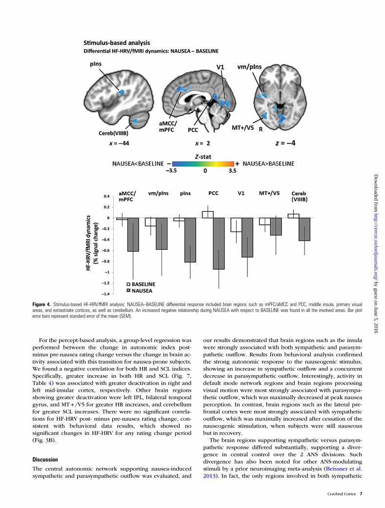
Compared with BASELINE, we found that NAUSEA pro-duced an increased negative relationship between HF-HRVand fMRI signals in a diffuse network of brain regions includ-ing medial prefrontal cortices (mPFC)/anterior mid-cingulatecortices (aMCC) and posterior cingulate cortices (PCC), ventralmiddle/posterior (vm/pIns) and posterior (pIns) insulae,para-hippocampus (PHG), supramarginal gyrus (SMG), andsuperior temporal gyrus, and lateral temporal cortex (LTC).Other regions showing this pattern included fusiform gyrus(FuG), inferior parietal lobule (angular gyrus, AnG), primaryand secondary somatosensory (S1, S2), premotor and motor(preMC, M1), and primary visual (V1) and extrastriate corticesconsistent with MT + /V5, as well as cerebellum (lobules V, VI,VIIIB, IX) (Fig. 4, Table 1).
Compared with BASELINE, we found that RECOVERY pro-duced an increased negative relationship between SCL and
fMRI signals in the dorsomedial (dmPFC), ventrolateral (vlPFC),and dorsolateral (dlPFC) prefrontal and orbitofrontal (OFC) cor-tices, dorsal and ventral anterior insula (daIns, vaIns), inferior(AnG) and superior (SPL) parietal lobules, and precuneus(PCun) (Fig. 5, Table 2). There were no significant differences inSCL-associated central autonomic network activity for theNAUSEAversus BASELINE differential window analysis.
In addition to the difference map analyses reported earlier, acloser linkage between peripheral autonomic outflow and brainactivity was evaluated by correlating individual differencesof ANS–fMRI parameter estimates with matching variations inautonomic responses. Difference maps for both HF-HRV andSCL/fMRI analyses showed negative correlation with the meanchange in autonomic indices within brain regions previouslyidentified by the original differential analysis. For the HF-HRVcorrelation analysis, all of the brain areas noted in Table 1 (withthe exclusion of PCC) demonstrated a significant correlationwith the change in HF-HRV between the NAUSEA and BASE-LINE windows (Fig. 6, Table 3). For the SCL correlational ana-lysis, left dlPFC (middle frontal gyrus) and vlPFC demonstrateda significant correlation with the change in SCL between the RE-COVERY and BASELINE windows (Fig. 6, Table 3).
Figure 3. (A) Autonomic response to nausea for the stimulus-based analysis: HR, HF-HRV, and SCL variations (mean ± SEM) with respect to BASELINE for both nausea-prone andcontrol subjects. (B) Autonomic response to nausea for the percept-based analysis: HR, HF-HRV, and SCL transitions to higher nausea states (mean ± SEM). Note: *P< 0.05;**P< 0.001; statistical significance testing was Bonferroni-corrected for multiple comparisons.
6 Central Autonomic Response to Nausea • Sclocco et al.
by guest on June 5, 2016http://cercor.oxfordjournals.org/
Dow
nloaded from
For the percept-based analysis, a group-level regression wasperformed between the change in autonomic index post-minus pre-nausea rating change versus the change in brain ac-tivity associated with this transition for nausea-prone subjects.We found a negative correlation for both HR and SCL indices.Specifically, greater increase in both HR and SCL (Fig. 7,Table 4) was associated with greater deactivation in right andleft mid-insular cortex, respectively. Other brain regionsshowing greater deactivation were left IPL, bilateral temporalgyrus, and MT + /V5 for greater HR increases, and cerebellumfor greater SCL increases. There were no significant correla-tions for HF-HRV post- minus pre-nausea rating change, con-sistent with behavioral data results, which showed nosignificant changes in HF-HRV for any rating change period(Fig. 3B).
Discussion
The central autonomic network supporting nausea-inducedsympathetic and parasympathetic outflow was evaluated, and
our results demonstrated that brain regions such as the insulawere strongly associated with both sympathetic and parasym-pathetic outflow. Results from behavioral analysis confirmedthe strong autonomic response to the nauseogenic stimulus,showing an increase in sympathetic outflow and a concurrentdecrease in parasympathetic outflow. Interestingly, activity indefault mode network regions and brain regions processingvisual motion were most strongly associated with parasympa-thetic outflow, which was maximally decreased at peak nauseaperception. In contrast, brain regions such as the lateral pre-frontal cortex were most strongly associated with sympatheticoutflow, which was maximally increased after cessation of thenauseogenic stimulation, when subjects were still nauseousbut in recovery.
The brain regions supporting sympathetic versus parasym-pathetic response differed substantially, supporting a diver-gence in central control over the 2 ANS divisions. Suchdivergence has also been noted for other ANS-modulatingstimuli by a prior neuroimaging meta-analysis (Beissner et al.2013). In fact, the only regions involved in both sympathetic
Figure 4. Stimulus-based HF-HRV/fMRI analysis: NAUSEA–BASELINE differential response included brain regions such as mPFC/aMCC and PCC, middle insula, primary visualareas, and extrastriate cortices, as well as cerebellum. An increased negative relationship during NAUSEA with respect to BASELINE was found in all the involved areas. Bar ploterror bars represent standard error of the mean (SEM).
Cerebral Cortex 7
by guest on June 5, 2016http://cercor.oxfordjournals.org/
Dow
nloaded from

and parasympathetic modulation were the insula and inferiorparietal lobule, regions also implicated as having more gener-alized function by this previous meta-analysis. Interestingly,while mid/posterior insula was implicated in parasympatheticprocessing, anterior/mid insula was implicated in sympatheticprocessing. This posterior shift of insular subregions in pro-cessing different divisions of the ANS appears to also be con-sistent with sympathetic/parasympathetic differentiation inprevious ANS–fMRI studies (Beissner et al. 2013). In additionto ANS division specialization, insula subregion involvementmay also relate to organ specificity (i.e. skin versus heart) aswell as differential temporal response properties. In general,autonomic processing via insula activity is consistent with theinsula’s oft-mentioned role in interoception (Craig 2002;Critchley et al. 2004; Wiens 2005), defined as conscious aware-ness of internal body states (Craig 2002), as interoceptive affer-ence is commonly carried by general visceral afferent(autonomic) fibers. Since insular (anterior, middle, and poster-ior) along with mid-cingulate cortex activation was also foundin response to nausea (Napadow et al. 2012), our results
demonstrate that these interoception processing brain regions(Mayer et al. 2009) also play a critical role in autonomic modu-lation by nausea.
Importantly, for both the HF-HRV and SCL analyses, an in-creased negative correlation between ANS metric and fMRIsignal was found during the NAUSEA and RECOVERY timewindows relative to BASELINE, respectively. This suggests thatmany of the regions implicated by our analysis had inhibitoryinfluence on premotor autonomic nuclei in the medulla andthat this inhibitory influence was magnified during periods ofnausea perception, when autonomic outflow was significantlymodulated. Hence, this altered brain response linked to auto-nomic outflow may serve as a coping mechanism in nausea-prone subjects, trying to limit the magnitude of efferent auto-nomic response and ultimately nausea sensation in response tothe visual stimulus. Despite early studies questioning ANS ac-tivity as a marker for nausea (Money 1970), multiple lines ofevidence support the importance of autonomic activity in thegeneration and perception of nausea (see review, Muth 2006).Our own human research found that “dynamic HF-HRV . .
Figure 5. Stimulus-based SCL/fMRI analysis: RECOVERY–BASELINE differential response included brain regions such as vlPFC prefrontal and orbitofrontal cortices, anterior insula,inferior and superior parietal lobule, and precuneus. An increased negative relationship during RECOVERY with respect to BASELINE was found in all the involved areas. Bar plot errorbars represent SEM.
8 Central Autonomic Response to Nausea • Sclocco et al.
by guest on June 5, 2016http://cercor.oxfordjournals.org/
Dow
nloaded from

. bursts of cardiovagal modulation precede rating transitions tohigher nausea” (LaCount et al. 2011), allowing for a potentialcausal effect. Interestingly, Stern (2002) suggested a model bywhich motion sickness produces autonomic outflow, whichinduces gastric tachyarrhythmia and feeds back to the brain toproduce nausea perception. Such feedback may be interpretedin the context of the James–Lange theory of emotion (Lang1994), which suggests a causal linkage between autonomic/humoral outflow (higher adrenaline level, heartbeat, etc.) andaffective response to arousal stimuli, though such connectionsare likely more complex than a simple one-way causal link. Insum, while cardiac and sudomotor activity may or may not bepart of a causal chain of events leading up to the conscious per-ception of nausea, ANS outflow is at least an important androbust marker, and a better understanding of the underlyingbrain activity may aid in understanding individual variability inboth conscious nausea perception and heterogeneity in ANSresponse to that perception.
Negative correlation was found for both the stimulus-basedanalysis between m/pIns signal and HF-HRV power duringNAUSEA, and the percept-based analysis between mIns signaland both HR and SCL response for transitions to higher levelsof nausea. Interestingly, for the stimulus-based analysis,greater HF-HRV decrease with respect to BASELINE was asso-ciated with reduced anti-correlation between fMRI signal in m/pIns and HF-HRV power (i.e. parasympathetic modulation).This might be explained by the fact that reduced HF-HRVpower leads to reduced signal dynamics and ultimatelyreduced correlation to mIns fMRI signal. In contrast, for thepercept-based analysis, greater HR or SCL increase to increas-ing nausea (i.e. increased sympathetic outflow) was associatedwith greater mIns deactivation, consistent with a direct inhibi-tory role of mIns activity on sympathetic premotor activity inthe brainstem.
Pooled together, many of the brain regions supporting ANSresponse to nausea are consistent with those proposed as keyregulatory areas of autonomic outflow in recent reviews andmeta-analyses concerning the central autonomic network(Beissner et al. 2013; Shoemaker et al. 2012; Thayer et al.
2012). These regions include cingulate, insular, prefrontal, andorbitofrontal cortices. Conversely, many subcortical and brain-stem areas associated with central autonomic control, suchas amygdala, thalamus, hippocampus, and brainstem nuclei(Macefield et al. 2013; Jänig 2006; Saper 2002), were not identi-fied by our analysis. Physiological artifacts from cardiorespira-tory sources hamper neuroimaging in brainstem regions,requiring a more focused approach (Brooks et al. 2013), whichin our case is especially acute as there is a need to account fornon-neuronal cardiorespiratory pulsatile artifact without sacri-ficing signal variance attributable to neural activity associatedwith cardiorespiratory (i.e. autonomic) outflow.
In our stimulus-based analysis, parasympathetic-associatedbrain activity was found to be much more widespread thansympathetic-associated activity and included brain areas nottypically associated with autonomic regulation. These areas in-cluded visual processing regions such as V1 and MT + /V5.This was probably due to an interaction between brain re-sponse to the nauseogenic visual stimulation during NAUSEAand dynamics in parasympathetic outflow. In contrast, duringthe RECOVERY time window, when sympathetic outflow wasmost increased, visual stimulation was confined to simple cross-hair fixation and produced less robust primary visual and extra-striate activation. Interestingly, a recent study has implicatedmany of the regions identified by our parasympathetic-fMRIanalysis during NAUSEA as areas coding specific parameters ofvisually induced self-motion (Becker-Bense et al. 2012). Specif-ically, cerebellar vermis and parieto-occipital regions showedsignificantly enhanced activation during circular vection,whereas parietal areas (IPL, bilateral PCun) were found toprocess the intensity of perceived vection, attributable to thedorsal stream (“where or how” pathway), which is mediated byinformation transfer from primary visual areas to the parietallobe (de Haan and Cowey 2011). Moreover, Lobel et al. (1998)found that vection induced by galvanic (i.e. nonvisual) stimuliproduces activation in superior and inferior posterior parietalcortex, whereas we found ANS-BOLD correlations in the leftAnG and SMG for parasympathetic-related structures and in theleft SPL for sympathetic-related structures. In sum, in our study,greater visual and self-body motion processing due to the nau-seogenic stimulus likely led to higher activity in these visual andvection processing areas and was associated with altered para-sympathetic or sympathetic modulation. Hence, while visualprocessing areas such as V1 and MT+ /V5 and vection process-ing areas in the posterior parietal lobule are likely not directlylinked with premotor autonomic nuclei in the medulla, ANS
Table 1Stimulus-based HF-HRV/fMRI analysis: NAUSEA–BASELINE-associated brain regions
Brain region Side Location (MNI) Z-score
x y z
aMCC/mPFC R 2 32 36 −3.31vm/pIns R 44 2 −10 −3.05pIns L −42 −26 20 −3.27M1 L −56 −8 48 −2.99preMC L −57 16 26 −3.25S1 L −58 −10 40 −3.05S2 L −64 −20 16 −2.99PCC L −2 −54 14 −3.84AnG L −62 −32 36 −3.21SMG L −60 −18 36 −3.5LTC R 58 −20 4 −3.37— L −54 −20 8 −3.34PHG L −34 −32 −20 −3.05V1 R 6 −78 4 −3.09— L −4 −80 2 −3.01MT + /V5 R 42 −78 16 −3.24FuG R 38 −56 −10 −3.26Cereb(V) R 16 −46 −22 −3.13Cereb(VI) L −38 −42 −32 −3.29Cereb(VIIIB) L −26 −42 −40 −3.33Cereb(IX) R 14 −56 −40 −3.21
Table 2Stimulus-based SCL/fMRI analysis: RECOVERY–BASELINE-associated brain regions
Brain region Side Location (MNI) Z-score
x y z
OFC L −28 48 −12 −3.6vlPFC L −42 48 −4 −4.08dmPFC L −6 56 10 −3.01dlPFC L −46 38 24 −3.23vaIns L −38 16 −12 −3.53daIns L −36 24 0 −3.31SPL L −20 −70 48 −3.56AnG R 42 −68 30 −3.31PCun R 4 −66 50 −3.7— L −6 −68 50 −3.4
Cerebral Cortex 9
by guest on June 5, 2016http://cercor.oxfordjournals.org/
Dow
nloaded from

outflow due to nauseogenic visual stimulation likely involves in-formation transfer from these brain areas to areas more intimate-ly linked with ANS outflow, such as the insula.
Consistent with our previous study (LaCount et al. 2011),the stimulus-based analysis showed increased HR and skinconductance, as well as decreased HF-HRV, during a 4-minwindow when nausea was most intense. Previous studies havenoted a heterogeneous HF-HRV response to nausea (Dowecket al. 1997; Kim et al. 2005; LaCount et al. 2011; Mullen et al.1998; Yokota et al. 2005), and Lin et al. recently suggested thatthis heterogeneity may be due to variability in nausea-inducedchanges in respiratory rate and/or volume between studies(Lin et al. 2011, 2013). In our study, subjects were asked tomaintain constant respiration, which likely limited the influ-ence of respiratory changes on HF-HRV response to nausea. Infact, our previous study did not find any significant change inrespiratory rate for similar duration windows (LaCount et al.2011). Interestingly, our percept-based SCL analysis showed
the greatest sympathetic increase following the 0–1 nauseatransition, consistent with McClure et al. (1971), which foundthat skin conductance, measured from the palmar aspect of thehand, may be a marker of early arousal during motion sick-ness. However, as reported by our previous study (LaCountet al. 2011) and as shown in Figure 1 for a representativesubject and confirmed by the analysis of tonic SCL group data(Fig. 3A), the activity recorded on the palmar aspect of thefinger tips clearly increases, gradually, as the visual stimuluscontinues and increasing nausea sensations are reported bythe subjects. Moreover, all transition to higher nausea levels(including 1-to-2 and 2-to-3 transitions) also resulted in phasicincreases following transitions (Fig. 3B), supporting the ideathat SCL increase accompanies both phasic transitions andgradually increasing nausea sensation.
Another interesting finding was that the greatest changes inHF-HRV and SCL were found for the final 4 min of nauseogenicstimulation (NAUSEA–BASELINE) and for the 4 min after
Figure 6. Stimulus-based analysis: inter-individual correlation between (A) change in HF-HRV/fMRI insular cortex response and change in HF-HRV (NAUSEA–BASELINE); (B)change in SCL/fMRI vlPFC prefrontal cortex response and change in SCL (RECOVERY–BASELINE. Note: r= Pearson’s correlation coefficient; r* =−0.63, Pearson’s correlationcoefficient removing the outlier (−0.77; 2.43), as determined by Grubb’s test for outliers.
10 Central Autonomic Response to Nausea • Sclocco et al.
by guest on June 5, 2016http://cercor.oxfordjournals.org/
Dow
nloaded from

removal of this stimulus (RECOVERY–BASELINE), respectively.A similar result was also found in our previous study (LaCountet al. 2011), where the instantaneous estimate of HF-HRV wasfound to increase back toward baseline values within tens ofseconds after cessation of the nauseogenic stimulus, whereasSCL showed persistent elevated values into this post-stimulus re-covery period. The persistence of sympathetic activation intothe recovery period is likely a manifestation of the known longlatency characterizing this division of the ANS (Boucsein 2012).Also, the work by Sheehan et al. (2011) noted the difficulty ofclearly attributing motion sickness to the unique modulation ofeither sympathetic (“fight or flight”) or parasympathetic (“restand relax”) autonomic responses, thus supporting our resultsshowing the absence of coordination between the 2 ANSbranches. For instance, we found greater increase in sympathet-ic activity after the cessation of the visual stimulus, whereasparasympathetic (cardiovagal) activity decreased most signifi-cantly during peak nausea and rapidly returned toward baselinevalues after stimulus cessation. Moreover, our results did notfind significant correlation between HF-HRV and SCL changes.That is, subjects with greater HF-HRV change did not also dem-onstrate greater SCL change during the same time window (i.e.NAUSEA or RECOVERY), nor did subjects with greater HF-HRVchange during NAUSEA also demonstrate greater SCL change
Table 3Stimulus-based analysis: inter-individual correlation between change in ANS/fMRI association andchange in ANS response
Brain region Side Location (MNI) Z-score
x y z
HF-HRVaMCC/mPFC L −2 32 34 −2.99m/pIns R 44 −12 −4 −3.16pIns L −42 −24 16 −2.49M1 L −56 −8 50 −3.09preMC L −54 10 26 −3.01S1 L −58 −10 40 −3.05S2 L −64 −20 16 −2.99AnG L −62 −32 36 −3.21SMG L −60 −18 36 −3.5LTC R 58 −20 4 −3.37
— L −54 −20 8 −3.34PHG L −34 −32 −20 −3.05V1 L −6 −72 −2 −3.61— R 6 −70 −2 −3.39MT + /V5 R 42 −70 −2 −3.09FuG R 38 −56 −10 −3.26Cereb(V) R 16 −46 −22 −3.13Cereb(VI) L −38 −46 −32 −2.77Cereb(VIIIB) L −26 −46 −40 −3.13Cereb(IX) R 14 −56 −40 −3.21SCL
vlPFC L −48 36 10 −3.12dlPFC L −56 14 28 −2.58
Figure 7. Percept-based analysis: inter-individual correlation between change in mid-insular cortex response and (A) change in HR and (B) change in SCL for increasing nauseasensation. Note: r= Pearson’s correlation coefficient; r* =−0.25, Pearson’s correlation coefficient removing the outliers (4.21; −1.58) and (3.30; −1.80), as determined byGrubb’s test for outliers.
Cerebral Cortex 11
by guest on June 5, 2016http://cercor.oxfordjournals.org/
Dow
nloaded from

during RECOVERY. This lack of correlation suggests independ-ent processes in ANS response associated with sympathetic andparasympathetic modulation during nausea. Hence, the inde-pendence between sympathetic and parasympathetic responsesto motion sickness necessitates the monitoring of both ANS divi-sions when investigating the complex phenomenon of nausea.
Several limitations to our study should be noted. First, whilesignificance was noted for many of the autonomic changesevaluated with our percept- and stimulus-based analyses, therewas a lack of significance for some of the more highly variableautonomic indices (e.g. HF-HRV) in our percept-based ana-lysis. This may have been due to insufficient sample size forthis combination of analysis method and autonomic index. Inaddition, we were not able to demonstrate significantautonomic-associated activity in the brainstem. Many auto-nomic processing nuclei are known to exist in medullary,pontine, and mesencephalic regions, and future studies shouldadopt specific neuroimaging methods (e.g. higher fieldstrength and smaller voxel size) that focus on brainstem activ-ity in relation to nausea-induced autonomic change. Anotherlimitation was that SCL is known to be an indirect measure ofsympathetic activity and is certainly delayed with respect tocentral signaling of peripheral sympathetic response (Boucsein2012). Future studies could consider the recovery of the under-lying nerve activity relative to the SCL signal, in order toimprove the accuracy of modeling for combined ANS/fMRIanalyses. As a final note, a recent study showed that magneticfields associated with the MRI environment produce nystag-mus, reportedly interacting with ionic currents in the labyrin-thine endolymph fluid, resulting in magnetic vestibularstimulation (Roberts et al. 2011). While our study was per-formed at 1.5 T and it is unlikely that magnetic field vestibularstimulation influenced the present results (Roberts et al. foundmuch greater effects at 7 T compared with 3 T), future studiesshould consider this potential confound when choosing MRIfield strength.
In conclusion, we found that the increased sympathetic anddecreased parasympathetic modulation caused by nausea wasassociated with a broad central autonomic network includingbrain regions such as the insula. Specificity in brain activitysupporting parasympathetic versus sympathetic modulationwas also noted. Activity in default mode network regions andbrain regions processing visual motion were most strongly as-sociated with parasympathetic outflow, which was maximallydecreased at peak nausea perception. In contrast, brainregions such as the lateral prefrontal cortex were most strongly
associated with sympathetic outflow, which was maximally in-creased after cessation of the nauseogenic stimulation, whensubjects were still nauseous but in recovery. These resultssuggest divergent central autonomic control for sympatheticand parasympathetic response to nausea, while highlightingthe role of the insula in mediating both phasic and sustainedautonomic response during nausea.
Funding
This work was supported by the National Institutes ofHealth (grant numbers P01-AT006663, R01-AT007550, and R01-AR064367 to VN, K23-DK069614 to BK, and R21-AR057920 toVN, BK and RB); the National Center for Research Resources(P41RR14075; CRC 1 UL1 RR025758, Harvard Clinical and Trans-lational Science Center); the MGH Department of Anesthesia,Critical Care and Pain Medicine and the International Foundationof Functional Gastrointestinal Disorders.
NotesThe content is solely the responsibility of the authors and does notnecessarily represent the official views of our sponsors.
ReferencesBarbieri R, Matten EC, Alabi AA, Brown EN. 2005. A point-process
model of human heartbeat intervals: new definitions of heart rateand heart rate variability. Am J Physiol Heart Circulat Physiol.288(1):H424–H435.
Becker-Bense S, Buchholz H, zu Eulenburg P, Best C, Bartenstein P,Schreckenberger M, Dieterich M. 2012. Ventral and dorsal streamsprocessing visual motion perception (FDG-PET study). BMC Neuro-sci. 13(1):81.
Beckmann CF, Jenkinson M, Smith SM. 2003. General multilevellinear modeling for group analysis in FMRI. Neuroimage. 20(2):1052–1063.
Beissner F, Meissner K, Bär K, Napadow V. 2013. The autonomic brain:an activation likelihood estimation meta-analysis for central pro-cessing of autonomic function. J Neurosci. 33(25):10503–10511.
Boucsein W. 2012. Electrodermal activity. New York: Springer.Brooks JC, Faull OK, Pattinson KT, Jenkinson M. 2013. Physiological
noise in brainstem fMRI. Front Hum Neurosci 7(623).Chang C, Cunningham JP, Glover GH. 2009. Influence of heart rate
on the BOLD signal: The cardiac response function. Neuroimage.44(3):857.
Cowings PS, Suter S, Toscano WB, Kamiya J, Naifeh K. 1986. Generalautonomic components of motion sickness. Psychophysiology. 23(5):542–551.
Craig AD. 2002. How do you feel? interoception: the sense of the physio-logical condition of the body. Nat Rev Neurosci 3(8):655–666.
Critchley HD, Wiens S, Rotshtein P, Öhman A, Dolan RJ. 2004.Neural systems supporting interoceptive awareness. Nat Neurosci.7(2):189–195.
de Haan EH, Cowey A. 2011. On the usefulness of ‘what’and ‘where’-pathways in vision. Trends Cogn Sci. 15(10):460–466.
Doweck I, Gordon CR, Shlitner A, Spitzer O, Gonen A, Binah O,Melamed Y, Shupak A. 1997. Alterations in R–R variability asso-ciated with experimental motion sickness. J Auton Nerv Syst.67(1):31–37.
Golding JF. 1998. Motion sickness susceptibility questionnaire revisedand its relationship to other forms of sickness. Brain Res Bull.47(5):507–516.
Gray MA, Minati L, Harrison NA, Gianaros PJ, Napadow V, CritchleyHD. 2009. Physiological recordings: basic concepts and implemen-tation during functional magnetic resonance imaging. Neuroimage.47(3):1105–1115.
Table 4Percept-based analysis: inter-individual correlation between change in brain response and changein ANS response
Brain region Side Location (MNI) Z-score
x y z
HRmIns R 34 −10 10 −4.4AnG L −40 −50 50 −3.82LTC L −50 12 −18 −3.41
−52 −22 12 −3.7R 58 −38 −10 −3.65
MT + /V5 L −38 −62 18 −4.26SCLmIns L −48 6 −12 −3.67Cerebellum L −22 −82 −18 −3.22
12 Central Autonomic Response to Nausea • Sclocco et al.
by guest on June 5, 2016http://cercor.oxfordjournals.org/
Dow
nloaded from

Graybiel A, Lackner JR. 1980. Evaluation of the relationship betweenmotion sickness symptomatology and blood pressure, heart rateand body temperature. Aviat Space Environ Med. 51(3):211–214.
Ishii M, Igarashi M, Patel S, Himi T, Kulecz W. 1987. Autonomic effectson RR variations of the heart rate in the squirrel monkey: an indica-tor of autonomic imbalance in conflict sickness. Am J Otolaryngol.8(3):144.
Jänig W. 2006. The integrative action of the autonomic nervous system:neurobiology of homeostasis. New York: Cambridge UniversityPress, Cambridge.
Kim YY, Kim HJ, Kim EN, Ko HD, Kim HT. 2005. Characteristicchanges in the physiological components of cybersickness. Psycho-physiology. 42(5):616–625.
LaCount LT, Barbieri R, Park K, Kim J, Brown EN, Kuo B, Napadow V.2011. Static and dynamic autonomic response with increasingnausea perception. Aviat Space Environ Med. 82(4):424.
Lang PJ. 1994. The varieties of emotional experience: a meditation onJames-Lange theory. Pshychol Rev. 101(2):211–221.
Levine ME, Stern RM, Koch KL. 2006. The effects of manipulatingexpectations through placebo and nocebo administration on gastrictachyarrhythmia and motion-induced nausea. Psychosom Med.68(3):478–486.
Lin C, Jung T, Chuang S, Duann J, Lin C, Chiu T. 2013. Self-adjustmentsmay account for the contradictory correlations between HRV andmotion-sickness severity. Int J Psychophysiol. 87(1):70–80.
Lin Chin-Teng, Lin Chun-Ling, Chiu Tzai-Wen, Duann Jeng-Ren, JungTzyy-Ping. 2011. Effect of respiratory modulation on relationshipbetween heart rate variability and motion sickness. Engineering inmedicine and biology society, EMBC, 2011 annual internationalconference of the IEEE. Boston, MA: IEEE. p. 1921.
Lobel E, Kleine JF, Bihan DL, Leroy-WIllig A, Berthoz A. 1998. Func-tional MRI of galvanic vestibular stimulation. J Neurophysiol.80(5):2699–2709.
Macefield VG, James C, Henderson LA. 2013. Identification of sites ofsympathetic outflow at rest and during emotional arousal: concur-rent recordings of sympathetic nerve activity and fMRI of the brain.Int J Psychophysiol. 89(3):451–459.
Mayer E, Aziz Q, Coen S, Kern M, Labus J, Lane R, Kuo B, Naliboff B,Tracey I. 2009. Brain imaging approaches to the study of functionalGI disorders: a Rome working team report. NeurogastroenterolMotility. 21(6):579–596.
McClure JA, Fregly AR, Molina E, Graybiel A. 1971. Response fromarousal and thermal sweat areas during motion sickness. Naval Aero-space Medical Research Lab, Pensacola (FL), USA. No. NARML-1142.
Money KE. 1970. Motion sickness. Physiol Rev. 50(1):1–39.Mullen TJ, Berger RD, Oman CM, Cohen RJ. 1998. Human heart rate
variability relation is unchanged during motion sickness. J VestibRes. 8(1):95–105.
Muth ER. 2006. Motion and space sickness: intestinal and autonomiccorrelates. Autonomic Neurosci. 129(1):58–66.
Muth ER, Stern RM, Thayer JF, Koch KL. 1996. Assessment of the mul-tiple dimensions of nausea: the nausea profile (NP). J PsychosomRes. 40(5):511–520.
Napadow V, Li A, Loggia ML, Kim J, Schalock PC, Lerner E, Tran T,Ring J, Rosen BR, Kaptchuk TJ. 2012. The brain circuitrymediating antipruritic effects of acupuncture. Cerebral Cortex. 24(4):873–882.
Napadow V, Sheehan JD, Kim J, LaCount LT, Park K, Kaptchuk TJ,Rosen BR, Kuo B. 2013. The brain circuitry underlying thetemporal evolution of nausea in humans. Cerebral Cortex. 23(4):806–813.
Ohyama S, Nishiike S, Watanabe H, Matsuoka K, Akizuki H, Takeda N,Harada T. 2007. Autonomic responses during motion sicknessinduced by virtual reality. Auris Nasus Larynx. 34(3):303–306.
Oldfield RC. 1971. The assessment and analysis of handedness: theedinburgh inventory. Neuropsychologia. 9(1):97–113.
Roberts DC, Marcelli V, Gillen JS, Carey JP, Della Santina CC, Zee DS.2011. MRI magnetic field stimulates rotational sensors of the brain.Current Biology. 21(19):1635–1640.
Saper CB. 2002. The central autonomic nervous system: consciousvisceral perception and autonomic pattern generation. Annu RevNeurosci. 25(1):433–469.
Sheehan SE, Oman CM, Duda KR. 2011. Motion sickness: a cholinomi-metic agent hypothesis. J Vestib Res. 21(4):209–217.
Shoemaker JK, Wong SW, Cechetto DF. 2012. Cortical circuitry asso-ciated with reflex cardiovascular control in humans: Does the cor-tical autonomic network “speak” or “listen” during cardiovasculararousal. Anat Rec. 295(9):1375–1384.
Stern RM. 2002. The psychophysiology of nausea. Acta BiologicaHungarica. 53(4):589–600.
Stern RM, Koch KL, Andrews PL. 2011. Nausea: Mechanisms and man-agement. New York: Oxford University Press.
Thayer JF, Åhs F, Fredrikson M, Sollers III JJ, Wager TD. 2012. Ameta-analysis of heart rate variability and neuroimaging studies:Implications for heart rate variability as a marker of stress andhealth. Neurosci Biobehav Rev. 36(2):747–756.
Wiens S. 2005. Interoception in emotional experience. Curr OpinNeurol. 18(4):442.
Wiggins G, Triantafyllou C, Potthast A, Reykowski A, Nittka M,Wald L. 2006. 32‐channel 3 tesla receive‐only phased‐array headcoil with soccer‐ball element geometry. Magn Reson Med. 56(1):216–223.
Yokota Y, Aoki M, Mizuta K, Ito Y, Isu N. 2005. Motion sicknesssusceptibility associated with visually induced postural instabilityand cardiac autonomic responses in healthy subjects. Acta Otolar-yngol. 125(3):280–285.
Cerebral Cortex 13
by guest on June 5, 2016http://cercor.oxfordjournals.org/
Dow
nloaded from
Copyright © 2022 FDOKUMEN