Kappa-Opioid Receptor Blockade Ameliorates Obesity ... - MDPI
Upload
khangminh22Category
view
3download
0
ENHANCING THE USE OF OPIOIDS IN PAIN
MANAGEMENT : ANTINOCICEPTIVE
POTENTIATION WITH OPIOID
AGONIST/ANTAGONIS T COMBINATIONS
Sophie La Vincente (BPsych(Hons))
Department of Clinical and Experimental Pharmacology
The University of Adelaide
(Faculty of Health Science)
February,2005
A thesis submitted for the degree of Doctor of Philosophy
Table of Contents
Abstract.. ....... i
Publications and presentations in support of this thesis ................ v
Abbreviations, prefixes and symbols ........... vii
1.1.
1.2.
1.3.
Background ...............
The terminology of pain......
The development of pain theories..
1
1.3.1. Specifrcity theory
1.3.2. Pattern theory
1.3.3. Gate Control theory
1.3.4. Neuromatrix theory
1.4. Neurobiological mechanisms ofnociception
1.4.1. The detection of noxious stimuli in the periphery
I.4.2. Activation of nociceptors in non-cutaneous tissue
1.4.3.
7.4.4.
1.4.5.
aJ
aJ
4
6
7
Ascending transmi s sion of nociceptive signals
Chemical modulators and transmitters in the nociceptive pathways..
Descending modulation.............
9
.. 10
.11
t2
76
18

1.5. Types of pain....
1.5.1. Transientpain.....
1.5.2. Acute pain
1.5.3. Chronicpain..........
r.5.4. Experimental pain ........
1.5.5. Techniques for experimental pain induction...
1.5.5.1. Coldpressor...............
1.5.5.2.
1.6. Opioids
1.6.3.
1.6.4.
1.6.5.
Electrical stimulation
1.6.1. The history of opioids
1.6.2. Opioid classification...
Opioid receptors
Endogenous opioid ligands
Second messengers and effectors ...
1.7. Opioid effects.
1.7.1. Analgesia.........
1.7.2. Respiratorydepression.
1.7.3. Sedation
1.7.4. Nausea and vomiting...
1.7.5. Effects on mood
1.7.6. Constipation
I.7.7. Pupillary miosis.......
L7.8. Cardiovasculareffects
1.7.9. Cough suppression
1.7.10. Pruritus
1.8. Barriers to adequate pain management with opioids ........
1.8.1. Adverse effects .39
1.8.2. Tolerance ..40
L9. Enhancing analgesia through drug combinations .............42
1.9.1. Opioid agonist-NMDA antagonist combinations........ .................44
1.9.2. Opioid agonist-antagonist combinations.............. ......45
1.9.2.1. Enhanced opioid sensitivity following chronic antagonist pre-treatment46
L9.2.2. Analgesic actions of low-dose opioid antagonists ................ 46
1.9.2.3. Reduction in side effects with the addition of low-dose antagonist.......49
1.9.2.4. Enhanced analgesia and attenuation of opioid tolerance with co-
administration of low-dose antagonist......... ....... 5l
1.9.2.4.1. Invivo animal studies 51
7.9.2.4.2. In vitro studies: the basis of the bimodal opioid receptor model
for enhanced opioid analgesia with ultra-low dose antagonist...... 53
1.9.2.4.3. Putative role of excitatory G.-coupled opioid receptors in the
development of tolerance..... 55
1.9.2.4.4. Human models of analgesic potentiation with low-dose antagonist5T
1.9.3. Limitations of the human studies 61
1.9.4. Buprenorphine and antagonist combinations 67
1.10. Summary
1.1 1. The present research.
1.11.1. Aims
.71
..73
2. General Methods 75
2.1. Introduction............
2.2. Nociceptivetesting.
70
75
2.2.1. Cold pressor (CP) test .
75

2.2.1.1. Materials
2.2.1.2. Set-up procedure
2.2.1.3. Test administration
2.2.2. Electrical stimulation (ES) test
..76
76
2.2.2.t.
)))')
2.2.2.3.
Materials...
Set-up procedure....
Test administration.
77
79
80
80
80
82
83
83
84
85
85
2.2.3. Procedures for repeated testing
2.2.4. Testing environment.........
2.3. Methods coÍìmon to drug studies (Chapters 4,5 and6).
2.3.1. Drugadministration
2.3.2. Testing time points.
2.3.2.1. Blood sampling.....
2.3.2.2. Monitoring of physiological parameters ..........
2.3.2.3. Monitoring of nausea and sedation
2.3.2.4. Monitoring of other opioid effects .
2.3.2.5. Nociceptive testing....
2.3.3. Testing schedule....
2.4. Methods of statistical inference
86
86
87
87
87
3. ESTABLISHING NORMAL VALUES FOR TIIE COLD PRESSOR TEST
AND ELECTRICAL STIMULATION TEST IN HEALTITY VOLUNTEERS.. 39
3.1. Introduction 89
3.1.1. The validity of experimental pain.... .. 89
3.1.2. Considerations in experimental pain induction 90
3.1.3. Determinants ofpain response.. 92
3.1.3.l. Sex...
3.1.3.2. Ethnicity/race
3.1.3.3. 49e..................
3.1.3.4. CNS stimulants
3.1.3.5. Menstrual cycle.........
3.1.3.6. Body weighlsize
3.1.3.7. Psychological/cognitive factors .....
3.1.4. Cold pressor test.......
3. 1 .5. Electrical stimulation test............
3.2. Purpose and aims of the present study
3.3. Methods
3.3.l Participants..
3.3.1.1. Inclusion criteria ...
3.3.1.2. Exclusion criteria.
3.4. Procedures
3.4.1. Recruitment and screening procedures
3.4.2. Experimental procedures...
3.4.3. Statistical analyses ....
3.5. Results
3.5.1. Sample characteristics................
3.5.2. Normative data..........
3.5.3. Intra-subject variability...................
3.5.4. Factors impacting upon test performance ...........
3.5.4.1. Cold pressor tolerance (CPTOL)
3.5.4.2. Electrical stimulation tolerance (ESTOL)....
3.5.4.3. Impact of menstrual phase
92
92
93
94
94
95
95
96
98
100
101
101
101
101
t02
t02
t02
105
106
106
110
r12
rt2
tt4
116

3.5.4.4. Test order effects . tt6
3.6. Discussion .. 116
4. ANTINOCICEPTTVE ACTIVITY OF BUPRENORPHINE IN
EXPERIMENTAL PAIN: DOSE FINDING STIIDY.... .... 131
4.1. Overview 131
4.2. Pharmacology ofbuprenorphine... 133
4.2.I. Interaction with the ORL1 receptor.. 139
4.2.2. Human pharmacokinetics r4t
4.2.2.1. Oral administration t42
4.2.2.2. Sublingual administration 142
4.2.2.3. Intravenous administration r44
4.2.2.4. Intramuscular administration 145
4.2.3. Metabolism and excretion 146
4.2.4, Safety and toxicity r46
4.2.5. Subjective and physiological effects of buprenorphrne 148
4.3. Buprenorphine as an analgesic..
4.4. Clinical trials with pain patients
4.4.1. Acutepain..........
4.4.2. Chronic pain..........
4.5. Buprenorphine in the treatment of neuropathic pain
4.6. Buprenorphine in human experimental pain
4.7. The current study....
4.7.1. Hypothesis......
4.7.2. Aims
150
151
151
1s2
153
154
155
155
156
4.8. Methods...... 156
4.8.1. Participants................
4.8.1.1. Subject inclusion criteria...
4.8.1.2. Subject exclusion criteria..
4.8.2. Study design
4.9. Pilot study
4.9.1. Sample characteristics
4.9.2. Procedures
4.9.2.I. Screening procedures .........
4.9.2.2. Experimental procedures....
ts6
ls6
r57
r58
160
160
t59
. 159
. 159
.159
160
4.9.2.2.1.
4.9.2.2.2.
4.9.4. Discussion
4.10. Principal study
Drug administration..
Testing protocol and schedule....
4.9.2.3. Statistical analysis......
4.9.3. Results
4.9.3.1. Antinociception
4.9 .3 .2. Physiologi cal parameters ...........
4.9.3.3. Adverse and other drug effects .
162
r62
. t63
765
. t67
..169
..170
170
17t
t7l
t7r
172
. 174
.174
4.10.1. Sample characteristics
4.10.2. Procedures
4.10.2.1. Screening procedures.
4.10.2.2. Experimental procedures.
4.10.2.3. Statistical analyses
4.10.3. Results
4. 10.3. 1. Measures pre- and post-saline
4.10.3.2. Practice/order effects 174

4.10.3.3. Antinociception
4.10.3.3.1. Cold pressor threshold..
t76
176
t77
179
t79
4.10.3.3.2.
4.r0.3.3.3.
4.10.3.3.4.
Cold pressor tolerance
Electrical stimulation threshold......
Electrical stimulation tolerance ......
4.10.3.4. Physiological parameters and adverse effects.
4.10.3.4.1. Respiration.
4.10.3.4.2. Arterial oxygen saturation..
4.10.3.4.3. Heart rate ...........
182
4.10.3.4.4. Blood pressure 183
4.10.3.4.5. Nausea .... 184
4.10.3.4.6. Sedation........ 184
4.10.3.4.7. Other adverse effects 185
4.10.4. Discussion 188
5. ANTINOCICEPTTVE ACTIVITY OF BI]PRENORPHINE AND NALOXONE
COMBINATIONS IN HT]MAN EXPERIMENTAL PAIN: RATIO STUDY 1 193
5.1. Proposed mechanisms of enhanced analgesia with low dose antagonists .......... 195
5.2. Buprenorphine/antagonist combinations in animal models of nociception........ 196
5.3. Summary.. .. 198
...199
782
183
183
200
201
202
5.4. Naloxone
5.4.1. Pharmacology ofnaloxone...
5.5. Purpose and aims of the present research ....
5.5.1. Hypothesis
5.5.2. Aims..... 202
2025.6. Methods

5.6.1. Study design
5.6.2. Participants................
5.6.2.1. Inclusion and exclusion criteria
5.6.3. Samplecharacteristics
5.6.4. Procedures 205
..20s
202
203
203
204
205
205
206
207
207
208
5.6.4.I. Screening procedures
5.6.4.2. Experimental procedures..
5.6.4.2.1. Drug administration..
5.6.4.2.2. Testing protocol and schedule
5.6.5. Statistical analyses ...
5.6.5.1. Justification for use of parametric or non-parametric methods
5.6.5.2 Est¿blishing baseline response...
5.6.5.3. Assessing the effect associated with each drug condition.................... 208
5.6.5.4. Comparing the effect of each buprenorphine:naloxone ratio with
buprenorphine alone... 209
5.6.5.5. Comparing the effect of each ratio... 210
5.6.5.6. Subjective and other effects 2tt
5.7. Results.. 2tr
5.7 .1. Missing data and participant withdrawal/exclusion post-recruitment ......... 2ll
5.7.1.1. Participant withdrawal .......
5.7.1.2. Missing data..........
5.7.2. Pre- and post-saline infusion.....
5.7.3. Cold pressor threshold
5.7.3.1. Effect of each condition.....
2tl
5.7.3.2. Effect of buprenorphine:naloxone ratio compared to buprenorphine
212
.212
...212
...212
alone 214

5.7.4. Cold pressor tolerance................ ...216
5.7.4.1. Effect of each condition 216
5.7.4.2. Effect of buprenorphine:naloxone ratio compared to buprenorphine
alone 218
5.7 .4.3. Difference between o/o changen rn6 and %o changesup oNLy.. 220
5.7.5. Electrical stimulation threshold ... 22r
5.7.5.1. Effect of each condition 22r
5.7.5.2. Effect of buprenorphine:naloxone ratio compared to buprenorphine
alone ...222
5.7.6. Electrical stimulation tolerance.
5.7.6.L Effect of each condition
5.7.6.2.
alone 225
5.7.6.3. Difference between oá change¡,¡¡e and %o changesup oNLy'
5.7.7. Respiration 228
5.7.7.1. Effect of each condition ..228
5.7.7.2. Effect of buprenorphine:naloxone ratio compared to buprenorphine
alone 229
5.7.8. Heart rate 230
5.7.8.1. Effect of each condition.. 230
5.7.8.2. Effect of buprenorphine:naloxone ratio compared to buprenorphrne
alone 231
5.7 .9. Oxygen saturation 232
5.7 .9.1. Effect of each condition..... 232
5.7.9.2. Effect of buprenorphine:naloxone ratio compared to buprenorphine
224
224
Effect of buprenorphine:naloxone ratio compared to buprenorphine
227
alone 233
5.7.10. Blood pressure.... 235
5.7.10.1. Effect of each condition 235
5.7.10.2. Effect of buprenorphine:naloxone ratio compared to buprenorphine
alone236
5.7.1I. Subjective effects . 238
5.8. Discussion
6. OPTIMISING TIIE BIIPRENORPHINE:NALOXONE DOSE RATIO IN
HT]MAN EXPERIMENTAL PAIN....... .............247
6.1. Introduction 247
6.1.1. Hypothesis .247
6.1.2. Aim ...248
6.1.3. Study design..
240
...248
6.1.4. Participants
6.1.4.I. Inclusion and exclusion criteria
6.1.4.2. Sample characteristics
6.1.5. Procedures
6.1.5.1. Screening procedures
6.1.5.2. Experimental procedures
6.1.5.2.I. Drug administration
6.1.6. Testing procedure and schedule........
6.1.6.1. Statistical analyses
6.2. Results
6.2.1. Participant withdrawal and missing data ...
6.2.1.1. Participant withdrawal
248
248
249
2s0
250
250
250
252
2s3
2s4
254
254
6.2.L2. Missing data 2s5

6.2.2. Pre- and post-saline infusion..
6.2.3. Cold pressor threshold............
6.2.3.1. Effect of each condition..
255
256
256
6.2.3.2. Effect of buprenorphine:naloxone ratio compared to buprenorphine
alone 258
6.2.4. Cold pressor tolerance... 259
6.2.4.1. Effect of each condition 2s9
6.2.4.2. Effect of buprenorphine:naloxone ratio compared to buprenorphine
alone 260
6.2.4.3. Difference between o/o change¡,ane and %ó changesupoNly ..................262
6.2.5. Respiration 263
6.2.5.1. Effect of each condition.. 263
6.2.5.2. Effect of buprenorphine:naloxone ratio compared to buprenorphine
alone ............264
6.2.6. Heart rate 267
6.2.6.1. Effect of each condition ..................267
6.2.6.2. Effect of buprenorphine:naloxone ratio compared to buprenorphine
alone 268
6.2.7. Oxygen saturation 269
6.2.7.1. Effect of each condition 269
6.2.7.2. Effect of buprenorphine:naloxone ratio compared to buprenorphine
alone 270
6.2.8. Bloodpressure.... 272
6.2.8.1. Effect of each condition 272
6.2.8.2. Effect of buprenorphine:naloxone ratio compared to buprenorphine
alone 273

6.2.9. Subjective effects 274
6.3. Combined cold pressor results for ratio studies I and2 ... 27s
6.4. Discussion... ..277
6.5. Limitations associated with the buprenorphine studies
7. Conclusions, clinical implications and future directions ..29L
7.1. Overview of findings 292
7.r.1
7.t.2
7.1.3
Normative data and inter-individual variability in experimental pain testing292
Buprenorphine in experimental pain...... ..................294
Buprenorphine combined with ultra-low dose naloxone.............................295
7.1.3.1. "Proof of concept"
7.1.3.2. Clinical implications of the findings
7.1.3.3. Directions for future research .....
7.2. Summary
.295
.297
.298
. 300

List of Tables
Table l-1. Ascending pathways transmitting nociceptive information
Table l-2. Primary functions of opioid receptors (adapted from Dhawan et al, 1996).
15
30
Table 1-3. Summary of clinical reports and human studies of potentiation of analgesia with
opioid agonist:antagonist combinations... .. 60
Table 2-1. Grass stimulator settings for ES test. 80
Table 2-2. Sedatíon scale. 86
Table 3-1. Assessment of average alcohol and caffeine consumption and cigarette
smoking 103
Table 3-2. Demographic parameters of normative study sample (n:100).. .... 107
Table 3-3. Phase of menstrual cycle reported by female subjects (n:50) at initial testing.107
Table 3-4. Comparison of subjects according to number of testing sessions completed: all
subjects completing one session vs. subjects completing two sessions............ 109
Table 3-5. Comparison of subjects according to number of testing sessions completed:
subjects completing one session but given option of 2nd vs. subjects
completing two sessions. .................... . 110
Table 3-6. Descriptive data of cold pressor and electrical stimulation test parameters
(n:100) l1t
Table 3-7. Proportion of sample(n:l00) who would be excluded from future trials on the
basis of baseline nociceptive tolerance outside the determined normal range. 128
Table 4-1. Apparent Ki values (nM) of BUP and norBUP for the opioid receptors and the
ORLI receptor 135
Table 4-2. Description of pilot study testing time points.
Table 4-3. Incidence and severity of adverse effects experienced by subjects in the pilot
study (rr2) at each time point (baseline {pre-drug}, following each of 4IV
162

BUP infusions {0.5, 1.0,2.5,5.0 pg/kg}, andhourly post-drug administration
for 6-hours {lVashout 6}) over the testing day
Table 4-4. Age (years) and CP parameters (seconds) at screening for the active (BUP, n:6)
168
l7tand control (saline, n:3) groups. ....
Table 4-5. Description of principal study testing time point references. ...........172
Table 4-6. Mean (+SEM) nociceptive and physiological parameters for the BUP group
(n:6) at pre- and post-saline infusion (10 ml over 3O-minutes) time points....174
Table 4-7.Mean(+SEM) CPTOL (seconds) ateachtime point (baseline, following each of
4 cumulative 3O-minute IV BUP infusions {0.125,0.25 0.5 and 1.0 pg/kg}, and
hourly throughout BUP washout to 6 hrs) over one day for subjects receiving
BUP (n:6), with corresponding mean (+SEM, range) Yo change from baseline.lT9
Table 4-8. Mean (+SEM) ESTOL (volts) at each time point (baseline, following each of 4
cumulative 3O-minute IV BUP infusions {0.125,0.25 0.5 and 1.0 pglkg}, and
hourly throughout BUP washout to 6 hrs) for the BUP group (n:6), with
corresponding mean (+SEM, range) Yo change from baseline.. ........'.......'....'. 181
Table 4-9. Mean (+SEM) breaths per minute at eachtime point (baseline, following each
of 4 cumulative 3O-minute IV BUP infusions {0.125, 0.25 0.5 and 1.0 pglkg},
and hourly throughout BUP washout to 6 hrs) for the BUP group (n:6)'.'..'... I 83
Table 4-10. Incidence and severity of adverse effects among subjects in the BUP condition
(n:6) 187
Table 5-1. Age (years) and CP parameters (seconds) at screening among the entire group
(n:6), and the group classified according to sex (3 males, 3 females). ............204
Table 5-2. Doses administered for each condition based on a 70 kg subject. ...205
Table 5-3. Randomisation schedule for 7 healtþ volunteers. .........206
Table 5-4. Description of testing time point references. 207

Table 5-5. One-way repeated measures analysis of variance (ANOVA) of CPTHR
(seconds) for each drug condition (BUP only {0.5 pglkg}, BUP:NLX in a 15:1
{n:6}, 20: I {n:5 } or 25;1 {n:6} ratio) over Baseline, Post-bup and 'Washout
lhr time points.
Table 5-6. Mean rank for each condition (BUP only 0.5 Vglkg {n:6}, and BUP{0.5
pglkgÌ:NLX in a 15:1 {n:6}, 20:1 {n:5} and25:l {n:6} ratio) at Baseline,
Post-Bup and Washout I hr among healtþ volunteers. .................217
Table 5-7. One-way repeated measures analysis of variance (ANOVA) of ESTHR (volts)
for each condition (BUP only {0.5 Fg/r:g, n:6}, and BUP:NLX in a l5:1
{n:6}, 20:1 {n:5} or 25:1 {n:6} ratio) over Baseline, Post-bup and Washout
thr time points among healtþ volunteers. 222
Table 5-8. Mean (+SEM) ESTOL for each condition (BIIP only {0.5pgkg,n:6},
BUP{0.5¡re/kg}:NLX in a 15:1 {n:6},20:l {n:5} or25:1 {n:6} ratio) at
Baseline, Post-bup and V/ashout lhr time points among healtþ volunteers... 225
Table 5-9. Incidence of subjective effects among healtþ volunteers following IV infusion
of BUP only (0.5 pglkg), or BUP(0.5 pg/kg):NLX in a I5 1,20 l or 25:l ratio..239
Table 6-1. Age (years) and CP parameters (seconds) at screening among the entire group
(n:7), and the group classified according to sex (3 males, 4 females)............. 250
Table 6-2. Doses of BUP andNLX administeredbased ona70 kg subject. ....25I
Table 6-3. Randomisation schedule for 7 healtþ volunteers 252
Table 6-4. Description of testing time point references.
Table 6-5. Mean(+SEM) CPTHR (seconds) at Baseline, Post-bup and Washout thr time
points for each condition (BUP only {n:7}, BUP:NLX in a 5:1 {n:7}, 10:l
214
253
{r:7} or 12.5:1 {n:6} ratio). 257

Table 6-6. Mean rank for each condition (BUP only 0.5 pelkg {rr-7\, and BUP{0.5
pglke):NLX in a 5:l {r=7\,10:1 {n:7} and 12.5:l {t:6} ratio) at Baseline,
Post-Bup and Washout I hr among healtþ volunteers 260
Table 6-7. Incidence of subjective effects among healtþ volunteers following IV infusion
ofBUPonly(0.5 pglkg)orBUP:NLXina5:1, 10:1 or 12.5:l ratio...............275

List of Figures
Figure l-1. The chemical environment of sensory nerve fibres. ....... 19
Figure l-2.Putative sites of action of opioid analgesics. .................. 35
Figure 1-3. Bimodal opioid receptor model, including GMI ganglioside-induced
interconversion of opioid receptors between the GrGo-coupled inhibitory mode
(right) and the G.-coupled excitatory mode (left) in sensory DRG neurons. . ... 56
Figure 2-l . Cold pressor test administration. .. ........ 79
Figtxe2-2
Figure 2-3
Figure 3-1
Electrical stimulation test administration......
Graseby Syringe Pumps used for drug infusions............
Frequency distributions for cold pressor and electrical stimulation test
parameters in 100 healtþ volunteers.
.82
.85
111
Figure 3-2. Percent survival on the CP test bV (A) fear of pain (FPQ-III) and (B) sex in 100
healtþ volunteers. 113
Figure 3-3. Percent survival on the ES test bV (A) fear of pain (FPQ-IID and (B) sex
ethnicity interaction in 100 healtþ volunteers. 115
Figure 4-1. The effects of buprenorphine and methadone are shown for respiratory rate
(upper panel) and arterial oxygen saturation (lower panel). Each vertical bar
represents +1 SEM. 138
Figure 4-2. CPTHR (seconds) and CPTOL (seconds) for each pilot subject(r2) at each
time point over one day, starting at baseline (pre-drug), at the end of each of 4
cumulative IV BUP infusions (0.5, 1.0, 2.5,5.0 pgll<g), and hourly following
drug administration until 6-hours post-infu sion (Washout 6)
Figure 4-3. ESTHR (volts) and ESTOL (volts) for each pilot subject(n:2) at each time
point over one day, starting at baseline (pre-drug), at the end of each of 4 IV
163

BUP infusions (0.5, 1.0,2.5,5.0 pglkg), and hourly following drug
administraion until 6-hours post-infusion (Washout 6) 164
Figure 4-4. CPTOL (seconds) and ESTOL (volts) expressed as percent change from
baseline for pilot subjects (n:2) at each time point (baseline, at the end of each
of 4 IV BUP infusions.{0.5, 7.0,2.5,5.0 pglkg}, and hourly following drug
administration until 6-hours post-infusion {Washout 6}) over one day........... 165
Figure 4-5. Physiological parameters for each pilot subject Qr2) at each time point
(baseline {pre-drug}, at the end of each of 4IV BUP infusions {0.5, 1.0,2'5,
5.0 pglkg), and hourly following drug administration until 6-hours post-
infusion {Washout 6}) over one day.
Figure 4-6. Mean(+SEM) CPTOL (seconds) and ESTOL (volts) at each time point
(baseline, following each of 4 saline infusions feach 10 ml over 30 minutes],
and hourly for 6 hrs) over one day for subjects receiving saline (n:3) ............175
Figure 4-7.Mean(+SEM) respiration, artenal oxygen saturation, heart rate and blood
pressure at each time point (baseline, following each of 4 saline infusions [each
10 ml over 30 minutes], and hourly for 6 hrs) over one day for subjects
receiving saline (n:3).. 176
Figure 4-8. Mean(+SEM) CPTHR (seconds) and CPTOL (seconds) at each time point
(baseline, following each of 4 cumulative 3O-minute IV BUP infusions {0.125,
0.25 0.5 and 1.0 pglkg), and hourly throughout BUP washout to 6 hrs) over
one day for subjects receiving BUP (n:6).... 178
Figure 4-9. Mean(+SEM) ESTHR (volts) and ESTOL (volts) at each time point (baseline,
following each of 4 cumulative 3O-minute IV BUP infusions {0.125,0.25 0.5
and 1.0 pglkgÌ, and hourly throughout BUP washout to 6 hrs) for subjects
t66
receiving BUP (n:6) 180
Figure 4-10. Mean (+SEM) percent change from baseline CPTOL and ESTOL for the BUP
(n:6) and saline (n:3) groups across all time points (baseline, following each
of 4 cumulative 3O-minute IV BUP infusions {0.125 , 0.25 0.5 and 1.0 ¡rg/kg},
and hourly throughout BUP washout to 6 hrs). .'.........' 182
Figure 4-1 l. Physiological parameters for subjects in the BUP condition (n:6) at each
time point (baseline, following each of 4 cumulative 3O-minute IV BUP
infusions {0.125,0.25 0.5 and 1.0 pglkg}, and hourly throughout BUP washout
to 6 hrs) 186
Figure 5-1. Mean (+SEM) CPTHR (seconds) for each condition (BUP only {n:6},
BUP:NLX in a l5:1 {n:6}, 20:l {n:5} and25:l {n:6} ratio) over all time
points (baseline, following an IV BUP infusion {0.5 lLg/kg over 30 minutes},
and hourly throughout BUP washout to 6 hrs). ...'..'.....213
Figure 5-2. Mean (+SEM) CPTHR (seconds) percent change from Baseline for BUP:NLX
ratios (15:1 {n:6},20:7 {n:5} and25;1 {n:6}) compared to BUP only {n:6}
across all time points (baseline, following an IV BUP infusion {0.5 pglkg over
30 minutes), and hourly throughout BUP washout to 6 hrs)........'..'......'..'...". 215
Figure 5-3. Mean (+SEM) CPTOL (seconds) for each condition (BUP only {0.5 þglkg,
n:6), BUP:NLX in a 15:1 {n:6}, 20:1 {n:5} or 25:1 {n:6} ratio) over all
time points (baseline, post-BUP infusion, and hourly throughout BUP washout
to 6 hrs) 217
Figure 5-4. Mean (+SEM) CPTOL expressed as percent change from Baseline for BUP(0.5
pglkg):NlX ratios (15:1 {n:6},20:l {n:5}, 25:l {n:6\) compared to BUP
only (0.5 þgkg,n:6) IV infusion over 30 minutes across all time points
(Baseline, Post-bup, and hourly throughout BUP washout to 6 hours). ...........279
Figure 5-5. A. Mean (ISEM) CPTOL percent change difference from BUP only (0.5
Fdkg,n:6) for eachBUP(0.5 pglkg):NlX ratio (15:1 {n:6\,20:I {n:5} and

25:1 {n:6}) over all time points. B. Mean (+SEM) CPTOL AUC percent
change difference from BUP only (0.5 pglkg) for each BUP:NLX ratio.
p>0.05, no significant differences between ratios, V/ilcoxon signed ranks. ....220
Figure 5-6. Mean (+SEM) ESTHR (volts) for each condition (BIIP only{0.5 pg/r.g, n:6},
BIIP:NLX in a 15:1 {n:6}, 20:1 {n:5} or 25:l {t:6} ratio) over all time
points (Baseline, Post-bup, and hourly throughout BUP washout to 6 hours)
among healthy volunteers. 221
Figure 5-7. Mean (+SEM) ESTHR percent change from baseline for BUP(0.5 pglkg):NlX
ratios (15:l {n:6},20:l {n:5} and25:l {n:6}) compared to BUP only (0.5
Fg/rg,n:6) over all time points (Baseline, Post-bup, and hourly over the BUP
washout period to 6 hrs) among healtþ volunteers. 223
Figure 5-8. Mean (+SEM) ESTOL (volts) raw data for each condition (BUP only
{0.5pg/kg, î:6}, BUP{0.5pg/kg}:NLX in a 15:1 {n:6},20:1 {n:5} or25:l
{n:6} ratio) over all time points (Baseline, Post-bup, and hourly during the
BUP washout period to 6 hrs) among healtþ volunteers. ..............224
Figure 5-9. Mean (+SEM) ESTOL percent change from baseline for BUP (0.5 ¡rglkg):NlX
ratios (15:1 {n:6},20:l {n:5} and25:l {n:6}) compared to BUP only (0.5
pg/rg, n:6) across all time points (Baseline, Post-bup, and hourly over BUP
washout period to 6 hrs) among healtþ volunteers. ....226
Figure 5-10. A. Mean (+SEM) ESTOL percent change difference from BUP only for each
BUP(0.5 pg/kg):NLX ratio (15:1 {t:6}, 20:l {n:5} and25:l {tt:6}) over all
time points among healtþ volunteers. B. Mean (+SEM) ESTOL AUC percent
change difference from BUP only for each BUP:NLX ratio.. ........227
Figure 5-11. Mean (+SEM) breaths per minute for each condition (BUP only 0.5 þglkg
{n:6}, BUP:NLX in a 15:1 {n:6}, 20:1 {n:5} or 25:.l {n:6} ratios) over all
time points (Baseline, Post-bup and hourly over the BUP washout period to 6
hrs) among healthy volunteers... 228
Figure 5-12. Mean (+SEM) breaths per minute percent change from Baseline for BUP (0.5
pglkg):NlX ratios (15:1 {n:6),20:l {n:5} and25:1 {n:6}) compared to
BUP only (0.5 ¡rglkg, n:6) across all time points (Baseline, Post-bup, and
hourly over BIIP washout to 6 hrs) among healtþ volunteers. .....229
Figure 5-13. Mean (*SEM) heartrute (beats per minute) for each condition (BUP only 0.5
pglkg {n:6}, BUP 0.5 pgikg:NlX in a l5:1 {n:6}, 20:l {n:5} or 25:l {t:6}
ratio) over all time points (Baseline, Post-bup, and hourly over the BUP
washout period to 6 hrs) among healtþ volunteers. ... 230
Figure 5-14. Mean (+SEM) percent change from Baseline heart rate for BUP(0.5
¡rglkg):NlX ratios (15:1 {n:6),20l {n:5} and,25:l {n:6}) compared to
BUP only (0.5 pg/kg, n:6) across all time points (Baseline, Post-bup, and
hourly during BUP washout to 6 hrs) among healtþ volunteers.............,....... 231
Figure 5-15. Mean (+SEM) arterial oxygen saturation (%) for each condition (BUP only
0.5 ¡rglkg {n:6}, BIIP:NLX in a 15:l {n:6}, 20:1 {n:5} ot 25:1 {n:6} ratio)
over all time points (Baseline, Post-bup, and hourly during BUP washout
period to 6 hrs) among healthy volunteers. ..................232
Figure 5-16. Mean (ISEM) arteial oxygen saturation percent change from baseline for
BUP(0.5 ¡rglkg):NlX ratios (15:1 {n:6\,20:l {n:5} and25:1 {n:6})
compared to BUP only (0.5 þglkg, n:6) over all time points (Baseline, Post-
bup, and hourly during BUP washout period to 6 hrs) among healtþ
volunteers 234
Figure 5-17. Mean (+SEM) blood pressuro (mmHg) for each condition (BUP only {0.5
þglkg,rF6\,BUP{0.5 pglkg}:NLX in a 15:1 {n:6},20:l {n:5} or25:l {t:6}
ratio) over all time points (Baseline, Post-bup, and hourly during the BUP
washout period to 6 hrs) among healtþ volunteers. ...236
Figure 5-18. Mean (+SEM) percent change from baseline blood pressure for BUP(0.5
¡rglkg):NlXratios (15:1 {n:6),20:l {n:5} and25:1 {n:6}) comparedto
BUP only (0.5 pglkg, n:6) across all time points (Baseline, Post-bup, and
hourly during BUP washout to 6 hrs) among healthy volunteers. ..237
Figure 6-1. Mean (ISEM) CPTHR (seconds) for each condition (BUP only 0.5 þglkg
{n:7} , BUP {0.5 pglkg} :NLX in a 5: | {n:7} , 10: 1 {n:7} or 12.5;l {n:6}
ratio) over all time points (Baseline, Post-bup, and hourly post-infusion to 10
hrs) among healtþ volunteers. ...................257
Figure 6-2.Mean (+SEM) CPTHR percent change from Baseline for BUP(0.5pg/þ):NLX
ratios (5:1 {n:7}, l0:1 {n:7} andl2.5:l {n:6}) compared to BUP only (0.5
pg/rg, n:7) across all time points (Baseline, Post-bup, and hourly over BUP
washout to l0 hrs) among healtþ volunteers. .............. 258
Figure 6-3. Mean (+SEM) CPTOL (seconds) for each condition (BUP only {0.5 Wg/r.g,
n:'ll¡,BUP{0.5 pglkg}:NLX in a 5:1 {n:7}, 10:l {n:7} andI2.5:l {t:6}
ratio) over all time points (Baseline, Post-bup, and hourly during BUP washout
to l0 hrs) among healtþ volunteers...... 259
Figure 6-4.Mean (ISEM) CPTOL percent change from Baseline for BUP(0.5pg/kg):NLX
ratios (5:1 {n:7}, 10:l {r-7} andl2.5:l {n:6}) compared to BUP only
(0.5¡rg/kg, n:7) across all time points (Baseline, Post-bup, and hourly over
BUP washout period to 10 hrs) among healthy volunteers. 261
Figure 6-5. A. Mean (+SEM) CPTOL percent change difference from BUP only (0.5
þglkg,n:7) for each BUP(0.5 ¡rglkg):NlX ratio (5:1 {n:7}, l0:l {n:7} and
12.5:I {n:6}) across all time points among healtþ volunteers. B. Mean

(+SEM) CPTOL AUC percent change difference from BUP only for each
BUP:NLX ratio.. 263
Figure 6-6. Mean (+SEM) breaths per minute for BUP only (0.5 þg1rrg, n:7) and each
BUP(0.5 ¡rgikg):NlX ratio (5:1 {n:7),10:1 {n:7} and12.5:1 {n:6}) across
all time points (Baseline, Post-bup, and hourly over BUP washout to 10 hrs)
among healthy volunteers. ....... 264
Figure 6-7.Mean (+SEM) percent change from Baseline breaths per minute for each
BUP(0.5 ¡rglkg):NlXratio (5:1 {n:7),10:1 {n:7} and72'5:1 {n:6})
compared to BUP only (0.5 pglkg) across all time points (Baseline, Post-bup,
and hourly over BUP washout to 10 hrs) among healtþ volunteers. .'..'....'.'..266
Figure 6-8. Mean (+SEM) heart rate (beats per minute) for each condition (BUP only {0.5
Vg/xg, rF'l\ , BUP {0.5 pglkg} :NLX in a 5:1 {r=7} , l0:1 {n:7} and 12.5:l
{n:6} ratio) over all time points (Baseline, Post-bup and hourly over BUP
washout to 10 hrs) among healtþ volunteers 267
Figure 6-9. Mean (+SEM) percent change from Baseline heart rate for each BUP(0.5
pglkg):NlX ratio (5: I {n:7}, l0:l {n:7} and 12.5:l {n:6}) compared to BUP
only (0.5 Flglkg, n:7) over all time points (Baseline, Post-bup and hourly over
BUP washout to 10 hrs) among healtþ volunteers. ..268
Figure 6-10. Mean (ISEM) arterial oxygen saturation(o/o)for BUP only (0.5 ¡rg/kg, n:7)
and each BUP(0.5 pglkg):NlX ratio (5:1 {n:7}, 10:1 {n:7} andl2.5:l
{n:6}) over all time points (Baseline, Post-bup and hourly over BUP washout
to l0 hrs) among healtþ volunteers. 269
Figure 6-11. Mean (+SEM) percent change from Baseline arterial oxygen saturation for
BUP(0.5 ¡rg/kg):NLX ratios (5: 1 {n:7}, 10: I {n:7} and 12.5:l {n:6})
compared to BUP only (0.5 pglkg, n:7) across all time points (Baseline, Post-
bup and hourly over BIIP washout to 10 hrs) among healtþ volunteers.. ......27I
Figure 6-l2.Mlean (+SEM) blood pressure (mmHg) for BUP only (0.5 Pglkg, n:7) and
each BUP(0.5 ¡rglkg):NlX ratio (5:1 {n:7}, 10:1 {n:7} and 12.5:l {n:6})
across all time points (Baseline, Post-bup and hourly over BUP washout to l0
hrs) among healthy volunteers
Figure 6-13. Mean (+SEM) percent change from Baseline blood pressure for BUP(0.5
pg/xg,n:7):Nl,X ratios (5:1 {n:7}, 10:l {n:7} and 12.5:l {n:6}) compared
to BUP only (0.5 pglkg) across all time points (Baseline, Post-bup and hourly
over BUP washout to 10 hrs) among healthy volunteers. ..........'....273
Figure 6-14. Mean (ISEM) area under the difference in%ochange from baseline CPTOL
for each BUP:NLX ratio (5: | {n:7), 10:l {n:7}, 12.5:l {n:6}, l5:1 {n=6},
20: I {n:5 } , 25 l {n:6} ) and %ochange from baseline CPTOL for BUP only
curve (AUC)............ 276
272
Abstract
While opioids are the most effective and widely used class of drug for the management of
moderate to severe pain, their use may be limited by adverse effects that arc unpleasant
and potentially dangerous. Research is increasingly directed towards strategies to improve
the use of opioids in pain management, investigating methods by which the analgesia
afforded by an opioid may be enhanced, while minimising adverse effects. One approach
that has produced promising findings in animal studies and some clinical reports is the
combination of an opioid agonist and "ultra-low" (nanomole) doses of an opioid
antagonist. A recent animal study reported that antinociception may be significantly
enhanced with the combination of the pafüal opioid agonist/antagonist buprenorphine and
ultra-low doses of the antagonist naloxone. The central aim of the studies described herein
was to investigate the effect of this drug combination on response to experimental
nociceptive stimuli and the incidence and severity of adverse effects among healthy
volunteers.
The flrrst sfudy established normative responses to two commonly used nociceptive tests,
the cold pressor and electrical stimulation tests, in 100 healtþ volunteers. The effect of
buprenorphine on nociceptive test performance had not previously been determined,
therefore a dose-ranging study of buprenorphine was conducted to establish a dose-
response relationship. The subsequent two studies investigated the effect of a range of
buprenorphine:naloxone IV dose ratios (5:1, 10:1, 12.5:1, 15:1, 20:1 and 25:l) on
nociception and adverse effects among healtþ volunteers. These studies are the first to
investigate the combination of buprenorphine and ultra-low dose antagonist in humans,
and the first to assess the agonist:antagonist combination in an experimental model of
human nociception. Antinociception was significantly enhanced with the combination of
buprenorphine and naloxone in the 72.5:l and 15:l ratios. Moreover, this enhanced
Sophie La Vincente, PhD Thesis 2005 i
antinociception occurred without a simultaneous increase in adverse effects and indeed
with a reduction in the severity of some effects. An agent that produces greater analgesia
and reduces adverse effects has the potential to overcome some of the barriers that limit the
use of opioids in pain management. The current findings indicate that further investigation
of this drug combination is warranted.
uSophie La Vincente, PhD Thesis 2005

Acknowledgements
I would firstly like to extend my thanks to my three supervisors, Professors Jason White,
Andrew Somogyi and Felix Bochner of the Department of Clinical and Experimental
Pharmacology, University of Adelaide. I am extremely grateful for your wisdom,
guidance and commitment throughout this research.
I would also like to thank the other staff and students of the Department, in particular the
occupants of the Green Room, for their friendship, support and generosity. Special thanks
must go to Dr Janet Coller, Dr Mark Hutchinson and Andrew Menelaou for their
assistance and support.
For their contribution to the smooth running of the research trials I would like to
acknowledge the support of the numerous individuals, including research nurses, doctors
and research subjects, who have given their time and expertise to the project. Special
thanks to Charlotte Smith, the best research nurse one could hope for, and the staff of the
Royal Adelaide Hospital Pharmacy, in particular Virginia Sharley
I would finally like to thank my family and füends for their enduring encouragement,
patience and love. To my darling Angelo, my parents, Marcus and Rosalie, my sister
Renée, and my dear friends Camilla, Liz, Johannaand Sarah - thank you!
Reckitt Benckiser supported the research described in Chapters 4-6. A Royal Adelaide
Hospital Dawes Scholarship supported candidature throughout this research.
Sophie La Vincente, PhD Thesis 2005 tv

Publications and presentations in support of this thesis
La Vincente, S.F.,'White, J.M., Bochner, F., Somogyi, 4.4., Ling W. (2003). Cold pressor
pain sensitivity in opioid-dependent and non-dependent individuals. Australian
Professional Society on Alcohol & other Drugs (APSAD) and National Methadone Annual
Conference, November, 2 00 3, Bris bane, Australia.
La Vincente, S.F., White, J.M., Bochner, F., Somogyi, A.4., Ling V/. (2003). The
antinociceptive responses of opioid dependent and opioid naïve individuals to
experimentally induced pain. College on Problems of Drug Dependence (CPDD) 65th
ASM, June, 2003, Florida, U.S.A.
La Vincente, S.F., 'White, J.M., Bochner, F., Somogyi, A.A. (2003). Establishing normal
values for the cold pressor and electrical stimulation tests in healtþ volunteers.
Australian Pain Society 24'o ASM and New Zealand Pain Society 2g'' ASM, March, 2003,
Christchurch, New Zealand.
Additionat publications and presentations associated with the work contained in this
thesis
Hutchinson, M.R., La Vincente, S.F., Somogyi, A.A. (2004). In vitro opioid induced
proliferation of peripheral blood immune cells correlates with in vivo cold pressor pain
tolerance in humans: a biological marker of pain tolerance. Pain,ll0:751-755.
Editorial: Brack, 4., Stein, C. (2004). Potential links between leukocytes and
antinocicep tion P ain, I I | :l -2
Sophie La Vincente, PhD Thesis 2005 v
Hutchinson, M.R., La Vincente, S.F., Somogyi, A.A. (2003). Human pain tolerance
correlates wilh in vitro immvnological mitogenesis assay. The Society for Neuroscience
33'd Annual Meeting, November, 2003, New Orleans, (JSA.
Sophie La Vincente, PhD Thesis 2005 vl

AC
Abbreviations, prefixes and symbols
5HT
ANOVA
APD
5 -hydroxytryptamine (serotonin)
Adenyl cyclase
Analysis of variance
Action potential duration
Adeno sine tripho sphate
Area under the curve
Body mass index
Buprenorphine
Calcium
Cyclic adenosine monophosphate
Chronic constriction inju.y
Calcitonin gene related peptide
Clearance
Maximum plasma concentration
Central nervous system
Cold pressor
Cold pressor threshold
Cold pressor tolerance
Cholera toxin
Coefficient of variation
Double blind
Dorsal column nuclei
Dorsal funiculus
ATP
CL
CP
AUC
BMI
BUP
Ca2*
cAMP
CCI
CGRP
C,,,*
CNS
CPTHR
CPTOL
CTX
DB
DCN
DF
CV
Df
Sophie La Vincente, PhD Thesis 2005
Degrees of freedom
yu

DLF
DRC
DRG
DRN
ES
ESTHR
ESTOL
FPQ-III
GRK
hr(s)
IM
IP
IQR
IT
IV
K
kg
Ki
K-
L
LCN
LRN
LSN
MDvc
mg
min(s)
Dorsolateral funiculus
Dose response curve
Dorsal root ganglion
Dorsal raphe nuclei
Electrical stimulation
Electrical stimulation threshold
Electrical stimulation tolerance
Fear of Pain Questionnaire-Ill
G-protein coupled receptor kinase
Hour(s)
Intramuscular
Intraperitoneal
Interquartile range
Intrathecal
Intravenous
Kurtosis
Kilogram
Inhibition constant
Potassium
Litres
Later al cervical nucleus
Later al reticular nuc leus
Lateral spinal nucleus
Medial dorsal thalamus
Milligram
Sophie La Yincente, PhD Thesis 2005
Minute(s)
vut

ML
ml
MPE
MTP
N/OFQ
NCF
NGF
NH&MRC
NLX
NM
NMDA
NON-N
NoTBUP
NPY
NRM
NRPG
NSAIDS
NTX
ORLI
PAG
PBN
PCA
PCP
Median lemniscus
Millilitre
Maximum possible effect
Maximum tolerated pain
Nociceptin / Orphanin FQ
Nucleus cuneiformus
Nanograms
Nerve growth factor
National Health & Medical Research Council
Naloxone
Nanomolar
N-metþl-D-aspartate
Nitric oxide
Nonnociceptive (neurons)
Nor-buprenorphine
Neuropeptide Y
Nucleus raphe magneus
Nucleus reticularis paragigantocellularis
Nociceptive specific (neurons)
Non-steroidal anti-inflammatory drugs
Naltrexone
Opioid receptor like
Periaqueductal grey
Parabrachial nucleus
Patient-controlled analgesia
Phencyclidine
ng
NO
NS
Sophie La Vincente, PhD Thesis 2005 tx
PKC
PKC
PM
PO
PTX
RCT
sec(s)
SC
SCL
SEM
SSRI
STAI
Ttn
T-*
VAS
Vd
VLF
VMH
VMPo
VPI
VPL
VPM
WDR
Protein kinase C
Protein kinase C
Picomolar
Oral
Pertussis toxin
Randomised controlled trial
Skewness
Second(s)
Subcutaneous
Superior colliculus
Standard deviation
Standard error of the mean
Substance P
Selective Serotonin Reuptake Inhibitor
State Trait Anxiety Inventory
Half-life
Time to maximum plasma concentration
Visual analogue scale
Volume of distribution
Ventrolateral funiculus
Ventromedial h¡pothalamus
Ventromedial posterior thalamus ;
Ventroposterioinferior thalamus ;
Ventropo sterolateral thalamus
Ventroposteromedial thalamus
Wide dynamic range
S
SD
SP
Sophie La Vincente, PhD Thesis 2005 x

Chapter I - Introduction
1. INTRODUCTION
l.l. Background
Pain has been described as "a more terrible lord of mankind than even death itself'
(Schweitzer,1932, in Melzack and Wall 1996). Pain serves an essential protective role in
our lives, alerting us to tissue damage and often provoking a reflex reaction to prevent
further damage, or motivating us to seek medical attention. Notwithstanding, pain can be a
chronic, debilitating affliction associated with stress, anxiety and depression. Pain is the
most common reason for seeking medical advice, and the treatment of pain has been touted
as the greatest challenge of medicine (Melzack and V/all 1996).
In the last 45 years a virhral explosion has occurred in the area of pain management. Prior
to 1960, pain was regarded by clinicians and patients alike as simply an unpleasant but
inevitable consequence of disease or injury. It was viewed as a symptom that would be
resolved with the appropriate treatment of the disease or healing of the injury. Since that
time, the specialisation of pain medicine has emerged, pain research has flourished, the
original biomedical concept of pain has given way to the broader biopsychosocial
approach, considerable progtess has been made in elucidating the molecular biology of
pain, and standards of clinical training and patient care have been established (Loeser
2000).
Despite these advances, it is recognised that the management of pain is often inadequate
(NIHMRC 1999; Kamming et aL.2004; Primm et al.2004; Viscusi 2004). Findings indicate
ISophie La Vincente, PhD Thesis 2005
Chapler I - Introduction
widespread unsatisfactory management of both acute (Wilder-Smith et al. 2002; Shang et
al. 2003; Stomberg et al. 2003; Rupp and Delaney 200$ and chronic pain (Lister 1996;
Davies and McVicar 2000). Moreover, it has been demonstrated that the under-treatment
of pain has significant negative implications for the health, overall wellbeing and course of
recovery for patients. Unsatisfactory treatment of pain has been shown to increase
morbidity following trauma and surgery (Wattwil 1989), and lead to negative affective
states, frustration, stress, anxiety and craving for medication to relieve pain (McCaffery and
Vourakis 1992). Findings also indicate that the perception of pain is only one of a range of
related physiological responses triggered by the activation of nociceptors (sensory fibres
stimulated by noxious, or potentially noxious, stimuli - see discussion in section 1.4.1). For
example, nociception (see section 1.2) has been implicated in the secretion of stress-related
hormones involved in tissue breakdown; cardiovascular responses such as tachycardia,
ischemia, hypertension and ventricular anh¡hmias; slowing of peristalsis; and immune
impairment (Can 1993; NHMRC 1999). Inadequate pain control has been described as
"unethical, clinically unsound, and economically wasteful" (Phillips 2000).
In recent years there has been an increasing international focus on the problem of
inadequate pain management, with an increase in basic and clinical pain research, as well
as goveÍtment and institutional initiatives to draw attention to the problem and the
promulgation of therapeutic guidelines. The United States Congress declared January I't
2001 to be the beginning of "The Decade of Pain Control and Research". This sentiment
has been echoed in Europe, with the European Federation of the International Association
for the Study of Pain (IASP) Chapters convening from 2001 an annual "European Week
Against Pain". The problem has also been recognized in Australia, with the National
Health and Medical Research Council endorsing in 1998 the first Australian multi-
2Sophie La Vincente, PhD Thesis 2005

Chapter I - Introduclion
disciplinary report on the management of acute pain, with the acknowledgement that acute
pain must "rank with the more serious causes of contemporary morbidity in our society,
and be one of the most expensive" (NHMRC 1999).
L2. The terminology of pain
In order to understand the complex phenomenon of pain and the issues involved in pain
managemerrl, aî understanding of pain-related terminology is crucial. A distinction must
be drawn between the terms "pain" and "nociception". Sir Charles Sherrington first
proposed the term nociception, which was derived from the perception of noxious stimuli,
in the early 1900s. Nociception is the process by which noxious stimulation in the
periphery is transmitted to the central nervous system, while pain is the subjective
experience. Nociception is not pain (Loeser and Cousins 1990), and can occur in the
absence of the perception of pain, just as pain may be perceived in the absence of
nociception (Compton and Gebhart 1998). Thus, we speak of nociceptors, receptors that
are preferentially sensitive to noxious or potentially noxious stimuli, rather than speaking
of "pain receptors", as it is only when this sensory input reaches the brain that it is
perceived as pain. Similarly, we refer to nociceptive stimull, stimuli that activate sensory
receptors to a potentially injurious degtee, rather than "painful stimuli" which would imply
that the stimulusper se is directly responsible for the experience of pain. This distinction is
important given that there are many complex processes involved in the actual experience of
pain.
1.3. The development of pain theories
Pain is defined by the International Association for the Study of Pain (IASP) as "an
unpleasant sensory and emotional experience associated with actual or potential tissue
Sophie La Vincente, PhD Thesis 2005 3

Chapter I - Introduction
damage, or described in terms of such damage" (Merskey et al. 1979). Our understanding
of the mechanisms involved in the perception of pain has developed greatly over time.
With the evolution of pain theories, a gradual shift in focus from the periphery to the
central nervous system (CNS) is evident, as the brain becomes regarded as a functional
component of the pain experience, rather than merely a passive recipient of sensory input.
1.3.1. Speciflrcitytheory
Traditionally, pain was explained by the Specificþ theory, first described in its most basic
form by the French scientist and philosopher Descartes in 1664 (Melzack and Wall 1996).
It was held that the pain system was a direct channel from the skin to the brain. 'When
exposed to a noxious stimulus, specific skin receptors carried a message directly to a pain
centre in the brain. Descartes illustrated this concept by comparing it to the ringing of a
bell in a church - the rope is pulled down below and the bell rings above. Descartes
proposed that a noxious stimulus to the foot activates particles in the foot, which are then
transmitted up the leg and body to the brain. The individual then feels the pain and
responds to it.
This theory remained relatively unchanged until the 19th century when physiology
developed as an experimental science. Various physiologists and physicians throughout the
late 1800s refined the theory, notably von Frey (1894, in Melzack and Wall 1996), whose
research formed the basis of the "modern" Specificity theory. It was proposed that free
nerve endings were "pain receptors" which, upon stimulation, would generate pain
impulses. These impulses were carried by A-delta and C fibres to a pain centre in the
thalamus. An integral part of the theory was the notion of physiological specificity. It was
purported that these receptors specifically responded to painful stimuli andthat there exists
4Sophie La l/incente, PhD Thesß 2005
Chapter I - Introduction
a direct connection between the skin where the stimulus is applied and the pain centre in
the brain where the pain is "felt". Hence, stimulation of this receptor wlll always produce
this effect, and only this effect.
It became apparent that this theory was unable to explain the complex phenomenon of pain.
A considerable amount of clinical, psychological and physiological evidence refuted the
theory (see Melzack and Wall 1996). Clinically, pathological pain syndromes such as
phantom limb pain and peripheral neuralgias could not be reconciled with the theory.
Surgical lesions both in the periphery and the central nervous system were unsuccessful in
permanently eradicating these pains, despite lesions having been made at almost every
level. In many cases, pain was still felt when a stimulus was applied below the level of the
lesion.
Psychological evidence further refuted the notion of a direct relationship between stimulus
intensity and pain perception. A great deal of research has demonstrated that pain is not
only a function of sensory input, but is also determined by a variety of psychological
variables. Pavlov illustrated perhaps the most famous case of this in his conditioning
experiments. When a painful stimulus was applied to a dog, pain behaviours would be
elicited. However, when this stimulus was paired with a positive reinforcer, in this case the
provision of food, there was no evidence of pain behaviours. Instead, the stimulus
provoked salivation and excitement for the anticipated reward (Pavlov 1928). HK Beecher
fuither demonstrated the psychological component of pain with soldiers wounded in battle
on the Anzio beachhead. In treating these wounded soldiers it became apparent to Beecher
that the men did not complain of pain from their wounds, and often would decline the offer
of analgesic medication despite extensive injuries, which, under normal circumstances,
5Sophie La Vincente, PhD Thesis 2005
Chapter I - Introduction
would be very painful (Beecher 1946; Beecher 1959). The lack of pain experienced by
these soldiers was interpreted as a consequence of the absolute relief at having escaped
from the battleheld alive. This observation further suggested that the experience of pain
could be significantly mediated by psychological and situational factors.
1.3.2. Pattern theory
In response to the deficits of the Specifrcity theory, several alternative theories emerged,
which were collectively termed the "Pattern theory". The principle that is common to these
theories, that both stimulus intensity and central summation are critical in the experience of
pain, was first proposed by Goldscheider in 1894 (Melzack and V/all 1996). Following
observations from earlier studies of pathological pain, in particular demonstrations of
temporal and spatial summation, Goldscheider concluded that mechanisms of central
summation in the dorsal horn were fundamental to understanding pain. From this model,
several theories were proposed, all of which incorporated the notion of patterns of sensory
input in the experience of pain.
The Simple Pattern theory proposed by V/eddell (Weddell 1955) and Sinclair (Sinclair
1955) was based on the earlier work of Nafe (Murchison 1934), which asserted that pain is
associated with patterns of nerve impulses rather than separate specific transmission
pathways. Excessive peripheral stimulation of non-specific receptors activates a pattern of
nerve impulses that is interpreted by the brain as pain. This theory, however, overlooked
the established phenomenon of physiological specialisation*.
* An important distinction must be drawn between the notion of physiologicat specificity and specialisation.
Specificity asserts that a receptor or fibre serves one speciJìc modality alone, a concept purported by the
flawed Specificity theory outlined above. Specialisation, on the other hand, is the notion that receptors orother components of a sensory system are highly specialised, such that activation results in characleristic
6Sophie La l/incente, PhD Thesis 2005
Chapter I - Introduction
To account for the summation observed in pain syndromes such as phantom limb pain,
Livingstone (Livingstone 1943) proposed the existence of circuits in the dorsal horn. Some
years later another theory emerged asserting that small diameter, slow conducting fibres
carry the sensory impulse patterns that produce pain. Under normal circumstances, these
frbres are inhibited by larger diameter, rapidly conducting fibres. A shift in the ratio of
large-to-small fibres in favour of small fibres, though, would produce an increase in
transmission, summation, and pain (Noordenbos, 7959, in Melzack and Wall 1996).
Despite the progress that had been made, there lacked a single uniffing theory. Each of the
theories proposed could explain certain aspects of the pain experience, but could not
adequately address others. It has been noted, however, that while the pattern theories were
generally poorly defined and inadequate in their capacity to explain the experience of pain,
they did provide the foundation for the next major step in our understanding of this
complex phenomenon (Melzack I 993).
1.3.3. Gate Control theory
A major revolution in our understanding of the mechanisms of pain occurred in the 1960s
with the emergence of Melzack and Wall's "Gate Control" theory (Melzack and Wall
1965). This was the first pain theory that implicated the brain as an active component
involved in the transmission and modulation of nociception. It was proposed that there are
three spinal cord systems that receive nerve impulses following stimulation of the skin: the
pqtterns of neural signals. Other sensory input or cognitive processes, however, moy alter the quality of the
experrcnce.
7Sophie La Vincenle, PhD Thesis 2005
Chapler I - Introduction
cells of the substantia gelatinosa in the dorsal horn, the dorsal-column fibres that project
towards the brain, and the first central transmission cells (T cells) in the dorsal horn.
This theory holds that the experience of pain is determined by interaction between three
systems. (i) The cells of the substantia gelatinosa of the dorsal horn, which have a "gate
keeper" function, modulating the synaptic transmission of nerve impulses from peripheral
fibres before they reach the T cells; (ii) The dorsal-column system, which acts as a "central
control" that, when exposed to afferent impulses, triggers certain brain processes that exert
an influence on the gate control system; and (iii) the T cells, which activate brain
mechanisms associated with perception and response.
Even in the absence of evident stimuli, the spinal cord constantly receives nerve impulses,
which are carried predominantly by small fibres. These continuous incoming impulses
keep the "gate" in an open position. Upon stimulation of the skin, many more fibres will be
activated, including the larger diameter fibres. As these larger fibres are generally inactive
in the absence of a significant stimulus, the activity that follows from stimulation will result
in a proportionally greater increase in large fibre activity than small fibre activity. This
banage of large-fibre impulses results in a partial closing of the gate, and a consequent
reduction in the firing of T cells. If either the stimulus is prolonged, or there is an increase
in stimulus intensity, ouþut from the T cells will increase. This is due in the first instance
to the adaptation of the large fibres, and the consequent increase in small fibre activity,
which partially reopens the gate. In the second instance, an increase in stimulus intensity
creates an increase in the number of active receptor f,rbres. The positive and negative
effects of the small and large f,rbres counteract each other causing the gate to open further
and the ouþut of T cells slowly rises.
ISophie La Vincente, PhD Thesis 2005
Chapter I - Introduction
Three features of sensory input, then, are involved in the experience of pain: the ongoing
activity in the absence of a stimulus, the activity resulting from the stimulus, and the
relative proportion of large and small fibres activated.
1.3.4. Neuromatrix theory
While the Gate Control Theory incorporated many aspects of the pain experience, it did not
account for long-term changes in the response of the nervous system to noxious stimuli.
Several pieces of evidence have led to the proposition of the Neuromatrix theory. Firstly,
research indicated that anociceptive stimulus of moderate intensity could permanently alter
spinal cord function, leading to possible development of chronic pain following injury
(Dubner and Ruda 1992). It was also demonstrated that environmental influences could
alter response to noxious stimuli (Rainville et al. 7996) and that pain behaviours could be
elicited by certain environmental cues and by the expectation of pain. Furthermore, there
remained the question of phantom limb pain and other cases in which pain is experienced in
the absence of input from the periphery. It became apparent that learning plays a
considerable role in the pain experience. The Neuromatrix theory was developed to
account for these factors. The theory proposed that a pattern-generating mechanism exists
in the brain, which holds an image of self, created by genetics and memories of previous
experiences (Melzack 1990; Loeser and Melzack 1999; Melzack 1999). Sensory input
feeds into the neuromatrix, as well as information from other areas of the brain that are
involved in cognitive and affective activities. From the combined input from the periphery
and other brain regions, the neuromatrix then produces patterns of nerve impulses, which
result in the experience of pain. A variety of factors such as stress, past experience and
expectation may moderate the relationship between the periphery and the neuromatrix, such
that pain may be generated in the absence of peripheral input.
Sophie La Vincente, PhD Thesis 2005 9

Chapter I - Introduction
Our understanding of the mechanisms involved in the perception of pain has developed
considerably since the initial theory proposed by Descartes in the 1600s, and our
knowledge of this complex phenomenon continues to evolve. The physiological
mechanisms associated with the experience of pain may be divided into two categories
according to the source of the experience: nociceptive pain, which is produced by exposure
to noxious stimuli, and neuropathic pain, which is associated with damage to sensory
fibres, or to the CNS itself (Millan 1999). The following section will outline our current
understanding of the mechanisms involved in nociceptive pain, or nociception.
1.4. Neurobiologicalmechanisms ofnociception
Contrary to the early interpretation of pain as the result of activation of a direct channel
from the skin to the 'þain centre" of the brain, we now understand that between the
exposure of the skin or other tissue to "noxious" stimuli and the conscious experience of
pain, there is an intricate sequence of mechanisms involved in the peripheral receipt,
central transfer and supraspinal integration of nociceptive input. Furthermore, the
subjective experience of pain is determined by the modification and integration of
nociceptive signals in the periphery, the spinal cord, and the higher centres (Dray 1997). In
order to understand the complex events that lead to the perception of pain, one must
consider three vital components of the pain projection system: the fibres that respond to
noxious or potentially damaging stimuli, the peripheral and CNS systems that are activated,
and the mechanisms by which various components of the process may be sensitised or
suppressed.
Sophie La Vincente, PhD Thesis 2005 t0
Chapler I - Introduction
1.4.1. The detection of noxious stimuli in the periphery
First hypothesised by Sherrington in the early-1900s and described by Perl and colleagues
in the 1960s, nociceptors are primary afferent fibres that are preferentially sensitive to
noxious or potentially noxious stimuli (Sherrington, 1906, in Melzack and Wall 1996).
Nociceptors have naked sensory endings in peripheral tissues, and have a higher threshold
than other nerves, such that they are only activated by noxious stimuli that are likely to
result in some tissue damage. Nociceptors have been described in skin, joints, muscle and
some visceral structures (Willis 1995). Unlike most other afferent fibres, which are subject
to adaptation (a decreased response with repeated stimulation), nociceptors are sensitised
by repeated stimulation, which may involve a decrease in the threshold for activation,
increased and prolonged firing to a suprathreshold stimulus, and an increase in spontaneous
activity (Levine et al. 1993).
Cutaneous afferent fibres involved in the transmission of nociceptive information are
classified as C, Aõ or Ap according to their diameter, structure and conduction velocity
(Millan 1999). The speed of neural transmission is related to the size and myelination of
the nerve fibre (Markenson 1996; Millan 1999). C-fibres are thin, unmyelinated fibres with
slow conduction velocity (< 2 m/s). Aô-fibres are myelinated, and of intermediate diameter
and conduction veloclry (12-30 m/s), while AB-fibres are large, myelinated and have a
faster conduction velocity (30-100 m/s). While all three classes of cutaneous flrbres can
transmit non-nociceptive information, in the absence of tissue or nerve injury only C- and
Aô-fibres transmit nociceptive messages. Under these conditions, Ap-fibres are responsive
only to innocuous, low-intensity mechanical stimuli such as touch and vibration
(Markenson 7996; Millan 1999). Activation of Aô-fibres will elicit sharp localised pain,
whereas C-fibres will induce dull, burning, aching pain (Ochoa and Torebjork 1989;
SophielaVincente, PhD Thesis 2005 l1
Chapter I - Inlroduction
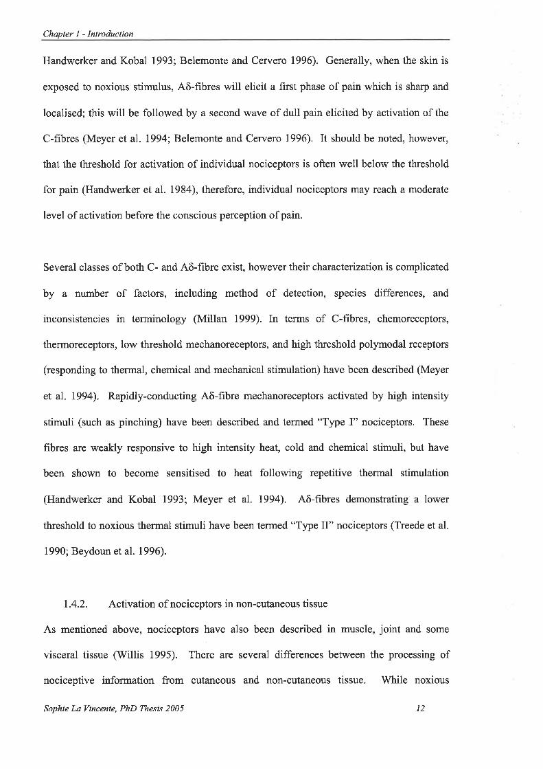
Handwerker and Kobal1993; Belemonte and Cervero 1996). Generally, when the skin is
exposed to noxious stimulus, Aô-fibres will elicit a first phase of pain which is sharp and
localised; this will be followed by a second wave of dull pain elicited by activation of the
C-fibres (Meyer et al. 1994; Belemonte and Cervero 1996). It should be noted, however,
that the threshold for activation of individual nociceptors is often well below the threshold
for pain (Handwerker et al. 1984), therefore, individual nociceptors may reach a moderate
level of activation before the conscious perception of pain.
Several classes of both C- and Aô-fibre exist, however their charucteÅzation is complicated
by a number of factors, including method of detection, species differences, and
inconsistencies in terminology (Millan 1999). In terms of C-fibres, chemoreceptors,
thermoreceptors, low threshold mechanoreceptors, and high threshold polymodal receptors
(responding to thermal, chemical and mechanical stimulation) have been described (Meyer
et al. 1994). Rapidly-conducting Aõ-fibre mechanoreceptors activated by high intensity
stimuli (such as pinching) have been described and termed "Type I" nociceptors. These
fibres are weakly responsive to high intensity heat, cold and chemical stimuli, but have
been shown to become sensitised to heat following repetitive thermal stimulation
(Handwerker and Kobal 1993; Meyer et al. 1994). Aô-fibres demonstrating a lower
threshold to noxious thermal stimuli have been termed "Type Il" nociceptors (Treede et al.
1 990; Beydoun et al. 1996).
1.4.2. Activation of nociceptors in non-cutaneous tissue
As mentioned above, nociceptors have also been described in muscle, joint and some
visceral tissue (V/illis 1995). There are several differences between the processing of
nociceptive information from cutaneous and non-cutaneous tissue. While noxious
SophieLaVincente,PhDThesis 2005 12

Chapler I - Inlroduction
stimulation of cutaneous tissue is generally associated with first (Aô-fibre) and second (C-
fibre) phases of pain, these phases are not as distinct in nociceptive input from other tissue.
For example, muscle pain mediated by both Aô- and C-fibres is experienced as dull, aching
and cramp-like (Millan 1999). A further difference is that transmission of nociceptive input
from viscera is often associated with an unpleasant autonomic component, such as
hypotension, nausea and perspiration, which is indicative of the involvement of
sympathetic and parasympathetic pathways.
1.4.3. Ascending transmission of nociceptive signals
Nociceptive information is transmitted synaptically to interneurons of the spinal cord and
dorsal horn (Willis and Coggeshall 1991). The fibres carrying nociceptive impulses enter
the spinal cord via the dorsal roots, ending in the grey matter of the dorsal horn. The dorsal
horn comprises six laminae. Nociceptive afferent fibres primarily terminate in the
superficial region of the dorsal horn, generally in laminae I and II. The cells of laminae II
form the substantia gelatinosa. The cells of the substantia gelatinosa are predominantly
short inhibitory interneurons, which project to lamina I and V, and regulate transmission
between the primary afferent fibres and the spinothalamic tract transmission neurons,
hence, the "gate keeper" function proposed by the Gate Control theory as described above.
Dorsal horn neurons with nociceptive responses have been classified into several groups,
although the criteria for each category varies between laboratories and according to the
neuron under investigation (Willis 1995). The taxonomy includes "wide dynamic range"
(WDR) neurons, which respond maximally to noxious stimuli but also respond to
innocuous stimuli, "nociceptive specific" (NS) neurons, which respond exclusively to
noxious stimuli, and non-nociceptive neurons (NON-N) (Price and Dubner 1977). More
Sophie La Tincente, PhD Thesis 2005 13
Chapter I - Introduction
recent studies have identified neurons in the marginal layer (I) of the dorsal horn that
respond specifically to cold, as well as polymodal neurons responding to thermal and
mechanical stimuli (Dostrovsþ and Craig 1996;Zhang and Craig 1997), indicating that the
aforementioned taxonomy may be too simplistic to account for the encoding properties of
dorsal horn neurons (Morgan 1998).
Following integration in the dorsal horn, nociceptive information is transmitted via
projection neurons to the higher centres in the brain. The anatomy and organisation of
ascending pain projection pathways is complex (Millan 1999). The ventrolateral funiculus
channel of the spinothalamic tract innervating the thalamus has long been considered the
most important in the transmission of nociceptive input to the higher centres, though this
no\Ã/ appears to be an oversimplification (Millan 1999). Several other pathways are
involved in the transmission of nociceptive information, including neurons belonging to the
spinoreticular, spinomesencephalic and spinocervical tracts and postsynaptic dorsal-column
pathway (Willis 1995). There has been suggestion that a specific pain pathway exists (Perl
1998), though this notion is controversial (Besson 1999). An overview of ascending
nociceptive pathways is displayed in Table 1-1.
Sophie La Vincente, PhD Thesß 2005 14

Tract
Spinothalamic tract
Spinoreticular tact
Spinomesencçhalic tact
Spinoparabrachio-amygdaloid tract
Spinoparabrachio-hypothalamic tract
Spinohypothalamic(spinotelencephalic) tract
Spinocewical Eact
Postsynaptic donal columnpathway
l,emi¡¿s gforicin
IilTVV/VIVIVVIIILSNIVA/IVIYVIIIx
NSWDRNon-N
NSWDRNon-N
NSWDRNon-N
NS
NS
NST¡/DR
Non-N
WDRNon-N
NSWDRNon-N
SkinVisceraJoints/muscle
SkinVisceraMuscle
SkinVisce¡¿Joints/muscle
SkinVisceraJoints/muscleSkinVisce¡aJoints/muscleSkinViscera
SkinJoints/muscle
SkinVisce¡aJoints/muscle
CelltvDes
Tissue input Ascending pathways Principal sub-cortical targets Axon types
UnmyelinatedSmall and largemyelinated
Small and largemyelinated
UnmyelinatedSmall and largemyelinated
UnmyelinatedSmall, myelinated
UnmyelinatedSmall, myelinated
UnmyelinatedSmall, myelinated
Small and largemyelinated
Small and mediummyelinated
Phylogeneticdistribution
AllmammalsProminent inprimates
All vertebrates
All vertebrates
Mamm¿ls
Mammals
Mammals
All vertebratesProminent incamivo¡es andprimates
Not fishProminent inmammals
Possitrle roles
Discriminative-sensory (VLF)Motivational-affectiveDescending inhibition
Motivational-affective (?)Descending inhibition
Motivati onal-affectiveAutonomic, motor
Motivational-affectiveAutonomic
Motivational-affectiveEndocrine
Sleep, autonomic and
endocrine fi¡nctionThermoregulation
Di scriminative-sensoryMotivational-affectiveAutonomic (?)
Discriminative-sensory(vPL)Motivational-affective(VMPo)
I-IIIv/VVIIxTSN
MainlyVLFDLF (r, LSìÐMainly contalateral
MainlyVLFMainly contalateral butipsilateral Q-V) via dorsalcolulms to DRNMainlyVLFDLF (I, LSN)Mainly contalateral
DLF-LFMainly contralateral
DLF-LFMainly contalateral
VLFMainly contalateral
DLFIpsilateral - thencontalateral (from LCN)
DF (and DLF)Ipsilateral - thencontralate¡al (fr om DCN)
Thalamus:VLF +\?L/V?MDLF -+ VMPo/VPLMDvcAlso PAG and collaterals-+ Reticular structures
RF of brainstem -+ LRN,medial thalamus and DRN
Midbrain and PAGDeç SCL, NCF and PBNThalamus
PBN -+ amygdala and
Stria terminalis
PBN -+ hypothalamus (VMH)
Hlpothalarnus and thalamus.Also pons, amygdala, stiatum@ilateral)
Relay LCN-+ contralateral thalamus and
midbrainSome LCN cells -+ sDinal cordRelay DCN of caudate medulla: viaML-+ contralateral tlalamusAlso SCL and spinal cord
Itr
Iil
I
XLSNIIII,iTV
ilI-VVIVII
DCN dorsal column nuclei; DF dorsal firniculus; DLF dorsolateral fi:niculus; DRN dorsal raphe nuclei; LCN lateral cervical nucleus; LRN lateral reticular nucleus; LSN lateral spinal nucleus; MDvc medial dorsal thalamus;
order oroiection. lAdaoted from Millan. 1999)second
Table 7-1. Ascending pøthwøys transmittíng nociceptive ínþrmation
I5Sophie La Vincente, PhD Thesß 2005

Chapter I - Introduction
1.4.4. Chemical modulators and transmitters in the nociceptive pathways
Chemicals play a vital role in the transmission and modulation of nociceptive information
at all stages of the pain projection system. In the periphery, chemicals are involved in the
stimulation of nociceptive fibres and also contribute to pain associated with inflammation
and ischaemic changes, which can persist long after the noxious stimulus has been
removed. Excitatory amino acids and Substance P are released by the primary afferent
fibres at their terminals in the dorsal horn. Nociception is modulated in the dorsal horn by
the release of these modulators, both from primary afferent fibre terminals and from other
sources, such as intrinsic neurons, terminals of descending pathways and glial cells.
Excitatory amino acids are also involved in the transfer of nociceptive information from the
spinothalamic tract to the thalamus, and from the spinomesencephalic tract to the
periaqueductal grey (Ericson et al. 1995; Azkue et al. 1997).
Tissue damage, inflammation and nerve injury are associated with local biochemical
changes effected by the liberation of intracellular substances into the extracellular fluid
surrounding the primary afferent fibres, evoking local pain, tenderness and hyperalgesia
(Bonica 1987). These biochemical changes can modiff the activity of nociceptors, either
by directly activating them, or sensitising them to different types of stimuli. This is of
considerable importance as the mechanisms leading to abnormal excitation of peripheral
afferent fibres are thought to be a major factor in the development of chronic pain (Rang
and Urban 1995). An example of the impact of chemical changes on the activity of
nociceptors is the sleeping or silent nociceptor. This class of nociceptor was described
during the late-1980s and is resistant to activation under normal conditions, but becomes
sensitised under pathological conditions such as inflammation (Schaible and Schmidt 1988;
Sophie La Vincente, PhD Thesis 2005 I6
Chapter I - Inlroduction
Handwerker 1991). This class of nociceptor has been described in joint, cutaneous and
visceral tissue (Treede et al. 1992; Schaible and Grubb 1993; Schmidt et al. 1995). It is
considered to be a chemoreceptor which becomes sensitised to mechanical stimuli due to
the activation of second messenger systems by chemical agents such as prostaglandins and
bradykinin, which are released in damaged tissue.
There is an extensive and expanding list of the chemicals that are contained and released by
primary afferent fibres, and are involved in the transmission and modulation of nociceptive
signals. Such chemicals include excitatory amino acids such as glutamate; neuropeptides
such as Substance P (SP) and calcitonin gene related peptide (CGRP); the cellular energy
source adenosine triphosphate (ATP); nitric oxide (NO); and the phospholipid metabolites,
prostaglandins and neurotrophins (growth factors). Furthermore, the release of
inflammatory mediators, such as bradykinin, prostaglandins and histamine, activate second
messenger systems that also act to sensitise the nociceptors. The chemical composition of
primary afferent fibres varies depending on tissue type, the state of the tissue (intact vs.
injured or inflamed) and the class of fibre (Millan 1999).
Of the many substances that affect the excitability of the primary afferent fibres, kinins,
which are potent algogenic þain producing) peptides, are of considerable importance (Dray
1997;' McHugh and McHugh 2000). These peptides are produced rapidly following injury
and initiate a range of chemical interactions impacting on both peripheral and central
neurons. Bradykinin, which is released from kininogens in the circulation, and lysyl-
bradykinin (kallidin) produce pain and contribute to hyperalgesia (an increased response to
a painful stimulus) by the activation of two major kinin receptors: Br and Bz. Bradykinin
can both directly activate nociceptors, and sensitise nociceptors by the excitation of
Sophie La Vincente, PhD Thesis 2005 17

Chapter I - Introduclion
postganglionic sympathetic neurones, causing the release of prostanoids such as
prostaglandinB2 (Rang and Urban 7995; Besson 1999). Kinins are also produced during
acute inflammation (Dray 1997), and bradykinin Bz receptor antagonists have been
demonstrated to be analgesic during these conditions (Steranka et al. 1988; Griesbacher et
al. 1994). Bl receptors have also been shown to have a role during prolonged
inflammation, and 81 receptor antagonists can attenuate the development of the associated
hyperalgesia (Perkins et al. 1993).
Countless other neuromodulators and neurotransmitters from a range of sources have also
been implicated in modulating nociception at peripheral, spinal and supraspinal stages, and
thus altering the subjective qualities of pain. These transmitters and modulators are too
numerous to detail herein, but include cytokines, endorphins, enkephalins, gamma-
aminobutyric acid, dopamine, acetylcholine, epinephrine and norepinephrine. Figure 1-l
depicts the chemical environment of sensory nerve fibres (Dray 1997). It should be noted
that these transmitters and modulators may have pronociceptive or antinociceptive actions.
For comprehensive reviews, see Millan (1999) and Dray (1997).
1.4.5. Descending modulation
The experience of pain is also subject to modulation by both descending inhibitory (Fields
and Basbaum 1994) and facilitatory pathways, which have been the subject of a recent
review (Millan 2002).
A series of studies conducted in the late 1960s and early 1970s revealed an important
feature of pain processing and modulation, with the discovery that analgesia could be
produced upon stimulation of the periaqueductal grey region of rat brain (Reynolds 1969;
Sophie La Vincente, PhD Thesis 2005 18

Chapter I - Introduction
Mayer and Liebeskind 1974). Studies later revealed that stimulation in the same areas of
the human brain in patients with intractable pain also produced analgesia (Adam s 1976;
Hosobuchi et al. 1977). It was also demonstrated that the opioid antagonist naloxone
attenuated the analgesia produced by stimulation (Akil et al. 1976), suggesting the
involvement of opioid systems.
Neurotrophins Sympathetic
nervesNGFProstanoids
Amines, NPY
Vascular
Amines, kinins
nitric oxide
Immune cell
products
/1SENSORY
FIBRE
Neurogenic
factors
Products oftissue iniury
Kinins,5HT,histamine
Protons, free radicals
-+/
\
Cytokines
Endorphins
Enkephalins
Neuropeptides
Figure I-1. The chemìcøl environment of sensory nerve Jìbres. A large number ofmediøtors produced by severøl lissues can aÍÍect the excitability ønd phenotype of sensoryneurons. NGF nerve groiltth føctor, NPY neuropeptide Y, sHT S-hydrorytryptømìne(serotonin). (Reproduced from Dray, 1997).
Descending pathways projecting from cerebral structures to the dorsal horn play a
significant role in the integration of nociceptive messages in the dorsal horn, modulating
the release of neurotransmitters from primary afferent fibres and inhibiting the activity of
the projection neurones that transmit nociceptive messages to the higher centres (Millan
leee).
Descending pathrù/ays involved in the modulation of nociception do not have a uniquely
inhibiting role in the dorsal horn. Individual neurotransmitters can exert either inhibitory or
Sophie La Vincente, PhD Thesis 2005 19

Chapter I - Introduction
excitatory effects in the dorsal horn depending, for example, on the receptor activated
(Millan 1999).
1.5. Types of pain
The pain experience may be classified into four subgroups or types of pain. These are
divided according to the time course and nature of the experience.
l 5.1. Transient pain
Transient pain occurs frequently in everyday life and rarely leads to the need for medical
attention. It is the fleeting pain experienced, for example, during venepuncture. Activation
of nociceptive transducers in the skin or other tissues elicits transient pain, although tissue
damage seldom occurs. The function of this pain pertains to its rapid speed of onset, and
the speed with which the pain dissipates once the noxious stimulus has been removed
(Loeser andMelzack 1999). This type of pain presumably has a protective role, evoking a
reflex response to remove the stimulus that is impinging on the body, thus reducing the
likelihood of tissue damage.
1.5.2. Acute pain
Acute pain is associated with significant injury of body tissue, which activates nociceptive
transducers at the site of tissue damage (Loeser and Melzack 1999). It is often seen after
trauma, surgical procedures, and following some diseases, and will usually result in the
individual seeking medical attention. While medical interventions may assist in both
reducing the pain and accelerating the healing process, it is often not necessary, and healing
can occur without treatment. Acute pain may persist for several days or weeks as the
healing process takes place.
Sophie La Vincente, PhD Thesis 2005 20

Chapter I - Introduction
1.5.3. Chronic pain
Chronic pain has previously been defined as pain which persists beyond a six-month period
(Russo and Brose 1998). It has been argued, however, that duration should not be the
distinguishing feature of chronic pain, but rather the ability of the body to restore
functioning to normal homeostatic levels (Loeser and Melzack 1999). Indeed recent
reports define chronic pain by its character rather than by duration (Schaible and Richter
2004). Chronic pain states include back pain and postherpetic neuralgia. While all types of
chronic pain typically lead individuals to seek medical care, often the pain is not treated
effectively and due to the unrelenting nature of the pain, stress and environmental factors
may further contribute to the problem (Loeser andMelzack 1999). A common form of
chronic pain is neuropathic pain, which is associated with damage to the nociceptive
pathway, rather than a result of excessive peripheral stimulation as described in transient
and acute pain.
1.5.4. Experimental pain
Producing pain in an experimental setting is an important part of research, particularly in
the assessment of analgesic drugs. However, it is impossible to produce pain in an
experimental setting that is comparable to "real life" pain. For many years the validity of
experimental pain, especially in assessing analgesic medication, has been a challenging and
contentious issue amongst clinicians and researchers (Keats et al. 1950; Beecher 1953;
Moore et al. 1997). Perhaps the most prominent of critics was Henry Beecher, who argued
that experimentally induced pain was qualitatively different from the pain produced by
injury or disease (Beecher 1962). He asserted that experimentally induced pain is without
significance or meaning to the individual, whereas pain that results from injury or disease
Sophie La Vincente, PhD Thesis 2005 21
Chapter I - Introduction
has significance to the patient and involves other parameters, particularly psychological
factors, such as anxiety
Notwithstanding, several methods of experimental pain induction have been developed
over time that yield reliable and reproducible data (Stacher et al. 1986), and are widely
used in the assessment of pain sensitivity and tolerance.
1.5.5. Techniques for experimental pain induction
Techniques that have been developed for the induction of experimental pain in humans
include tests of ischemic, heat, cold, pressure and electrical pain. Most frequently, study
participants are required either to rate the pain experienced, or are instructed to proceed
with the testing as long as the pain can be tolerated. These tests have not been
standardised, normative values have not been established, and tests are typically subject to
different methods according to the investigative group and the outcome measures of the
study. The cold pressor and electrical stimulation tests are two of the most widely used
human pain induction techniques. Experimental pain induction is described in greater
detail in Chapter 3.
I .5.5. I . Cold pressor
The cold pressor test is a common experimental pain induction technique used in clinical
investigation. Originally used in the 1930s as a measure of blood pressure variation (Hines
and Brown 1933), it is now used in a vaiety of experimental settings. It became
particularly prevalent in experimental pain research with the pioneering study conducted by
Wolff and co-workers (1940), which initially demonstrated that intramuscular morphine
produced a dose-dependent increase in cold pressor pain tolerance. Later studies have
Sophie La Vincente, PhD Thesis 2005 22

Chapter I - Introduction
consistently confirmed that the test is a highly sensitive assay for opioids, including
morphine (Wolff et al. 1966; Jones et al. 1988), dipipanone (Posner et al. 1985), and
codeine (Garcia de Jalon et al. 1985), and that this response can be distinguished from both
placebo (Posner et al. 1985; Jones et al. 1988) and non-opioid analgesics (Jones et al.
l9S8). The cold pressor test is of particular value as a model of experimental pain
induction as it is produces prolonged, deep sensations that are characteristic of many
clinical pain states (Wolff 1984), being likened to dental and back pain (Chen et al. 1989),
and has high reproducibility for repeated applications (Walsh et al. 1989; Grach et al.
2004).
There have been many variations on the cold pressor technique, but the test essentially
involves the immersion of a limb (usually the forearm) into a bath of very cold water. The
participant is typically required to indicate when pain is first experienced (threshold), and
when pain can no longer be tolerated (tolerance), both of which are measured in terms of
time (seconds) elapsed from initial immersion into the cold water. Alternatively, subjects
may be asked to continuously rate pain intensity during cold water immersion.
The cold-water bath may be preceded by the immersion of the limb in a warrn water bath
for two minutes, and the presence of a water pump in the cold water to keep the water
moving, reducing the effect of laminar warming around the limb.
1.5.5.2. Electrical stimulation
Electrical stimulation has been described as one of the most suitable methods of
experimental pain induction as it is simple to apply and easy to reproduce (Harris and
Blockus 1952). A number of studies have successfully used electrical stimulation to test
Sophie La Vincente, PhD Thesis 2005 23
Chapter I - Introduction
the analgesic efficacy of opioids, including morphine (Willer 1985), codeine (Stacher et al.
1936) and methadone @yer et al. 1999), and other analgesic compounds, including non-
steroidal anti-inflammatory agents (NSAIDs) (Stacher et al. 1979; Stacher et al. 1986;
Walker et al. 1993; Walker and Carmody 1998).
Electrical stimulation typically involves attaching electrodes to some area of the body,
often the earlobe, hand or finger, and conducting square wave pulses of electricity in
increasing volts. Resembling the cold pressor test, the subject indicates the level of pain
experienced in terms of both pain sensitivity (when pain is first felt) and tolerance
(maximum tolerable pain), however these parameters are measured in volts or amps rather
than time.
1.6. Opioids
1.6.1. The history of opioids
In 1915, the eminent pharmacologist D.L Macht wrote "If the entire matena medica at our
disposal were limited to the choice and use of only one drug, I am sure that a gteat many, if
not the majority, of us would choose opium" (Macht 1915). Opium is the milky sap
extracted from the seed pod of the poppy, Papaver somniferum, more commonly referred to
as the opium poppy. First classified by the botanist Linnaeus in 1753, the species takes its
name from the Latinword meaning "sleep-inducing". However, the properties of this plant
' The term opioid denotes all compounds that interact with opioid receptors in the central and peripheral
nervous system (Foley, 1993). The term opiate refers only to drugs that are derived from the juice of the
Sophie La Vincente, PhD Thesis 2005 24

Chapter I - Introduclion
have been known for thousands of years. In 3400 BC, the Sumerians, the world's first
civilisation and agriculturalists, were cultivating the opium poppy throughout the Tigris-
Euphrates river systems of lower Mesopotamia. The ideograms used by the Sumerians for
the poppy translate as 'Joy plant", suggesting that this ancient civilisation was aware of the
psychological and pain-relieving qualities of the plant. It is thought that, just as the
Sumerian invention of writing gradually spread to other societies, so too did knowledge of
opium. By the end of the second millennium BC, opium use was prevalent in much of the
ancient world. Throughout the ages, the use of opium flourished. In its time, it has been
hailed as a magicalpanacea. used for pain relief, as an antidote to sorrow, in the treatment
of insomnia, as a cure for stomach ailments, to treat coughs and colds, as a crude
anaesthetic during surgery, in religious rites, as a form of recreation, and as a convenient
poison in murder and suicide (Booth 1996). However, as early as the fifth century BC, the
dangers of this universal remedy were recognized. Erasistrafus advocated complete
abstinence from opium and later, in the third century BC, the philosopher Diagoras of
Melos proclaimed that it is better to suffer pain than to become dependent upon opium.
By the sixteenth century AD, opium was well established in Europe, however its use was
predominantly medicinal. Publications of the mid-late 1700s describe aspects of opium
addiction and the diff,rculty of withdrawal; however there lacked any notion of moral
denunciation for either medicinal or recreational use. In fact, at this time, there was a
widespread movement, particularly in the literary community, away from the restrained
ethos of classicism in favour of spontaneity, imagination, a gteater awareness of the natural
opium poppy. Hence, the synthetic compound methadone is classed as an opioid due to its interaction with
opioid recsptors, however it is not an opiate, as it is not derived from the opium poppy.
Sophie La l/incente, PhD Thesis 2005 25

Chapter I - Introduction
world, aîda more liberated expression of passion. Opium played a significant role in the
growth of this movement, which has become known as the Romantic Revival, and included
literary figures such as Samuel Taylor Coleridge, Lord Byron,Elizabeth Barrett Browning,
Goethe, and Thomas De Quincey, author of the notorious "Confessions of an English
Opium-eater" of 1821.
In 1806 the German pharmacist Friedrich Serttirner isolated a pure alkaline substance from
opium. The compound was named morphine, after the Greek god of dreams Morpheus.
Coupled with the invention of the hypodermic syringe in 1853, and the synthesis in 1874 of
the highly potent opioid, diacetylmorphine, the magnitude of opioid dependence presented
a significant public health concern. Diacetylmorphine, considered at the time to be a non-
addictive alternative to morphine, was marketed by the Bayer pharmaceutical company in
the late 1800s as a cough syrup, and named "Heroin". In response to the mounting problem
of opioid dependence, US Congress passed legislation in the early 1900s restricting the
medical and recreational use of opioids. This action has since been echoed throughout
most of the world.
The quest for potent, non-addictive analgesics led to the synthesis of drugs such as
methadone and pethidine in the late-1930s and 1940s; however, these compounds displayed
typical morphine-like properties, including the potential to produce dependence. It was
also during this time that the first opioid antagonist was synthesised. Pohl (1915, see
Gonzalez and Brogden 1988) first noted the pharmacology of opioid antagonists, drugs that
block the effects of opioids, in the early 1900s.
Sophie La Tincente, PhD Thesis 2005 26

Chapter I - Introduction
1.6.2. Opioidclassification
Opioids exert their effects by interacting with specific receptors on nerve cells in the CNS
and periphery (see discussion below). Opioids are classified according to their interaction
with these receptors and the pharmacological effects they produce. These compounds are
classed as agonists, antagonists or partial agonists (Zacny and Walker 1998). Opioid
agonists, such as methadone, morphine and heroin, exert a very strong or maximal effect at
the receptor, inducing the cellular actions described below (section 1.2.4). Antagonists
have minimal or no intrinsic action themselves, but act to block the effects of opioid
agonists. Opioid antagonists include the short-acting naloxone and longer-acting
naltrexone. Partial agonists such as buprenorphine have less intrinsic activity at the
receptor, and thus may exert agonist activity, but also may exert some degree of antagonist
activity by displacing full agonists that have higher intrinsic activity at the receptor.
1.6.3. Opioidreceptors
The mounting social problems associated with opiate dependence in the 1940s and 1950s
stimulated a surge of research focused on the still unrealised goal of developing a potent,
non-addictive analgesic. However, from this research emerged the finding that many of the
pharmacological effects of opioids were highly stereospecific. This generated the
hypothesis that these drugs exert their effects by binding with specific sites on nerve cells.
The first indication of stereospecific opioid binding in mouse brain was published in l97l
(Goldstein et al. l97l). ln 7973, three research groups independently produced decisive
evidence for specific opioid receptors in animal brain (Pert and Snyder 1973; Simon et al.
1973; Terenius 1973), and soon after in human brain (Hiller et al. 1973). With the
observation that morphine and several of its analogues had different pharmacological
profiles, and that opioid antagonists blocked various agonists differentially, it was
Sophie La Vincente, PhD Thesis 2005 27

Chapter I - Introduction
postulated that, rather than opioid receptors being one homogeneous group as first thought,
there existed different types of opioid binding sites. Evidence for multiple receptor types
was soon produced, and three receptor types proposed: p, rc and o (Gilbert and Martin
1976; Martin et al. 1976). Later research revealed a fourth receptor type, which was
labelled ô (Lord et al. 1977). Further investigation revealed that the o receptor was not
opioid, as effects mediated by this receptor were not reversed by opioid antagonists, even at
very high doses. Furthermore, findings indicated that the o receptor was a binding site for
non-opioid drugs such as phencyclidine (PCP) (Vincent et al. 1979; Zlkin and Zttkin
1979). More recent investigation has demonstrated that this class of receptor represents a
group of binding sites with high affrnity for a range of non-opioid compounds (Henderson
and McKnight 1997).
Molecular cloning of first the ô (Evans et al. 1992; Kieffer et al. 1992} and then rc (Yasuda
et al. 1993) and p receptor (Chen et al. 1993) has facilitated the pharmacological
characterisation of these binding sites (Reisine and Bell 1993; Raynor et al. 1994; Satoh
and Minami 1995). Subtypes of these receptors have been proposed (Dhawan et al. 1996);
however pharmacologically defined subclasses of the main receptor types have not been
well established (Connor and Christie 1999; V/illiams et al. 2001).
ln 1994, a fourth receptor clone was isolated from a number of species, including mouse
(Nishi et al. 7994), rat (Fukuda et al. 1994; Lachowicz et al. 1995) and human (Mollereau
et al. 1994). Although this novel receptor shares a high level of sequence homology with
traditional opioid receptors, in mammalian cells the receptor demonstrated very little
binding affinity with opioids (Henderson and McKnight 1997; Barlocco et al. 2000).
Hence, the novel receptor was considered by many to be an orphan receptor (Mogil and
Sophie La Vincente, PhD Thesis 2005 28
Chapter I - Introduction
Pasternak 2001), and labelled ORLI (opioid-receptorJike 1). In 1995 an endogenous
ligand for this receptor, orphanin FQ or nociceptin, was identified (see discussion below)
(Meunier et al. 1995; Reinscheid et al. 1995). There remains, however, some controversy
regarding this receptor t¡re, and whether it should be considered an opioid receptor
(Henderson and McKnight 1997).
Extensive investigation has revealed that the three traditional opioid receptor types (¡r, rc
and ô) demonstrate distinct anatomical distributions and functions in the CNS and
periphery (see Table l-2), and that there is generally a correlation between receptor density
in a particular brain region and the functional importance of opioids in that area (Mao
1999). Opioid receptors are distributed widely in the mammalian CNS (Mansour and
'Watson 1993). Dense ¡r-receptor binding is found in regions important for nociceptive
regulation, basal ganglia, limbic structures and thalamic nuclei, however there is
considerable intra-species variation in distribution. Similarly, while rc sites are widely
distributed throughout the forebrain, midbrain and brainstem, considerable differences exist
between mammalian species. By comparison, the distribution of delta receptors is more
restricted, being concentrated in forebrain regions, and is more consistent between species.
While the main effects of opioids are mediated by the CNS, receptors distributed
throughout the periphery produce hormonal, immunological and some analgesic effects
(Kit g et al. 2001).
Sophie La Vincente, PhD Thesis 2005 29

Chapter I - Introduction
Receptor type Primary functions
tr Nociception (J)Respiration (J)GI motility (J)Feeding (J)Learning and memory (ü)Locomotor activity (J)*Hormone secretion (1)Cardiovascular regulationImmune functionThermoregulation
Associated with positivesubj ective effects, highlyreinforcing
ô Nociception (J)GI motility (J)Respiration (J)_ -4.Locomotoractlvtty(l)Cognitive tunction (J)Cardiovascular regulationOlfactionImmune function
Associated with some
reinforcing properties
K Nociception (J)Diuresis (1 urinary excretion)Feeding (J)Neuroendocrine secretionImmune functionThermoregulation
No positive subjective effects,can produce dysphoria inhumans
*effects depend on dose and species
J decreased activity/effect, I increased activity/effect
Tøbte 7-2. Prímøry fanclìons of opìoid rcceptors (adøptedfrom Dhawøn et aL, 1996).
1.6.4. Endogenous opioid ligands
V/ith the detection of stereospecific binding sites for opioids in the early 1970s, it was
hypothesised that there must exist an endogenous ligand for these receptors. This notion
\ilas flrst supported with evidence of opiate-like activity in brain extract (Kosterlitz and
Sophie La Vincente, PhD Thesis 2005 30
Chapter I - Introduction
Waterheld 1975), and soon after the enkephalins were isolated and characterised (Hughes
1975; Hughes et al. 1975). Following these studies, a fragment of pituitary hormone B-
lipotropin was found to have high affinity for opioid receptors, and was renamed B-
endorphin (from endogenous morphine) (Bradbury et al. 1976). Some years later, a third
class of endogenous opioid was discovered, and labelled the dynorphins (Goldstein et al.
1979). These three classes of endogenous ligands each demonstrate greater binding affinity
with one of the receptor types: enkephalins with the ô-receptor, endorphins with the ¡r-
receptor, and dynorphins with the rc-receptor (van Ree et aL 1999). However, it should be
noted that these ligands also bind with other opioid receptor types, for example, the
enkephalins have affinity for the ¡r-receptor (Mansour et al. 1995), and p-endorphin binds
potently with ô-receptors (Reisine 1995).
In recent years, several advances have been made in our understanding of endogenous
opioids. Recently discovered in mammalian cells, ligands termed endomorphin 1 and 2
demonstrate potent binding affinity and are highly selective for the p-receptor (Zadina et al.
1997; Goldberg et al. 1998; Zadina2002). As mentioned, a novel neuropeptide, orphanin
FQ or nociceptin, was identified in 1995, and demonstrated to be the endogenous ligand for
the ORLI receptor (recently renamed NOPr, or nociceptin/orphanin FQ peptide). There is
still controversy regarding whether NOPr should be considered a fourth member of the
opioid receptor family.
1.6.5. Second messengers and effectors
Cloning has confirmed that opioid receptors belong to the superfamily of G-protein-
coupled receptors, which activate and regulate multiple second messenger pathways
associated with effector coupling, receptor trafficking and nuclear signalling (Williams et
Sophie La Vincente, PhD Thesis 2005 31

Chapter I - Introduclion
al. 2001). Signalling via these receptors has been shown to be mediated by interaction with
guanine nucleotide-binding proteins, or G-proteins. Opioids have been shown to interact
preferentially with pertussis-toxin (PTX)-sensitive Gi/Go-proteins, although evidence
indicates that opioids may also interact with other families of G-proteins, for example the
PTX-insensitive G, and G16 (see Connor and Christie 1999 for review). The cellular
actions produced by opioid receptor activation are coÍrmon to all three types of receptor.
The most common actions are inhibition of adenylyl cyclase (reducing intracellular cAMP),
activation of potassium conductance, inhibition of voltage-gated calcium channels, and
inhibition of transmitter release. Recent observations suggest that activation of opioid
receptors is also associated with activation of protein kinase C (PKC), release of calcium
from extracellular stores, nuclear signalling (e.g. the activation of the mitogen-activated
protein kinase (MAPK) cascade) and receptor trafficking (Williams et al. 2001). The
overall consequence is a reduction in the excitability of the cell due to hyperpolarization
and the inhibition of neurotransmitter release, however, opioids may also produce an
excitatory effect by preventing the release of inhibitory neurotransmitters (V/illiams et al.
2001).
1.7. Opioid effects
Opioids are predominantly used for the euphoria and feelings of well-being they inspire,
and for their exceptional analgesic qualities. Most clinically used opioid agonists exert
their effects primarily through ¡r receptors. While the pharmacokinetic profile of these
compounds varies widely, many share a similar pharmacodynamic profile, exerting both
desirable and undesirable effects (Mather 1990). Opioids affect a wide range of
physiological systems, including the respiratory, cardiovascular, genitourinary and
gastrointestinal systems. Typical opioid effects include analgesia, euphoria or feelings of
Sophie La Tincente, PhD Thesis 2005 32
Chapter I - Introduction
well-being, respiratory depression, reduced gastrointestinal motility, pupillary miosis,
prurifus, sedation, nausea, vomiting and cognitive impairment. These effects are typically
dose-dependent. While clinical use of opioids is principally for their powerful analgesic
actions, they are also prescribed for their anti-tussive and anti-diarrhoeal effects. The
discussion below will focus primarily on the effects of morphine, the prototypic ¡r receptor
agonist used in the management of pain.
1.7.I. Analgesia
Opioids are the mainstay of pain management. They are the most effective and frequently
used class of analgesic drug for the treatment of moderate to severe pain (Gutstein and Akil
2001). Referred to as "God's o\ryn medicine" by Sir William Osler, morphine remains the
standard by which drugs with analgesic actions are assessed.
The analgesic effects of opioids are mediated through spinal, supraspinal and peripheral
mechanisms (see Figure 1-2). Opioid receptors are prominent in the brain and spinal cord
regions involved in the transmission and modulation of pain (see Ossipov et al. 2004).
Binding of opioid agonists to these specific receptors inhibits nociceptive activity, resulting
in potent analgesia (Fields 1993; Codd etaI.1995; McNally 1999).
Opioids inhibit the release of Substance P (SP) by nociceptive afferent neurons at the dorsal
horn, and directly inhibit the pain transmission neuron. Opioids further produce analgesia
by activating descending inhibitory pathways. Opioids excite neurons in the periaqueductal
grey (PAG) and in the nucleus reticularis paragígantocellularis OIRPG), which in turn
project to the rostroventral medulla, including the nucleus raphe magneus (NRM). From
the NRM, neurons containing 5HT and enkephalin run through fibres in the dorsolateral
Sophie La Vincente, PhD Thesis 2005 33
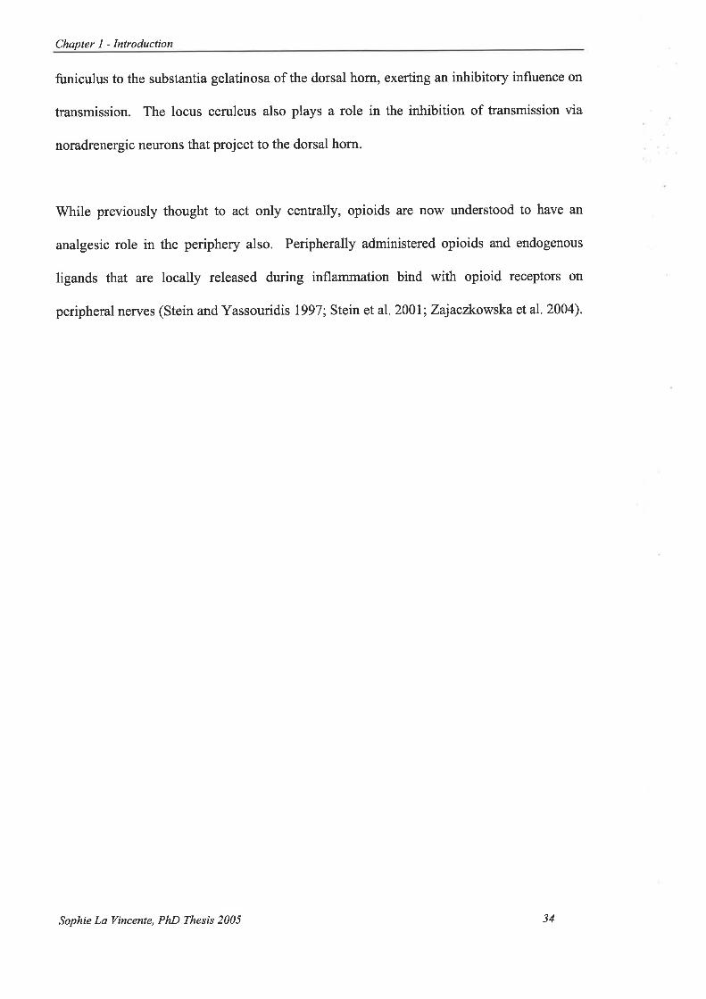
Chapter I - Introduction
funiculus to the substantia gelatinosa of the dorsal horn, exerting an inhibitory influence on
transmission. The locus ceruleus also plays a role in the inhibition of transmission via
noradrenergic neurons that project to the dorsal horn.
While previously thought to act only centrally, opioids are now understood to have an
analgesic role in the periphery also. Peripherally administered opioids and endogenous
ligands that are locally released during inflammation bind with opioid receptors on
peripheral nerves (Stein and Yassouridis 1997; Stein et al.2001; Zajaczkowska et al.2004).
Sophie La Vincente, PhD Thesis 2005 34
Chapter I - Introduction
Trdñsñ¡sBlon l¡lûdutãtloñ
Ve ntrå1cáildatthåiËmus
c
Þûîsål hgrnE
0ônêr
ÐlencüÊhË!dn
M¡dþîålft
Þlñdullå
$p¡ñal 6ôrd
Farìaquêduclalgrây
L
¿Fostr¿lvBñlrafmôdullå
å
Fígure 1-2. Putative sites of actíon of opioìd ønølgesics. Leftz Sites of øclion on pøintrsnsmissíon pøthway from lhe periphery to lhe higher cenlres. Right: Actìon on paìnmodulatíng neurons secondarily affects pain transmission pathways. A.: Possihle dìrectsction of opioids on ìnflømed perìpheral tissues. B: Inhìbition occurs ìn the spinal cord"
C: Possíble sìte of action in the thalømus. On the right, actions of pøin-modulatingneurons in the mìdhrsìn (D) and med.ulla (E) secondaríly øffect pain trønsmissionpathwøys. (úYøy et aL 2000)
1.7.2. Respiratorydepression
Respiratory depression is one of the most serious adverse effects associated with opioid
analgesics (Hill 1993). It is one of the principal adverse effects that limits their therapeutic
use (Florez and Hurle 1993), and death from morphine toxicity is typically the result of
respiratory arrest (Gutstein and Akil 2001). Respiration is controlled primarily through the
medullary respiratory contres, namely the dorsal respiratory group and ventral respiratory
group of neurones, while chemoreceptors and stretch receptors in the periphery contribute
to the control of breathing rate and pattern (White and Irvine 1999). Agonists at ¡.r- or ô-
Sophie La Vincente, PhD Thesß 2005 3s
Chapter I - Inlroduction
opioid receptors can result in respiratory depression. Such agents can cause a decrease in
respiratory rate, a decrease in tidal volume, irregular respiratory rhythm and obstructive
apnea during sleep (Macintyre and Ready 2001). The medullary and pontine centres
involved in the regulation of respiration are inhibited by opioid interaction with both ¡r- and
ô-receptors (Martin l9S3). Opioids inhibit chemoreceptors in the periphery, reducing
response to changes in oxygen and, importantly, increases in carbon dioxide levels. This
effect is chiefly mediated by interaction with ¡r-receptors (White and Irvine 1999).
Following IV administration of morphine, respiratory depression peaks within
approximately 10 minutes and depression may persist for up to 5 hours. While therapeutic
doses of morphine depress all phases of respiratory activity, rarely do such doses produce
clinically significant depression (Gutstein and Akil 2001). A greater risk occurs with the
combination of opioids and other CNS depressants, such as sedative-hypnotics, general
anaesthetics and alcohol, the use of which may be problematic in both acute and chronic
pain patients.
1.7.3. Sedation
Respiratory depression is often preceded by sedation (Macintyre and Ready 2001).
Sedation occurs primarily in the early stages of treatment (Foley 1993), and can
compromise analgesia by limiting dose escalations that may be required for adequate pain
relief (Macintyre and Ready 2001).
L7.4. Nausea and vomiting
All clinically used opioid analgesics produce nausea and vomiting to some extent, although
the magnitude of this effect varies considerably between individuals. These side effects
Sophie La Vincente, PhD Thesis 2005 36
Chapter I - Introduction
are caused by direct stimulation of the chemoreceptor trigger zone for emesis. The
likelihood of nausea and vomiting is also increased in ambulatory patients compared to
recumbent patients. This suggests the involvement of the vestibular system, and indeed
there is evidence that opioids may be involved in the modulation of neurotransmission in
the peripheral vestibular system (Andrianov and Ryzhova 1999). Opioids can increase
vestibular sensitivity, and in some patients even slight movements can provoke nausea and
vomiting (Macintyre and Ready 2001).
1.7.5. Effects on mood
Opioid use is associated with euphoria and decreased emotional distress. The mood
altering properties of opioids are considered one of the primary reasons for illicit (non-
medical) use of this class of drug (van Ree et al. 1999). Opioid abuse and dependence
presents a significant public health challenge throughout much of the world, being
associated with signif,rcant morbidity and mortality. While the prevalence of illicit opioid
use is considerably lower than other drugs such as alcohol and cannabis, the economic cost
is staggering. The mechanisms by which opioids exert their rewarding effects have not
been fully elucidated (O'Brien 2001); however it is thoughtthat the systems involved in
mediating mood and reward are distinct from those associated with analgesia and the
development of physical dependence (Koob and Bloom 1988). The rewarding effects of
drugs of abuse, including opioids, are thought to be mediated by the midbrain dopaminergic
system, which is also involved in the rewarding effects of other stimuli such as food
(Hyman and Malenka 2001).
Sophie La Vincente, PhD Thesis 2005 37

Chapter I - Introduction
1.7 .6. Constipation
Opioids can cause delayed gastric emptying, inhibition of bowel motility and constipation.
This inhibition of smooth muscle activity is mediated by both central opioid receptors and
receptors on the bowel wall (Gutstein and Akil200l).
L7.7. Pupillary miosis
Most ¡r and r agonists cause pupillary constriction by exerting an excitatory effect on the
parasympathetic nerve innervating the pupil (Gutstein and Akil2001).
1.7.8. Cardiovasculareffects
Therapeutic doses of opioid analgesics are associated with peripheral vasodilation, reduced
peripheral resistance and inhibition of baroreceptor reflexes. Arterial or venous
vasodilation may occur by a direct effect of vascular muscle or by histamine release. While
in a supine position, there is no significant effect on blood pressure or cardiac rate, though
upon movement to a sitting or standing position, hypotension and fainting may occur.
Opioid agonists such as morphine are also known to have utility in the treatment of angina
pectoris and acute myocardial infarction, and have more recently been reported to have a
cardioprotective effect (Schultz and Gross 2001).
1.7.9. Cough suppression
The mechanism by which opioids suppress the cough reflex is thought to involve a direct
effect on the cough centre in the medulla (Gutstein and Akil 2001). The dose required for
cough suppression is lower than that required for analgesia (Foley 1993).
Sophie La Vincente, PhD Thesis 2005 38

Chapter I - Introduction
1.7.10. Pruritus
Opioids can trigger histamine release from mast cells, which can create itching and flushing
of the skin. Opioid-related pruritus primarily occurs on the face, neck and trunk, or at the
injection site (Macintyre and Ready 2001). Pruritus does not occur with all opioids
(Duthie and Nimmo 1987), and has been reported to occur more coÍtmonly following
epidural or spinal opioid administration (Ballantyne et al. 1988).
1.8. Barriers to adequate pain management with opioids
There are a number of barriers to effective pain control with opioids. Several of these are
societal barriers, including a stigma associated with the use of opioids that fails to
adequately distinguish between legitimate and illegitimate use, and an apprehension
amongst clinicians to prescribe opioids for fear of the patient developing a psychological
dependence on the drug (Hill 1993; Rupp and Delaney 2004).
Moreover, significant limitations in the use of opioids for pain control are the adverse
effects associated with this class of drug, andthe adaptations that occur with prolonged use.
1.8.1. Adverse effects
Adverse effects associated with opioids can present significant limitations to pain
management. As described above, the most significant adverse effects are respiratory
depression and sedation, as these are potentially dangerous and can also limit dose
escalation required for adequate pain control. While tolerance develops to the emetic
effects of opioids (see 1.8.2), constþation is an ongoing adverse effect that can impact
upon comfort and quality of life, especially in cases of prolonged opioid administration for
chronic pain.
SophieLqVincente, PhD Thesis 2005 39

Chapter I - Introduction
1.8.2. Tolerance
Chronic opioid administration results in tolerance, which is characterised by a decrease in
activity of a drug after a previous exposure to the same or a similar drug (Foley 1993).
Opioid tolerance is associated with decreased magnitude and duration of effects such as
analgesia and euphoria, as well as several of the adverse effects associated with opioids,
including respiratory depression, nausea and sedation (Collett 1998). The decreased effect
associated with opioid tolerance can be very pronounced, with morphine doses many times
greater than would be administered for analgesia producing only mild effects in chronic
opioid users (Gregory et al. 1992; O'Brien 2001). However tolerance to different opioid
effects does not develop at the same rate (Ling et al. 1989); for example, tolerance to the
euphoric effects of opioids develops much earlier than tolerance to the gastrointestinal
effects.
Several different types of tolerance have been described, which may be innate or acquired
(O'Brien 2001). Innate tolerance refers to a genetic predisposition towards a lesser (or
greater) response to a drug. Acquired tolerance may be classified as pharmacokinetic,
pharmacodynamic or learned tolerance. Pharmacokinetic tolerance describes the increase
in clearance (CL) of an agent with repeated use, resulting in a lower plasma drug
concentration. Learned tolerance refers to the development of behaviours or mechanisms
that reduce the magnitude of drug effect. For example, compensatory behaviours may be
developed in order to maintain functioning despite intoxication. Pharmacodynamic
tolerance describes adaptive changes occurring in systems that are affected by the drug,
which result in a reduction in effect.
Sophie La Vincente, PhD Thesis 2005 40
Chapter I - Introduction
The majority of investigations of opioid tolerance have focused on the p receptor, as this is
the major site of action of interest. The involvement of rc- and ô-opioid receptors in
tolerance has received less attention.
The mechanisms underlying tolerance have not been fully elucidated. While a number of
cellular and synaptic adaptations have been demonstrated to occur with chronic
administration of an opioid agonist, how these adaptations result in the physiological and
behavioural phenomena of tolerance remains unclear. A blunting of opioid receptor
response, by receptor de-coupling or internalisation (Borgland 2001; Williams et al. 2001),
or an increase in opioid receptor coupling with excitatory G-proteins (Crain and Shen
2000) have been implicated in the development of tolerance. However it is apparent that
there are a number of other adaptations that occur with the chronic administration of
opioids, and are increasingly understood to play a role in tolerance.
Phosphorylation of occupied receptors is thought to play a significant role in receptor
desensitisation and internalisation by uncoupling receptors from their G-proteins. G-
protein coupled receptor kinase (GRK)-mediated receptor phosphorylation allows binding
of the cellular protein arrestin, which disrupts G-protein binding with receptors. A number
of other kinases may mediate receptor desensitisation by phosphorylation, including protein
kinase C (PKC) (Inoue and Ueda 2000; Ueda et al. 2001), calcium/calmodulin-dependent
protein kinase II (CaM kinase II) and tyrosine kinase (Borgland 2001). Mu-receptor
desensitisation is associated with a decrease in binding sites on the plasma membrane (Pak
et al. 1996). Receptor phosphorylation by GRKs is considered to play a central role in
receptor internalisation (Capeyrou et al. 1997).
Sophie Lø Vincente, PhD Thesis 2005 4I

Chapter I - Introduction
These cellular adaptations have been demonstrated to occur rapidly upon administration of
an opioid, often within minutes of administration (V/illiams et al. 2001). The role these
initial changes play in the development of chronic tolerance with prolonged dosing is
unclear. It has been reported that the degree of change observed at the cellular level is
much smaller in magnitude than the effects observed clinically (Harrison et al. 1998;
V/illiams et al. 2001), suggesting the involvement of other adaptational changes in the
development of tolerance. Long-term adaptations are thought to involve functional
uncoupling of p receptors from signalling pathways as a consequence of downregulation of
opioid receptors on the surface of cell membranes (Williams et al. 2001).
A well-established adaptation occurring with chronic opioid administration of opioids is the
upregulation of cAMP pathways (Nestler 2001). Opioids inhibit activity of the cAMP
pathway. V/ith chronic administration, activity of cAMP pathways recovers with the
induction of adenylyl cyclase and protein kinase A.
The N-metþl-D-asparate (NMDA) receptor has also been implicated in the development
of tolerance (Elliott et al. 1994; Mao 1999; Trujillo 2000), and indeed NMDA receptor
antagonists have been demonstrated to attenuate the development of tolerance (Trujillo and
Akil 1991; Tiseo and Inturrisi 1993). There is also evidence for the involvement of nitric
oxide and nitric oxide synthase (Pasternak 1995; Aley and Levine 1997).
1.9. Enhancing analgesia through drug combinations
While opioids are considered the gold standard in the management of moderate to severe
pain, their use is limited by the adverse effects described. Due to these limitations, research
is increasingly focusing on ways in which to improve the use of opioids in pain
Sophie La Vincente, PhD TVtesis 2005 42
Chapter I - Introduction
management; that is, to enhance the analgesic effect while minimising the incidence and
severity of adverse effects. The notion of combining agents for enhanced analgesic effect
and reduced incidence of adverse effects is increasingly being viewed as an important
consideration for effective pain treatment. The management of post-operative pain has
received particular attention, with the recognition that inadequate post-operative pain relief
can adversely influence organ functioning and contribute to morbidity (Kehlet 1994; Kehlet
1997). It has been acknowledged that optimal pain relief is unlikely to result from a single
drug or treatment modality (Kehlet and Dahl 1993; Mehlisch 2002). Drug combinations
producing either additive or synergistic analgesic effects, which can lower doses of
individual drugs and thus moderate the incidence and severity of side effects, may
significantly reduce postoperative morbidity, hospital stay and convalescence (Kehlet 1995;
Kehlet 1997).
One approach is the addition of a non-opioid analgesic to opioid treatment, the rationale
being that a lower dose of opioid would thus be required to achieve antinociception,
thereby reducing the incidence and severity of adverse effects.
An increasing body of literature has demonstrated that in some circumstances, the addition
of an alternative agent to opioid treatment can potentiate the analgesic effect of an opioid.
Importantly, it has also been demonstrated that some combinations may attenuate the
development of tolerance. Two drug classes that have received considerable attention for
the therapeutic advantages observed in concomitant administration with an opioid agonist
are NMDA receptor antagonists, and paradoxically, ultra-low doses of opioid receptor
antagonists.
Sophie La Vincente, PhD Thesis 2005 43
Chapter I - Introduction
1.9.1. Opioidagonist-NMDAantagonistcombinations
NMDA receptors, a class of excitatory glutamatergic receptors, have been implicated in the
neural ptasticity associated with the development of opioid tolerance and physical
dependence (Mao 1999; Trujillo 2000). NMDA receptor antagonists are a diverse range of
compounds, including the over-the-counter antifussive dextromethorphan and the
anaesthetic ketamine. The addition of an NMDA receptor antagonist to opioid treatment
has received considerable attention since the pioneering work of Trujillo and Akil reported
that the repeated co-administration of the NMDA receptor antagonist MK-801 and
morphine attenuated the development of tolerance to the antinociceptive effects of the
morphine in the rat (Trujillo and Akil 1991). Subsequent investigation has substantiated
this finding in animal models (Tiseo and Inturrisi 1993; Allen and Dykstra 1999; Allen and
Dykstra 2000), and demonstrated that attenuation of antinociceptive tolerance by NMDA
receptor antagonists may be observed with a variety of other opioids, including the ¡r-
receptor agonists etorphine and dezocine (Allen and Dykstra 2000), as well as some ô-
(Bhargava and Zhao 1996; Zhao and Bhargava 1996) and rc-receptor agonists (Bhargava
and Thorat 1994).
There have also been conflicting reports suggesting that NMDA receptor antagonists can
modulate acute opioid antinociception. It has been demonstrated that the addition of
NMDA receptor antagonists caÍr, in some circumstances, potentiate morphine
antinociception in animal models (Plesan et al. 1999; Belozertseva et al. 2000; Carlezon et
al. 2000; Kozela et al.200l; Alvarez et aL.2003; Redwine and Trujillo 2003), and in some
clinical trials of healtþ volunteers and pain patients (Bell et al. 1999; Caruso 2000; KaIz
2000; Mercadante et al. 2000; Weinbroum et al. 2002; Sveticic et al. 2003). In contrast,
other studies have reported no effect of the addition of an NMDA receptor antagonist on
Sophie La Vincente, PhD Thesis 2005 44

Chapter I - Introduction
opioid antinociception (Trujillo and Akil 1991; Trujillo and Akil 1994; Gonzalez et al.
1997; Allen and Dykstra 1999). Indeed, a small number of studies has reported that the
addition of an NMDA receptor antagonist inhìbited the acute antinociceptive effects of
morphine (Lutff et al. 1993; Plesan et al. 1999). There is no identifiable factor
distinguishing investigations that demonstrate potentiation and those that do not, and it has
been suggested that due to the idiosyncratic nature of the synergism, it is unlikely that the
effects are related to NMDA receptor blockade (Redwine and Trujillo 2003).
1.9.2. Opioidagonist-antagonistcombinations
An alternative strategy that has received some attention is the co-administration of an
opioid agonist and ultra-low doses of an opioid antagonist. It is widely accepted that opioid
antagonists such as naltrexone (NTX) and naloxone (NLX) are "pure" opioid antagonists
(Blumberg and Dayton 1973); that is, they have no intrinsic agonist action. Given the
blockade of opioid binding associated with administration of an opioid antagonist, it would
be anticipated that administration of such an agent would either produce no analgesic
effect, or conceivably increase paín sensitivity due to blockade of endogenous opioid
ligands that may mediate nociception. This has been consistently demonstrated in
experimental pain models with healthy volunteers showing NLX (0.4-8 mg IV) (El-Sobþ
etal. 1976; Grevert and GoldsteinlgTT; Davis etal. 1978; Grevert and Goldstein 1978;
McCubbin and Bruehl 1994) and NTX (50-100 mg orally) (Volavka et al. 1979) either
increased or had no significant impact on pain response.
However, the role of opioid antagonists in the modulation of pain has been shown to be
considerably more complex, and the potential role of these agents in pain management
strategies is increasingly compelling (McNicholas and Martin 1984). This section will
Sophie La Vincente, PhD Thesis 2005 45

Chapter I - Introduction
review evidence for the role of opioid antagonists in modulating nociception and response
to opioids, and enhancing pain management.
1.9.2.1. Enhanced opioid sensitivity following chronic antagonist pre-treatment
There is a wealth of findings from animal studies indicating that chronic exposure to an
opioid antagonist results in the upregulation of opioid systems (Pert and Snyder 1976:'
Ztftin et al. 1982; Yoburn et al. 1985; Cote et al. 1993; De Vries et al. 1993; Marley et al.
1995; Daws and White 1999) with resultant supersensitivity to the agonist effects of
morphine (Hollt et al. 1978). While several putative mechanisms underlie these changes,
increased opioid receptor density has been most intensively studied. The development of a
quantitative immunohistochemical assay, used in conjunction with radioligand binding,
investigated whether this upregulation of the opioid system is simply a function of an
increase in the total number of opioid receptors, or if there is also an increase in the number
of receptors in an active binding formation (Unterwald et al. 1998). It was revealed that the
changes are brain region specific. In some brain regions (for example, hþocampus,
amygdala, thalamus) there was found to be an increase of between 35%o and 130% in the
total number of opioid receptors, as well as a substantial increase in the proportion of
receptors in active binding conformation (between 35%o and 195%o). However, in other
areas (for example, hypothalamus, central grey, globus pallidus), an increase in the
proportion of binding receptors (between 43%o and200%) was evident, without an increase
in the total number of receptors per se.
1.9.2.2. Analgesic actions of low-dose opioid antagonists
As described, administration of high doses of opioid antagonists has consistently been
shown to either increase or have no impact on sensitivity to noxious stimuli in healthy
Sophie La Yincente, PhD Thesis 2005 46

Chapter I - Introduction
volunteers across a range of pain induction techniques (El-Sobþ et al. 1976; Grevert and
Goldstein 1977; Davis et al. 1978; Grevert and Goldstein 1978; McCubbin and Bruehl
1994). In contrast, numerous authors have reported that, in certain conditions,
administration of NLX in low doses itself produces analgesia. Early investigation with
healtþ volunteers demonstrated analgesia and hyperalgesia to ischemic pain with low (2
mg SC) and high dose (8-10 mg SC) NLX, respectively (Lasagna 1965). Subsequent
investigations substantiated this bi-directional dose-dependent effect in studies with pain-
free animals (Kokka and Fairhurst 1977; V/oolf 1980; Ueda et al. 1986; Taiwo et al. 1989;
Miaskowski et al. 1990) and in animals suffering from acute (Kayser et al. 1988; Iwasaki et
al. 1991) or chronic (arthritic) (Kayser and Guilbaud 1981; Kayser et al. 1986; Kayser et al.
1987) pain.
A certain degree of analgesia may be attributed to the expectation of pain relief simply by
virtue of drug administration (placebo analgesia). It has been consistently demonstrated
that clinical pain relief may be achieved by administering a neutral agent that a patient
believes has analgesic properties, and that this pain relief can be antagonised by high dose
NLX (Levine et al. 1978). The possibility that the analgesia associated with antagonist
administration was in fact the result of expectation, rather than a drug effect, is an
important consideration. To distinguish antagonist analgesia from placebo analgesia,
Levine and colleagues employed the use of a hidden programmable infusion pump that had
previously been reported to eliminate placebo analgesia (Levine and Gordon 1986). The
findings demonstrated selective analgesia produced by low-dose NLX, but also revealed
that the h¡peralgesia associated with higher doses was absent, suggesting that the increase
in pain normally evident with higher doses is the result of blocked placebo analgesia
(Levine and Gordon 1986).
Sophie La Vincente, PhD Thesis 2005 47

Chapter I - Introduction
Trials in clinical pain have confirmed the bi-directional analgesic effect of opioid
antagonists (Levine et al. 1979). However, it has been noted that the effect of antagonist
administration may depend upon the characteristics of the patient sample. Individual
differences have been observed in the effect of antagonists on pain response. In most cases,
variation in the activity of endogenous opioid systems is implicated. For example, in a
study of post-operative dental pain that distinguished placebo responders from placebo non-
responders, it was revealed that aNLX bi-directional dose-response was evident only in the
cohort of placebo responders (Levine et al. 1979). In this group, the degree of analgesia
associated with low-dose NLX (0.4 and 2 ^g)
was significantly greater than was associated
with placebo analgesia. Amongst the placebo non-responders NLX had a minimal effect on
pain response though, in comparison with the effect of placebo, NLX demonstrated a trend
towards hyperalgesia.
The effects of antagonist administration on pain sensitivity have also been assessed
according to variation in baseline pain response. Buchsbaum and colleagues divided a
sample of healtþ volunteers into two groups on the basis of basal sensitivity to electrical
pain. NLX (2 mg) was associated with antinociception, but only in the cohort classiflred as
pain sensitive. Those grouped as pain insensitive (i.e. higher baseline tolerance of pain)
demonstrated a greater level of hyperalgesia to electric shock following NLX
administration (Buchsbaum et al. 1977). These findings are in accord with a later animal
study revealing that acute doses of NLX (0.1-0.2 mg/kg [V) were associated with
hlperalgesia only in rats classified as 'good adaptors' to noxious stimuli (Satoh et al.
1979). In contrast, an investigation by Volavka and colleagues reported that among a
cohort of healtþ male volunteers, comparatively high acute doses of NLX (50 mg and 100
Sophie La Vincente, PhD Thesis 2005 48

Chapter I - Inlroduction
mg) were associated with analgesia in those participants with lower baseline pain threshold
(Volavka et al. 1979)
Mechanisms proposed to explain the paradoxical antinociception observed with low
concentrations of antagonist have included the selective blockade of a putative endogenous
opioid system that is antagonistic to analgesia (Gillman and Lichtigfeld 1985; Gillman and
Lichtigfeld 1989), and blockade of an endogenous dynorphin "anti-analgesia" system (Wu
et al. 1983; Fujimoto and Rady 1989; Holmes and Fujimoto 1993).
1.9.2.3. Reduction in side effects with the addition of low-dose antagonist
Early reports demonstrated that the co-administration of an opioid antagonist in low doses
with morphine could reduce opioid side effects. This was first established in animal
models, with reports of the reversal of respiratory depression without a reduction in
analgesia (Hensel et al. 1983). These findings were substantiated in subsequent trials with
human pain patients. Brookshire and colleagues reported that the IV infusion of low-dose
NLX (0.4 mg bolus, then 0.6 mg/hr for 23 hours) significantly decreased the incidence of
pruritus, urinary retention and vomiting in 31 women receiving intrathecal morphine for
painful labor (Brookshire et al. 1983). It was also demonstrated that intermittent
administration (Korbon et al. 1983) or continuous infusion (Rawal et al. 1986) of low-dose
NLX could reverse respiratory depression without reducing analgesia from epidural
morphine. Gueneron and colleagues investigated whether reduced incidence of opioid side
effects associated with NLX infusion could be extended to epidural fentanyl analgesia
(Gueneron et al. 1988). While drowsiness, pruritus and nausea were reduced with the
administration of a low dose NLX infusion, respiratory depression was not reversed. As
anticipated, high dose NLX effectively reversed both opioid induced side effects and
Sophie La Vincente, PhD Thesis 2005 49

Chapter I - Introduction
analgesia. The authors proposed that the failure of low-dose NLX to reverse respiratory
depression following epidural fentanyl was related to the differing lipid solubility of
morphine and fentanyl.
An intriguing finding emerged from a 1997 study of 60 post-hysterectomy patients
investigating the addition of a low-dose NLX infusion to morphine in patient controlled
analgesia (PCA). It was revealed that the continuous infusion of low-dose NLX (0.25
pglkg/hr) not only attenuated opioid side effects, but was also associated with reduced
opioid requirements (42.3!24.1 mg) compared with morphine alone (59.1+27.4 mg)
(p<0.05) over the 24-hottr study period. Continuous infusion of a higher dose of NLX (1.0
VgA:glt:r) was associated with greater morphine requirements (64.7+33.0 -g); however this
was not significantly different from morphine requirement without NLX (Gan et al. 1997).
The authors proposed that the reduction in side effects may be due to different
concentration-response curves for different opioid effects, that is, a lesser concentration of
an opioid antagonist is required to antagonise effects such as nausea, vomiting and pruritus
while a higher concentration is required to antagonise analgesia.
A 1999 study investigating the effect of varying doses of the antagonistnalmefene (15 or
20 pg IV) on the incidence of patient controlled morphine-related side effects in the post-
operative period revealed a significant reduction in the need for anti-emetic and anti-
pruritic medications among those receiving nalmefene as compared to placebo. There was
no difference in total morphine consumption between the nalmefene and placebo groups,
though patients who received nalmefene retrospectively reported less severe pain (Joshi et
al.1999).
Sophie La Vincente, PhD Thesis 2005 50

Chapter I - Introduction
Recent studies have also demonstrated that epidural naloxone can attenuate pruritus
(p<0.05) and nausea þ<0.05) in post-hysterectomy patients (Choi et al. 2000) and
intestinal hypomotility (constipation) in post-gastrectomy patients (p<0.001) (Lee et al.
2001) without compromising epidural morphine analgesia. There were no significant
differences between groups in pain ratings in either of these investigations.
1.9.2.4. Enhanced analgesia and attenuation of opioid tolerance with co-
administration of low-dose antagonist
The opioid sparing effect revealed in the study by Gan and colleagues (Gan et al. 1997) and
the reduction in pain severity reported by Joshi and colleagues (Joshi et al. 1999) are
perhaps not unexpected when considered in the context of, not only the reported analgesic
actions of opioid antagonists, but also the large body of in vitro and rodent in vivo studies
investigating the combined effects of opioid agonists and low-dose antagonists. Results
from many of these studies have formed the basis of the bimodal opioid receptor model, the
principal model postulated to account for observations of analgesic potentiation with co-
administration of opioid agonists and ultra-low dose antagonists.
7.9.2.4.I. In vivo animal studies
Combining morphine with ultra-low (nanomole) doses of the opioid antagonist naloxone
(lng/kg) in mice has been found to increase the analgesic potency of morphine in a radiant
heat tail flick test (Holmes and Fujimoto 1993). Acute, concomitant administration of IP
morphine (l-3 mg/kg) and ultra-low dose naltrexone (10-100 nglkg) in mice revealed
significantly enhanced antinociception using the hot-water tail-flick assay, while chronic
co-treatment with morphine (30-50 mg/kg) and low dose naltrexone (10 pglkg) markedly
attenuated the development of tolerance (Crain and Shen 1995; Shen and Crain 1997).
Sophie La I/incente, PhD Thesis 2005 51

Chapter I - Introduction
In a more recent series of investigations, the effect of the co-administration of ultra-low
dose naltrexone on morphine antinociception was investigated following acute dosing, in
attenuating the development of tolerance, and in reversing established tolerance (Powell et
al.2002). Responses to thermal (radiant heat tail-flick test) and mechanical (paw pressure
test) nociceptive stimuli were assessed in rats, using both systemic and spinal (IT) routes of
administration. Acute administration of IT (0.05 and 0.1 ng) or systemic (10 nglkg IP.)
naltrexone enhanced the antinociception associated with an acute submaximal dose of IT (5
pg) or systemic (7.5 mg/kg) morphine in the tail-flick test (results for the mechanical
stimuli were not reported). Chronic IT (0.005 and 0.05 ng) or systemic (10 ng/kg)
naltrexone combined with IT (15 pg) or systemic (15 mg/kg) morphine over a 7-day period
inhibited the decline in morphine analgesia and prevented the loss of morphine potency in
both nociceptive tests.
In studying the reversal of established tolerance by naltrexone, tolerance was induced by
administration of IT (15 pg) or systemic (15 mglkg) morphine daily over 5 or 7 days for the
IT and systemic gfoups, respectively. Subsequent IT (0.05 ng) co-administration of
naltrexone produced a progtessive recovery of antinociception to approximately 70Yo of
baseline in thermal stimuli, and 50Yo of baseline in mechanical stimuli, by day 10.
Systemic co-administration of naltrexone also significantly restored antinociception.
However, the 10 nglkg dose was significantly more effective than the 50 ng/kg. In both
attenuating and reversing the development of tolerance, IT administration of morphine and
naltrexone was significantly more effective than systemic administration, suggesting that
this effect is expressed at a spinal level, or that the peripheral effects of naltrexone may
interfere following systemic administration.
Sophie La Vincente, PhD Thesis 2005 52

Chapter I - Introduction
7.9.2.4.2. In vitro studies: the basis of the bimodal opioid receptor model for
enhanced opioid analgesia with ultra-low antagonist
Classically, opioids exert their effects by binding with specific receptors belonging to the
superfamily of G-protein coupled receptors, interacting preferentially with receptors linked
to pertussis-toxin (PTX)-sensitive Gi/Go-proteins (see 1.6.5). At the cellular level, opioids
inhibit voltage-gated calcium channels, activate potassium channels and inhibit adenylate
cyclase (North and Williams 1983; Ikeda et al. 7995). The overall result of this activity is
neuronal inhibition and analgesia.
The inhibitory effects of Gi/Go--coupled receptor activation, such as shortening of the
calcium component of the action potential and inhibition of neurotransmitter release,
provide a cellular model of opioid analgesia (Mudge et al. 19791' Werz and Macdonald
1933). In nociceptive type dorsal root ganglion (DRG) neurons in culture it has been
demonstrated that micromolar and nanomolar concentrations of opioid agonists decrease
and increase action potential duration (APD), respectively (Chen et al. 1988; Shen and
Crain 1989). The mechanism proposed to explain this dual action has been termed the
"bimodal opioid receptor model" (Crain and Shen 2000). Electrophysiological studies of
the effects of opioids on nociceptive types of DRG neurons in culture have revealed that a
subgroup of opioid receptors is coupled to an excitatory second messenger system. These
studies have established that treatment of these DRG neurons with extremely low
concentrations þicomolar to nanomolar) of an opioid agonist elicits excitatory effects, such
as prolongation of the APD, which are mediated by a cholera-toxin (CTX)-sensitive G'-
coupled opioid-receptor (Crain and Shen 1995) (see Figure l-3). This G,-coupled mode of
receptor is reported to elicit excitatory effects via an adenylate cyclase/ oAMP/ protein
kinase A-mediated transduction system. These excitatory effects may attenuate the
Sophie La Vincente, PhD Thesis 2005 53

Chapter I - Introduction
inhibitory effects of concurrent activation of Gi/Go-coupled opioid receptors on these cells
(see Figure 1-3). The excitatory effects are typically overlooked as they are masked by the
inhibitory effects of the higher concentrations (micromolar) used therapeutically.
Several opioid alkaloids and peptides have been identified as selective antagonists of
excitatory G.-coupled opioid receptor functions, including clinically used opioid
antagonists such as NTX and NLX, as well as opioid agonists such as the potent analgesic
etorphine. At therapeutic (millimolar) concentrations, agents such as NTX and NLX
antagonise activity at the inhibitory Gi/Go-coupled opioid receptors, while etorphine is a
robust agonist at these receptors. When present in extremely low þicomolar)
concentrations, however, these agents have been shown to selectively antagonise
excitatory, but not inhibitory, opioid-receptor mediated functions in DRG. It is postulated,
then, that the enhanced analgesia associated with low- or ultra-low doses of an opioid
antagonist is a result of this selective blockade of excitatory, anti-analgesic opioid effects.
It should be noted that while this model may account for the enhanced antinociception
apparent when morphine is co-administered with ultra-low dose antagonist, it does not
readily account for all the paradoxical actions observed with low or ultra-low dose
antagonists. For example, it does not explain the antinociceptive action of low dose
antagonists administered alone. In response to this, it may be argued that the putative
endogenous pronociceptive opioid system proposed by earlier authors (Gillman and
Lichtigfeld 1985; Gillman and Lichtigfeld 1989) is, in fact, the activation of excitatory G.-
coupled receptors by endogenous opioids, which is selectively antagonised by low
concentrations of an antagonist, thus enhancing the antinociceptive action of the
endogenous system. The potential clinical benefit of opioid agonist-antagonist combination
Sophie La Tincente, PhD Thesß 2005 54

Chapter I - Introduction
depends on the enhanced analgesia occurring without a simultaneous increase in adverse
effects. The bimodal opioid receptor model would not predict the selective enhancement of
only the antinociceptive effect of opioids without an increase in other opioid effects. While
there have been reports of enhanced analgesia without a simultaneous increase in adverse
effects (see section 1.9.2.4.4), this approach has not been well studied in humans.
1.9.2.4.3. Putative role of excitatory G,-coupled opioid receptors in the
development of tolerance
It has been suggested that activation of G.-coupled receptors and the resultant excitatory
effects may provide insight into the mechanisms underlying opioid tolerance and
hyperalgesia. As described previously, the mechanisms underlying opioid tolerance are not
fully understood. One mechanism that has been proposed is that sustained opioid exposure
increases excitatory G.-coupled receptor binding, leading to a reduction in agonist potency
(Crain and Shen 1998). GMI ganglioside is an endogenous glycolipid occurring in
abundance on the surface of neuronal cells. Recent studies with cloned opioid receptors
indicate that these receptors are readily converted from the inhibitory G/Go-coupled mode
to excitatory Gr-coupled mode when the concentration of GMI ganglioside on the surface
of the neuron increases (Wu et al. 1997; Wu et al. 1998). GMI ganglioside levels are
regulated by a cAMP/protein kinase A-dependent glycotransferase (Scheideler and Dawson
1986), which may be activated following G,-mediated increases in cAMP and protein
kinase A (Crain and Shen 1990; Crain and Shen 1992). A positive feedback loop is
therefore postulated, whereby G,-coupled receptor binding increases cAMP and PKA,
which can result in an increase in GMI ganglioside levels, triggering the conversion of
additional inhibitory Gi/Go-coupled receptors to excitatory Gr-coupled mode, and so forth.
Thus GMI ganglioside is considered to play a key role in the regulation of Gr-coupled
Sophie La Vincente, PhD Thesis 2005 55
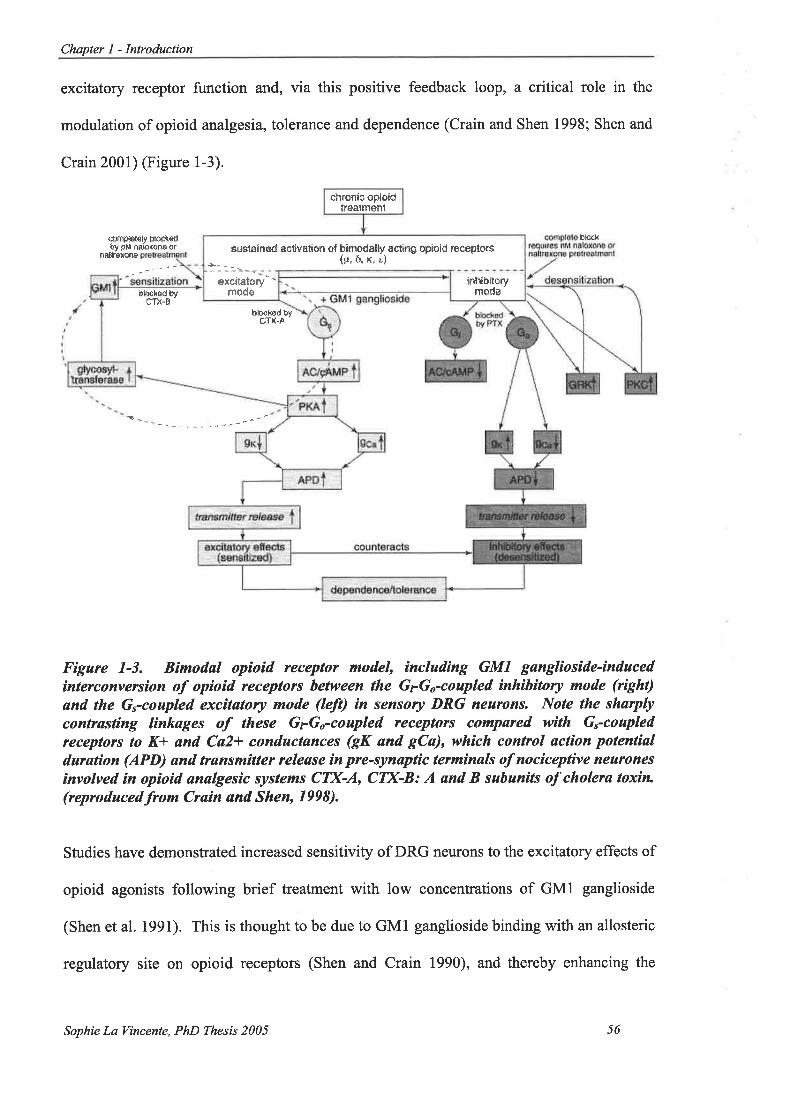
Chapter I - Introduction
excitatory receptor function and, via this positive feedback loop, a critical role in the
modulation of opioid analgesia, tolerance and dependence (Crain and Shen 1998; Shen and
Crain 2001) (Figure l-3).
nallÍErsna
{ðm@l€iy Þ!)ohoíât ÐM nabxaftÐ ar
þlrßL
Figure 7-3. Bimodøl opioid receptor model, íncluding GMI gangliosìde-inducedìnterconversion of opìoíd receptors between the GrGo-coupled ìnhìbìtory mode (r¡Sht)
ønd the Gycoupled excìtøtory mode (leÍt) ¡n sensory DRG nearons. Note the sharplycontrøsting linkages of these GyG,-coupled receptors compared wìth Gycoapledreceplors lo K* ønd Ca2+ conductances (gK ønd gCa), whìch control action potentíaldurøtion (APD) and trønsmìtter release ín pre-synaptìc terminals of nociceptìve nearonesinvolved ìn opíoid ønalgesic systems CTX-A, CT)GB: A ønd B subunits of cholera toxín(reproduced.from Crøin and Shen, 1998).
Studies have demonstrated increased sensitivity of DRG neurons to the excitatory effects of
opioid agonists following brief treatment with low concentrations of GMI ganglioside
(Shen et al. 1991). This is thought to be due to GMI ganglioside binding with an allosteric
regulatory site on opioid receptors (Shen and Crain 1990), and thereby enhancing the
chronicopioidtrÉåInìent
b¡môdä¡ty aatng oploid reëeptôrË{+¡, rT, x, ¿)
---a'--o--excitätÕru"
modø'
sustÊlnÈd äctivâΡön of
inhibitorymods
blDck۟ 3fCTX-A
þlrrckÈd þttTl{-Ê '
Sophie La Vincente, PhD Thesis 2005 56

Chapter I - Introduction
efficacy of excitatory G.-coupled opioid receptor functions (Crain and Shen 1998). This is
consistent with evidence that injection of low doses of GMI in mice reduces the analgesic
effect of morphine, producing what has been termed "acute tolerance" (Crain and Shen
2000).
Furthermore, unlike most G-protein coupled receptors that become desensitised with
chronic exposure to an agonist, Gr-coupled opioid receptors become increasingly sensitised
during sustained exposure of DRG neurons to bimodally acting opioid agonists (Crain and
Shen 1992). Indeed, Gi/Go-coupled receptors have been shown to become progressively
desensitised by activation of G-protein receptor kinases and arrestins, and by activation of
PKC (Hanis and Williams l99l1' Ueda et al. 1995; Chuang et al. 1996), and it is thought
that this may also contribute to the development of tolerance with chronic opioid treatment.
1.9.2.4.4. Human models of analgesic potentiation with low-dose antagonist
Conflicting findings have emerged from human studies investigating the use of opioid
antagonists in conjunction with opioid agonists to enhance analgesia (summarised in Table
1-3). In contrast to the opioid-sparing effect reported by Gan and colleagues (Gan et al.
1997) (see 1.9.2.3), Cepeda and colleagues reported no decrease in opioid requirements
combining low-dose (Cepeda et aL.2002) or ultra-low dose (Cepeda et al.2004) NLX with
morphine in patient controlled analgesia (PCA) during the post-operative period. Low-
dose NLX (an average of 0.5 ¡rglkglhour in.the first hour, followed by an average of 0.06
¡rglkglhour afterwards) combined with morphine did not produce a decrease in opioid
requirements, and pain intensity ratings were in fact higher in the group receiving NLX
(Cepeda et aI.2002). However, this study was limited in that only one dose of NLX was
Sophie La Vincente, PhD Thesis 2005 57

Chapter I - Introduction
used. Recent findings (see below and Table 1-3) indicate that both the dose of agonist and
the agonist:antagonist ratio are crucial in producing potentiation. In a more recent study,
the same group trialled the same methodology utilising a lower dose of NLX. In this study
patients received an average of 0.05 pLglkglh NLX in the first 2 hours, and an average of
0.009 Vglkglh subsequently. At this dose, it was revealed that NLX still failed to reduce
opioid requirements, but signifrcantly decreased the incidence of opioid side effects
(Cepeda et al. 2004).
A case study of a 61-year old diabetic with painful polyneuropathy revealed that ultra-low
dose naltrexone potentiated the analgesia afforded by methadone, and decreased the
incidence and severity of side effects (Cruciani et al. 2003).
Several studies have also investigated other opioid agonists in combination with low dose
antagonists. There have been reports of NLX potentiating the effects of opioids that
mediate antinociception via other opioid receptor types, such as the r partial agonists
nalbuphine and pentazocine. In an early investigation, analgesia produced by morphine (8
or 15 mg) andpentazocine (60 mg) administered individually and in combination with low-
dose NLX (0.a mg) were compared (Levine et al. 1988). Analgesia produced by
pentazocine (60 mg) was potentiated by the addition of NLX, while morphine analgesia (8
mg) was attenuated with the addition of the antagonist. The analgesia associated with the
combination of pentazocine and NLX was significantly greater than was associated with
administration of either NLX, pentazocine or high-dose morphine (15 mg) alone. Patient
use of díazepam was considered a potentially confounding effect in these results; therefore
a correlative study was conducted with rats. Findings from the animal study substantiated
the results of the clinical trial. More recent investigation has focused on another rc agonist,
Sophie La Yincente, PhD Thesis 2005 58

Chapter I - Introduction
nalbuphine. It has been reported that the addition of low-dose NLX (itself not associated
with analgesia) to low-dose nalbuphine produced marked post-operative analgesia (Gear et
al. 2000), and a report of three case studies describes improved relief of neuropathic
trigeminal pain with the combination of nalbuphine and low-dose NLX (Schmidt et al.
2003).
Sophie La Vincente, PhD Thesis 2005 59

Nalbuphine+NlX associated with enhanced analgesia
in both males and females compared to eithernalbuohine o¡ NLX alone (p=0.0001)
Nalbuphine + NLX reduced pain ratings by 50%comoa¡ed to prior freatment (varied) (NB case report)
No difference in morphine requirement betweenmorphine+naloxone (25.4 mg) and morphine (27.0 mg)groups þ>0.05)Nausea and pruritus reduced in NLX group (P:0.01)Other side effects similar between groups
Potentiated analgesia
Decreased incidence and severity ofopioid side effectsfNB Case ¡eoort)Pentazocine * NLX produced greater analgesia thanpentazocine 0<0.01) and high-dose morphine (15 mg)(p<0.01)Significant enhancement maintained at end ofstudy (3
h l0 min post-dosing) þ<0.01)Addition of NLX attenuated morphine (8 mg) analgesia
lo<0.051
Outcome35% reduction in nausea and vomiting, 30% reduction
in pruritus þ<0.05), no difference in respiratory rate
Morphine requirernent reduced by one third (59.1 mg to42.3 mg) in low-dose NLX groupþ<0.05) High dose
NLX srouo had hieher momhine requirements (p>0.05)
No difference in morphine requirement þ>0.05)Reduced refrospective rating ofpain intensity (p<0.05).
Reduced need for antiemetic and antþruritic medication
(p<0.05)
Morphine requirement higher in morphine+NlX group(15.6 mg) compared to morphine group (13.4 mg)(p=0.00e)More treatement failures due to inadequate analgesia inmorphine+NlX group (n:38) compared to morphinegrouP (n:22)(P=0.025)Higher percentage ofpatients in naloxone group not
satisfied with pain treatrnent þ=0.01)No differences between goups in side effects (p>0.05)
Pain ratings
Pain ratings, males vs.
females
Pain ratings
Cumulative morphinerequirement in l" 24 h post-surgery @CA)Pain intensity ratingsPatient satisfaction with pain
teatmentSide effects
Cumulative moçhinerequirement in I't 24h post-
surgery @CA)Pain ratings
Pain ratingsMethadone dose
Side effects
Outcome measureCumulative morphinerequirement over 24h period(PcA)
Cumulative moçhinerequirement in 11 24 h post-surgeryA¡ti-emetic and anti-pnriticdrug requirementRefr osDective Dain intensitv
TV
TV
TV
TV
IV
Oral
RouteIV
TV
NLX (0.4 mg)
NLX
NLX (average of 0.05
pg/kg/hr in I't 2 hours,
0.009 pg/kg/hrsubsequently)Intermittent infusion(in combination withmomhine)NTX
NLx (0.a mg)
Antasonist ldose)
Low (0.25 ¡rglkg/h) orhigh (1.0 pdke/h)dose NLX.Conti¡uous infusion
Nalmefene (15 mg or25 mg)
NLX (average of0.5¡rglkglh lr 2 hours;
0.06 pg/kglhsubsequently)intermittent infusion(administered incombination withmorphine)
Methadone
Pentazocine(60 mg) Morphine (8mg combined withNLX & l5mg alone)
Nalbuphine (5 mg)
Nalbuphine
Morphire (PCA)
Morphine (PCA)
Morphine (PCA)
AsotristMorphine (PCA)
DBRCT
DBRCT
CasereDort
DBRCT
DBRCT
Case
report
DesienDBRCT
DBRCT
Post-surgical patients(removal of impacted thirdmolars) (n:105)
Post-surgical patients(removal of impacted thirdmolars) t'¿=88 )Tri geminal neuropathic¡ún h=3)
Post-surgical patients(abdominal, thoracic,orthopedic surgery of< 3 hduration) (n=166)
Post-surgical patients(abdominal thoracic,orthopedic, craniofacialsurgery) of < 3 h duration)(n=265)
6l-year old diabetic,painful neuropathy (n :1)
SamplePost-surgical patients(hysterectomy)(n:60)
Post-surgical patients(lower abdominal surgery),fenales only (n:120)
Cruciani etat.,2003
Levine et al.1988
Gear et al.2000
Schmidt et
al..2003DB Doubte-blind RCT Randomised controlled trial PCA Patient-contolled
Joshi et al.,1999
Cepeda et al.,2002
Cçeda et al.,2004
StudvGan et a1.,
1997
Tøbte 1-3. Summary oÍ clinicøl repofis and human stud¡es of potentíøtion of anølgesiø with opíoid agon¡st:øntagoníst combínøtions.
60Sophie La Tincente, PhD Thesß 2005
Chapter I - Inlroduction
1.9.3. Limitations of the human studies
The inconsistencies associated with the findings from human studies of analgesic
potentiation with opioid agonist:antagonist combinations may be related to differences in
trial methodology, outcome measures and doses used. For example, Gan and colleagues
showed analgesic potentiation with the administration of NLX by continuous infusion
(Gan et al. 1997), whereas Cepeda and colleagues administered NLX intermittently in
combination with morphine and detected no enhancement of analgesia (Cepeda et aI.2002;
Cepeda et al.2004). There were also differences between studies in the amount of NLX
administered. The average NLX dose administered to patients in the first 2 hours of the
initial study by Cepeda and colleagues (0.5 Wekelh) (Cepeda et al. 2002) was midway
between the low (0.25 Vg/rglh) and high (1.0 ¡rg/kg/h) doses used by Gan and colleagues,
which were associated with lower and higher morphine requirements, respectively.
However, the amount of NLX administered by Cepeda and colleagues was decreased to an
average of 0.06 pg/rglh after the initial 2 hours of observation (Cepeda et aI. 2002). It has
also been noted that the amount of NLX administered by Cepeda and colleagues was
higher than the equipotent dose of nalmefene used by Joshi and colleagues (Joshi et al.
1999), which was associated with enhanced analgesia (Mehlisch 2003). The subsequent
study by Cepeda and colleagues aimed to investigate whether the failure to detect
enhanced analgesia was related to the comparatively higher doses of NLX administered
(Cepeda et al. 2004). In this study, patients received an average of 0.05 $g/rrglh in the flrrst
2 hours, and an average of 0.009 þgkglh subsequently (compared to an average of 0.5 and
0.06 ¡rglkg/h administered in the initial study, as described above). While a decrease in
side effects was observed with this combination, there \ryas no evidence of analgesic
potentiation.
Sophie La Vincente, PhD Thesis 2005 61
Chapter I - Introduclion
Signif,rcant variation in average cumulative morphine requirement between studies has also
been noted (Mehlisch 2003). The average amount of morphine administered in the 24
hours post-surgery in the first study by Cepeda and colleagues (Cepeda et aL.2002) (13.4-
15.6 mg) was considerably lower than used over an equivalent time period by post-
hysterectomy patients in the studies by both Gan and colleagues (42.3-64.7 mg) (Gan et al.
1997) and Joshi and colleagues (45-56 mg) (Joshi et al. 1999). It has been proposed that
the difference in morphine requirement is indicative of fundamental differences between
studies in terms of type of surgical procedure, level of pain associated with the procedure,
and/or method of drug administration (Mehlisch 2003). Cepeda and colleagues have
defended the design of their study, responding that morphine requirements during the first
2 hours post-surgery 00.7-12 mg) were similar to those reported by Joshi and colleagues
(8 mg), and that differences in opioid consumption subsequent to this are likely due to the
PCA dose used (0.5 mg rather than 1.0 mg) (Carr and Cepeda 2003). Notwithstanding,
both human studies that have shown enhanced analgesia with the morphine and NLX
combination recruited patients undergoing the same surgical and anaesthetic procedures
(Gan et al. 1997; Joshi et al. 1999), whereas both studies that failed to demonstrate
analgesic potentiation recruited patients undergoing a wide range of different surgical
procedures (Cepeda et al. 2002; Cepeda et al. 2004). The relative advantages and
disadvantages of these approaches are discussed below. Further, those studies reporting
enhanced analgesia administered the antagonist by continuous infusion (Gan et al. 1997).
In contrast, studies reporting no analgesic potentiation administered NLX in combination
with morphine in intermittent boluses (Cepeda et al. 2002; Cepeda et al. 2004). The
implications of this discrepancy are discussed below.
There are several limitations associated with the human studies of analgesic potentiation
using opioid agonist:antagonist combinations. All studies were conducted with clinical
Sophie La Vincente, PhD Thesis 2005 62

Chapter I - Introduction
pain rather than experimental pain. As discussed previously, the validity of experimental
pain, particularly in assessing analgesic medication, has been a challenging and
contentious issue amongst clinicians and researchers (Keats et al. 1950; Beecher 1953;
Moore et al. 1997). It has been argued that experimental pain is qualitatively different
from pain associated with injury or disease (Beecher 1962). While it is recognised that the
effectiveness of an analgesic intervention must be assessed in the circumstances for which
it is intended, there is a strong argument that the measurement of dose-response, evaluation
of optimal dosage and comparison of relative efficacy with known substances are best
conducted with a sample of healtþ volunteers who are as homogeneous as possible
(Bromm 1985).
There are two broad justifications for the use of an experimental pain paradigm prior to
clinical evaluation of agonist:antagonist combinations. Firstly, clinical pain involves
considerably more variability in both patient characteristics and pain experience. As
described in Chapter 3, the experience of pain is subject to considerable inter- and intra-
individual variability. Numerous factors have been shown to impact upon the pain
experienced and reported in an experimental or clinical setting. In an experimental
context, factors such as sex, age, ethnicity, psychological variables, current or past
substance dependence, presence of medical conditions, cigarette smoking, previous
chronic exposure to opioids and history of chronic pain can be better controlled for in
participant recruitment. Importantly, the type and intensity of noxious stimuli can also be
standardised. In a clinical context, not only is it considerably more difficult to control for
these factors, but even greater variability is introduced. The type of pain frequently
investigated in trials with clinical populations is post-surgical pain, and indeed all
randomised trials of agonist:antagonist combinations for enhanced analgesia have assessed
this type of pain. The use of post-operative pain introduces numerous sources of
Sophie La Vincente, PhD Thesis 2005 63
Chapter I - Introduction
variability. These may include fear of surgery and associated stress, differing anaesthetic
techniques and recovery from the anaesthetic procedure, prior experience with surgical
procedures, .or.o-itant medication (both administered for surgery and for coexisting
medical conditions) and the purpose and type of surgical procedure itself, incorporating
factors such as time in theatre and differences in baseline pain, pain duration and general
discomfort typically associated with a certain procedure. Furthermore, side effects (which
have been used as an outcome measure indicative of treatment quality) observed in the
post-operative period may be related to the surgery, the anaesthetic technique or
concomitant medications rather than the analgesic agent(s) administered. Several of the
studies in question attempted to reduce potential sources of variability by recruiting
patients who had undergone equivalent surgical and/or anaesthetic procedures (Levine et
al. 1988; Gan et al.19971' Joshi et aI. 1999; Gear et al. 2000). An alternative strategy is to
recruit patients who have undergone surgical procedures lasting within a certain time
Íaîge, ostensibly as time in theatre provides a degree of standardisation of the gravity of an
operation, anticipated degree of post-surgical pain or recovery process (Cepeda et aI.2002;
Cepeda et al. 2004). Despite these approaches to reducing variability, the use of clinical
pain significantly reduces the investigator's control over the many sources of variability
within a sample.
It has been argued that recruiting patients who have undergone a wide range of surgical
procedures enhances the generalisability of the findings and provides a more naturalistic
study in terms of the likely clinical utility of the intervention (Carr and Cepeda 2003).
However, in the case of agonist:antagonist combinations for enhanced analgesia, where
application to humans remains in the preliminary stages and 'þroof of concept" is yet to be
adequately demonstrated, it may be considered that establishing optimal dose and
agonist:antagonist ratio using experimental pain would be preferable. Indeed, the findings
Sophie La Vincente, PhD Thesis 2005 64

Chapter I - Introduction
from one human study that failed to detect analgesic potentiation with the
agonist:antagonist combination have been censured for utilising a flawed methodology
(Mehlisch 2003). It has been argued that this study (Cepeda et aL.2002), which aimed to
investigate the use of agonist:low-dose antagonist combinations using intermittent
administration (described in section L.9.2.4.4) suffered a number of significant flaws both
in terms of departure from the methodology of previous studies and confounding factors
that were not adequately controlled (Mehlisch 2003).
The second justification for using an experimental pain paradigm prior to clinical pain
trials in the investigation of agonist:antagonist combinations relates to the importance of
identiffing the optimal agonist dose and agonist:antagonist dose ratio. As discussed in
greater detail below (see 1.9.4), animal studies have demonstrated that both the dose of
agonist administered and the agonist:antagonist ratio are critical in producing
antinociceptive potentiation. While it is recognized that the optimal dose ratio in
managing clinical pain may be different from that demonstrated experimentally, in a
clinical pain setting it is considerably more difficult to establish dose-response curyes for
the agonist alone and in combination with different antagonist ratios. The human studies
of agonist/antagonist combinations for enhanced analgesia have been limited by a failure
to vary the antagonist dose relative to the agonist dose, or have at best administered either
"high" or "lo\ry" antagonist doses (Gan et al. 1997). Furthermore, the use of PCA
introduces additional variability in terms of the amount of each agent administered to each
patient. PCA has been used in several studies examining agonist/antagonist combinations
in analgesia, offering the advantage of an accurate representation of common clinical
procedure, and the useful outcome measure of a reduction in opioid requirements to
indicate analgesia. However, in studies that have administered the antagonist in
continuous infusion (Gan et al. 1997) irrespective of patients' use of morphine, the ratio
Sophie La Vincente, PhD Thesis 2005 65

Chapter I - Introduction
\Mould differ between patients according to their use of the agonist. In other studies, the
antagonist has been combined with morphine, such that the antagonist was administered
simultaneously with the morphine and only if the morphine was accessed. In these
circumstances, while the ratio is consistent between patients, the absolute amount of both
agonist and antagonist received varies according to the need for additional doses.
Moreover, if the ratio is not optimal, such that the dose of antagonist is too high relative to
the agonist, as has been postulated in one study (Cepeda et al. 2002), it is conceivable that
the antagonist may attenuate analgesia, resulting in an increase in reported pain intensity
and the need for another bolus, thereby further enhancing the hyperalgesia. The
intermittent administration of low-dose antagonist according to the methodology of
Cepeda and colleagues (Cepeda et al. 2002) has also provoked criticism (Mehlisch 2003)
as it has been demonstrated in acutely and chronically morphine treated rodents that
intermittent administration of a low-dose antagonist precipitates long-lasting h¡.peralgesia
(Celerier et al. 1999; Shen and Crain 2001). Indeed, the 2002 study by Cepeda and
coworkers that used intermittent antagonist dosing reported that patients admininstered
morphine plus naloxone experienced less pain relief and used more opioids than those
administered morphine plus saline (Cepeda et al.2002).
The valid assessment of outcome measures is complicated further by the requirement in
some protocols of the mandatory administration of a rescue dose when reported pain
intensity exceeded a set point, and that the PCA dose be augmented by 20%. These studies
also incorporated a mandatory decrease in PCA dose by 20% if side effects attributed to
the PCA were experienced (Cepeda et al.2002; Cepeda ef al.2O04). While these measures
are reasonable for ethical reasons, they further complicate the valid comparison between
groups. In these studies, the requirement for rescue doses and the incidence of side effects
were recorded. However, as noted by Mehlisch (2003), side effects may have been related
Sophie La Vincente, PhD Thesis 2005 66

Chapler I - Introduction
to other factors (i.e. the surgical procedure, the anaesthetic procedure or concomitant
medication). Thus rescue doses may have been administered when they were not, in fact,
required.
A further concern is the difference in time course and duration of effect associated with the
agonist and antagonist administered, and the lack of control over the timing of drug
administration in clinical pain settings.
In summary, the use of agonist:antagonist combinations for enhanced analgesia in humans
has produced inconsistent findings. It is proposed that differences in trial methodology,
outcome measures and doses used may account for these differences. Moreover, the
failure to assess a wide range of dose ratios and the degree of variability and lack of
control associated with the clinical pain trials described, reduces the capacity for valid
assessment between treatment groups and may prevent the observation of enhanced
analgesia. There have been no published studies of opioid agonists combined with ultra-
low or low-dose antagonists in a human experimental pain paradigm.
Given the importance of agonist dose and agonist:antagonist ratio in observing analgesic
potentiation, it is proposed that the investigation of agonist/antagonist combinations be
fîrst assessed in an experimental pain paradigm with healtþ volunteers to establish the
optimal opioid dose and agonist:antagonist ratio. In an experimental setting many of the
sources of variability that complicate assessment in clinical pain studies may be controlled.
1.9.4. Buprenorphine andantagonistcombinations
Buprenorphine (BUP) is an opioid receptor pafüal agonist with potent analgesic effects
(see 4.2). Chronic exposure to BUP may be associated with mild physical dependence,
Sophie La Vincente, PhD Thesis 2005 67
Chapter I - Introduction
with limited withdrawal signs and symptoms. The adverse effects of BUP are similar to
other opioid agonists. Findings suggest that BUP may have a ceiling effect for respiratory
depression and other effects in humans. Thus, it may be safer than other opioids (Walsh et
al. 1995).
There have been very few investigations of BUP in combination with a low-dose
antagonist. An early investigation reported that prior treatment of rats with NLX
significantly reduced the analgesic action of low-dose BUP in a tail flick assay (Rance et
al. 1980). A subsequent study demonstrated that pretreatrnent with NTX shifted the BUP
analgesic dose-response curve to the right (Dum and Herz 1981). Following a report
describing potent analgesia resulting from the addition of NLX in two patients recovering
from cholecystectomy (Pedersen et al. 1985), Bergman and colleagues conducted a trial
investigating this drug combination in a rabbit tooth pulp assay. This study revealed
enhanced antinociception with the combination of NLX (0.001 mg/kg IV) and BUP (0.10
mgn<g IV), with a peak o/o maximum possible effect (MPE) of 78Yo compared to 48o/o
produced with BUP only þ<0.05). NLX at a higher dose (0.1 mg/kg) did not alter
antinociception (p>0.05).
There have been no published investigations of BUP combined with ultra-lo\¡/ or low-dose
antagonists in humans.
While the utilþ of opioids in the management of neuropathic pain is controversial
(Portenoy et al. 1990; McCormack 1999), mounting evidence suggests that BIJP may have
a special role in this capacity (Benedetti et al. 1998; McCormack et al. 1998; Kouya et al.
2002; Radbruch 2003). Given this potential for BUP in the treatment of neuropathic pain,
Cougnon-Aptel and colleagues (Cougnon-Aptel et a1., unpublished) recently investigated
Sophie La Vincente, PhD Thesis 2005 68
Chapter I - Introduction
the use of BLIP combined with low-dose opioid antagonists in rats with peripheral
neuropatþ induced by the chronic constriction injury model (CCI) (Bennett and Xie
1988). This model involves loose ligation of the sciatic nerve to induce neuropatþ. Over
the 7-10 days following ligation, animals develop pain behaviours including thermal and
mechanical hyperalgesia and allodynia in the hindpaw ipsilateral to the site of nerve
ligation. These behaviours can persist for up to 7 weeks following ligation (Bennett and
Xie 1988; Attal et al. 1990). Nociceptive testing coÍìmenced one week following nerve
ligation, and following confirmation of neuropatþ. Thermal nociceptive threshold, as
determined by hindpaw withdrawal latency, was assessed for increasing doses of BIIP, and
two BUP:NLX ratios, l5:1 and 20:1.
As described in Chapter 4, one of the most intriguing features of the pharmacological
profile of BLIP is the bell-shaped dose-response curve observed with many effects,
including antinociception in animal models. Antinociception peaked at a dose oî20 ¡tgkg,
decreased to a trough at 40 pgkg, but then increased at 50 pglkg, which may be indicative
of the biphasic dose-response curve that has previously been associated with BUP (see
Cowan 1995). The combination of BUP and naloxone was associated with significantly
enhanced antinociception, but this effect was dependent upon the BUP dose (Cougnon-
Aptel et al. unpublished). Interestingly, the combination l¡/as associated with significant
antinociception only when the dose of BUP alone produced minimal or no antinociception.
This was evident at both ends of the BUP dose-response curve. When the dose of BUP
alone was associated with significant antinociception, the addition of naloxone reduced the
magnitude of antinociception, ostensibly having an antagonistic effect. The enhanced
antinociception associated with the drug combination was observed with both ratios,
though the greatest effect was evident with the 15:1 ratio. In light of the suggestion in
previous findings that reduced opioid requirements can occur without a simultaneous
Sophie La Vincente, PhD Thesis 2005 69
Chapter I - Introduction
increase in adverse effects (Rawal et al. 1986; Gan et al. 1997), the potentiation evident at
the lower end of the BUP dose-response curve is of particular clinical relevance. The
optimal dose of BUP in the lower range of the dose-response curve associated with
enhanced antinociception when combined with NLX was 2.5 Wglkg. This dose was then
tested in combination with NLX and the long-acting opioid antagonist naltrexone, as well
as morphine and naloxone combinations, in a range of ratios. The BUP:naloxone
combination in a 15:l ratio was the most effective in enhancing antinociception. These
data indicate that both the BUP dose and the agonist:antagonist ratio are critical in
producing potentiation of BUP antinociception by ultra-low doses of antagonist.
1.10. Summary
The negative consequences of pain managemenl are costly to the community and
associated with increased morbidity for the patient. While opioid analgesics are highly
effective in the management of pain, they are associated with a number of unpleasant and
dangerous adverse effects that limit their use. One approach by which the use of opioids
may be improved is the administration of an opioid in combination with another agent that
either itself produces analgesia or potentiates the analgesia associated with the opioid. A
lower dose of the opioid is therefore required to achieve an adequate level of analgesia,
thus reducing the incidence and severity of adverse effects.
The combination of an opioid agonist with ultra-low dose opioid antagonist has been
reported to potentiate the pain relieving effect of the opioid in animal studies and in some
clinical investigations. Critical to the clinical utility of this drug combination is whether
the adverse effects are potentiated in the same manner as has been observed with pain
relief. Previous experience with the addition of low doses of opioid antagonists to opioid
treatment suggests that adverse effects may be reduced without a simultaneous reduction in
Sophie La Vincente, PhD Thesis 2005 70

Chapter I - Introduclion
analgesia, or unaffected despite enhanced analgesia. If the combination of an opioid
agonist and ultra-low dose antagonist can potentiate analgesia without a simultaneous
increase in adverse effects, the negative effects that limit the use of opioids in pain
management may potentially be overcome.
BUP is a highly potent opioid analgesic, with low abuse liability and a potentially better
safety profile than other opioid analgesics such as morphine. BUP is also considered to
have potential in the management of neuropathic pain, for which other opioids have been
less effective (Kouya et al. 2002). Recent animal data suggest that BUP antinociception
may be potentiated with the addition of ultra-low dose NLX in a model of neuropathic pain
(Cougnon-Aptel et al. unpublished). The antinociceptive effect of BUP combined with
ultra-low doses of an opioid antagonist has not been investigated in humans.
I .1 l. The present research
The broad aim of the present research was to determine whether BUP combined with ultra-
low doses of NLX enhances antinociception in a human model of experimental pain. A
further aim was to determine whether any antinociceptive potentiation observed with the
drug combination was associated with a simultaneous increase in the incidence or severity
of adverse opioid effects, including respiratory depression, sedation and nausea.
Due to the ratio-dependent nature of the enhanced antinociception observed in the animal
study of BUP:NLX combinations (Cougnon-Aptel et al., unpublished), a further aim was
to identiff the optimal ratio for antinociceptive potentiation, if this occurred.
This thesis will describe four studies, which are organised by chapter:
Sophie La Vincente, PhD Thesis 2005 7I
Chapter I - Introduclion
1. Normative study of the nociceptive tests (Chapter 3)
The nociceptive tests to be used in this series of trials had not been standardised, and
normative values had not been established. Furthermore, numerous variables have been
reported to contribute towards inter-subject variability in response to experimental noxious
stimuli, and it is unclear to what extent these factors should be controlled for in subject
recruitment. The purpose of the initial study was to determine normative values for the
two nociceptive tests to be used, the cold pressor and electrical stimulation tests, and to
assess the contribution of a range of biological and psychosocial variables to inter-
individual variability in test performance. The findings from this study guided subject
selection and exclusion criteria for the subsequent drug studies.
2. Buprenorphine dose-finding study (Chapter 4)
Unlike many other opioid analgesics, BUP had not been evaluated in a human
experimental pain paradigm. A dose-finding study was required to determine whether the
nociceptive tests to be used are sensitive assays for BIIP antinociception, and to identiff a
sub-analgesic dose of BUP to be used in the subsequent BUP:NLX ratio studies.
3. Antinociceptive activity of buprenorphine and naloxone combinations (Chapter 5)
The effects of BUP combined with NLX were compared with the effect of BUP combined
with saline. The BUP:NLX combination was administered in the following ratios: 15:1,
20:l and 25:7. These ratios were selected on the basis of findings from the
BUP:antagonist animal study (Cougnon-Aptel et al., unpublished).
4. Optimising the BUP:NLX ratio for antinociception (Chapter 6)
Based on the results of the first ratio study, a second BUP:NLX ratio study was conducted
to further investigate the effects of this drug combination over a wider range of ratios
Sophie La Vincente, PhD Thesis 2005 72

Chapter I - Introduction
This study investigated the effects of BUP combined with NLX in a 5:1, 10:1 and 12.5:l
ratio, compared to the effects of BUP combined with saline.
1.1 1.1. Aims
CHAPTER 3:
1. To establish normative values for healtþ volunteers on the cold pressor and electrical
stimulation tests.
2. To establish an upper and lower limit in baseline pain response to guide subject
selection in subsequent studies.
3. To determine the factors that contribute to intra-individual variation in pain response,
and may thus be important to control in subject recruitment.
CHAPTER 4:
1. To determine whether the pain induction tests selected for use in these studies are
sensitive assays for BUP antinociception.
2. To determine a BUP dose-response curve for doses in the lower range and below
therapeutic doses for analgesia
3. To select a sub-antinociceptive dose of BUP for use in the subsequent BUP:NLX ratio
studies.
CHAPTER 5
1. To investigate whether the addition of ultra-low doses of NLX significantly enhances
antinociception to experimental pain compared to the same dose of BUP alone in healtþ,
pain-free volunteers
Sophie La Vincente, PhD Thesis 2005 73
Chapler I - Inlroduction
2. To determine whether the addition of ultra-low doses of NLX significantly enhances
adverse opioid effects compared to the same of BUP alone in healtþ, pain-free volunteers
3. To identiff the optimal BUP:NLX ratio for enhanced antinociception to experimental
pain.
CHAPTER 6
l. To investigate further the effect of BUP:NLX combinations on antinociception and
adverse effects.
Sophie La Vincente, PhD Thesis 2005 74
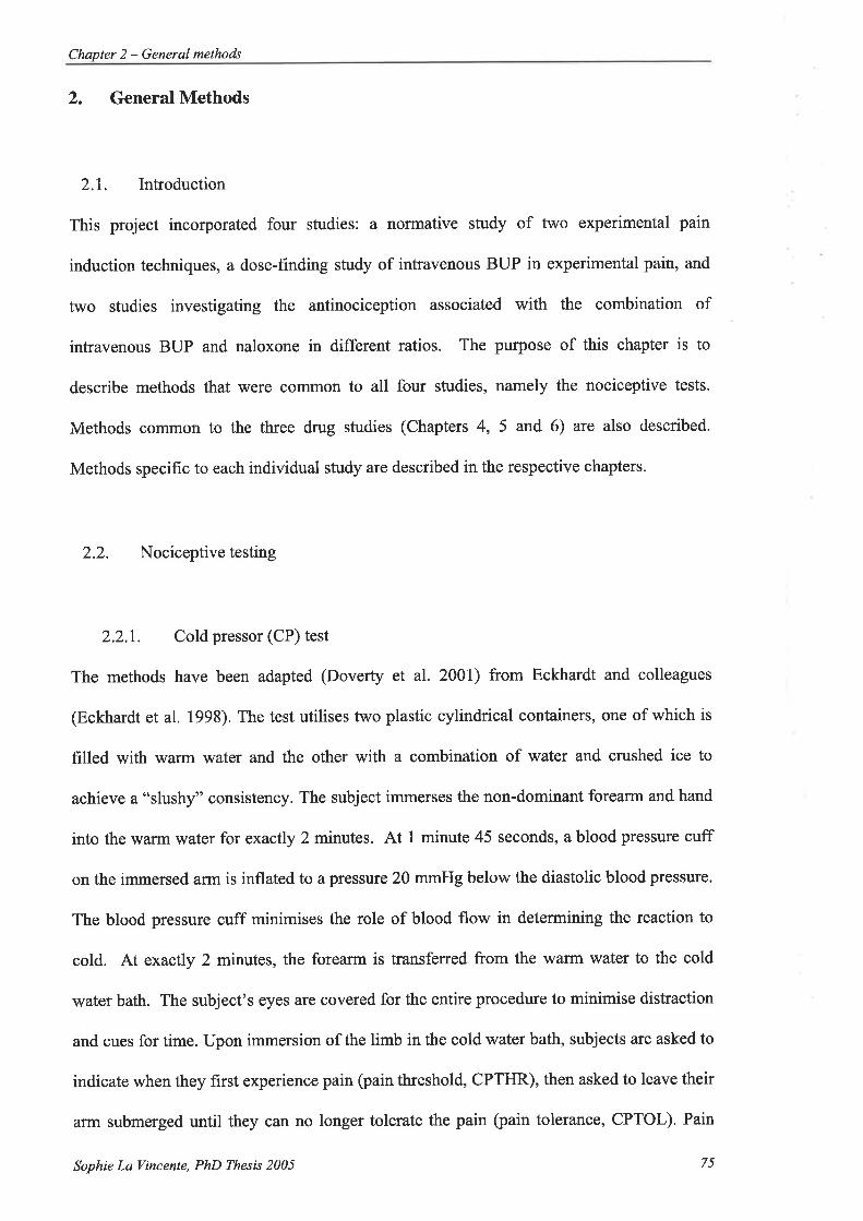
Chapter 2 - General methods
2. General Methods
2.1. Introduction
This project incorporated four studies: a normative study of two experimental pain
induction techniques, a dose-finding study of intravenous BUP in experimental pain, and
two studies investigating the antinociception associated with the combination of
intravenous BUP and naloxone in different ratios. The purpose of this chapter is to
describe methods that were coÍrmon to all four studies, namely the nociceptive tests.
Methods common to the three drug studies (Chapters 4, 5 and 6) are also described.
Methods specific to each individual study are described in the respective chapters.
2.2. Nociceptive testing
2.2.1. Cold pressor (CP) test
The methods have been adapted (Doverty et al. 2001) from Eckhardt and colleagues
(Eckhardt et al. 1998). The test utilises two plastic cylindrical containers, one of which is
filled with warm water and the other with a combination of water and crushed ice to
achieve a "slushy" consistency. The subject immerses the non-dominant forearm and hand
into the warrn water for exactly 2 minutes. At I minute 45 seconds, a blood pressure cuff
on the immersed arm is inflated to a pressure 20 mmHg below the diastolic blood pressure.
The blood pressure cuff minimises the role of blood flow in determining the reaction to
cold. At exactly 2 minutes, the forearm is transferred from the warm water to the cold
water bath. The subject's eyes are covered for the entire procedure to minimise distraction
and cues for time. Upon immersion of the limb in the cold water bath, subjects are asked to
indicate when they first experience pain þain threshold, CPTHR), then asked to leave their
arm submerged until they can no longer tolerate the pain þain tolerance, CPTOL). Pain
Sophie La Vincente, PhD Thesis 2005 75
Chapter 2 - General methods
threshold and tolerance times are recorded in seconds from immersion in cold. An
undisclosed cut-off of 180 seconds is imposed, after which time pain tolerance can no
longer be accurately assessed due to numbness.
2.2.I.1. Materials
2x20litre plastic cylindrical containers (38cm in depth; 30cm in diameter)
Digital Thermometer
Blindfold
Sphygmomanometer
Digital timer (with second display)
Thermoregulator (Unistat I 1 0, Thermoline Scientific, Sydney, Australia)
Aquatic water pump (Brotga MV 1500, Brolga Australia Pty. Ltd., Haberfield,
NSW, Australia)
Towel
2.2.1.2. Set-upprocedure
One container was frlled with warm water (to 5 cm from top of container), and the
thermoregulator immersed in container and set at 35oC. The second container was filled
with crushed ice (to 10 cm below top of the container). Water was added until the
container was filled to 5cm below the top of the container, and stirred to ensure water and
ice were mixed evenly and there were no large clumps of ice. The temperature of the
water/ice combination was then checked with a digital thermometer, and water or ice
added as required to achieve a temperature between 0.5 and 1.0oC. Containers were
placed on the trolley l0 cm apart with the warm water container on the left hand side. The
water pump was placed at the bottom of the cold water container on the far side of the
container (away from where the subject will stand) with the water jet facing upwards.
Sophie LaVincente, PhD Thesis 2005 76
I
I

Chapter 2 - General methods
2.2.1.3. Testadministration
The experimenter described to the subject the purpose of the test and the procedure as
follows:
This is the cold pressor test. It is a test of your tolerance to cold pain. Here are two
water containers, one filled with warm water, one filled with ice and cold water. You
will place your non-dominant arm into the warm water containerfor two minutes, then
take it out and put it immediately into the cold water container. When your arm is in
the cold water container, there are two things I will ask you to tell me: tell me when
you first feel pain, then leave your arm in the cold water as long as you can possibly
tolerate the pain. Tell me when you feel you can no longer tolerate the pain, and
remove your arm from the water. I will pass you a towel, which you may use to dry
your arm. While you are completing the test you will be blindfolded, and I will inflate
a blood pressure cuff on your arm just beþre you transfer your arm to the cold water
container. This is to control þr other factors that may interfere with the results. There
is a water pump in the cold water container to keep the water circulating and stop the
ice from clumping together. l4lhen you put your arm in each water container, immerse
your arrn quickly but carefully. As you will be blindfolded, I will help you transfer your
arm from the warm water to the cold water. Keep your fingers straight and spread
apart. Do not touch the sides or the bottom of the container and try not to move your
arm around too much in the water.
I will not speak to you during the test except to give you reminder instructions. You
should not speak during the test unless you ltave an urgent question or concern. The
pain you experience from the test disappears quickly øfter removing your arm from the
coldwater, and there is no risk of permanent damage.
Every person is different in terms of his or her pain sensitivity. It is very important that
we obtain an accurate and honest assessment of your pain tolerance. There is no
Sophie La Vincente, PhD Thesis 2005 77

Chapter 2 - General methods
reward for setting a record time, but please try to perþrm the test honestly and leave
your arm in the cold water as long as you can tolerate the pain.
The experimenter then ensured that the subject understood the instructions and enquired
whether the subject had any questions before commencing the test. The subject was seated
in a comfortable chair and hisÆrer blood pressure taken. The subject then stood in front of
the containers at an appropriate distance such that the non-dominant arm could be fully
immersed in the container (see Figure 2-I)., The thermoregulator was then switched off.
The temperature of the water in each container was checked with a digital thermometer,
and adjusted if necessary to ensure the temperature was within the required range (warm
water: 34.5-35.5"C; cold water: 0.5-1'C). A blood pressure cuff was attached to the non-
dominant arm, and a blindfold placed over the eyes. With the assistance of the
experimenter, the subject rapidly immersed the non-dominant arm into the warm water
container. The fingers of the immersed hand were spread apart comfortably, the arm held
vertically and immersed such that there was no contact with the sides of the container and
the flrngertips were just above the bottom of the container. The digital timer was activated
as soon as the aÍn was immersed. At I minute 45 seconds, the blood pressure cuff was
inflated to 20mmHg below diastolic (obtained when blood pressure was taken just prior to
the cold pressor testing) and remained inflated for the subsequent duration of the test. At
exactly 2 minutes, the subject was assisted in transferring the immersed arm to the cold
water container. The digital timer was started as soon as the aÍn was immersed in the cold
water. The experimenter reminded the subject "Tell me when you first feel pain". The
time was recorded (in seconds from the immersion of the arm in cold water) when the
subject verbally indicated the onset of pain (CPTHR). The experimenter then instructed
the subject "Now leave your arm in the water as long as you can tolerate the pain". The
subject verbally indicated when the pain could no longer be tolerated (CPTOL), the time
Sophie La Tincente, PhD Thesis 2005 78
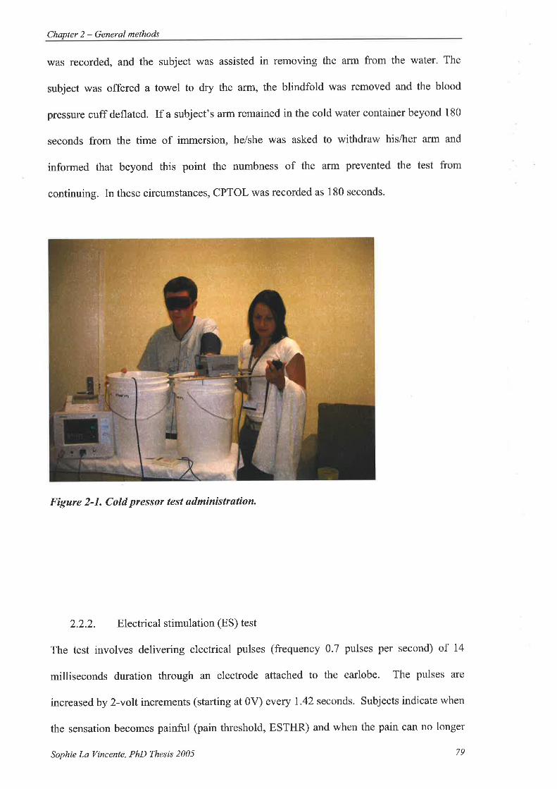
Chapler 2 - General methods
was recorded, and the subject was assisted in removing the arm from the water. The
subject was offered a towel to dry the arm, the blindfold was removed and the blood
pressure cuff deflated. If a subject's arm remained in the cold water container beyond 180
seconds from the time of immersion, he/she was asked to withdraw his/her arm and
informed that beyond this point the numbness of the afin prevented the test from
continuing. In these circumstances, CPTOL was recorded as 180 seconds.
Figure 2-1. Cold pressor test administrøtion.
2.2.2. Electrical stimulation (ES) test
The test involves delivering electrical pulses (frequency 0.7 pulses per second) of 14
milliseconds duration through an electrode attached to the earlobe. The pulses are
increased by 2-volt increments (starting at 0V) every 1.42 seconds. Subjects indicate when
the sensation becomes painful (pain threshold, ESTHR) and when the pain can no longer
Sophie La Vincente, PhD Thesis 2005 79

Chapter 2 - General methods
be tolerated (pain tolerance, ESTOL). Pain threshold and pain tolerance are recorded in
volts, with a maximum of 100 volts.
2,2.2.1. Materials
Conductive gel (Livingstone Conductive Gel, Livingstone International Pty Ltd,
Sydney, NSW, Australia)
Grass stimulator (Grass Instrument Co., Model S6C, Quincy, M.4., USA)
2.2.2.2. Set-upprocedure
The Grass stimulator is adjusted to the following settings outlined in Table 2-1 (also see
Figne2-2).
Frequencv 7 (x.1)
Delay 7 (x10)
Duration 14 ms (xl)Volts 0 (xlO)Output MonoPolarity NormalMode Repeat
Stimulus RezularTable 2-1. Grass slímuløtor settings for ES test.
2.2.2.3. Testadministration
The subject was seated in a comfortable chair and the procedure explained as follows:
This is the electrical stimulation test. It is a test of your tolerance to electrical pain. I
will smear some gel on your earlobe and then attach this earclip. The earclip is an
electrode connected to this machine [indicate], which delivers electrical current in
pulses. Wen you are ready to commence, I will ask you to close your eyes and will
start sending very low voltage curcent through the electrode. At first you won't feel
anything. I will slowly increase the voltage and you will begin to feel a sensation
through the clip. It won't hurt at first; it will feel like someone is lightly pinching your
Sophie La l/incente, PhD Thesis 2005 80

Chapter 2 - General melhods
earlobe in pulses. I will continue to slowly increase the voltage and I would like you to
tell me when that sensøtion becomes painful. I will then continue to increase the
voltage until you tell me you can no longer tolerate the pain, at which time I will
deactivate the machine and remove the earclip.
So, there are two things I'd like you to tell me: when the sensation you feel becomes
painful, and when you can no longer tolerate that pain. Aside from indicating these
things, do not speak during the test unless you have an urgent question or concern. I
will not speak to you during the test except to give you reminder instructions.
Electro-conductive gel was applied to the earlobe and electrode, and the electrode clipped
onto the lobe. The subject closed his/Ìrer eyes and upon verbally indicating readiness to
commence, the experimenter increased the pulses by 2-volt increments (starting at 0v)
every 1.42 seconds (as determined by the light indicator) (Figure 2-2). The subject
verbally indicated onset of pain (ESTHR). The experimenter then instructed the subject
"Now tell me when you can no longer tolerate the pain", aÍrd continued to increase the
voltage as described. The subject verbally indicated when the pain could no longer be
tolerated (ESTOL) and the stimulator was immediately turned off. The electrode was
removed from the earlobe and the gel wiped from both the earlobe and the electrode.
Sophie La Vincente, PhD Thesis 2005 81

Chapter 2 - General melhods
Fìgure 2-2. Electricul stimulation test sdministrution.
2.2.3. Procedures for repeated testing
In several of the studies described herein, subjects were tested repeatedly over one or
several days. In these circumstances the procedure for preparing the test and instructing
the subject varied, by necessity, from the procedures described above.
The descriptions provided to the subject prior to the first testing occasion are detailed and
lengthy, and not warranted on each occasion a subject repeats the test, particularly
considering the studies described herein required the subject to complete the test up to 13
times in one day. In these circumstances, the experimenter described the full details of the
test on the first testing occasion, and gave brief reminder instructions prior to each
subsequent testing.
Sophie La Vincente, PhD Thesis 2005 82

Chapter 2 - General methods
The set-up of the tests according to the details above was conducted at the beginning of a
testing day, and this apparatus used for testing at multiple time points throughout that day.
For the cold pressor, the thermoregulator maintained the warm water at the appropriate
temperature between testing occasions, and immediately prior to each testing occasion, the
cold water container was assessed by digital thermometer for temperature, and by visual
inspection for consistency. Ice was added to achieve the correct temperature and the same
consistency as at initial set-up. The electrical stimulation machine was switched off
between each testing occasion, and remaining conductive gel wiped from the ear clip.
2.2.4. Testing environment
Nociceptive testing during each study was conducted in the same environment, with
minimal background noise, audible voices and no clock with audible ticking. Ambient
room temperature and lighting was consistent for each study. At no time did the
experimenter discuss with the subject hislher performance on the test, or answer any
questions related to the average pain tolerance time or any previous results.
2.3. Methods common to drug studies (Chapters 4,5 and'6)
Common testing procedures were employed for all drug studies. Upon arrival at the
testing centre participants provided a urine sample, which was tested for drugs of abuse
(opioids, cannabinoids, benzodiazepines and sympathomimetic amines) by an independent
laboratory and, for female subjects, pregnancy. A 22 gatge indwelling venous catheter
(InsyterM) was inserted into the best available forearm vein on each arm (above the CP
immersion line for the non-dominant arm). A male luer lock adaptor injection site
(Interlink' Injection site, Baxter Healthcare Cotp, Deerheld, IL, USA) was attached to
each catheter. One catheter was used for blood sampling throughout the testing day, and
the other for the infusions. The participant was then connected to an Agilent A3@
Sophie La Vincente, PhD Thesis 2005 83

Chapter 2 - Generql methods
(Phillips) monitor, which was set to continuously monitor physiological parameters for the
duration of the testing day.
2.3.I. Drug administration
On each testing day, pafücipants received a 30-minute unblinded intravenous infusion of
saline, followed by one or more 30-minute drug (or placebo) infusions. The purpose of the
initial saline infusion was two-fold: to establish whether any changes in pain or
physiological parameters would occur as a response to the infusion process itself, and to
ensure that there was no obstruction to venous access via the catheter and the infusion
pump was operating correctly.
Infusions were administered using Graseby Syringe Pump 3100 (SIMS Graseby Ltd.,
Herts, UK) (see Figure 2-3). Drugs and saline were prepared by the Royal Adelaide
Hospital Pharmacy in 30ml BD Plastipak syringes. Infusions were run at a :late of 20ml
per hour for 30 minutes. Each syringe was attached to a minimum volume extension set
(150cm tubing, female luer lock, male luer lock, 0.5mll30cm) (Tuta Healthcare,Lane
Cove, NSV/ Australia). The male luer lock was attached to a lever lock cannula (BD
Interlink', Franklin Lakes, NJ, USA). The extension set was primed with the drug/saline,
and inserted into the injection site (Interlink' Injection site, Baxter Healthcare Corp,
Deerfield, IL, USA). In studies requiring the simultaneous infusion of two drugs (via one
cannula), a Y-type catheter extension set (Interlink' System Baxter Healthcare Co.p,
DeerFreld, IL, USA) with two injection sites was attached to the catheter, and the lever lock
cannulas (connected via the minimum volume extension set to each syringe) were inserted
in each of the injection sites.
Sophie La Vincente, PhD Thesis 2005 84

Chapter 2 - General methods
Figure 2-3. Graseby Syringe Pumps usedÍor drug infusions.
2.3.2. Testing time points
Measurements were taken at numerous time points during each testing day. Each testing
time point consisted of the following measures in the order listed: nausea and sedation
recorded, blood sample taken, physiological parameters recorded (pulse, oxygen saturation
and blood pressure), nociceptive testing completed, and respiration recorded (breaths per
minute counted for one full minute during wann water component of CP). The schedule
for testing time points is outlined below.
2.3.2.1. Blood sampling
Plasma samples taken during the studies were for use in an investigation unrelated to this
thesis. Blood sampling (10 ml on each occasion) was conducted immediately prior to
nociceptive testing throughout each testing day. Each sample was obtained via the catheter
inserted for blood sampling at the beginning of the testing day. Following each sample,
the catheter was flushed with 5ml saline. 'Where sampling from the catheter was not
possible, the blood sample was taken, with the subject's consent, by venepuncture.
Sophie La Vincente, PhD Thesis 2005 85
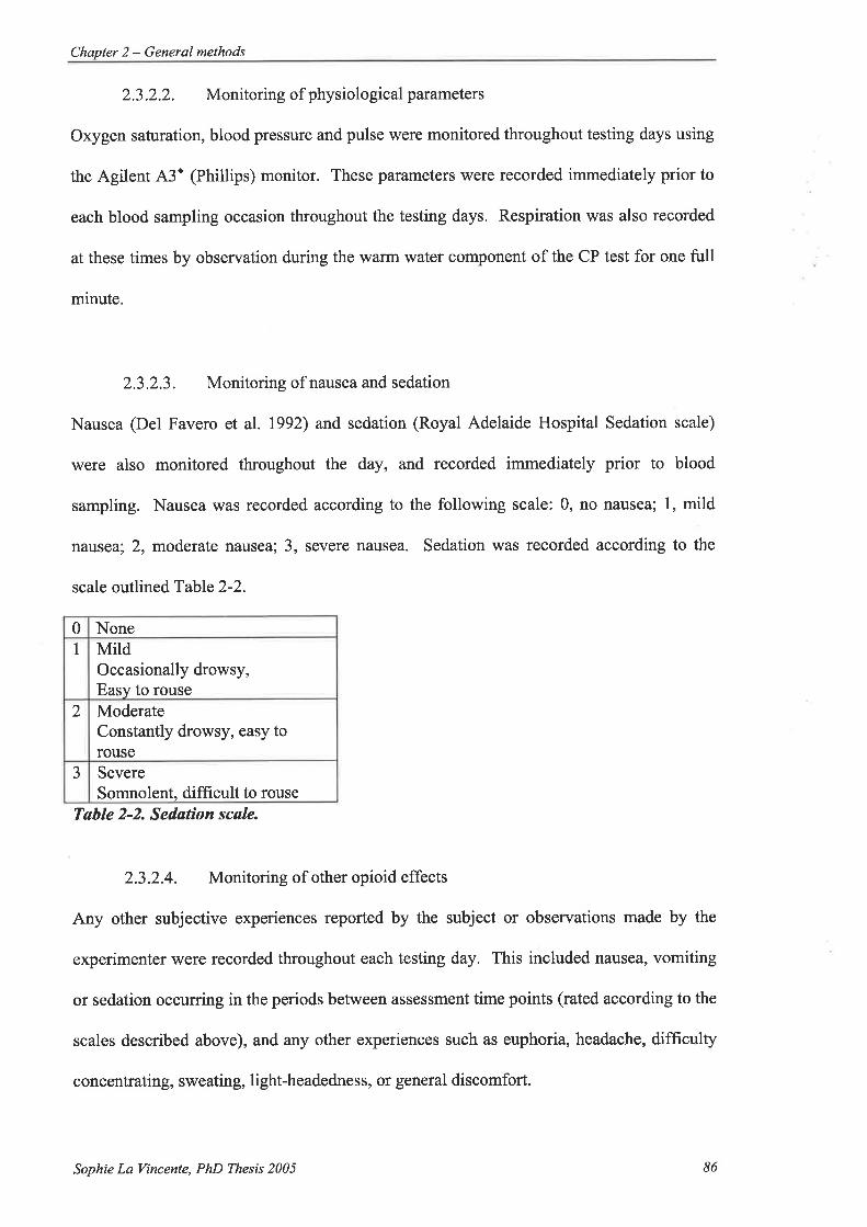
Chapter 2 - General methods
2.3.2.2. Monitoring ofphysiological parameters
Oxygen saturation, blood pressure and pulse were monitored throughout testing days using
the Agilent A3' (Phillips) monitor. These parameters were recorded immediately prior to
each blood sampling occasion throughout the testing days. Respiration was also recorded
at these times by observation during the warm water component of the CP test for one full
minute.
2.3.2.3. Monitoring of nausea and sedation
Nausea (Del Favero et al. 1992) and sedation (Royal Adelaide Hospital Sedation scale)
were also monitored throughout the day, and recorded immediately prior to blood
sampling. Nausea was recorded according to the following scale: 0, no nausea; 1, mild
nausea; 2, moderate nausea; 3, severe nausea. Sedation was recorded according to the
scale outlin ed T able 2-2.
0 NoneI Mild
Occasionally drowsy,Easy to rouse
2 ModerateConstantly drowsy, easy torouse
3 SevereSomnolent, difficult to rouse
Tøble 2-2. Sedatìon scøle.
2.3.2.4. Monitoring of other opioid effects
Any other subjective experiences reported by the subject or observations made by the
experimenter were recorded throughout each testing day. This included nausea, vomiting
or sedation occurring in the periods between assessment time points (rated according to the
scales described above), and any other experiences such as euphoria, headache, difficulty
concentrating, sweating, li ght-headedne s s, or general discomfort.
Sophie La Vincente, PhD Thesis 2005 86

Chapter 2 - General methods
2.3.2.5. Nociceptivetesting
Nociceptive testing was conducted as described above. To reduce the impact of any order
effects in studies using both CP and ES tests, participants were randomised to receive
either CP followed by ES, or vice versa, for the duration of the study.
2.3.3. Testing schedule
The above measures were taken at set intervals throughout each testing day. These testing
time points were as follows: l Prior to the commencement of infusions; 2. Twenty minutes
after the coÍtmencement of the 30 minute saline infusion; 3. Twenty minutes after the
commencement of the 30 minute drug infusionl, and hourly following the cessation of the
(last) drug infusion for a period of 6 (Study 2 and,3) or l0 (Study 4) hours. This is referred
to as the washout period. The purpose of conducting the testing 20 minutes after
commencing each 30 minute infusion was to allow time for testing to be completed before
starting the subsequent infusion.
2.4. Methods of statistical inference
Methods of statistical inference used in the current series of experiments are described for
each study in the relevant chapter. In several of the experiments, multiple tests of
statistical inference have been conducted with a set of data. The practice of adjusting the
alpha level used to determine statistical significance is a coÍtmon practice in cases where
multiple comparisons are undertaken within a data set (Tukey 1977). The rationale for
adjusting the alpha level is that the chance of Type I error (a false positive, that is, a
statistically significant (p<0.05) result occurring by chance) increases with the number of
1 The dose finding study described in Chapter 4 involved four consecutive 3}-minute drug infusions. The
testing battery was conducled 20 minutes afier the commencement of each infusion.
Sophie La Vincente, PhD Thesis 2005 87

Chapter 2 - General methods
comparisons made. However, the ethical and scientifrc validity of this practice has been
questioned, and is the subject of ongoing debate (Perneger 1998; Aickin 1999; Bender and
Lange L999;Perneger 1999). Several problems with the practice of alpha level adjustment
for multiple comparisons have been identifred (Perneger 1998). Firstly, it has been
proposed that the practice defies coÍrmon sense, creating a scenario whereby the number
of tests performed determines the findings of a study rather than the data. Moreover while
an alpha level adjustment can prevent an increase in Type I error rate, this entails an
increase in the rate of Type II error (a false negative, that is, not finding a significant
relationship where one does exist), which can lead to practical and ethical dilemmas.
Describing what tests have been used and why, allowing the reader to reach a reasonable
conclusion without the use of alpha level adjustment, has been proposed as the best
approach to addressing the problem. To avoid the problems described, an alpha level of
p<0.05 has been used throughout the studies described herein. 'Where relevant, the
potential impact of this approach to the clinical implications of the findings will be
considered in the interpretation of results.
Sophie La Tincente, PhD Thesis 2005 88

Chapter 3 - Normative study of nociceptive lests
3. ESTABLISHING NORMAL VALUES FOR THE COLD PRESSOR
TEST AND ELECTRICAL STIMULATION TEST IN HEALTHY
VOLUNTEERS
3.1. Introduction
3.1.1. The validity of experimental pain
The use of experimental pain models in the evaluation of analgesic interventions has for
many years been a challenging and contentious issue amongst clinicians and researchers
(Keats et al. 1950; Beecher 1953; Beecher 1957; Moore etaI. 1997). Perhaps the most
prominent of critics was Henry Beecher, who argued that experimentally induced pain was
qualitatively different from the pain produced by injury or disease (Beecher 1962). He
asserted that experimentally induced pain is without significance or meaning to the
individual, whereas pain that results from injury or disease has significance to the patient
and involves other parameters, most notably psychological factors such as anxiety.
While it is recognised that analgesic interventions must be evaluated in the clinical setting
to which they will be applied, experimental pain models capable of evaluating analgesic
efficacy under standardised conditions are of considerable utility and importance. As
described below, the experience of pain is subject to numerous sources of intra- and inter-
individual variability. Experimental pain research with pain-free humans allows a level
of control and standardisation not possible in a clinical pain setting (Gracely 1999). It has
been argued that investigations of dose-response, optimal dosage or comparison of relative
efficacy with known substances are best conducted with a sample of healtþ volunteers
who are as homogeneous as possible (Bromm 1985). The evaluation of analgesic
interventions in an experimental pain paradigm prior to assessment in a clinical pain
Sophie La Vincente, PhD Thesis 2005 89
Chapter 3 - Normative study of nociceptive tests
population allows far greater control over factors that may contribute to variability and
compromise the validity of findings.
Numerous pain induction techniques have been developed for use in experimental pain
studies with humans. These techniques can typically be classified according to the type of
pain induced, such as mechanical þressure), chemical, thermal (heat or cold), electrical
and ischaemic pain. The electrical stimulation (ES) and cold pressor (CP) tests were
selected for use in the series of experiments described herein as they each provoke a
different type of pain (tonic and phasic), both tests have consistently been demonstrated to
be sensitive to the antinociceptive effects of opioids, have been shown to produce reliable
and valid outcomes under controlled conditions, are applied easily, produce a distinct pain
sensation, are associated with rapid onset and ofßet of pain (though in the case of the CP
offset is more prolonged), are reproducible and suitable for multiple administration
(Gracely 1991). For example, a recent study compared the sensitivity of five pain tests to
clinical doses of the opioid alfentanil. Antinociceptive effects were observed with
electrical, CP and pressure pain while no significant antinociceptive effect was observed
with ischemic or heat pain (Luginbuhl et al. 2001).
3.1.2. Considerations in experimental pain induction
The development of new pharmacological strategies for pain management depends upon
techniques for the induction and accurate assessment of experimental pain in healtþ, pain-
free humans. The valid assessment of experimental pain is limited by several factors.
Firstly, the methods used for common pain induction techniques have not been
standardised. Consequently, there is a poor understanding of the normal pain response
using these techniques and it is difficult to make meaningful comparisons between findings
from different investigative groups (Eccleston 1995). Secondly, the sensitivity of a test in
Sophie La Vincente, PhD Thesis 2005 90
Chapter 3 - Normative study of nociceptive tests
detecting a drug effect can be compromised by inter- and intra-individual variation in pain
response. While a substantial body of research has been concerned with the impact of
biological and psychosocial factors on experimental pain response, we lack an appreciation
of the relative contribution of these variables to test performance and, moreover, to what
extent these factors should be taken into consideration in the design of experimental pain
studies in order to reduce inter-subject variability. To maximise both the statistical power
of a study and the likelihood of observing an analgesic effect, a pain induction technique
should be associated with minimal variation, both in terms of variation between
individuals, and variation within an individual over time.
Pain induction techniques typically have a maximum time or stimulus limit, after which
point the assessment of maximum tolerated pain (MTP) is no longer valid (for example,
due to numbness when inducing cold pain) or safe (for electrical stimulation or ischaemic
pain). For the evaluation of analgesic interventions an individual's baseline pain response
must be sufficiently below this ceiling to allow a significant increase to be measured.
There are two approaches by which the magnitude of MTP may be reduced: modiffing the
pain induction methods and excluding subjects whose baseline MTP is too close to the
censor point to allow a significant increase to be measured. Such an approach may induce
a bias in subject selection. An alternative is to exclude all potential subjects outside a
notional normal range, whether at the extreme high or extreme low ends of the
distributions. This approach has previously been employed (Eckhardt et al. 1998).
However, there have been no standardised data upon which to justiff the inclusion range.
In establishing the upper and lower boundaries of MTP for inclusion in an experimental
pain trial it is preferable that standardised data be available to assess individual
performance relative to the distribution within the population.
Sophie La Vincente, PhD Thesis 2005 9I

Chapter 3 - Normative study of nociceplive tests
3.1.3. Determinants of pain response
Numerous studies have examined the impact of biological (physiological and genetic) and
psychosocial variables on pain response, though in many cases findings have been
inconsistent. Much of this research has been instigated by clinical observations, such as
the apparent difference in pain sensitivity between men and women.
3.1.3.1. Sex
Sex differences in pain sensitivity and tolerance have been a major focus of pain research.
While it is often reported that there is minimal sex difference in terms of pain threshold,
pain tolerance is consistently higher in males compared to females (Berkley 1997; Riley et
al. 1998). Lower pain tolerance or greater pain report in females compared to males has
been demonstrated using araîge of pain induction techniques including the CP test (Walsh
et al. 1989; al'Absi ef al. 1999), noxious heat stimuli (Feine et al. 1991; Fillingim et al.
1998), pressure (Jensen et al. 1992; Chesterton et al. 2003) and mechanical pain (Sarlani
and Greenspan 2002). These differences may be attributed to a variety of factors, which
may include sociocultural influences such as gender role expectation (Robinson et al.
2001; Wise et al.2002) and participant and experimenter gender (Levine and De Simone
I99l; Robinson and 'Wise 2003; Kallai et al. 2004; Robinson and'Wise 2004) as well as
biological þhysiological and genetic) influences such as hormonal changes (Riley et al.
1999; Hellstrom and Lundberg 2000) and differences in central pain processing (Paulson et
al. 1998; France and Suchowiecki 1999;Naliboff et aL.2003; Sarlani etal.2004).
3.1.3.2. Ethnicityirace
Ethnicity, or race, has also been shown to influence tolerance to experimentally induced
pain. Studies of ethnicity and pain have focused predominantly on differences between
African-Americans and Caucasians, with early studies concluding that Caucasians
Sophie La Vincente, PhD Thesis 2005 92
Chapter 3 - Normative study ofnociceplive tests
demonstrated greater pain tolerance than African-Americans (Chapman and Jones 1944;
Woodrow et al. 1972). The results of more recent studies continue to support the notion of
differences in pain tolerance between ethnic groups (Edwards et al. 2001). Studies of
experimentally induced pain have generally not focused on Asian populations. In the late
1980s, Zatzick and Dimsdale conducted a review of studies investigating ethnicity
differences in pain (Zatzick and Dimsdale 1990). Thirteen English language articles
published between 1943 and 1989 were identified. Of these thirteen papers, only four
included an Asian group, and findings were inconsistent. Two of the studies reported
greater pain tolerance amongst Caucasians than Asians using cold pressor (Knox et al.
1977) and mechanical pressure tests (V/oodrow et al. 1972; Knox eI" al. 1977). One study
used small numbers of Nepalese (n:6) and Caucasians (n:5) and revealed a higher pain
threshold in' the Nepalese group. The fourth study reported a higher pain threshold
amongst North American Asians than North American Caucasians, but the latter group
evidenced a higher pain threshold than native Asians. These inconsistent results may be
attributed in part to different methods of pain induction, differences in subject numbers,
and variation in the operational definition of ethnicity as distinct from race. The
distinction between race and ethnicity is discussed in further detail below (section 3.6). A
recent study investigated differences in heat pain threshold between three East Asian
Ethnic groups (Malay, Indian and Chinese), and revealed no significant differences
between the groups (Yosipovitch et al. 2004).
3.1.3.3. Age
Investigations of age-related differences in response to noxious stimulation have generally
demonstrated that older subjects are less tolerant of experimentally induced pain
('Woodrow et al. 1972; Edwards et al. 2003). However, it has been observed that age-
related differences are influenced by the method of pain induction and the outcome
Sophie La Vincente, PhD Thesis 2005 93
Chapter 3 - Normative study of nociceptive tests
measure (for example, whether subjects are required to rate the intensity of the pain
elicited by a stimulus, or are asked to report at what point a stimulus becomes intolerable)
(Gibson and Helme 2001).
3.1r3.4. CNS stimulants
Studies examining the impact of stimulants such as caffeine and nicotine on pain response
have produced variable findings. An early study reported that cigarette smoking did not
influence performance on either the CP test or ES test (Sult and Moss 1986), while a 1993
study revealed that, amongst habitual smokers, elevated thermal pain threshold occurred
when they were cigarette-deprived (Pauli et al. 1993). A recent study revealed that the
administration of 250 mg of caffeine was associated with higher CP pain threshold and
tolerance than placebo (Keogh and Witt 2001), and a later study suggested that caffeine-
induced hypoalgesiamay be related to anxiety sensitivity (Keogh and Chaloner 2002).
3.1.3.5. Menstrual cycle
Several studies have also investigated the impact of menstrual cycle on response to
experimental pain stimuli. A meta-analytic review of 16 published studies revealed a
relatively consistent pattern of changes in pain sensitivity, but this was dependent upon the
modality of stimulus used. Studies using ES found that the luteal phase was associated
with higher pain threshold, while studies employing other pain induction techniques found
higher pain threshold and tolerance to be evident during the follicular phase (Riley et al.
1999). A recent study of 500 healtþ volunteers found no significant effect of menstrual
cycle on heat or cold pain (Kim et at.2004).
Sophie La Vincente, PhD Thesis 2005 94
Chapter 3 - Normalive study of nociceptive tests
3.1.3.6. Body weight/size
The role of body weight in pain response has been examined, with an early investigation
demonstrating that "obese" individuals were more sensitive to pressure pain than "non-
obese" individuals (McKendall and Haier 1983). The presence of an eating disorder such
as anorexia nervosa has also been associated with altered pain sensitivity (de Zwaan et al.
1996; de Zwaan et al. 1996; Raymond et al. 1999), though findings have been inconsistent
and it remains unclear whether any effect observed is a physiological correlate of the
disorder itself or related to body weight.
3.1.3.7. Psychological/cognitive factors
Psychological factors that have been considered include the role of attention (de Wied and
Verbaten 2}}L;Villemure and Bushnell2002), mood (Weisenberg et al. 1998) and coping
strategies (Baker and Kirsch 1991) in modiffing the experience of pain (see Chen et al.
1989; Fields 2000), as well as emotional constructs such as catastrophizing (Sullivan et al.
2001; Edwards et al.2004), fear of pain (Crombez et al. 1999; Vlaeyen and Linton 2000;
Keefe et al. 2004; Roelofs et al. 2004) and anxiety (Janssen and Arntz 1996; Rhudy and
Meagher 2000). Taken together, this research provides strong support for the notion that
cognitive factors impact considerably on the pain experienced in both clinical and
experimental settings, although the exact nafure of the effect on pain response, as well as
potential interactions with other variables such as gender, are yet to be clearly elucidated.
While numerous reports describe the impact of a variety of factors on pain response, absent
from the literature is a comparison of the relative contribution of these variables to
experimentally induced pain, and an indication of the degree to which these factors should
be controlled for in the desigu of experimental pain studies.
Sophie La Vincente, PhD Thesis 2005 95
Chapter 3 - Normative study of nociceptive tests
3.1.4. Cold pressor test
The cold pressor (CP) test was flrst studied as a pain induction technique in the 1940s
(Wolf and Hardy 1941) and established as a method of analgesic evaluation some years
later (Wolff et aL 1966; Wolff et al. 1966). The test is a tonic pain model, activating slow-
conducting, unmyelinated C-fibres. The pain experienced has been likened to dental or
back pain (Chen et al. 1989). There have been many variations on the CP technique, but
the test essentially involves the immersion of a limb (usually the hand or forearm) in a bath
of very cold water. The participant is generally required to indicate when pain is first
experienced (pain threshold), and when pain can no longer be tolerated (tolerance), or is
required to continuously rate pain throughout the procedure using a VAS. Threshold and
tolerance are measured in terms of latency (seconds) from initial immersion in the cold
water.
The CP test has become one of the most widely used methods of experimental pain
induction for the evaluation of analgesic interventions, including pharmacological
(Berntzen et al. 1985;Garcia de Jalon et al. 1985; Posner et al. 1985; Jones et al. 1988;
Compton 1994; Eckhardt et al. 1998; Yuan et al. 1998; Compton et al. 2000; Compton et
aL.2001; Doverty et al.2001; Compton et al. 2003), cognitive (Gilligan et al. 1984; Spanos
et al. 1984; Dolce et al. 1986) and other approaches (Hilgard et al. 1974; Ashton et al.
1984). The test has also increasingly been used to characterise pain response in different
groups, such as individuals with a current or past history of opioid-dependence (Compton
1994; Compton 7998; Compton et al. 2000; Compton et al. 2001; Doverty et al. 2001;
Doverty et al. 2001). The effect of NSAIDs on CP response has been inconsistent.
Therapeutic doses of ibuprofen (Jones et al. 1988) and indomethacin (Telekes et al. 1987)
have failed to increase VAS ratings of CP pain. However, in a double blind, placebo
controlled investigation, Yuan and colleagues demonstrated a significant increase in VAS
Sophie La Vincente, PhD Thesis 2005 96

Chapter 3 -Normative study ofnociceptive tesls
pain ratings with acetaminophen þaracetamol, 1000 mg p.o.) in 18 healthy volunteers
(Yuan et al. 1998). Compton and colleagues investigated the influence of sex on the effect
of ketorolac (10 mg p.o.) on CP pain tolerance in healthy volunteers, and revealed a
moderate (though not statistically signifrcant) analgesic effect in women. A considerable
increase in CP tolerance was evident in the male sample. However, this could not be
distinguished from the comparably large placebo effect (Compton et al. 2003). Studies
have consistently confirmed that the test is a highly sensitive assay for opioids, including
morphine (Wolff et al. 1966; Jones et al. 1988), dipipanone (Posner et al. 1985), and
codeine (Garcia de Jalon et al. 1985), and that this response can be distinguished from both
placebo (Posner et al. 1985; Jones et al. 1988) and non-opioid analgesics (Jones et al.
lgSS). Furthermore, the test is associated with high reproducibility for repeated
applications (Walsh et al. l9S9) and, in terms of subjective experience, the test is
considered comparable to clinical pain (Wolff 1984).
The lack of standardised procedures for the CP test provoked concern regarding the
reliability of the technique (Blasco and Bayes 1988). It was observed that there was
significant variation in the methods used by different investigative groups, and that these
procedural differences hampered replication of results and comparison between studies.
This lack of standardisation compromises the reliability of the test as a number of the
procedural differences relate to factors that may impact on test performance. These
methodological differences include a lack of consistency in the temperature of the cold
water, the immersion of the arm in warm water for a period of time prior to cold water
immersion, the proportion of the limb immersed (i.e. hand or forearm), the induction of
ischemia in the immersed arm by inflation of a blood pressure cuff, the laterality of the arm
used (i.e. dominant, non-dominant or unspecified), the instructions given to the subject,
subject blinding (i.e. eyes open, closed or blindfolded), the use of a water pump to
Sophie La Vincenle, PhD Tttesis 2005 97
Chapter 3 - Normative study ofnociceptive tests
circulate the cold watsr þreventing laminar wanning around the immersed limb), and the
outcome measures used (i.e. pain threshold and tolerance, or visual analogue scales of pain
intensity).
A 1989 normative model of the CP test established by V/alsh and colleagues represents the
only published study of this kind to date (V/alsh et al. 1989). The methods used in this
investigation required subjects to immerse the non-dominant hand and arm into cold water
(l-2 "C) until the pain could no longer be tolerated. Walsh and colleagues used Cox
regression to develop a normative mathematical model for CP pain tolerance according to
this simplified technique. This analysis revealed the best model for predicting
performance on the test contained the following covariates and interactions: sex, race
(Anglo-Saxon vs. non-Anglo-Saxon) by sex, sex by age, and race by age. Findings
indicated that, when controlling for age, pain tolerance was gteatest in Anglo-Saxon males,
followed by non-Anglo-Saxon males, Anglo-Saxon females and non-Anglo-Saxon
females. Since that time, however, several features have been incorporated by various
experimental groups (for example, Garcia de Jalon et al. 1985) to the current CP
methodology with the aim of reducing variability and mean tolerance time. Standardised,
normative data according to these methods have not been established.
3.1.5. Electrical stimulation test
The electrical stimulation (ES) test is a phasic pain model, activating fast-conducting Aô-
fibres, and associated with sharp, localised pain. Despite its extensive use in human pain
investigation, no noÍnative studies have been published. As with the CP test, a number of
methods have been used to induce pain by electrical stimulation in humans, varying both in
terms of the apparatus used to deliver electrical current and area of the body to which the
stimulus has been applied. Most commonly, ES has been applied cutaneously to the
Sophie La Vincente, PhD Thesis 2005 98
Chapter 3 - Normative study of nociceptive tests
finger, hand or earlobe, and has been relatively consistent in demonstrating sensitivity to
analgesic compounds. An early study by Wolff and colleagues demonstrated a significant
increase in tolerance to ES applied cutaneously to two fingers with the administration of 60
mg of codeine as compared to placebo (Wolff et al.1966). These findings were replicated
in a later study demonstrating a significantly greater increase in threshold and tolerance to
electrical stimulation of the earlobe with 60 mg codeine than placebo (Stacher et al. 1986).
A significant increase in analgesia compared to placebo has also been demonstrated using
the same method with a range of other compounds (Stacher et al. 1979; Stacher et al. 1982;
Stacher et al. 1983). As described above, Luginbuhl and colleagues also recently reported
that clinical doses of alfentanil were associated with significant analgesia to electrical
stimulation of the toe (Luginbuhl et al. 2001). Evidence of a signifrcant increase in ES
performance compared to placebo with the administration of NSAIDs has been less
consistent. Several studies have reported a significant effect (Stacher et al. 1979; Stacher
et a|. 1986; Walker et al. 1993; Walker and Carmody 1998), while others have failed to
detect an increase in ES response (Wolff et al. 1966; Moore et al.797l; von Graffenried et
al. 1978). It has been suggested that this failure to detect a significant effect may be due to
insufficient subject numbers, given the substantial variability in response to analgesic
drugs (Stacher et al. 1986).
There are several advantages associated with the application of electrical current to the
earlobe in ES testing. In comparison with other areas of the body that have previously
been used, it has been argued that the use of the earlobe offers advantages in reducing
stimulus detection threshold and reducing variability by minimising individual differences
and confounding factors (Walker et al. 1993; Walker and Carmody 1998). For example,
results from stimulation of sites that overlie muscle (e.g. the hand) may be compromised
due to muscle contraction. Variation in skin thickness may also be minimised with use of
Sophie La Vincente, PhD Thesis 2005 99

Chapter 3 - Normative study of nociceptive tests
the earlobe. For example, " /ith use of the hand, skin thickness may vary considerably
according to a number of factors, including the sex of the subject and participation in
occupational or recreation activities involving manual activity. The impact of stress-
induced sweating may also be reduced with use of the earlobe'
The application of ES to the earlobe has recently been used in a number of studies of pain
response in opioid dependent individuals and healtþ volunteers. Dyer and colleagues
assessed the antinociceptive activity of methadone prescribed as a maintenance
pharmacotherapy to opioid dependent individuals. This study reported a decrease in pain
sensitivity with increasing plasma methadone concentrations, further substantiating both
the capacity of the ES method to evaluate the antinociceptive activity of opioids, and the
reproducibility of the technique (Dyer et al. 1999). Subsequent studies have also applied
this technique to opioid-dependent populations in comparing pain response to that of
healthy volunteers (Dyer et aI. 1999; Doverty et al. 2001).
3.2. Purpose and aims of the present study
The aims of the present study were: to demonstrate the distribution of values for the CP
and ES tests, which may guide in the selection of subjects in future studies; to establish
whether the current methods produce less variation and a lower mean than earlier
techniques; to determine the replicability of the tests, and to assess the factors that may be
important to control for when using these techniques in order to minimise variability.
Sophie La Vincente, PhD Thesis 2005 100
Chapter 3 - Normative study of nociceptive tests
3.3. Methods
3.3.1. Participants
This study was conducted with the approval of the Research Ethics Committee of the
Royal Adelaide Hospital, South Australia ßAH Protocol 011119). Subjects were 100
healtþ, drug-free volunteers, who met the criteria detailed below. Participation in the
study was on a voluntary basis. Participants were financially remunerated $AUl5 for
participation. Participants who completed a second testing session were remunerated
$4U40 for the two sessions (i.e. $4U15 for the first session, $4U25 for the second). The
pulpose of offering higher remuneration for the second tesiing session was to enhance the
incentive to return, and to reduce a potential bias in the sample by attracting only the
subjects who did not find the tests overly aversive. Written informed consent was obtained
from all participants prior to commencing the trial.
3.3.1.1. Inclusion criteria
Aged between 18 and 65 years
Males and females in equal numbers
Signed informed consent provided
3.3.1.2. Exclusioncriteria
o A history of opioid dependence according to DSM-IV criteria
o History of significant chronic pain
. Prior chronic opioid use (in excess of one week)
o Current regular use of any other drug, including recreational and non-prescription
drugs such as allergy medication (with the exception of the contraceptive pill) (Ne
Regular use considered in excess of once per weeþ
Sophie La Vincente, PhD Thesis 2005 101
a

a
a
Chapter 3 - Normalive studv ofnociceptive tests
Any history of substance dependence
Alcohol consumption exceeding National Health and Medical Research Council
(NHMRC) guidelines
Prior knowledge of, or exposure to, the CP or ES tests
Suffering from Raynaud's Syndrome
3.4. Procedures
3.4.1. Recruitment and screening procedures
Participants were recruited using an advertisement flyer posted in key areas (e.g. university
and hospital notice boards), and through word of mouth. The experimenter conducted a
brief screening interview with each participant prior to organising the testing session.
3.4.2. Experimentalprocedures
The subject was provided with a study information sheet, and was given the opportunity to
read the details.and purpose of the study, discuss the procedures with the investigator and
ask any questions. All participants attended the testing centre on an individual basis for
one testing session of approximately 20 minutes. Participants were instructed to refrain
from taking any drug (excluding nicotine, alcohol and the contraceptive pill) in the 24
hours prior to testing. Testing was conducted in a quiet room at controlled temperature
(approximately 23oC), with only the investigator present. Participants completed a
questionnaire providing demographic information, including age, sex, ethnicity, average
daily alcohol and caffeine consumption, cigarette smoking, and phase of menstrual cycle
(where appropriate). Height and weight were recorded and body mass index (BMI)
calculated (weight (kg)/height (r")t). Average daily alcohol and caffeine consumption and
Sophie La Yincente, PhD Thesis 2005 102
a

Chapter 3 - Normative study of nociceplive tests
cigarette smoking were established by the questions displayed in Table 3-1. Caffeine
consumption was calculated as follows: instant coffee 95 mg; brewed/percolated coffee
135 mg; espresso coffee 100 mg ; decaffeinated coffee 3 -g ; tea 50 mg. These values
were based on the rangos determined by Bunker and McWilliams (Bunker and
McWilliams 1979). Data were collected between March and May 2002.
you smoke cigarettes? yes / no
if yes, on average how many do you smoke per day? (a) less than l0þ) between 10 and 20(c) between 20 and 30
(d) between 30 and 40
(e) more than 40
the past 30 days, on how many days did you have at least one alcoholic drink?(a) 0 days(b) 1 or 2days(c)3to5days(d)6to9days(e) 10 to 19 days
(f) 20 to 29 days
(g) every day
these days, how many drinks did you have on average?(a)1to2(b)2to5(c)5to8(d) more than eight
the past 30 days, on how many days did you have 5 or more drinks in a row
is, within a few hours)?(a) 0 days(b) I day(c) 2 days
(d)3to5days(e)6to9days(f) l0 to 19 days
(g) 20 or more days
write in each box the number of cups of each "type" of coffee you would
normally drink in a day
[instant
f lun8er/nercolate
l"rp."rtoldecaffeinated
many cups of tea would you normally drink in a day?
Tsbte 3-7. Assessment oÍ øverage alcohol ønd caffeine consumption and cigørette
smok¡ng.
Sophie La Vincente, PhD Thesis 2005 r03
Chapter 3 - Normative study of nociceplive tests
Phase of menstrual cycle \¡/as recorded for those female participants who reported a regular
menstrual cycle (n:44). Regularity of menstrual cycle was established by self-report, with
subjects indicating whether they had four menses in the previous six months, and if they
considered their cycle to be regular. For those reporting a regular cycle, phase at testing
was determined by self-report, with phases categorised according to the following
classification: menstrual (days 1-5), follicular (days 6-12), ovulation (days 13-16) and
luteal (days 17-28) (Sherwood 1997).
A skinfold thickness measure was taken around the volar aspect of the right forearm using
skinfold calipers (Holtain Ltd., Crymych, UK). This was performed on three consecutive
occasions and a mean calculated. This variable was assessed as skinfold thickness around
the arm was considered to be a potential source of variability in CP performance.
Participants then completed the State Trait Anxiety Questionnaire (STAD (Spielberger
19S3). This inventory independently assesses two constructs of anxiety: state anxiety and
trait anxiety, using two 20-item scales. The state anxiety scale assesses sifuational anxiety,
requiring subjects to respond to a series of items according to how they feel "right now at
this moment". The trait anxiety scale aims to assess the more enduring, stable personality
trait of proneness to anxiety. This scale requires participants to respond to items according
to how they "generally feel". Each scale has a possible scoring range of 20 - 80, with a
higher score indicative of greater anxiety. This inventory is used widely in research and
clinical practice, and has been demonstrated to be psychometrically sound (Spielberger
re83).
Participants then completed the nociceptive tests, the CP and ES, once in a randomised
order. These tests were conducted according to the methods described in Chapter 2.
Following the nociceptive testing, pafücipants completed the Fear of Pain Questionnaire-
III (FPQ-III). The FPQ-III is a 3O-item questionnaire assessing the degree to which an
Sophie La Vincente, PhD Thesis 2005 104
Chapter 3 - Normative study of nociceptive lesls
individual fears the pain associated with a raîge of different experiences. Participants
indicate how much they fear the pain associated with a range of situations, such as
breaking an arrn or receiving an injection, by rating a 5-point Likert-type scale with the
categories "not at all", "a little", "a fair amount", "a lot" and "extreme"' Scores may be
summed to produce a Total Fear of Pain score (with a possible scoring range of 30-150), or
may be grouped into three subscales of l0-items each: Fear of Medical Pain, Fear of
Severe Pain, and Fear of Minor Pain. The FPQ-III has been shown to be psychometrically
sound, demonstrating high reliability and validity (McNeil and Rainwater 1998).
To determine the replicability of the techniques, a cohort of thirty subjects completed the
pain tests a second time, between 14 and2l days after the initial session.
3.4.3. Statistical analyses
Statistical analyses were conducted using SPSS@ for Windows version 11.5 (SPSS Inc.,
Chicago, United States of America). To assess the intra-subject variability between two
sessions, 'Wilcoxon signed rank tests for related samples were conducted to identiff
differences in nociceptive test performance between first and second testing for the cohort
of 30 subjects who completed two sessions. This non-parametric test was used due to
deviations from the normal distribution and the presence of censored data as a result of the
test upper limits. A number of subjects did not report reaching MTP before the upper limit
of the tests (180 seconds for CP, 100 volts for ES), and are therefore censored data. The
percent change between first and second testing for both CP tolerance and ES tolerance
were also calculated for each of the 30 returning subjects. Survival analyses (Cox
proportional hazards regression) were conducted for CP tolerance and ES tolerance to
assess the relative contribution to test performance of the biological and psychosocial
variables assessed. The presence of censored data necessitated the use of survival analysis
Sophie La Vincente, PhD Thesis 2005 105
Chapter 3 - Normative study of nociceptive lests
to determine the relative contribution of each variable to test performance. Cox regression
assesses the effect of each variable on the tikelihood of a subject "suryiving" until the
maximum limit of the test, that is, not rcaching pain tolerance during the observation
period. To determine whether there were any interactive effects between the variables,
Cox regression analysis was performed with the main effects and interaction terms for each
possible pair of variables. Where significant interactions rürere detected, the interaction
term was also entered into the full Cox regtession.
Among the female participants who reported a regular menstrual cycle, no data were
censored due to reaching the maximum limit. To analyse the effect of menstrual phase on
test performance among this cohort, simple linear regression with self-reported phase of
menstrual cycle at time of testing was conducted. Unless otherwise indicated, data are
described as mean (tSEM, interquartile range).
3.5. Results
3.5.1. Sample characteristics
The sample comprised 100 healthy unrelated volunteers (50:50 sex ratio), who ranged in
age from 18 to 56 (26.1+0.88) years, with a BMI between l6 and 42 (23+0.44), and body
weight between 40 and 108 kg (66.8+1.5). Ethnicity was established by self-identification,
with 63 identiffing as Caucasian and 37 identifuing as Asian. Subject demographics are
detailed in Table 3-2. The proportion of female subjects at each phase of the menstrual
cycle at first testing is detailed in Table 3-3.
Sophie La Vincente, PhD Thesis 2005 106

Chapter 3 - Normative study of nociceptive tests
Mean +SEM Median IQRAse (vears) 26.11 0.88 24.00 8.00
BMI 23.15 0.44 22.50 5.18
Skinfold thick.(mm)
6.84 0.26 6.37 3.19
Daily caffeine(me)
148.55 14.8 135.00 2tr.25
FPQ-III (total) 88.30 1.63 88.00 21.75
STAI Trait 35.85 0.81 3s.50 17.75
STAI State 32.47 0.81 32.00 r0.75
Ethnicity Caucasian63Yo Asian3T%;o
CigarettesmokerIfyes, howmanylday?
Yes 10% No 90%
< l0: 9o/o
10-20: I%o
Alcohol
During the past 30days, on how manydays did you haveat least onealcoholic drink?(n:100)
On these days, howmany drinks didyou have onaverage? (n:66)
During the past 30days, on how manydays did you have5 or more drinks ina row (n:100)
Vo Vo
0 days 34 10 to 19 days 9
1 or 2 days 16 20 to 29 days 2
3 to 5 days t9 every day 0
6 to 9 days 20
Ito2 37.8 5to8 4.5
2to 5 56. I >8 1.5
0 davs 6l 6 to 9 days J
1 day 11 l0 to 19 days 0
2 days 6 20 or > days 0
3 to 5 days 19
IQR Interquartile range; BMI Body mass Index (kg)/height (m)2; FPQ-III Fear of Pain
Questionnaire-Ill, scoring range 30-150; STAI State Trait Anxiety Inventory, Trait and State
subscale scores, scoring range 20-80 on each scale. See section 3.4.2 for descriotion of measures.
Table 3-2. Demogrøphic parametersInterquarlile range.
of normative stady sample (n:100). IQR,
N Vo
Menstrual 5 10
Follicular 74 28
Ovulation 5 10
Luteal 20 40
Irregular (or absent due tocontraception)
4 8
Absent due to hysterectomy 2 4
Table 3-3. Phase of menstrual cycle reported by femøle subjects (n:50) al ínítisl testing.
Sophie La Vincente, PhD Thesis 2005 107

Chapter 3 - Normative sludy of nociceptive tests
As described, a cohort of 30 participants completed a second testing between 14 and 2l
days later. Prior to testing, participants were offered the opportunity to enrol for one or
two testing occasions. The offer of a second testing occasion was made to all participants
until 15 males and 15 females had been recruited for two testing sessions. All subjects
who initially agreed to participate in a second testing session did so. The cohort of 30 who
were tested on two occasions ranged in age from 18 to 56 (25.3+1.69), with a BMI
between 17.3 and 42 (23.28t0.91). There were no significant differences between those
who participated in one or two testing sessions in terms of age, BMI, pain parameters,
average alcohol or caffeine consumption, fear of pain or cigarette smoking. There was,
however, a significant difference between those who participated in one or two testing
sessions in terms of state anxiety as measured by the STAI, such that those who agreed to
return for a second testing reported lower state anxiety than those who enrolled for one
testing occasion only (see Table 3-4). However, a comparison of only the subjects who
were offeredthe opportunity of returning for a second session (i.e. until 15 males and 15
females had been recruited for two sessions) revealed no significant differences between
those who did and did not enrol for two sessions (see Table 3-5). This suggests that the
difference in state anxiety between the sample returning for a second testing (n:30) and
those participating in only one testing session (n:70) does not reflect a sampling bias.
Sophie La Vincente, PhD Thesis 2005 108
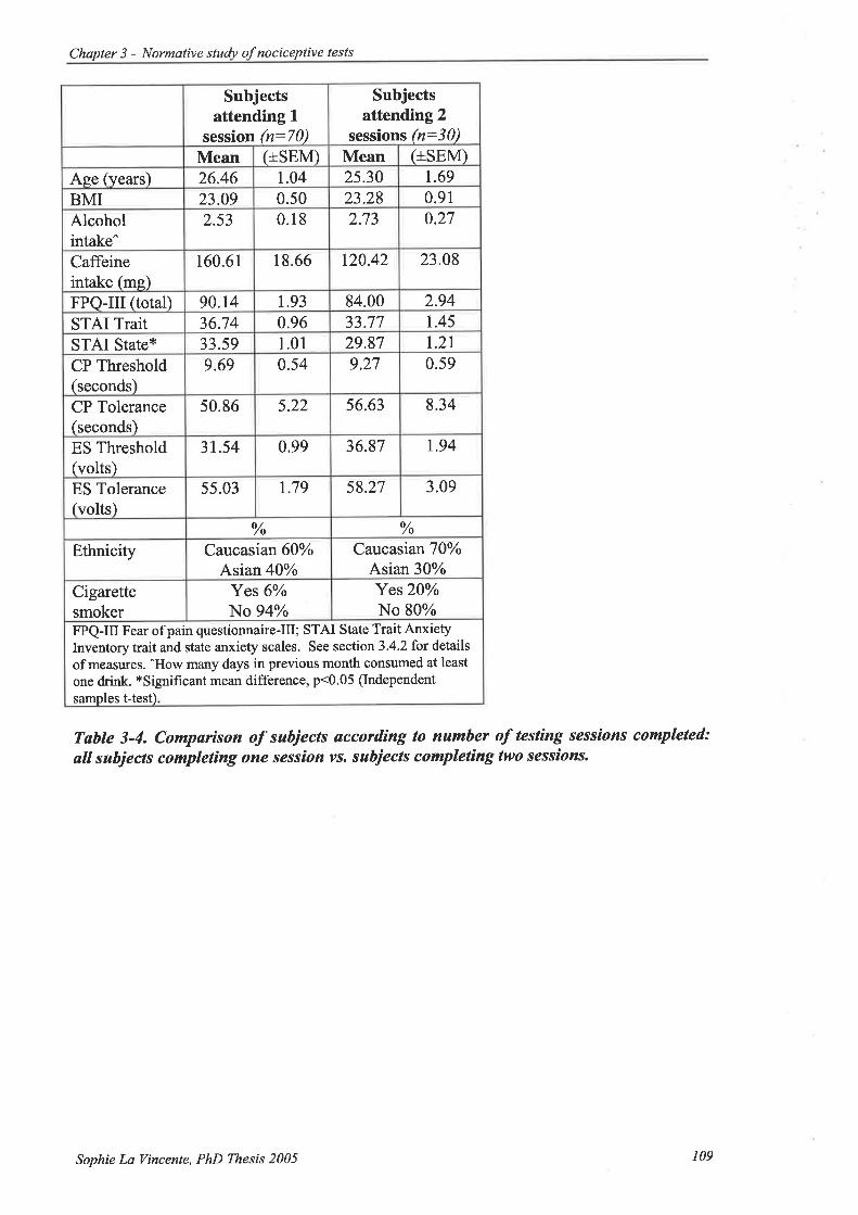
Chapter 3 - Normative study of nociceptive tests
Subjectsattending L
session (n:70)
Subjectsattending 2
sessions (n:30)Mean (+sEM) Mean (+sEM)
Aee (years) 26.46 1.04 2s.30 1.69
BMI 23.09 0.50 23.28 0.91
Alcoholintake^
2.53 0.18 2.73 0.27
Caffeineintake (mg)
160.61 18.66 r20.42 23.08
FPQ-III (total) 90.14 1.93 84.00 2.94
STAI Trait 36.74 0.96 33.77 1.45
STAI State* 33.59 1.01 29.87 1.21
CP Threshold(seconds)
9.69 0.54 9.27 0.59
CP Tolerance(seconds)
50.86 5.22 56.63 8.34
ES Threshold(volts)
31.54 0.99 36.87 1.94
ES Tolerance(volts)
55.03 1.79 58.27 3.09
o//o o//o
Ethnicity Caucasian609ioAsian40%;o
CatcasianT0o/oAsian 30%
Cigarettesmoker
Yes 6%o
No 94%Yes20VoNo 80%
FPQ-III Fear of pain questionnaire-Ill; STAI State Trait AnxietyInventory trait and state anxiety scales. See section 3.4.2 fot details
of measures. ^How many days in previous month consumed at least
one d¡ink. *Signifrcant mean difference, p<0.05 (Independent
samples t-test)
Table 3-4. Comparison of subjects øccordìng to number of testing sessions completed:
ølI sabjects complet¡ng one session vs. subiects completìng two sessions.
Sophie La Vincente, PhD Thesis 2005 109

Chapter 3 - Normative stuþ of nociceptive tesls
Subjects offered2"d session,
performed 1
orùy (n:26)
Subjectsperforming 2
sessions (n:30)
Mean (+sEM) Mean (+sEM)
Aee (years) 24.77 1.48 2s.30 1.69
BMI 23.34 0.91 23.28 0.91
Alcoholintake^
2.50 0.29 2.73 0.27
Caffeineintake (me)
142.88 26.02 120.42 23.08
FPQ-III (total) 88.08 2.97 84.00 2.94
STAI Trait 36.3r 1.56 33.77 1.45
STAI State 32.00 1.53 29.87 t.2lCP Threshold(seconds)
10.77 1.22 9.27 0.59
CP Tolerance(seconds)
60.04 9.74 56.63 8.34
ES Threshold(volts)
33.08 1.18 36.87 1.94
ES Tolerance(volts)
53.62 2.69 58.27 3.09
% o//o
Ethnicity Catcasian53%ioAsian 47o/o
CaucasianT0qioAsian 30%
Cigarettesmoker
Yes 8%No 92%
Yes20%oNo 80%
FPQ-III Fear of pain questionnaire-Ill; STAI State Trait AnxietyInventory trait and state anxiety scales. See section 3.4'2 for details
of measures, ^How many days in previous month consumed at least
one drink. No significant mean differences (Independent samples t-test)
Table 3-5. Comparíson of subjects øccordìng to numher of testing sessions completed:
subjects completìng one session but gìven option of 2nd vs. suhiects completing two
sessions.
3.5.2. Normative data
Normative data and frequency distributions for the pain tests in all 100 subjects are
displayed in Table 3-6 and Figure 3-1. Distributions for ES threshold and tolerance
approximated a normal distribution, with both skewness (0.8 and 0.9, respectively) and
kurtosis (0.8 and 0.5) near zero. CP threshold and tolerance distributions demonstrated a
moderate degree of positive skew (1.6 and 1.8, respectively) and kurtosis (4.5 and 2.3,
respectively).
Sophie La Vincente, PhD Thesis 2005 110

Chapter 3 - Normative study of nociceptive tests
Mean SD 95Vo ClUpperLower
Median IQR CY (7o) S K
Cold PressorThreshold (seconds) 9.6 4.1 8.7 10.4 9.0 4.0 42.7 1.6 4.5
Tolerance lseconds) 52.6 44.1 43.8 61.3 35.5 31.8 83.8 1.8 2.3
Electrical StimulationThreshold (volts) 35.9 9.0 34.1 37.3 35,0 t2.o 25.1 0.8 0.8
Tolerance (volts) s6.0 15.6 52.9 59.9 s3.0 21.5 27.9 0,9 0.5
SD St¿ndard deviation; CI Confrdence Intervals; IQR Interquadile range; CV Coefficient of variation;
S Skewness: K Kurtosis.
Tahle 3-6. Descrþtive datø of cold pressor and electrical stimuløtion test patameterc
(n:100). Note that due to the potential for censoríng on CP and ES tolersnce' med¡øn
and IQR more accarøtely rE resent the dístribution for these parømeters thøn meøn and
^sD.
oo
Il!
a)
o=Ilt
Cold Pressor Threshold
s ry Þ 6 6 *s "$ J *6 *5 û,9ù rV. ry.
Seconds
Cold Pressor Tolerance
oeoÞs"o.o*SSoSoSo*o"Seconds
Electrical St¡mulat¡on Threshold
s loNs*r.vo* ,o,f vsI,,rst?os8,\oVolts
Electrica I Stimulat¡on Tolerance
G *s rys ,'rs vs oro os ,ts os os $Volts
o(¡)
aott t0
()tr(,fIlt
Figare 3-1. Frequency d¡sî¡but¡ons for cold pressor ønd electrícal sthnulatíon test
parameteß in 100 heølthy volanteers. Note that the values of 180 seconds for CP
tolerønce ønd 100 volts Íor ES tolerønce tepresent censored døtø (maximum l¡m¡ts).
Sophie Lø Vincente, PhD Thesis 2005 111
Chapter 3 - Normative study of nociceptive lests
3.5.3. Intra-subjectvariability
There were no statistically significant differences between testing sessions for CPTHR (1't:
9.3 (t0.6, 4.3) seconds; 2nd: 8.7 (t0.7, 5.0) seconds; z:-0.51, p:0.61); CPTOL (l't: 56.6
(+8.3, 44.0) seconds; 2od: 52.6 (+7.7,39.3) seconds; z:-1.034, p:0.30); ESTHR (l't: 36.9
(+1.9, 15.0) volts;2"d;35.7 (+1.5, 13.5) volts; z:-0.822, p:0.41) or ESTOL (l't: 58.3
(+3.1,25.0) volts; 2"d: 58.I (12.9,23.0) volts; z:-0.239, p:0.81). The mean percent
change between first and second testing was 15.1% (tSEM 2.37) and ll.6% (tSEM 1'72)
for CPTOL and ESTOL, respectively.
3.5.4. Factors impacting upon test performance
3.5.4.1. Cold pressor tolerance (CPTOL)
There were no significant interactions between variables in CPTOL. Eleven variables
were entered in the Cox regression model for CPTOL: sex, age, BMI, skin-fold thickness,
ethnicity, cigarette smoking, average alcohol and caffeine consumption, fear of pain, state
anxiety and trait anxiety. Two of these variables were significant predictors of survival on
the CP test after adjustment for other variables in the model: fear of pain and sex (see
Figure 3-2). The likelihood of an individual reaching pain tolerance within the observation
period (i.e. not being censored) increased by 1.027 times for every unit increase in total
FPQ score (exp(0.026):1.027, p:0.001). For the predictor variable sex, females were
approximately half as likely to reach the pain tolerance cut-off point, relative to males
(exp(-0. 7 89):0.45 4, p:0. 005).
Sophie La Vincente, PhD Thesis 2005 It2

Chapter 3 - Normative study of nociceptive tests
A
---e-- Low fear----+- Med fear* High fear
0 20 40 60 80 100 120 140 160 180 200
Cold pressor tolerance (seconds)
B
----.- Male---s- Female
0 20 40 60 80 100 120 140 160 180 200
Cold pressor tolerance (seconds)
Figure 3-2. Percent survivøI on the CP test by (Q feør of pøín (FPQ-IID ønd (þ sex in100 heøtthy volunteers. Note that, for the pu{pose of clarity, the FPQ-^III variable has been
converted to a categorical variable. This was performed by the SPSS@ categorize variable
function, producing three variables: low (n:34), medium (n:33) and high (n:33) fear ofpain. Survival analyses were conducted with the raw data (continuous variable).
1
1
(E
.¿
os
1
1
(ú.¿
Ø
s
Sophie La Tincente, PhD Thesis 2005 113
Chapter 3 - Normalive study of nociceptive tests
3.5.4.2. Electrical stimulation tolerance (ESTOL)
A signifrcant interaction effect for ESTOL r¡/as detected between ethnicity and sex
(F1,e6:5.87, p:0.017). Ten' individual variables (sex, age, BMI, ethnicity, cigarette
smoking, alcohol and caffeine consumption, fear of pain, state anxiety and trait anxiety)
and one interaction term (sex*ethnicity) were entered into the Cox regression for ESTOL.
Fear of pain and the sex*ethnicity interaction were found to significantly predict survival
on the ES test (see Figure 3-3). The likelihood of reaching pain tolerance prior to the cut-
off point increasing by 1.021 times for every unit increase in FPQ score, after adjustment
for the effects of the other variables in the model (exp(0.021):7.021, p:0.006). The
interaction between ethnicity and sex also contributed signif,rcantly to ESTOL (e*p(-
1.53):0.216, p:0.004). This interaction indicated that while Caucasian females were less
tolerant of ES pain than Caucasian males, Asian females were more tolerant of ES pain
than Asian males.
'Skin¡old thiclcness was excludedfrom the electrical stimulation tolerance regression as this variable was
taken around the þrearm qnd considered only 1o be a potentiøl source of variance in cold pressor
perþrmance.
Sophie La Vincente, PhD Thesß 2005 114

Chapter 3 - Normative studv of nociceptive tests
A
----e- Low fear
--+- Med fear
- High fear
01020304Ú 50607080$100Electrical stimulation tolerance (volts)
B
-+- Male Caucasian---e- Male Asian----t- Female Caucasian+- Female Asian
0102û 30Æ$607080901Electrical stimulation tolerance (volts)
00
Figure 3-3. Percent survìval on the ES test by (Ð feør of paìn (FPQ-III) and (þ sex
ethnicity interøctìon ùn 100 heøtthy volunteers. Note that, for the purpose of clarity, the
FPQ-III variable has been converted to a categorical variable. This was performed by the
SPSS@ categonze variable function, producing three variables: Low (n:34), medium
(n:33) and high (n:33) fear of pain. Survival analyses were conducted with the raw data
(continuous variable).
I
1
50
õL
|t,
àe
1
1
G
¿ãID
s
Sophie LaVincente, PhD Thesß 2005 115
Chapter 3 - Normative study of nociceptive tests
3.5.4.3. Impact of menstrual phase
Menstrual phase was not a significant predictor of CPTOL or ESTOL in the 43 normally
menstruating females subjects. Simple linear regression analysis revealed that menstrual
phase described 3.5Yo of variance in CPTOL (*u¿¡:1.2) (Fr,¿r:l .50,p:0.228) and l'9%o of
variance in ESTOL $3"4:0.5) (F1,a1:0.78, p:0.382).
3.5.4.4. Test order effects
There were no significant differences on any nociceptive parameter according to the order
in which the two tests were performed. Independent samples t-tests of CPTHR and
ESTHR revealed no significant differences in response between those who completed the
CP followed by the ES test, and those who completed the tests in the reverse order (CP:
t(98):0.75, p:0.45; ES: t(98):-0.19, p:0.84). Mann-'Whitney U tests revealed no test
order effects for CPTOL (2:-1.55, p:0.12) or ESTOL (2:-0.27,p:0.79).
3.6. Discussion
The central aims of this study were to establish normal values for two human pain
induction techniques, and to determine which factors should be controlled for in the
selection of subjects for clinical trials of healtþ, pain-free humans using these techniques.
The CP and ES tests are commonly used techniques, but the methods used for each vary
considerably between investigative groups. Standardisation enables comparison between
studies, and provides an index of normal values by which to gauge performance.
Moreover, due to the maximum limit imposed on pain induction tests, it is imperative
when investigating an analgesic intervention that a subject's baseline MTP is sufficiently
below the maximum limit to allow a significant increase to be measured. In this respect,
the establishment of a normal response range for these tests provides a basis from which to
impose inclusion criteria for the upper and lower boundaries of pain response. On the
Sophie La Vincente, PhD Thesis 2005 I 16

Chapter 3 - Normative study ofnociceptive lests
basis of these normative data, a baseline MTP cut-off in subject selection of one standard
deviation above and below the log mean for each test was decided upon for use in
subsequent studies. This produces a range of 2l-85 seconds and 42-68 volts for subject
inclusion on the CP and ES tests, respectively. This range was selected as the upper limit
permits an increase of 50Yo in ESTOL and over 100% in CPTOL before the censor point
on each test. In a previous study of healthy volunteers, a steady state plasma morphine
concentration of 23 nglml was associated with mean increase in CPTOL of 52%o and in
ESTOL of l5%o (Athanasos et aI. 2002). The exclusion of subjects according to baseline
response to experimental pain induction has previously been reported in a study of the CP
test. However, the inclusion range in that study was not based on standardised data
(Eckhardt et al. 1998).
Modification of the pain induction technique is another approach by which mean MTP
may be reduced. The CP method used in the present study was adapted from Eckhardt and
colleagues (Eckhardt et al. 1998), and was developed with the aim of reducing intra-
subject variability and reducing the mean MTP. The results suggest that these measures
have been effective in reducing CP MTP, with the mean tolerance time associated with the
current method (52.6 seconds) being considerably lower than the mean withdrawal latency
reported in healtþ volunteers using the simplifred method, which has been as high as 138
seconds in healthy controls (Compton et al. 2001).
The current technique differs from the model evaluated by V/alsh and colleagues (Walsh et
al. 1989) in the addition of several features. The inflation of a sphygmomanometer cuff
prior to cold water immersion and for the subsequent duration of the test reduces the role
of blood flow in determining reaction to cold. It has been reported that restricting blood
flow to the immersed limb does not impact upon response to cold (Wolf and Hardy l94l).
Sophie La Vincente, PhD Thesis 2005 It7

Chapter 3 - Normative studY ofnociceptive tests
However, Garcia de Jalon and colleagues proposed that better control over the
haemodynamic condition of the immersed arm may decrease the inter-subject variability
that compromises the applicability of the technique to small sample sizes (Garcia de Jalon
et al. 1985). In their investigation, a sphygmomanometer cuff attached to the subject's arm
was inflated to 20 mm Hg below diastolic l0 seconds prior to immersion in cold and for
the subsequent duration of the test. Using a sample of 6 healthy volunteers, a significant
difference in pain response was observed between codeine (60 mg) and placebo. An
earlier report failed to detect a significant difference between the same dose of codeine,
placebo and aspirin (1 g) with a much larger sample size (n:56) (V/olff et al. 1969)' The
restriction of venous return may also contribute to reducing the mean pain tolerance time'
Indeed, the investigation by Garcia de Jalon reported a mean baseline pain tolerance of
approximately 30 seconds, which is considerably lower than has been associated with
studies that do not induce ischemia (for example Compton et al. 2001).
The immersion of the arm in waÍn water for two minutes prior to cold water immersion is
an adaptation procedure considered to modiff the response to the cold water (Blasco and
Bayes 19SS). The methodology used by Garcia de Jalon and colleagues also involved a
brief warm water immersion prior to cold and this may have contributed to the lower mean
pain tolerance. The blindfold covering the subject's eyes during the test prevents visual
distraction and time cues, and the water pump in the cold water container circulates the ice
and cold water to prevent laminar warming around the immersed limb. Consistent with the
procedures described in the normative study by V/alsh and colleagues, subjects in the
current study immersed the forearm and hand in the cold water. This differs from several
other studies requiring only the immersion of the hand. While an early investigation
reported that immersion of a finger in the cold water induces an equivalent degtee of pain
Sophie La Vincente, PhD Thesis 2005 118
Chapter 3 - Normative study of nociceptive tests
to the immersion of the entire hand (V/olf and Hardy l94l), subsequent findings support
the notion of spatial summation in CP pain (Westcott et al.1977; Martikainen et al. 2004).
As discussed, there have been no normative studies of the ES test. Comparison of ES
results from the current study with different methods is more complicated as many earlier
studies have used current (amps) as the outcome measure (Gracely 1999), whereas voltage
was used in the present study. However, ESTOL in the current study is consistent with
other studies of healthy volunteers that have assessed response to electrical stimulation of
the earlobe in volts (Dyer et al. 1999; Doverty et al.200l; Athanasos et aL.2002). Walker
and Carmody reported a lower mean pain tolerance to electrical stimulation of the earlobe
(24+0.4 and27+0.4 volts in males and females, respectively) compared to the findings of
the current study (60+2.6 and 52+1.6 volts in males and females, respectively), though this
study assessed a relatively small sample size (n:10 males, n:10 females) (Walker and
Carmody 1998).
Findings from the present study indicate that both the CP and ES tests are associated with
low intra-subject variability, with no significant differences detected between testing
occasions 74 to 2l days apart, for either test. While it has been reported that there is a
novelty factor associated with the pain tests used, such that the first test will produce a
different result from subsequent tests, the mean intra-subject variation between pain
tolerance at hrst and second testing was minimal (15.l% for the CP, 1 l.60/o for the ES). It
may be argued that this is perhaps due to the length of time between testing. To the
author's knowledge, the capacity for restoration of the novelty response associated with
first testing, given sufficient time between testing occasions, has not been investigated.
Variation in pain response between individuals has been identified as a limitation of pain
induction techniques testing MTP. Comparison with other studies is limited, as many
Sophie La Vincente, PhD Thesis 2005 119

Chapter 3 - Normative study of nociceptive lests
publications do not report mean MTP and standard deviation (SD) or SEM from which to
calculate coefficient of variation (CV). Comparison is further complicated by the many
variations in methodology, outcome measure, sample size and characteristics.
Notwithstanding, the CP tolerance CV associated with the present method (83.8%) is
lower than has been observed with the method established by Walsh and colleagues
(Walsh et al. 1989), which has produced a CV as high as 124%o (Compton 1994). Another
report describes a substantially lower CY (23.5%), though this method used a continuous
visual analogue scale (VAS) of pain intensity, and the area under the curve of the VAS-
time curve was used as the outcome measure (Luginbuhl et al. 2001). The CV for ES
tolerance in the current study was 27.9o/o. The CV associated with the ES tolerance in
earlier studies has varied considerably. Luginbuhl and colleagues reported a CV of 32.2%
(Luginbuhl et al. 2001) from ES of the toe, while a study by Walker and Carmody found a
CV of 5.25% and 60/o from ES of the earlobe for males and females, respectively (V/alker
and Carmody 1993). This may be considered to support the assertion that the use of the
earlobe in electrical stimulation is preferable to the use of other stimulation sites (Walker
et al. 7993; Walker and Carmody 1998). It must be noted that in the distribution of all
parameters, most notably the CP parameters, there is some deviation from the normal
distribution, having a moderate degree of positive skew and kurtosis. Moreover, due to
censoring of data at the maximum limit of the tests, mean and SD (from which CV is
calculated) are less reliable indices of tolerance distribution than median and IQR. In this
respect, CV for these parameters should be considered with due appreciation of these
limitations.
Despite any decrease in inter-subject variability that may be associated with the current
methods, the level of inter-subject variability observed remains high. As described
previously, variables implicated in determining pain sensitivity include sex (Riley et al.
Sophie La Vincente, PhD Thesis 2005 120

Chapter 3 - Normative study of nociceptive tests
1998; Fillingim 2000), age (Edwards et al. 2003; Gibson and Farrell 2004), ethnicity
(Zatzick and Dimsdale 1990; Edwards et al. 2001), caffeine consumption (Keogh and
Chaloner 2002), cigarette smoking (Pauli et al. 1993), menstrual cycle (Riley et al. 1999),
psychological parameters (Chen et al. 1989; Baker and Kirsch 1991; McGrath 1994;Turk
1999) and body size (McKendall and Haier 1983; Lautenbacher and Strian 1993).
However, evidence is inconsistent and often contradictory, and there is little indication of
the extent to which each should be considered in the design of experimental pain studies
with healtþ, pain-free subjects. A recent study investigated the involvement and
interaction of sex, ethnicity, 'þsychological temperament" and genetic variation on
response to experimental noxious stimuli among 500 healtþ volunteers. Sex, ethnicity
and "psychological temperament" (as measured by the Temperament and Character
Inventory) were found to contribute significantly to response to noxious cold and heat
stimuli by interactions with polymorphisms of molecules involved in pain perception (Kim
et at.2004).
Of the variables measured in the present study, only sex and fear of pain contributed
significantly to CPTOL. Fear of pain and the interaction between ethnicity and sex
together contributed significantly to ESTOL. The findings of the current study, that sex,
ethnicity and fear of pain are significant determinants of response to experimentally
induced pain, are in line with the recent report that sex, ethnicity and psychological factors
contribute significantly to individual variation in response (Kim et aI.2004). Our finding
that phase of menstrual cycle did not impact significantly on CP or ES response is also
consistent with this investigation, which reported no statistically significant effect of
menstrual phase on heat or cold pain (Kim et aL.2004).
Sophie La Vincente, PhD Thesis 2005 121

Chapter 3 - Normative study of nociceptive tests
It is widely accepted that psychological factors play a significant role in determining pain
response (McGrath 1994). Pain-related fear has been shown to contribute to morbidity
among chronic pain patients, and in particular in the adjustment to persistent pain (Vlaeyen
and Linton 2000; Keefe et a1.2004). In studies of selective attention, chronic pain patients
have demonstrated a bias for pain-related material (Pincus and Morley 2001), though it has
been suggested that this attentional bias may be mediated by emotional state, in particular
pain-related anxiety or fear (Asmundson et al. 1997). Important in the context of this
investigation, selective attention bias for pain-related material has also been demonstrated
among healtþ, pain-free volunteers with high fear of pain (Keogh et al. 2001). It has been
suggested that this preferential processing of pain-related material among high-fear, pain-
free individuals may be indicative of a greater disposition towards negative responses to
pain (Keogh et al. 2001), analogous to the attentional biases that are observed in cases of
non-clinical anxiety and considered a vulnerability factor in the development of emotional
disorders (Eysenck 1992; Williams et al. 1997). It must be noted that evidence for the
existence of selective attentional bias for pain-related material among high-fear, pain-free
individuals has been inconsistent (Keogh et al. 2003; Roelofs et al. 2003) and further
research is required. Nonetheless, if there were such a cognitive bias in the processing of
pain-related information, it may interfere with performance not only on pain induction
tests, but also on other tasks performed within experimental pain studies. Thus, while
controlling for fear of pain in experimental pain trials may reduce inter-individual
variability on pain tests, it may also address the potentially confounding effects of
cognitive processing bias on other tasks. Indeed, a recent study reported that the effect of
distraction or sensory focusing in modiffing subject rating of CP pain among pain-free
females was moderated by fear of pain (Roelofs et al' 2004).
Sophie La Vincente, PhD Thesis 2005 122

Chapter 3 - Normative study of nociceptive lesls
Despite the role of pain-related fear in tolerance to nociceptive stimuli, neither state- nor
trait-anxiety contributed significantly to test performance. The role of anxiety in
experimental pain has not been fully elucidated and findings have been inconsistent (James
and Hardardottir 2002). Further, it has been suggested that fear and anxiety represent
qualitatively different emotional constructs (Rhudy and Meagher 2000). The current
findings support a recent report that fear of pain correlated significantly with ratings of CP
pain intensity, while no correlation existed between fear of pain and trait anxiety (Sullivan
et al. 2004). It should also be noted that attentional bias for pain-related material among
pain-free individuals with high fear of pain was not related to anxiety sensitivity, defined
as the fear of anxiety-related sensations (Keogh and cochrane2002).
The signifrcant contribution of sex to CPTOL, with males being more tolerant of CP pain
than females, is consistent with extensive clinical and experimental reports of sex
differences in pain. As described, greater pain sensitivity in females compared to males
has been demonstrated in cold (Walsh et al. 1989),heat (Feine et al. l99I; Fillingim et al.
Tggg), pressure (Jensen et al.1992; Chesterton et al. 2003), electrical (Lautenbacher and
Rollman 1993) and mechanical pain (Sarlani and Greenspan}}02)'
The influence of ethnicity on test performance in the current study raises several issues'
Firstly, the interaction between sex and ethnicity in ESTOL was such that males were more
tolerant of ES pain than females, but only among Caucasians. Asian females were more
tolerant of ES pain than Asian males. While the greater tolerance of ES among Asian
females compared to Asian males is not consistent with the majority of literature on
differences in pain between the sexes, sex differences within specific ethnic groups have
not been well defined. Secondly, the lack of significant effect of ethnicity on CPTOL in
the current study, either individually or as an interaction with sex, is not consistent with
Sophie La Vincente, PhD Thesis 2005 123

Chapter 3 - Normative study of nociceptive tests
several reports of the impact of ethnicity on CP performance (Knox et al. 1977; Walsh et
al. 1989; Kim et al. 2004). However, the role of ethnicity in the cunent study must be
considered in the context of limitations associated with this investigation as well as those
inherent to the study of ethnicity and pain. Many investigations have examined ethnicity
and pain, yet there remains a lack of consensus regarding the role of ethnicity in response
to noxious stimuli. In experimental pain research, findings often vary according to the
type of stimulus employed (Lautenbacher and Rollman 1993). A further difference is the
ethnic groups investigated. In a 1990 review of studies examining pain and ethnicity, it
was noted that, of the thirteen studies reviewed, no two investigations studied the same
combination of cultural groups (Zatzick and Dimsdale 1990). It is therefore difficult to
make meaningful comparisons with other investigations. It should also be considered that
the current study included only Caucasians and Asians, and thus the generalisabilþ of
findings to other ethnic or racial groups is uncertain.
Several additional problems are encountered in the study of ethnicity and pain that
complicate the interpretation of f,rndings. Edwards and colleagues have described the
important distinction between ethnicity and race (Edwards et al. 2001), though defining
and classi$ing the ethnicity of a sample itself is complicated (Aspinall 1997). In the
current study, ethnicity was established by self-identification. More correctly, this
classification determined race, as it failed to take into consideration the numerous social,
linguistic and cultural factors that characterise ethnicity. Cultural beliefs and practices
related to the experience of pain and the individual's response to pain caî vary
considerably (Melzack and Wall 1996). It would, therefore, have been preferable to
distinguish between the cultural identity or heritage of the participants, in addition to the
biological and physical characteristics associated with race. It would have been beneficial
to differentiate, for example, between Asians who had been born in Australiaand those
Sophie La Vincente, PhD Thesis 2005 r24

Chapler 3 - Normative study of nociceptive tests
who had immigrated at alater age. Indeed, it is probable that subjects in the current study
who identified as Caucasian also derived from different cultural backgrounds and
influence. The lack of recognition of the heterogeneity within ethnic groups is a common
limitation (Morris 2001). It is unlikely that such broad classihcations as Caucasian, Asian
and African will elicit a true representation of the impact of ethnicity on test performance,
given not only the broad genetic variation within these groups, but also the role of cultural
experiences and upbringing (Melzack and V/all 1996). It becomes apparent, then, that the
classification of participants according to race or ethnicity is a complex and multifaceted
undertaking, and this classification is inadequately addressed in the majority of
experimental pain studies. Indeed the diff,rculties involved in defining and classiffing the
ethnicity of a sample have previously been recognised (Aspinall 1997). In this respect, the
flrndings from the current study regarding the impact of ethnicity should be interpreted with
due appreciation of these limitations.
The method by which nociception is evaluated in experimental pain trials has been the
subject of significant debate. There is a significant limitation inherent to the assessment of
nociception in humans. Pain is a subjective and private experience, which may be
modulated by many variables, and for which there is no reliable, objective marker (Kumar
et aL.2002). Determining the level of pain experienced by an experimental subject or by a
clinical pain patient is, for the most part, dependent on the report of the individual in pain.
While the noxious input and testing environment may be standardised, the method by
which the degree of nociception or antinociception produced is quantified remains a
complicated and contentious issue. Physiological correlates such as autonomic indices
(e.g. skin conductance and resistance, pulse rate, and skin temperature) (Bromm and
Scharein 1982; Dowling 1983), direct recording from peripheral nerves (Hallin and
Torebjork 1974; Fors et aI. 7984), and evoked potentials (Bennett and Jannetta 1980;
Sophie La I/incente, PhD Thesis 2005 125

Chapter 3 - Normative study of nociceptive tesls
Coger et al. 1980; Stohr et al. 1981) have been investigated as indices of pain. Such
measures in isolation are tlpically not considered to be reliable indices of the pain
experience. Neuroimaging has increasingly received attention as a measure of pain
(Bradley et al. 2000; Bornhovd et aL.2002). However, these methods are costly and are
typically not readily accessed or used. Notwithstanding, the field of pain measurement has
experienced considerable development in recent years and several promising approaches
are emerging (Gracely 1999). The use of rating scales, such as category judgements and
visual analogue scales (VAS) requiring subjects to report on the intensity and
unpleasantness of the stimulus is a common approach as it is economical and
straightforward, both to administer and for the subject to understand. The validity and
reliability of these techniques have been the subject of several investigations. These
assessments have, for the most part, focused on the repeatability of VAS measurements of
pain. Early reports concluded that the use of VAS in pain measurement exhibited very
high test-retest correlations (Gracely et al. 1978; Price et al. 1983). There has been
considerable debate regarding the validity of the correlational method of establishing
repeatability, which has recently been outlined by Rosier and colleagues (Rosier et al.
2002). This investigation sought to determine methods by which reproducibility of VAS
pain assessment may be maximised, concluding that session-to-session variability in VAS
may be minimised by incorporating several features into the structure of the assessment
procedure.
The use of threshold and tolerance, as employed in the present study, is another
widespread approach, though it has received some criticism. It has been argued that such
methods are highly subjective, that responses vary widely according to the instructions
given, and that subjects can too easily be biased in their response (Gracely 1999).
However, under carefully controlled conditions, the use of threshold and tolerance can
Sophie La Vincente, PhD Thesß 2005 126

Chapter 3 - Normative study of nociceptive tesls
produce valid outcomes (see Chapman et al. 1985). It has been observed that opioid
analgesia is less evident in pain threshold than pain tolerance, and that tolerance more
reliably detects analgesic effects (Luginbuhl et al. 2001). Indeed, tolerance has been
demonstrated to be reliably altered by morphine and other opioids as discussed earlier.
However, tolerance has been criticised as it represents the level of pain a subject is willing
to endure, and this may alter as the subject becomes more comfortable with the technique,
or more familiar with the experimenter and experimental environment (Chapman et al.
l9S5). It has also been suggested that testing paradigms requiring subjects to endure a
stimulus until the pain can no longer be tolerated does not measure pain tolerance but
rather an alternative construct related to endurance (Wolff l97l; Cleeland et al. 1996).
Notwithstanding, experimental pain paradigms using tolerance have been reported to
approximate clinical pain more than other experimental pain techniques, as there is
typically a level of anxiety or arousal involved in testing (Chapman et al. 1985). Overall,
it is apparent that there is a lack of an objective, reliable index of the pain experience. The
current study demonstrates the reproducibility of pain threshold and tolerance on the CP
and ES tests, supporting evidence that these indices can be associated with valid, reliable
outcomes in a carefully controlled study.
Excluding subjects according to the baseline pain response criteria suggested (i.e. 21-85
seconds and 42-68 volts for CP and ES tolerance, respectively) would have precluded29%o
and 32%o of those tested from participation in future trials, based on MTP for the CP and
ES, respectively (see Table 3-7). Interestingly, there was very little consistency between
those who were outside the normal range (determined to be the standard deviation above
and below the log meaÐ on the CP and the ES. That is, those who fall into the CP normal
range are typically not the same individuals who fall into the normal range on the ES.
Only five of the subjects tested (5%) were outside the normal range on both CP and ES (4
Sophie La I/incente, PhD Thesis 2005 127
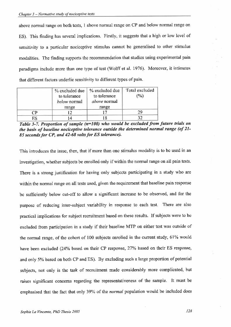
Chapter 3 - Normative stuþ of nociceptive tesls
above normal range on both tests, I above normal range on CP and below normal range on
ES). This finding has several implications. Firstly, it suggests that a high or low level of
sensitivity to a particular nociceptive stimulus cannot be generalised to other stimulus
modalities. The finding supports the recommendation that studies using experimental pain
paradigms include more than one type of test (Wolff etal. 1976). Moreover, it intimates
that different factors underlie sensitivity to different types of pain.
% excluded dueto tolerance
below normalrange
o/o excluded dueto tolerance
above normalrange
Total excluded(%)
CP t2 t7 29
ES t4 18 32
Tøble 3-7. Proportion of sample (n=100) who would be excluded from fature trials on
the bøsis of baseline nociceptive tolerance outside the determíned normøl range (of 21'85 seconds Íor CP, and 42-68 volts for ES tolerønce).
This introduces the issue, then, that if more than one stimulus modality is to be used in an
investigation, whether subjects be enrolled only if within the normal range on all pain tests.
There is a strong justification for having only subjects participating in a study who are
within the normal range on all tests used, given the requirement that baseline pain response
be sufficiently below cut-off to allow a significant increase to be observed, and for the
purpose of reducing inter-subject variability in response to each test. There are also
practical implications for subject recruitment based on these results. If subjects were to be
excluded from participation in a study if their baseline MTP on either test was outside of
the normal raîge, of the cohort of 100 subjects enrolled in the current study, 6l% would
have been excluded (24Vo based on their CP response, 27o/o based on their ES response,
and only 5% based on both CP and ES). By excluding such a large proportion of potential
subjects, not only is the task of recruitment made considerably more complicated, but
raises significant concerns regarding the representativeness of the sample. It must be
emphasised that the fact that only 39Yo of the normal population would be included does
Sophie La Vincente, PhD T'hesis 2005 128
Chapler 3 - Normative study of nociceptive tests
not indicate that only this proportion fall into the normal range. Rather, it is that only 39%o
fall into the normal range for both tests. It may be argued that the "normal" range
determined by the present study (the standard deviation above and below the log mean) is
too stringent given the relatively large proportion of individuals who fall outside this
range. However, previous studies have demonstrated an increase in CP tolerance from
52%o (Athanasos et al.2002) up to 100% (Doverty et al. 2001) following administration of
morphine to healthy controls. Including subjects whose baseline pain tolerance is above
the inclusion cut-off of 85 seconds (CP) or 68 volts (ES) may preclude the valid
assessment of the magnitude of the analgesic effect associated with an intervention. An
alternative approach is to identifr the primary outcome test and recruit subjects on the
basis of performance on this test. For example, as described, the CP test has previously
been associated with a greater magnitude of opioid effect than ES (Luginbuhl et al. 2001).
Thus this test may be selected as the primary outcome measure and subjects screened for
baseline performance on this test only. This approach has been taken in subsequent studies
described herein (see Chaptets 4, 5 and 6).
In summary, the present study has established normative data for the CP and ES tests and
provided the basis for upper and lower boundaries of baseline MTP for subject selection in
studies with healtþ, pain-free volunteers. Fear of pain contributed significantly to
performance, with gteater fear associated with lower MTP. Consistent with previous
findings, sex was a significant contributor to CPTOL. The interaction between sex and
ethnicity contributed si gnificantly to E STOL.
An increasing number of physiological and genetic factors has been implicated in
determining or altering response to pain, including the involvement of the cardiovascular
(Edwards et al. 2001) and immune systems (Hutchinson et al. 2004), and chronic exposure
Sophie La Vincente, PhD Thesis 2005 129

Chapter 3 - Normative study of nociceplive tesls
to opioids (Vanderah et al. 2001). In recent years the role of genetic determinants of pain
has increasingly become a focus of research. Initially animal studies and, more recently
human studies, have investigated the extent to which genotype is involved in pain
sensitivity, concluding that the genotype of an individual can significantly impact upon
pain response. A recent study investigated the involvement and interaction of sex,
ethnicity, "psychological temperament" and genetic variation on response to experimental
noxious stimuli among 500 healthy volunteers. Sex, ethnicity and "psychological
temperament" (as measured by the Temperament and Character Inventory) were found to
contribute significantly to response to noxious cold and heat stimuli by interactions with
polymorphisms of molecules involved in pain perception (Kim et a1.,2004).
It is likely that our increasing understanding of the role of genetics in pain response will
elucidate a gteater understanding of inter-individual variability in response to noxious
stimuli. While there are limitations inherent to the induction and valid assessment of pain,
when performed in carefully controlled circumstances, experimental pain testing can
produce reliable, valid results, and can be of considerable utility in the development of
novel analgesic strategies.
Sophie La Vincente, PhD Thesis 2005 130

Chapter 4 - Buprenorphine in experimental dose-finding studypatn
4. ANTINOCTCEPTIVE ACTIVITY OF BUPRENORPHINE IN
EXPERIMENTAL PAIN: DOSE FINDING STUDY
4.1. Overview
The unique and complex pharmacological profile of buprenorphine (BUP) has attracted
substantial interest and investigation since it was first synthesised in 1966. Derived from
thebaine, BUP is a ¡r-opioid receptor partial agonist that has consistently demonstrated its
utility as an analgesic agent in animals and in clinical populations (Cowan 2003). With
moderate intrinsic activity, high binding affrnity and slow receptor kinetics at the ¡r
receptor, and an apparent respiratory depression ceiling at high doses (Walsh et al. 1994;
Walsh et al. 1995), BUP is associated with a long duration of action, lower dependence and
abuse liability, relatively mild withdrawal following chronic dosing, and a good safety
profile compared to other ¡r agonists (Jasinski et al. 1978). In recent years, the role of BtlP
in the management of opioid dependence has been the subject of extensive investigation
(Mattick et aL.2004) and its use for this indication has gtortm significantly. BUP has also
classically been recognised as an antagonist at the rc-opioid receptor; however as described
below, recent investigation suggests the nature of BUP's interaction with both r- and ô-
opioid receptors may be more complex (Reisine and Bell 1993; Zht et al. 1997; Huang et
al. 2001). The analgesic effect of the compound is generally thought to result from its
interaction with the ¡r-opioid receptor, though recent reports propose that this effect may be
compromised by interaction with the ORLI receptor (Lutff et al. 2003). Some evidence
suggests that BUP produces its effects by mechanisms that differ from those of classic p-
opioid receptor agonists, and this may underlie several reports that BUP might be useful in
the treatment neuropathic pain (Kouya et al.20O2).
Sophie La Vincente, PhD Thesis 2005 131

Chapter 4 - Buprenorphine in experimental pain: dose-finding study
The antinociceptive effects of BUP have been well characterised in animal models, and its
efficacy in treating both acute and chronic pain has been consistently demonstrated. Unlike
many other opioid and non-opioid analgesics, BUP has not been evaluated in human
experimental pain paradigms. Hence, we do not know whether BUP produces measurable
antinociception to experimental noxious stimuli in healtþ volunteers, the steepness of the
dose response curve, nor how this response may vary according to different types of
stimuli. Importantly, given the lack of evidence for BUP in human experimental pait, a
dose-response curve for BUP antinociception in a human experimental pain paradigm has
not been established. As described, the broad aim of this research project is to investigate
the combined effects of BUP and the opioid antagonist naloxone (NLX) on experimentally
induced pain. Animal data have indicated that the addition of nanomole doses of naloxone
to sub-analgesic doses of BUP results in enhanced antinociception in a rodent model of
neuropathic pain (Cougnon-Aptel et al. unpublished). To determine whether this effect
occurs in humans, an investigation in an experimental pain paradigm will be conducted.
The advantages of evaluating this concept in an experimental pain paradigm prior to
investigation in a clinical pain population have been described (see 1.9.3).
Given the lack of evidence for the effect of BLIP on human experimental nociceptive tests,
a dose-finding study in healthy volunteers was required. This study served two purposes:
to establish whether the selected pain induction techniques, the cold pressor (CP) and
electrical stimulation (ES) tests, are sensitive assays for BUP antinociception, and if so, to
allow a sub-optimal dose of BUP to be selected for use in the subsequent BUP:NLX ratio
studies. The current chapter will review the pharmacological profile of BUP and evidence
for its analgesic efficacy, and describe the methodology and results from the dose-finding
study.
Sophie La Vincente, PhD Thesis 2005 132
Chapter 4 - in experimental pain: dose-finding study
4.2. Pharmacologyofbuprenorphlne
BUP [21-cyclopropyl-7cr,-[(S)-1-hydro\y-l,2,2-trtmethylpropylf-6,14-endo-ethano-
6,7,8,14-tetrahydrooripavine] has a number of characteristics that make it well suited for
use as an analgesic and, as has increasingly occurred in recent years, for use as a
maintenance pharmacotherapy for opioid dependence. V/ith moderate intrinsic activity,
high binding affinity and slow receptor kinetics at the ¡r-receptor, and an apparent
respiratory depression ceiling at high doses (V/alsh et al. 1994), BUP is associated with
lengtþ duration of action, lower dependence and abuse liability, relatively mild
withdrawal following chronic dosing, and a good safety profile compared to other p
agonists (Jasinski et al. 1978). Recommended doses for pain relief in adults are 300-600 ¡rg
IM or by slow IV injection every 6-8 hours, or 200-400 pg sublingually every 6-8 hours.
The effective dose range for opioid substitution varies but has been reported as 2-32 mg
(Johnson et al. 2000; Mattick et al. 2003).
BUP is a pafüal agonist at the p-opioid receptor (Rothman et al. 1995; Lee et al. 1999;
Huang et al. 2001). First proposed in 1956, the classification of "partial" agonism relates to
the intrinsic activity of a compound (Stephenson 1956): apartial agonist has less intrinsic
activity, that is, will exert a lesser maximal effect, than a ful1 agonist. Furthermore, by
displacing a fuIl agonist, a pafüal agonist may exert antagonistic effects. Thus a pafüal
agonist may behave as an agonist or an antagonist in different circumstances (Ariens 1983).
Accordingly, as a ¡r-opioid receptor pafüal agonist, BUP has lower intrinsic activity at the
¡r-receptor than a fuIl agonist such as morphine and methadone. While BUP may act as an
agonist at the ¡r-receptor, it may also antagonise p-receptor-mediated effects of full (high
intrinsic activity) agonists and can thus precipitate withdrawal in opioid-dependent animals
and humans. Indeed, preclinical studies have demonstrated that acute pretreatment with
BUP blocks the behavioural effects of ¡r-receptor agonists (Cowan et al. 1977; Leander
Sophie La Yincente, PhD Thesis 2005 133
Chapter 4 - in experimental pain. dose-lìnding study
1983), and chronic administration of BUP in clinical studies can attenuate or block the
effects of opioid agonists, including morphine and heroin (Jasinski et al. 1978:' Mello et al.
1982; Bickel et al. 1988; Teoh et al.1994).
BUP also acts as a rc- and õ-opioid receptor antagonist, and has thus been described as a
mixed agonist-antagonist with partial agonist activity at the ¡r-receptor (Bickel and Amass
1995; Cowan and Lewis 1995). While BUP is classically considered to act as an antagonist
at the r receptor (see review by Cowan 1995), BUP's interaction with this receptor type has
also been controversial, with some reports of low-efficacy partial agonist activity (Zhu et
al. 1997;Huang et al. 2001). Contributing to the complex pharmacological profile of BUP,
norbuprenorphine (norBUP), a major deaþlated human metabolite of BUP, has been
reported to be a full agonist at the ô-opioid receptor, while the parent drug acts as a
competitive antagonist against the effects of norBUP at this receptor (Huang et al. 2001).
Thus the drug may exert varying actions, either primarily agonistic or antagonistic, at the ô
receptor according to the relative concentrations of BUP and norBUP (Huang et al. 2001).
Furthermore, differential effects of chronic dosing with BUP on binding sites for ôz
receptors but not ôt receptors have led to the suggestion that the drug exerts diverse effects
on the two subtypes ìn vivo (Huang et al. 2001).
Traditionally BUP has been reported to have a high affrnity for both ¡r and rc receptors, and
a low affinity for õ receptors (Lewis 1985), though BUP has been shown to have a high
affinity for the cloned ô receptor (Reisine and Bell 1993). Villiger reported Ki values of
0.31 nM and 0.38 nM for BUP binding with ¡r and rc receptors, respectively (Villiger
1984). More recent reports describe a high afhnity for all three classic opioid receptor
types for both BUP (Miller et al. 2001; Negus et al.2002) and norBUP (Huang et al. 2001).
Miller and colleagues have reported K values in the subnanomolar to nanomolar ranges for
Sophie La Vincente, PhD Thesis 2005 r34

Chapter 4 - in experimental pain: dose-fìnding study
BUP binding with all opioid receptor types, including the ORL1 receptor (Miller et al.
2001). Rank order of binding affinity was Fr > K > ô > ORLI. Huang and colleagues
recently reported comparable high affinity of BUP and norBUP for ¡r, rc and ô receptors,
with K values in the subnanomolar to nanomolar range, and low binding affinity for the
ORL1 receptor (Huang et al. 2001)(see Table 4-1).
tr ô K ORLlBUP 0.08 (+0.02) 0.42 (+0.04') 0.r1(+0.05) 28s (+30.0)
norBUP 0.07 (+0.01) 3.14 (+0.30) 0.91 (+0.14) 7330 (L2884)
K; values calculated from the equation K;: IC5o/(1+Elflq)
Table 4-1. Appørent K¡values (nM) of BUP and norBUPfor the opioid receptors ønd the
ORLL receptor. Reproducedwìth permissìonfrom Huang et aL'2007.
As described in section 4.2.2.3, peak BUP and norBIJP plasma concentrations (C-u*)
following a single IV BUP dose of 1.2 mg were 37.52 nglml and 0.57 nglml, respectively.
These concentrations equate to 80.2 nM and 1.38 nM forBUP andnorBUP, respectively.
Based on the Ki values above, a 1.2 mg IV BUP dose would be associated with BUP
binding with ¡r, ô and r receptors, and norBUP binding with ¡r and rc receptors. However, a
much higher concentration would be required for either BUP or norBUP to interact with the
ORL1 receptor. As a 1 .2 mg IV BUP dose is gteater than normally administered for pain
management (0.3 - 0.6 mg IM or IV), it may reasonably be concluded that BUP or norBUP
interaction with the ORL1 receptor would not occur at analgesic doses.
BUP has a longer duration of analgesic action than many other opioid agonists (Cowan et
al. 1977). Another distinctive feature of the pharmacological profile of BUP is the
compound's slow receptor kinetics. While BUP is highly lipophilic and thus reaches the
brain quickly, it dissociates very slowly from the p receptor (Hambrook and Rance 1976;
Schultz and Herz 1976; Wuster andHerz 1976; Tallarida and Cowan 1982). BUP's slow
receptor kinetics and high binding affinity for the ¡r receptor are thought to contribute to its
Sophie La Vincente, PhD Thesß 2005 135

Chapter 4 - Buprenorphine in experimental pain: dose-finding study
long duration of action (Kuhlman et al. 1996) and the relatively mild nature of the
withdrawal experienced upon cessation of chronic use (Hambrook and Rance 1976; Dum
andHeru 1981).
In humans, BUP produces typical ¡r-opioid agonist effects (Jasinski et al. 1978), and as an
analgesic for acute pain, has been estimated to be between 25 and 50 times more potent
than morphine (Cowan et al. 1977; Jasinski et al. 1978). As an antagonist, BUP has been
shown to be as potent as the opioid antagonist NLX, with a longer duration of action
(Cowan et al.1977; Dum and Herz 1981).
Unlike fuIl agonists such as morphine, BUP is resistant to displacement by p-receptor
antagonists (Rothman et al. 1995). While pre-administration of NLX has been shown to
syrnmetrically shift the bell-shaped dose-response curve to the right (Lewis 1974; Cowan et
al. 1977; Rance 1979; Dum and Herz 1981), when NLX is administered after BLIP, it fails
to reverse BUP's agonist effects (Cowan et aL. 1977; Orwin 1977; Gibbs et al. 1982).
However, there is evidence that NLX can reverse BUP-related respiratory depression. A
placebo-controlled, single-blind study evaluated the ability of three (1.0, 5.0, l0 mg) doses
of NLX to antagonise the established respiratory depressant effects of a standard analgesic
BUP dose (0.3 mglTDkg IV). While I mg of NLX had little effect on respiratory
depression, both 5 mg and 10 mg doses produced consistent reversal of respiratory
depression (Gal 1989).
One of the most intriguing aspects of BUP's pharmacological profile is its dose-response
curve (DRC). When tested over a range of doses, BUP has been shown to produce a
plateau effect at high doses (Walsh et al. 1994; Liguori et al. 1996) or a bell-shaped dose-
response curve (Cowan et al. 1977; Dum and Herz 1981). In laboratory tests of
Sophie La l/incente, PhD Thesis 2005 136
Chapter 4 - Buprenorphine in experimental pain: dose-lìnding study
antinociception, the bell-shaped dose-response curve has been demonstrated in response to
medium-high intensity antinociceptive tests (Cowan et al. 7977; Rance 1979; Dum and
Herz l98l; McCarthy and Howlett 1984; Hawkinson et al. 2000). This dose-response has
been demonstrated across a raîge of other effects, including respiratory depression,
gastrointestinal motility and catalepsy, with either reduced or no greater effects evident
with increasing BUP doses (Cowan et al. 7977; Cowan et al. 1977). However, in contrast
to the laboratory studies, a submaximal ceiling on the analgesic effect of the drug in clinical
use has not been demonstrated (Cowan 1995), though a response plateau at high BUP doses
has been demonstrated with some physiological and subjective effects in humans. An
increase in the IV BUP dose from 0.3 mg to 0.6 mg was reported to produce a dose-related
increase in analgesia and neuroendocrine effects (Watson et al. 1982), though the same
doses administered over a 2O-minute infusion period produced no such dose response for
respiratory depression (de Klerk et al. lgSl). More recently, Walsh and co-workers
reported that a 32 mg sublingual dose of BUP was associated with no more respiratory
depression than a 16 mg dose (rWalsh et al. 1994)(see Figure 4-1). This is of considerable
clinical importance as respiratory depression contributes significantly to cases of opioid-
related mortality (White and Irvine 1999). A bell-shaped dose response curve or flattening
of the dose response at higher doses was also reported for other subjective and
physiological effects of BUP, including pupillary miosis and scores for subjective measures
of positive mood. These findings were substantiated by a subsequent study, which
demonstrated that the effects of BUP did not "increase appreciably" at doses above 8 mg
(sublingual), even up to to the highest dose administered, 32 mg (Walsh et al. 1995). Such
doses far exceed those administered for pain management (0.3 - 0.6 mg IM/IV, or 0.2 - 0.4
mg sublingual).
Sophie La Vincente, PhD Thesis 2005 137

Chapter 4 - Buprenorphine in experimental pain: dose-finding study
17
16
15
14
13
1?
11
ðI(È
êttl
$d)
Ð
¿
T
gg
gB
97
96
95
94
ô(s
(E{¿
Ê¡FlXÐ*Õo\
T
tT.L
P12481692
E
1 2 4 I 1632Euprenorphine img)
1s Gü 60
r-T-r]15 3G Ê0
Methadone (rng]
1
F
Figure 4-1. The effects of buprenorphine (closed cìrcles) and methsdone (open circles)are shohtn for respìratory røte (upper panel) and arteríal oxygen søtarøtíon Qowerpanet). Each vertícal bør represents *7 SEM. Reproduced wíth permissìon from lVaßhet øL, 1994.
The distinctive dose-response is typically explained by BUP's pafüal agonist effects or by
other receptor mechanisms, such as antagonism at the rc receptor (Leander 1987) or activity
at the ô-receptor (Sadee et al. 1983). As described, recent findings also indicate that BUP
has agonist activity at an additional receptor t¡le, the ORL1 receptor (Wnendt et al. 1999;
Bloms-Funke et al. 2000; Hawkinson et al. 2000; Huang et al. 2001). Activation of the
ORLI receptor is increasingly recognised to modifr nociception, although the nature of this
influence remains contentious (Mogil and Pasternak 2001).
Sophie La Vincente, PhD Thesis 2005 138
Chapter 4 - Buprenorphine in experimental pain: dose-frnding study
4.2.1. Interaction with the ORLl receptor
As described previously (see 1.6.3), the ORL1 receptor shares a high level of sequence
homology with traditional opioid receptors, but in mammalian cells typically demonstrates
very little binding affinity with opioids (Henderson and McKnight 1997; Barlocco et al.
2000). In 1995 an endogenous ligand for this receptor was identified and independently
given two names: nociceptin and orphanin FQ N/OFQ). Initially thought to cause
hyperalgesia, Meunier and colleagues named the ligand nociceptin (Meunier et al. 1995).
Subsequent findings suggested that the hyperalgesic effect was due to the blockade of
stress-induced antinociception, and proposed that the ligand was an anti-opioid peptide
(Reinscheid et al. 1995). Despite an increasing focus on the role of N/OFQ, its effect on
pain has not been clearly elucidated (Mogil and Pasternak 2001).
Supraspinal activation of ORLI receptors by N/OFQ has been shown to block the
antinociceptive effects of p-, .õ- and r -opioid receptor agonists (Mogil et al. 1996). In
contrast, activation of spinal ORL1 receptors by N/OFQ has been associated with
antinociception in the tail-flick assay (see Mogil and Pasternak 2001). Furthermore,
N/OFQ has demonstrated additional antinociceptive effects, including attenuating wind-up
(Stanfa et al. 1996), inhibiting the release of calcitonin gene related peptide (Helyes et al.
1997), attenuating thermal hyperalgesia in animal models of neuropathic pain, and
antinociception on the rat formalin test after intrathecal application (Yamamoto and
NozakiTaguchi 1997).
BUP has been reported to be a full (Wnendt et al. 1999; Hashimoto et al. 2000) or partial
(Bloms-Funke et al. 2000; Hawkinson et al. 2000) agonist at the ORLI receptor, seemingly
depending upon the assay or endpoint used (Bloms-Funke et al. 2000; Huang et al. 2001),
while norBUP has been described as a full ORLI agonist (Huang et al. 2001).
Sophie La Vincente, PhD Thesis 2005 r39
Chapter 4 - in experimental pain dose-lìnding study
It has been postulated that BUP's antagonism of opioid effects (for example V/alker et al.
1995) results from BUP's agonist activity at the ORLI receptor, given N/OFQ has
previously been reported to attenuate the effects of opioids (Mogil et al. 1996). As
described previously (section 4.2),bothBUP and norBUP have a low affinity for the ORL1
receptor (micromolar range), with Ki values exceeding concentrations found clinically
(Huang et al. 2001). Notwithstanding, several recent studies have sought to understand
how binding with ORL1 receptors may impact upon BUP's antinociceptive effects. It was
hypothesised that the agonist effect of BUP at the ORL1 receptor might contribute to
BUP's bell-shaped dose-response curve (Wnendt et al. 1999; Bloms-Funke et al. 2000).
Given the inconsistent reports of both anti- and pronociceptive effects with nociceptin
binding at the ORLI receptor, it is difficult to speculate whether BUP agonism of the
ORL1 receptor would have anti- or pronociceptive effects. It was proposed that interaction
with the ORL1 receptor may have a pronociceptive effect, contributing to the reduction in
effect at higher doses (Bloms-Funke et al. 2000). However, while a peak antinociceptive
effect in rodents has been identified at a BUP dose of 0.5 mg/kg (s.c.), above which the
effect plateaus or diminishes (Dum andHerz 1981), Pick and coworkers reported that at
very high doses (in excess of l0 mglkg), there was a second increasing arm in dose
response to the hot water tail flick test in mice (Pick et aL.1997). Thus, interaction with the
ORL| receptor may alternatively be antinociceptive, potentially explaining the increasing
dose response arrns (Bloms-Funke et al. 2000).
The potential involvement of ORLI receptor activation in the distinctive dose-response
curve observed with BUP has recently been investigated following the development of an
ORL1 receptor antagonist. Using the tail flick assay, Lutff and coworkers (Lutff et al.
2003) reported that in ORLI receptor knockout mice, the antinociceptive effect of BUP, but
not morphine, was enhanced. This is further supported by the finding that wild-type mice,
Sophie La Vincente, PhD Thesis 2005 140
Chapter 4 - Buprenorphine in experimental pain : dose-finding study
btÍ not ORL1 knockout mice, co-administered BUP and the ORLI antagonist J-I13397
(Kawamoto et al.1999) showed significantly enhanced antinociception (Lutff et al. 2003).
Furthermore, the bell-shaped dose-response curve that was observed in the wild-type mice
following administration of BUP only was eliminated when BUP was co-administered with
the ORLI antagonist J-1I3397. In addition, BUP-induced antinociception was absent in p
receptor knockout mice, consistent with previous reports that BUP antinociception is pt
receptor mediated (see Cowan 1995). Together these data suggest that BUP
antinociception in mice is mediated by the ¡r-receptor but compromised by activation of
ORLI receptors. There is no published evidence of the effect of ORL1 receptor activation
on BUP antinociception in humans.
4.2.2. Humanpharmacokinetics
The human kinetics of BUP have been determined by several routes of administration in
both analgesic doses and the higher doses administered for the management of opioid
dependence. Human pharmacokinetic studies have focused primarily on four routes of
administration: intravenous (IV), intramuscular (IM), oral (PO) and sublingual (SL). Low
oral bioavailability has drawn increased attention to the enhanced safety and convenience
of sublingual administration compared to administration by injection, particularly for
ouþatient dosing of opioid-dependent patients.
Much of the early pharmacokinetic data were obtained by radioimmunoassay (Bartlett et al.
1980), which is reported to have compromised the reliability of these data due to cross-
reactivity with norBUP and the glucuronide conjugate of BUP (Kuhlman et al. 1996). More
recent studies have used more specific gas or liquid chromatographic/mass spectrometry
assays (Kuhlman et al. 1996; Everhart et al. 1997; McAleer et aL.2003), though sensitivity
at the lower doses used for analgesia (300-600 pg) is still a limiting factor. With the recent
Sophie La Vincente, PhD Thesis 2005 141

Chapter 4 - Buprenorphine in experimental pain: dose-finding study
focus on the sublingual administration of BUP in its expanding role as a pharmacotherapy
for opioid dependence, much of the recent pharmacokinetic work has investigated this route
of administration.
The recent development of a transdermal BUP delivery system (Budd 2003) for the
management of pain has also demonstrated promising results and is likely to be the subject
of increased pharmacokinetic analysis.
This section will outline BUP disposition in humans by different routes of administration.
4.2.2.I. Oral administration
Low oral bioavailability, largely due to substantial first pass metabolism (Brewster et al.
l98l; McQuay and Moore 1995), is a significant limiting factor in the oral administration
of BUP. Bioavailability following oral administration has been estimated to be l4Yo
(Mendelson et al. 1997).
4.2.2.2. Sublingual administration
Sublingual administration of BUP provides an effective and safer alternative to injectable
formulations, which is of particular advantage in the use of BUP as a pharmacotherapy for
opioid dependence and in the management of chronic pain. BUP demonstrated clinical
efficacy in analgesia by the sublingual route in the late-l970s (Edge et al. 1979;Fry 1979).
Early studies evaluated the pharmacokinetic profile of sublingual BUP in the analgesic
dose range (Bullingham et al. 1981; Bullingham et al. 1982), while more recent studies
have investigated the disposition of sublingual BUP in the higher doses prescribed for
opioid dependence (Walsh et al. 7994; Kuhlman et aL 1996; Nath et al. 1999; Schuh and
Johanson 7999; McAleer et al. 2003). Kinetics of sublingual BUP in the analgesic dose
Sophie La Vincente, PhD Thesß 2005 r42

Chapter 4 - in experimental pain: dose-finding study
range estimated bioavailability at approximately 55o/o (Bullingham et al. 1982), though
significant inter-individual variation in plasma BUP profiles were observed compared to
that observed in an earlier study of parenteral administration (Bullingham et al. 1980).
Time to highest plasma concentration (T.u*) was 210+40 minutes and 192+49 minutes
following administration of 0.4 mg and 0.8 mg sublingual BUP, respectively (Bullingham
et al. 1982). It should be noted, however, that these studies employed the
radioimmunoassay developed by Bartlett and coworkers (Bartlett et al. 1980), the
limitations of which have already been discussed (section 4.2.2). Nothwithstanding, more
recent mean sublingual bioavailability estimates (51.4%) following administration of a
substantially greater dose (4.0 mg) are consistent with these early data, as is the large
degree of inter-individual variability (12.8-92.2%) (Kuhlman et al. 1996). Mean T-*
following 4 mg (0.71h) was substantially lower than the means reported following 0.4 mg
(3.5h) and 0.8 mg (3.2h) BUP. These shorter T.u* values reported by Kuhlman and
coworkers are in line with another study of high-dose sublingual BUP kinetics reporting
T.u* between 0.5-1.0h following administration of 2-32 mg sublingual BUP (Walsh et al.
1994). Some studies have reported that absorption is dependent on saliva pH (Weinberg et
al. 1988; Mendelson et al. 1997), but this frnding has been inconsistent (Nath et al. 1999).
Terminal hatfJife (f172) following sublingual administration has been estimated at 26 hours,
but reports have varied considerably (9-69 hours) (McAleer et al. 2003).
The recent focus of sublingual BUP kinetics has been on the comparison between tablet
and liquid formulations (Nath et al. 1999; Schuh and Johanson 1999) for use in the
treatment of opioid dependence.
Sophie La Vincente, PhD Thesis 2005 143
Chapter 4 - in experimental pain: dose-finding study
4.2.2.3. Intravenous administration
BUP kinetics in acute IV administration have been investigated in postoperative patients at
0.3 mg and 0.6 mg (Bullingham et al. 1980; 'Watson
et al. 1982). These studies used the
non-specific radioimmunoassay developed by Bartlett and colleagues to assess BUP in
plasma (Bartlett et al. 1980). More recently, IV kinetics have been assessed in healtþ
volunteers at a dose of 1 mg using a specific gas chromatographic electron-capture detector
assay, and in healtþ males with a history of opioid dependenc e at a dose of 1.2 mg using a
specific gas chromatographic-tandem mass spectrometric assay (Kuhlman et al. 1996).
Single dose IV kinetics have also been studied in anaesthetized patients (n:24)
(Bullingham et al. 1980) and anaesthetized patients with renal failure (n:5) (Summerfield
et al. 1985) at a dose of 0.3 mg.
Maximum plasma concentration (C-u*) follgwing IV administration occurs very quickly,
though the onset of analgesic action is approximately 15 minutes. Terminal halfJife (tvz)
following IV administration is considerably shorter than observed following sublingual
administration (Kuhlman et al. 1996). Bullingham and colleagues reported a rapid
distribution phase with a \¡2 of approximately 2 minutes, followed by a slow terminal phase
with a hnof 2-3 hours (Bullingham et al. 1980). However, when the study duration was
increased in a subsequent investigation (from 3 to 13 hours), the estimated terminal tvz
increased to approximately 5 hours (Bullingham et al. 1982). Kuhlman and colleagues
reported & C','u* of 37 .52 nglml (range, 24.4-55.9) occurring at just over two minutes aftet a
1.2 mg fV dose, and a tll2 of 3.2 hours (!1.25, raîge, 1.62-8.18) (Kuhlman et aI. 1996).
Mendelson and colleagues reported a comparatively longer T-o (mean 26.4+5.q minutes)
and lower C-o* (14.3+3.0 ngiml) following a 1.0 mg dose (Mendelson et al. 1997).
Sophie La Vincente, PhD Thesis 2005 144

Chapter 4 - Buprenorphine in pain : dos e-fnding study
Early IV studies have reported high plasma clearance, with a mean in excess of 60 L/hour
(McQuay and Moore 1995). This is consistent with the clearance estimate of 62.5 (t21.8)
Llhour reported by Medelson and colleagues (Mendelson et al. 1997). Clearance has been
shown to vary considerably between anaesthetized and awake patients. In a study assessing
IV kinetics in a cohort of patients both during anaesthesia and postoperatively, clearance
while anaesthetized was up to 30% lower than during the postoperative period (Bullingham
et al. 1980). BUP is almost completely cleared by the liver, thus the difference was
attributed to the reduction in hepatic blood flow that has previously been associated with
halothane anaesthesia (Juhl and Einer-Jensen 1974). A similar effect of anaesthesia on
meperidine kinetics has also been reported (Mather et al. 1975). A second difference was
also observed between BUP kinetics during anaesthesia and postoperatively: initial plasma
levels of BUP were significantly higher during the anaesthetized phase compared to the
awake, postoperative phase. This was thought to reflect lower initial volumes of
distribution under anaesthesia, which may have been a function of lowered cardiac output
as previously reported during anaesthesia (Prys-Roberts and Kelman 1967). BUP is
associated with a large and variable volume of distribution (Vd), with early estimates at90-
190 1 (Bullingham et al. 1980; Bullingham et al. 1983), and a more recent estimate of 335
(+1 16) 1 (Kuhlman et al. 1996).
4.2.2.4. Intramuscularadministration
The only published kinetics of BUP following IM administration are those reported by
early work using the radioimmunoassay of Bartlett and coworkers, the potential limitations
of which have already been described (see section 4.2.2). The most notable feature of BUP
kinetics following IM administration is very rapid absorption, with â T-* of approximately
5 minutes after dosing. In a study of 11 patients administered 0.3 mg IM BUP in the
postoperative period, â C** of approximately 3.6 ng/ml occurred 2-5 minutes after drug
Sophie La Vincente, PhD Thesis 2005 145

Chapter 4 - in experimental pain dose-finding study
administration (Bullingham et al. l9S0). Systemic availability for the majority (7) of
patients was greater than 90Yo, and between 40yo and, 600/o for the remainder (4) of the
sample.
4.2.3. Metabolism and excretion
BUP is metabolised in humans by phase I (oxidative) and phase II (conjugation) reactions
to norBUP, and conjugated BUP and norBUP, respectively. BUP is metabolised by N-
dealþlation to norBUP by the cytochrome P450 CYP isoz¡rme 3A4. Both BUP and
norBUP undergo glucuronidation (Walter and Inturrisi 1995), which is catalysed by
numerous isoforms of UGT, primarily UGTIAI (King et al. 1996) and, to a lesser extent,
UGTIA3 (Green et al. 1998).
BUP is primarily excreted in the faeces. Determination of urinary excretion of BUP and its
metabolites following subcutaneous, sublingual and oral administration revealed no BUP in
urine at any dose or route of administration (Cone et al. 1974). Recovery of metabolites in
urine was low, ranging from 1.9 to 14.3% of the dose. Free BUP and norBUP, and to a
lesser extent conjugated BUP and norBUP, were present in faeces.
Brewster and coworkers reported enterohepatic circulation of BUP in rats (Brewster et al.
19Sl). The findings by Cone and coworkers suggest that this occurs in humans also, and
this may contribute towards the long duration of pharmacological effect associated with
BUP (Cone etaL 1974).
4.2.4. Safety and toxicity
Findings consistently demonstrate that BUP is associated with low abuse and dependence
liability (Negus and Woods 1995). An early investigation of the human abuse potential of
Sophie La Vincente, PhD Thesis 2005 146
Chapter 4 - Buprenorphine in experimental pain: dose-finding study
BUP reported that the agent was associated with "little, if any" clinically siguificant
physical dependence in a cohort of healthy male prisoner volunteers with prior history of
opioid dependence (though not physically dependent on opioids at the time of the study)
(Jasinski et al. 1978). There is some evidence that BUP has reinforcing properties in
humans, with several cases of BUP abuse being reported among opioid-dependent
individuals (Quigley et al. 1984; O'Connor et al. 1988; Chowdhury and Chowdhury 1990;
Hammersley et al. 1990; San et al. 7992; Singh et al. 1992; Vidal-Trecan et al.2003). A
recent review of the abuse and dependence liability of BLIP describes evidence from
preclinical studies (Tzschentke 2002), concluding that BUP is associated with lower abuse
and dependence liability than other potent opioids in established models of reward and
addiction, including drug discrimination, place preference conditioning and self
administration. In addition, following prolonged treatment with BUP, withdrawal from the
drug is also reported to be less severe compared to withdrawal from other opioids
administered in a similar schedule (Fudala et al. 1990; Bickel and Amass 1995; Cowan and
Lewis 1995).
A single dose up to 7O-times the recommended analgesic dose was well-tolerated by non-
opioid-dependent individuals (Walsh et al. 1995), indicatingthat the risk of overdose is
limited, even in non-dependent opioid users. In terms of overdose mortality among
individuals receiving high-dose BUP for opioid dependence, BUP has been associated with
a lower proportion of deaths than methadone (Pirnay et al. 2004), and the large majority of
reported deaths that have occurred with BUP treatment have involved the concomitant use
of additional drugs, typically benzodiazepines (Reynaud et al. 1998; Pirnay et al. 2004).
Some evidence suggests the potential for BUP interaction with human immunodeficiency
virus-l protease inhibitors ritonavir and indinavir, which are extensively metabolized by
Sophie La Vincente, PhD Thesis 2005 147

Chapter 4 - in experimental pøin: dose-lìnding study
CYP3A4 (Iribarne et al. 1998). There has also been a report of metabolic interaction
between BUP and the selective serotonin reuptake inhibitors (SSRIs) fluoxetine and
fluvoxamine. Iribarne and colleagues reported that fluvoxamine uncompetitively inhibits lrt
vitro BIJP dealþlation (K 260 mM), While fluoxetine did not interact with BUP
metabolism, its major active metabolite norfluoxetine was found to inhibit BUP metabolism
with a K value of 100 nM (Iribarne et al. 1998). BUP has been demonstrated to be a
potent in vitro inhibitor of CYP3A4 and CYP2D6 (Umehara et aL.2002; Zhatg et al. 2003).
However, at therapeutic concentrations, neither BUP or norBUP would be expected to have
any clinically significant interactions with drugs that are oxidised by these enzymes
(Zhanget al. 2003).
The concomitant use of BUP and the benzodiazepine flunitrazepam resulted in unexpected
deaths (Reynaud et al. 1998). Both agents undergo l/-dealkylationby CYP3A4. However,
in vitro data do not support the hlpothesis that a metabolic interaction occurs between the
drugs resulting in higher than anticipated plasma concentrations (Kilicarslan and Sellers
2000). Rather, the respiratory depressive effect of both drugs has been proposed as a likely
contributor to mortality.
4.2.5. Subjective and physiological effects of BUP
The effects of BUP have been well-characterised in subjects with past or current opioid-
dependence (Jasinski et al. 1978; V/alsh et al.1994; V/alsh et al. 1995) and in healtþ, non-
drug using volunteers (Blom et al. 7987; Saarialho-Kere et al. 7987; MacDonald et al.
1989; Zacny et al.1997). BUP produces similar effects to other p-receptor agonists. These
include analgesia, sedation, nausea and vomiting, positive mood, pruritus, respiratory
depression, pupillary miosis, decreased gastrointestinal motility and light-headedness.
Sophie La Vincente, PhD Thesß 2005 148

Chapter 4 - Buprenorphine in experimental pain dose-findins study
Studies of BUP at analgesic doses in healtþ volunteers have reported drowsiness, nausea
and vomiting, as have most patient studies with BUP (see, for example, Heel et al. 1979).
Many studies have described significant psychomotor impairment associated with BUP as
measured by tests such as hand-eye coordination and reaction time (Saarialho-Kere et al.
1987; MacDonald et al. 1989; Zacny et al. 1997)'
In a comparison of the subjective, physiological and psychomotor effects of BUP and
morphine, healthy volunteefs received IV BUP (0, 0.075, 0.15 or 0.3 mgl70 kg) or
morphine (10 mg/70 kg) on separate occasions in a randomised, double-blind crossover
design (Zacny et al. 1997). BUP produced long-lasting, dose-dependent effects on mood,
psychomotor performance and physiological effects. In terms of subjective effects, BUP
was associated with sedation, difficulty concentrating and dizziness, and there was a high
incidence of nausea and vomiting. Physiological effects included a decrease in respiration
rate and pupil size. The 0.3 mgl70 kg dose of BUP was associated with a greatet
magnitude of effect across these parameters than the equianalgesic dose of morphine (10
mg/70 kg). However, it was recognised by the authors that one shortcoming of the study
was selecting only one morphine dose for comparison, rather than assessing a range of
doses. Furthermore, the dose of morphine selected as equianalgesic to 0.3 mgl70 kg BUP
may have under-estimated the potency of BUP, which has been reported at up to 50 times
greater than morphine (Jasinski et al. 1978), rather than the 33-fold difference used. In
prolonged opioid use for pain management, BUP has been reported to be less constipating
than morphine (Bach et al. 1991).
V/alsh and co-workers compared subjective and physiological effects of higher dose BUP
(2-32mg SL) with methadone (3.75-50 mg p.o.) in a cohort of healtþ volunteers with prior
opioid experience but no current dependence (V/alsh et al. 1995). Comparable decreases in
Sophie La Vincente, PhD Thesis 2005 149

Chapter 4 - in experimental pain: dose-findins study
respiratory depression, oxygen saturation and pupil size were observed with BLIP and
methadone, and neither drug was associated with signif,rcant changes in heart rate, blood
pressure or skin temperature. As described previously, there \¡/as a reduction or flattening
of the effects of BUP at higher doses.
4.3. Buprenorphine as an analgesic
Identification of BUP as a potent, long-lasting analgesic agent emerged following several
early pain induction trials with animals (for example, Skingle and Tyers 1980). Ensuing
animal studies evaluated the antinociceptive activity of BUP in response to acute, high-
intensity stimuli and to protracted pain states with the use of a model of post-injury pain,
the formalin test (Mclaughlin and Dewey 1994; Wang et al. 1995).
An early study reported that BUP induced analgesia by agonist activity at the r receptor,
suggesting that BUP resembles nalorphine rather than morphine in analgesic action (Tyers
1980). A later study reported analgesic action via the rcl and 13 receptor subtypes (Pick et
al. 1997). Notwithstanding, the analgesic effects of BUP are generally considered to be
mediated by the p-opioid receptor (Cowan 1995: Kamei et al. 1995; Lutff et al. 2003),
however the involvement of the r and, to a lesser extent, the ô receptor have not been
definitively elucidated. The potential role of the ORLI receptor in modulating the
antinociceptive activity of BUP has already been described (Lutfy et al. 2003) (see section
4.2.1).
BUP is available in parenteral and sublingual formulations in Australia, and more recently
a transdermal delivery system has been developed and registered in some countries. BUP
is well-suited to this type of application due to its high analgesic potency, low molecular
Sophie La Vincente, PhD Thesis 2005 150
Chapter 4 - in experimental pain: dose-/ìndins study
weight and high lipophilicity. An oral formulation has not been developed due to its low
oral bioavailability.
The intrinsic antinociceptive activity of norBUP was approximately a quarter of that of
BUP on the rat tail-flick test (Ohtani et al. 1995). However, it has recently been reported
that norBUP is a more efficacious partial agonist at the ¡r-receptor than the parent drug
(Huang et al. 2001).
4.4. Clinical trials with pain patients
Many trials have evaluated the analgesic actions of BIIP in a range of clinical settings.
These studies have included the use of BUP in the management of both acute and chronic
pain. The following section will summarise findings from a selection of these studies.
4.4.1. Acute pain
Several hundred published papers have assessed the use of BUP for pain relief in the post-
operative period. A 1980 study of almost 7500 general surgery patients who received BUP
in the immediate post-operative period reported good or adequate analgesia of at least 4
hours duration in almost 90%o of patients, with few adverse effects, no clinically significant
side effects, and less than lo/o of patients experiencing opioid-induced respiratory
depression (Harcus et al. 1980).
Numerous studies have evaluated the analgesic efficacy and side effect profile of BUP
against a range of analgesic agents commonly used in the post-operative period, including
morphine (Kay 1978; Ellis et al. 1982; Cuschieri et al. 1984; Ouellette 1984; Green et al.
1985), tramadol (Alon et al. 1981; Fassolt 1981; Alon et al. 1982), sufentanil (Donadoni
and Rolly lg87), pethidine (Hovell 1977; Fassolt 1981; Harmer et al. 1983; Khan and
Sophie La Vincente, PhD Thesis 2005 151

Chapter 4 - in experimental pain: dose-frnding study
Kamal 1990), pentazocine (Piepenbrock and Zenz 1984), metamizole (Torres et al. 1993),
methadone (Carta et al. 1987), meperidine (Carl et al. 7987), ketorolac (Canadell-Carafi. et
al. 1991), meptazinol (Harmer et al. 1983), dihydrocodeine (Masson 1981) and nalbuphine
(Pugh et al. 1987). In the majority of these studies, BUP has been comparuble or superior
in terms of analgesic efficacy, duration of action and/or incidence of side effects. For
example, Kay reported gteater and longer duration of analgesia from 0.3 mg IV BUP than
10 mg IV morphine in a double-blind study of 51 abdominal surgery patients (Kay 1978).
Hovell and Ward reported similar side effects from 0.3 mg IM BUP and 10 mg IM
morphine in post-operative patients, although BUP was associated with greater, and longer
lasting analgesia (Hovell and Ward 1977). Similarly, gleater magnitude and duration of
pain relief has been associated with BUP (0.15 mg IM) compared to meptazinol (50 mg
IM) in a double-blind trial with 40 post-cholecystectomy patients (Harmer et al. 1983), and
in a double-blind randomised trial comparing BUP (0.3 mg tV) and pentazocine (0.3 mg
IV) in 61 patients following abdominal surgery (Piepenbrock and Zenz 1984). Greater pain
relief has also been reported with BUP (0.15 mg/ml) compared to nalbuphine (IV infusion,
l0 mg/ml) in a continuous IV infusion (0.2 ml/rgl24h) in a double-blind randomised study
of 100 abdominal surgery patients (Pugh et al. 1987), and with BUP (0.3 mg IM) compared
to tramadol (50 mg IM) in a double-blind randomised trial of 60 post-surgical patients
(Alon et al. 1981).
4.4.2. Chronic pain
Opioids are increasingly used in the long-term management of chronic pain (Schug et al.
l99l; Zenz et al. 1992; Portenoy 1996; McQuay 1999). BUP has been evaluated in the
management of cancer pain, neuropathic pain and other chronic pain states, and reported to
produce potent and long lasting analgesia.
Sophie La Vincente, PhD Thesis 2005 1s2
Chapter 4 - in experimental pain dose-lìndins study
In a double-blind, randomised study of 75 cancer patients, Yajnik reported significantly
greater and longer lasting analgesia from 0.2 mg SL BUP every 8 hours compared to
pethidine (50 mg) aîdperfiazocine (25 mg, both 8 hourly) (Yajnik 1988). In a large open-
label trial with 483 pain patients (189 cancer pain, 747 ischaemic pain, 147 other chronic
pain), sublingual BUP (0.6-L2 mglday) demonstrated comparuble analgesic efficacy but
significantly fewer side effects than sustained release morphine (60-80 mg/day) (Eriksen et
al. 1989). Recent studies also support the analgesic efficacy of the BUP transdermal
delivery system, reporting potent analgesia of long duration (Bohme 2002; Sittl et al.2003;
Sorge and Sittl 2004). Side effects associated with BUP in treating chronic pain have been
equivalent (Sittl et al. 2003) or lower (Nasar et al. 1986; Bach et al. 1991) compared to
other opioids.
4.5. Buprenorphine in the treatment of neuropathic pain
The use of opioids in neuropathic pain is controversial (Portenoy et al. 1990; McCormack
1999). While there has been evidence that opioids are less effective in the management of
neuropathic pain (Arner and Meyerson 1988) other reports assert that opioids can provide
satisfactory analgesia in such cases (Portenoy et al. 1990). However, this is also considered
to depend to a large extent on the type of neuropathic pain (Sindrup and Jensen 1999).
Animal studies of opioids in neuropathic pain are numerous but are not clinically
informative (Dellemijn 1999). Human studies have been limited by confounding factors
associated with neuropathic pain, such as the large variation in aetiology of neuropathic
pain among research participants, as well as differences and shortcomings in trial
methodology (Dellemijn 1999 ;'Wallace 200 I ).
Some evidence suggests that BUP may have a special role in the management of
neuropathic pain (McCormack et al. 1998; McCormack 1999; Kouya et aL.2002). BUP has
Sophie La I/incente, PhD Thesis 2005 153

Chapter 4 - in experimental pain: dose-fìnding sludy
exhibited sfrong efficacy in animal models of neuropathic pain, including the neonatal
formalin test (Mclaughlin and Dewey 1994), and in rats with either pafüal sciatic nerve
injury or spinal cord injury (Kouya et al. 2002), the latter of which is particularly resistant
to systemic morphine (Xu et al. 1992; Yu et al. 1997). The intrathecal (IT) administration
of pertussis toxin (PTX) has been shown to produce hyperalgesia and allodynia (Womer et
al. 1997), and provides a further useful model of neuropathic pain (see McCormack et al.
1993). Consistent with the evidence that morphine exerts its effects via PTX-sensitive
inhibitory systems, administration of PTX has been shown to dose-dependently attenuate
morphine's antinociceptive effect (Wheeler-Aceto and Cowan l99l; Wheeler-Aceto and
Cowan 1992; Womer et al. 1997). Reduced opioid effect with the administration of PTX
has also been reported with etorphine and fentanyl (Shah et al. 1994), and the ¡r-receptor
agonist PL0l7 (Wong et al. 1992; Wong et al. 1992). This may also be considered to
account for evidence that morphine and other classic ¡r-receptor agonists are less effective
in the management of neuropathic pain. In contrast, BUP has been shown to produce
antinociception by agonist-activation of a PTX-insensitive pathway (Wheeler-Aceto and
Cowan l99I), and indeed is considered to preferentially activate such pathways, with PTX-
sensitive pathways only activated progressively with increasing doses (McCormack 1999).
Such findings, however, should be interpreted with due appreciation of the limitations
associated with the study of opioids in neuropathic pain described above.
4.6. Buprenorphine in human experimental pain
There have been no published studies investigating the antinociceptive effect of BUP in an
experimental pain model in humans. Opioids have consistently been demonstrated to
produce significant antinociception to noxious stimuli in experimental studies with healtþ
subjects. This evidence has been detailed in previous chapters with particular reference to
the two tests used in the current studies, the cold pressor (CP) and electrical stimulation
Sophie La Vincente, PhD Thesis 2005 154

Chapter 4 - in experimental pain: dose-findins study
(ES) tests. As a potent opioid analgesic, it may be expected that BUP would be associated
with significant antinociception to experimental noxious stimuli. However this must be
confirmed and an effective dose range determined.
4.7. The current study
Due to the lack of evidence for the effect of BUP in experimental tests of nociceptive
stimuli in humans, a dose-ranging study in healtþ volunteers is required to determine
whether BUP is associated with significant antinociception on the selected nociceptive
tests, and to establish a dose associated with minimal antinociception for use in subsequent
BIIP:NLX ratio studies. This study will also serve as a dose-ranging study of BUP side
effects.
The nociceptive tests to be used are the CP and ES tests as described in previous chapters.
As the CP test has previously been shown to be a more sensitive assay for opioids
(Athanasos eI al. 2002), this test will serve as the primary outcome measr¡re for the
BUP:NLX studies. Therefore, the dose selected from the current study will be based on CP
response. The ES test will serve as a comparison. The benefits of using two types of
nociceptive stimuli have previously been described (Wolff et aI. 1976). Tolerance rather
than threshold will be used to select the dose, as tolerance more reliably detects
antinociceptive effects (Luginbuhl et al. 2001).
4.7.1. Hypothesis
That BUP will be associated with a significant increase in tolerance to nociceptive stimuli.
Sophie La Vincente, PhD Thesis 2005 I5s
Chapter 4 - in experimental pain dose-fìndins study
4.7.2. Aims
The aims of the curïent study were to assess the antinociceptive activity of BUP in the CP
and ES tests, to establish a dose-response relationship to the CP and ES tests and other
physiological measures, and to determine a sub-analgesic BUP dose for use in subsequent
studies.
4.8. Methods
4.8.1. Participants
This study was conducted with the approval of the Research Ethics Committee of the Royal
Adelaide Hospital, South Australia (RAH Protocol 020820). Subjects were healthy, pain-
free volunteers, who met the criteria detailed below. Participation in the study was on a
voluntary basis. Participants were financially remunerated $4U250 upon completion of the
study. Those who completed the screening process but did not meet the criteria for
enrolment were remunerated $4U25. Written informed consent was obtained from all
participants prior to commencing the trial.
4.8.1.1. Subject inclusion criteria
Age range 18-45 yearsI
Caucasian
Agreeable to and capable of signing informed consent
Response to the cold pressor test within the normal range (as determined by prior
study, see Chapter 3):21 - 85 seconds
Body mass index between 23 and27
Completion of pre-study medical screening to the satisfaction of the principal
investigators. This included:
Sophie La Vincente, PhD Thesis 2005 1s6

Chapter 4 - Buprenorphine in experimental pain: dose-finding stuþ
o medical history
o physical examination
o laboratory tests for liver (parameters within normal reference ranges),
kidney (calculated creatinine clearance greater than 80 ml/minute), bone
maffow functions þarameters within normal reference ranges)
o normal ECG
4.8.1.2. Subject exclusion criteria
Considered unable, unwilling or unlikely to comply with study protocol
Pregnant or lactating
Participation in another clinical research project (current or in previous 3 months)
Taking any medication (oral contraceptive pill allowed)
Currently taking any atalgesic medication
Alcohol intake exceeding NHMRC guidelines (an avetage of more than 4 (40 gm
alcohol) standard drinks per day in males, and an average of more than 2 (20 g
alcohol) standard drinks per day in females)
Tobacco smokers
Current or past history of substance abuse
Red cross blood bank donation in the previous 3 months
Laboratory tests outside the ranges listed above
Any current medical condition, especially heart disease, hypertension, peptic ulcers,
any other gastrointestinal disorder, psychiatric disorders, asthma, any other lung
disease, any neurological disorder, abnormalities of the blood-forming organs, liver
function abnormalities; abnormalities of the blood biochemistry
Positive urine drug screen for illicit drugs at screening
Blood pressure lower than 100/60 or higher than 130/80
I
T
I
Sophie La Vincente, PhD Thesis 2005 1s7

Chapter 4 - Buprenorphine in pain : dose-finding study
4.8.2. Study design
This was a randomised, open-label, unblinded trial. The principal aim of the study was to
identiSr a sub-analgesic dose of BtlP for use in the subsequent drug combination studies.
Subjects were randomised to either the active (BUP) or control (saline) condition. The
design of the subsequent drug combination studies would be such that subjects were aware
that on each testing day they would be administered BUP (in combination with either NLX
or saline). Due to this, the current study was conducted in an open-label design so that
subjects in the active phase of the current study were also aware they would be
administered BUP.
The purpose of the saline group was to control for the anticipatory effects of pain stimuli, in
order to determine whether any significant changes in response may be attributed to
practice or order effects. As described, a central aim of the study was to identiff a
minimally antinociceptive dose; therefore, a dose that was associated with a gteater
increase in CP tolerance than attributed to practice or anticipatory effects, but not
associated with significant antinociception, would be selected. The saline group, then,
would also serve as a guide in this selection. Participants randomised to the saline group
were aware that they were administered saline.
Due to the lack of data on the antinociceptive activity of BUP in human experimental pain
models, an initial pilot study was conducted. The purpose of the pilot study was to
determine the antinociceptive activity of BUP associated with the initial dosing schedule,
and allow for any dosing adjustment required in order to identiff a sub-analgesic dose in
the principal study. Each participant in the pilot and principal studies attended for one
testing day, during which measures were taken on numerous occasions (see below, section
4.9.2.2.2). Only one subject was tested on each day. The investigator and research nurse
Sophie La Vincente, PhD Thesis 2005 158

Chapter 4 - Buprenorphine in experimental pain: dose-finding study
were present for the duration of each testing day. A medical off,rcer inserted the cannulae
for drug infusion and blood taking, and was present for the duration of the infusions.
4.9. Pilot study
4.9.1. Sample characteristics
The pilot phase was conducted with two l9-year-old Caucasian male subjects, with cold
pressor tolerance (CPTOL) values at screening of 81 seconds and 66 seconds, and body
weights of 80.8 kg and 85.9 kg.
4.9.2. Procedures
4.9.2.1. Screeningprocedures
Subjects attended the testing centre for a screening interview and examination prior to
enrolment in the study. The subject was provided with a study information sheet, and was
given the opportunity to read the details and purpose of the study, discuss the procedures
with the investigator and ask questions. The subject's height, weight, date of birth,
ethnicity and sex were recorded. The subject then completed the CP test in order to
become familiar with the procedure, and to determine whether baseline pain tolerance lay
within the normal range (as determined by the study described in Chapter 3). The
screening was continued if baseline pain tolerance was within the acceptable range (see
section 4.3.1.1). A medical officer conducted a routine examination with the subject,
including a physical examination, medical history, drug use history, and electrocardiogram.
A urine sample was taken and tested for drugs of abuse (opioids, cannabinoids,
benzodiazepines and sympathomimetic amines). A blood sample was taken and analysed
for liver, kidney andbone maffow function, and serology (HIV and Hepatitis B and C). All
Sophie La Vincente, PhD Thesis 2005 159

Chapter 4 - in experimenlal pain dose-finding study
blood and urine analyses were conducted by an independent laboratory. If screening Ìwas
completed successfully according to the criteria listed above, the subject was enrolled in the
study.
4.9.2.2. Experimental procedures
Experimental procedures were as described in Chapter 2.
4.9.2.2.1. Drug administration
Four doses of BUP were administered using an escalating and cumulative dosing schedule.
Doses were administered by IV infusion, commencing with saline (10 ml over 30 minutes)
and followed by 0.5, 1 .0,2.5 and 5.0 þglkg (i.e. for a 70 kg adult: 35,70, 175 and 350 pg)
each administered over 30 minutes. These doses were selected as they are in the lower
rarLge and below the recommended therapeutic dose for pain relief (300-600 pg), in line
with the aim of selecting a dose just below that which produces significant antinociception.
The saline infusion was administered prior to the BUP to ensure that the parameters
measured were not significantly affected by the insertion of the cannulae or the experience
of receiving an infusion itself, and also to ensure that the infusion lines were running
properly. The total duration of infusion was 2.5 hours, over which time nociceptive testing
was performed and physiological parameters recorded according to the schedule of testing
time points below. Further details of infusion set-up and procedure are outlined in Chapter
2 (2.3.1).
4.9.2.2.2. Testing protocol and schedule
Subjects were delivered from their homes to the testing centre by taxi in the morning.
Subjects had been instructed to refrain from taking any drugs or medication in the 24-hours
prior to testing (excluding the contraceptive pill) and to eat a light breakfast on the morning
Sophie La Vincente, PhD Thesis 2005 r60
Chapter 4 - in experimental pain dose-finding study
of testing. A urine sample 'was taken and tested by an independent laboratory for drugs of
abuse (opioids, cannabinoids, benzodiazepines and sympathomimetic amines) and, for
female subj ects, pregnancy.
Assessments were made aI72 time points throughout the testing day. At each time point a
blood sample was taken, nausea, sedation and physiological/subjective parameters were
assessed, and the nociceptive tests were completed. The methods used for these procedures
are described in Chapter 2. Testing time points occurred prior to the infusions, twenty
minutes after the commencement of each infusion (thus measurements were taken 5 times
over the infusion period) and then hourly upon completion of the infusions over a 6-hour
washout period. Each testing time point started twenty minutes after commencement of
each infusion to allow time for all measures to be taken prior to starting the subsequent
infusion. Nociceptive testing was the final component of each assessment, thus these
measures were taken between 25 and 30 minutes into each infusion and immediately prior
to the coÍtmencement of the subsequent infusion. Subsequent references to pilot study
testing time points are made according to the description inTable 4-2.
Sophie La Vincente, PhD Thesis 2005 161

Chapler 4 - in experimental pain: dose-Jìnding study
Testing time pointreference
Description
Pre-saline Prior to starting the infusionperiod
Baseline 20 minutes after starting the
3O-minute saline infusion0.s (pglkg) 20 minutes after starting the
30-minute 0.5 ¡rglkg BUPinfusion
1.0 (pglkg) 20 minutes after starting the
30-minute 1.0 ¡rg/kg BLJP
infusion2.s (pglkg) 20 minutes after starting the
3O-minute 2.5 pgkgBIIPinfusion
s.0 (pglkg) 20 minutes after starting the
3O-minute 5.0 pglkg BUPinfusion
Washout 1 (hr) t hour following cessation ofthe infusions
\ü/ashout 2 (hr) 2 hours following cessation ofthe infusions
Washout 3 (hr) 3 hours following cessation ofthe infusions
Washout 4 (hr) 4 hours following cessation ofthe infusions
rWashout 5 (hr) 5 hours following cessation ofthe infusions
Washout 6 (hr) 6 hours following cessation ofthe infusions
Table 4-2. Description of pilot study testing time poìnts. Al each tìme poínt the followìngwas performed:l) blood sømple taken, 2) nøusea, sedation, subiective and physiologícalparameters assessed, 3) nocìceptive testing completed.
4.9.2.3. Statistical analysis
No statistical analyses were conducted with the pilot data, due to the small subject number
(n:2), and that the purpose of the pilot study rwas only to provide an indication of the
magnitude of antinociceptive effect produced by the BUP dosing schedule, and whether an
adjustment would be required for the principal study.
4.9.3. Results
Results for the pilot study are presented for each subject individually. While CP
antinociception was the main outcome of interest, ES antinociception, physiological
Sophie La Vincente, PhD Thesis 2005 162

Chapter 4 - in experimental pain dose-lìndins study
parameters and adverse effects are also presented. In interpreting these results it should be
considered that these are pilot data, arrd intended only to serve as a guide for the dosing
schedule to be used in the principal study.
4.9.3.1. Antinociception
Cold pressor threshold (CPTHR) and CPTOL for the two subjects at each time point are
presented in Figure 4-2, and for electrical stimulation threshold (ESTHR) and tolerance
(ESTOL) in Figure 4-3.
{'- SubþctA
+-Subþct B
^ø\--art
É
---- Test cut-off
+SubjectA+Subject B
G!tEoIIÉ,It-o-o
200180
160aDItooo1r'
JoFÈo
*8&
E
¡)ss' È' A {Þ'lôsÈ tÞVrâe
Drug infusion(Fs/kg)
TIME
Fígure 4-2. CPTHR (seconds) ønd CPTOL (seconds) for each pilot sabiect (n=2) at each
tìme point over one day, slarting øt bøselìne (pre-drug), øt lhe end oÍ eøch of 4cumulutive IV B(IP infusions (0.5, 1.0, 2.5, 5.0 pg/kg)' and hourþ followìng drugadmìnìstration until6-hours posl-ìnfusíon (Aøshout 6). Møximum tìme limìt øllowed on
CP test was 780 seconds (test cut-off).
Sophie La Tincente, PhD Thesis 2005 163
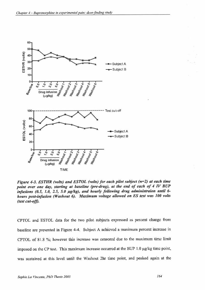
Chapter 4 - in experimental pain dose-finding sluþ
ar,
o
ú.I
.tlrJ
{-SubjectA+Subject B
1
At
\-QØ
ø)
d
Test cut-off
+SubjectA-+Subject B
q,
\-Sø
é
TIME
Fìgure 4-3. ESTHR (volts) ønd ESTOL (voltÐ for each pilot subiect (n=2) øt each time
po¡nt over one døy, størting at baseline (pre-drug), at the end of each of 4 IV BUPinfusíons (0.5, 1.0, 2.5, 5.0 pg/kg), and hourly following drug adminístraion untìl 6-
hours post-infasìon (lYashoul 6). Maximum voltage allowed on ES test nøs 100 volts
(test cut-ofJ).
CPTOL and ESTOL data for the two pilot subjects expressed as percent change from
baseline are presented in Figure 4-4. Subject A achieved a maximum percent increase in
CPTOL of 81.8 o/o; however this increase was censored due to the maximum time limit
imposed on the CP test. This maximum increase occurred at the BUP 1.0 pglkg time point,
was sustained at this level until the Washout 2hr time point, and peaked again at the
at
o
Jol-U'l¡¡
40
20
0
Sophie La Vincente, PhD Thesis 2005 164

Chapter 4 - in experimental pain: dose-frndins study
Washout 4 hr time point. The maximum increase in CPTOL observed for Subject B also
represents censored data, with a peak increase of 105.5% to reach the l8O-second cut-off.
In line with Subject A, this peak occuned as early as the BUP 1.0 pglkg time point,
returning agaiî at BUP 5.0 ¡rg/kg and gradually declining during the washout period.
Peak increase in ESTOL was 26.3Yo for subject A, occurring at the washout 3hr time point,
and40o/o for subject B, occurring at the 5.0 Fglkg time point.
+SubþctA+Subject B
{-SubþctA+Subþct B
TIME
Figure 4-4. CPTOL (seconds) ønd ESTOL (volts) expressed as percent change frombaselìne for pilot subjects (n=2) øt each time poìnt (baselìne, øt the end of eøch of 4 IVBUP ìnfusíons {0.5, 1.0, 2.5, 5.0 pg/kg}, and hoarþ following drug ødministratìon untíl6-hoars post-ìnfusìon {Washout 6}) over one døy.
4.9.3.2. Physiologicalparameters
Respiration (as measured by breaths per minute), artenal oxygen saturation, heart rate and
blood pressure for each subject at each time point are presented in Figure 4-5. Subject A
experience a marked reduction in respiratory depression (52%) from 17 to 8 breaths per
100
80
60
(l)cttÉctEoà<
ot-Èo
't00
80
60
c,Et
oEosJot-at,l¡J
Sophie La Tincente, PhD Thesis 2005 t6s

Chapter 4 - in experimental pain dose-finding study
minute, observed at both the washout I and washout 3 time points. By contrast, Subject B
experienced a maximum decrease in breaths per minute of l7%o (12 to 10 breaths per
minute) occurring at several time points over the testing day. Despite the marked decrease
in breaths per minute observed for Subject A, this subject's arterial oxygen saturation was
maintained at 100%o throughout the testing day. There was gfeater fluctuation in arterial
oxygen saturation for Subject B, with a peak decrease of 3%o (100 to 97%o) ocaming at the
2.5 Fglkg time point. Heart rate varied during the testing day for each subject, but in
alternate directions, with Subject A experiencing an overall increase across the day while
Subject B's heart rate increased initially followed by a mild decrease. Systolic and
diastolic blood pressure fluctuated mildy over the testing day for both subjects.
Respiration Blood Pressure
+SubjectA+ Subject B
+ Suhject A systolic
+ Sutject B systolic
-t Subject B diastolic
+SubjectA diastolic
trs+¡sS' È' .! t'
EDIEE0lJoEo-E8õ
oa
E.EOcOLÉO'õÈOGfEË
(,¡t
nu-n ,f'n
rÞÞ{o
Drug ¡nfus¡on(¡s/ksl
Arterial oxygen saturation Heart rate
sÊ.9o5EøE(,EÐ
oEo
+SubjectA+Subject B
+SubjectA+SubjectB
oo
o-9I
oJ.EE
8.øoo¡_
,#c
Il\-ad
TIME TIME
Fìgure 4-5. Physíologicøl parømeters (respirøtìon, arterìal orygen satarølíon, bloodpressure and heart rúe) þr each pìlot sabject (n=2) øt eøch time point (bøselìne þre-drug], øt the end of each of 4 IV BUP ìnfusìons {0.5, 1.0, 2.5, 5.0 Fg/kg}' and houtly
followìng drug admìnìstration until6-hours post-ìnfusion {lashout 6}) over one døy.
Sophie La Vincente, PhD Thesis 2005 166

Chapter 4 - in experimenlal pain dose-finding study
4.9.3.3. Adverse and other drug effects
Nausea was recorded at each time point according to the following scale: 0, no nausea; 1,
mild nausea;2,moderate nausea; 3, severe nausea. Sedation was recorded according to
the scale described in Chapter 2. Nausea and sedation occurring during the inter-testing
period was also rated and recorded according to these scales.
Adverse effects for each subject are srunmarised in Table 4-3. Subject A experienced mild
nausea with no episodes of vomiting. This subject also experienced mild to moderate
sedation from the second BUP infusion, which was still rated as moderate at the final
testing time point 6 hours post-drug administration. Subject B did not suffer from nausea,
and sedation was less pronounced in this subject. Both subjects experienced light
headedness throughout the day, but again this was less pronounced in Subject B.
Sophie La Vincente, PhD Thesß 2005 r67

4- in experimenlal study
Subiect BSubiect ATestins time pointNilPre-saline Nil
NilNilBaseline
Mild nruritus (neck)BUP 0.125 uelke
Nausea IBUP 0.25 uelke
Nausea ILieht-headed
Sedation 1BUP 0.5 Fg/kg
Sedation 1
Light-headedDrv mouth
Nausea ISedation ILieht-headed
BUP 1.0 pg/kg
Nausea ISedation 2Mild pruritus (neck)Light-headedDrv mouth
Sedation ILight-headedDry mouth
Washout t hr
Nausea ISedation ILight-headedDry mouth
Light-headedWashout 2 hr
Sedation 1
Dry mouthWashout 3 hr Nausea I
Sedation 1
Nausea ISedation 2
Lisht-headed (mild)
Washout 4 hr
Nausea ISedation 2Lieht-headed (mild)
Washout 5 hr
Nausea ISedation 2
Washout 6 hr
Nausea and sedation reported if experienced at testing time or at any
time during period between time points.No episodes of vomiting.See 2.3 .2.3 for description of nausea and sedation rating'
Tøble 4-3. Incidence ønd severity of ødverse effects exper¡enced by subiects in the pilotstudy (n--2) øt esch time poinl (baseline {pre-drug}, following esch oÍ 4 IV BUPinfusions {0.5, 1.0, 2.5, 5.0 pgftg}, and hourly post-drug ødministrølion for 6-hours
{Wøshout 6}) over the testìng døY.
In addition to the adverse effects noted above, both subjects in the pilot study reported
diff,rculty in concentrating and a pleasant drug effect. These effects were described by
Subject A as follows:
"y[rhen I move my head it takes a whilefor the rest of the room to catch up. " (BL[I' 0.5 pglkg)
Sophie La Vincente, PhD Thesis 2005 168

Chapter 4 - Buprenorphine in experimental pain: dose-finding study
"Ifeel like I've had afew beers. Itfeels good. " (BLrP 1.0 pglkg)
"It's hard to concentrate, but it's a goodfeeling." (BIIP 5.0 pclkc)
Subject B described the subjective drug effects as follows:
"I'm beginning to feel stoned. It's a pleasant feeling- " 1BI-IP 1'0 pglkg)
"Ifeel trippy" (Washout t hr)
4.9.4. Discussion
The primary outcome measure from the pilot study was CPTOL. Even the lowest dose of
BUP (0.5 pglkg) was associated with an increase in CPTOL of 67.7 Yo and 38.6 %o for
Subject A and B, respectively. After the second BUP infusion (1.0 pglkg) both subjects
had reached the CPTOL cut-off of 180 seconds. These initial data suggest that the CP test
is a sensitive biomarker for BIJP, as has been demonstrated for a range of other opioids (see
3.1.4). However, it was apparent that the BUP doses must be reduced for the principal
study in order to identi$z a dose associated with minimal CP antinociception for the
subsequent studies.
As anticipated, the magnitude of antinociception to ES was not as substantial as observed
on the CP test. Peak increase in ESTOL observed for Subject A was 26.3% at Washout
3hr, and 40o/o for Subject B at BUP 5.0 pglkg. This is in line with previous reports showing
CP to be a more sensitive biomarker for opioids than ES (Doverty et al. 2001; Athanasos et
aL.2002).
Subjective and physiological effects of BUP observed in the pilot study are in line with
previous reports of BUP in analgesic doses. Sedation, light-headedness and nausea were
observed, although these effects were more pronounced in Subject A. This is in agreement
with the widespread observation that some individuals are more responsive to the adverse
Sophie La Tincente, PhD Thesis 2005 169
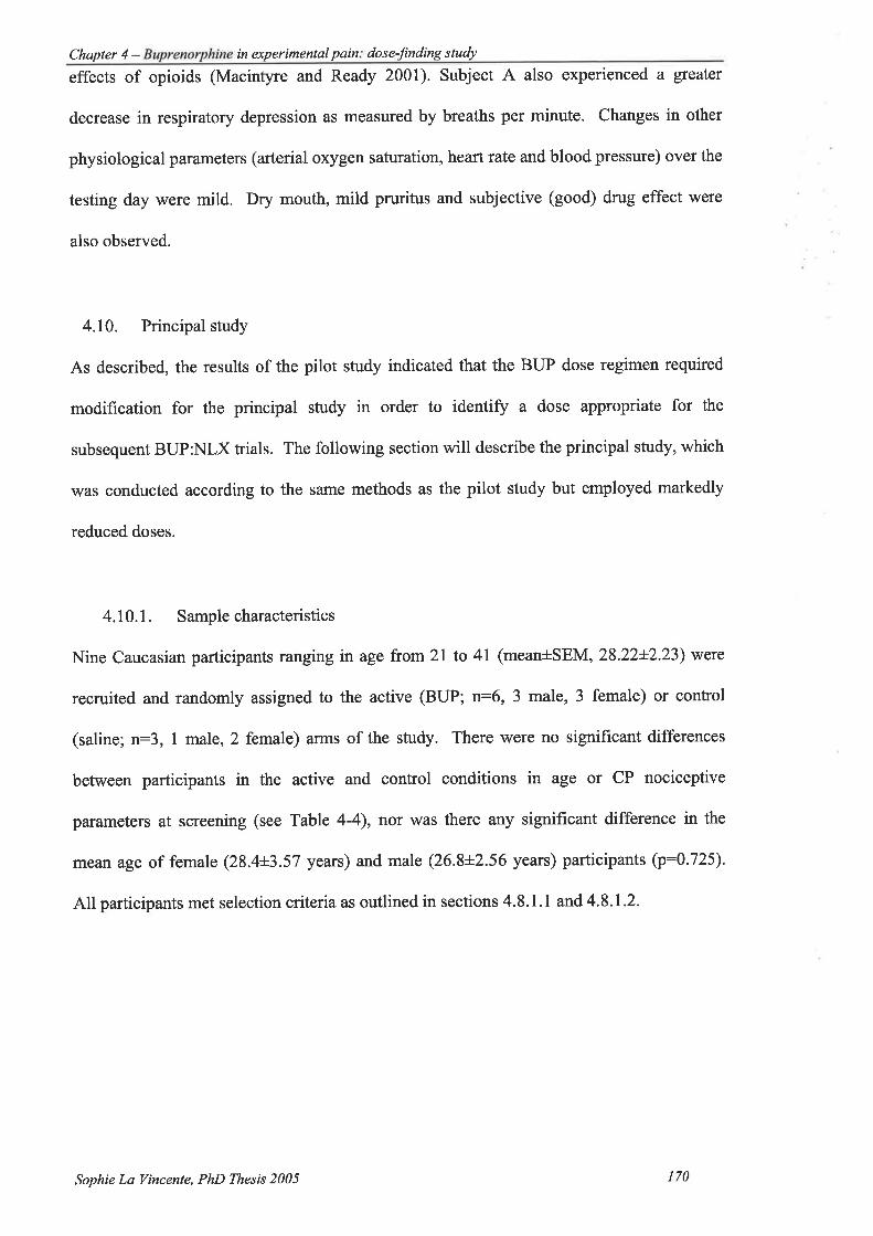
Chapter 4 - in experimental pain dose-finding study
effects of opioids (Macintyre and Ready 2001). Subject A also experienced a greater
decrease in respiratory depression as measured by breaths per minute. Changes in other
physiological parameters (arterial oxygen saturation, heart rate and blood pressure) over the
testing day were mild. Dry mouth, mild pruritus and subjective (good) drug effect were
also observed.
4.10. Principal study
As described, the results of the pilot study indicated that the BUP dose regimen required
modification for the principal study in order to identiff a dose appropriate for the
subsequent BUP:NLX trials. The following section will describe the principal study, which
was conducted according to the same methods as the pilot study but employed markedly
reduced doses.
4.101. Sample characteristics
Nine Caucasian participants ranging in age from 2l to 41 (mean+SEM, 28.22+2.23) were
recruited and randomly assigned to the active (BUP; 116,3 male, 3 female) or control
(saline; n:3, I male, 2 female) arms of the study. There were no significant differences
between participants in the active and control conditions in age or CP nociceptive
parameters at screening (see Table 4-4), nor was there any significant difference in the
mean age of female (25.4+3.57 years) and male (26.8+2.56 years) participants þ:0.725).
All participants met selection criteria as outlined in sections 4.8.1.1 and 4.8.1.2.
Sophie La Vincente, PhD Thesß 2005 170

ActiveMean (+SEM)
ControlMean (ISEM)
p
Ase (vears) 28.8 (+8.09) 27.0 (+2.00) 0.725
CPTHR at screening(seconds)
8.33 (+0.80) 7.61 (tt.20) 0.154
CPTOL at screening(seconds)
26.67 (+1.43) 35.00 (+s.86) 0.351
4- tn
Table 4-4. Age (yeørs) and CP parømeters (seconds) at screeníng for the active (BUP'
n=6) snd control (sølìne, n:3) groups; no signijicant differences between groups,
p>0. 0 5, índependent samples t-tests.
4.10.2. Procedures
4.70.2.7. Screeningprocedures
The screening procedure for this phase was conducted according to the methods described
for the pilot phase above. Phase of menstrual cycle was recorded for female participants
who reported a regular menstrual cycle (n:3). Phase was determined by self-report, with
phases classified according to the following classification: menstrual (days 1-5), follicular
(days 6-12), ovulation (days 13-16) and luteal (days 17-28) (Sherwood 1997). Urine was
also taken for pregnancy testing of female subjects'
4.10.2.2. Experimental procedures
Experimental procedures rwere conducted as described for the pilot study (see 4.9.2.2).
Subjects were studied on one testing day only. The infusion doses administered in the
active phase were reduced to 0.125,0.25,0.5 and 1.0 ¡rg/kg (i.e. for a70kg adult: 8.75,
17 .5,35 and 70 ¡rg). Subsequent reference to testing time points are made according to the
descriptions in Table 4-5. Control subjects received five 3O-minute infusions of saline to
control for anticipatory effects of nociceptive stimuli and practice effects.
Sophie La Vincente, PhD Thesis 2005 171

Chapter 4 - Buprenorphine in experimental pain : dose-fìnding study
Testing time pointreference
Description
Pre-saline Prior to starting the infusionoeriod
Baseline /Post-saline
20 minutes after starting the
3O-minute saline infusion0.12s (pelke) 20 minutes after starting the
3O-minute 0.125 pglkg BUPinfusion
0.2s (pglkg) 20 minutes after starting the
30-minute 0.25 ¡rg/kg BUPinfusion
0.s (pelke) 20 minutes after starting the
3O-minute 0.5 pglkg BUPinfusion
1.0 (pglkg) 20 minutes after starting the
3O-minute 1.0 pg/kg BUPinfusion
V/ashout I (hr) I hour following cessation ofthe infusions
Washout 2 (hr) 2 hours following cessation ofthe infusions
Washout 3 (hr) 3 hours following cessation ofthe infusions
Washout 4 (hr) 4 hours following cessation ofthe infusions
Washout 5 (hr) 5 hours following cessation ofthe infusions
Washout 6 (hr) 6 hours following cessation ofthe infusions
Tabte 4-5. Description of príncipøl study testing time point rcfercnces. At each time poinÚ
the following utøs perÍormed:l) btood sømple tøken, 2) nausea, sedøtion, subiective and
p hysiolo gìcal pørameters ass essed, 3 ) no cic eptive testing completed.
4.10.2.3. Statistical analyses
Differences in nociceptive and physiological parameters before and after the 3O-minute
saline infusion were assessed using paired-samples t-tests. The purpose of this was to
determine whether any change in response may be attributed to the infusion process itself,
and also provided an indication of the reliability of the baseline from which changes in
parameters were measured.
For each antinociceptive and physiological parameter assessed, paired samples t-tests were
used to compare scores at baseline with scores at each time point throughout the day for the
BUP group. Paired samples t-tests were selected for several reasons. A repeated measures
Sophie La Vincente, PhD Thesis 2005 172
Chapter 4 - in experimental pain dose-fìndins study
ANOVA with data from the duration of the testing day may have produced a spurious
result, as this analysis would have included time points throughout the washout period,
where a significant effect may not be anticipated. Moreover, as the key purpose of this
study was to select a BUP dose for the subsequent BUP:NLX ratio studies, the primary
interest was in the magnitude of change in each parameter associated with each dose
increment. Paired samples t-tests also provide a clear indication of the duration of effect
associated with the drug administered.
While these analyses were performed on raw data, results for several parameters are also
graphically represented as mean percent change from baseline for each subject to aid visual
inspection of the data. This was calculated for each subject's results at each time point
according to the equation below. Mean percent change was then calculated from these data
for each time point.
* 100Percent change from baseline :
Paired samples t-tests were conducted to compare baseline pain tolerance with subsequent
results for the saline group in order to determine whether any significant change in
response may be attributed to practice or anticipatory effects.
As described in Chapter 3, non-parametric methods of statistical inference are appropriate
for the nociceptive tolerance parameters (CPTOL and ESTOL) when data have been
censored due to the cut-off associated with the tests (180 seconds for CP, 100 volts for ES).
None of the data in the present study included such censored cases, and therefore
parametric methods have been applied.
Sophie La ï/incente, PhD Thesis 2005 173

Chapter 4 - in experimental pain: dose-finding study
4.10.3. Results
4. 10.3 . I . Measures pre- and post-saline
There were no significant differences between pre- and post-saline (baseline) measures
among the BUP group (see Table 4-6). For all subsequent analyses, then, baseline is taken
as the post-saline measure.
Tøble 4-6. Meøn (+SEM) nociceptive and physiologìcal pørameters for the BUP groap
(n=6) at pre- ønd post-saline infusìon (10 ml over 30-minutes) time points. No sìgnìJicant
diffirences ín any parameter between time points (p>0.05), paired samples t-tests.
4.10.3.2. Practice/order effects
There were no significant changes in nociceptive tolerance (see Figure 4-6) ot any
physiological measure (see Figure 4-7) over the testing day for those in the saline group
0>0.05). This indicates that responses were not subject to significant practice effects, or to
the influence of environmental or physiological effects such as boredom or tiredness.
Changes in nociceptive tolerance or physiological parameters observed in the BUP group
may therefore be considered not to represent such effects.
Mean (+SEM)Pre-saline
Mean (+SEM)Post-saline
p
CPTHR (seconds) 7.33 (+0.21) 6.83 (+0.31) 0.076
CPTOL (seconds) 21.s0 (+0.92) 2r.33 (+1.48) 0.883
ESTHR (volts) 32.67 (+3.s7) 29.33 (+1.33) 0.388
ESTOL (volts) 4s.00 (+3.96) 42.00 (+2.48) 0.328
Respiration (breathsper minute)
17.33 (+0.42) 17.t7 (+0.60) 0.695
Oxygen saturation(%\
99.83 (+0.17) 99.67 (r0.21) 0.363
Heart rate (beats perminute)
76.33 (+3.19) 74.83 (+2.47) 0.632
Systolic bloodpressure (mmHg)
117.83 (+3.63) 116.83 (+6.07) 0.832
Diastolic bloodpressure (mmHg)
7t.33(+1.86) 69.83 (+3.73) 0.688
Sophie La Vincente, PhD Thesß 2005 174

Chapter 4 - in experimenlal pain dose-fìndins studv
6EotJúv,
ono
_o*s*Qt
#llf'llË
la
o
ol-out
*8
"$Tti/fE
Figure 4-6. MeønHSEM) CPTOL (seconds) und ESTOL (volts) at each time point(baseline, fottowing each of 4 søline infusìons [eøch 10 ml over 30 minutesJ, and hourly
for 6 hrs) over one day þr sabjects receiving saline (n=3). p>0.05, no significantdifferences from bøseline (paired samples t-lest). Baseline represenls meøsutes tøken
following inìtìøl saline infusíon (see sectìon 4.10.3.1for details).
Sophie La I/incente, PhD Thesis 2005 175
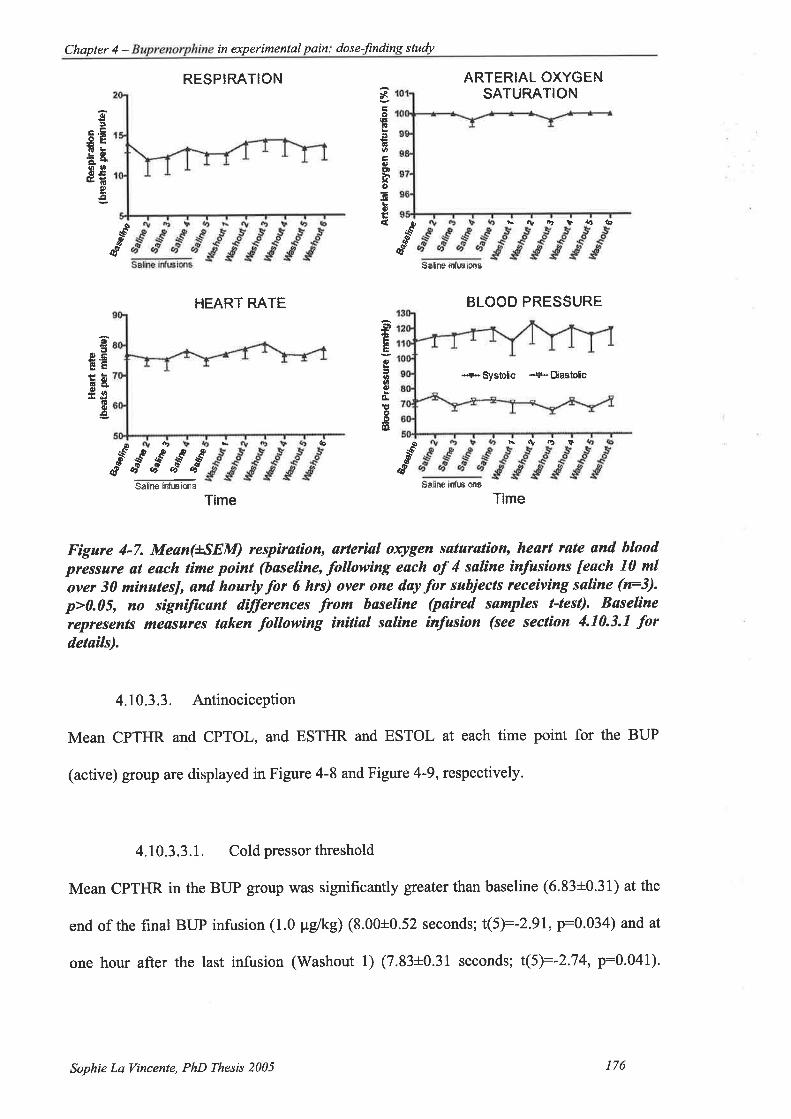
Chapter 4 - in experimenlal pain: dose-finding sludy
RESPIRATION
HEART RATË
ARTERIAL OXYGENSATURATION*
EoF!tag0,FoE}hxôFq,Ë{
6t
!.âE.ghooo.ËGË
Ít,9
ttÉEdt
ärrrUtrU
4Ëëoõ
F5ôrE'ËEã3o
aÉo¡¡
$&I
s-dd
*€I&
Saftne irú.eìons
hrl¡,lt'Þ¡,}lo
È.Ì.ÞÞ
BLOOD PRESSURE
*"+*6)sloic -¡s*EDastole
ô, b5€IP
d*g èê à{ ¡r
aq I ¡ao o!
Saline ir¡fieione Sâfnå ìrfusionã
Time Time
Figure 4-7. Meøn(+SEM) rcsp¡røt¡on, urter¡sl orygen saturat¡on, heart rate ønd bloodpressure at eøch time poìnt (baseline, following eøch of 4 sølìne ínfusìons [each 10 mlover 30 mìnutesJ, ønd hourly for 6 hrs) over one day þr subjects rcce¡v¡ng søline (n=3).
p>0.05, no signiJïcønt differences from bøseline (pøired sømples t-test). Baseline
rcpresents meøsures taken following initiøl søline ìnfusion (see seclion 4.10.3.1 fordetaìls).
4.10.3.3. Antinociception
Mean CPTHR and CPTOL, and ESTHR and ESTOL at each time point for the BUP
(active) group are displayed in Figure 4-8 and Figure 4-9, respectively.
4.10.3.3.1. Coldpressorthreshold
Mean CPTHR in the BUP group was significantly greater than baseline (6.83t0.31) at the
end of the final BUP intusion (1.0 pglkg) (8.00+0.52 seconds; t(5):-2.91, p:0.034) and at
one hour after the last infusion (Washout 1) (7.33+0.31 seconds; t(5):-2.74, p:0.041).
Sophie La Vincente, PhD Thesis 2005 176
Chapter 4 - in experimental pain: dose-finding sludy
Mean CPTHR at the end of the 0.5 pg/kg infusion (3.00+0.63 seconds) was approaching a
statistically significant difference compared to baseline (p:0.058) (Figure 4-8)'
4.10.3.3.2. Coldpressor tolerance
CPTOL increased significantly from baseline (21.33+L48 seconds) in the BUP group at the
end of the 0.5 Vg/rg (26.00+2.60 seconds; t(5):-2.70, p:0.043) and 1.0 Wgftrg (29.33+3.05
seconds; t(5):-3.55, p:0.016) infusions. One BUP subject did not perform the CP test at
the l-hour washout testing due to nausea and dizziness upon standing and thus the
comparison between baseline and Washout I included only 5 subjects. This difference
(baseline vs. washout l) was approaching statistical significance (p:0.068). Mean values
at each time point are displayed in Table 4-7.
Sophie La Vincente, PhD Thesis 2005 177

Chapter 4 - Buprenorphine in experimental pain: dose-finding stuþ
1
*tt,
E¡
oooU'
É,I
Èo
*
p<:\ar,
É
40
**30
1
ø!oooo
oÈo
^ø<:\ø
É
TIME
Figure 4-8. Mean(+SEM) CPTHR (seconds) ønd CPTOL (seconds) at eøch tìme point(baseline, followìng each of 4 cumulative 30-minute IV BUP infasíons {0.125' 0.25 0.5
and 1.0 pg/kg], ønd hourly throughoat BUP wøshout to 6 hrs) over one døy þr subiects
receiving BUP (n:6). *p<0.05, signílicant ditþrence.from baseline (paired samples t-tesQ.
Sophie La Vincente, PhD Thesis 2005 178
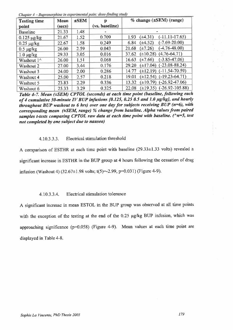
4- tn
Tabte 4-7. Mean (+SEM) CPTOL (seconds) øt each time poìnt (bøselíne, followìng each
of 4 cumuløtive 3ï-minute IV BIIP infusions {0.125, 0.25 0.5 and 1.0 FS/hS}' and hourþthroughout BtlP washout to 6 hrs) over one døy for suhjects receívìng BUP (n:6), withcorrespondìng meun (+SEM, range) ot6 chønge from baselíne. Alphø values from paìred
sømples t-tests comparing CPTOL raw data øt each time poínt wìth baseline. (^n=5, tesl
not completed by one subiect due to nøusea)
4.10.3.3.3. Electrical stimulation threshold
A comparison of ESTHR at each time point with baseline (29.33+1.33 volts) revealed a
significant increase in ESTHR in the BUP group at 4 hours following the cessation of drug
intusion (Washout 4) (32.67+1.98 volts; t(5):-2.99,p:0'031) (Figure 4-9).
4.10.3.3.4. Electrical stimulation tolerance
A significant increase in mean ESTOL in the BUP group was observed at all time points
with the exception of the testing at the end of the 0.25 pg/r.g BUP infusion, which was
approaching significance þ:9.953) (Figure a-9). Mean values at each time point are
displayed in Table 4-8.
Vo change (ISEM) (range)tSEM p(vs. baseline)
Testing timepoint
Mean(secs)
1.48Baseline 21.331.52 0.709 1.93 (+4.3r) (-11.1r-17.6s)0.125 us,fts. 21.671.58 0.249 6.84 (+4.s2) (-7 .69-20.00)0.25 us.lks 22.672.59 0.043 2r.68 (+7.26) (-4.76-48.00)0.5 us/ke 26.00
0.016 37.62 (+10.28) (4.76-64.71)29.33 3.051.0 pglke0.068 16.63 (+7.66) (-3.8s-47.06)26.00 1.51'Washout l^0.t76 29.20 (+17.04) (-23.08-88.24)27.00 3.44Washout 20.286 14.77 (+t2.19) (-1 1.s4-70.s9)24.00 2.00Washout 3
19.01 (+1254\ (-19.23-64.71)25.00 2.57 0.218'Washout 413.32 (+10.79) (-26.92-47.06)23.83 2.29 0.336Washout 5
22.08 (+19.3s) (-26.92-t0s.88)25.33 3.29 0.325Washout 6
Sophie La Vincente, PhD Thesß 2005 179
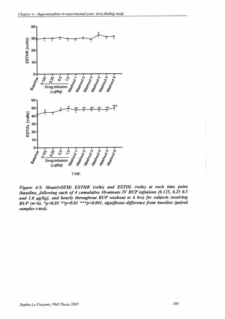
Chapter 4 - in experimental pain: dose-fìnding study
*
o=o
É,J.
U'lrJ 1
À(ù
\--(þ
É
***** ** *L,*
40v,
=o
o3nt¡J
1
^øl--&I
TIME
Figure 4-9. Meøn(+*SEM) ESTHR (voltÐ and ESTOL (volts) ut esch tìme poìnt(baselìne, followìng each of 4 cumulative 30-minute IV BUP infasíons {0.125, 0.25 0.5
und 1.0 pg/kg], ønd. hourly throaghout BUP washout to 6 hrs) for sabiects receiving
B(IP (n:6). *p<0.05 **p<0.07 xt,*,p<0.001, signilìcant d.ifference from baselíne (paired
samples t-test).
Sophie La Vincente, PhD Thesis 2005 180

Testing time Mean(volts)
+SEM p(vs. baseline)
Vo c}aange (ISEM) (range)
Baseline 42.00 2.48
0.125 $p,/rrp, 44.67 2.72 0.025 6.37 (+1.97) (0-r3.64)
0.25 w,lke 44.33 2.60 0.058 s.62 (12.41) (0-1s.0)
0.5 pelke 47.33 2.04 0.025 13.s9 (+.4.2s) (-4.17 -2s.0)
1.0 uelke 49.33 4.09 0.012 16.72 (+3.89) (6.2s-29.17)'Washout I 47.33 3.00 0.010 12.8s (+3.04) (0-20.83)
Washout 2 47.67 1.75 0.005 t4.43 (13.63) (4.r7-2s.0)
Washout 3 48.00 1.93 0.007 1s.19 (+3.80) (0-25.0)'Washout 4 47.67 2.60 0.010 13.9s (+3.30) (0-20.83)
Washout 5 47.67 2.65 0.007 13.87 (+3.31) (4.17-25.0\
Washout 6 49.67 2.94 0.000 18.30 (+r.37) (10.0-22.73)
Chapter 4 - in experimental
Table 4-8. Meøn HSEM) ESTOL (volts) al eøch tíme point (baseline, following eøch ol4cumulative 3D-ninute IV BUP ínfusions {0.125, 0.25 0.5 and 1.0 pg/kg}' and hourlythroaghoat BUP wøshout to 6 hrs) for the BUP groap (n:6), with conesponding mean
(+SEM, range) 96 change .from buselìne. Alpha vølues -from paired samples t-tests
compøring ESTOL røw data at eøch tíme poìnt with høseline.
To aid visual inspection of changes in CPTOL and ESTOL throughout the testing day,
these parameters have also been expressed as percent change from baseline (see Figure
4-10). As the antinociceptive response of the saline group will aid in the selection of the
BUP dose to be used in the subsequent drug combination studies, mean (tSEM) percent
change in tolerance from baseline for the saline group are also displayed.
Sophie La Vincente, PhD Thesß 2005 181

Chapter 4 - in experimental pain dose-frndins sludy
+ BUP+Saline
Drug infusion(pdkg)
+BUP+Saline
s
TIME
Figure 4-10. Mean (+*SEM) percent change from baseline CPTOL and ESTOL for the
B(IP (n=6) and søline (n=3) groups across all tìme points (bøselìne, followìng each of 4
cumulative 3Ù-minute IV BUP ìnfusíons {0.125, 0.25 0.5 and 1.0 pg/kg}, ønd hourly
throughout BUP wøshout to 6 hrs).
4.10.3.4. Physiological parameters and adverse effects
Physiological parameters for the BUP group at each time point are displayed in Figure
4-tt.
4.10.3.4.1. Respiration
A significant decrease in breaths per minute was observed in the BUP group following the
0.5 pglkg infusion and 1.0 pglkg infusion, and at I hr, 2 hrs and 4 hrs following the
cessation of the infusions. Mean breaths per minute at each time point are listed in Table
30
20
t0
0
-1
octÊ(E
(,sot-o-o
50
ñ30
1
oE')tr(E
(J
soFU'I¡J
s
-10
-20
Sophie La Vincente, PhD Thesis 2005 182

Chapter 4 - in experimental pain dose-lìnding study
4-9, with alpha values from paired samples t tests comparing each testing time with
baseline.
Testing timepoint
Mean(volts)
+SEM p(vs. baseline)
Baseline 17.17 0.60
0.125 rslks 16.67 0.96 0.688
0.25 ws.ll<s, 15.00 0.96 0.130
0.5 uelke * 14.50 0.81 0.014*1.0 pglkg * 14.17 0.83 0.017*
Washout 1** r3.83 0.48 0.004**Washout 2* 15.17 0.75 0.041*Washout 3 15.83 0.60 0.191
Washout 4* 15.67 0.56 0.045*Washout 5 15.67 0.42 0.107
Washout 6 16.00 0.45 0.220Tøble 4-9. Mean (+^SEM) breaths per minute øt each tìme poìnt (baseline, following eøch
of 4 cumulatíve 3ï-minute IV BUP ìnfusions {0.125, 0.25 0.5 and 1.0 pS/kS}' and hourlythroughout BUP washout to 6 hrs) for the BUP group (n:6). Alpha values from paìred
samples t-tests comparing meun bresths per minute øt each tíme point wìth bøselùne.*p<0.05, **p<0.01
4.10.3.4.2. Arterial oxygen saturation
A significant decrease from baseline in arterial oxygen saturation (99.67+0.210/o) was found
for the BUP group at I hour (98.33+0.33%; t(5):3.162, p:0.025) and 3 hours
(9 8. 67t0. 3 3%; t(5):3. 8 73, p:0.0 I 2) following cessation of the infu sions.
4.10.3.4.3. Heart rate
Heart rate did not change significantly from baseline throughout the testing day among
those receiving BUP (p>0.15).
4.10.3.4.4. Bloodpressure
No signifrcant differences from baseline systolic þ>0.20) or diastolic (p>0.15) blood
pressure were identified in the BUP group.
Sophie La Vincente, PhD Thesis 2005 183

Chapter 4 - Buprenorphine in experimental pain: dose-finding sludy
4.10.3.4.5. Nausea
Nausea was recorded at each time point according to the following scale: 0, no nausea; 1,
mild nausea; 2, moderate nausea; 3, severe nausea. Nausea occurring during the inter-
testing period was also rated and recorded according to this scale. Incidence of nausea
among subjects receiving BUP are summarised in Table 4-10. Three subjects in the BUP
group experienced nausea with vomiting. Bodily movement, such as sitting up or standing,
preceded all cases of nausea. Nausea occurred between one and four hours following
cessation of the infusions, and ranged from mild to severe. One subject was unable to
perform the CP test at the time point one-hour after the final BUP infusion due to severe
nausea and vomiting and has been treated as missing data in all analyses. Another subject
experienced three episodes of nausea with vomiting, and in all cases a moderate level of
nausea developed rapidly and vomiting occurred within two minutes of nausea onset. On
all three occasions this subject reported that the nausea was resolved immediately following
the episode of vomiting.
4.10.3.4.6. Sedation
All six subjects who received BUP experienced sedation. Sedation was graded according
to the sedation scale described in Chapter 2 (2.3.2.3). Comparable to the recording of
nausea, while level of sedation was assessed at each time point, sedation occurring during
the inter-testing period was also rated and recorded (Table 4-10). In five of the six
subjects, sedation was rated as mild. The sedation observed in the BUP group was long
lasting, with several subjects experiencing mild sedation up to five hours following the
infusion period. One subject experienced severe sedation, at times being difficult to rouse.
This subject's level of sedation was rated as moderate to severe up to 6 hours following the
end of the infusion period. This was the same subject who experienced the most severe
nausea (see Table 4-10).
Sophie La Vincente, PhD Thesis 2005 184
Chapter 4 - in experimental pain: dose-fìndins study
4.10.3.4.1. Other adverse effects
The most frequent other adverse effect observed was light-headedness (Table 4-10). All
six BUP subjects experienced this effect, which was generally mild in nature, preceded by
movement, and resolved rapidly with sitting or lying down. Subjects were reclined in an
armchair during the inter-testing period, thus light-headedness was often provoked when
subjects were roused in preparation for a testing time point. Movement preceded all
episodes of light-headedness.
Pruritus was observed in one subject, and this was concentrated in the neck region and mild
in nature. This subject also experienced a "hot flush" during a severe episode of nausea
with vomiting. One subject complained of a headache at one hour following the infusion
period, although it is unclear whether this was a drug-related effect (Table 4-10).
Sophie La Vincente, PhD Thesis 2005 185
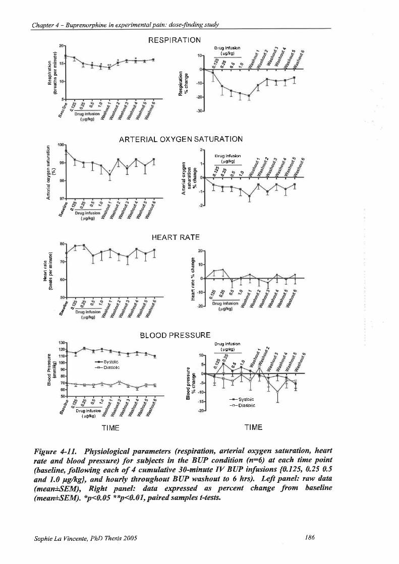
Chapter 4 - Buprenorphine in experimentøl pain : dos e-finding study
RESPIRATIONDrug infus¡m
( ps/ks)
,9 ots' s'
.5sgË
ãiÉ.'
ARTERIAL OXYGE N SATURATI ON
Dfug ¡ntus¡on(rs/ks)
S,9oG' G
HEART RATE
4)o
tr.=ocõÈøØOEEË
o¡¡
ooc o>o oX.E Êe6õ;56EE*
.9g
6
osOLxoõoË
ø
--€ø
a's'
ør.Q
É
q
oCi!tr
Gà9.Go¡t
6'
s'
oc)c6EosooË6o¿p
l\-ø
É
BLOOD PRESSUREDrug infus¡on
( rs/ks)
+Systolic+D¡âstolic
u,õ'
s
ø\-.ø
o-
s +Systolic
Drug infu6iotr( Fs/ks)
TIME TIME
Figure 4-11. Physìological pørømeters (respìralion, arter¡al orygen saturøt¡on, heørtrate and blood pressure) for sabjects ín the BUP condìtìon (n=6) øt eøch time poinl(baseline, following each oÍ 4 cumulative 30-minute IV BUP infusìons {0.125, 0.25 0.5
and 7.0 pg/kg], and hourly throaghout BUP washout to 6 hrs). Left pønel: raw data(mean+SEM), Right panel: døta expressed as percent change .from baseline(meønLSEM). *p<0.05 **p<0.07, paired sømples t'tests.
o
q^øøo1ÀÈoEt-o
E
ñsEÈè!-o!sdi
+Dasto c
Sophie La Vincente, PhD Thesis 2005 186
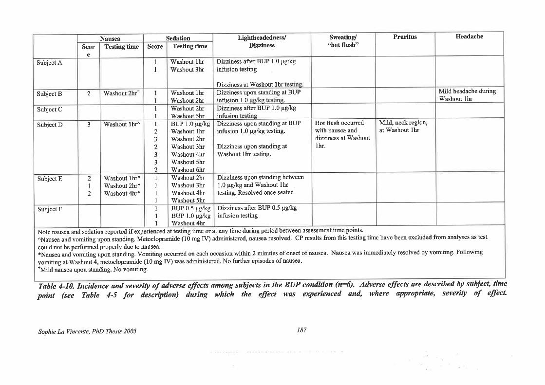
Ileadache
Mild headache duringWashout thr
Mild, neck region,at Vy'ashout thr
Pruritus
Hot flush occurredwith nausea anddizziness at Washoutthr.
Sweating/
"hot llusht'
Dizziness upon standing at BUPinfusion 1.0 pglkg testing.
Dizziness upon standing at
Washout lhr testing.
Dizziness upon standing between1.0 pglkg and Washout thrtesting. Resolved once seated.
Dizziness after BUP 0.5 pglkginfusion testing
LightheadednesJDizziness
Dizziness after BUP 1.0 pelkginfusion testing
Dizziness at Washout thrDizziness upon standing at BUPinfusion 1.0
Dizziness after BUP 1.0 pg/kginfusion
BUP 1.0 pglkgWashout thrWashout 2hrWashout 3hrWashout 4hrWashout 5hrWashout 6hrrüashout 2hrWashout 3hrWashout 4hrWashout 5hrBUP 0.5 ¡rglkgBUP 1.0 ¡rglkgWashout 4hr
Testing time
Washout thrWashout 3hr
rWashout lhrWashout 2hrWashout 2hrWashout 5hr
II1
1
II2J
2
J
3
2
1
III
SedationScore
1
I
Washout thr*Washout 2hr*Washout 4hr*
Testing time
Washout
Washout lhr^
2
I2
e
Scor
2
3
Nausea
Subject E
Subject F
at any time during period between assessment time points'
administered, nausea resolved. CP results from this testing time have been excluded from analyses as test
could not be performed properly due to nausea*Nausea and vomiting upon standing. Vomiting occurred on each occasion within 2 minutes of onset of nausea. Nausea was immediately resolved by vomiting. Following
vomiting at Washout 4, metoclopramide (10 mg IV) was administered. No further episodes of nausea.*Mild
nausea upon standing. No vomiting.
Note nausea and sedation reported if experienced at testing time or
^Nausea and vomiting upon standing. Metoclopramide (10 mg fV)
Subject A
Subject B
Subject C
Subject D
Tøble 4-10. Incidence ønd severtU of adverse eÍlects among subjects ìn the BUPpoint (see Table 4-5 for descrþtíon) duríng which the eÍÍect was
cond¡t¡on (n=6). Adverse effects are descrtbed by subiect, timeeqrer¡enced and, where appropr¡ate, severity of effecL
Sophie La llincente, PhD Thesis 2005 187

Chapler 4 - in experimental pain: dose-findins studv
4.10.4. Discussion
The purpose of the cüïent study was to determine the BUP dose response for CP and ES
antinociception. BUP has consistently demonstrated its potent analgesic effects in acute
(for example, Harcus et al. 1980; Harmer et al. 1983) and chronic (for example, Sittl et al.
2003; Sorge and Sittl 2004) pain. The activity of many clinically used analgesic agents has
been assessed in experimental pain paradigms with healthy volunteers. The capacity for
opioid analgesics to produce significant antinociception to experimental pain has been
established in a number of experimental pain induction techniques, including tests of cold
(Posner et al. 1985; Jones et al. 1988; Doverty et al. 2001) and electrical (Wolff et al.
1966; Stacher et al. 1986) stimulation.
There have been no published reports of the effect of BUP in human models of nociceptive
stimulation. In this respect, while BUP is a well-characterised analgesic agent, nothing
was reported of its effect in an experimental setting with healtþ volunteers. It may be
surmised that BUP would produce significant antinociception to experimental stimuli
given the potent analgesia observed clinically and the existing evidence for other opioid
analgesics with experimental techniques. However, as described previously, the
pharmacological profile of BUP differs markedly from opioid agonists such as morphine.
Moreover, the BUP dose range associated with significant antinociception to experimental
stimuli had not been determined.
The current study assessed the antinociceptive effects of IV BUP in a cumulative dosing
schedule in a cohort of healtþ volunteers using two common nociceptive tests, the CP and
ES tests. Findings demonstrated that BUP is associated with signifrcant antinociception on
both the CP and ES tests, with CP showing the largest response.
Sophie La Vincente, PhD Thesis 2005 r88
Chapter 4 - in experimental pain. dose-finding study
In a previous study with healtþ volunteers, a steady state plasma morphine concentration
of 23 (+1) îglmlwas associated with a mean increase in CPTOL of 52%o and in ESTOL of
l5%o (Athanasos et al. 2002). A plasma morphine concenffation of 1l ng/ml increased
mean CPTOL by approximately 31%, while a plasma morphine concentration of 33 nglml
increased mean CPTOL by approximately 97%o (Doverty et al. 2001). A plasma morphine
concentration of 15 nglml is required for minimum effective post-operative pain relief
(Dahlstrom et aI. 1982 Gourlay et al. 1986), while a concentration in the order of 50 nglml
is sufficient to provide relief from moderate to severe post-operative pain (Berkowitz et al.
1975). By comparison, subjects in the current study received BUP doses lower than would
normally be administered for pain management (300-600 Fg by slow IV injection every 6-
8 hours). Subjects received atotal of 1.875 pglkg over a 2-hour period, equating to a total
dose in a 70 kg subject of 131 .25 pg. This dosing schedule produced a significant peak
mean increase in CPTOL of 37.6Yo (+10.3) (p:0.016).
ESTOL increased significantly at almost all post-BUP time points. In line with previous
reports, the mean magnitude of the antinociception was not as gteat as CPTOL; however
these differences were statistically significant due to the small degree of inter-subject
variability associated with ESTOL compared to CPTOL. The maximum mean increase in
ESTOL was 18.3(+1.6)%, occurring at the Washout 6 hr time point 0<0.001).
Notwithstanding, CPTOL will remain the major outcome measure of the subsequent
studies as it has been reported that the CP test is the human nociceptive test that most
closely resembles clinical pain (Wolff 1984), and the findings of the current study have
revealed a greater effect with this test than with the ES test.
Sophie La Vincente, PhD Thesis 2005 189
Chapter 4 - Buprenorphine in pain : dos e-finding study
Consistent with typical opioid effects, a significant decrease in respiration (as measured by
breaths per minute) was observed, with a mean trough of 13.8 breaths per minute
compared to l7.2 breaths per minute at baseline.
Sedation and light-headedness were the most prevalent adverse-effects, with all subjects
experiencing these effects at least once during the testing day. Nausea was experienced by
50o/o of the BUP subjects. In line with previous reports, nausea and light-headedness
occurred upon ambulation or with significant body movement (Macintyre and Ready
2001), supporting the involvement of the vestibular system in these effects (Andrianov and
Ryzhova 1999). Sedation was observed to be of particularly long duration, with several
subjects being moderately sedated at the final time point 6-hours post-infusion.
The plateau or reduction in effect with increasing dose that is often associated with BUP
was not observed for any effect (including nociception and respiration) in the current
study. A plateau in effects such as respiration and pupillary miosis has previously been
reported to occur at doses exceeding 8 mg (SL) (Walsh et a1.,1995), which is greater than
doses typically administered for pain management (0.2-0.4 mg SL, 0.3-0.6 mg IMiIV) and
far exceeds the comparatively lower doses administered in the present investigation (in a
70 kg adult, 0.131 mg IV over 2 hours). That increasing doses of BUP were not associated
with a plateau or reduced effect in the current study is therefore not unexpected, given the
comparatively lower doses administered in this study. It may be considered then, that at
the doses administered in the current study, BUP produced effects typical of a full opioid
agonist.
The open-blind design of this study was necessary due to the fact that all subjects in the
subsequent drug combination studies will be aware that they are receiving BUP on each
Sophie La Vincente, PhD Thesis 2005 r90
Chapter 4 - in experimental pain dose-finding study
testing day (the design of these studies is described in more detail in Chapter 5). As the
response to BUP in the current study is the basis for anticipated response in subsequent
studies, it was necessary to maintain the same subject conditions (i.e. aware that they are
receiving BUP). In addition, it is highly unlikely that blinding in this study would have
been successful given the obvious subjective and objective effects associated with BUP.
Due to the lack of blinding, the influence of subject and experimenter bias, and importantly
the impact of placebo analgesia, may have contributed to response.
As described, the principal objective for conducting this dose finding study was to identify
a sub-analgesic dose of BUP for use in the subsequent BUP:NLX ratio studies. A dose
that was associated with greater CP antinociception than saline but not the maximal effect
would be selected. Based on the data presented here, the 0.5 pglkg dose was selected for
the subsequent studies. As can be seen in Figure 4-10 (upper panel), testing at the 0.5
pg/r.g infusion was the f,rst to produce an effect above that observed in the saline group.
While the time point at the 0.5 Vglkg dose was associated with a statistically significant
increase from baseline (21.68%(+7.26), range -4.76-48.00, p:0.043), this infusion had
been preceded by a 0.125 and 0.25 pglkg infusion. Thus the effect of a single 0.5 pg/kg
30-minute infusion would be anticipated to be lower than observed with the cumulative
dosing schedule employed in the current study, and is therefore considered suitable for use
in the subsequent studies.
It may be argued that determining BUP plasma concentrations following each infusion and
selecting a target concentration would have produced a more precise indication of BUP
effect than basing the selection on dose. However, there are several practical limitations
associated with this approach. Firstly, BUP is difficult to quantiff at the very low doses
such as those used in the current study, and the equipment required to quantiff BUP
Sophie La Vincente, PhD Thesis 2005 191

Chapler 4 - Buprenorphine in experimental pain: dose-finding study
concentrations with the required specificity r'¡/as not available at the time. Moreover, such
an approach would require quantification of both BUP and its potent, active metabolite,
norBUP. This would entail the selection of BUP and norBUP target concentrations for
subsequent studies and that would be extremely difficult to achieve.
In conclusion, this study has demonstrated that both the CP and ES are sensitive tests for
BUP antinociception, with doses below those that are used therapeutically for pain relief
producing significant antinociception. A dose of 0.5 |rg/r.g has been selected for use in the
subsequent BUP:NLX ratio studies.
Sophie La Vincente, PhD Thesis 2005 192

Chapter 5 - Buprenorphine and naloxone combinations in antinociception Ratio study l
5. ANTINOCICEPTIVE ACTIVITY OF BUPRENORPHINE AND
NALOXONE COMBINATIONS IN HUMAN EXPERIMENTAL PAIN:
RATIO STUDY 1
The management of pain is one of the principal tasks of health care practitioners, and has
been described as one of the greatest challenges facing medicine. Despite the substantial
advances that have been made in our understanding of the complex mechanisms of pain, it
is acknowledged that inadequate treatment of pain remains a significant problem (NHMRC
1999; Kamming et al.2004; Primm et aL.2004; Viscusi 2004). The consequences of poor
pain management are costly to both the individual and the community (Phillips 2000).
Opioids are considered the "gold standard" in moderate to severe pain management, and
are the most widely used class of drug in clinical practice for the treatment of moderate to
severe pain (Gutstein and Akil 2001). The use of opioids, however, is limited by a number
of factors, including the development of tolerance, concerns regarding abuse liability, and
unpleasant and potentially dangerous side effects such as respiratory depression, gastro-
intestinal problems and pruritus (see section 1.8).
Research is increasingly focusing on ways in which to improve the use of opioids,
investigating approaches to enhance the analgesic actions of the drug, while attenuating the
development of tolerance and minimising adverse side effects.
A promising drug combination for improved pain management is the co-administration of
opioid agonists and ultra-low doses of opioid antagonists. While in millimolar plasma
concentrations opioid antagonists have either no effect or enhance pain sensitivity (E1-
Sobþ et al. 1976; Grevert and Goldstein 1977 Davis et al. 1978; Grevert and Goldstein
SophieLaVincente, PhD Thesis 2005 193

Chapter 5 - Buprenorphine and naloxone combinations in antinociception: Ratio study I
1978; McCubbin and Bruehl1994), findings increasingly suggest that opioid antagonists
have a more complex role in the modulation of pain.
Firstly, chronic exposure to an opioid antagonist enhances sensitivity to the analgesic
effects of subsequent agonist administration (Daws and White 1999). This has been
reported to result from the upregulation of opioid systems, and has the obvious clinical
advantage of lower opioid requirements, and as a consequence, potentially fewer adverse
effects. Secondly, the paradoxical finding has emerged that opioid antagonists in low
doses can themselves produce analgesia (Buchsbaum et al. 1977; Levine et al. 1979; 'Woolf
1980; Levine and Gordon 1986; Ueda et al. 1986; Taiwo et al. 1989; Miaskowski et al.
1990). A bi-directional dose response has been identified, whereby opioid antagonists can
be analgesic when administered in low doses and hyperalgesic in higher doses. Thirdly,
small (¡rg) doses of an opioid antagonist co-administered with an agonist can significantly
reduce adverse opioid side-effects without reducing analgesia (Brookshire et al. 1983;
Korbon et al. 1983; Rawal et al. 1986; Gueneron et al. 1988; Gan et al. 1997; Joshi et al.
1999; Choi et al. 2000; Lee et al. 2001). Moreover, evidence from animal studies has
demonstrated that this drug combination can enhance analgesia (Lasagna 1965; Bergman
et al. 1988; Levine et al. 1988; Vaccarino et al. 1989). Particular emphasis has more
recently been drawn to the use of antagonists in "ultra-low" doses (ca. 10 ng/kg) (rather
than the "low" doses (ca. 10 pglkg) employed in other studies) and the reported benefit in
terms of enhanced analgesia (Holmes and Fujimoto 1993) as well as attenuation of the
development of opioid tolerance (Shen and Crain 7997; Crain and Shen 2001; Powell et al.
2002).
Enhancing the analgesic effect of a given dose of an opioid by the co-administration of an
antagonist has the obvious clinical utility of lower dose requirements, and thus fewer
Sophie La Vincente, PhD Thesis 2005 194
Chapter 5 - Buprenorphine and naloxone combinations in antinociception Ratio study l
adverse effects. However, critical to this rationale is whether the opioid-related adverse
effects would, akin to the analgesia, also be potentiated. As described, several clinical
studies have reported the agonist/antagonist combination to be associated with a reduction
in opioid side effects without a reduction in analgesia (Rawal et al. 1986; Joshi et al. 1999;
Cepeda et al. 2004). There have also been a small number of reports of enhanced
analgesia in clinical pain patients, witii either equivalent (Levine et al. 1988) or a reduced
(Gan et al. 1997) incidence of adverse effects, with the addition of low or ultra-low dose
antagonists to opioid agonist treatment. These data suggest that the potentiation may not
be general to all opioid effects. However, the clinical studies of this drug combination
have been few in number, findings have been inconsistent (Levine et al. 1988; Cepeda et
al. 2002; Cepeda et al. 2004), and studies have typically been associated with significant
limitations (see discussion in 1.9.3). The potential for agonisVantagonist combinations in
pain management has thus not been adequately evaluated. A drug combination that could
enhance the analgesia afforded by a given dose without a simultaneous escalation in
adverse effects could potentially overcome some of the limitations that compromise pain
management with opioids.
5.1,. Proposed mechanisms of enhanced analgesia with low dose antagonists
Of particular relevance in the context of this investigation are the reports of paradoxical
analgesia associated with ultralow dose opioid antagonist administration, and the
enhanced effect of agonist and ultra-low dose antagonist co-administration. A number of
mechanisms have been proposed to explain this paradoxical analgesia. Animal studies
suggest that low dose NLX selectively blocks a putative endogenous opioid system that is
antagonistic to analgesia (Gillman and Lichtigfeld 1985; Gillman and Lichtigfeld 1989), or
an endogenous dynorphin "anti-analgesia" system (Wu et al. 1983; Fujimoto and Rady
1989; Holmes and Fujimoto 1993). Alternatively, it has been postulated that low dose
Sophie La Vincente, PhD Thesis 2005 195

Chapter 5 - Buprenorphine and naloxone combinations in antinociception : Ratio study l
opioid antagonist blockade of presynaptic opioid receptors involved in autoinhibition of
enkephalin release may augment release of endogenous opioid peptides (Ueda et al. 1986).
However, there have been reports of analgesia produced by doses of NLX (Vaccarino et al.
l9S8) and NTX (Vaccarino et al. 1989) that were also considered to have postsynaptic
actions.
As described in section 1.9.2.4, the principal hypothesis that has emerged to explain the
paradoxical actions of antagonists postulates that enhanced analgesia and attenuated
tolerance with the addition of a low dose opioid antagonist results from selective
antagonism of excitatory opioid-receptor functions (Crain and Shen 2000), the effect of
which is reported to be most profound with ul*a-low doses ftrM - nM) of an antagonist
(Shen and Crain 1997). This has been termed the bimodal opioid receptor model (see
Figure 1-3). It is proposed that in low doses, opioid antagonists and indeed some opioid
agonists, selectively antagonise ligand binding with these excitatory, anti-analgesic G.-
coupled receptors, without affecting inhibitory GiGo-coupled receptor binding. Enhanced
antinociception is thus observed as the anti-analgesic effects of G,-coupled receptor
binding are blocked. Furthermore, opioid receptors can be readily converted between
inhibitory G/Go-coupled mode and excitatory G.-coupled mode, and this conversion is
initiated by increases in the concentration of the glycolipid GMl ganglioside. It has been
postulated that the interaction between GMl ganglioside and excitatory G.-coupled
receptors may underlie the mechanisms involved in tolerance and hyperalgesia (Crain and
Shen 1998).
5.2. Buprenorphine/antagonist combinations in animal models of nociception
BUP is a potent opioid analgesic that is reported to be safer and have less abuse liability
than many other opioid analgesics (Jasinski et al. 1978). Mounting reports indicate that
Sophie La Vincenle, PhD Thesis 2005 196
Chapler 5 - Buprenorphine and naloxone combinations in antinociception : Ratio study I
BUP may be particularly useful in the treatment of neuropathic pain (Kouya et al. 2002).
In a rat model of neuropathic pain, it has recently been demonsfated that the co-
administration of BUP and ultra-low doses of NLX significantly enhanced thermal pain
threshold, but this effect was dependent on the BUP dose (Cougnon-Aptel et al.
unpublished). The drug combination enhanced antinociception only when the dose of BUP
alone was not antinociceptive. As described previously, one of the most intriguing
features of the pharmacological profile of BUP is the bell-shaped dose-response curve
observed with many effects, including antinociception in animal models. The enhanced
antinociception observed by Cougnon-Aptel and co-workers occurred only at the low- and
high-dose BUP troughs of the bell-shaped dose-response. When BUP alone produced
significant antinociception, the addition of naloxone reduced the maguitude of the
antinociception, effectively having an antagonistic effect. In light of previous findings
suggesting that enhanced analgesia can occur without a simultaneous increase in adverse
effects (Gan et al. 1997; Cruciani et al. 2003; Gear et al. 2003; Schmidt et al. 2003), the
potentiation at the lower, sub-antinociceptive BUP doses is of particular potential clinical
relevance, Antinociception was assessed at BUP:NLX ratios of 15:1 and 20:1, and
potentiation found to be most profound at the l5:1 ratio, with a mean increase in paw
withdrawal latency approaching2}}% compared to the effect of the BUP alone þ<0.005).
These data indicate that the agonist:antagonist dose ratio are critical in producing
potentiation of antinociception. This may provide some basis for the failure of other
studies to detect analgesic potentiation (Cepeda et aL.2002; Cepeda et aL.2004), though it
is also possible that the dose-dependence of the effect is unique to BUP. Most studies to
date have investigated morphine and, more recently, nalbuphine. As described (see section
4.2), the pharmacological profile, mechanism of action and dose-response relationship
associated with BUP are increasingly recognised to differ substantially from those of other
opioid p-receptor agonists.
Sophie La Vincente, PhD Thesis 2005 197

Chapler 5 - Buprenorphine and naloxone combinations in antinociception: Ralio study I
5.3. Summary
Inadequate pain relief can be associated with increased morbidity and significant cost to
the individual and the community. Multimodal approaches to pain relief involving the
combination of different drugs are increasingly being investigated for the advantages they
offer in terms of enhanced analgesia and reduced incidence of side effects. Opioids remain
the optimal treatment for the management of moderate to severe pain. There have been
several promising findings from in vivo and in vitro animal and human studies
demonstrating that the addition of an alternative agent to opioid agonist analgesia can
improve treatment and attenuate the development of tolerance. Numerous reports have
documented a paradoxical analgesic effect with the administration of low- or ultra-low-
dose opioid antagonists, and enhanced analgesia with the combination of an agonist and
ultra-low dose antagonist. Human studies have produced inconsistent findings, likely due
to methodological differences and limitations associated with those investigations.
BUP is apafüal p-opioid receptor agonist with potent analgesic effects (Kay 1978). While
the use of opioids in the treatment of neuropathic pain is controversial (Portenoy et al.
1990; McCormack 1999), evidence suggests that BUP may have potential in the
management of this complex pain condition.
A compelling finding recently emerged from an investigation of BUP combined with NLX
in a neuropathic pain model in rats (Cougnon-Aptel et al. unpublished). These findings
demonstrate that NLX can significantly enhance antinociception, but that this effect was
evident only when BUP was administered in sub-analgesic doses. These data indicate that
the BUP:NLX dose ratio is critical to producing antinociceptive potentiation. The effects
of BUP combined with a low- or ultraJow-dose antagonist have not previously been
Sophie La Vincente, PhD Thesß 2005 198

Chapter 5 - Buprenorphine and naloxone combinations in antinociception: Ratio stuþ I
investigated in humans, and the agonist:antagonist combination has not previously been
evaluated in an experimental pain paradigm.
Based on the findings of Cougnon-Aptel and colleagues, that BUP combined with the short
acting opioid antagonist NLX produced greater antinociceptive potentiation than the
BUP:NTX combination (Cougnon-Aptel et al. unpublished), NLX will be used in the
cuffent ratio studies.
5.4. Naloxone
Like other opioid antagonists, the effects of NLX vary depending upon the presence of
exogenous opioid agonists or the activation of endogenous opioid systems. Small doses of
NLX (0.4-0.8 mg) can rapidly and effectively reverse the effects of opioids, and thus have
considerable clinical utility in cases of overdose, and in diagnosing opioid physical
dependence (Gutstein and Akil 200D2. NLX is administered for acute opioid toxicity, and
in such circumstances has been associated with adverse effects, including pulmonary
oedema (Flacke et al. 1977; Prough et al. 1984; Partridge and Ward 1986; Johnson et al.
1995), hypertension (Tanaka 1974; Azar and Turndorf 1979; Levin et al. 1985) nausea,
vomiting, hypotension, ventricular tachycardia and fibrillation, bradycardia (MIMS 2004)
and cardiac arrest (Cuss et al. 1984). NLX dose can be titrated to reverse respiratory
depression without precipitating a full withdrawal syndrome, and as outlined previously,
NLX in low doses can attenuate opioid adverse effects without attenuating analgesia in
pain patients.
2 The small doses of NLX that reverse the effects of opioids (0.4-0.8 mg) exceed those described as "ultra-
low". In the current study, the ultra-low NLX doses administered to a 70kg adult would be 2.33, 1.75 and
Sophie La Vincente, PhD Thesis 2005 199

Chapter 5 - Buprenorphine and naloxone combinations in antinociception Ratio studv I
In a non-dependent person and in the absence of opioid agonists (or activation of
endogenous opioid systems) NLX is understood to have few effects. In the absence of
opioid agonists, doses of up to 12 mg of subcutaneous (SC) NLX have produced no
evident effects in non-dependent humans (Gutstein and Akil 2001).
As described previously (see 1.9.2.2), opioid antagonists including NLX have been
reported to produce a dose-dependent biphasic effect, being associated with analgesia in
low doses and either producing no effect or reducing pain tolerance at higher doses
(Buchsbaum et al. 1977; Levine et al. 1979; V/oolf 1980; Levine and Gordon 1986; Ueda
et al. 1986; Taiwo et al. 1989; Miaskowski et al. 1990). Opioid antagonists such as NLX
can attenuate placebo analgesia and produce effects in other circumstances where the
endogenous opioid system has been activated, such as in stress (Gutstein and Akil 2001).
5.4.L Pharmacology of naloxone
NLX is a "pure" opioid antagonist that binds non-selectively with all three classic opioid
receptor types (Goldstein and Naidu 1989). Reported Ki values for NLX binding to ¡r-
opioid sites have ranged from 10.2 nM in avian brain tissue (Magnan et al. 1982), to 1.78
in guinea pig (Magnan et al. 1982) and 1.9 nM in mouse brain homogenates
(Lewanowitsch and Irvine 2003). Binding with ô sites is less potent, with K values
reported between 17.7 rNl and27 .0 nM (Magnan et al. 1982; Deviche 1997).
NLX is readily absorbed from the gastrointestinal tract, but is almost completely
metabolised by the liver before reaching the systemic circulation and must therefore must
1.4 pg for the BUP:NLX !5:1, 20:l and 25:l ratios, respectively and, as such, are more than two orders of
magnitude lower than doses administered for antagonism of opioid effects.
Sophie La Vincente, PhD Thesis 2005 200

Chapter 5 - Buprenorphine and naloxone combinations in anlinociception Ratio study l
be administered parenterally. NLX is metabolised rapidly, primarily by glucuronide
conjugation. NLX has a htz of approximately an hour (Evans et al. 1974; Gutstein and
Akil 2001) , aîd a similarly short duration of action, although this is dependent upon dose
and route of administration. Longnecker and colleagues reported reversal of morphine-
induced respiratory depression vp to 79 minutes following an IV dose of 5 þg/r.g and up to
99 minutes following l0 ¡rg/kg (Longnecker et al. 1973). A similar duration of effect has
been reported by other investigators (Hasbrouck 1977; Evans et al. 7974). Onset of action
following IV administration is rapid (l-2 minutes) (Ngai et al. 1976), and time to peak
effect between 5 and 15 minutes.
5.5. Purpose and aims of the present research
The purpose of the present study was to investigate whether the addition of ultraJow doses
of NLX significantly enhances antinociception to experimental pain compared to the same
dose of BUP alone in healtþ, pain-free volunteers. A further aim was to determine
whether adverse opioid effects are enhanced by the drug combination. This study also
sought to determine whether enhanced antinociception with the BUP:NLX combination is
ratio dependent.
This drug combination has not previously been studied in pain-free humans. As described,
animal data indicate that there is a naffow BUP dose and agonist:antagonist ratio range
over which enhanced antinociception is produced in a neuropathic pain model. It is
therefore proposed that a sub-analgesic dose of BUP combined with NLX in a range of
ratios be assessed using an experimental pain paradigm in pain-free humans. This
investigation will demonstrate whether the enhanced antinociception observed in the study
by Cougnon-Aptel and colleagues can be achieved in humans. By using an experimental
pain modet with pain-free volunteers the many potential sources of variability associated
Sophie La Vincente, PhD Thesis 2005 201

Chapter 5 - Buprenorphine and naloxone combinations in antinociception: Ratio stuþ I
with a clinical pain paradigm are reduced, allowing effective dose ratios to be identified
prior to investigation in a clinical pain population.
5.5.1. Hypothesis
That there will be a significant increase in antinociception, but not adverse effects, with the
combination of BUP and NLX compared to BUP alone.
5.5.2. Aims
The aims of the present study were to determine whether the addition of ultra-low dose
NLX can potentiate BUP antinociception in two human experimental pain models, and to
determine the effect of the addition of NLX on the incidence of adverse effects.
5.6. Methods
5.6.1. Study design
This study was a double-blind, randomised controlled trial. Each participant was studied
on four occasions approximately two weeks apart. Participants received, in a randomised
manner, each of the following combinations over the four testing days: (i) BUP:saline; (ii)
BUP:NLX in a 15:l dose ratio; (iii) BUP:NLX in a 20:l dose ratio; (iv) BUP:NLX in a
25 l dose ratio. These ratios were selected on the basis of the animal data that
demonstrated optimal antinociception at a BUP:NLX ratio of 15:l (Cougnon-Aptel et al.
unpublished). BUP was administered at a dose of 0.5 pglkg body weight based on the
results of the dose-f,rnding study described in the previous chapter (see 4.10.3.3). Only one
subject was tested on each day, with the investigator and research nurse present for the
duration of each testing day. A medical officer inserted the cannulae and was present for
the duration of the infusions (see 1.4.4.2.1).
Sophie La Vincente, PhD Thesis 2005 202
Chapter 5 - Buprenorphine and naloxone combinations in antinociception; Rqtio study I
While the inclusion of a saline only condition in the protocol would have controlled for any
placebo effect, it was considered that such a control would have been futile given the
subjective opioid effects associated with the BUP. Furthermore, the central aim of the
investigation was not to assess the effect of the BUP, but rather to investigate the
magnitude of difference in response with the drug combination compared to the BUP
alone.
5.6.2. Participants
This study was conducted with the approval of the Research Ethics Committee of the
Royal Adelaide Hospital, South Australia (RAH Protocol 020820). Participants were
healthy, drug-free volunteers recruited through word of mouth. Participants from the dose-
finding study (Chapter 4) were precluded from participating in the current study.
Participation in the study was on a voluntary basis. Participants were financially
remunerated $4U1000 upon completion of the study. If a participant withdrew from the
study for personal reasons prior to completion of the 4 testing days, he/she was
remunerated $4U100. If a participant withdrew due to adverse effects related to the study
drug or procedures, or the investigator considered it unethical to continue with testing due
to adverse effects, the participant was remunerated on a pro-rata basis (i.e. $AU250 per
testing day). Those who completed the screening process but did not meet the criteria for
enrolment were remunerated $4U25. V/ritten informed consent was obtained from all
participants prior to commencing the trial.
5.6.2.1. Inclusion and exclusion criteria
Inclusion and exclusion criteria for this study were as described in Chapter 4 (see 4.8.1.1
and4.8.1.2)
Sophie La Vincente, PhD Thesis 2005 203

Chapter 5 - Buprenorphine and naloxon e combinations in antino ciception : Ratio study l
5.6.3. Samplecharacteristics
The sample comprised 6 healtþ Caucasian volunteers. One male subject withdrew from
the study following the first testing day (see details in section 5.7.1.1) and an additional
subject recruited in his place. The sample characteristics described represent the final
cohort of 6 who completed the trial. Demographic characteristics are displayed in Table
5-1. There were no significant differences between male and female participants in terms
of age or CP performance at screening.
Complete sample (N:6)
Mean (+SEM) Range CV
Age (years) 30.33 (+3.9s) t9-45CPTHR atscreening(seconds)
8.17 (+0.47) 7 -10 14.31
CPTOL atscreening(seconds)
st.67 (+10.77) 22 - 83 57.07
Sample divided by sexo(Mean+SEM)
Male (n:3) Female (n:3)Ase 29.33 (+4.49) 3r.33 e7.54\CPTHR atscreening
8.67 (+0.88) 7.67 (+0.33)
CPTOL atscreeninguNo significant differences, p>0.05, independent samples t-tests. CVCoeffi cient of variationTable 5-1. Age (years) ønd CP parameters (seconds) at screening among the entiregtoup (n:6), and the group cløssiJìed according to sex (3 males, 3 females). Nosignificant differences hetween males and females in øge or CP pørømeters øt screeníng
(p>0.0s).
66.67 (+13.91) 36.67 (+12.72)
Sophie La Tincente, PhD Thesis 2005 204
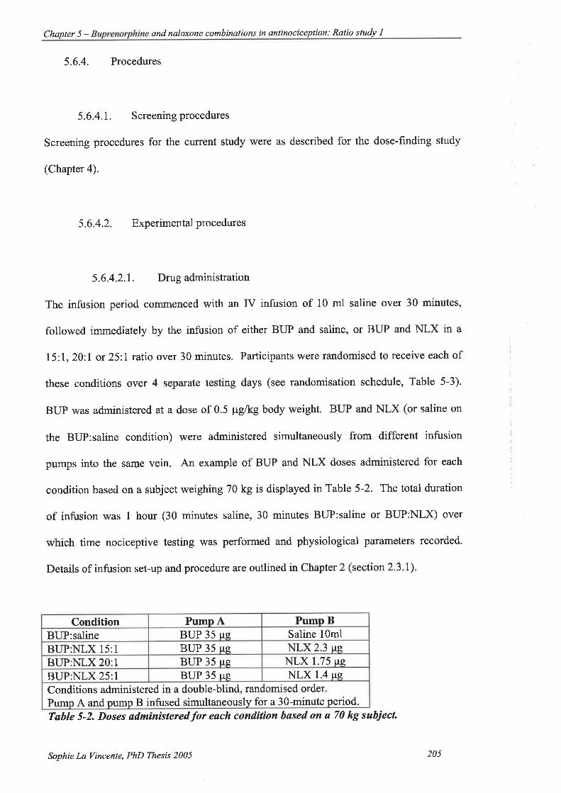
Chapter 5 - and naloxone combinations in antinociception: Ratio study I
5.6.4. Procedures
5.6.4.1. Screeningprocedures
Screening procedures for the curïent study were as described for the dose-finding study
(Chapter 4).
5.6.4.2. Experimentalprocedures
5.6.4.2.1. Drug administration
The infusion period coÍtmenced with an IV infusion of 10 ml saline over 30 minutes,
followed immediately by the infusion of either BUP and saline, or BUP and NLX in a
l5 1, 20 l or 251 ratio over 30 minutes. Participants were randomised to receive each of
these conditions over 4 separate testing days (see randomisation schedule, Table 5-3).
BUP was administered at a dose of 0.5 ¡rg/kg body weight. BUP and NLX (or saline on
the BUP:saline condition) were administered simultaneously from different infusion
pumps into the sar.ne vein. An example of BUP and NLX doses administered for each
condition based on a subject weighing 70 kg is displayed in Table 5-2. The total duration
of infusion was I hour (30 minutes saline, 30 minutes BUP:saline or BUP:NLX) over
which time nociceptive testing was performed and physiological parameters recorded.
Details of infusion set-up and procedure are outlined in Chapter 2 (section 2.3.1).
Condition Pump A Pump BBUP:saline BUP 35 pg Saline 10ml
BUP:NLX 15:1 BUP 35 pe NLX 2.3 lteBUP:NLX 20:1 BUP 35 ttg NLX 1.75 pe
BUP:NLX 25:l BUP 35 pe NLX 1.4 tte
Conditions administered in a double-blind, randomised order
Pump A and pump B infused simultaneously for a 30-minute period.
Table 5-2. Doses ødminìsteredfor each condition bøsed on a 70 kg subiecL
Sophie La Vincente, PhD Thesis 2005 205

Chapter 5 - Buprenorphine and naloxone combinations in anlinociception Ratio study I
Session I Session 2 Session 3 Session 4
Subiect A BUP only l5: I 25:7 20:lSubiect B 20:l 15:1 BUP only 25:lSubiect C 20:l BUP only 15:1 25:lSubiect D BUP only l5: I 20:.7 25:ISubiect E 15:1 25:l 20:l BUP onlySubiect F 25:l BUP only 15:1 20:lTøble 5-3. Røndomìsation schedule for 7 heølthy volunteers. Drug condìtions were IVBUP:søtine (BIIP only) 0.5 pg/kg; IV BUP(0.5 pg/kg):NLX ìn a 75:7, 20:1 and 25:1
røtio. BUP and sølineî,{LX infused simultøneously into the same vein over 30-minutes.
Subjects røndomised to receive each conditìon across 4 indíviduøI testing days.
Rsndomisation schedule was created and administered by hospital pharmacy
responsible for drug preparøtìon.
5.6.4.2.2. Testing protocol and schedule
Subjects were delivered from their homes to the testing centre by taxi in the morning.
Subjects had been instructed to refrain from taking any drugs or medication in the 24-hours
prior to testing (excluding the contraceptive pill) and to eat a light breakfast on the
morning of testing. A urine sample was taken and tested by an independent laboratory for
drugs of abuse (opioids, cannabinoids, benzodiazepines and sympathomimetic amines)
and, for female subjects, pregnancy.
Assessments were made at nine time points throughout each testing day according to the
procedures described in section 2.3.2. Each assessment time point involved taking a blood
sample, recording nausea, sedation and physiological parameters, and completion of the
nociceptive tests. This testing procedure took place prior to infusion, twenty minutes after
the commencement of each infusion (saline and then BUP with saline or BUP with NLX),
and then hourly upon completion of the infusions over a 6-hour washout period.
Subjective effects were also recorded throughout the testing day. Subsequent reference to
testing time points are made according to the description in Table 5-4'
Sophie La Vincente, PhD Thesis 2005 206
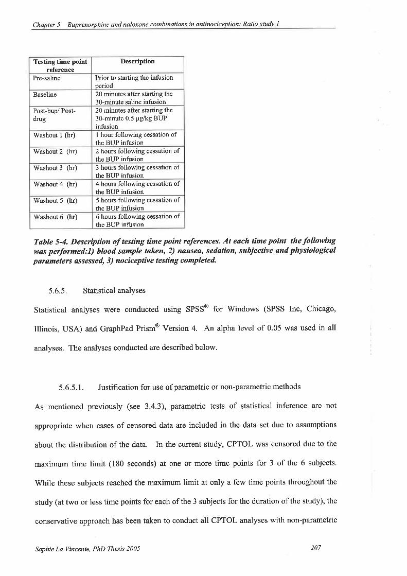
Chapter 5 - Buprenorphine and naloxone combinations in antinociception Ratio study l
Testing time pointreference
Description
Pre-saline Prior to starting the infusionperiod
Baseline 20 minutes after starting the30-minute saline infusion
Post-bup/ Post-drug
20 minutes after starting the
30-minute 0.5 pglkg BUPinfusion
Washout I (hr) I hour following cessation ofthe BUP infusion
Washout 2 (hr) 2 hours following cessation ofthe BUP infusion
Washout 3 (hr) 3 hours following cessation ofthe BUP infusion
Washout4 (hr) 4 hours following cessation ofthe BUP infusion
Washout 5 (hr) 5 hours following cessation ofthe BUP infusion
Washout 6 (hr) 6 hours following cessation ofthe BUP infusion
Table 5-4. Description of testing time poínt references. At each lìme point thefollowìngwas performed:I) hlood sample taken, 2) nauseø, sedatìon, subjective and physiologicalpørameteß øssessed, 3) nociceptíve testìng completed.
5.6.5. Statistical analyses
Statistical analyses were conducted using SPSS@ for Windows (SPSS Inc, Chicago,
Illinois, USA) and GraphPad Prism@ Version 4. An alpha level of 0.05 was used in all
analyses. The analyses conducted are described below.
5.6.5.1. Justification for use of parametric or non-parametric methods
As mentioned previously (see 3.4.3), parametric tests of statistical inference are not
appropriate when cases of censored data are included in the data set due to assumptions
about the distribution of the data. In the crürent study, CPTOL was censored due to the
maximum time limit (180 seconds) at one or more time points for 3 of the 6 subjects.
While these subjects reached the maximum limit at only a few time points throughout the
study (at two or less time points for each of the 3 subjects for the duration of the study), the
conservative approach has been taken to conduct all CPTOL analyses with non-parametric
Sophie La Vincente, PhD Thesis 2005 207

Chapter 5 - Buprenorphine and naloxone combinations in antinociception: Ratio study I
techniques. Other parameters met the assumptions for parametric methods (Tabachnick
and Fidell 2000) and have therefore been analysed with these methods.
5.6.5.2. Establishingbaseline response
Differences in each parameter before and after the 3O-minute saline infusion were assessed
using paired samples t-tests. The purpose of this was to determine whether any change in
nociceptive response or physiological parameters may be attributed to the infusion process
itself, and also provided an indication of the reliability of the baseline from which changes
in nociception were measured.
5.6.5.3. Assessing the effect associated with each drug condition
To assess the effect associated with each drug condition (BUP only, BUP:NLX l5;7,20:I
and25;l), changes from baseline were assessed for each parameter using one-way repeated
measures analysis of variance (ANOVA) for each condition (BUP only, and each
BUP:NLX ratio). These analyses included results from assessment time points at Baseline,
Post-drug and Washout t hour. Results from subsequent time points were not included as
drug effect may reasonably be expected to be most distinct at the assessments immediately
following drug administration (Post-bup and Washout thr). Including all time points in
these analyses (to 6 hours following cessation of drug administration, by which time the
drug effect may be minimal) may have masked a significant effect early in the post-drug
period. Furthermore, it has previously been reported that ANOVA using all time points is
not appropriate when a biphasic response is anticipated due to a potential overestimation of
variance (Angst et al. 2003), such as in cases where effects during both administration and
washout of a drug are assessed. Tukey's Multiple Comparison Test was used for post-hoc
analyses to identifu significant relationships between individual time points when a
significant effect had been observed with ANOVA.
Sophie La Vincente, PhD Thesis 2005 208

Chapter 5 - Buprenorphine and naloxone combinations in anlinociception: Ratio study I
As described, non-parametric tests of statistical inference were used to assess changes in
CPTOL due to the potential for censored data included in these results (i.e. cases where the
subject has not reached maximum tolerable pain before the forced cut-off point on the
tests). Friedman's analysis of ranks (a non-parametric equivalent to the one-way repeated
measures analysis of variance) was used to analyse these data. Consistent with the other
analyses, only Baseline, Post-bup and'Washout thr were included in these analyses. When
significant differences were found, a series of Dunn's Multiple Comparison tests were
carried out to provide post-hoc comparisons of the Mean ranks'
5.6.5.4. Comparing the effect of each BUP:NLX ratio with BUP alone
As baseline values were different between conditions, data \ilere expressed as percent
change from baseline in order to compare the effect associated with each ratio with the
effect of BUP alone. Each participant's nociceptive and physiological response at each
time point for each condition was expressed as a percent change from baseline response
according to the equation below. Data are expressed as the mean (+SEM) of these values
at each post-drug time point for each condition.
Post-drug latency - baseline latency *100baseline latency
As mentioned, ANOVA including all time points is considered inappropriate due to the
nature of the data. Two-way repeated measures ANOVA comparing BUP alone with each
BUP:NLX ratio using only the 3 time points described above would assess the effect of the
addition of the NLX at each ratio early in the post-drug period. However, the magnitude
of effect during the latter phase of the washout period was also of interest. Furthermore,
no non-parametric equivalent (to anaþse the CPTOL data) of the two-way repeated
measures ANOVA was available with either of the statistical programs employed.
Sophie La Vincente, PhD Thesis 2005 209

Chapter 5 - and naloxone combinations in antinociception: Ralio study I
Therefore, to assess the effect of the addition of NLX at all time points, and for the sake of
consistency in analysis of nociceptive and physiological parameters, paired samples t-tests
(and the non-parametric equivalent, Wilcoxon signed ranks, for the CPTOL data) were
used to compare parameters at each time point for BUP alone and each BUP:NLX ratio.
5.6.5.5. Comparing the effect of each ratio
In addition to comparing the effect of each ratio compared to BUP alone significant
differences between the antinociceptive effects of each BUP:NLX ratio were investigated
across all time points. As tolerance to the nociceptive stimuli was the primary outcome
measufe, these further analyses were only conducted with CPTOL and ESTOL.
To investigate differences in antinociception between the BIIP:NLX ratios, the percent
change from baseline associated with BUP alone at each time point was subtracted from
the percent change in nociceptive tolerance associated with each BUP:NLX ratio at the
corresponding time point. While this illustrates the time course of antinociception
associated with each ratio, the area under the percent change BUP:NLX minus percent
change BUP only curve (AUC) from baseline to the end of the monitored washout period
was calculated for each ratio to demonstrate the magnitude of change associated with the
addition of NLX over the entire dosing and washout period3. AUC was calculated by the
trapezoidal method. As the statistical software package used to calculate AUC did not not
assign a negative value to peaks below the x axis, negative and positive peaks were
calculated separately, and negative peak area subtracted from positive peak area for each
subject. V/ilcoxon signed rank tests and paired samples t-tests were used to assess mean
differences in AUC between the ratios for CPTOL and ESTOL, respectively.
3 Aucl%changesup,Nlx- o/ochangesup oory) for each subject in each ratio
Sophie La Vincente, PhD Thesis 2005 210
Chapter 5 - Buprenorphine and naloxone combinations in antinociception: Ratio study 1
5.6.5.6. Subjective and other effects
These effects are reported in tabular form according to effect, subject and time point during
which the effect was experienced. Where appropriate the severity of the effect is also
reported. No tests of statistical inference have been applied to these data.
5.7 . Results
This section is divided into five parts: participant withdrawal and missing data, analysis of
pre- and post-saline results to establish baseline, results from nociceptive testing,
physiological measurements, and the incidence of adverse effects. The antinociceptive
parameters are outlined according to each test, in the order CPTHR, CPTOL, ESTHR, and
ESTOL.
5.7 .1. Missing data and participant withdrawal/exclusion post-recruitment
5.7.I.1. Participant withdrawal
A 33-year old male subject attended for the first testing day, but was subsequently
excluded due to nausea (without vomiting) and lighfheadedness. The participant received
BUP:NLX in a 15:1 ratio. The nausea and light-headedness were of short duration
(approximately 5 minutes) but the participant reported "not feeling too good" at several
subsequent time points, stating on each of these occasions that he felt too ill to undergo the
pain testing. Throughout the intervals between nociceptive testing (50-55 minutes) the
participant did not complain of any significant adverse effects. It is considered that this
participant's failure to continue with the testing is due in part to an unwillingness to
undergo testing procedures for the three subsequent scheduled testing days. The subject
stated that he was disinclined to continue with the study due to the adverse effects he had
Sophie La Vincente, PhD Thesis 2005 2II

Chapler 5 - Buprenorphine and naloxone combinations in antinociception. Ratio sludy l
experienced. The subject was remunerated $4U250 for his participation. An additional
male participant was recruited in his place.
5.7.1.2. Missing data
One participant (45-year old female) retumed an opioid positive urine on the 20:1 testing
day. Data from this participant for the 20:1 testing day have been excluded. Where
comparison is made with the BUP only condition (such as in mean difference between
performance during a BUP:NLX condition and BUP only condition), the results from this
participant for the BUP only condition have also been omitted to avoid confounding
results.
5.7 .2. Pre- and post-saline infusion
Paired samples t-tests were conducted to detect any significant differences between
nociceptive and physiological parameters before and after the 30-minute saline infusion.
No significant differences were detected on any parameter at pre-saline testing and post-
saline testing þ>0.05). Thus it is considered that the infusion process itself had no
significant impact on nociceptive or physiological parameters. In all subsequent analyses,
then, post-saline values have been used as baseline response.
5.7.3. Cold pressor threshold
5.7.3.1. Effect of each conditton
Mean (+SEM) CPTHR values for each condition across all time points are presented in
Figure 5-1. One-way ANOVA conducted for each condition over three time points
(Baseline, Post-bup and Washout lhr) revealed a significant increase in the BUP only
condition (F2Js:4.52, p:0.039) (see Table 5-5). Tukey's multiple comparison test
SophieLaTincente, PhD Thesis 2005 212

Chapter 5 - Buprenorphine and naloxone combinations in øntinociception Ratio study I
revealed a significant difference between Baseline and Post-bup (p<0.05). No significant
differences were observed in any of the BUP:NLX ratios. While there was a considerable
increase in CPTHR in the 25 l ratio condition, there was a high degree of inter-individual
variability in response. This variability may be attributed to the results of one subject (30
year old female) who experienced a peak increase in CPTHR approaching 300 %. This
subject demonstrated a marked increase in CPTHR in the l5:1 condition also, although this
was of a lesser magnitude (approximately 50%).
+BUP only -+15:l
tt,t,coIØ
É.!t-(!o
thttcoIoÉ.IFùo
C:".""s-"t-"c-"o-T""'-TIME
-"S"J-t"""o.yds'.vd-""'TIME
thttcoIøÉ.IFf!o
thItcoIoÉ.IFo-o
""o-""-nc"---"-.""'-TIME
"lþ.
"4:t'
-e-20.1 +25 1
".,ìþ.""".
os'
"dTIME
Fìgure 5-1. Mean (+SEM) CPTHR (seconds) for each condition (BUP only {n:6},B(IP:NLX in a 15:1 {n:6}, 20:1 {n:5} ønd 25:1 {n:6} røtio) over all time points(bøselìne, foltowing un IV B(IP ìnfusion {0.5 pg/kg over 30 minutes}, and hourlythroughout BUP wøshout to 6 hrs). *p<0.05, signítìcant change over Baseline, Post-bup
and Washoat Lhr, one-wøy repeated-meøsates anølysis of vatíønce.
Sophie La Vincente, PhD Thesis 2005 213
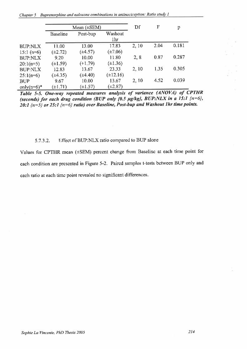
Chapter 5 - and naloxone combinations in anlinoeiception: Ratio study I
Mean (tSEM) Df F p
Baseline Post-bup 'Washout
thrBUP:NLX15:1 (n:6)BUP:NLX20:1(n:5)BUP:NLX25:l(n:6)BUPonly(n:S)x
11.00(+2.72)
9.20(+1.5e)
12.83(+4.3s)
9.67(+1.71)
13.00(+4.s7)10.00
(+r.7e)t3.67
(+4.40)10.00
(+1.s7)
17.83(+7.06)11.80
(+1.36)23.33
(+12.16)13.67
e2.87\
2,10 2.04 0.181
2,8 0.87 0.287
2, t0 1.35 0.305
2,10 4.52 0.039
Table 5-5. One-way repeøted meøsures ønalysis of variønce (ANOVA) of CPTHR(seconds) for each drug condition (BUP only {0.5 þg/kg}, BUP:NLX ín ø 15:1 {n:6},20:1 {n:5} or 25:1 {n:6} ratio) over Baseline, Post-bap ønd Washout thr time points.
5.7.3.2. Effect of BUP:NLX ratio compared to BUP alone
Values for CPTHR mean (+SEM) percent change from Baseline at each time point for
each condition are presented in Figure 5-2. Paired samples t-tests between BUP only and
each ratio at eachtime point revealed no signif,rcant differences.
Sophie La Tincente, PhD Thesß 2005 214

Chapter 5 - and naloxone combinations in antinociception: Ratio study I
o,E).E
osÉ,¿o-o
{- BUP only
+15:1
./".rs'"rs'"rs'bst aro'
oau setos'
$Pu
{- BUP only+2O:1
--r- BUP only+25.1
TIME
Fígure 5-2. Meøn (+*SEM) CPTHR (seconds) percent change -from Baselìne forBUP:NLX røtìos (15:1 {n:6}, 20:1 {n:5} and 25:1 {n:6}) cornpøred to BUP only {n:6}across all time points (bøseline, followìng øn IV BUP infusion {0.5 þg/kg over 30
mìnutes], and hourly throughoat B(IP washout to 6 hrs). p>0.05' no signift'cønt
difference -fro* BUP only, pøíred sømples t-tests. Note thøt data for the BUP only
condìtíon øre presented in all 3 grøphs.
o,ct)(Ú
(,)
sÉ.IFo.o
oo)Lq
!()
sÉ.4Fo-o
."""".$.""J."S"S"S"S
.""""s-"J"-"rs-r$.u*-$
Sophie La l/incente, PhD Thesis 2005 2Is

Chqpter 5 - Buprenorphine and naloxone combinations in antinociceplion Ratio study I
5.7 .4. Cold pressor tolerance
5.7.4.I. Effect of each condition
Mean (+SEM) CPTOL for each condition is presented in Figure 5-3. Friedman's two-way
analysis by ranks for each condition (BUP only, l5:1, 20:l and 25:l BUP:NLX ratios)
over three time points (Baseline, Post-drug and Washout lhr) revealed a significant
increase in CPTOL in the 15:1 condition only dQ):g.Z| p:0.005) (see Table 5-6).
Dunn's multiple comparison tests revealed significant differences in Baseline vs. Washout
lhr 0<0.05). There were no significant differences in CPTOL in the 20:l (p:0.367) or
25:l þ:0.142) conditions. The difference in CPTOL over these three time points in the
BUP only condition was approaching significance þ:0.072).
Sophie La Vincente, PhD Thesis 2005 216

Chapter 5 - Buprenorphine and naloxone cornbinations in antinociception: Ratio study I
tttttEoo0)tl,
Jot-o-o
o!to(¡c)tt,
Jot-o-o
.rS(
"È-
,.s{ os{
C"Aoncnncnn$*¡doocTIME
øIttrooottJot-o-o
anItoo(,tt,
JoFo-o
+'15: I
{- BUP only
S ,o\' os(
.A .rd" C
,oS* oS(
"f .*dons"oc.ocTIME
*s*o$onco¡s ,4:."TIME
*¡$*¡c
"ly TME
+25'.1+20'.1
,4ys-'
sr-
Fígure 5-3. Meøn (+SEM) CPTOL (seconds) þr eøch condìtion (BUP only {0.5 Fg/kg,n:6], BUP:NLX in a 15:1 {n:6}, 20:1 {n:5} or 25:1 {n:6} ratio) over all time poìnts(baseline, post-BtlP ìnfusion, and hourly throughout BUP washout to 6 hrs). **p<0.007,
signift.cant difference over Bøseline, Post-bup and Wøshout 7hr, Friedman's tesl
2Mean rank v
Baseline Post-drug Washoutlhr
15:1 (n:6)20:1(n:5)25:lQ=6)BUPonly(n:6)
1.001.90r.751.83
2.832.703.2s3.17
3.002.902.s82.83
9.33**0.370.144.73
Tahle 5-6. Meøn rank for eøch conditíon (BUP only 0.5 þg/kg {n=6}, and BUP{0.5pg/kg]:NLX ìn a 75:7 {n:6}, 20:1 {n:5} and 2_5:1 {r:6} ratìo) øt Baseline, Post-Bup
and Washout 7 hr among heøtthy volanteers. f sntitti" of Friedmøn's tesl of CPTOLover Bøselíne, Post-bup and Wøshout thr for each condition, **p<0.07.
Sophie La l/incente, PhD Thesis 2005 217
Chapter 5 - Buprenorph ine and naloxone combinations in anlinociception. Ratio sludy l
5.7.4.2. Effect of BUP:NLX ratio compared to BUP alone
Figure 5-4 displays mean (+SEM) percent change from baseline CPTOL at each time
point for each ratio compared to BUP only. The increase in CPTOL observed in the l5:1
ratio condition was significantly greater than observed with the BUP only condition at
Washout 2ltr (z:-2.20, p:0.028), 'Washout 3hr (z=-1.99, p:0.046) and Washout 5fu (z:-
2.20, p:0.028). A significantly greater increase in CPTOL was observed in the 20:l rutio
condition compared to BUP only at the Washout 3hr time point (z:-2.02, p:0.043).
Sophie La Vincente, PhD Thesß 2005 218

Chapter 5 - Buprenorphine and naloxone combinations in antinociception Ratio study I
(l)
=(I)ar,(u-oEo(l)o)(¡Eos
5
4
3
2
I
*BUP ONLY
+15'.1
*BUP ONLY
*20'.1
*BUP ONLY*25.1
(l).=õØ(õ_o
Eo(1)
o)c(!(.)
s
3
2
I
ÈooÈÈ
-1
ooolÈÈ
oo*Ë
JJJooo*SÈ
I
2
1
oÈ
oU'(Ù
@
cc)U)o
@
(l)
.gõØ(l'-oEoL
(l)o)c(E-cos
(t,oa
oË
55ooÈË
TIME
Fìgure 5-4. Meøn (+SEM) CPTOL expressed as percent change from Baseline forBUP(0.5 pgftþ:NLX rat¡os (15:1 {n:6}, 20:1 {n:5}, 25:1 {n:6}) compøred to BUPonly (0.5 Fg/kg, n:6) IV ìnlusíon over 30 minutes across øll time points (Baselìne, Post-
bap, ønd hourly throughout BUP washout to 6 hours). *p<0.05, sígnìJicønt difference
-from BUP only, Wilcoxon signed ranks. Nole thøt døtø for the BUP only condìtìon arepresented ìn øll3 graphs.
Sophie La Vincente, PhD Thesis 2005 219
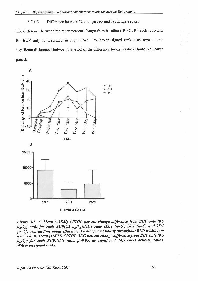
Chapter 5 - and naloxone combinations in antinociception: Ratio study I
5.7 .4.3. Difference between oá change¡,qs6 and o/o changesup oNLY
The difference between the mean percent change from baseline CPTOL for each ratio and
for BUP only is presented in Figure 5-5. V/ilcoxon signed rank tests revealed no
significant differences between the AUC of the difference for each ratio (Figure 5-5, lower
panel).
A
""e- 15:1
+20:1+25:1
ão,È È
TIME
1
15;f 2t:1
BUP:NLX RATIO
25:1
Fìgare 5-5. 4. Mean (uSEM) CPTOL percent change difference from BUP only (0.5
F{kg, n:6) for each B(IP(0.5 pg/kþ:NLX røtio (15:1 {n:6}, 20:1 {n:5} ønd 25:7
{n:6}) over øll tìme points (Baselíne, Post-bup, and hourþ throughout BUP wøshout to
6 hours). þ. Mean (+*SEM) CPTOL A(IC percent change difference from BUP only (0.5
pg/kÐ for each BIIP:NLX ratio. p>0.05, no signijicønt differences between ratios,
Wilcoxon signed ranks.
-1
àöû-ffoEoL{t)çco{l)Fõ{¡)ö)$rJ
ñ
2
1
*È9:¡J::9??ÈÈÈ
?È
Ë
1
Sophie La Vincente, PhD Thesis 2005 220

Chapter 5 - and nøloxone combinations in antinociception: Ratio study I
5.7.5. Electricalstimulationthreshold
5.7.5.1. Effect of each condition
Mean (ISEM) ESTHR values for each condition are presented in Figure 5-6. One-way
analysis of variance (ANOVA) was conducted for each condition (BUP only, 15:1, 20:1
and25:I BUP:NLX ratios) over three time points (Baseline, Post-bup and Washout thr) to
determine whether a significant change in ESTHR was associated with any condition post-
drug administration (see Table 5-7). These analyses revealed no significant differences in
any condition over the three time points assessed.
+BUP only +15:1
/ìåc
øo
É.IFattl¡J
øo
É.¿Þv>IJJ
,f-'
sPoTIME
r.S( ,tr-'
*P" c
Ø
o
É.Il-at,t¡l
øo
ú.¿l-anl¡J
sPo seu
ñ'dseu sPo
dsPu
..ct-sa ,.S( 4t't ,C ñ( d d.r{d o¡* seo seo s¡* s¡u s¡*
TIME
+20:1
6-'
+25:1
6-' 6-',ft .f-t ñ( 6.'
sPo seo sPo s¡* sPs"4å"""$..rJor.f-*.'tr'TIME TIME
Fígare 5-6. Meøn (uSEM) ESTHR (volts) for each condition (BUP only{0.S pg/kg' n=6},
BUP:NLX in a 15:1 {n:6}, 20:1 {n:5} or 25:1 {r:6} ratío) over all thne points(Baseline, Post-bup, and hourly throaghout BUP washout to 6 hours) among heølthyvolunteers. p>0.05, no signìJìcant vøriønce over Baseline, Post-bup snd Washoul 7hr,
one-way repeøted measures ANOVA.
Sophie La Vincente, PhD Thesis 2005 221

Chapter 5 - Buprenorphine and naloxone combinations in antinociception: Rqlio study l
MeanBaseline Post-drug
Df F p
Washoutthr
l5:1 (n:6)
20:1(n:5)
25:1(n:6)
32.33(+3.70)36.00
(+4.6e)35.33
(+2.s1)29.33
32.00(+3.23)36.00
(+3.7e)35.33
(+2.40)31.33
(+2.61\
33.00(+3.86)36.00
(+3.35)33.00
(+1.77)3r.33
(+2.1l)
5, 15 0.393 0.685
4, 12 0.000 1.000
5, 15 0.683 0.527
5, 15 0.750 0.497BUPonly(n:6) (+3 .4e)
Tøble 5-7. One-way repeated measures analysis of varìønce (ANOVA) of ESTHR (volrs)
for eøch condition (BUP only {0.5 Fg/kg, n=6}, and BUP:NLX in a 15:1 {n=6}' 20:1
{n:5} or 25:1 {r:6} røtio) over Bøseline, Post-bup und ryashout thr time points amonghealthy volunteers, p>0.05, no signiftcant dífferences.
5.7.5.2. Effect of BUP:NLX ratio compared to BUP alone
ESTHR mean (+SEM) percent change from baseline at each time point for each BUP:NLX
ratio compared to BUP only are presented in Figure 5-7. Paired samples t-tests revealed
no significant differences between BUP only and any of the BUP:NLX ratios.
Sophie La Vincente, PhD Thesis 2005 222
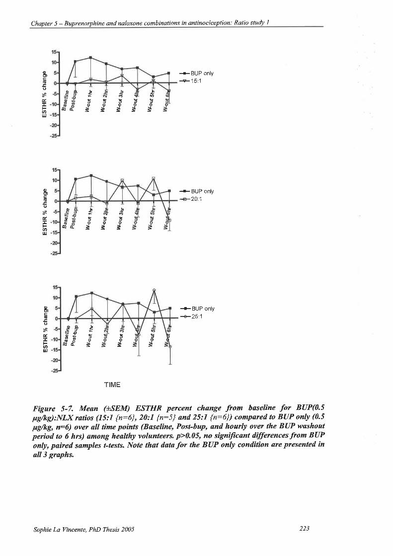
Chapter 5 - Buprenorphine and naloxone comb inations in antinoc iceplion : Ratio study I
L
4)
oË
L
fÞ
l!l!ÈoooËËË
L
oË
oc''(ú
osÉ.ÅFanIJJ
ocoU'o
4¡
+BUP only
+15:1
+- BUP only.+201
+BUP only
+251
aÞ
5o
Ëo
Ë5o
Ë
oE)
ooñÉ.I
(tt¡J
ocott,tù
@
5o
Ë
oCll-:õ Èo¿Ê
fo8 s
oct)e.(ü
osÉ.I
.hul
5o
Ë
Jo
Ë
TIME
Fígure 5-7. Mean (+SEM) ESTHR percenl chønge from bøseline for BUP(0.5pg/kg):NLX rutios (15:1 {n:6}, 20:1 {r:5} and 25:7 {n:6}) compared to BUP only (0.5
ltg/kg, n=6) over all time po¡nts (Bøseline, Posl-bup, ønd hourly over the BUP washoulperiod to 6 hrs) ømong heølthy volunteers. p>0.05, no signiJicønt dìfferences from BUPonly, pøìred samples t-tests. Note thøt data for the BUP only condition are presented ín
all3 graphs.
Sophie La Vincente, PhD Thesis 2005 223
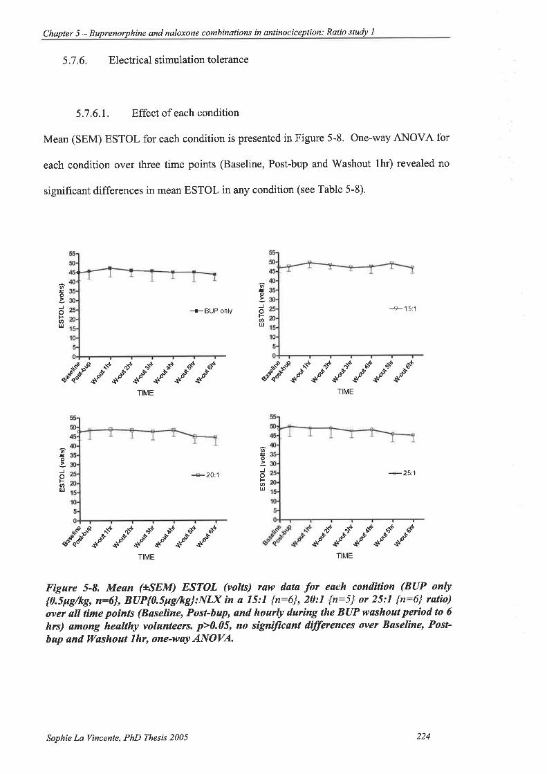
Chapter 5 - Buprenorphine and nøloxone combinalions in antinociception: Ratio study I
5.7.6. Electricalstimulationtolerance
5.7.6.1. Effect of each condition
Mean (SEM) ESTOL for each condition is presented in Figure 5-8. One-way ANOVA for
each condition over three time points (Baseline, Post-bup and Washout lhr) revealed no
significant differences in mean ESTOL in any condition (see Table 5-8).
+BUP only +15:1
Ø
o
-Jot-.J'IJJ
ttt
o
JoÞatr¡J
Cþ--r$.d."-..".o-..s'.r*". -"ìå"o"-."-."-."*-"-.".-TME TIME
oo
Jot-U'l¡l
oo
JoÞat)Lrl
+2O..1 +25:.1
C:c.d."s"s.,s"-."."*"r$*rSC**"$
65'
$fTIME TME
Figure 5-8. Mean (+SEM) ESTOL (volrs) raw data þr each condition (BUP only
{Q.Spg/kg, n:6}, BUP{0.5pg/kg}:NLX in a 15:7 {n:6}, 20:1 {n:5} or 25:1 {n:6} ratìo)
over all tíme points (Bøselìne, Post-bup, and hourly daríng the BUP washout perìod to 6hrs) ømong healthy volunteers. p>0.05, no sìgnijicønt differences over Baseline, Post-
hup and W'ashout lhr, one-way ANOVA.
Sophie La Tincente, PhD Thesis 2005 224

Chapter 5 - and naloxone combinations in antinociception: Ratio stuþ I
Mean
Baseline Post-drug
Df F p
Washoutthr
15:l (n:6)
20:1(n:5)
25:1(n:6)
47.0(+2.67)47.60
(L4.4e)48.67
(+s.26)45.0
47.67(+2.60)48.40
(+4.79)50.0
(+5.68)45.67
(+4.27)
49.67(+2.7s)48.80
(+3.4e)49.0
(+4.19)47.33
(L4.72)
2, t0 1.429 0.285
2,8 0.528 0.609
2,10 0.389 0.687
2, t0 1.032 0.391BUPonly(n:6) (t4 .34',,
Table 5-8. Meøn (+-SEM) ESTOL for eøch conditìon (BUP only {0.5p9/kg, n=6},
BUP{0.5ps/ks}:NLX in a 15:1 {n:6}, 20:1 {n:5} or 25:1 {n:6} ratio) at Baseline, Post-
bup ønd Wøshout thr tìme points ømong healthy volunteers. p>0.05' no signiftcantdifference across tìme poinls in øny condìtíon.
5.7.6.2. Effect of BUP:NLX ratio compared to BUP alone
Paired samples t-tests revealed no significant differences in percent change from baseline
for ESTOL between BUP only and any BUP:NLX ratio (see Figure 5-9).
Sophie La Vincente, PhD Thesis 2005 225

Chapter 5 - and naloxone combinations in antinociception: Ratio study I
(¡,Ðtr(!
!¡J
sJoØt¡J
c,,so,at,lÙ
@
t=ÈËoôoËsË
+BUP only+15:1
+BUP only
+2O:1
+BUP only
+25'.1
oË
oÐ.U
osJoFU't¡J
oQ,ÊJo¿U'6
d o-o
5os
5555ooooËÉËË
5o
Ë
oÞ)co(J
soFant¡¡
oQ.
EÈdf ooËË
TIME
Fígure 5-9. Mean (+SEM) ESTOL percent change .from høselìne for BUP (0.5
pg/kg):NLX røtios (15:1 {n:6}, 20:1 {r:5} and. 25:1 {n:6}) cornpared to BUP only (0.5
Fg/kg, n:6) øcross all tûme points (Bøseline, Post-bup, ønd hourly ovet BUP iltashoatperiod to 6 hrs) among heøtthy volunteers. p>0.05, no sìgniftcønt dífferences from BUPonly, pa¡red sømples t-tests. Note thøt datø for the BUP only condition are ptesented inøll3 graphs.
Sophie La Vincente, PhD Thesis 2005 226

Chapler 5 - and naloxone combinations in antinociception: Ratio study I
5.7 .6.3. Difference between o/o change¡,q¡s and o/o changesuP oNLY
The difference between the mean percent change from baseline CPTOL for each ratio and
for BUP only is presented in Figure 5-10. Paired samples t-tests revealed no significant
differences between the AUC of the difference for each ratio (p<0.05).
+15:1+20.1+25.1
TIME
I 5:1
BUP:NLX RATIO
Fígure 5-10. A. Mean (+SEM) ESTOL percent change difference -from BUP only foreach B(IP(0.5 pg/kg):NLX ratio (15:1 {n:6}, 20:1 {n:5} and 25:I {n:6}) over all tíme
points (Baselìne, Post-bup, and hourly during BUP wsshout for 6 hrs) ømong healthy
volunteers. B. Mean erSEM) ESTOL AUC percent change difference -from BUP only
for eøch BUP:NLX røtio. p>0.05, no sígníJìcant dffirences hetween ratios, pøired
sømples t-tesls.
A
ss sJ èts ss sJ ss.-8oSø-{
É"e
S **' t*t o*t ,o*t a*'
coÈft0EoÈ(1,(,Ê0,oÈ!loE')
(É
oàR
B
1
,l1 25:1
Sophie La Vincente, PhD Thesis 2005 227
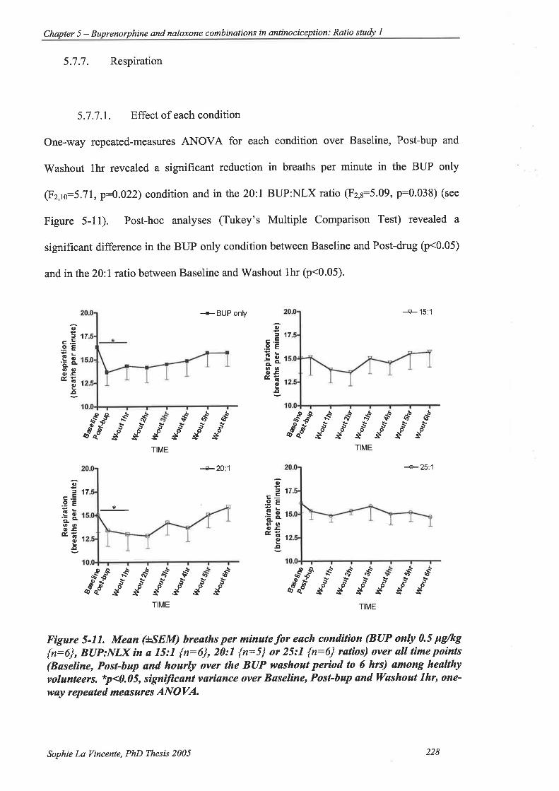
Chapter 5 - Buprenorphine and naloxone combinations in antinociceplion: Ratio study I
5.7.7. Respiration
5.7.7.1. Effect of each condition
One-way repeated-measures ANOVA for each condition over Baseline, Post-bup and
Washout thr revealed a significant reduction in breaths per minute in the BUP only
(F2Js:5.71, p:0.022) condition and in the 20:l BUP:NLX ratio (F2,s:5.09, p:0.038) (see
Figure 5-11). Post-hoc analyses (Tukey's Multiple Comparison Test) revealed a
significant difference in the BUP only condition between Baseline and Post-drug þ<0.05)
and in the 20:l ratio between Baseline and'Washout thr (p<0.05).
+ BUP onV +15:1
+25:1
7a=1c.E
.9EEõr'ao-'u, aDosÉË.
Ol-o
17
1
o
c.E.9EEõ'õ o-øttC' .CEË
0¡
.o
*s* .n*t ,*t o*t or*t "*'s*s*s*s*5s*sosssss
TIME
o*t or*t a*'s*SSooosss5osTIME
*{ ,vs
ooss
0,
5l
.9EËbn'ã o.ano{,GÉË.
()l
I
17
d'.-*so$o$*$TIME
PQ\\€g^ii
8sq
"'Ìd
.-€osg*ii
+20 I
(,
=.9EEõ'aa]t u,O'GÉË
o-o
7
1
a*' n*' "*'s*Sooss
Fùgure 5-11. Mean (+SEM) breaths per mínute for eøch condition (BUP only 0.5 Fg/kg{n:6}, BUP:NLX ìn a 15:1 {n:6}, 20:1 {n:5} or 25:1 {n:6} ratíos) over all time poìnts(Baselìne, Post-bup and hourþ over the BUP washoat period to 6 hrs) ømong heølthy
volunteeß. *p<0.05, signiJìcønt vøriance over Basel¡ne, Post-bup and W'ashout 7hr, one-
way repeøted meøsures ANOVA.
S **t r*'s*.S.Ys*s*Sdooooo.ssssssTME
Sophie La Vincente, PhD Thesis 2005 228
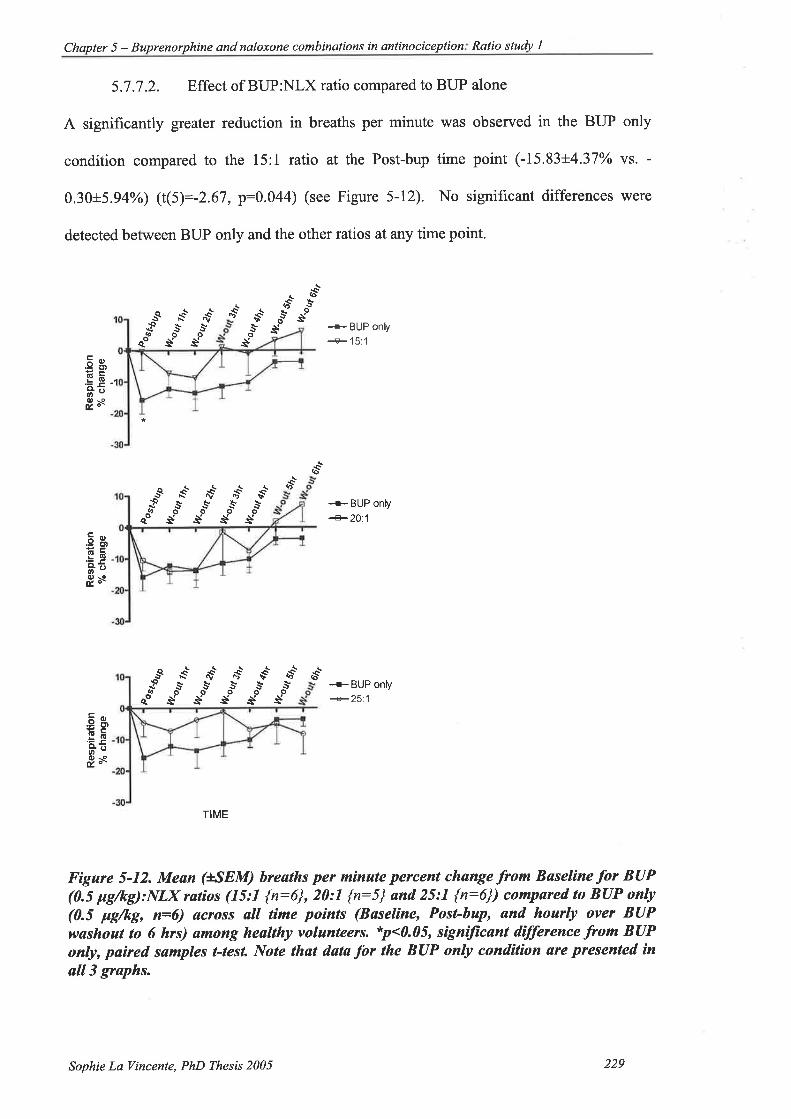
Chapter 5 - Buprenorphine and naloxone combinations in antinociception Ratio study l
5.7.7.2. Effect of BUP:NLX ratio compared to BUP alone
A siguificantly greater reduction in breaths per minute was observed in the BUP only
condition compared to the 15:1 ratio at the Post-bup time point (-15.83+4.37Yo vs. -
0.30+5.94yo) (t(5):-2.67, p:0.044) (see Figure 5-12). No significant differences were
detected between BUP only and the other ratios atany time point'
sQ€{qeo
s*oss*os +BUP only
+15:l
oQOrS
Eg'gË "*5 ''o\o
Éo-
.,*'t*'** .!*so
s5* ù*00ss
S *o' -*t oot n*t ao's* st .l* s* s*ooooasssss
.o{
o*_se S *ot ^,*t o*'
"g= s .ì* l* õ-
oSsoJs"so--r- BUp onv+2O:1
.ãsEEfi-oO-cÉo-
-+BUP only+25:1
TIME
Figare 5-12. Mean ($SEM) hreaths per m¡nate percent change from Baseline for BUP(0.5 pg/kg):NLX røtios (15:1 {n:6}, 20:1 {n:5} and 25:1 {n:6}) compured to BUP only(0.5 pg/kg, n=6) øcross all time points (Bøseline, Post-bup, and hourly over BUPwøshout to 6 hrs) among heølthy volanteers. *p<0.05, signíJìcant dìfference from BUPonly, paìred sømples t-tesL Note that data þr the BUP only condition ate presented inall3 graphs.
s*,o-6
qo
,E g'
.EEâõd) rodor
Sophie La Vincente, PhD Thesis 2005 229
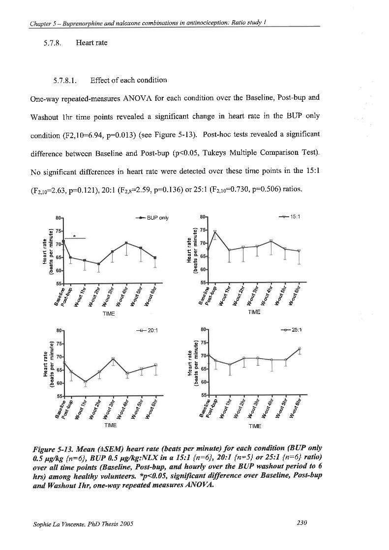
Chapter 5 - and naloxone combinalions in antinociception: Ratio stuþ I
5.7.8. H.eartratæ
5.7.8.1. Effect of each condition
One-way rspeated-measures ANOVA for each condition ovor the Baseline, Post-bup and
Washout thr time points revealed a significant change in heart rate in the BUP only
condition (F2,10:6.94, p:0.013) (see Figure 5-13). Post-hoc tests revealed a significant
difference between Baseline and Post-bup þ<0.05, Tukeys Multiple Comparison Test).
No significant differences in heart rate were detected over these time points in the l5:1
(F2,¡s:2.63, p:0.121), 20:l (Fz,s:2.59,p:0.136) or 25:1 (F2,16:0.730, p:0.506) ratios.
+BUP onV +15:1
+20:1 +25:1
()
=artfuFtbõo-9ø
(lr¡,¡
o={rtr
6E
:brú CL9ø
ñ(,¡t
or*t a*'55oossssoossi"/.*so$ /y*$u$ S,Ss*Soooossss
,*t o*t n*'
TME TME
oaoc
rúEËbOCL9ø
tú(,¡¡
()
(rÊaEttbOCL9o
(lc¡¡¡
8sa
rir* s*osTê5os st st st st
t*t ,r*'p-q
/:"so
.sooss
or" a*'*to- o-ssTIME TIME
Figure 5-13. Mean @SEM) heørt rate (beals per mìnute) for each condìtíon (BUP only0.5 pgftg {n:6}, BUP 0.5 pg/kg:NLX ìn a 15:1 {n:6}, 20:1 {n:5} or 25:1 {r:6} rutìo)over all time points (Baseline, Post-bup, and hoarþ over the BUP wøshout períod to 6hrs) among heølthy volunteers. *p<0.05, sìgnìftcant dílþrence over Baselìne, Post-bup
ønd Washout 7hr, one-wøy repeated meøsuÍes ANOVA.
Sophie La Vincente, PhD Thesis 2005 230
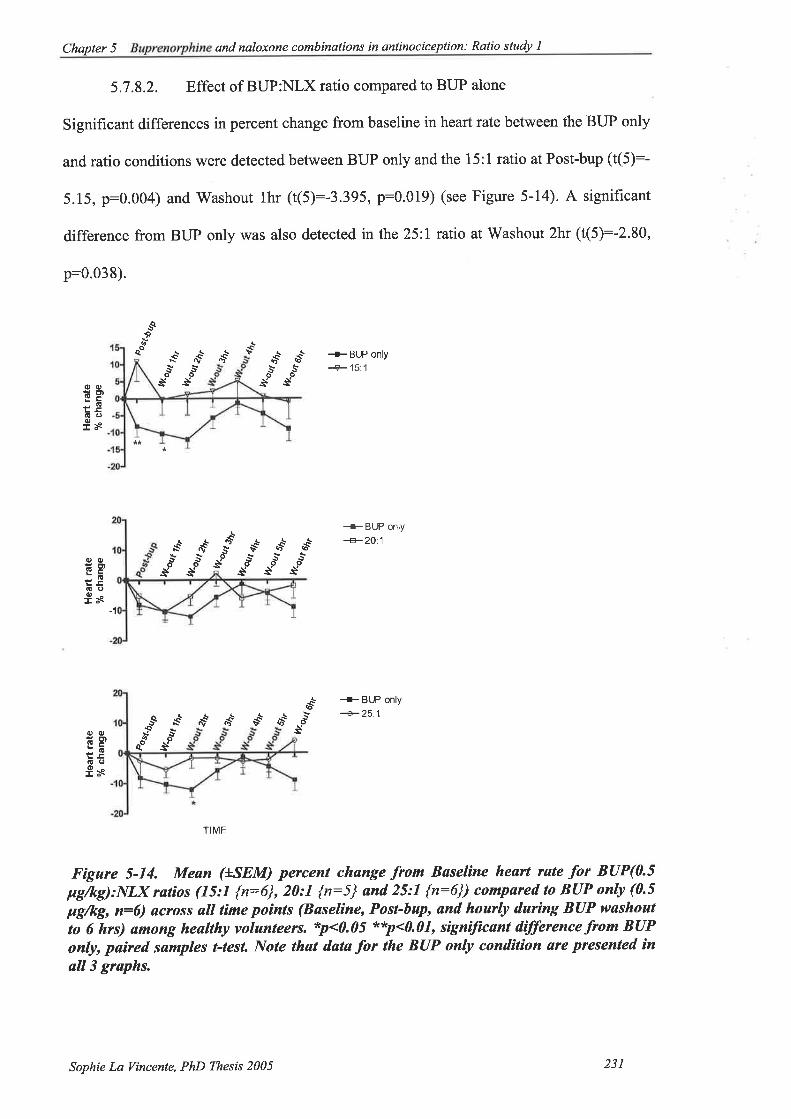
Chapter 5 - qnd naloxone combinations in antinociception: Ralio study I
5.7.8.2. Effect of BUP:NLX ratio compared to BUP alone
Significant differences in percent change from baseline in heart rate between the BUP only
and ratio conditions were detected between BUP only and the 15:1 ratio at Post-bup (t(5):-
5.15, p:0.004) and Washout thr ((5):-3.395, p:0.019) (see Figure 5-14). A significant
difference from BUP only was also detected in the 25ll ratio at Washout 2hr (t(5):-2.80,
p:0.038).
+BUP only+15:1
+BUP only+2O:1
*o' où
s
+BUP only
+25.1-Q.o
¿*qo
*s' r*t $ .*'
rqt
eo vtS *ot r*' or*t ao'
oE)cf!os
oG
Èoo!
ol* osssos osss
o*t tot a*'
s" *J *JootG
os
ol!
Gq,I
t*'o-s
*{* ,1"55ooss
oss
-1
a*'
octtcG
os
o(!
(!oI
TIME
Fígure 5-14. Meøn (LSEM) percent chønge from Baseline heart rste Íor BUP(0.5pg/kg):NLX ratios (15:1 {n:6}, 20:1 {n:5} ønd 25:1 {n:6}) compøred to BUP only (0.5
þg/kg, n:6) across all tíme poìnts (Bøseline, Post-bup, and hourly during BUP washout
to 6 hrs) among heølthy volunteers. *p<0.05 **p<0.01, signiJìcant difference from BUPonly, paired samples t-tesl Note that data for the BUP only condìlion øre presented ¡n
øll3 graphs.
Sophie La Vincente, PhD Thesß 2005 231

Chapter 5 - and naloxone combinations in antinociception: Ratio study I
5.7.9. Oxygen saturation
5.7.9.1. Effect of each condition
Mean arterial oxygen saturation over all time points for each condition is displayed in
Figure 5-15. One-way repeated measures ANOVA over the three initial time points
(Baseline, Post-bup and Washout thr) revealed no significant effect for BUP only
(F2,1s:2.83, p:0.106), 15:1 (F2,1e:0.66, p:0.538), 20:I (Fz,s:l .46, p:0.289) or 25:l
(Fz,ro:1 . I 5, p:0.354).
+BUP only -r 15:1
0t
Ê.9(E
=a!ttt(,ED
xo(,a,(,ô-
toIE
).EotoED
xo
{,a)q)À
c.9tú
GØt(,CD
xol,l,6)À
c.9l!
6ØEoCD
xoEo(,a,o.
-å-t
^ø -Q
a\o*o-
8oe
^9 rct'o *rõ
éoe
.a".S
8*"9
as'5o
.*
S **t r*t o*t "*'&!¡rkþ
ooooosssssss55'S5oooooossssss
**t **' .o*t o*t "*'
a*'
S **t ,*t o*t ,o*t a**
TME TME
+20:'l
or*' a*'
+25:1
sss5s5oooooos-s-ë s s sssoos*
S *** r*t o*'ssSsoooossssTME TME
Fìgure 5-15. Mean (+SEM) arterial orygen saturat¡on (%) for each cond¡l¡on (BUPonty 0.5 pg/kg {n:6}, BUP:NLX ìn a 75:7 {n:6}, 20:I {n:5} or 25:1 {r:6} ratio) over
øIl time points (Bøseline, Post-bup, ønd hourly during BUP washout perìod to 6 hrs)
among heatthy volunteers. p>0.05, no sìgnìftcant vsr¡ance over Baseline, Post-bap øndWøshout 7hr, one-way repeøted meøsuÍes ANOVA.
Sophie La Vincente, PhD Thesis 2005 232

Chapter 5 - Buprenorphine and naloxone combinations in antinociception Ratio study I
5.7.9.2. Effect of BUP:NLX ratio compared to BUP alone
Paired-samples t-tests revealed a significantly greater reduction in arterial oxygen
saturation in the BUP only condition compared to the 15:l ratio condition at Post-bup (-
0.67+0.33 vs. 0.34+0.34, t(5):-3'89, p:0.011), Washout 4hr (-0.84+0.31 vs' 0'00+0'26,
t(5)---2.70, p:0.04), and Washout 6hr (-0.67+0.34 vs. 0.68+0.21, t(5):-3.15, p:0'025).
There were no significant differences in oxygen saturation change from baseline between
BUP only and any other ratio (see Figure 5-16).
Sophie La Vincente, PhD Thesis 2005 233
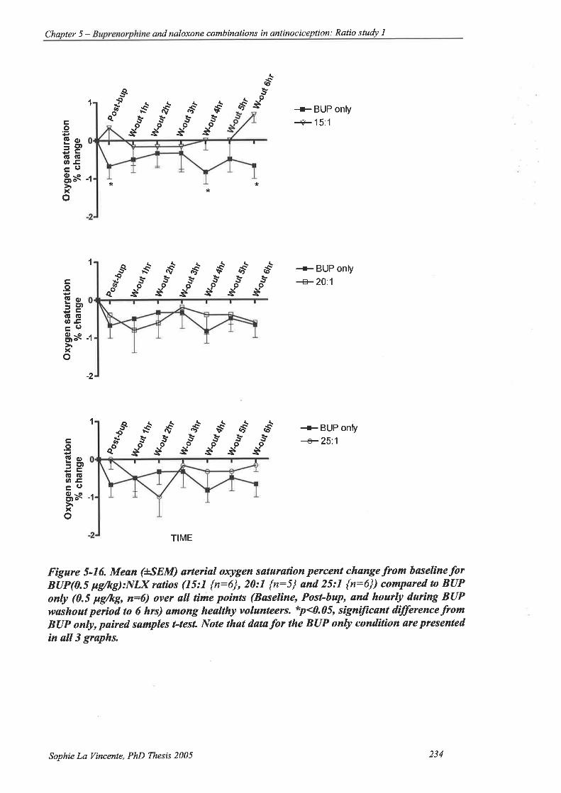
\3 È (ù \ I
Oxy
gen
satu
ratio
n%
cha
nge
I
Oxy
gen
satu
ratio
n%
cha
nge
itI
Oxy
gen
satu
ratio
n%
cha
nge
¡
:È
-{ m
ss*È
*ËëÈ
\èÈ
ì
'xiÌÈ
sÈ
Èì$
ËÈ
ñ sÊ
R
$ÈÈ
eÈ
ÈÈ
Fì$
G\í*
s ñ
E A
¡"
È.H
s"ÌÈ
Ì $
ËH
"ìi s
ÈÈ
ÈÈ
ËS
\ w
¡u.Ñ
N.S
EE
Þ
!a È ß F,r Þ o (\ $ ! û s $ s. lv s a ¡.) \
poV
.baþ
Þos
t bU
þÞ
%f%
Þ o G o ù ø a o c) (\ \ o Þ ! .a-
=
++
r@ f+c
rTo
++
l\) c
D9C a!
o ¿
++
NID
ot t
-:!
o f
lhr
2h¡
3h¡
4hr
6hr
6hr
6hr
fu.o
et
wn* %*
wo*
ho*
AV
.out
Itout
ù?.o
t t
þlao
ut
%*
lV.o
rrt
wo*
wo*
llÀou
t th
,Lk
oq¿
h,
L?.o
ut eh
r
Ikoq
qh,
¡l
lh¡
2hr
3h¡
4h¡
5hr
6hr
,t
6hr

Chapter 5 - and naloxone combinations in antinociception: Ratio study 1
5.7.10. Blood pressure
5.7.10.1. Effect of each condition
Mean (+SEM) systolic and diastolic blood pressure for each condition at each time point
are displayed in Figure 5-17. One-way repeated measures analysis of variance for each
condition over Baseline, Post-bup and Washout thr revealed a significant effect on systolic
blood pressure in the BUP only condition (F2,10:5.07, p:0.03). Post-hoc analyses
revealed a significant difference between Post-bup and Washout thr (Tukey's Multiple
Comparison Test, p<0.05). There were no other significant differences in systolic blood
pressure over these time points for any other condition. A significant difference in
diastolic blood pressure over the first three time points was found in the 25:I tatio
(F2,10:8.49, p:0.007). Tukey's Multiple Comparison Test revealed a significant
difference between Baseline and Post-bup þ<0.001).
Sophie Lø Vincente, PhD Thesß 2005 23s

Chapter 5 - and naloxone combinations in antinociception: Ratio sludy I
(,
øOCDo-o-ÈrEo-9o
(,
øøCÐo-o-ÈllEo-9o
*'ê ,vôs5,oossssssoooossssTIME
9tø1øCD IOTELÈoEo-9o
oIøoaDOIo-È¡Eo-eo
.yosTIME
*ô ,vôssooss
^øs
"u-%n-
¿i-^ø3
¿,"
*é ,vst nô \nê
+BUP only systolic
+BUP only diastolic
5ôs
-+20:1 systolic
+2j:l diastolic
6ê,sos
v+sos
\ôs
S ,1.ð ,,rê vó b+ 6+s.yss55clooooos-ss s s ëvtô tr'ê 6êslsooosss
6+5os
^@ -e
rrÌ--{oôs
cls
+15:1 systolic
+ 15:1 diastolic
+25:1 systolic
+25:1 diastolic
TME
TME
Fígure 5-17. Mean (uSEM) blood pressarc (mmHg) for each condit¡on (BUP only {0.5
Fg/kg, n:6], BUP{0.5 pg/kg}:NLX in a 75:1 {n:6}, 20:1 {n:5} or 25:7 {n:6} røtío) over
all time points (Bøseline, Post-bup, and hourly darìng the BUP washoul period to 6 hrs)
ørnong heøIthy volanteers. *p<0.05, signìJìcant var¡ance over Baseline, Post-bup ønd
ll/øshout 7hr, one-wøy repeated measures ANOVA.
5.7.I0.2. Effect of BUP:NLX ratio compared to BUP alone
The only significant difference between BUP only and the BUP:NLX ratios in percent
change in blood pressure from Baseline was between BUP only and the 25:l ratio at Post-
bup. The mean percent change in systolic blood pressure at Post-bup in the BUP only
condition (-5.44+0.33) was signif,rcantly different from that observed in the 25:l ratio
(3. 8 8+2.73) (t(5):-3.07 6, p:0.028) (see Figure 5- I 8).
Sophie La Vincente, PhD Thesis 2005 236

Chapter 5 - Buprenorphine and naloxone combinqtions in antinociceplion: Ralio studv I
Systolic
qtu{
a*t a*'
*å sà
+BUP only--+- l5:1
+BUP only+n1
+BUPonly+25.1
Diastolic
r+
s,vê
s*o.rÈq{È*
Þê bô
oS où*ës,Þê
.ù'osdso
ãoóOotÈoÀ!¡O8sa!
IosE
isEGcL!!lJlso
ds
Þ{
ds
E
BgLGOt!l)lso
ooÈõÊo
o¡øEo!
EÍo
Jé or*'õ!S
*'dr
q€
ds.vs
d$
I$E
=øogHÊspO8s.D
sQIù
eo
,\*
ðt*so*
ds(oê
Ios
a*'to
s{rS
¡*os
ìôs*o
è
-'l
:l
*+5os
oQIù
qo
S ,v{s
ss sàas às
àe-5
-10
Jl
sQIù
eoøt
as
6+s*os
-1
TME
Fìgure 5-18. Mean (+SEM) percent chønge -from bøselìne blood pressure fü BUP(0.5pg/kg):NLX tøt¡os (15:1 {n:6}, 20:1 {n:5} and 25:l {n:6}) compared to BUP only (0.5
ltg/kg, n=6) øcross all time points (Baseline, Post-bup, ønd hourþ durìng BUP washoutto 6 hrs) ømong heølthy volunteers. *p<0.05, signìtícønt dffirence from BUP only,pø¡red sømples t-tesl Note thøt systolíc and disstolic datafor the BUP only conditíon are
presented ìn all graphs on the lelt snd r¡ght panel, respectively.
Èç
.ôs'os
*+¡toë
sQ.
c $ùose
fisLõcl¡!olso
(D+
os osss
TME
_.+{
Sophie La I/incente, PhD Thesis 2005 237
Chapter 5 - Buprenorphine and naloxone combinations in anlinociception: Ratio study I
5.7.11. Subjective effects
Subjective effects experienced by each subject in each condition are outlined in Table 6-7.
Overall, subjects experienced a similar subjective effect profile in all conditions, with no
greater prevalence of subjective effects during the BUP:NLX ratio conditions than during
the BUP only condition. Subjective effects observed in the current study are consistent
with the earlier dose-finding study (Chapter 4) and previous reports (Macintyre and Ready
2001) that some individuals experience more side effects than others. For example, subject
1 experienced a greater number of effects than other subjects, and this was consistent
throughout all drug conditions.
Sophie La Vincente, PhD Thesis 2005 238

Chapter 5 - ine and naloxone combinations in antinociception: Ratio study I
BUP only Subject I Subject 2 Subject 3 Subject 4 Subject 5 Subject6 Vo ofsubiects
Sedation^ W-out 3hrW-out 4hrW-out 5hr
W-out 4hrW-out 5h¡
-tJ
Nausea^ W-out 5h¡ l7Pleasantfeeling
Post-buP \ü-out thr
W-out lhr 33
Dizziness W-out lhr W-out thr Post-bupW-out lhr
50
Difficultyconcentrating
0
Pruritus 0
"Hot sweats" W-out 5hr W-out 5hr -t -t
BUP:NLX15:1
Subject I Subject 2 Subject 3 Subject 4 Subject 5 Subject 6 Vo ofzubjects
Sedation^ W-out lhrW-out 2hrW-out 4hrW-out 6hr
Post-bup W-out 5h¡W-out lhrW-out 3hr
50
Nausea^ W-out 2hr W-out lh¡ JJ
Pleasantfeeling
Post-bup t7
Dizziness W-out 2hr W-out lhrW-out 2hr
JJ
DifFrcultyconcentrating
W-out 2h¡ W-out lh¡ 33
Pruritus W-out 2hr * t7
"Hot sweats" W-out 2hr t7
BUP:NIX Subject I Subject 2 Subject 3 Subject 4 Subject 5 Subject 6 Vo ofsobjectsI
Sedation^ W-outlh¡W-out 5h¡
Nausea^ W-out 4hrPleasant
Dizziness W-out 3hrDiffrculty
Pruritus"Hot sweats" W-out 3hr
20
20
40
0
W-out lhr0
BUP:N/J(25:1
Subject I SubJeet 2 Subject 3 Subject 4 Subject 5
020
Subject 6 Vo ofsubjects
Sedation^ Post-bupW-out thrW-out 2hrW-out 5hrW-out 6h¡
W-out 4hrW-out 5hr
W-out 5hr 50
Nausea^ l7Pleasantfeelins
W-out lh¡ t7
Dizziness W-out lhr W-out lhr W-out thrW-out 5hrW-out 6hr
50
Diffrcultyconcentrating
0
Pruritus 0
"Hot sweats" 0
^All nausea and sedation mild (1). No epìsodes of vomiling; nausea ¡esolved without anli-emelícmedícatíon *Míld, neck regìon. Shaded areø represenls mìssíng dala due lo opioíd positive uríne on this
lesling day.
Tahle 5-9. Incídence oÍ subjective effects among healthy volanteers following IVinfusion oÍBUP only (0.5 pg/kg), or BUP (0.5 pgftg):NLx in a 75:7, 20:7 or 25:1 røtio.
See Tuble 5-4 for time poinf descriptions.
Sophie La Vincente, PhD Thesis 2005 239

Chapter 5 - and naloxone combinalions in antinociception: Ratio study I
5.8. Discussion
The principal aim of this study was to determine whether the co-administration of BUP
and ultralow dose NLX would be associated with signif,rcantly greater antinociception
than the same dose of BUP alone in healthy, pain-free volunteers. Greater analgesia,
typically demonstrated by reduced opioid requirement and/or pain ratings, has been
reported in pain patients with the combination of an opioid agonist and ultra-low dose
antagonist compared to the agonist alone (Levine et al. 1988; Gear et al. 2000; Cruciani et
al. 2003; Gear et al. 2003; Schmidt et al. 2003); however findings have been inconsistent
(see discussion in 1.9.2.4.4). BUP combined with ultra-low dose NLX demonstrated
significantly increased antinociception in a rodent model of neuropathic pain (Cougnon-
Aptel et al. unpublished). The combination of BUP and an antagonist had not previously
been investigated in humans.
The second key aim of the current study was to determine whether the addition of ultra-
low dose NLX to BUP would impact upon the prevalence of adverse effects such as
respiratory depression, sedation and nausea. Increasing the pain relief associated with a
given dose of an analgesic has the obvious clinical advantage of lower dose requirements
to achieve a given effect. However, this is based on the supposition that the enhancement
is selective for the analgesic effect of the agent only, and does not extend to unwanted
effects. Previous reports from clinical pain patients have described a reduced incidence of
side effects with the administration of low- or ultra-low dose antagonist without
compromising analgesia (Korbon et al. 1983; Rawal et al. 1986; Gueneron et al. 1988;
Cepeda et al. 2004) or enhanced analgesia with either no increase or a reduction in side
effects (Gan et al. 1997 ; Gear et al. 2000; Cruciani et al. 2003; Schmidt et al. 2003).
Sophie La Vincente, PhD Thesis 2005 240
Chapter 5 - Buprenorphine and naloxone combinations in ønlinociception: Ratio study I
The current study is the first known study to investigate BUP combined with ultra-low
dose NLX in humans, and the first investigation of effects associated with opioid
agonist:antagonist combinations in an experimental pain paradigm.
Findings from the current study suggest that antinociception may be significantly enhanced
with the combination of BUP and NLX compared to BUP alone. The co-administration of
BUP and NLX in a l5 l ratio was associated with a significantly greater increase in
CPTOL compared to BUP alone. Indeed, while comparison between studies is limited, the
l5:1 BUP:NLX ratio was associated with an equivalent mean percent increase in
antinociception (see Figure 5-4) as the higher doses of BUP administered in the dose-
ranging study (cumulative dose of 1.875 pglkg, see Figure 4-10), but with a marked
reduction in the incidence of adverse effects (see Table 4-10 and Table 5-9).
In the present study, BUP alone initially demonstrated a moderate degree of CP
antinociception with a mean increase from baseline of approximately l5Yo in CPTOL, a
difference that was approaching statistical significance (p:0.072), before decreasing to
reach baseline levels by the 'Washout 3hr time point. In contrast, CPTOL associated with
the 15:1 BUP:NLX ratio increased to reach a peak mean increase from baseline of 37%o at
the 'Washout 3 hr time point, at which point the BUP only condition had returned to
baseline CPTOL (p:0.046). Significantly greater CP antinociception compared to BUP
only was also observed at the 2 hr 1p:6.928) and 5 hr (p: 0.028) washout time points.
It is particularly interesting to note that the significant enhancement of antinociception
observed in the 15:1 BIIP:NLX ratio was observed up to 5 hours following drug
administration, suggesting not only a significant enhancement in antinociception but also
an increased duration of effect. Levine and colleagues reported that patients receiving
Sophie La Vincente, PhD Thesis 2005 241

Chapter 5 - Buprenorphine and naloxone combinations in anlinocic epl ion : Ratio study I
pentazocine combined with low-dose NLX (see Table 1-3) experienced significantly
greater and more prolonged analgesia at the final observation time (3 h 10 min post-
dosing) than was associated with pentazocine or even high-dose (15 mg) morphine alone
(Levine et al. 1988). Gear and colleagues have also reported a signif,rcant prolongation of
nalbuphine analgesia with the administration of NLX (0.2 mg) compared to the nalbuphine
alone up to the end of the experiment 3 h post-dosing (Gear et al. 2003). In their report of
enhanced analgesia among 3 patients with neuropathic trigeminal pain treated with low-
dose nalbuphine and naloxone, Schmidt and colleagues reported that the magnitude of
analgesia associated with the combination remained maximal at 3 h in 2 out of 3 patients
(Schmidt et aL.2003). To my knowledge, the curent study is the first to have monitored
response to a single dose of agonist combined with a single dose of ultra-low-dose
antagonist for as long as 6 hours. Gan and colleagues reported prolonged analgesia among
patients receiving the combination of morphine and the lower dose of naloxone (see Table
l-3) as evidenced by reduced cumulative opioid requirement in PCA over a 24-hour study
period. However, the Gan study administered a continuous infusion of NLX throughout
the study period. Thus, a comparison cannot be made with the extended duration observed
in the present study following a 3O-minute infusion of NLX. Given the extended duration
of effect observed in the current study, the washout monitoring period will be extended for
the subsequent BUP:NLX ratio study.
No significant effect was observed in CPTHR, which is in line with earlier reports that
opioid antinociception is less evident in pain threshold than pain tolerance, and that
tolerance more reliably detects analgesic effects (Luginbuhl et al. 2001). There were also
no significant differences associated with ES parameters. While this is consistent with
previous reports indicating that this technique is a less sensitive test for opioid effects than
the CP test (Athanasos et aL.2002), the dose-finding study (Chapter 4) revealed that the ES
Sophie La Vincente, PhD Thesis 2005 242

Chapter 5 - and naloxone combinations in anlinociception: Ratio study I
test is a sensitive assay for BUP, with statistically significant changes in ESTOL following
BUP administration. Notwithstanding, the mean magnitude of increase in ESTOL in the
dose-finding study was considerably smaller than was associated with CPTOL.
Differences between the CP and ES tests in the antinociceptive effect of opioids have
previously been described, with methadone maintained patients demonstrating
hyperalgesia to cold but not electrical stimulation (Doverty et al. 2001). The sensations
associated with phasic stimuli (such as electrical stimulation) are qualitatively different
from those associated with the deep, prolonged sensations produced by tonic stimuli (such
as the cold pressor test) that are often associated with clinical pain (Chen et al. 1989).
Indeed Chen and colleagues have postulated that these experiences involve different
neurophysiological pathways and may be differentially affected by opioids (Chen et al.
1e8e).
Importantly, findings from the present study indicate that the enhanced antinociception
associated with the BUP:NLX combination does not extend to the other opioid effects
measured. Both BUP alone and BUP:NLX 25 l ratio were associated with a significant
decrease in respiratory depression over the first 3 time points (Baseline, Post-bup and
Washout lhr), while no such decrease occurred with the other BUP:NLX ratios.
Moreover, the BUP:NLX l5:1 ratio was associated with significantly less respiratory
depression (as measured by breaths per minute) than BUP only at the initial Post-bup time
point (16%o decrease in breaths per minute with BUP only vs. no decrease with BUP:NLX
l5:1). Furthermore, the decrease in breaths per minute associated with the l5:1 ratio
peaked at Washout 2hr with a decrease of \Yo, and returned to baseline levels by Washout
3hr. By comparison, the BUP only condition was associated with a decrease of more than
l\Yo at every time point until V/ashout 5hr. These respiratory data are supported by the
Sophie La I/incente, PhD Thesis 2005 243

Chapter 5 - Buprenorphine and naloxone combinations in antinociception: Ralio study I
arterial oxygen saturation findings, which demonstrate a significant decrease in saturation
in the BUP only condition compared to the BUP:NLX 15:1 ratio at several time points.
In terms of other physiological parameters, there was a significant decrease in heart rate in
the BUP only condition over the first three time points (Baseline, Post-bup and Washout
thr), and a significantly greater decrease in heart rate in the BUP only condition compared
to the 15:1 and 25:l BUP:NLX ratios for at least one time point. There were no significant
differences between BUP only and any BUP:NLX ratio in change in blood pressure
throughout all time points.
Other adverse effects, including sedation, nausea and light-headedness, were mild and did
not require treatment (i.e. anti-emetic medication) in any drug condition. Overall, these
adverse effects were no more prevalent or severe in the BUP:NLX ratio conditions than in
the BUP alone condition. The incidence of subjective effects is largely consistent with
previous observation (Macintyre and Ready 2001) that some individuals are more
responsive to adverse opioid effects than others, and will respond in such a manner each
time opioids are administered. One subject experienced an episode of mild nausea
following drug administration in all conditions. A second subject experienced a mild
episode of nausea in the 15:l ratio only. Dizziness/lightheadedness occulred in all
conditions, with the lowest proportion of subjects experiencing this effect during the 15:1
ratio condition(33o/ovs.50% {BUP only}, 40yo4 {BUP:NLX 20:l},and50%o {BUP:NLX
25:l\). Three of the 6 subjects experienced sedation in the 15:1 and 25:l ratío conditions,
* Note that data are missing for one subject for the BUP:NLX 20:l ratio due to an opioid positive urine on
this day. It is considered unlikely that this subjet would have experienced dizziness on this day as she had
not experienced this effect with any other condition. It is likely, then, that the 20:1 and 15:1 ratios would
have been associated with an equally low incidence of dizziness.
Sophie La Vincente, PhD Thesis 2005 244
Chapter 5 - Buprenorphine and naloxone combinations in anlinociception: Ratio study l
while 2 of 6 subjects, and I of 5 subjects experienced sedation in the BUP only and 20;l
conditions, respectively
Overall, the adverse opioid effect data from the current study, in particular the rates of
respiratory depression, are consistent with findings suggesting that ultra-low or low-dose
opioid antagonist can reduce the incidence and severity of adverse opioid effects (Korbon
et al. 1983; Rawal et al. 1986; Gueneron et al. 1988; Cepeda et al, 2004), or that enhanced
analgesia can occur without a simultaneous increase in adverse effects (Gan et al. 1997;
Gear et al.2000; Cruciani et aL.2003; Schmidt et al. 2003).
There were no significant differences between the BUP:NLX ratios in magnitude of
antinociceptive effect compared to BUP alone. However, the finding that BUP:NLX in a
15:l ratio produced significantly greater antinociception than BUP alone, while ratios
either side did not, further supports the finding by Cougnon-Aptel and colleagues
(Cougnon-Aptel et al. unpublished) and Gear and colleagues (Gear et aL.2003) that dose
ratio is critical for enhanced antinociception with combinations of BUP:NLX and
nalbuphine:Nlx, respectively. It is noted, however, that a statistically significant increase
in antinociception relative to the same dose of BUP alone was observed with small subject
numbers (n:6), contributing to the large variability in cold pressor response. It is possible
that investigation with a larger sample size may produce a similar effect with other
BUP:NLX ratios.
As the mechanism for enhanced antinociception has not been elucidated it is difficult to
speculate why dose ratio appears so critical to enhanced antinociception. While it is also
unclear whether the mechanisms underlying BUP:NLX potentiation are the same
mechanisms that underlie morphine or other agonist:antagonist potentiation, it may be that
Sophie La Vincente, PhD Thesis 2005 245
Chapter 5 - Buprenorphine and naloxone combinations in antinociception. Ratio study I
the failure of other studies to observe enhanced analgesia with an agonist:antagonist
combination in pain patients relates to the ratio. This underscores the advantage of
assessing this drug combination in an experimental pain paradigm, in order to establish
"proof of concept" and identiff the effective dose ratio range prior to assessment in a
clinical setting.
Sophie La Vincente, PhD Thesis 2005 246
Chapter 6 - Buprenorphine and naloxone combinations in antinociception: Ratio sluþ 2
6. OPTIMISING THE BUPRENORPHINE:NALOXONE DOSE RATIO
IN HUMAN EXPERIMENTAL PAIN
6.1. Introduction
As described in the previous chapter, the addition of NLX to BUP in a 15:1 (BUP:NLX)
ratio was associated with significantly enhanced tolerance to cold pain as measured by the
cold pressor (CP) test, with less respiratory depression compared to BUP alone.
The animal study of antinociception with BUP:NLX combinations revealed that BUP:NLX
dose ratio was critical in observing enhanced antinociception (Cougnon-Aptel et al.
unpublished). The previous study assessed the antinociceptive and physiological effects of
three BUP:NLX combinations (15:1, 20 l and 25:l) compared to BUP alone. In line with
the animal data, only the 15:1 ratio was associated with significant antinociception.
These findings lead to the suggestion that a lower ratio may produce a similar or possibly
greater effect than the l5:1 ratio. In order to define the range of BUP:NLX ratios that are
associated with enhanced antinociception, the present study replicates the first ratio study
described in Chapter 5 with lower BUP:NLX ratios. The ratios selected for use in the
current study are 12.5:1,10:l and 5:1.
6.1.1. Hlpothesis
That there will be a significant increase in antinociception, but not adverse effects, with the
combination of BUP andNLX compared to BUP alone.
Sophie La Tincente, PhD Thesis 2005 247

Chapter 6 - and naloxone combinations in antinociception: Ratio study 2
6.1.2. Aim
To investigate further the effect of BUP:NLX combinations on antinociception and adverse
effects over a lower dose ratio range and for an extended duration post-drug administration
than previously investigated.
6.1.3. Study design
This study was a double-blind, randomised controlled trial. The protocol replicated that of
the first ratio study (Chapter 5). Subjects were administered 3 BUP:NLX combinations
and BUP:saline by IV infusion over 4 separate days. As opioid effects were evident in the
previous ratio study even at the final assessment time point 6 hours after drug
administration, the washout monitoring period was extended to l0 hours for the current
study.
6.1.4. Participants
This study was conducted with the approval of the Research Ethics Committee of the
Royal Adelaide Hospital, South Australia (RAH Protocol 030923). 'Written informed
consent was obtained prior to commencing the trial. Subject recruitment and remuneration
were as described for the first ratio study (see 5.6.2).
6.1.4.1. Inclusion and exclusion criteria
Inclusion and exclusion criteria for this study \¡/ere as described in Chapter 4 ( see 4'8.1.1
and 4.8.1.2).
Sophie La Vincente, PhD Thesis 2005 248
Chapter 6 - and naloxone combinations in antinociception: Ratio stuþ 2
6.1.4.2. Sample characteristics
The sample comprised 7s healtþ Caucasian volunteers, ranging in age from 2l to 37 years
(see Table 6-l). The mean age of female and male participants \¡/as 27.25C3.25) and
22.33(+0.88) years, respectively (p:0.265). Participants undertook a screening interview
and medical examination prior to commencing the trial to ensure that they were in good
health. Subjects also completed the cold pressor (CP) test at the screening session to
ensure that pain response was within the normal range as determined by La Vincente and
colleagues (La Vincente et al. 2003). Participants had no prior exposure to the pain tests,
had no history of chronic pain or drug use, and were not taking any medication on a
regular basis (excluding the contraceptive pill). For the purpose of this study, regular use
of a medication constifuted more than once per week and included recreational use of
drugs. Participants did not use any medication or drug (excluding the contraceptive pill in
3 of the 4 females) in the 24 hours prior to each testing day.
t night subjects completed the study (4F:4M), however data from one male subject were excluded due to an
opioid positive urine on the BUP only testing day (see 6.2.1.2). The sample demographics det¿iled here
describe the frnal sample of 7.
Sophie Lø Vincente, PhD Thesis 2005 249
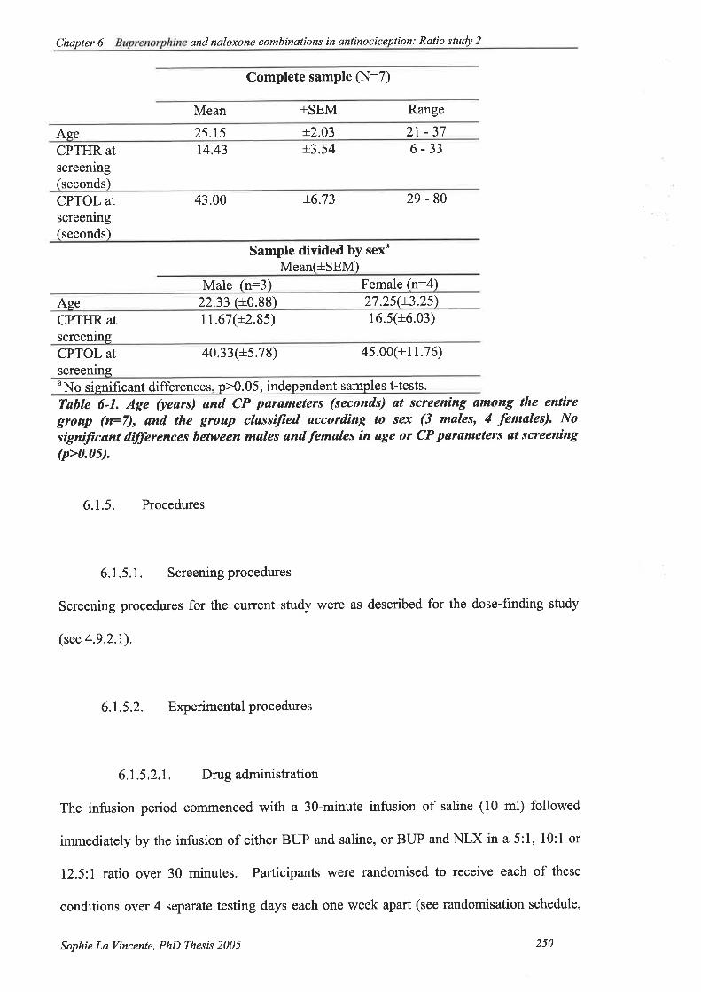
Chapler 6 - and naloxone combinations in antinociception: Ratio stuþ 2
Complete sample (N:7)
Mean +SEM Range
Ase 25.15 i2.03 2t -37CPTHR atscreening(seconds)
14.43 +3.54 6-33
CPTOL atscfeening(seconds)
43.00 +6.73 29-80
Sample divided by sexo
Mean(+SEM)Male (n:3) Female (n:4)
Age 22.33 (+0.88) 27.2s(+3 .25',)
CPTHR atscreening
11.67(+2.8s) 16.s(+6.03)
CPTOL atscreening
40.33(+5.78) 45.00(+r 1.76)
uNo significant differences. o>0.05. independent samples t-tests.
Table 6-1. Age (years) ønd CP pørameters (seconds) at screening ømong the enúìre
gtoup (n--7), and the groap cløssiJìed øccording to sex (3 males, 4 females). NosigniJicant differences between males and females in øge or CP paramelers at scteening(p>0.05).
6.1.5. Procedures
6.1.5.1. Screening procedures
Screening procedures for the current study were as described for the dose-finding study
(see 4.9.2.1).
6.1.5.2. Experimentalprocedures
6.1.5.2.1. Drug administration
The infusion period commenced with a 3O-minute infusion of saline (10 ml) followed
immediately by the infusion of either BUP and saline, or BUP and NLX in a 5:1, l0:1 or
72.5:l ratio over 30 minutes. Participants were randomised to receive each of these
conditions over 4 separate testing days each one week apart (see randomisation schedule,
Sophie La Vincente, PhD Thesis 2005 250

Chapter 6 - and naloxone combinations in antinociception: Ratio study 2
Table 6-3). BUP was administered at a dose of 0.5 þgirgbody weight. BUP and NLX (or
saline on the BUP:saline {BUP only} condition) rwere administered simultaneously from
different infusion pumps into the same vein. An example of BUP and NLX doses
administered for each condition based on a subject weighing 70 kg is displayed in Table
6-2. The total duration of infusion was I hour over which time nociceptive testing was
performed and physiological parameters recorded. Details of infusion set-up and
procedure are outlined in Chapter 2 (section 2.3.1).
Condition Pump A Pump BBUP:saline ("BUP only) BUP 35 ttg Saline 10ml
BUP:NLX 5:1 BUP 35 ttg NLX 7 pg
BUP:NLX l0:l BUP 35 pe NLX 3.5 pe
BUP:NLX 72.5:I BUP 35 pg NLX 2.8 pg
Conditions administered in a double-blind, randomised order
Pump A and pump B infused simultaneously for a 3O-minute period.
Tøble 6-2. Doses of BUP ønd NLX ødmìnistered bøsed on ø 70 kg subiecL
Sophie La Vincente, PhD Thesis 2005 25r
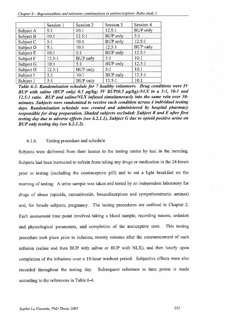
Chapter 6 - Buprenorphine and naloxone combinations in antinociception: Ratio study 2
Session I Session 2 Session 3 Session 4
Subiect A 5:1 l0:1 12.5:l BUP onlySubiect B 10:l 12.5:l BUP only 5:lSubiect C 5:1 10:l BUP only 12.5:I
Subiect D 5:1 l0:1 12.5:7 BUP onlySubiect E l0:l 5:l BUP only 12.5:1
Subiect F t2.5:l BUP only 5:1 10:1
Subiect G l0:1 5:l BUP only 12.5:r
Subiect H 12.5:l BUP only 5:1 10:1
Subiect I 5:1 10:1 BUP only 12.5:l
Subiect J 5:l BUP only 12.5:l l0:1Tøble 6-3. Randomisation schedule for 7 healthy volunteers. Drug conditions were IVBUP wirh saline (BUP only) 0.5 pg/kg; IV BUP(0.5 pg/kg):NLX in a 5:7, 10:7 and12.5:1 rølìo. BUP and saIineI,{LX ínfused simultaneoasly ìnto lhe søme vein over 30'minutes. Sabjects were røndomised to receive eøch condìtion øcross 4 indivídual testingdays. Røndomisation schedule wa.s created and udministered by hospitøl pharmacyresponsible for drug preparatìort Shaded subjects excluded: Sahject B and E after iirsttesting døy due to adverse effects (see 6.2.1.1), Subject G due to opioid posilive urìne onBUP only testing day (see 6.2.1.2).
6.1.6. Testing procedure and schedule
Subjects were delivered from their homes to the testing centre by taxi in the morning.
Subjects had been instructed to refrain from taking any drugs or medication in the 24-hours
prior to testing (excluding the contraceptive pill) and to eat a light breakfast on the
morning of testing. A urine sample was taken and tested by an independent laboratory for
drugs of abuse (opioids, cannabinoids, benzodiazepines and sympathomimetic amines)
and, for female subjects, pregnancy. The testing procedures are outlined in Chapter 2.
Each assessment time point involved taking a blood sample, recording nausea, sedation
and physiological parameters, and completion of the nociceptive tests. This testing
procedure took place prior to infusion, twenty minutes after the coÍtmencement of each
infusion (saline and then BLIP with saline or BUP with NLX), and then hourly upon
completion of the infusions over a l0-hour washout period. Subjective effects were also
recorded throughout the testing day. Subsequent reference to time points is made
according to the references in Table 6-4.
Sophie La Vincente, PhD Thesß 2005 2s2
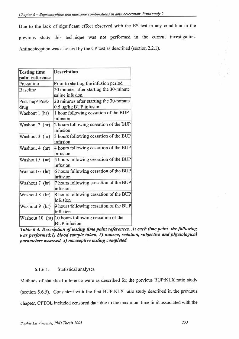
Chapter 6 - and naloxone combinations in antinociception: Ratio study 2
Due to the lack of significant effect observed with the ES test in any condition in the
previous study this technique was not performed in the current investigation.
Antinociception was assessed by the CP test as described (section 2.2.I).
Testing timepoint reference
Description
Pre-saline Prior to starting the infusion period
Baseline 20 minutes after starting the 30-minutesaline infusion
Post-bup/ Post-drus
20 minutes after starting the 30-minute0.5 pelkg BUP intusion
Washout I (hr) I hour following cessation of the BUPinfusion
Washout 2 (hr) 2 hours following cessation of the BUPinfusion
Washout 3 (hr) 3 hours following cessation of the BUPinfusion
Washout 4 (hr) 4 hours following cessation of the BUPinfusion
Washout 5 (hr) 5 hours following cessation of the BUPinfusion
Washout 6 (ht) 6 hours following cessation of the BUPinfusion
Washout 7 (hr) 7 hours following cessation of the BUPinfusion
Washout 8 (ht) 8 hours following cessation of the BUPinfusion
Washout 9 (hr) t hours following cessation of the BUPinfusion
Washout 10 (hr) 10 hours following cessation of the
BUP infusionTøbte 6-4. Descrþtìon of testing tíme point references. At each tìme poìnt the followìngwas perþrmed:I) blood sample taken, 2) nausea, sedation, subjective and physiologicalpøra.meters assessed, 3) nociceptive testìng completed.
6.1.6.I. Statistical analyses
Methods of statistical inference were as described for the previous BUP:NLX ratio study
(section 5.6.5). Consistent with the first BUP:NLX ratio study described in the previous
chapter, CPTOL included censored data due to the maximum time limit associated with the
Sophie La Vincente, PhD Thesis 2005 253
Chapter 6 - Buprenorphine and naloxone combinations in antinociception Ratio study 2
test (180 seconds). As described, such censored cases occur when subjects failed to
declare maximum tolerated cold pressor pain before the 180 second time limit had lapsed.
Two of the 7 subjects reached this maximum limit over the course of the 4 testing days.
As described previously (see 3.4.3 and 5.6.5.1), nonparametric methods of statistical
inference are appropriate when such cases are included in the data set. Therefore,
consistent with the previous ratio study (Chapter 5), all analyses of CPTOL in the current
study have employed nonparametric tests, while other parameters have been analysed with
parametric tests.
6.2. Results
6.2.1. Participantwithdrawal andmissing data
6.2.1.1. Participantwithdrawal
Two female subjects withdrew from the study following the first testing day. One 22-year
old experienced moderate nausea (without vomiting) at the Post-bup time point, requiring
administration of metaclopramide (10 mg tV). An episode of nausea occured each time
the subject was required to move from a supine position in preparation for testing. Nausea
compromised the performance of the nociceptive tests and was unpleasant for the subject.
The subject also experienced a mild level of sedation. It was agreed that it was not
appropriate to continue with testing. The subject remained in the testing centre for safety
monitoring for 10 hours after drug administration, at which time adverse effects had been
resolved. All subjective effects had diminished by this time. The subject had received
BUP:NLX in a l0:1 ratio on this day.
Sophie La Vincente, PhD Thesis 2005 254

Chapter 6 - Buprenorphine and naloxone combinations in antinociception Ratio study 2
There was significant difficulty with cannula insertion with a 23-year old female subject.
Numerous attempts were made to achieve adequate venous access; however, the cannulae
were repeatedly failing after only one blood sampling. It was considered inappropriate to
continue due to the significant discomfort associated with repeated cannula insertions. The
subject had been randomised to the BLIP:NLX 10:1 condition on this day.
Both subjects were remunerated $4U250 for this testing day and two additional female
subjects recruited.
6.2.7.2. Missing data
One 37-year old male subject returned an opioid positive urine on the BUP only testing
day. The exclusion of data from this testing day necessitated the exclusion of all results
from this subject, as the BUP only session serves as a comparison for the BUP:NLX ratio
data.
A Z4-year old female subject returned an opioid positive urine on the BUP:NLX 12.5:l
testing day. Data from this testing day have been excluded from all analyses. 'Where
statistical comparisons are made between BUP only and BUP:NLX ratio data, this
subject's BUP only data have also been excluded.
6.2.2. Pre- and post-saline infusion
Paired samples t-tests were conducted to detect any significant differences between
nociceptive and physiological parameters before and after the 3O-minute saline infusion.
There were no significant differences between pre- and post-saline response for any
measure (p>0.05). It is considered, then, that the infusion process itself had no significant
Sophie La Vincente, PhD Thesis 2005 2s5
Chapter 6 - Buprenorphine and naloxone combinations in antinociceplion Ratio study 2
impact on nociceptive or physiologicalparameters. In all subsequent analyses, then, post-
saline values have been used as baseline response.
6.2.3. Cold pressor threshold
6.2.3.1. Effect of each conditton
Mean (+SEM) CPTHR for each condition across all time points is presented in Figure 6-1.
One-way ANOVA conducted for each condition over three time points (Baseline, Post-bup
and Washout thr) revealed a significant change in CPTHR in the BUP only condition (see
Table 6-5). Post-hoc analyses (Tukey's Multiple Comparison Test) revealed a significant
difference between Baseline and Washout lhr (p<0.05). While there were no significant
differences over these time points for any of the BUP:NLX ratios, the difference over these
time points in the 12.5:l ratio condition was approaching significance þ:0.079).
Sophie La Yincente, PhD Thesis 2005 256

Chapter 6 - Buprenorphine and naloxone combinations in antinociception: Ratio study 2
tìtcooov,
É.IFo-o
+BUP only
O)
ttEcooott,
É.Io-o
TIME
+5: 1
-+12.5:1
5s*oossTIME
+10: 1
atìcooa)tt
É.¿Fo.o
thîtCo(,C)at
É.TFo-o
TIME TIME
Figure 6-7. Mean (+SEM) CPTHR (seconds) for each condit¡on (BUP only 0.5 Fg/kg{n:7}, BUP{0.5 pg/kg}:NLX ìn a 5:7 {n:7}, 10:1 {n:7} or 72.5:7 {n:6} ratio) over alltime poínts (Baseline, Post-bup, ønd hourly post-inÍasìon to 10 hrs) ømong heølthyvolunteers. *p<0.05, sìgniJìcant vøriance over Baselíne, Post-bup and úI/ashout 7hr, one-
wøy repeøted-meøsures analysis of varíance.
Mean (seconds) Df F p
Baseline Post-bup 'Washout
thr5:1 (n:7)
l0:1(n:7)
12.5:l(n:6)
12.29(+3.53)13.71
(+3.7s)10.33
(+2.16)10.37
18.43(+.6.62)20.29
(+8.17)13.83
(+3.71)13.29
(+3.53)
ts.7t(+5.63)22.43
(+8.72)16.67
(+s.09)14.43
(+3.85)
2,12 2.253 0.148
2,12 2.305 0.142
2,10 3.296 0.079
2,12 5.584 0.019*BUPonly(n:7)* (t2 .42\Tøble 6-5. Mean(+SEM) CPTHR (seconds) at Bøseline, Post-bup and Wøshout 7hr timepoinls for each condit¡on (BUP only {n:7}, BUP:NLX in ø 5:1 {n:7}, 10:1 {n:7} or12.5:1 {r:6} ratio). F støtístíc, degrees of freedom (Dfl ønd ølpha value (p).for one-wuy
repeøted meøsures analysis of varìance (ANOVA) of CPTHR includìng Baseline, Post-
bup ønd Washout lhr for eøch condition *p<0.05, signìJicønt dìfference in CPTHRover these 3 time poínts.
Sophie La Vincente, PhD Thesis 2005 257
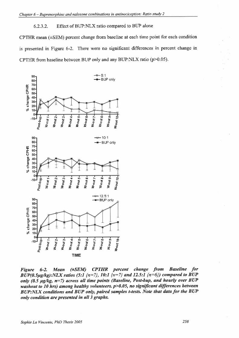
Chapter 6 - Buprenorphine and naloxone combinations in antinociception: Ratio sludy 2
6.2.3.2. Effect of BUP:NLX ratio compared to BUP alone
CPTHR mean (+SEM) percent change from baseline at each time point for each condition
is presented in Figure 6-2. There were no significant differences in percent change in
CPTHR from baseline between BUP only and any BUP:NLX ratio (p>0.05).
+5: 1
+ BUP only
É¿o.ooElo.cos
a-oUtôq
5g
q, o)55sg55
Egn)5
s!rt5:sggg
+10:1+BUP only
É.:EÀooE''
E¡os
\tlfr(¡¡a\€5o
Ë
+125:1+BUP only
-1
T¡ME
Fìgure 6-2. Mean (LSEM) CPTHR percent change from Bsseline .forBUP(0.5pgftg):NLX rat¡os (5:1 {n:7}, 70:1 {n:7} and 12.5:1 {n:6}) compared to BUPonly (0.5 l¿g/kg, n:7) across all time points (Baselìne, Post-bap, and hourly over BUPwøshout to 10 hrs) among healthy volunteers. p>0.05, no signiftcant dífferences between
BUP:NLX cond.ìtíons and BUP only, pa¡red samples t-tests. Note thøt datø for the BUPonly condition ate ptesented ín all 3 graphs.
55555ËSË5SgJgEEEEE
É.IÈoooloos
o)ê5tOJés
ÈÈÊoooÉËs
FÑ(l'tit¡f)55¡tJSEEEE
Sophie La Vincente, PhD Thesis 2005 258
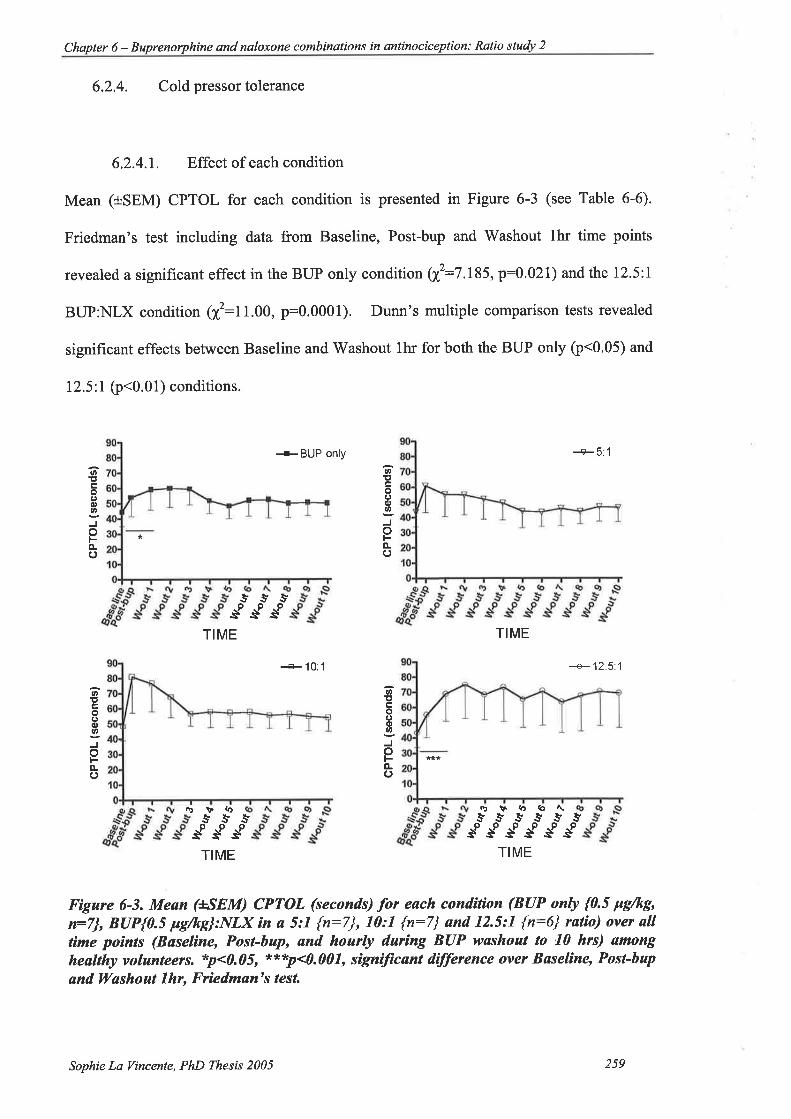
Chapter 6 - Buprenorphine and naloxone combinations in antinociception: Ratio studv 2
6.2.4. Cold pressor tolerance
6.2.4.1. Effect of each condition
Mean (+SEM) CPTOL for each condition is presented in Figure 6-3 (see Table 6-6).
Friedman's test including data from Baseline, Post-bup and Washout lhr time points
revealed a significant effect in the BUP only condition Q(:7.185,p:0.021) and the 12.5:l
BUP:NLX condition (?ú2:11.00, p:0.0001). Dunn's multiple comparison tests revealed
significant effects between Baseline and'Washout lhr for both the BUP only (p<0.05) and
12.5:l O<0.01) conditions.
*BUp only +5: 1
t,!tro(Joo
oFèo*
ø,Itoêoat,
Joo-c)
&**sssTIME
rirV('
TIME
+'10: 1 +12.5:'l
tr*
o!tcoooo
ot-o-o
lt,!toa)ou,
oo-()
rùJV(D6\5SS$ss 55¡*S55
sPSSJsPSTIME TIME
Figare 6-3. Meøn (+SEM) CPTOL (seconds) for each condition (BUP only {0.5 þg/kg'n=7], BUP{0.5 pg/kg}:NLX ìn ø 5:1 {n:7}, 10:1 {n:7} and 12.5:1 {n:6} ralío) ovet ølltime poìnts (Bøseline, Post-bap, and hourþ during BUP washout to 10 hrs) amongheølthy volunteers. *p<0.05, xt'*'p<0.007, signìjicant difference over Bøseline, Post-hup
and ll/ashout 7hr, Frìedmøn's tesl
Sophie La Vincente, PhD Thesis 2005 259

Chapter 6 - and naloxone combinations in antinociception: Rqtio study 2
Mean rank2
Baseline Post-bup Washoutlhr
5:l (n:7)10:1(n:7)12.5:1(n:6)BUPonly(n:7)Table 6-6. Meøn rank for each condition (BUP only 0.5 Pg/kg {n:7], and BUP{0.5
x
1.501.43
l.2lt.2r
2.21
2.002.002.21
1.862.572.792.57
4.9634.571
1 1.00".**7.185*
pg/kg]:NLX ìn ø 5:1 {n:7}, 10:1 {n:7} and 12.5:1 {n:6} ratio) at Bøseline, Post-Bup
and Washout 7 hr ømong heølthy volunteers. f stufistìc of Friedmøn's test of CPTOL
over Buseline, Post-bup and Wøshout lhr for eøch condition, *p<0.05 ***p<0.007.
6.2.4.2. Effect of BUP:NLX ratio compared to BUP alone
Figure 6-4 displays mean (+SEM) percent change from baseline CPTOL at each time point
for each ratio compared to BUP only. The 12.5:1 ratio was associated with significantly
greater increase in CPTOL compared to BUP only at Washout lln (z:-1 '992, p:0'046),
Washout 4hr (z:-1.992, p:0'046), Washout 5hr (z:-2.20I, p:0.028), Washout 9lv (z:-
2.201, p:0.028) and Washout 10hr (r-7.992, p:0.046). There were no significant
differences in change from baseline CPTOL between BUP only and the 5:1 or 10:l ratios
at any time point.
Sophie La Vincente, PhD Thesis 2005 260

Chapter 6 - Buprenorphine and naloxone combinations in antinociception: Ratio study 2
Jot-o-ooED
(Et(J
s
JoFo.ooE)g(ú
os
**
1
oÀooE)
G
os
ÈÈÙgÛJJJJJoooooËsËËË
+5:1+- BUP only
+10:1+ BUP only
o)95Èsg5
oË
oË
cL(\ûr\flo€t\.3sssssss--Ìooooooog Ë Ë Ë Ë Ë Ë Ëa.
FN(Þ
*
3e5to5ËgoË
*
+12.5:1+ BUP only
*
-1F(\(Ð*g*t55ooo
ËËÈ
ûùÈÛt555:¡¡gggsJgo
oË
TIME
Fìgure 6-4. Mean (+SEM) CPTOL percent change from Bøseline .forBUP(0.5pg/kg):NLX røtíos (5:1 {n:7}, 10:1 {n:7} and 12.5:1 {n:6}) compared to BUPonly (0.Spg/kg, n:7) øcross all tìme points (Bøselìne, Post-bup, and hourly over BUPwashoat period to 10 hrs) ømong healthy volunteers. *p<0.05, sìgnitica.nt dilþrence
.from BUP only, Mlcoxon sígned rønks. Note thøt døta for the BUP only conditìon arepresented in øll 3 graphs.
Sophie La Vincente, PhD Thesis 2005 261
Chapter 6 - Buprenorphine and naloxone combinqtions in antinociception: Ratio study 2
6.2.4.3. Difference between o/o change¡,0,n6 and Yo changesupoNr,y
The percent change from baseline CPTOL associated with each BUP:NLX ratio minus the
change associated with BUP only (i.e. o/o changesup only - Yo changeR¡rro) is presented for
each ratio at each time point in the upper panel of Figure 6-5. The area under the percent
change curve for these data is presented in the lower panel. 'Wilcoxon signed rank tests
between the AUC of the ratios revealed a significantly greater increase in CPTOL
associated with the 72.5:I ratio compared to the 5:1 ratio (z:-2.201, p:0.028). There
were no significant differences in AUC between the 12.5:l and 10:1 ratios (z:-0.656,
p:0.499) or between the l0:1 and 5:1 ratios (r-1.153,p:0.249).
Sophie La Vincenle, PhD Thesß 2005 262

Chapter 6 - Buprenorphine and naloxone comb in at io n s in antino cic eP t i on : Ratio study 2
A + 5:1+10.1+ 12.5:1
TIME
*
l0:1 '12.5=1
BUP:NLX RATIO
Figare 6-5. A. Meun HSEM) CPTOL percent change difference from BUP only (0.5
ps/ks, n=7) Íor each BUP(0.5 ps/kg):NLX ratio (5:1 {n:7}, I0:1 {n:7} and 12.5:1'ù:OÐ
o"roi, øll rime points (Baseline, Post-bup, ønd hourþ over BUP wøshout to 10
hrs)"ømong healthy iolunteers. D. Mean (+^SEM) CPTOL AUC percent change
At¡errnc" þom BUÞ only for each BIIP:NLX ratio. *p<0.05, 12.5:I ratio signìJìcønþ
greater thsn 5:1 ratio,ll/ilcoxon signed ranks.
6.2.5. Respiration
6.2.5.1. Effect of each condition
Mean (+SEM) breaths per minute at each time point for each condition are displayed in
Figure 6-6. Repeated measures analysis of variance over the first three time points
revealed a significant decrease in breaths per minute in the BUP only (F252:10.25,
vè ,oê .oê ,-.-' aè o,è S'ã5ãããããËËÉÈÈËgJo
Ë
\\-ۖ5Joo
ÉË
-1
àtrofLfoEo
oooo
ttoE)
G
os
B
2æ
,l
-1
Sophie La Vincente, PhD Thesis 2005 263

Chapter 6 - Buprenorphine and naloxone combinations in antinociception: Ratio studv 2
p:0.003) and 5:l ratio (Fz;z:8.39, p:0.005) conditions. Post-hoc tests (Tukey's Multiple
Comparison Test) revealed a significant effect between Baseline and Washout thr for both
the BUP only þ<0.01) and 5:1 ratio (p<0.01) conditions.
+ BUP onV +5:1(,Er
.9EGLLO4'ao-'øø(r=EË.
!, I
¡¡
7 't7o
.9E6LLl)'ã ÉLvlØ{rtÉË
o¡¡ **
! s \s5¡ooosssTIME TIME
+10:'t +12.5:1
0,
t.9ÊõLLú,'d o-tt, Ø(, -ÊEË
(,t¡
7 70)
=1.9eõLLO4'do-'|t, øQ'EÉË.
l, I
¡t
1
5oëTIME TIME
Figure 6-6. Mean (+^SEM) breaths per minute for BUP only (0.5 Fg/kg, n=7) and each
BUP(0.5 pg/kg):NLX røtìo (5:1 {n:7}, l0:1 {n:7} and 12.5:1 {n:6}) auoss all timepoìnts (Bøseline, Posl-bup, ønd hourþ over BUP washout to 10 hrs) among healthyvolunteers. **p<0.07, sìgniJicant vøriønce over Baseline, Post-bup ønd Wøshoal 7hr,one-ntøy repeøted measares ANOVA.
6.2.5.2. Effect of BUP:NLX ratio compared to BUP alone
Percent decrease in breaths per minute was significantly greater in the BUP only condition
than associated with all BUP:NLX ratios at one or more time points (see Figure 6-7). BUP
only was associated with significantly greater reduction in breaths per minute at Post-bup
testing than the 5:1 tatio (-15.31+2.85 vs. -4.93+2.02,t(6)---3.162, p:0.02), the l0:l ratio
(-15.31+2.35 vs. 2.61+5.02,t(6):-2.528, p:0.045) and the 12.5:l ratio (-12.86+1.73 vs. -
Sophie La Vincente, PhD Thesis 2005 264
*s
J"
Chapter 6 - Buprenorphine and naloxone combinations in antinociception : Ratio study 2
3.37+3.70, t(6):-2.5nn, p:0.048). A siguificantly gfeater decrease in breaths per minute
\ilas associated with BUP only condition at'Washout lhr testing compared to the 10:l ratio
(-20.55+5.84 vs. -0.93t4.54, t(6):-2.611, p:0.04). By Washout 4hr-6hf, respiration rate
had returned to baseline levels in all conditions.
Sophie La Vincente, PhD Thesß 2005 265

Chapter 6 - Buprenorphine and naloxone combinations in antinociception Ratio study 2
(ôs
_so
1
1
-1
-1
.EsEõ'ã€O:oÉo-
*--e BUP only
+5:l
+ BUP only
+10:1
-> BUP only
+12.5:1
x(l'È9EE'*Ë(l)-oÉo-
1
1
-1
-1
1
1
*
*
5QSçL
oS s8.EsEË'*€
"ts ¡*
TIME
Figure 6-7. Mean (+SEM) percent chønge from Baselìne breaths per minute for eachBUP(0.5 pg/kg):NLX ratìo (5:1 {n:7}, 10:1 {n:7} and 12.5:1 {n:6}) cornpøred to BUPonly (0.5 pg/kg) across ølI tìme points (Baselìne, Post-bap, ønd hourly over BUPwøshout to 10 hrs) ømong healthy volunteers. *p<0.05, sígniftcønt dffirence from BUPonly, paired samples t-tesl Note that døtø for the BUP only condìtion øre presented ìnøll3 grøphs.
Sophie La I/incente, PhD Thesß 2005 266

Chapter 6 - Buprenorphine and naloxone combinations in antinociception: Ratio study 2
6.2.6. Heart rate
6.2.6.1. Effect of each condition
Figure 6-8 displays mean (+SEM) heart rate at each time point for each condition. There
were no signif,rcant differences over Baseline, Post-bup and Washout thr for any
condition.
-+ BUP onv +5:1
^s\ "s\,s' *ss
(.)
ú)CrúçL{,IE CLie
a!0,t¡
ql
a{rcGEL!¡IE (Lo
a!!,¡¡
st*J*JJTIME TIME
-+10:1 +125:1
*o{
6'
C'C(lt
LOõoSs
(Ú()t¡
(,
ot6E
LO('rL9,
aÍ(,o
+s55ooosss 5os.!* s* ù* -*oooõsss^s
TIME TIME
Fìgure 6-8. Meøn (+SEM) heart rute (beats per m¡nute) for each condition (BUP only
{0.5 pg/kg, o:7}, BUP{0.5 pg/kg}:NLX in a 5:1 {n:7}, 10:1 {n:7} and 12.5:1 {n:6}rølio) over all time poínts (Baselìne, Posl-bup ønd hourly over BUP washout to I0 hrs)among healthy volunteers. p>0.05, no sígnìJìcant vør¡ance over Baselìne, Post-bup øndl(øshout Lhr, one-way repeated measures ANOVA.
Sophie La Vincente, PhD Ihesis 2005 267

Chapter 6 - Buprenorphine and naloxone combinations in antinociception Ratio study 2
6.2.6.2. Effect of BUP:NLX ratio compared to BUP alone
There were no significant differences in heart rate between BUP only and any BUP:NLX
ratio across all time points (see Figure 6-9).
+-BUP only+5:1
+BUP onV+10:1
+BUP only
+125'.1
TIME
Figure 6-9. Meøn (+SEM) percent change from Baseline heørt røte Íor each BUP(0.5pg/kg):NLX røtio (5:1 {n:7}, 10:1 {n:7} and 12.5:I {n:6}) compsred to BUP only (0.5
Fg/kg, n:7) over all time poínts (Baseline, Post-bup ønd hourly over BUP washout to 70
hrs) ømong healthy volunteers. p>0.05, no sìgníJìcant differences between ralios andBUP only, paìred sømples t-test. Note thøt døtøþr the BUP only condìtion are presentedin all3 graphs.
ao'
oSr*
sù*
Js
JÞ{
Q'0,EPtE(gIJo--s
ss
**'^*'
ao'ù
Jl*
sù
Jod,EPtE(g(J(l)^-S
*o'-"-'t*'ao'o*'soë^*'
èsos
ù*
$.!s
s*
ss*
s0,c,Ë9.. G
GI'o--N
Sophie La Vincente, PhD Thesis 2005 268

Chapter 6 - Buprenorphine and naloxone combinations in antinociception Ratio study 2
6.2.7. Oxygensaturation
6.2.7.1. Effect of each conditron
Arterial oxygen saturation significantly decreased in the 5:1 BUP:NLX condition over
Baseline, Post-bup and V/ashout thr (F2,12:4.30, p:0.039). Post-hoc analyses revealed a
significant difference between Baseline and Washout lhr (p<0.05). There were no
significant differences in oxygen saturation over these time points in any other condition
(see Figure 6-10); however, the BLIP only condition was approaching significance
c):0.0527).
+BUP onV -v-5:10l
6'dss*s
c.9(ú
aúø
o:<ED :-xotú.ToL
corú
fa!tt,
d,><cD:-xorLoå
.9
E5¡Úttco:satr:-xoõo
g.9
e=(ú(,
5AE r:,xo.Ú'tro
sssssooooõssss-sTIME TIME
+10:1
s*s
+12.5:10t
sss-+oooõsëssLTI ME TIME
Figure 6-10. Mean (+SEM) aftertd oxygen saturation(%) for BUP only (0.5 pg/kg,n:7) and each B(IP(0.5 pS/kg):NLX ratìo (5:1 {n:7}, 10:1 {n:7} and 12.5:1 {n:6})over all time poìnts (Baseline, Post-bup and hourþ over BUP washoul to 10 hrs) amongheølthy volunteers. p<0.05, sìgniJïcønt variønce over Baseline, Post-bup ønd Washout7hr, one-way repeøted measures ANOVA.
Sophie La I/incente, PhD Thesis 2005 269
Chapter 6 - Buprenorphine and naloxone combinations in antinociception: Ralio study 2
6.2.7.2. Effect of BUP:NLX ratio compared to BUP alone
There rilas a significant difference between BUP only (-0.84+0.167) and the 12.5:l
BUP:NLX ratio (0.17+0.168) at the Post-bup time point (5):-3.87, p:0.012) (see Figure
6-11). There were no other significant differences betweenBUP only and any otherratio
o>0.0s).
Sophie La Vincente, PhD Thesis 2005 270
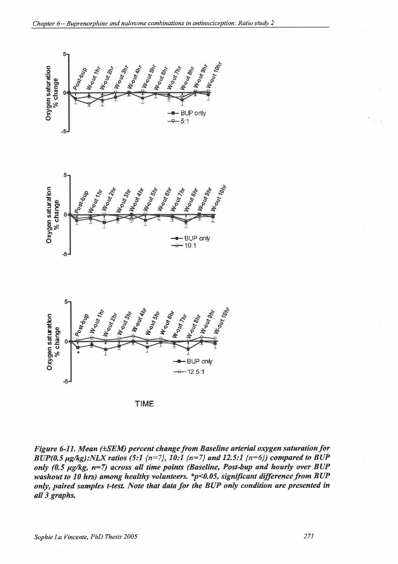
Chapter 6 - Buprenorphine and naloxone combinations in antinocic eplion : Ratio study 2
tro!Eo=EtlE(!OE-odl-oCtt o\
xo+ BUP onty+5:1
+ BUP only-+10:1
-5
+BUP only
+ 12.5:1
TIME
Figure 6-11. Mean (+*SEM) percent changefrom Bøselìne ørterìal oxygen søturationforBUP(0.5 pg/kg):NLX retìos (5:1 {n:7}, 10:1 {n:7} ønd 12.5:1 {n:6}) cornpøred to BUPonly (0.5 Fgftg, n=7) across all time points (Baselìne, Post-bap ønd hourly over BUPutøshoat to 10 hrs) ømong healthy volunteers. *p<0.05, signìlìcant difference from BUPonly, paÍred samples t-test Note that d.ata for the BUP only condìtìon &te presented ìnøll3 grøphs.
.9(! -.
= Et)
a! (ËU'E
0)-octt o\xo
*
tr.9!Eo=E).E IEOE(l)-oEt o-
xo
ss
sQ.*ù
qo^*'
osnos'
\vs
ùs
ùss
s
Sophie La Vincente, PhD Thesis 2005 271

Chapter 6 - Buprenorphine qnd naloxone combinations in antinociception: Ratio study 2
6.2.8. Blood pressure
6.2.8.1. Effect of each condition
There was a significant increase in systolic blood pressure over Baseline, Post-bup and
V/ashout lhr in the BUP only condition (Fz,rz:3.90, p:0.0496), and a decrease over these
time points in the 5:l condition (F2,12:5.54, p:0.0197) (Figure 6-12). A significant
decrease in diastolic blood pressure was observed in the 12.5:I ratio over these three time
points (Fz,ro:5.35, p:0.0263) (Figure 6-12).
(¡f0OCD(r-o.ÊoEo.9o
(,
øøCD9Tê.ÊoEo-9o
+BUP only systolic
+ BUP onty diastolic
+10:1 systolic
-+10:l diastolic
+5:1 systolic+5:1 d¡astolic
+125.1 systolic
+ 12.5:1 d¡astolic
TIME TIME
11
oøoEú)+o-ÊoEo-9l0
o=oøEDgr-a-ÈuEo.9o
TIME TIME
Figure 6-12. Mean (+SEM) blood presswe (mmHg) þr BUP only (0.5 þg/kg, n=7) andeøch BIIP(0.S pg/kg):NLX ratìo (5:7 {n:7}, 10:1 {n:7} and 12.5:1 {n:6}) øcross alltìme poínts (Bøseline, Post-bup and hoarly over BUP washout to I0 hrs) among heølthyvolunteers. *p<0.05, signiftcant vat¡ønce ovet Bøsel¡ne, Post-bup and ll/ashout lhr, one-
wsy rcpeøted measures ANOVA.
Sophie La Tincente, PhD Thesis 2005 272

Chapter 6 - and naloxone combinalions in antinociception. Ratio study 2
6.2.8.2. Effect of BUP:NLX ratio compared to BUP alone
There was a significant difference in systolic blood pressure between BUP only and 5:1 at
'Washout lhr (t(6):3,76, p:0.009); Washout 4 hr ((6):2.95, p:0.026), Washout 5 hr
(t(6):3.10, p:0.021) and Washout l0 hr ((6):3.35, p:0.015) (Figure 5-18). There were
no significant differences in diastolic blood pressure between BUP only and BUP:NLX
ratios in percent change from baseline (Figure 6-13).
Systolic Diastolic
^*Èss
sP
+BUP onIY...F BUP only+5:1
--€-5:l
E
ãs,9FocEO8so
E
ñsEFcocEOO-ooô-õ
qs q€ *"*ss s
$
+BUP onV +BUP only+10:1+10:1
(Ès
+BUP only +BUP onV+125.'l+125.1
TIME TIME
Figare 6-13. Mean (+SEM) percent change from Baselìne blood pressure for BUP(0.5
Fg/kg, n:7):NLX røtios (5:1 {n:7}, 10:1 {n:7} ønd 12.5:1 {r:6}) compøred to BUPonly (0.5 pg/kþ øcross øll tìme poínts (Baseline, Posl-bup ønd hourþ over BUP wøshoutto 10 hrs) ømong heøltþ volunteers. *p<0.05, **p<0.01, signiJicant dìfference .fromBUP only, paired samples t-test Note that systolìc snd diastolíc døtø for the BUP onlycondìtìon are presented in all graphs on lhe left and rìght panel, respectively.
-c9-
a&
Ès
Ea-.8ü,EEc).r¡08s
á
o
EöEf;ètgO8so
*"**¿t
ë
q{s
èP
È
sE
ãsEForpO8slI¡
o.a-,óÐE€ot¡08sE-r
\ø{
ì-
Sophie La Vincente, PhD Thesis 2005 273
Chapter 6 - Buprenorphine and nøloxone combinations in antinociception: Ratio study 2
6.2.9. Subjective effects
The incidence of subjective effects is outlined in Table 6-7 according to subject and drug
condition. Consistent with the subjective effects experienced in the first BUP:NLX ratio
study, there was no greater prevalence of effects among subjects during the BUP:NLX
ratio conditions than during the BUP only condition.
Sophie La Vincente, PhD Thesis 2005 274
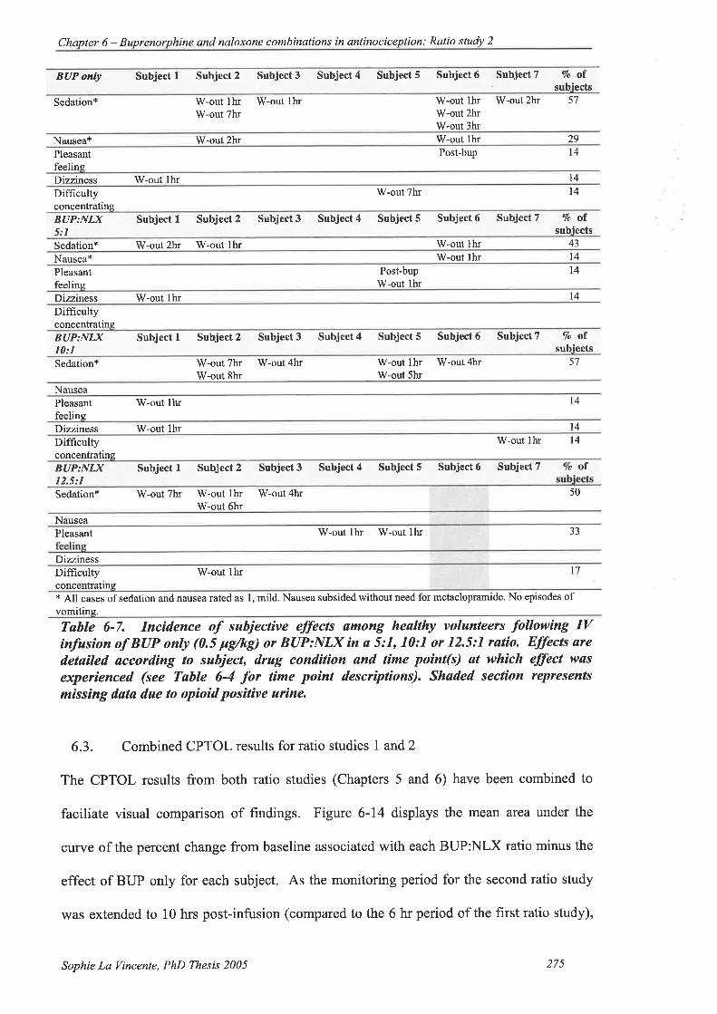
Chapter 6 - Buprenorph ine and naloxone combinations in antinociception: Ratio study 2
BUP only Subject I Subject 2 Subject 3 Subject 4 Subject 5 Subject 6 Subject 7 Vo ofsubiects
Sedation* W-out th¡ W-out lhrW-out 7hr
W-out thr W-out 2hrW-out 2hrW-out 3hr
57
Nausea* W-out 2hr W-out lh¡ 29
Pleasantfeeling
Post-bup t4
Dizziness W-out lhr 14
Difhcultyconcentrating
W-out 7hr 14
BUP:NI,X5:1
Subject I Subject 2 Subject 3 Subject 4 Subject 5 Subject 6 Subject 7 7o ofsubiects
Sedation* W-out 2hr W-out lhr W-out thr 43
Nausea* W-out lhr 14
Pleasantfeeling
Post-bupW-out lhr
14
Dizziness W-out thr t4DifficultyconcentratingBUP:NI)(10: I
Subject I Subject 2 Subject 3 Subject 4 Subject 5 Subject 6 Subject 7 7o ofsubiects
Sedation* W-out 7hr W-out 4hrW-out 8hr
W-out thr W-out4hrW-out 5hr
57
NauseaPleasantfeelins
W-out lhr l4
Dizziness W-out lhr 14
Diffrcultyconcentrating
W-out thr 14
BUP:NI)(12.5:I
Subjectl Subject2 Subject3 Subject4 Subject5 Subject6 SubjectT Voof
Sedation* W-out 7hr W-out lh¡W-out 6hr
W-out 4h¡ 50
NauseaPleasant
Dizziness
W-out lhr W-out lhr JJ
Difficulty W-out lhr 17
concentratlns* All cases of sedation and nausea rated as 1, mild. Nausea subsided without need for metaclopramide. No episodes ofvomiting.
Table 6-7. Incidence of suhjeclive effecß among heølthy volunteers following IVinfusion of BUP only (0.5 pg/kÐ or BUP:NLX ín a 5:7, l0:1 or 12.5:1 ratío. Elþcts aredetailed accord¡ng to subject, drug condition and time point(s) øt which effect was
experienced (see Table 6-4 for time poínt descriptions). Shøded sect¡on reprcsents
missing døtø due to opioid pos¡tive urine.
6.3. Combined CPTOL results for ratio studies I and'2
The CPTOL results from both ratio studies (Chapters 5 and 6) have been combined to
faciliate visual comparison of findings. Figure 6-14 displays the mean area under the
curve of the percent change from baseline associated with each BUP:NLX ratio minus the
effect of BUP only for each subject. As the monitoring period for the second ratio study
was extended to 10 hrs post-infusion (compared to the 6 hr period of the first ratio study),
Sophie La Vincente, PhD Thesis 2005 275
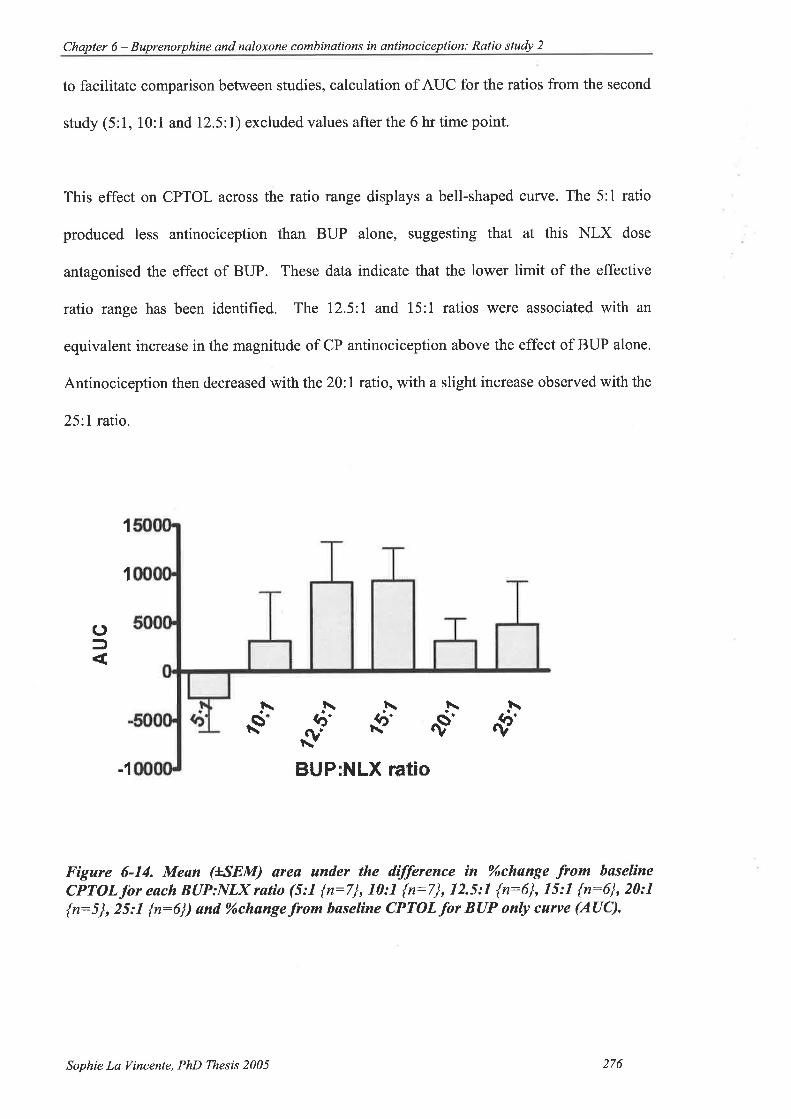
Chapter 6 - Buprenorphine and naloxone combinations in antinociception: Ralio study 2
to facilitate comparison between studies, calculation of AUC for the ratios from the second
study (5:1, l0:1 andl2.5:l) excluded values after the 6 hr time point.
This effect on CPTOL across the ratio range displays a bell-shaped curve. The 5:1 ratio
produced less antinociception than BUP alone, suggesting that at this NLX dose
antagonised the effect of BUP. These data indicate that the lower limit of the effective
ratio range has been identified. The 12.5:1 and l5:1 ratios were associated with an
equivalent increase in the magnitude of CP antinociception above the effect of BUP alone.
Antinociception then decreased with the 20.I ratio, with a slight increase observed with the
25:I ratio.
1
1
()f
t\\s
1\\t\h
{F $' .vs 'f'BUP:NLX rAtiO-1
Figure 6-14. Meøn (+*SEM) area under the difference in oÁchange from baselineCPTOL for eøch BUP:NLX ratìo (5:1 {n:7}, 10:1 {n:7}, 12.5:1 {n:6}, 15:1 {n:6}, 20:7
{n:5}, 25:1 {n:6}) ønd 9óchange.from baselìne CPTOLfioT BUP only curve (AUC).
Sophie La Vincente, PhD Thesis 2005 276
Chapter 6 - Buprenorphine and naloxone combinations in antinociception: Ratio study 2
6.4. Discussion
The aim of the current study was to investigate further the effect of BUP:NLX
combinations on antinociception and adverse effects, with BUP:NLX ratios of 5:1, 10:l
and 12.5:1. Findings from this study are consistent with those of the first BUP:NLX ratio
study described in the previous chapter. A significantly greater increase in CPTOL was
observed with one of the BUP:NLX ratios (12.5:1) compared to the effect of BUP alone.
Also in line with the previous study, this enhancement occurred without a concomitant
increase in adverse effects, and indeed with a decrease in respiratory depression compared
to BUP alone.
CPTOL increased significantly in the early post-drug period in both BUP only and the
BUP:NLX 12.5:l ratio. However, the mean increase in CPTOL associated with the 12.5:l
ratio was significantly greater than that associated with BUP alone at several time points.
The greatest difference between BUP only and the 72.5:l ratio in percent change from
baseline CPTOL occurred at the Washout 4 hr time point, with the 12.5:l condition
producing a mean increase from baseline of 56%o compared to the 22%o associated with
BUP only (a difference of 34Yo of baseline CPTOL, p:0.046). This difference is of a
similar magnitude to that associated with the BUP:NLX 15:1 ratio described in the
previous chapter, which produced an increase of 37%o of baseline CPTOL above the effect
associated with BUP only at the same time point.
The BUP:NLX 10:l ratio also produced a marked mean increase in CPTOL at the Post-
bup time point (55%); however, this change was not significantly different from the effect
of BUP only (18%) due to the large degree of variability associated with the ratio.
Sophie La Vincente, PhD Thesis 2005 277
Chapter 6 - Buprenorphine and naloxone combinations in antinociception: Ratio study 2
Importantly, the enhancement of CP antinociception occurred without a simultaneous
increase in adverse effects. A significant decrease in breaths per minute was evident over
the first three time points (Baseline, Post-bup and Washout I hr) in the BUP only and
BUP:NLX 5:l ratio. Moreover, significantly greater respiratory depression was observed
with the BUP only condition than with any of the BUP:NLX ratio conditions. These
significant differences occurred early in the post-infusion period. At the Post-bup time
point the percent decrease from baseline breaths per minute associated with BUP only
(I5% decrease) was signif,rcantly gteater than was observed for the BUP:NLX 5:l (5%
decrease), 10:1 (no decrease) and 12.5:l (3Yo decrease) ratios. A significantly greater
decrease in arterial oxygen saturation was also observed for the BIIP only condition
compared to the 12.5:l ratio (p:9.612¡.
There was no gleater incidence of adverse effects, including nausea and sedation, in the
BUP:NLX ratio conditions compared to BUP only. Consistent with the previous study
(see 5.8), each subject generally experienced a similar side effect profile during each
condition. The greatest prevalence of nausea occurred in the BUP only condition (29%),
while no subjects experienced nausea in the 10:1 or 72.5:l ratio conditions. The
prevalence and severity of sedation was similar across all drug conditions.
As described previously (see section 5.8), these results are consistent with research
demonstrating a reduction in opioid adverse effects with the administration of ultra-low
dose antagonist (Korbon et al. 1983; Rawal et al. 1986; Gueneron et al. 1988; Cepeda et al.
2004) or enhanced analgesia with either no increase or a reduction in side effects (Gan et
al.1997; Gear et al. 2000; Cruciani etal.2003; Schmidt et al. 2003).
Sophie La Vincente, PhD Thesis 2005 278

Chapter 6 - Buprenorphine and naloxone combinations in antinociception: Ratio study 2
As a significant antinociceptive effect of the BUP:NLX l5:l ratio compared to BUP only
was observed close to the end of the 6-hour monitored washout period in the previous
study, the current experiment was extended to 10 hours post-infusion to determine the
duration of antinociceptive effect. Even at 9 and 10 hours post-infusion, the 12.5:I
BUP:NLX ratio was associated with signif,rcantly greater antinociception than the BUP
only condition. This may be considered to support findings from the previous study
(Chapter 5) and earlier reports (Levine et al. 1988; Gear et al. 2003; Schmidt et al. 2003),
of prolonged analgesia with the addition of ultra-low dose antagonist to agonist treatment.
However, the reason for the lack of significant effect (compared to BUP only) at the
Washout 6hr,7hr and 8hr time points while a significant difference was observed at both
thr and l0hr time points is unclear. The lack of significant effect at these time points may
be attributed in part to greater variation in response. One potential explanation may be that
subjects were experiencing what is commonly referred to as the 'þost-lunch dip", that is,
an increased level of sleepiness in the early afternoon (Carskadon and Dement 1992).
While level of sleepiness has not directly been associated with changes in pain response,
poor sleep and sleep deprivation have been associated with heightened pain sensitivity
(Hakki Onen et al. 2001; Onen et al. 2001). However, it is unlikely that this or other
environmental or physiological factors caused the apparent decline in antinociception over
these time points as this effect was not observed in the other drug conditions, nor was it
observed among subjects receiving saline in the dose-ranging study (see 4.10.3.2).
Furthermore, circadian rythyms have not been reported to impact upon response to
experimental pain (Koltyn et al. 1999).
Of greater interest, perhaps, is the reason for the increase in antinociception observed at
the V/ashout thr time point. Given that the mechanism for enhanced analgesia with opioid
Sophie La Vincente, PhD Thesis 2005 279
Chapler 6 - Buprenorphine and naloxone combinalions in antinociception: Ratio stuþ 2
agonist:antagonist combinations is not well understood (discussed in detail below), it is
difficult to speculate why the enhanced antinociception in the current study is apparent up
to 10 hours post-drug administration, particularly when the duration of action of NLX is
approximately one hour, and BUP in the comparatively higher doses used for analgesia
(0.3 - 0.6 mg) is typically required 6-8 hourly. A better understanding of the mechanisms
involved in enhanced antinociception with opioid agonist:antagonist combinations will
provide gteater insight into the time course and dose response for BUP combined with
NLX. It would, however, be useful for future investigations of these drug combinations to
extend the monitoring period beyond 10 hours in order to define the duration of effect.
Findings from both ratio studies described are in line with earlier reports (Gear et aI.2003;
Cougnon-Aptel et al. unpublished) that dose ratio is critical in observing the
antinociceptive potentiation in agonist:antagonist combinations. In the current study,
BUP:NLX in a 12.5:1 ratio was associated with significantly greater CP antinociception
than the 5:1 ratio. This highlights the importance of dose ratio in achieving
antinociceptive potentiation, particularly as the difference in the dose of NLX administered
for each of these ratios would have been small (a 70 kg subject would have been
administeredT ¡tg NLX in the 5:1 ratio compared to 2.8 ttg NLX administered in the
12.5:l ratio). The results of the ratio studies described suggest that even a minor change in
BUP:NLX ratio will impact upon the effect observed (Figure 6-14). These findings further
account for the failure by several clinical investigations of opioid agonist:antagonist
analgesia to observe an enhanced effect with the combination. This again underscores the
advantages of assessing this combination initially in an experimental pain model to define
the effective dose ratio prior to testing in a clinical pain population (see 1.9.3), but also has
implications for the clinical use of these combinations in terms of achieving the required
dose ratio to manage pain. These and other implications are discussed below.
Sophie La Vincente, PhD Thesis 2005 280

Chapter 6 - Buprenorphine and naloxone combinations in antinociception: Ratio study 2
In the cuffent study, a significant increase in CPTHR was observed in the BUP only
condition over the Baseline, Post-bup and Washout thr time points. While non-significant
due to the large variation in response, increases in CPTHR were observed in all ratios
across the testing day with the lowest mean values occruring at baseline. The gteater
effect of CPTHR observed in the current study compared to the previous study may be
attributed to subjects in the cuffent study being more responsive to the antinociceptive
effects of opioids. Indeed, the CPTOL response in the current study demonstrated a
response curve similar to that of the previous study, but in the current study the magnitude
of response was in the order of 20 to 40o/o greater than baseline in the early post-drug
period compared with the increase of l5To and lower observed in the previous study.
Consistent with the previous study, however, no significant differences in CPTHR were
observed between BUP only and the BUP:NLX ratios in CPTHR.
The findings from both BUP:NLX ratio studies described indicate that significantly
enhanced antinociception may be achieved with the combination of BUP and ultra-low
dose NLX, and that the adverse side effects that often limit the use of opioids are either
unchanged or reduced. The mechanisms that underlie antinociceptive potentiation with
agonist:antagonist combinations have not yet been elucidated. Based on our curent
understanding of opioid receptor binding and transduction, it is possible to describe a
number of potential mechanisms that may be involved. However, it should be noted that
there is currently a paucity of evidence to adequately explain all aspects of the observed
potentiation, and thus the following discussion of potential mechanisms is highly
speculative.
Given the evidence for the biphasic effect of NLX described previously (specifically,that
it may be antinociceptive in low doses and pronociceptive in high doses, see section
Sophie La Vincente, PhD Thesis 2005 281
Chapter 6 - Buprenorphine and naloxone combinalions in antinociception: Ratio study 2
1.9.2.2), it should initially be considered whether the increased effect represents the
additive effect of BUP and NLX antinociception, rather than an interaction between them.
The study by Cougnon-Aptel and colleagues revealed that the BtlP and NLX administered
together produced greater antinociception than the combined effects of the two drugs
administered individually. Furthermore, the results suggest that the effect of NLX is
influenced by the dose of BUP, as the antinociceptive potentiation in that study was
observed only when the BUP dose alone was not antinociceptive. Based on these results,
it may be considered that the increase in antinociception is not simply an additive effect of
BUP and NLX, and thus a NLX-only condition was not included in the current BUP:NLX
ratio studies.
As detailed in section 1.9.2.4, the primary mechanism that has been proposed to account
for the enhanced antinociception observed with morphine/antagonist combinations is the
bimodal opioid receptor model. Classically, opioid agonists exert their effects by binding
with inhibitory PTX-sensitive Gt/Go-coupled opioid receptors. In low doses, it has been
demonstrated that opioid agonists bind with a divergent, excitatory G,-coupled mode of
receptor, the anti-analgesic effects of which are typically masked by the inhibitory
analgesic effects observed at higher, therapeutic doses (Crain and Shen 2000). When
present in extremely low þM) concentrations, agents such as NLX and NTX have been
shown to selectively antagonise excitatory, but not inhibitory, opioid-receptor mediated
functions in dorsal root ganglion (Crain and Shen 2000). The bimodal opioid receptor
model postulates that the enhanced antinociception associated with low or ultra-low doses
of an opioid antagonist is a result of this selective blockade of excitatory, anti-analgesic
opioid effects.
Sophie La Vincente, PhD Thesis 2005 282

Chapter 6 - Buprenorphine and naloxone combinations in antinociception Ratio study 2
Given the unique pharmacology of BUP, it is interesting to speculate whether the
mechanisms that underlie BUP:antagonist antinociceptive potentiation are the same as
those underlying morphine or other agonistrantagonist potentiation. For example, a
potential limitation of the bimodal opioid receptor model in explaining the potentiation
observed with the BUP:NLX combination is that BUP has been reported to interact
preferentially with PTX-insensitive G-protein coupled receptors, only interacting with
PTX-sensitive proteins at higher doses (Wheeler-Aceto and Cowan 1991).
An interesting finding from the animal study by Cougnon-Aptel and colleagues was that
BUP:NLX potentiation was only observed when BUP itself did not produce significant
antinociception. Potentiation with sub-maximal agonist doses has not been reported with
other p-opioid receptor agonists that have been investigated in combination with an
antagonist; however, it should also be noted that these combinations have not been
rigorously tested over a range of doses and ratios. It has been reported that the opioid
analgesic nalbuphine, which is thought to produce analgesia primarily through interaction
with the r-receptor, produces marked analgesia when administered in low doses and
combined with 0.4 mg naloxone (Gear et al. 2003).
ORL1 receptor activation is thought to compromise BUP analgesia, and animal evidence
suggests that BUP's bell-shaped dose-response curve may result from interaction with this
receptor. Lutff and colleagues recently reported that the administration of an ORLI
receptor antagonist both eliminated BUP's bell-shaped curve and enhanced antinociception
using the tail-flick assay (Lutff et al. 2003). It would seem plausible then to contend that
NLX antagonises interaction with the ORLI receptor, particularly as pretreatment with
NLX symmetrically shifts the BUP dose-response curve to the right (Dum and Herz 1981),
suggesting that NLX has the same binding affrnity as BUP for the receptor or effector
Sophie La Vincente, PhD Thesis 2005 283

Chapter 6 - Buprenorphine and nal oxone combinations in antino ciception : Ratio sludy 2
system producing the bell-shaped curve. However, NLX has not demonstrated any
binding affinity for the ORL1 receptor. Furthermore, the involvement of ORLI receptor
binding in the BUP dose-response in humans has not been clearly demonstrated and, as
described previously (see 4.2.1), animal studies have indicated that BUP has a very low
affinity for the ORLI receptor (Huang et al. 200I) and is unlikely to interact with this
receptor type at the lower concentrations required for analgesia.
Another possible mechanism of potentiation may involve BUP's interaction with the rc-
receptor. While BUP has classically been reported to antagonise r-receptor activation
(Cowan 1995), other investigations have reported low-efficacy pafüal agonist activity at
this receptor type (Zhu et al. 1997; Huang et al. 2001). The effect of rc-receptor activation
on nociception is also unclear. Kappa-receptor agonists can have analgesic action by
inhibiting glutamate synaptic currents (Ackley et al. 2001), and indeed there are several
opioids that are thought to produce analgesia primarily via agonist interaction with r-
receptors (nalbuphine, pentazocine and butorphanol) (Gutstein and Akil 2001). As
described previously, while it is generally considered that BIIP's analgesic effects are
mediated by the ¡r receptor, one report also contends that agonist activity at rc3 receptors
contributes to the drug's analgesic effects (Pick et al. 1997). However, r-receptor
activation can antagonise ¡r-receptor mediated effects (Bie and Pan 2003), and r-receptor
activity has been associated with the development of hyperalgesia (Wu et al. 1983). Gear
and colleagues have proposed that the rc-receptor agonist nalbuphine may exert both
analgesic and anti-analgesic effects, and that the enhanced analgesia observed with the
combination of low-dose nalbuphine and naloxone may result from selective antagonism
by naloxone of the anti-analgesic effects (Gear et al. 2003). It is possible that ultra-low
dose NLX antagonises anti-analgesic r-receptor mediated effects produced by BUP or
endogenous ligand interaction with this receptor.
Sophie La Vincenle, PhD Thesis 2005 284

Chapter 6 - Buprenorphine and naloxone combinations in antinociception: Ratio study 2
Notwithstanding, the possible mechanisms described above do not readily explain why the
potentiation observed in the Cougnon-Aptel study occurred only when BUP itself was not
antinociceptive. There is evidence that receptor interaction of BUP and other opioid
ligands can be dose-dependent. For example, as described, BUP only binds with PTX-
sensitive G-protein coupled receptors at higher doses (Wheeler-Aceto and Cowan 1991).
Moreover, if the trough observed in the BUP dose-response curye is related to ORLI
receptor interaction as proposed by Lutff and colleagues, it follows that this interaction is
dominant or only occurs at higher doses. It is conceivable then, that the nature of BUP's
interaction with the rc-receptor is dose-dependent, and that NLX selectively antagonises
anti-analgesic effects of BUP r-receptor activation, thus enhancing antinociception.
Interestingly, the Cougnon-Aptel study also investigated the effect of BUP combined with
naltrexone and found that this combination was not as effective as BUP combined with
NLX. This may provide support for the notion that rc-receptor interaction underlies the
potentiation, as NLX binds with high potency to both p- and rc-receptors, while naltrexone
has a greater binding affrnity for ¡r- than rc-receptors.
Previous reports have suggested that modulation of opioid effects by addition of ultra-low
dose antagonist may be selective, with some opioid effects being enhanced or reduced
while others are unaffected (for example, Gan et al. 1997, Cepeda et al. 2004, Cruciani et
al. 2003). Indeed, findings from both ratio studies described herein reveal that
antinociception to CP stimulation can be significantly enhanced with the administration of
BUP:NLX without a simultaneous increase in adverse effects. None of the potential
mechanisms described account for the selective potentiation of antinociception without
potentiation of other measured effects.
Sophie La Vincente, PhD Thesis 2005 285

Chapter 6 - Buprenorph ine and naloxone combinalions in antinociception: Ratio sludy 2
It has been proposed that the antagonism of adverse opioid effects with ultra-low dose
antagonist without antagonism of analgesia relates to different concenfration-response
curves for antagonism of different opioid effects (that is, a lesser concentration of
antagonist is required to antagonise adverse effects than is required to antagonise
analgesia) (Gan et al. 1997). Thus, both the excitatory effects of Gs-coupled receptor
binding and the adverse opioid effects are antagonised by (ultra)-low antagonist
concentration, resulting in potent analgesia as well as a reduction in adverse effects.
An alternative explanation may be that the mechanism underlying selective potentiation of
antinociception is brain-region specific, occurring only in areas of the brain involved in
pain processing. Indeed in vitro opioid binding to excitatory Gs-protein coupled receptors
(the basis for the bimodal opioid receptor model) has been investigated only on
nociceptive types of dorsal root ganglion neurones. Neuromodulation specific to certain
brain regions has previously been demonstrated in opioid systems. For example, as
described previously, upregulation of opioid systems upon cessation of chronic opioid
antagonist administration is brain region specific (Marley et al. 1995; Unterwald et al.
1998). In addition, opioid system downregulation occurring with chronic exposure to
opioids has also been reported to be brain region specific (Bhargava and Gulati 1990).
These examples demonstrate that receptor activity and adaptations are not homogeneous
throughout the brain and spinal cord, thus it is possible that the mechanism underlying
antinociceptive potentiation is specific to the brain regions involved in pain transmission
and modulation.
In summary, the current study reveals significantly greater antinociception to CP
stimulation with the combination of BUP and NLX in a 12.5:l ratio compared to the same
dose of BUP alone. This BLIP:NLX ratio was associated with an equivalent incidence of
Sophie La Vincente, PhD Thesis 2005 286

Chapter 6 - Buprenorphine and naloxone combinations in antinociception: Ratio stuþ 2
adverse effects, and significantly /ess respiratory depression, than the BUP alone. These
f,rndings support the results of the first ratio study, which together provide evidence for the
potential clinical utility of this combination.
6.5. Limitations associated with the BUP studies
There are several limitations associated with the BUP studies described that must be
addressed in consideration of the findings. Many of these limitations pertain not only to
the study described in the current chapter, but to all BUP studies detailed in this thesis
(Chapters 4,5 and6).
Numerous practical limitations are encountered in conducting research of this nature. As
described previously (see 3.6), pain is a subjective and private experience, which may be
modulated by many variables, and for which there is no reliable, objective marker (Kumar
et aL.2002). Determining the level of pain experienced by an experimental subject or by a
clinical pain patient is, for the most part, dependent on the report of the individual in pain.
While the research environment and procedures are standardised, there are numerous
factors that are difficult or impossible to control. For example, it was not possible to
control subjects' amount or quality of sleep the night before testing days. As mentioned
previously, poor sleep or sleep deprivation have been associated with reduced tolerance of
pain (Hakki Onen et al.200l; Onen et al. 2001), and a poor sleep the night before would
also be likely to contribute to the level of sedation during testing days, which were lengtþ
and featured prolonged periods of inactivity. This is of particular importance given that
sedation is monitored as an adverse opioid effect and was one of the outcome measures.
One approach to controlling this potentially confounding variable would be to have
subjects report the number of hours and rate the quality of their sleep on the previous
night.
Sophie La Vincente, PhD Thesis 2005 287
Chapter 6 - Buprenorphine and naloxone combinations in antinociceplion. Ratio studv 2
While subjects were instructed to eat a light breakfast before each testin9 day, they often
reported having no breakfast due to the testing days commencing at such an early hour.
Similarly, there was no control over food intake throughout the testing days. Previous
research has investigated a link between palatable food intake and activation of
endogenous opioid systems, and there have been reports that the intake of palatable foods
can enhance morphine antinociception in rats (Kanarek et al. 1997; Kanarek and
Homoleski 2000). While this has not been demonstrated in humans, food intake the
morning of and during testing days may have impacted upon nociceptive test performance.
To address this issue, subjects may be asked to fast overnight prior to each testing day and
food intake over the course of each day could be standardised. Similarly, while caffeine
consumption was minimised during each testing day (typically a coffee or tea was offered
during the morning and again during the afternoon), consumption of these beverages by
each subject was not recorded. While the normative study described in Chapter 3 found no
significant impact of average daily caffeine intake on CP or ES test performance, there
have been reports that pain response may be altered in the period immediately following
intake of a large dose (250 mg) of caffeine (Keogh and Witt 2001).
Another potential source of variation was subjects' level of activity during each testing
day. Testing days were long (in the second ratio study subjects attended for approximately
12 hours on each day), with relatively long intervals (approximately 50 minutes) of
inactivity between assessment time points. Between time points subjects were permitted to
read magazines, books, or other material (including study, as many participants were
university students), and were also permitted to erugage in other sedentary activities such as
needlework. V/atching television and receiving visitors was not permitted during testing
days. Some subjects maintained alertness and interest by engaging in activities of personal
relevance (such as bringing their own book to read or studying for exams), while others
Sophie La Vincente, PhD Thesis 2005 288

Chapter 6 - Buprenorphine and naloxone combinalions in antinociception. Ratio study 2
would fluctuate between reading magazineq engaging research staff in conversation and
sleeping. Level and tlpe of activity varied not only between subjects, but within subjects
across testing days. These varying levels of activity, and thus alertness and boredom, may
also have potentially introduced extra variation in nociceptive test performance and level
of alertness. Similar to the problem of food intake, one approach would have been to
standardise level and type of activity between time points, for example tasks could be
designed to engage subjects' attention during the inter-testing periods. One practical
limitation that may be problematic in standardising these factors however is subject
retention. Prohibiting subjects from engaging in activities of personal interest throughout
the numerous and lengtþ inter-testing periods would most likely compromise recruitment
and increase the rate of subject withdrawal.
A further potential source of error that must be considered in studies such as these is a
subject response bias. It has previously been demonstrated that research subjects may alter
their response or performance on experimental measures in order to please the investigator
or comply with perceived norms or expectations. This has been reported to occur
primarily in face-to-face research situations (for example, Chestnutt et al. 2004). This
response bias may be of particular concern in regard to the ratio studies, for which
numerous lengtþ data collection sessions were conducted with each subject and, a rapport
is established between the investigator/research staff and the subject. This may have
impacted upon subjects' motivation in the conduct of the nociceptive tests, possibly by
enhancing the desire to be positively regarded (i.e. endure the tests for longer), or
conversely by making the subject feel more comfortable and less concerned about
maintaining a stoic impression. However, it is considered that this response bias would not
have had a marked impact on results due to the randomisation of drug conditions.
Sophie La Vincente, PhD Thesis 2005 289

Chapter 6 - Buprenorphine and naloxone combinations in antino ciception : Ratio study 2
A final limitation to consider is the method by which subjective and adverse effects were
monitored throughout the testing days. While sedation and nausea scales were used to rate
the occurrence and severity of these effects, other subjective effects were, in general,
recorded as either present or not. It would have been preferable to have included a
questionnaire requiring subjects at each time point to rate the subjective and adverse
effects experienced since the previous time point. Such an approach would have facilitated
better quantification of the incidence and severity of subjective effects.
Sophie La Vincente, PhD Thesis 2005 290
Chapler 7 - Conclusions and clinical implications
7. Conclusions, clinical implications and future directions
The key aim of this thesis was to investigate the potential for the addition of ultra-low
doses of the opioid antagonist naloxone to enhance the antinociceptive effect of
buprenorphine without increasing the incidence or severity of adverse effects in healthy
humans. In order to achieve this aim a normative study of the experimental nociceptive
tests to be used, the cold pressor and electrical stimulation tests, was initially conducted.
The purpose of this normative study was three-fold: (1) to establish normative data for the
tests; (2) to establish upper and lower limits of response for subject inclusion in subsequent
studies; and (3) to determine the factors that contributed to inter-individual variability in
test performance.
Subsequent to the normative study, a dose-ranging study of IV buprenorphine in healtþ
volunteers was conducted in order to determine whether these nociceptive tests were
sensitive to IV doses of buprenorphine, and to select an appropriate dose for use in the
subsequent studies.
Two studies investigating the combined effects in healtþ volunteers of fV buprenorphine
(0.5 pglkg over 30 minutes) and ultra-low dose naloxone were then carried out. The first
study assessed the antinociceptive, physiological and subjective effects of buprenorphine
alone and buprenorphine administered simultaneously with naloxone in buprenorphine:
naloxone ratios of 15:1, 20 l and25:1. The second study replicated the procedures, but
with lower ratios (5:1, 10:l and 12.5:7).
The following discussion will summarise conclusions from each study, outline the clinical
implications of the findings and propose directions for future research.
Sophie La Vincente, PhD Thesis 2005 291
Chapter 7 - Conclusions and clinical implications
1.1. Overview of findings
7.1.1. Normative data and inter-individual variability in experimental pain testing
While the cold pressor and electrical stimulation tests are coÍrmonly used techniques for
experimental nociceptive stimulation, the methods associated with each of these
techniques had not been standardised. This limited both our understanding of the normal
response to these techniques and the potential for meaningful comparisons between
findings from different investigative groups (Eccleston 1995). The normative study of the
cold pressor and electrical stimulation tests described in Chapter 3 has defined normal
response to test performance according to the methods used by our research group. This is
the first normative study of the electrical stimulation test, and while a normative model of
the cold pressor test had previously been published (Walsh et al. 1989), this study had used
an earlier cold pressor technique that lacked many of the features that have since been
incorporated in order to reduce response variability and reduce mean pain tolerance
(Garcia de Jalon et al. 1985; Eckhardt et al. 1998; Doverty et al. 2001).
Due to the maximum limit associated with the cold pressor and electrical stimulation tests
it was also imperative that the upper and lower limits of test performance be identified for
subject inclusion in the subsequent buprenorphine studies. When evaluating an analgesic
agent, a subject's baseline response to a nociceptive test must be sufficiently below the
maximum limit on the test to allow a significant increase to be observed. Reducing mean
pain tolerance may be achieved by modifrcation of the test method, as has been described
for the tests used in the current investigations (see section 3.6), and by excluding subjects
whose baseline response is too close to the cut-off point. As the latter approach may
induce a bias in subject selection, an alternative is to exclude all potential subjects at the
extreme high or extreme low ends of the distributions. This approach has been employed
Sophie La Vincente, PhD Thesis 2005 292
Chapter 7 - Conclusions and clinical implicalions
previously (Eckhardt et al. 1998); however, the range in baseline response for subject
inclusion was not based on normative data. The normative study described herein
facilitated the identification of upper and lower boundaries of baseline response for
inclusion in the subsequent buprenorphine studies described. Upper and lower boundaries
of baseline cold pressor tolerance were established at 21 seconds and 85 seconds for
subject inclusion in the three subsequent buprenorphine studies described. Findings from
these buprenorphine studies suggest that the limits established were appropriate, with
censored data due to the maximum cold pressor limit being observed at few time points
over the course of testing. Furthermore, signif,tcant cold pressor antinociception was
observed in all studies indicating that the upper limit of baseline response for subject
inclusion (85 seconds) was sufficiently below the test cut-off to observe a statistically
significant effect in a cohort of as few as 6 individuals.
A further important issue in experimental pain induction is that the sensitivity of a test in
detecting a drug effect can be compromised by inter- and intra-individual variation in pain
response. While the test method may be adapted to reduce this variability, there are
numerous factors that have been reported to contribute to variability in test performance
between individuals. Such factors have included, but not been limited to, sex, ethnicity,
age and psychological or cognitive parameters. Reports of the impact of each of these
variables on test performance have varied widely, which may be attributed to the wide
range of nociceptive tests used, differences in outcome measure and subject group, and the
potential source of variability under investigation. Furthermore, these reports have
typically focused on the contribution of only one or a small number of variables to test
performance. Consequently, there was a poor understanding of the relative contribution of
these variables to experimentally induced pain, and little indication of the degree to which
these factors should be controlled in the design of experimental pain studies. The
Sophie La Vincente, PhD Thesis 2005 293

Chapter 7 - Conclusions and clinical implications
notmative study described assessed the contribution of a large range of variables (11 in
cold pressor perfonnance and 10 in electrical stimulation perfonnance) to variability in test
performance within the one cohort of subjects. Using Cox proportional hazards, fear of
pain and sex were identified as significant contributors to variability in cold pressor
performance, and fear of pain and the interaction between sex and ethnicity were found to
contribute significantly to electrical stimulation performance.
The findings of this study provide data on the response of healtþ volunteers to the
nociceptive tests, providing a basis for comparison with future data from different subject
groups and populations. These data will also assist in the design of future studies with
these techniques, firstly by allowing baseline test performance limits for subject selection
to be established, and secondly by establishing which variables may be controlled to
reduce inter-subject variability in test performance.
7.1.2. Buprenorphine in experimental pain
While the antinociceptive effects of buprenorphine have been well-characterised in animal
models of experimental noxious stimulation (for example, Skingle and Tyers 1980) and the
analgesic properties of the agent are well recognised clinically, no reported studies had
evaluated buprenorphine in a human experimental pain paradigm. In contrast, the
antinociceptive actions of many other agents, including opioids and NSAIDs, have been
evaluated with a range of nociceptive tests, including the cold pressor and electrical
stimulation tests.
The dose-ranging study assessed the antinociceptive effects of IV buprenorphine in a
cumulative dosing schedule in a cohort of healtþ volunteers using the cold pressor and
Sophie La Vincente, PhD Thesis 2005 294

Chapter 7 - Conclusions and clinical implications
electrical stimulation tests. Findings demonstrated that buprenorphine is associated with
significant antinociception in both cold pressor and electrical stimulation.
Peak increase in mean tolerance from baseline in this study was 37.6Yo (+10.3) for cold
pressor tolerance, and 18.3(t1.6)% for electrical stimulation tolerance). In the current
study buprenorphine was administered in doses lower than would normally be used for
pain management (300-600 Fg by slow fV injection every 6-8 hours). Subjects received a
total of 1.875 ¡rg/kg over a 2-hour period, equating to a total dose in a70kg subject of
131.25 ¡tg. Using the same cold pressor method, a plasma morphine concentration of 1l
nglml increased mean cold pressor tolerance by approximately 3IYo, while a plasma
morphine concentration of 33 nglml increased mean cold pressor tolerance by
approximately 97o/o (Athanasos et al. 2002). A plasma morphine concentration of 15
ng/ml is required for minimum effective post-operative pain relief (Dahlstrom et al. 1982;
Gourlay et al. 1986), while a concentration of 50 nglml is suffrcient to provide relief from
moderate to severe post-operative pain (Berkowitz et al. 1975). It may therefore be
concluded that buprenorphine is consistent with other opioids in producing signiflrcant
antinociception to cold and electrical pain in healthy volunteers.
7 .1.3. Buprenorphine combined with ultra-low dose naloxone
7.1.3.1. "Proof of concept"
The most significant contribution of this thesis is the demonstration that buprenorphine
combined with ultra-low dose naloxone can significantly enhance cold pressor
antinociception compared to buprenorphine alone, with a simultaneous reduction in
respiratory depression and no gteater incidence of other adverse effects. Previous human
studies of agonist:ultra-low dose antagonist combinations have produced inconsistent
Sophie La Tincente, PhD Thesis 2005 295
Chapter 7 - Conclusions and clinical implications
findings (Levine et al. 1988; Gear et al. 2000; Cepeda et al.2002; Cruciani et aL.2003;
Cepeda et al. 2004). These inconsistencies may be related to differences in trial
methodology, in particular to a failure to study a range of agonist:antagonist dose ratios.
Previous reports suggest that the dose ratio is critical to observe enhanced
analgesia/antinociception (Gear et al. 2003; Cougnon-Aptel et al. unpublished), aÍr
observation that is supported by the findings of the investigations described herein. The
current studies represent the first reported investigation of buprenorphine combined with
ultra-low doses of an opioid antagonist, and the first investigation of agonist:antagonist
combinations in human experimental pain. By investigating the buprenorphine:naloxone
combination in an experimental pain paradigm with healtþ, pain-free volunteers, intra-
individual response to a raîge of buprenorphine:naloxone ratios compared to
buprenorphine alone could be assessed, and a significant advantage of
buprenorphine:naloxone in two ratios (12.5:l and 15:1) has been demonstrated. The 15:1
and 12.5:l ratios were observed to be approximately equivalent, maximally increasing cold
pressor antinociception by a further 37%io and 34%o of baseline response, respectively,
above the effect associated with buprenorphine alone.
The electrical stimulation test was used in the first ratio study as a secondary nociceptive
test, and as a comparison of the effect of buprenorphine on a tonic (cold pressor) versus a
phasic (electrical stimulation) test. Previous reports have demonstrated that opioid
antinociception can be observed in electrical stimulation response, however the cold
pressor test has been found to be a more sensitive test for opioid effect (Luginbuhl et al.
2001; Athanasos et aL.2002). While the dose-ranging study demonstrated that significant
changes in electrical stimulation response may be observed with buprenorphine, the
magnitude of this effect was not as great as was associated with the cold pressor test.
Similarly, buprenorphine alone or in any buprenorphine:naloxone ratio was not associated
Sophie La Yincente, PhD Thesis 2005 296

Chapter 7 - Conclusions and clinical implications
with a significant effect on electrical stimulation response in the first ratio study (Chapter
5), and was therefore not included in the subsequent study.
7.1.3.2. Clinical implications of the frndings
The results of the two buprenorphine:naloxone ratio studies have demonstrated that this
combination can produce a statistically significant increase in antinociception in humans.
While proof of concept was the primary objective of the current studies the clinical
relevance of the findings is an important consideration. It is, however, difficult to quantiff
how meaningful is a given percent increase in tolerance to experimental noxious
stimulation. A recent study of 700 post-operative pain patients reported that a20o/o change
in pain score on a numeric rating scale was indicative of minimal improvement in
analgesia, a35%o change indicative of much improvement, and a45Yo change indicative of
very much improvement (Cepeda et al. 2003). It has further been reported that the percent
change in pain ratings following a given dose of fentanyl is equivalent in clinical and
experimental pain (Price et al. 1986). While it may be proposed, then, that a mean increase
in cold pressor tolerance of about 35%o above the effect of buprenorphine alone observed in
both the 15:1 and 12.5:l buprenorphine:naloxone ratios is indicative of a clinically
significant improvement, it is unclear to what extent a given percentage decrease in pain
rating relates to a given percentage increase in cold pressor pain tolerance.
An alternative approach is to consider the percent change in cold pressor tolerance
typically associated with therapeutic doses of the prototypic opioid analgesic, morphine. A
plasma morphine concentration of 15 ngml is required for minimum effective post-
operative pain relief (Dahlstrom et al. 1982; Gourlay et al. 1986). Using the same cold
pressor method, a mean plasma morphine concentration just below this (11 nglml)
Sophie La Vincente, PhD Thesis 2005 297

Chapter 7 - Conclusions and clinical implicalions
increased mean tolerance by approximately 31% (Doverty et al.200l), while a mean
plasma morphine concentration of 23 nglml increased mean tolerance by 52% (Athanasos
et a1.2002). Based on this evidence then, it may be argued that an increase from baseline
cold pressor tolerance of approximately 35%o beyond the effect of buprenorphine alone
represents a clinically significant improvement.
Notwithstanding, the key outcome from these ratio studies has been the demonstration that
this combination can potentiate antinociception in humans without a simultaneous increase
in adverse effects. In order to clariff the clinical signif,rcance of the magnitude of change,
this combination should undergo testing in a clinical pain setting.
7 .1.3.3. Directions for future research
The findings from the current buprenorphine:naloxone ratio studies indicate that this
combination warrants further investigation. As described, further elucidation of the
mechanisms involved in opioid agonist:antagonist analgesia would contribute significantly
to work in the area. In addition to this objective, however, based on the results presented
herein there are a number of experimental and clinical research directions that would
complement and expand our understanding of the effects of this drug combination.
The initial steps towards developing our understanding of buprenorphine and ultra-low
dose naloxone would involve further experimental work with healtþ volunteers. The
current studies have established a dose-ratio response to the cold pressor test over a range
of buprenorphine:naloxone ratios (5:l-25:l). This ratio response forms a bell-shaped
curve, with the optimal ratios of those tested being 12.5:l and 15:1. However, it is
interesting to note that at the 25:l ratio an increase in response is again observed, which
may be the beginning of a second increasing arm of the ratio response curve. It would be
Sophie La Vincente, PhD Thesis 2005 298

Chapter 7 - Conclusions and clinical implications
valuable, then, to initially replicate the ratio studies over a higher range of
buprenorphine:naloxone ratios in order to define the fuIl range of effective
buprenorphine:naloxone ratios in experimental pain. This would further guide the
selection of dose ratios to be tested clinically.
It would also be useful to assess the most effective buprenorphine:naloxone ratios with a
lower dose of buprenorphine. A submaximal buprenorphine dose was selected based on
the fîndings of Cougnel-Aptel and colleagues (unpublished) that enhanced antinociception
with this combination occurred only when the buprenorphine alone was not
antinociceptive. The 0.5 Wglr.g dose of buprenorphine was selected on the basis of the
dose-ranging study (see 4.9.4). This dose alone did produce significant antinociception
among one cohort of subjects (ratio study 2, see 6.2.4.I), suggesting that it would be
worthwhile investigating the combination using a lower dose of buprenorphine.
While the current studies have demonstrated that the combination of buprenorphine and
naloxone can enhance cold pressor antinociception, an interesting area of investigation for
future studies would be to determine the factors that contribute to inter-individual variation
in response to this drug combination. This would be of particularly interest in light of
growing evidence of the role of biological factors, such as sex (Fillingim and Gear 2004)
and baseline sensitivity to nociceptive stimuli (Elmer et al. 1998; Hutchinson et al. 2004),
in modulating response to opioids.
Following these proposed experimental studies, the most logical and appropriate action
would be to trial the most effective dose ratios in a cohort of clinical pain patients. This
could initially be undertaken with post-operative pain patients, with patients randomised to
receive buprenorphine alone according to therapeutic guidelines or the
Sophie La Vincente, PhD Thesis 2005 299

Chapter 7 - Conclusions and clinical implications
buprenorphine:naloxone combination in one of several selected ratios. Treatment outcome
couldbe measuredby factors such as (1) need for/time to rescue medication,(2)patient
pain rating scales (Farrar et al. 2000), and (3) incidence and severity of side effects
(observer and patient rated scales, need for medication to alleviate side-effects).
7.2. Summary
In conclusion, the major contributions and findings of this thesis are as follows:
The normative distributions of 2 commonly used nociceptive tests have been established
with standardized methods, allowing comparison with future work in other populations.
Importantly, several factors have been identified to contribute or not contribute
significantly to inter-individual variability in test performance, and may thus be
appropriately controlled for in the design of experimental pain studies with healtþ
volunteers.
The effect of IV buprenorphine on performance on these 2 tests has been established,
demonstrating that buprenorphine may be considered to produce similar effects on these
tests as other opioids. The cold prossor test, in particular, has been demonstrated to be a
sensitive assay for the antinociceptive effects of buprenorphine, though considerable
variability is evident in response to the cold pressor test relative to the electrical
stimulation test.
The most significant finding from this thesis is that the combination of buprenorphine and
ultra-low dose naloxone caî significantly enhance and prolong cold pressor
Sophie La Vincente, PhD Thesis 2005 300
Chapter 7 - Conclusions and clinical implications
antinociception with no gleater prevalence of adverse effects, and in the case of respiratory
depression a significant decrease, compared to buprenorphine alone.
These findings contribute significantly to the body of work in this area, being the first
investigation of buprenorphine in combination with ultra-low or low-dose opioid
antagonist, the first investigation in a model of experimental pain, and the first to
demonstrate enhanced antinociception and a clear simultaneous decrease in respiratory
depression without the potentially confounding variables encountered in research with
clinical pain.
By enhancing analgesia and reducing adverse effects, the combination of buprenorphine
and ultra-low dose antagonist has the potential to overcome barriers that limit the use of
opioids in pain management. These findings may lead to improved patient comfort and
quality of life, and reduce the cost and impact of pain on the individual and the
community.
Sophie La Vincente, PhD Thesis 2005 301

References
References
Ackley, M. ,A'., R. W. Hurley, D. E. Virnich and D. L. Hammond (2001). "A cellularmechanism for the antinociceptive effect of a kappa opioid receptor agonist." Pain
91(3): 377-388.
Adams, J . E. (197 6). "Naloxone reversal of analgesia produced by brain stimulation in the
human. " P ain 2(2): I 6l -l 66.
Aickin, M. (1999). "Other method for adjustment of multiple testing exists." BritishMedical Journal 318(7 17 6): 127 -128.
Akil, H., D. J. Mayer and J. C. Liebeskind (1976). "Antagonism of stimulation-producedanalgesia by naloxone, a narcotic antagonist." Science l9l(4230):961-962.
al'Absi, M., T. W. Buchanan, A. Marrero and W. R. Lovallo (1999). "Sex differences inpain perception and cardiovascular responses in persons with parental history forhypertension." Pain 83(2): 33 1 -338.
Aley, K. O. and J. D. Levine (1997). "Dissociation of tolerance and dependence for opioidperipheral antinociception in rats." J Neurosci 17(10): 3907-3912.
Allen, R. M. and L. A. Dykstra (1999). "The competitive NMDA receptor antagonistLY235959 modulates the progression of morphine tolerance in rats."Psychopharmacolo gy (Berl) L42(2): 209 -21 4.
Allen, R. M. and L. A. Dykstra (2000). "Attenuation of mu-opioid tolerance and cross-
tolerance by the competitive N-metþl-D-aspartate receptor antagonist LY235959is related to tolerance and cross-tolerance magnifude." J Pharmacol Exp Ther295(3): 7012-r02t.
Allen, R. M. and L. A. Dykstra (2000). "Role of morphine maintenance dose in the
development of tolerance and its attenuation by an NMDA receptor antagonist."Psychopharmacology (Berl) 148(l): 59-65.
Alon, E., I. Rajower, G. Schulthess and G. Hossli (1982). "fBuprenorphine in the treatment
of postoperative painl." Minerva Anestesiol a8(11): 791-794.
Alon, 8., G. Schulthess, C. Axhausen and G. Hossli (1981). "[A doubleblind comparisonof tramadol and buprenorphine in the control of postoperative pain (author's
transl)1. " Anaesthesist 30(1 2): 623-626.
^LIvarez, P., G. Saavedra, A. Hernandez, C. Paeile and T. Pelissier (2003). "Synergisticantinociceptive effects of ketamine and morphine in the orofacial capsaicin test inthe rat." Anesthesioloey 99($: 969-97 5.
Andrianov, G. N. and L V. Ryzhova (1999). "Opioid peptides as possible neuromodulatorsof the afferent synaptic transmission in the frog semicircular canal." Neuroscience93(2):801-806.
Sophie La Vincente, PhD Thesis 2005 302
References
Angst, M. S., W. Koppert, L Pahl, D. J. Clark and M. Schmelz (2003). "Short-terminfusion of the mu-opioid agonist remifentanil in humans causes hyperalgesia
during withdrawal." Pain 106(l-2): 49-57.
Ariens, E. J. (1983). "Intrinsic activity: pafüalagonists andpartíal antagonists." fCardiovasc Pharmacol5 Suppl 1: 58-15.
Arner, S. and B. A. Meyerson (19SS). "Lack of analgesic effect of opioids on neuropathic
and idiopathic forms of pain." Pain 33(l): ll-23.
Ashton, H., I. Ebenezer,J. F. Golding and J. W. Thompson (1984). "Effects of acupuncture
and transcutaneous electrical nerve stimulation on cold-induced pain in normal
subjects." J Psvchosom Res 28():301-308.
Asmundson, G. J., J. L. Kuperos and G. R. Norton (1997). "Do patients with chronic pain
selectively attend to pain-related information?: preliminary evidence for the
mediating role of fear." Pain 72(l-2):27-32.
Aspinall, P. J. (1997). "The conceptual basis of ethnic group terminology and
classifications." Soc Sci Med 45(5): 689-98.
Athanasos, P., C. Smith, J. White, A. Somogyi, F. Bochner, A. Menelaou, S. Edwards and
W. Ling (2002). High momhine concentrations do not provide antinociception to
methadone maintenance patients. College on Problems of Drug Dependence 64thAnnual Scientific Meeting, Quebec, Canada.
Attal, N., V. Kayser and G. Guilbaud (1990). "The bidirectional dose-dependent effect ofsystemic naloxone is also related to the intensity and duration of pain-related
disorders: a study in aratmodel of peripheral mononeuropathy." Brain Res 525(1):
170-r74.
Azar,I. and H. Turndorf (1979). "Severe hypertension and multiple atrial premature
contractions following naloxone administration." Anesth Analg 58(6): 524-5.
Azkue, J. J., T. Knopfel, R. Kuhn, J. M. Mateos and P. Grandes (1997). "Distribution ofthe metabotropic glutamate receptor subtype mGluR5 in rat midbrainperiaqueductal grey and relationship with ascending spinofugal afferents."
Neurosci Lett 228(l): l-4.
Bach, V., M.Kamp-Jensen, N.-H. Jensen and J. Eriksen (1991). "Buprenorphine and
sustained release morphine - effect and side-effects in chronic use." Pain Clinic4(2):87-93.
Baker, S. L. and I. Kirsch (1991). "Cognitive mediators of pain perception and tolerance."
J Pers Soc Psychol 61(3): 504-510.
Ballantyne,J.C.,A. B. Loach and D. B. Carr (1988). "Itching after epidural and spinal
opiates." Pain 33(2): 149-160.
Barlocco, D., G. Cignarella, G. A. Giardina andL. Toma (2000). "The opioid-receptor-likeI (ORL-I) as a potentíaltarget for new analgesics." Eur J Med Chem 35(3):275-282.
Sophie La Vincente, PhD Thesis 2005 303

Reþrences
Bartlett, A. J., J. G. Lloyd-Jones, M. J. Rance,I. R. Flockhart,G. J. Dockray, M'R.Bennett and R. A. Moore (1980). "The radioiÍrmunoassay of buprenorphine." Eur J
Clin Pharmacol 18(a): 339-345.
Beecher, H. K. (1946). "Pain in men wounded in battle." Annals of Sursery 123:96-105.
Beecher, H. K. (1953). "Pain - controlled and uncontrolled: A rejoinder to Dr. Hardy, Dr.
V/olff, and Miss Goodell." Science ll7: 166-167.
Beecher, H. K. (1957). "Measurements of pain." Pharmacol Rev 9:59-209'
Beecher, H. K. (1959). Measurement of Subjective Responses. New York, OxfordUniversity Press.
Beecher, H. K. (1962). "Pain, placebos and physicians." The Practitioner 189: 141-155.
Belemonte, C. and F. Cervero (1996). Neurobiology of nociceptors. London, OxfordUniversity Press.
Bell, R. F., I. V. Belozertseva, O. A. Dravolina, O. N. Neznanova,'W. Danysz and A. YBespalov (1999). "Low-dose subcutaneous ketamine infusion and morphinetolerance." Pain 83(1): 101-103.
Belozertseva, I. V., O. A. Dravolina, O. N. Neznanova, W. Danysz and A. Y. Bespalov(2000). "Antinociceptive activity of combination of morphine andNMDA receptor
antagonists depends on the inter-injection interval." Eur J Pharmacol 396(2-3):77-83.
Bender, R. and S. Lange (1999). "Multiple test procedures other than Bonferroni's deserve
wider use." Bmj 318(7183): 600-1.
Benedetti, F., S. Vighetti, M. Amanzio, C. Casadio, A. Oliaro, B. Bergamasco and G.
Maggi (1998). "Dose-response relationship of opioids in nociceptive and
neuropathic postoperative pain. " P ain 7 4(2 -3): 20 5 -21 I .
Bennett, G. J. and Y. K. Xie (1938). "A peripheral mononeuropatþ in rat that produces
disorders of pain sensation like those seen in man." Pain 33(1): 87-107.
Bennett, M. H. and P. J. Jannetta (1980). "Trigeminal evoked potentials in humans."
Electroencephaloer Clin Neurophvsiol a8(5) : 5 17 -526.
Bergman, S. 4., R. L. Wynn, D. E. Myers and F. G. Rudo (1988). "Low dose naloxone
enhances buprenorphine in a tooth pulp antinociceptive assay." Arch IntPharmacodyn Ther 291: 229-237 .
Berkley, K. J. (1997). "Sex differences in pain." Behav Brain Sci 20(3): 371-380.
Berkowitz, B. 4., S. H. Ngai, J. C. Yang, J. Hempstead and S. Spector (1975). "Thediposition of morphine in surgical patients." Clin Pharmacol Ther ll(6): 629-635.
Berntzen, D., E. Fors and K. G. Gotestam (1985). "Effect of an analgesic drug (paralgin
forte) upon laboratory pain under different cognitive manipulations: Anexperimental study. " Acta Neurol Sc and 7 2: 3 0-3 5.
Sophie La Vincente, PhD Thesis 2005 304

References
Besson, J. M. (1999). "The neurobiology of pain." @! 353(916a): 1610-1615'
Beydoun,4., D.B. Dyke, T. J. Morrow and K. L. Casey (1996). "Topical capsaicin
selectively attenuates heat pain and A delta fiber-mediated laser-evokedpotentials." Pain 65(2-3): I 89- I 96.
Bhargava, H. N. and A. Gulati (1990). "Down-regulation of brain and spinal cord mu-
opiate receptors in morphine tolerant-dependent rats." Eur J Pharmacol 190(3):
305-31 1.
Bhargava, H. N. and S. N. Thorat (1994). "Effect of dizocilpine (MK-801) on analgesia
and tolerance induced by U-50,488H, a kappa-opioid receptor agonist, in the
mouse." Brain Res 649(l-2): lll-l16.
Bhargava, H. N. and G. M. Zhao (1996). "Effects of N-methyl-D-aspartate receptor
antagonists on the analgesia and tolerance to D-Ala2, Glu4 deltorphin lI, a delta2-opioid receptor agonist in mice." Brain Res 719(l-2):56-61.
Bickel, W. K. and L. Amass (1995). "Buprenorphine treatment of opioid dependence: a
review." Exp Clin Psychopharmacol 3:477-489.
Bickel, Vy'.K., M. L. Stitzer, G. E. Bigelow, L A. Liebson, D.R. Jasinski and R' E.
Johnson (l93S). "Buprenorphine: Dose-related blockade of opioid challenge effects
in opioid dependent humans." J Pharmacol Exp Ther 247:47'53'
Bie, B. andZ. Z.Pan (2003). "Presynaptic mechanism for anti-analgesic and anti-hyperalgesic actions of kappa-opioid receptors." J Neurosci 23(19):7262-7268.
Blasco, T. and R. Bayes (19S8). "Unreliability of the Cold Pressor Test Method in pain
studies." Methods Find Exp Clin Pharmacol 10(12): 767-772.
Blom, Y., U. Bondesson and L. M. Gunne (1987). "Effects of buprenorphine in heroinaddicts." Drus Alcohol Depend 2O(l): l-7.
Bloms-Funke, P., C. Gillen, A. J. Schuettler and S. Wnendt (2000). "Agonistic effects ofthe opioid buprenorphine on the nociceptir/OFQ receptor." Peptides 2L(7): ll4l-t146.
Blumberg, H. and H. B. Dayton (1973). "Naloxone, naltrexone, and related
noroxymolphones. " Adv Biochem Psychopharmacol 8(0): 3 3-43.
Bohme, K. (2002). "Buprenorphine in a transdermal therapeutic system--a ne\il option."Clin Rheumatol21 Suppl 1: 513-516.
Bonica, J. J. (19S7). "Importance of effective pain control." Acta Anaesthesiol Scand
Suppl E5: l-16.
Booth, M. (1996). Opium A History. London, England, Simon and Schuster'
Borgland, S. L. (2001). "Acute opioid receptor desensitization and tolerance: is there a
link?" Clin Exp Pharmacol Physiol28(3): 147-I54.
Sophie La Vincente, PhD Thesis 2005 305

References
Bornhovd, K., M.Quante, V. Glauche, B. Bromm, C. Weiller and C. Buchel (2002).
"Painful stimuli evoke different stimulus-response functions in the amygdala,prefrontal, insula and somatosensory cortex: a single-trial fMRI study." Brain125(Pt 6):1326-1336.
Bradbury,4.F., D. G. Smyth, C. R. Snell, N. J.M. Birdsall and E. C. Hulme (1976)."C-fragment of lipotropin has a high affrnity for brain opiate receptors." Nature 260:
793-39s.
Bradley, L.4., N. L. McKendree-Smith, K. R. Alberts, G. S. Alarcon, J. M. Mountz and
G. Deutsch (2000). "IJse of neuroimaging to understand abnormal pain sensitivityin fibromyalgia." Curr Rheumatol Rep 2(\: 14l-148.
Brewster, D., M. J. Humphrey and M. A. Mcleavy (1981). "The systemic bioavailabilityof buprenorphine by various routes of administration." J Pharm Pharmacol33(8):s00-506.
Bromm, B. (1935). "Modern techniques to measure pain in healtþ man." Methods FindExp Clin Pharmacol T(3): 161-169.
Bromm, B. and E. Scharein (1982). "Response plasticity of pain evoked reactions in man."Physiol Behav 28(1): 109-1 16.
Brookshire, G.L., S. M. Shnider, T. K. Abboud, D. M. Kotelko, R. Rouiehed, J. W.Thigpen, S. S. Khoo, J. A. Raya, S. E. Foutz and R. V. Brizgys (1983). "Effects ofnaloxone on the mother and neonate after intrathecal morphine for labor analgesia."
Anesthesiolog-v 59(3): A4l7 .
Buchsbaum,M. S., G. C. Davis and V[. E. Bunney,Jr. (1977} "Naloxone alters painperception and somatosensory evoked potentials in normal subjects." Nafure
27O(5638): 620-622.
Budd, K. (2003). "Buprenorphine and the transdermal system: the ideal match in painmanagement." Int J Clin Pract Suppl(133): 9-14; discussion 23-4.
Bullingham,R. E., H. J. McQuay,D. Dwyer, M. C. Allen and R. A. Moore (1981).
" Sublingual buprenorphine used postoperatively: clinical observations and
preliminary pharmacokinetic analysis." Br J Clin Pharmacol l2(2): lI7-122.
Bullingham, R. E., H. J. McQuay, A. Moore and M. R. Bennett (1980). "Buprenorphinekinetics." Clin Pharmacol Ther 28(5): 667-672.
Bullingham, R. E., H. J. McQuay andR. A. Moore (1983). "Clinical pharmacokinetics ofnarcotic agonist-antagonist drugs. " Clin Pharmacokinet 8(4): 332-3 a3 .
Bullingham, R. E.,H. J. McQuay,E. J. Porter, M. C. Allen and R. A. Moore (1982).
"Sublingual buprenorphine used postoperatively: ten hour plasma drug
concentration analysis." Br J Clin Pharmacol 13(5): 665-673.
Bunker, M. L. and M. McWilliams (1979). "Caffeine content of common beverages." J AmDiet Assoc 7 4(l): 28-32.
Sophie La Vincente, PhD Thesis 2005 306
Reþrences
Canadell-C arafl, J., A. Moreno-Londono and B. Gonzalez-Caudevilla (1991). "Ketorolac,a new non-opioid analgesic: a single-blind trial versus buprenorphine in pain after
orthopaedic surgery." Curr Med Res Opin 12(6):343-349.
Capeyrou, R., J. Riond, M. Corbani, J. F. Lepage, B. Bertin and L. J. Emorine (1997).
"Agonist-induced signaling and trafficking of the mu-opioid receptor: role of serine
and threonine residues in the third cytoplasmic loop and C-terminal domain." FEBS
Lett 4L5(2):200-205.
Carl, P., M. E. Crawford, N. B. Madsen, O. Ravlo, V. Bach and A. I. Larsen (1987). "Painrelief after major abdominal surgery: a double-blind controlled comparison ofsublingual buprenorphine, intramuscular buprenorphine, and intramuscularmeperidine. " Anesth Analg 66(2): 142-146.
Carlezon,V/. 4., Jr., T. A. Kosten and E. J. Nestler (2000). "Behavioral interactions
caused by combined administration of morphine and MK-801 in rats."
Psychopharmacolo gy (Berl) L51.(2-3): 26 I -27 2.
Carr,D.B.(l993).''PainContro1:TheNew''.WhyS''and''Hows''.''@1(l): 1-9.
Carr, D. B. and M. S. Cepeda (2003). "The combination of low dose of naloxone and
morphine in patient-controlled (PCA) does not decrease opioid requirements in thepostoperative period: Reply to Letter to the Editor." Pain 101: 2ll-212'
Carskadon, M. A. and W. C. Dement (1992). "Multiple sleep latency tests during the
constant routine." Sleep 15(5): 396-399.
Carta,F., D. Cervi, R. Saracchi, M. L. Dal Pra and D. Bedoschi (1987). "Intraoperativeadministration of buprenorphine vs methadone in postoperative pain management."
Curr Ther Res a2$):1003-1010.
Caruso, F. S. (2000). "MorphiDex pharmacokinetic studies and single-dose analgesic
efficacy studies in patients with postoperative pain." J Pain Symptom Manage 19(1
Suppl): 53l-36.
Celerier, E., J. Laulin, A. Larcher, M. Le Moal and G. Simonnet (1999). "Evidence foropiate-activated NMDA processes masking opiate analgesia in rats." Brain Res
8a7(1): 18-25.
Cepeda, M. S., J. M. Africano, A. M. Manrique,'W. Fragoso and D. B. Carr (2002). "Thecombination of low dose of naloxone and morphine in PCA does not decrease
opioid requirements in the postoperative period." Pain 96(l-2):73-79.
Cepeda, M. S., J. M. Africano, R. Polo, R. Alcala and D. B. Carr (2003). "What decline inpain intensity is meaningful to patients with acute pain?" Pain L05(1-2): l5l-157.
Cepeda, M. S., H. Alvarez, O. Morales and D. B. Carr (2004). "Addition of ultralow dose
naloxone to postoperative morphine PCA: unchanged analgesia and opioidrequirement but decreased incidence of opioid side effects." Pain 107(l-2):41-46.
Chapman, C. R., K. L. Casey, R. Dubner, K. M. Foley, R. H. Gracely and A. E. Reading(1985). "Pain measurement: an overview." Pain22(l): 1-31.
Sophie La Vincente, PhD Thesis 2005 307

References
Chapman, W. and C. Jones (1944). "Variations in cutaneous and visceral pain sensitivity innormal subjects." J Clin Invest 23:81-91.
Chen, A. C., S. F. Dworkin, J. Haug and J. Gehrig (1989). "Human pain responsivity in atonic pain model: psychological determinants." Pain37(2): 143-160.
Chen, G. G., A. Chalazonitis, K. F. Shen and S. M. Crain (1988). "Inhibitor of cyclicAMP-dependent protein kinase blocks opioid-induced prolongation of the actionpotential of mouse sensory ganglion neurons in dissociated cell cultures." BrainRes 462(2):372-377.
Chen, Y., A. Mestek, J. Liu, J. A. Hurley and L. Yu (1993). "Molecular cloning and
functional expression of a mu-opioid receptor from rat brain." Mol Pharmacola4Q):8-r2.
Chesterton, L. S., P. Barlas, N. E. Foster, G. D. Baxter and C. C. Wright (2003). "Genderdifferences in pressure pain threshold in healthy humans." Pain 101(3):259-266.
Chesûrutt, I. G., M. Z.Morgan, C. Hoddell and R. Playle (2004). "A comparison of acomputer-based questionnaire and personal interviews in determining oral health-
related behaviours." Communitv Dent Oral Epidemiol 32(6):410-417.
Choi, J. H., J. Lee, J. H. Choi and M. J. Bishop (2000). "Epidural naloxone reduces
pruritus and nausea without affecting analgesia by epidural morphine inbupivacaine." Can J Anaesth 47(l):33-37.
Chowdhury, A.N. and S. Chowdhury (1990). "Buprenorphine abuse: report from India."Br J Addict 85(10): 1349-1350.
Chuang, T. T., L. Iacovelli, M. Sallese and A. De Blasi (1996). "G protein-coupledreceptors: heterologous regulation of homologous desensitization and itsimplications." @ 17(l 1): 416-421.
Cleeland, C. S., Y. Nakamura, E. 'W. Howland, N. R. Morgan, K. R. Edwards and M.Backonja (1996). "Effects of oral morphine on cold pressor tolerance time and
neuropsychological performance. " Neuropsychopharmacolo gy tS : 252 -262.
Codd, E. E., R. P. Shank, J. J. Schupsþ and R. B. Raffa (1995). "Serotonin and
norepinephrine uptake inhibiting activity of centrally acting analgesics: structuraldeterminants and role in antinociception." J Pharmacol Exp Ther 274(3):1263-1270.
Coger, R. Vy'., B. Kenton, J. J. Pinsþ, B. L. Crue, A. Carmon and Y. Friedman (1980).
"Somatosensory evoked potentials and noxious stimulation in patients withintractable, noncancer pain syndromes. " P sychiatry Re s 2(3) : 27 9 -29 4.
Collett, B. J. (1993). "Opioid tolerance: the clinical perspective." Br J Anaesth 81(1): 58-
68.
Compton, M. A. (1994). "Cold-pressor pain tolerance in opiate and cocaine abusers:
correlates of drug type and use status." J Pain Symptom Manaee 9(7):462-473
Sophie La Vincente, PhD Thesis 2005 308
References
Compton, P. (1998). "Pain tolerance in opioid addicts on and off naltrexonepharmacotherapy: a pilot study." J Pain Symptom Manage 16(l): 2I-28
Compton, P., V. C. Charuvastra, K. Kintaudi and W. Ling (2000). "Pain responses inmethadone-maintained opioid abusers." Journal of Pain and Symptom Management
20@):237-24s.
Compton, P., V. C. Charuvastra and W. Ling (2001). "Pain intolerance in opioid-maintained former opiate addicts: effect of long-acting maintenance agent." DrugAlcohol Depend 63(2): 139-146.
Compton, P., V. C. Charuvastra and W. Ling (2003). "Effect of oral ketorolac and gender
on human cold pressor pain tolerance." Clin Exp Pharmacol Physiol 30(10): 759-763.
Compton, P. and G. F. Gebhart (1998). The Neurophysiology of Pain in Addiction.Principles of Addiction Medicine. Chevy Chase, Maryland, American Society ofAddiction Medicine Inc: 901-917.
Cone, E. J., C. W. Gorodetzky and S. Y. Yeh (1974). "The urinary excretion profile ofnaltrexone and metabolites in man." Drug Metab Dispos 2(6): 506-512.
Connor, M. and M. D. Christie (1999). "Opioid receptor signalling mechanisms." Clin Exp
Pharmacol Physiol 26(7): 493 -499 .
Cote, T. E., S. Izenwasser and H. B. Weems (1993). "Naltrexone-induced upregulation ofmu opioid receptors on 7315c cell and brain membranes: enhancement of opioidefficacy in inhibiting adenylyl cyclase." J Pharmacol Exp Ther 267(l):238-244.
Cougnon-Aptel, N., R. Munglani and C. Chapleo (unpublished). "Low-dose naloxonepotentiates the antinociceptive effect of buprenorphine in a model of neuropathicpain in rats."
Cowan, A. (1995). Update on the general pharmacology of buprenorphine. Buprenorphine:Combating Drus Abuse With a Unique Opioid. A. Cowan and J. W. Lewis. NewYork, rWiley-Lissz 3l-47 .
Cowan, A. (2003). "Buprenorphine: New pharmacological aspects." International Journalof Clinical Practice Supplement 133: 3-8.
Cowan, 4., J. C. Doxey and E. J. R. Harry 0977). "The animal pharmacology ofbuprenorphine, an oripavine analgesic agent." Br J Pharmacol 60: 547-554.
Cowan, A. and J. W. Lewis (1995). Buprenorphine: Combatting drug abuse with a uniqueopioid. New York, V/iley-Liss.
Cowan, 4., J. W. Lewis and L R. Macfarlane (1977). "Agonist and antagonist properties ofbuprenorphine, a new antinociceptive agent." Br J Pharmacol 60(a): 537-545.
Crain, S. M. and K. Shen (2000). "Enhanced analgesic potency and reduced tolerance ofmorphine in 129lSvEv mice: evidence for a deficiency in GMl ganglioside-
regulated excitatory opioid receptor functions." Brain Res 856(1-2): 227-235.
Sophie La Vincente, PhD Thesis 2005 309

References
Crain, S. M. and K. F. Shen (1990). "Opioids can evoke direct receptor-mediated
excitatory effects on sensory neurons.rr Trends Pharmacol Sci Il(2):77-81
Crain, S. M. and K. F. Shen (1992). "After chronic opioid exposure sensory neurons
become supersensitive to the excitatory effects of opioid agonists and antagonists
as occurs after acute elevation of GMI ganglioside." Brain Res 575(1): 13-24.
Craín, S. M. and K. F. Shen (1995). "Ultra-low concentrations of naloxone selectively
antagonize excitatory effects of morphine on sensory neurons, thereby increasing
its antinociceptive potency and attenuating tolerance/dependence during chroniccotreatment." Proc Natl Acad Sci U S A92(23): 10540-10544.
Crain, S. M. and K. F. Shen (1998). "Modulation of opioid analgesia, tolerance and
dependence by Gs-coupled, GMI ganglioside-regulated opioid receptor functions."Trends Pharmacol Sci 19(9): 358-365.
Crain, S. M. and K. F. Shen (2000). "Antagonists of excitatory opioid receptor functionsenhance morphine's analgesic potency and attenuate opioid tolerance/dependence
liability." Pain 84(2-3): I 2 I -1 3 l.
Crain, S. M. and K. F. Shen (2001). "Acute thermal hyperalgesia elicited by low-dose
morphine in normal mice is blocked by ultra-low-dose naltrexone, unmaskingpotent opioid analgesia." Brain Res 888(1): 75-82.
Crombez, G., J. W. Vlaeyen, P. H. Heuts and R. Lysens (1999). "Pain-related fear is more
disabling than pain itself: evidence on the role of pain-related fear in chronic backpain disability." Pain 80(1-2): 329-339.
Cruciani, R. 4., D. Lussier, D. Miller-saultz and D. M. Arbuck (2003). "Ultra-low dose
oral naltrexone decreases side effects and potentiates the effect of methadone." J
Pain Symptom Manage 25(6): 491 -494.
Cuschieri, R. J., C. G. Morran and C. S. McArdle (1984). "Comparison of morphine and
sublingual buprenorphine following abdominal surgery." EJ Anaeg¡!þ 56(8): 855-
859.
Cuss, F. M., C. B. Colaco and J. H. Baron (1984). "Cardiac arrest after reversal of effects
of opiates with naloxone." Br Med J (Clin Res Ed) 288(6414):363-4.
Dahlstrom, 8., A. Tamsen, L. Paalzow and P. Hartvig (1982). "Patient-controlled analgesic
therapy, Part IV: pharmacokinetics and analgesic plasma concentrations ofmorphine. " Clin Pharmacokinet 7 (3): 266-27 9.
Davies, J. and A. McVicar (2000). "Issues in effective pain control. 1: Assessment and
education." Int J Palliat Nurs 6(2): 58-65.
Davis, G. C., M. S. Buchsbaum and'W. E. Bunney, Jr. (1978). "Naloxone decreases diurnalvariation in pain sensitivity and somatosensory evoked potentials." Life Sci ßQ4):1449-1459.
Daws, L. C. and J. M. rWhite (1999). "Regulation of opioid receptors by opioid antagonists:
implications for rapid opioid detoxification." Addiction Biology($:391-397.
Sophie La Vincente, PhD Thesis 2005 310

References
de Klerk, G., H. Mattie and J. Spierdijk (1981). "Comparative study on the circulatory and
respiratory effects of buprenorphine and methadone." Acta Anaesthesiol Belg32(2):131-139.
De Vries, T. J., G. H. lon Tien Ril, J. W. Van derLaan, A. H. Mulder and A' N'Schoffelmeer (1993). "Chronic exposure to morphine and naltrexone induces
changes in catecholaminergic neurotransmission in rat brain without altering mu-
opioid receptor sensitivity." Life Sci 52(21): 1685-1693.
de Wied, M. and M. N. Verbaten (2001). "Affective pictures processing, attention, and
pain tolerance." PAiU 90(1-2): 163-172.
deZwaan,M., D. Biener, M. Bach, S. Wiesnagrotzki and G. Stacher (1996). "Painsensitivity, alexithymia, anddepression in patients with eating disorders: are theyrelated?" J Psychosom Res a1(1): 65-70.
deZwaan,M., D. Biener, C. Schneider and G. Stacher (1996). "Relationship between
thresholds to thermally and to mechanically induced pain in patients with eating
disorders and healtþ subjects." Pain67(2-3): 511-512.
Del Favero, 4., M. Tonato and F. Roila (1992). "Issues in the measurement of nausea." BrJ Cancer Suppl 19:569-71.
Dellemijn, P. (1999). "Are opioids effective in relieving neuropathic pain?" Pain 80(3):4s3-62.
Deviche, P. (1997). "Affinity of naloxone and its quaternary analogue for avian central
delta and mu opioid receptors." Brain Res 757(2):276-279.
Dhawan,8.N., F. Cesselin, R. Raghubir, T. Reisine, P. B. Bradley, P. S. Portoghese and
M. Hamon (1996). "International Union of Pharmacology. XlL Classification ofopioid receptors." Pharmacol Rev 48(a): 567-592.
Dolce, J. J., D. M. Doleys, J.M.Raczynski, J. Lossie, L. Poole, M. Smith, R' M. Gilligan,L. M. Ascher, J. Wolper, C. Bochachevsþ, N. P. Spanos, D. C. Hodgins, H. J'
Stam and M. Gwynn (1936). "The role of self-efficacy expectancies in theprediction of pain tolerance. " P ain 27 (2) : 26 I -27 2.
Donadoni, R. and G. Rolly (1987). "Epidural sufentanil versus intramuscularbuprenorphine for postoperative analgesia. A double-blind comparative trial."Anaesthesia aZQl: ll7 l-ll7 5.
Dostrovsþ, J. O. and A. D. Craig (1996). "Cooling-specific spinothalamic neurons in the
monkey. " J Neurophvsiol 7 6(6): 3656-3665.
Doverty, M., A. A. Somogyi, J. M.'White, F. Bochner, C. H. Beare, A. Menelaou and'W'Ling (2001). "Methadone maintenance patients are cross-tolerant to the
antinociceptive effects of morphine." þþ 93(2):155-163.
Doverty, M., J.M.'White, A. A. Somogyi, F. Bochner, R. Ali and W. Ling (2001).
"H¡peralgesic responses in methadone maintenance patients." Pain 90(1-2):91-96
Sophie La Vincente, PhD Thesis 2005 311
References
Dowling, J. (19S3). "Autonomic measures and behavioral indices of pain sensitivity." PainL6(2):193-200.
Dray, A. (1997). "Agonists and Antagonists of Nociception. Proceedings of the 8th V/orldCongress on Pain." Progress in Pain Research and Management 8:279-292.
Dubner, R. and M. A. Ruda (1992). "Activity-dependent neuronal plasticity followingtissue injury and inflammation." Trends Neurosc! 15(3): 96-103.
Dum, J. E. and A.IJeru (1981). "In vivo receptor binding of the opiate partial agonist,
buprenorphine, correlated with its agonistic and antagonistic actions." Br JPharmacol 74: 627-633.
Duthie, D. J. and V/. S. Nimmo (1987). "Adverse effects of opioid analgesic drugs." Br JAnaesth 59(1): 6l-77.
Dyer, K.R., D. J. Foster, J. M. White, A. A. Somogyi, A. Menelaou and F. Bochner( I 999). " Steady-state pharmacokinetics and pharmacodynamics in methadone
maintenance patients: comparison of those who do and do not experience
withdrawal and concentration-effect relationships." Clin Pharmacol Ther 65(6)685-694.
Eccleston, C. (1995). "The attentional control of pain: methodological and theoreticalconcerns." Pain 63(l): 3-10.
Eckhardt, K., S. Li, S. Ammon, G. Schanzle, G. Mikus and M. Eichelbaum (1998). "Sameincidence of adverse drug events after codeine administration irrespective of thegenetically determined differences in morphine formation." Pain 76(I-2):27-33.
Edge, 'W. G., G. M. Cooper and M. Morgan (1979). "Analgesic effects of sublingualbuprenorphine. " Anaesthesia 34: 464-461 .
Edwards, C.L., R. B. Fillingim and F. Keefe (2001). "Race, ethnicity and pain." Pain9aQ):133-137.
Edwards, L, C. Htg, D. Mclntyre and D. Carroll (2001). "Modulation of the humannociceptive flexion reflex across the cardiac cycle." Psychophysioloey 38(4):712-8.
Edwards, R.R., R. B. Fillingim, W. Maixner, A. Sigurdsson and J. Haythornthwaite(2004). "Catastrophizing predicts changes in thermal pain responses afterresolution of acute dental pain." J Pain 5(3): ßa-fi0.
Edwards, R.R., R. B. Fillingim and T. J. Ness (2003). "Age-related differences inendogenous pain modulation: a comparison of diffi.rse noxious inhibitory controlsin healtþ older and younger adults." Pain L01(l -2): 155-165.
Elliott, K., A. Hynansþ and C. E. Inturrisi (1994). "Dextromethorphan attenuates and
reverses analgesic tolerance to morphine." Pain 59(3): 361-368.
Ellis, R., D. Haines, R. Shah, B. R. Cotton and G. Smith (1982). "Pain relief afterabdominal surgery--a comparison of i.m. morphine, sublingual buprenorphine and
self-administered i.v. pethidine. " þ-.]:þ4esth 54(): 421 -428.
Sophie La Vincente, PhD Thesis 2005 312
Reþrences
Elmer, G. I., J. O. Pieper, S. S. Negus and J. H. Woods (1998). "Genetic variance innociception and its relationship to the potency of morphine-induced analgesia inthermal and chemical tests." Pain 75(1): 129-140.
El-Sobþ, 4., J. O. Dostrovsþ and P. D. Wall (1976). "Lack of effect of naloxone on painperception in humans." Na1[4e 263(5580): 783-784.
Ericson, A. C., A. Blomqvist, A. D. Craig, O. P. Ottersen and J. Broman (1995). "Evidencefor glutamate as neurotransmitter in trigemino-and spinothalamic tract terminals inthe nucleus submedius of cats." Eur J Neurosci 7(2):305-17.
Eriksen, J., B. Bach and N.-H. Jensen (1989). Sustained release momhine and
buprenorphine - side effects in chronic use. 2nd European Meeting on ChronicPain, Strasbourg.
Evans, C. J., D. E. Keith, Jr., H. Morrison, K. Magendzo and R. H. Edwards (1992).
"Cloning of a delta opioid receptor by functional expression." Science 258(5090):
t952-1955.
Evans, J. M., M. I. Hogg, J.N.Lunn and M. Rosen (1974). "Degree and duration ofreversal by naloxone of effects of morphine in conscious subjects." BrlVþ[.l2(919): s89-91.
Everhart, E. T., P. Cheung, P. Shwonek,K. Zabel. E. C. Tisdale, P. Jacob, 3rd, J.
Mendelson and R. T. Jones (1997). "Subnanogram-concentration measurement ofbuprenorphine in human plasma by electron-capture capillary gas chromatography:
application to pharmacokinetics of sublingual buprenorphine." Clin Chem 43(12):
2292-2302.
Eysenck, M. W. (1992). Anxietv: The coenitive perspective. Hillsdale, NJ, LawrenceErlbaum Associates.
Farrar, J. T., R. K. Portenoy, J. A. Berlin, J. L. Kinman and B. L. Strom (2000). "Definingthe clinically important difference in pain outcome measures." Pain 88(3):287-294.
Fassolt, A. (1981). "[Buprenorphine for the relief of pain after operation (author's transl)]."Schweiz Rundsch Med Prax 70(7):2136-2141.
Feine, J. S., M. C. Bushnell, D. Miron and G. H. Duncan (1991). "Sex differences in the
perception of noxious heat stimuli." Pain aaQ):255-262.
Fields, H. L. (1993). Brainstem mechanisms of pain modulation: anatomy and physiologyOpioids II. Handbook of Experimental Pharmacology. A. Herz. Berlin, Springer-
Verlag. 2z 3-20.
Fields, H. L. (2000). "Pain modulation: expectation, opioid analgesia and virtual pain."Proe Brain Res L22: 245-253.
Fields, H. L. and A. I. Basbaum (1994). Central nervous system mechanisms of painmodulation. Textbook of Pain. P. D. Wall and R. Melzack. New York, ChurchillLivingstonez 243-357.
Sophie La Vincente, PhD Thesis 2005 313
Reþrences
Fillingim, R. B, (2000). "Sex, gender, and pain: women and men really are different." CurrRev Pain 4(l):24-30.
Fillingim, R. B. and R. \M. Gear (2004). "Sex differences in opioid analgesia: clinical and
experimental findings." Eur J Pain 8(5):413-425.
Fillingim, R. 8., V. Maddux and J. A. Shackelford (1999). "Sex differences in heat pain
thresholds as a function of assessment method and rate of rise." Somatosens MotRes 16(1): 57-62.
Fillingim, R.B., W. Maixner, S. Kincaid and S. Silva (1998). "Sex differences in temporal
summation but not sensory-discriminative processing of thermal pain." Pain 75(1):
t2t-127.
Flacke, J. 'W., W. E. Flacke and G. D. Williams (1977). "Acute pulmonary edema
following naloxone reversal of high-dose morphine anesthesia." Anesthesiologya7@):376-378.
Florez, J. and M. A. Hurle (1993). Opioids in Respiration and Vomiting. Opioids II. A.IJerz. Berlin, Springer-Verlag: 263 -292.
Foley, K. M. (1993). "Opioids." Neurol Clin 11(3): 503-522.
Fors,IJ., M. L. Ahlquist, R. Skagerwall,L. G. Edwall and G. A. Haegerstam (1984).
"Relation between intradental nerve activity and estimated pain in man--a
mathematical model." Pain 18(a): 397-408.
France, C. R. and S. Suchowiecki (1999). "A comparison of diffuse noxious inhibitorycontrols in men and women." Pain 81(1-2): 77-84.
Fry, E. N. S. (1979). "Relief of pain after surgery." Anaesthesia 34:549-551.
Fudala, P. J., J. H. Jaffe, E. M. Dax and R. E. Johnson (1990). "Use of buprenorphine inthe treatment of opioid addiction. II. Physiologic and behavioral effects of dailyand alternate-day administration and abrupt withdrawal." Clin Pharmacol Ther
47(): s2s-s34.
Fujimoto, J. M. and J. J. Rady (1939). "Intracerebroventricular physostigmine-inducedanalgesia: enhancement by naloxone, beta-funaltrexamine and nor-binaltorphimineand antagonism by dynorphin A (l-17)." J Pharmacol Exp Ther 251(3): 1045-1052
Fukuda, K., S.Kato, K. Mori, M. Nishi, H. Takeshima, N. Iwabe, T. Miyata, T. Houtaniand T. Sugimoto (1994). "cDNA cloning and regional distribution of a novelmember of the opioid receptor family." FEBS Lett 343(\: a2-6.
Gal, T. J. (1989). "Naloxone reversal of buprenorphine-induced respiratory depression."
Clin Pharmacol Ther 45(1): 66-71.
Gan, T. J., B. Ginsberg, P. S. Glass, J. Fortney, R. Jhaveri and R. Perno (1997). "Opioid-sparing effects of a low-dose infusion of naloxone in patient-administered
morphine sulfate. " Anesthesiologv 87(5): I 075- I 08 1 .
Sophie La Vincente, PhD Thesß 2005 3r4
References
Garcia de Jalon, P. D., F. J. Harrison, K. I. Johnson, C. Kozma and K. Schnelle (1985). "Amodified cold stimulation technique for the evaluation of analgesic activity inhuman volunteers." Pain 22(2): I83-189.
Gear, R. Vy'., N. C. Gordon, C. Miaskowski, S. M. Paul, P. H. Heller and J. D. Levine(2003). "Dose ratio is important in maximizing naloxone enhancement ofnalbuphine analgesia in humans." Neurosci Lett 351(1): 5-8.
Gear, R.W., N. C. Gordon, C. Miaskowski, S. M. Paul, P. H. Heller and J. D. Levine(2003). "Sexual dimorphism in very low dose nalbuphine postoperative analgesia."
Neurosci Lett 339(l): 1-4.
Gear, R.W., C. Miaskowski, N. C. Gordon, S.M.Paul, P. H. Heller and J. D. Levine(2000). "Action of naloxone on gender-dependent analgesic and antianalgesic
effects of nalbuphine in humans." J Pain t(2):122-127.
Gibbs, J.M., H. D. Johnson and F. M. Davis (1982). "Patient administration of t.v.
buprenorphine for postoperative pain relief using the "Cardiff' demand analgesia
apparatus." Br J Anaesth 54:279-284.
Gibson, S. J. and M. Fanell (2004). "A review of age differences in the neurophysiology ofnociception and the perceptual experience of pain." Clin J Pain 2O():227-39.
Gibson, S. J. and R. D. Helme (2001). "Age-related differences in pain perception and
report. " Clin Geriatr Med 17 (3): 433 -456, v -vi.
Gilbert, P. E. and W. R. Martin (1976). "The effects of morphine and nalorphine-like drugsin the nondependent, morphine-dependent and cyclazocine-dependent chronicspinal dog." J Pharmacol Exp Ther 198(1): 66-82.
Gilligan, R. M., L. M. Ascher, J. Wolper and C. Bochachevsþ (198a). "Comparison ofthree cognitive strategies in altering pain behaviors on a cold pressor task." Percept
Mot Skills 59(1): 235-240.
Gillman, M. A. and F. J. Lichtigfeld (1985). "A pharmacological overview of opioidmechanisms mediating analgesia and hyperalgesia." Neurol Res 7(3): 106-119.
Gillman, M. A. and F. J. Lichtigfeld (1989). "Naloxone analgesia: an update." Int JNeurosci 48(3-a): 321-324.
Goldberg, L E., G. C. Rossi, S. R. Letchworth, J. P. Mathis, J. Ryan-Moro, L. Leventhal,W. Su, D. Emmel, E. A. Bolan and G. W. Pasternak (1998). "Pharmacologicalcharacterization of endomorphin-l and endomorphin-2 inmouse brain." J
Pharmacol Exp Ther 286(2): 1007-101 3.
Goldstein, 4., L. I. Lowney and B. K. Pal (1971). "Stereospecific and nonspeciltcinteractions of the morphine congener levorphanol in subcellular fractions ofmouse brain." Proc Natl Acad Sci U S A 68(8): 1742-7.
Goldstein, A. and A. Naidu (1989). "Multiple opioid receptors: ligand selectivity profilesand binding site signatures." Mol Pharmacol 36(2):265-272.
Sophie La Vincente, PhD Thesis 2005 315
References
Goldstein, A., S. Tachibana, L. I. Lowney, M. Hunkapiller aîdL. Hood (1979).
"Dynorphin-(1-13), an extraordinarily potent opioid peptide." Proc Natl Acad Sci US A 76(12):6666-6670.
Gonzalez, J. P. and R. N. Brogden (1988). "Naltrexone. A review of its pharmacodynamic
and pharmacokinetic properties and therapeutic efficacy in the management ofopioid dependence." Drugs 35(3): 192-213.
Gonzalez, P., P. Cabello, A. Germany, B. Norris and E. Contreras (1997). "Decrease oftolerance to, and physical dependence on morphine by, glutamate receptor
antagonists. " Eur J Pharmacol 332(3) : 257 -262.
Gourlay, G.K., D. A. Cherry and M. J. Cousins (1986). "A comparative study of the
efficacy and pharmacokinetics of oral methadone and morphine in the treatment ofsevere pain in patients with cancer." þþ 25(3):297-312.
Gracely, R. H. (1991). Experimental pain models. Advances in Pain Research and
Therapv. R. Portenoy andE. Laska. New York, Raven Press: 33-53.
Gracely, R. H. (1999). "Pain measurement." Acta Anaesthesiol Scand a3Q):897-908.
Gracely, R.H., F. McGrath and R. Dubner (1978). "Ratio scales of sensory and affectiveverbal pain descriptors." Pain 5(1): 5-18.
Grach, M., W.Massalha, D. Pud, R. Adler and E. Eisenberg (2004). "Can coadministrationof oxycodone and morphine produce analgesic synergy in humans? Anexperimental cold pain study." British Journal of Clinical Pharmacology 58(3):235-242.
Green, D. V/., J. R. Sinclair and M. S. Mikhael (1985). "Buprenorphine versus morphine.A comparison of intra-operative and postoperative analgesia." Anaesthesia a0($:371-375.
Green, M.D., C. D. King, B. Mojarrabi, P. I. Mackenzie and T. R. Tephly (1998).
"Glucuronidation of amines and other xenobiotics catalyzedby expressed human
UDP-glucuronosyltransferase 1 43. " Drug Metab Dispos 26(6): 507 -512.
Gregory, R. E., S. Grossman and V. R. Sheidler (1992). "Grand mal seizures associated
with high-dose intravenous morphine infusions: incidence and possible etiology."Pain 51(2): 255-258.
Grevert, P. and A. Goldstein (1977). "Effects of naloxone on experimentally inducedischemic pain and on mood in human subjects." Proc Natl Acad Sci U S A 7aQ):t29t-1294.
Grevert, P. and A. Goldstein (1978). "Endorphins: naloxone fails to alter experimental painor mood in humans." Science 199(4333): 1093-1095.
Griesbacher, T., R. L. Sutliff and F. Lembeck (199a). "Anti-inflammatory and analgesic
activity of the bradykinin antagonist, icatibant (Hoe 140), against an extract fromPorphyromonas gingivalis." Br J Pharmacol ll2@): 1004-1006.
Sophie La Vincente, PhD Thesis 2005 316

References
Gueneron, J. P., C. Ecoffey, P. Carli, D. Benhamou and J. B. Gross (1988). "Effect ofnaloxone infusion on analgesiaandrespiratory depression after epidural fentanyl."Anesth Anals 67(1): 35-38.
Gutstein, H. B. and H. Akil (2001). Opioid Analgesics. Goodman and Gilman's ThePharmacological Basis of Therapeutics. J. G. Hardman, L. E. Limbird and A.Goodman-Gilman. New York, McGraw-Hill.
Hakki Onen, S., A.Alloui, D. Jourdan, A. Eschalier and C. Dubray (2001). "Effects ofrapid eye movement (REM) sleep deprivation on pain sensitivity in the rat." BrainRes 900(2):261-7 .
Hallin, R. G. and H. E. Torebj ork(1974). "Methods to differentiate electrically induced
afferent and sympathetic C unit responses in human cutaneous nerves." ActaPhysiol Scand 92(3): 3 I 8-33 1.
Hambrook, J. M. and M. J. Rance (1976). The interaction of buprenorphine with the opiatereceptor: lipophilicity as a determining factor in drug-receptor kinetics. Opiates and
endogenous opioid peptides. H. W. Kosterlitz. Amsterdam, Elsevier: 295-3 0 I .
Hammersley, R., T. Lavelle and A. Forsyth (1990). "Buprenorphine and temazepam--
abuse." Br J Addict 85(2): 301-303.
Handwerker, H. O. (1991). "Electrophysiological mechanisms in inflammatory pain."Agents Actions Suppl 32:91-99.
Handwerker, H. O., H. Adriaensen, J. M. Gybels and J. Van Hees (1984). Nociceptordischarges and pain sensations: results and open questions. Pain measurement inman : neurophysiological correlates of pain. B. Bromm. Amsterdam, Elsevier: 55-64.
Handwerker, H. O. and G. Kobal (1993). "Psychophysiology of experimentally inducedpain." Ph]¡siol Rev 73(3): 639-67L
Harcus, A. W., E. A. Ward and D. V/. Smith (1980). "Buprenorphine in post-operativepain: results in 7500 patients." Anaesthesia 35: 382-386.
Harmer, M., P.J. Slattery, M. Rosen and M. D. Vickers (1983). "Intramuscular on demand
analgesia: double blind controlled trial of pethidine, buprenorphine, morphine, and
meptazinol." Br Med J (Clin Res Ed) 286(6366):680-682.
Harris, G. C. and J. T. Williams (1991). "Transient homologous mu-opioid receptor
desensitization in rat locus coeruleus neurons.r' J Neurosci 11(8): 2574-2581
Harris, S. C. and L. E. Blockus (1952). "The reliability and validity of tooth pulpalgesimetry." Journal of Pharmcolosy and Experimental Therapeutics 104: 135.
Harrison, L. M., A. J. Kastin and J.E. Zadina (1998). "Opiate tolerance and dependence:
receptors, G-proteins, and antiopiates. " Peptides 19(9) : I 603- I 630.
Hasbrouck, J. D. (1971). "The antagonism of morphine anesthesia by naloxone." A4esthAnale 50(6): 954-9.
Sophie La Vincente, PhD Thesis 2005 317

References
Hashimoto, Y., G. Calo, R. Guerrini, G. Smith and D. G. Lambert (2000). "Antagonisticeffects of [Nphel]nociceptin(l-13)NH2 on nociceptin receptor mediated inhibitionof cAMP formation in Chinese hamster ovary cells stably expressing the
recombinant human nociceptin receptor." Neurosci Lett 278(l-2): 109-112.
Hawkinson, J. E.,M. Acosta-Bumrel and S. A. Espitia (2000). "Opioid activity profilesindicate similarities between the nociceptin/orphanin FQ and opioid receptors." EutJ Pharmacol 389: 107 -114.
Heel, R. C., R. N. Brogden, T. M. Speight and G. S. Avery 0979). "Buprenorphine: a
review of its pharmacological properties and therapeutic efficacy." Drugs l7(2)81-1 10.
Hellstrom, B. and U. Lundberg (2000). "Pain perception to the cold pressor test during the
menstrual cycle in relation to estrogen levels and a comparison with men." IntegrPhysiol Behav Sci 35(2):132-41.
Helyes, 2., J. Nemeth, E. Pinter and J. Szolcsanyi (1997). "Inhibition by nociceptin ofneurogenic inflammation and the release of SP and CGRP from sensory nerve
terminals." Br J Pharmacol l2L@):613-615.
Henderson, G. and A. T. McKnight (1997). "The orphan opioid receptor and itsendogenous ligand--nociceptin/orphanin FQ." Trends Pharmacol Sci 18(8): 293-
300.
Hensel, J. L, R. F. Albrecht and D. J. Meletich (1983). "The reversal of morphine mediated
respiratory depression but not analgesia in rats." Anesthesiology 59: 4195.
Hilgard, E.R., J. C. Ruch, A. F. Lange, J. R. Lenox, A. H. Morgan andL. B. Sachs (1974).
"The psychophysics of cold pressor pain and its modification through hypnoticsuggestion." Am J Psychol 87(l-2): l7-31.
Hill, C. S., Jr. (1993). "The barriers to adequate pain management with opioid analgesics."
Semin Oncol20(2 Suppl 1): l-5.
Hiller, J. M., J. Pearson and E. J. Simon (1973). "Distribution of stereospecific binding ofthe potent narcotic analgesic etorphine in the human brain: predominance in the
limbic system." Res Commun Chem Pathol Pharmacol 6(3):1052'62.
Hines, E. A. J. and G. E. Brown (1933). "A standard test for measuring the variability ofblood pressure: its significance as an index of the pre-hypertensive state." Annalsof Internal Medicine 7:209-217.
Hollt, V., R. Przewlocki and A. Herz (1978). 'lbeta-Endorphin-like immunoreactivity inplasma, pituitaries and hypothalamus of rats following treatment with opiates." LifeSci 23(10): 1057-1065.
Holmes, B. B. and J. M. Fujimoto (1993). "Inhibiting a spinal dynorphin A component
enhances intrathecal morphine antinociception in mice." Anesth Analg 77(6): 1166-
t173.
Sophie La Vincente, PhD Thesis 2005 3r8

References
Hosobuchi, Y., J. E. Adams and R. Linchitz (1977). "Pain relief by electrical stimulation ofthe central gray matter in humans and its reversal by naloxone." Science
ß7@299): 183-186.
Hovell, B. C. (1977). "Comparison of buprenorphine, pethidine andpentazocine for therelief of pain after operation." Br J Anaesth a9Q):913-916.
Hovell, B. C. and A. E. Ward (1977). "Pain relief in the post-operative period: a
comparative trial of morphine and a new analgesic buprenorphine." J Int Med Res
5(6):417-421.
Huang, P., G. B. Kehner, A. Cowan and L. Y. Liu-Chen (2001). "Comparison ofpharmacolo gical activities of buprenorphine and norbuprenorphine :
norbuprenorphine is a potent opioid agonist." J Pharmacol Exp Ther 297(2):688-695.
Hughes, J. (1975). "Isolation of an endogenous compound from the brain withpharmacological properties similar to morphine." Brain Res 88(2): 295-308.
Hughes, J., T. Smith, B. Morgan and L. Fothergill (1975). "Purification and properties ofenkephalin - the possible endogenous ligand for the morphine receptor." Life SciL6(12): t7s3-1758.
Hutchinson, M. R., S. F. La Vincente and A. A. Somogyi (2004). "In vitro opioid inducedproliferation of peripheral blood immune cells correlates with in vivo cold pressor
pain tolerance in humans: a biological marker of pain tolerance." Pain 110(3):751-755.
Hyman, S. E. and R. C. Malenka (2001). "Addiction and the brain: the neurobiology ofcompulsion and its persistence." Nat Rev Neurosci 2(10): 695-703.
Ikeda, K., T.Kobayashi, T. Ichikawa, H. Usui and T. Kumanishi (1995). "Functionalcouplings of the delta- and the kappa-opioid receptors with the G-protein-activatedK* channel." Biochem Biophvs Res Commun 208(1): 302-308.
Inoue, M. and H. Ueda (2000). "Protein kinase C-mediated acute tolerance to peripheral
mu-opioid analgesia in the bradykinin-nociception test in mice." J Pharmacol ExpTher 293(2): 662-669.
Iribarne, C., F.Berthou, D. Carlhant, Y. Dreano, D. Picart, F. Lohezic and C. Riche(1993). "Inhibition of methadone and buprenorphine N-deaþlations by three HIV-I protease inhibitors." Drug Metab Dispos 26(3):257-260.
Iribarne, C., D. Picart, Y. Dreano and F. Berthou (1998). "In vitro interactions betweenfluoxetine or fluvoxamine and methadone or buprenorphine." Fundam ClinPharmacol l2(2): 194-199.
Iwasaki, H., J. G. Collins, Y. Saito and A. Kerman-Hinds (1991). "Naloxone-sensitive,pregnancy-induced changes in behavioral responses to colorectal distention:pregnancy-induced analgesia to visceral stimulation." Anesthesiology 74(5):927-933.
Sophie La Vincente, PhD Thesis 2005 319

References
James, J. E. and D. Hardardottir (2002). "Influence of attention focus and trait anxiety on
tolerance of acute pain." Br J Health Psychol 7(Pt2):149-162.
Janssen, S. A. and A. Arntz (1996). "Anxiety and pain: attentional and endorphinergicinfluences." Pain 66(2-3): 145- I 50.
Jasinski, D. R., J. S. Pevnick and J. D. Griffith (1978). "Human pharmacology and abuse
potential of the analgesic buprenorphine: a potential agent for treating narcoticaddiction." Arch Gen Psvchiatry 35(a): 501-516.
Jensen, R., B. K. Rasmussen, B. Pedersen, L Lous and J. Olesen (1992). "Cephalic muscle
tendemess and pressure pain threshold in a general population." Pain 48(2): 197-
203.
Johnson, C., P. Mayer and D. Grosz (1995). "Pulmonary edema following naloxone
administration in a healtþ orthopedic patient." J Clin Anesth 7(4):356-357.
Johnson, R. E., M. A. Chutuape, E. C. Strain, S. L. Walsh, M. L. Stitzer and G. E. Bigelow(2000). "A comparison of levomethadyl acetate, buprenorphine, and methadone foropioid dependence." N-Eqgl J Med 343(18): 1290-1297.
Jones, S. F., H. J. McQuay, R.A.Moore and C. W. Hand (1988). "Morphine and ibuprofencompared using the cold pressor test." Pain 3aQ): ll7-122.
Joshi, G. P., L. Dufff, J. Chehade, J. 'Wesevich, N. Gajraj and E. R. Johnson (1999).
"Effects of prophylactic nalmefene on the incidence of morphine-related side
effects in patients receiving intravenous patient-controlled analgesia. "Anesthesiologv 90(a): 1007-101 1.
Juhl, B. and N. Einer-Jensen (197$. "Hepatic blood flow and cardiac ouþut duringhalothane anesthesia: an animal study." Acta Anaesthesiol Scand 18(2): ll4-122.
Kallai, I., A. Barke and U. Voss (2004). "The effects of experimenter characteristics onpain reports in women and men." PainLl2(l-2): 142-147 .
Kamei, J., A. Saitoh, T. Suzuki, M. Misawa, H. Nagase and Y. Kasuya (1995).
"Buprenorphine exerts its antinociceptive activity via mu l-opioid receptors." LifeSci 56(1s):PL285-290.
Kamming, D., F. Chung, D. Williams, B. M. McGrath and B. Curti (2004). "Painmanagement in ambulatory surgery." J Perianesth Nurs 19(3): 174'182.
Kanarek, R. B. and B. Homoleski (2000). "Modulation of morphine-inducedantinociception by palatable solutions in male and female rats." Pharmacol
Biochem Behav 66(3): 653-659.
Kanarek, R. B., J. Przypek, K. E. D'Anci and R. Marks-Kaufinan (1997). "Dietarymodulation of mu and kappa opioid receptor-mediated analgesia." Pharmacol
Biochem Behav 58(1): 43-49.
Katz, N. P. (2000). "MorphiDex (MS:DM) double-blind, multiple-dose studies in chronicpain patients." J Pain Symptom Manage 19(1 Suppl): 537-41.
Sophie La Vincente, PhD Thesß 2005 320
References
Kawamoto, H., S. Ozaki, Y. Itoh, M. Miyaji, S. Arai, H. Nakashima, T. Kato, H. Ohta andY. Iwasawa(1999). "Discovery of the first potent and selective small moleculeopioid receptor-like (ORL I ) antagonist: 1 -[(3R,4R)- I -cyclooctylmetþl-3 -
hydroxymetþl-4-piperidyll-3-etþl-1, 3-dihydro-2H-benzimidazol-2-one (J-
113397)." J Med Chem a2Q5):5061-5063.
Kay, B. (1978). "A double-blind comparison of morphine and buprenorphine in theprevention of pain after operation." Br J Anaesth 50(6): 605-609.
Kayser, V., J. M. Benoist, A. Neil, M. Gautron and G. Guilbaud (1988). "Behavioural and
electrophysiological studies on the paradoxical antinociceptive effects of an
extremely low dose of naloxone in an animal model of acute and localizedinflammation." Exp Brain Res 73(2):402-410.
Kayser, V., J. M. Besson and G. Guilbaud (1986). "Analgesia produced by low doses ofthe opiate antagonist naloxone in arthritic rats is reduced in morphine-tolerantanimals." Brain Res 371(l):37-41.
Kayser, V., J. M. Besson and G. Guilbaud (1987). "Paradoxical hyperalgesic effect ofexceedingly low doses of systemic morphine in an animal model of persistent pain(Freund's adjuvant-induced arthritic rats)." Brain Res 414(l): 155-157.
Kayser, V. and G. Guilbaud (1981). "Dose-dependent analgesic and hyperalgesic effects ofsystemic naloxone in arthritic rats." Brain Res 226(l-2):344-348.
Keats, A. S., H. K. Beecher and F. C. Mosteller (1950). "Measurement of pathological painin distinction to experimental pain." Journal of Applied Physioloey 3:35-44.
Keefe, F. J., M. E. Rumble, C. D. Scipio, L. A. Giordano and L. M. Peni (2004).
"Psychological aspects of persistent pain: current state of the science." J Pain 5(4):t95-211.
Kehlet, H. (1994). "Postoperative pain relief--what is the issue?" Br J Anaesth 72():375-378.
Kehlet, H. (1995). "Synergism between analgesics." AnnIVþ! 27(2):259-262.
Kehlet, H. (1997). "Multimodal approach to control postoperative pathophysiology andrehabilitation." Br J Anaesth 78(5): 606-617.
Kehlet, H. and J. B. Dahl (1993). "The value of "multimodal" or "balanced analgesia" inpostoperative pain freatment." Anesth Analg 77(5): 1048-1056.
Keogh, E. and N. Chaloner (2002). "The moderating effect of anxiety sensitivity oncaffeine-induced h¡p oalge sia in healtþ women. " P svchopharmacolo gv (B erl)ß4@):429-31.
Keogh, E. and M. Cochrane (2002). "Anxiety sensitivþ, cognitive biases, and the
experience of pain." J Pain 3():320-329.
Keogh, E., D. Ellery, C. Hunt and I. Hannent (2001). "Selective attentional bias for pain-related stimuli amongst pain fearful individuals." Pain 9L(I-2):91-100.
Sophie La Yincente, PhD Thesis 2005 321

References
Keogh, E., T. Thompson and I. Hannent (2003). "Selective attentional bias, consciousawareness and the fear of pain." Pain L04(1-2): 85-91.
Keogh, E. and G. Witt (2001). "Hypoalgesic effect of caffeine in normotensive men andwomen. rr Psvchophvsiolog.v 38(6): 886-895.
Khan, F. A. and R. S. Kamal (1990). "A comparison of buprenorphine and pethidine inanalgesic supplemented anaesthesia." Singapore Med J 31(a): 345-349.
Kieffer, B. L., K. Befort, C. Gaveriaux-Ruff and C. G. Hirth (1992). "The delta-opioidreceptor: isolation of a cDNA by expression cloning and pharmacologicalcharacterization." Proc Natl Acad Sci U S A 89(24):72048-12052.
Kilicarslan, T. and E. M. Sellers (2000). "Lack of interaction of buprenorphine withflunitrazepam metabolism." Am J Psvchiatrv L57(7): 1164-1166.
Kim, H., J. K. Neubert, A. San Miguel, K. Xu, R. K. Krishnaraju, M. J. Iadarola, D.Goldman and R. A. Dionne (2004). "Genetic influence on variability in humanacute experimental pain sensitivity associated with gender, ethnicity andpsychological temperament. " Pain 109(3 ) : 48 8-96.
Krg, C.D., M. D. Green, G. R. Rios, B. L. Coffrnan,I. S. Owens, W. P. Bishop and T. R.
Tephly (1996). "The glucuronidation of exogenous and endogenous compounds bystably expressed rat and human UDP-glucuronosyltransferase 1.1." Arch BiochemBiophvs 332(1): 92-100.
King, M., W. Su, A. Chang, A. Zuckerman and G. W. Pasternak (2001). "Transport ofopioids from the brain to the periphery by P-glycoprotein: peripheral actions ofcentral drugs." Nat Neurosci aQ):268-27a.
Knox, V. J., K. Shum and D. M. Mclaughlin (1977). "Response to cold pressor pain and
to acupuncture analgesia in Oriental and Occidental subjects." Pain aQ): a9-57.
Kokka, N. and A. S. Fairhurst (1977). "Naloxone enhancement of acetic acid-inducedwrithing in rats." Life Sci 2l(7):975-980.
Koltyn, K.F., B. C. Focht, J.M.Ancker and J. Pasley (1999). "Experimentally inducedpain perception in men and women in the morning and evening." Int J Neurosci98(1-2): l-11.
Koob, G. F. and F. E. Bloom (1988). "Cellular and molecular mechanisms of drugdependence." fu 242@87 9): 7 15 -723.
Korbon, G. 4., C. A.Difazio, J. M. Verlander, D. J. James, S. J. Levy, P. C. Perry and S.
M. Rosenblum (1983). "I.M. naloxone reverses respiratory depression fromepidural morphine while preserving analgesia. " Anesthesiolo g.v(5 9) : 42 1 8.
Kosterlitz, H. W. and A. A. WaterFreld (1975). "In vitro models in the study of structure-activity relationships of narcotic analgesics." Annu Rev Pharmacol 15: 29-47.
Kouya, P. F., J. X. Hao and X. J. Xu (2002). "Buprenorphine alleviates neuropathic pain-like behaviors in rats after spinal cord and peripheral nerve injury." Eur J
Pharmacol 450(1): 49-53.
Sophie La Vincente, PhD Thesis 2005 322

References
Kozela, E., Vy'. Danysz and P. Popik (2001). "Uncompetitive NMDA receptor antagonists
potentiate morphine antinociception recorded from the tail but not from the hindpaw in rats." Eur J Pharmacol anQ): 17-26.
Kuhlman, J. J., Jr., S. Lalani, J. Magluilo, Jr., B. Levine and \ù/. D. Darwin (1996).
"Human pharmacokinetics of intravenous, sublingual, and buccal buprenorphine." fAnal Toxicol 20(6): 369-7 8.
Kuhlman, J. J., Jr., J. Magluilo, Jr.,E. Cone and B. Levine (1996). "Simultaneous assay ofbuprenorphine and norbuprenorphine by negative chemical ionization tandem mass
spectrometry." J Anal Toxicol 20@):229-235.
Kumar, S., O. P. Tandon and R. Mathur (2002). "Pain measurement: a formidable task."
Indian J Physiol Pharmacol a6@):396-406.
La Vincente, S. F., J. M. White, F. Bochner, A. A. Somogyi and W. Ling (2003). The
experimentally induced pain. College on Problems of Drug Dependence 65thAnnual Scientific Meeting, Bal Harbour, Florida.
Lachowicz, J. E., Y. Shen, F. J. Monsma, Jr. and D. R. Sibley (1995). "Molecular cloningof a novel G protein-coupled receptor related to the opiate receptor family." lNeurochem 64(l): 34-40.
Lasagna, L. (1965). "Drug interaction in the field of analgesic drugs." Proc R Soc Med58(l l Part2):978-983.
Lautenbacher, S. and G. B. Rollman (1993). "Sex differences in responsiveness to painfuland non-painful stimuli are dependent upon the stimulation method." þþ 53(3):255-64.
Lautenbacher, S. and F. Strian (1993). "The role of body size in somatosensory testing."
Electromyoer Clin Neurophysiol 33(2): 113-8.
Leander, J. D. (19S3). "Opioid agonist and antagonist behavioural effects ofbuprenorphine." Br J Pharmacol 78: 607 -615.
Leander, J. D. (1987). "Buprenorphine has potent kappa opioid receptor antagonist
activity. " Neuropharmacolo gy 26(9): | 445 -I 447 .
Lee, J., J. Y. Shim, J. H. Choi, E. S. Kim, O. K. Kwon, D. E. Moon and M. J. Bishop(2001). "Epidural naloxone reduces intestinal hypomotility but not analgesia ofepidural morphine." Can J Anaesth a8(1): 54-58.
Lee, K. O., H.Akil, J. H. V/oods and J. R. Traynor (1999). "Differential binding properties
of oripavines at cloned mu- and delta-opioid receptors." Eur J Pharmacol3TS(3):323-330.
Levin, E. R., B. Sharp, J. I. Drayer and M. A. Weber (1985). "Severe hypertension induced
by naloxone." Am J Med Sci 290(2):70-2.
Levine, F. M. and L. L. De Simone (1991). "The effects of experimenter gender on pain
report in male and female subjects." Pain 44(1):69-72.
Sophie La Vincente, PhD Thesis 2005 323
References
Levine, J. D., H. L. Fields and A. I. Basbaum (1993). "Peptides and the primary afferentnociceptor. " J-Mq-Sci 13(6) : 221 3 -2286.
Levine, J. D. and N. C. Gordon (1986). "Method of administration determines the effect ofnaloxone on pain." Brain Res 365(2):377-378.
Levine, J. D., N. C. Gordon and H. L. Fields (1978). "The mechanism of placebo
analgesia." Lancet 2(8091): 654-657 .
Levine, J. D., N. C. Gordon and H. L. Fields (1979). "Naloxone dose dependentlyproduces analgesia and hyperalgesia in postoperative pain." Nature 278(5706)740-741.
Levine, J. D., N. C. Gordon, Y. O. Taiwo and T. J. Coderre (1988). "Potentiation ofpentazocine analgesia by low-dose naloxone." Jlliq-Iuves! 82(5): 1574-1577.
Lewanowitsch, T. and R. J. Irvine (2003). "Naloxone and its quaternary derivative,naloxone methiodide, have differing affinities for mu, delta, and kappa opioidreceptors in mouse brain homogenates." Brain Res 964(2):302-5.
Lewis, J. W. (1974). "Ring C-bridged derivatives of thebaine and oripavine." AdvBiochem Psychopharmacol 8: 123-136.
Lewis, J. W. (1985). "Buprenorphine." Drug Alcohol Depend l4Q-$:363-372.
Liguori,4., W.H. Morse and J. Bergman (1996). "Respiratory effects of opioid fullandpartial agonists in rhesus monkeys." J Pharmacol Exp Ther 277(l):462-472.
Ling, G. S., D.Paul, R. Simantov and G. W. Pasternak (1989). "Differential developmentof acute tolerance to analgesia, respiratory depression, gastrointestinal transit and
hormone release in a morphine infusion model." Life Sci 45(18): 1627-1636.
Lister, B. J. (1996). "Dilemmas in the treatment of chronic pain." Am J Med 101(14): 25-5S.
Livingstone, W. K. (1943). Pain Mechanisms. New York, Macmillan.
Loeser, J. D. (2000). "The Future: Will Pain be Abolished or Just Pain Specialists?" Pain
Clinical Updates 8(6): 1-9.
Loeser, J. D. and M. J. Cousins (1990). "Contemporary pain management. " Med J Aust153(a): 208-212,216.
Loeser, J. D. and R. Melzack (1999). "Pain: an overview." Lance! 353(916a): rc07-1609
Longnecker, D. E., P. A. Grazis and G. V/. Eggers, Jr. (1973). "Naloxone for antagonismof morphine-induced respiratory depression. " Anesth Anal g 52(3): 447 -453 .
Lord, J. 4., A. A. Waterfield, J. Hughes and H. W. Kosterlitz (1977). "Endogenous opioidpeptides: multiple agonists and receptors. " Nature 267 (561 l): 49 5 -499 .
Luginbuhl, M., T. V/. Schnider, S. Petersen-Felix, L. Arendt-Nielsen and A. M. Zbinden(2001). "Comparison of five experimental pain tests to measure analgesic effects ofalfentanil." Anesthesiology 95(1): 22-29.
Sophie La Tincente, PhD Thesis 2005 324

References
Lutff, K., S. Eitan, C. D. Bryant, Y. C. Yang, N. Saliminejad, W. 'Walwyn, B. L. Kieffer,H. Takeshima, F. L Carroll, N. T. Maidment and C. J. Evans (2003).
"Buprenorphine-induced antinociception is mediated by mu-opioid receptors and
compromised by concomitant activation of opioid receptor-like receptors." J
Neurosci 23(32): 1 033 I - I 0337.
LutS, K., D. E. Hurlbut and E. Weber (1993). "Blockade of morphine-induced analgesia
and tolerance in mice by MK-801." Brain Res 616(l-2):83-88.
MacDonald, F. C.,K. J. Gough, R. A. Nicoll and R. J. Dow (1989). "Psychomotor effectsof ketorolac in comparison with buprenorphine and diclofenac." Br J ClinPharmacol 27 (4): 453 -459.
Macht, D. I. (1915). "The history of opium and some of its preparations and alkaloids."Journal of the American Medical Association64:477-481.
Macintyre, P. E. and L. B. Ready (2001). Acute pain management: a practical guide.
London, Saunders.
Magnan, J., S. J. Paterson, A. Tavani and H. 'W. Kosterlítz (1982). "The binding spectrumof narcotic analgesic drugs with different agonist and antagonist properties."Naunyn Schmiedeberss Archives of Pharmacolo gy 319 : 197 -205 .
Mansour, 4., C.A. Fox, H. Akil and S. J. 'Watson (1995). "Opioid-receptor mRNA
expression in the rat CNS: anatomical and functional implications." TrendsNeurosci 18(1): 22-29.
Mansour, A. and S. J. Watson (1993). Anatomical Distribution of Opioid Receptors inMammalians: An Overview. Opioids I. A.Herz. Berlin, Springer-Verlag: 79-105
Mao, J. (1999). "NMDA and opioid receptors: their interactions in antinociception,tolerance and neuroplasticity." Brain Res Brain Res Rev 30(3): 289-304.
Markenson, J. A. (1996). "Mechanisms of chronic pain." Am J Med 101(14): 65-185.
Marley, R. J., K. Shimosato, M. Gewiss, E. Thorndike, S. R. Goldberg and C. V/.Schindler (1995). "Long-term sensitization to the behavioral effects of naltrexone is
associated with regionally specific changes in the number of mu and delta opioidreceptors in rat brain." Life Sci 56(10): 767-774.
Martikainen, I. K., M. V. Narhi and A. Pertovaara (2004). "Spatial integration of coldpressor pain sensation in humans." Neurosci Lett 361(l-3): 140-143.
Martin, W. R. (1983). "Pharmacology of opioids." Pharmacol Rev 35(a): 283-323
Martin, V/.R., C. G. Eades, J. A. Thompson, R. E.Huppler and P. E. Gilbert (1976). "Theeffects of morphine- and nalorphine- like drugs in the nondependent and morphine-dependent chronic spinal dog." J Pharmacol Exp Ther I97(3): 5I7-532.
Masson, A. H. (1981). "Sublingual buprenorphine versus oral dihydrocodeine in post-
operative pain." J Int Med Res 9(6): 506-510.
Sophie La Vincente, PhD Thesis 2005 32s

References
Mather, L. E. (1990). "Pharmacokinetic and pharmacodynamic profiles of opioidanalgesics: a sameness amongst equals?" Pain 43(1): 3-6.
Mather, L. E., G. T. Tucker, A. E. Pflug, M. J. Lindop and C. Wilkerson (1975).
"Meperidine kinetics in man. Intravenous injection in surgical patients and
volunteers." Clin Pharmacol Ther 17(1): 2l-30.
Mattick, R., J. Kimber, C. Breen and M. Davoli (2004). "Buprenorphine maintenanceversus placebo or methadone maintenance for opioid dependence." Cochrane
Database Syst Rev 3: CD002207.
Mattick, R. P., R. Ali, J. M. White, S. O'Brien, S. Wolk and C. Danz (2003).
"Buprenorphine versus methadone maintenance therapy: arandomized double-blindtrial with 405 opioid-dependent patients." Addiction 98(4): 441-452.
Mayer, D. J. and J. C. Liebeskind (1974). "Pain reduction by focal electrical stimulation ofthe brain: an anatomical and behavioral analysis." Brain Res 68(1): 73-93.
McAleer, S.D., R. J. Mills, T. Polack, T. Hussain, P.E.Rolan, A. D. Gibbs, F. G. MullinsandZ. Hussein (2003). "Pharmacokinetics of high-dose buprenorphine followingsingle administration of sublingual tablet formulations in opioid naive healtþ malevolunteers under a naltrexone block." Drue Alcohol Depend 72(l):75-83.
McCaffery, M. and C. Vourakis (1992). "Assessment and relief of pain in chemicallydependent patients." Orthop Nurs ll(2): 13-27.
McCarth¡ P. S. and G. J. Howlett (1984). "Physical dependence induced by opiate partialagonists." Neuropeptides 5: I 1-14.
McCormack, K. (1999). "Fail-Safe Mechanisms That Perpetuate Neuropathic Pain." Pain
Clinical Updates 7(3): 1-8.
McCormack, K. (1999). "Signal transduction in neuropathic pain, with special emphasis onthe analgesic role of opioids - Part II: Moving basic science towards a newpharmacotherapy. " Pain Reviews 6(2): 99 -I32.
McCormack, K., P. Prather and C. Chapleo (1998). "Some new insights into the effects ofopioids in phasic and tonic nociceptive tests." Pain78(2):79-98.
McCubbin, J. A. and S. Bruehl (1994). "Do endogenous opioids mediate the relationshipbetween blood pressure and pain sensitivity in normotensives?" Pain 57(1): 63-67
McGrath, P. A. (1994). "Psychological aspects of pain perception." Arch Oral Biol39Suppl: 555-625.
McHugh, J.M. and V/.8. McHugh (2000). "Pain: neuroanatomy, chemical mediators, and
clinical implications." !¡\tÇN QitlssuçS 11(2): 168-178.
McKendall, M. J. and R. J. Haier (1983). "Pain sensitivity and obesity." Psychiatry Res
8(2): 119-12s.
Sophie La Vincente, PhD Thesis 2005 326
References
Mclaughlin, C. R. and'W. L. Dewey (1994). "A comparison of the antinociceptive effectsof opioid agonists in neonatal and adult rats in phasic and tonic nociceptive tests."Pharmacol Biochem Behav a9@): 7017 -1023.
McNally, G. P. (1999). "Pain facilitatory circuits in the mammalian central nervoussystem: their behavioral significance and role in morphine analgesic tolerance."Neurosci Biobehav Rev 23(8): 1059-1078.
McNeil, D. W. and A. J. Rainwater, 3rd (1998). "Development of the Fear of Pain
Questionnaire--Ill. " J Behav Med 2l@\ 3 89-4 1 0.
McNicholas, L. F. and V/. R. Martin (1984). "New and experimental therapeutic roles fornaloxone and related opioid antagonists." Drugs 27(l):81-93.
McQuay, H. (1999). "Opioids in pain management." Lancet 353(9171): 2229-2232.
McQuay, H. J. and R. A. Moore (1995). Buprenorphine kinetics in humans.
Buprenorphine: Combatind Drue Abuse With a Unique Opioid. A. Cowan and J
W. Lewis. New York, Wiley-Lissz 127-147.
Mehlisch, D. R. (2003). "The combination of low dose of naloxone and morphine inpatient-controlled (PCA) does not decrease opioid requirements in thepostoperative period." Pain 101(l-2): 209-11; author reply 2ll-2.
Mehlisch, D. R. (2002). "The efficacy of combination analgesic therapy in relieving dentalpain." J Am Dent Assoc 133(7):861-871.
Mello, N. K., J. H. Mendelson and J. C. Kuehnle (1982). "Buprenorphine effects on humanheroin self-administration: An operant analysis." J Pharmacol Exp Ther 223:30-39.
Melzack, R. (1990). "Phantom limbs and the concept of a neuromatrix." Trends Neurosci13(3): 88-92.
Melzack,R. (1993). "Pain: past, present and future." Can J Exp Psychol a7@):615-629.
Melzack, R. (1999). "From the gate to the neuromatrix." þþ Suppt(6): 3121-5126
Melzack, R. and P. D. Wall (1965). "Pain mechanisms: a new theory." Science 150(699):971-979.
Melzack, R. and P. D. Wall (1996). The Challenge of Pain. London, Penguin.
Mendelson, J., R. A. Upton, E. T. Everhart, P. Jacob, 3rd and R. T. Jones (1997).
"Bioavailability of sublingual buprenorphine." J Clin Pharmacol3T(l): 3l-37.
Mercadante, S., E. Arcuri, W. Tirelli and A. Casuccio (2000). "Analgesic effect ofintravenous ketamine in cancer patients on morphine therapy: a randomized,controlled, double-blind, crossovor, double-dose study." J Pain Symptom Manage20($:246-2s2.
Merskey, H., D.G. Albe-Fessard, J. J. Bonica, A. Carmon, R. Dubner, F.'W. L. Kerr, ULindblom, J. M. Mumford, P. W. Nathan, W. Noordenbos, C. A. Pagni, M. J.
Renaer, R. A. Sternbach and S. Sunderland(1979). "Pain terms: a list with
Sophie La Vincente, PhD Thesis 2005 327

References
def,rnitions and notes on usage. Recommended by the IASP Subcommittee onTaxonomy." þþ 6(!: 2a7 -252.
Meunier, J. C., C. Mollereau, L. Toll, C. Suaudeau, C. Moisand, P. Alvinerie, J. L. Butour,J. C. Guillemot, P. Ferrara, B. Monsarrat and, et al. (1995). "Isolation and structureof the endogenous agonist of opioid receptor-like ORLI receptor." Nature
, 377(6549):532-5.
Meyer, R. 4., J. N. Campbell and S. N. Raja (1994). Peripheral neural mechanisms ofnociception. Textbook of Pain. P. D. V/all and R. Melzack. Edinburgh, Churchill-Livingstonez 13-44.
Miaskowski, C., Y. O. Taiwo and J. D. Levine (1990). "Intracerebroventricular naloxoneproduces a dose-dependent, monotonic increase in nociceptive threshold in the rat."Brain Res 515(1-2): 323-325.
Millan, M. J. (1999). "The induction of pain: an integrative review." Prog Neurobiol 57(1):t-164.
Millan, M. J. (2002). "Descending control of pain." Prog Neurobiol66(6): 355-474.
Miller, W., F. Hussain, S. Shan, M. Hachica, D. Kyle and K. J. Valenzano (2001). In vitro
receptors. Society for Neuroscience 3lst Annual Meeting, San Diego, California.
MIMS (2004). The MIMS Annual. Crows Nest, N.S.'W, ntercontinental Medical Statistics(Australasia).
Mogil, J. S., J. E. Grisel, G. Zhangs, J. K. Belknap and D. K. Grandy (1996). "Functionalantagonism of mu-, delta- and kappa-opioid antinociception by orphanin FQ."Neurosci Lett 2L4(2-3): l3l-134.
Mogil, J. S. and G. V/. Pasternak (2001). "The molecular and behavioral pharmacology ofthe orphanin FQ/nociceptin peptide and receptor family." Pharmacol Rev 53(3):381-415.
Mollereau, C., M.Parmentier, P. Mailleux, J.L.Butour, C. Moisand, P. Chalon, D. Caput,G. Vassart and J. C. Meunier (1994). "ORLI, a novel member of the opioidreceptor family. Cloning, functional expression andlocalization." FEBS Lett341(l): 33-38.
Moore, 4., O. Moore, H. McQuay and D. Gavaghan (1997). "Deriving dichotomousoutcome measures from continuous data in randomised controlled trials ofanalgesics: use of pain intensity and visual analogue scales." Pain 69(3): 311-315.
Moore, J. D., L. 'Weissman, G. Thomas and E. N. Whitman (197I). "Response ofexperimental ischemic pain to analgesics in prisoner volunteers." J Clin PharmacolNew Drugs 11(6): 433-439.
Morgan, M. M. (1998). "Direct comparison of heat-evoked activity of nociceptive neuronsin the dorsal horn with the hindpaw withdrawal reflex in the rat." J Neurophvsiol79(t):174-180.
Sophie La Vincente, PhD Thesis 2005 328
References
Morris, D. B. (2001). "Ethnicity and Pain." Pain: Cinical Updates 4(4).
Mudge, A. W., S. E. Leeman and G. D. Fischbach (1979). "Enkephalin inhibits release ofsubstance P from sensory neurons in culture and decreases action potentialduration." Proc Natl Acad Sci U S A 76(l): 526-530.
Murchison, C., Ed. (1934). Handbook of General Experimental Psvchology.Massachusetts, Clark University Press.
Naliboff, B. D., S. Berman, L. Chang, S. V/. Derbyshire, B. Suyenobu, B. A. Vogt, M.Mandelkern and E. A. Mayer (2003). "Sex-related differences in IBS patients:central processing of visceral stimuli." Gastroenterology 124(7): 1738-1747.
Nasar, M. 4., M. A. Mcleavy and J. Knox (1986). "An open study of subJingualbuprenorphine in the treatment of chronic pain in the elderly." Curr Med Res Opin10(a): 2st-2ss.
Nath, R. P., R. A. Upton, E. T. Everhart, P. Cheung, P. Shwonek, R. T. Jones and J. E.
Mendelson (1999). "Buprenorphine pharmacokinetics: relative bioavailability ofsublingual tablet and liquid formulations." J Clin Pharmacol39(6): 619-623.
Negus, S. S., J. M. Bidlack, N. K. Mello, M. S. Furness, K. C. Rice and M. R. Brandt(2002). "Delta opioid antagonist effects of buprenorphine in rhesus monkeys."Behav Pharmacol 13(7): 557-70.
Negus, S. S. and J. H. Woods (1995). Reinforcing effects, discriminative stimulus effectsand physical dependence liability of buprenorphine. Buprenomhine: CombattingDrug Abuse with aUnique Opioid. A. Cowan and J. W. Lewis. New York, Wiley-Liss,Inc.: 71-101.
Nestler, E. J. (2001). "Molecular neurobiology of addiction." Am J Addict 10(3): 201-217
Ngai, S.H., B. A. Berkowitz, J. C. Yang, J. Hempstead and S. Spector (1976).
"Pharmacokinetics of naloxone in rats and in man: basis for its potency and shortduration of action. " Anesthesiology 44(5): 3 98-40 I .
NHMRC (1999). Acute pain management: scientific evidence. Canberra, Australia,National Health and Medical Research Council.
Nishi, M., H. Takeshima, M. Mori, K. Nakagawara and T. Takeuchi (1994). "Structure andchromosomal mapping of genes for the mouse kappa-opioid receptor and an opioidreceptor homologue (MOR-C)." Biochem Biophys Res Commun 205(2):1353-1357.
North, R. A. and J. T. Williams (1983). "Opiate activation of potassium conductanceinhibits calcium action potentials in rat locus coeruleus neurones." Br J Pharmacol80(2):22s-228.
O'Brien, C. P. (2001).Drug Addiction and Drug Abuse. Goodman and Gilman's ThePharmacological Basis of Therapeutics. J. G. Hardman, L. E. Limbird and A.Goodman-Gilman. New York, McGraw-Hillz 621 -642.
Sophie La Vincente, PhD Thesis 2005 329

References
Ochoa, J. and E. Torebjork (1989). "Sensations evoked by intraneural microstimulation ofC nociceptor fibres in human skin nerves." J Physiol4l5: 583-599.
O'Connor, J. J., E. Moloney, R. Travers and A. Campbell (1988). "Buprenorphine abuse
among opiate addicts." Br J Addict 83(9): 1085-1087.
Ohtani, M., H.Kotaki, Y. Sawada and T. Iga (1995). "Comparative analysis ofbuprenorphine- and norbuprenorphine-induced anal gesic effects based onpharmacokinetic-pharmacodynamic modeling." J Pharmacol Exp Ther 272(2):505-5r0.
Onen, S. H., A. Alloui, A. Gross, A. Eschallier and C. Dubray (2001). "The effects of totalsleep deprivation, selective sleep intemrption and sleep recovery on pain tolerancethresholds in healtþ subjects." J Sleep Res 10(1): 35-42.
Orwin, J.M. (1977). Pharmacological aspects in man. Pain- New Perspectives inMeasurement and Management. A. W. Harcus, R. Smith and B. Whittle. London,Chwchill-Livingstone: l4l -159 .
Ossipov, M. H., J.La| T. King, T. W. Vanderah, T. P. Malan, Jr., V. J. Hruby and F.
Porreca (2004). "Antinociceptive andnociceptive actions of opioids." J-@161(1): 126-148.
Ouellette, R. D. (1984). "Comparison of analgesic activity of buprenorphine hydrochlorideand morphine in patients with moderate to severe pain postoperatively." SurgGynecol Obstet 159(3): 201-206.
Pak, Y., A. Kouvelas, M. A. Scheideler, J. Rasmussen, B.F. O'Dowd and S. R. George(1996). "Agonist-induced functional desensitization of the mu-opioid receptor ismediated by loss of membrane receptors rather than uncoupling from G protein."Mol Pharmacol 50(5): 1214-1222.
Partridge, B. L. and C. F. V/ard (1986). "Pulmonary edema following low-dose naloxoneadministration. " Anesthesiology 65(6) : 7 09 -7 10.
Pasternak, G. W. (1995). "Nitric oxide and opioid tolerance." NIDA Res Monogr L47:
182-794.
Pauli, P., H. Rau, P. Zhuang, S. Brody and N. Birbaumer (1993). "Effects of smoking onthermal pain threshold in deprived and minimally-deprived habitual smokers."Psychopharmacology (Berl) 111(a) : 47 2-47 6.
Paulson, P. E., S. Minoshima, T. J. Morrow and K. L. Casey (1998). "Gender differencesin pain perception and patterns of cerebral activation during noxious heat
stimulation in humans. " Pain 7 6(l -2) : 223 -229 .
Pavlov, I. P. (1928). Lectures on Conditioned Reflexes. New York, InternationalPublishers.
Pedersen, J. E., B. Chraemmer-Jorgensen, J. F. Schmidt and A. Risbo (1985). "Naloxone -a strong analgesic in combination with high-dose buprenorphine." Br J Anaesth 57:1045-1046.
Sophie La Vincente, PhD Thesis 2005 330

References
Perkins, M. N., E. Campbell and A. Dray (1993). "Antinociceptive activity of thebradykinin B1 and 82 receptor antagonists, des-Arg9, [Leu8]-BK and HOE 140, in
' two models of persistent hyperalgesia in the rat." Pain 53(2): l9l-197.
Perl, E. R. (1998). "Getting a line on pain: is it mediated by dedicated pathways?" NatNeurosci 1(3): 177 -17 8.
Perneger, T. V. (1998). "What's wrong with Bonferroni adjustments." British MedicalJournal 316: 1236-1238.
Perneger, T. V. (1999). "Adjusting for multiple testing in studies is less important thanother concerns. " British Medical Journal 318(7 I 93) : I 2 8 8.
Pert, C. B. and S. H. Snyder (1973). "Opiate receptor: demonstration in neryous tissue."Science 179(77): 101 1 -4.
Pert, C. B. and S. H. Snyder (1976). "Opiate receptor binding--enhancement by opiateadministration in vivo. " Biochem Pharmacol 25(7): 847 -853.
Phillips, D. M. (2000). "JCAHO pain management standards are unveiled. JointCommission on Accreditation of Healthcare Organizations." Jama284(4):428-429.
Pick, C. G., Y. Peter, S. Schreiber and R. Weizman (1997). "Pharmacologicalcharacterization of buprenorphine, a mixed agonist-antagonist with kappa 3analgesia." Brain Res 7 44(l): 4l-46.
Piepenbrock, S. and M. Zenz (1984). Post-operative pain relief with intravenous opiatesBuprenorphine vs. pentazocine. Buprenomhine and Anaesthesiology. P. L. T.Bevan and M. Firth, Royal Society of Medicine Int. Congress and SymposiumSeries no 65: 95-105.
Pincus, T. and S. Morley (2001). "Cognitive-processing bias in chronic pain: a review andintegration." Psychol Bull L27 (5): 599-617 .
Pirnay, S., S.'W. Borron, C. P. Giudicelli, J. Tourneau, F. J. Baud and I. Ricordel (2004)"A critical review of the causes of death among post-mortem toxicologicalinvestigations: analysis of 34 buprenorphine-associated and 35 methadone-associated deaths. " Addiction 99(8) : 978 -9 8 8.
Plesan,4., O.Hoffrnann, X. J. Xu and,Z. V/iesenfeld-Hallin (1999). "Genetic differencesin the antinociceptive effect of morphine and its potentiation by dextromethorphanin rats." Neurosci Lett263(l): 53-56.
Portenoy, R. K. (1996). "Opioid therapy for chronic nonmalignant pain: a review of thecritical issues." J Pain Symptom Manage 11(a): 203-217.
Portenoy, R.K., K. M. Foley and C. E. Inturrisi (1990). "The nature of opioidresponsiveness and its implications for neuropathic pain: new hypotheses derivedfrom studies of opioid infusions." Pain 43(3):273-286.
Posner, J., A. Telekes, D. Crowley, R. Phillipson and A. V/. Peck (1985). "Effects of anopiate on cold-induced pain and the CNS in healtþ volunteers." Pain 23(l):73-82.
Sophie La Vincente, PhD Thesis 2005 331
References
Powell, K. J., N. S. Abul-Husn, A. Jhamandas, M. C. Olmstead, R. J. Beninger and K.Jhamandas (2002). "Paradoxical effects of the opioid antagonist naltrexone onmorphine analgesia, tolerance, and reward in rats." J Pharmacol Exp Ther 300(2):588-s96.
Price, D. D. and R. Dubner (1977). "Neurons that subserve the sensory-discriminativeaspects of pain." Pain 3(4): 307-338.
Price, D.D., S. W. Harkins, A. Rafii and C. Price (1986). "A simultaneous comparison offentanyl's analgesic effects on experimental and clinical pain." Pain24(2):197-203,
Price, D. D., P. A. McGrath, A. Rafii and B. Buckingham (1983). "The validation of visualanalogue scales as ratio scale measures for chronic and experimental pain." Pain17(1): 4s-s6.
Primm, B. J., L. Perez, G. C. Dennis, L. Benjamin,'W. Clark, K. Keough, W.D.Leak, R.Payne, D. Smith and L. W. Sullivan (2004). "Managing pain: The Challenge inUnderserved Populations: Appropriate Use Versus Abuse and Diversion." J NatlMed Assoc 96(9): 1152-116l.
Prough, D. S., R. Roy, J. Bumgarner and G. Shannon (1984). "Acute pulmonary edema inhealtþ teenagers following conservative doses of intravenous naloxone."Anesthesiolog,v 60(5): 485-486.
Prys-Roberts, C. and G. R. Kelman (1967). "The influence of drugs used inneuroleptanalgesia on cardiovascular and ventilatory function." Br J Anaesth 39(2):134-14s.
Pugh, G. C., G. B. Drummond, R. A. Elton and C. C. Macintyre (1987). "Constant i.v.infusions of nalbuphine or buprenorphine for pain after abdominal surgery." Br JAnaesth 59(1 l): 1364-137 4.
Quigley, A. J., D. E. Bredemeyer and S. S. Seow (1984). "A case of buprenorphine abuse."Med J Aust M0(7): 425-426.
Radbruch, L. (2003). "Buprenorphine TDS: use in daily practice, benefits for patients." IntJ Clin Pract Suppl(I33): 19-22; discussion 23-4.
Rainville, P., G. H. Duncan, P. D.D. and M. C. Bushnell (1996). "Selective modulation ofpain unpleasantness alters activity in the human cerebral cortex." NeurosciAbtr(614):2.
Rance, M. J. (1979). "Animal and molecular pharmacology of mixed agonist-antagonistanalgesic drugs." Br J Clin Pharmacol T: 2815-2865.
Rance, M.J., J. A. Lord and T. Robinson (1980). Biphasic dose-response to buprenorphinein the rattail flick assay. Effect of naloxone pretreatment. Exogenous OpiateAgonists adn Antagonists. E. L. 'Way. New York, Pergamon Press: 387-390.
Rang, H. P. and L. Urban (1995). "New molecules in analgesia." Br J Anaesth 75(\: ru5-756.
Sophie La Vincente, PhD Thesis 2005 332
References
Rawal, N., U. Schott, B. Dahlstrom, C. E. Inturrisi, B. Tandon, U. Sjostrand and M.'Wennhager (1986). "Influence of naloxone infusion on analgesia and respiratorydepre s sion following epidural morphine. " Anesthesiolo gy 64(2) : 19 4 -20 l .
Raymond, N. C., P. L. Faris, P. D. Thuras, B. Eiken, L.A.Howard, R. D. Hofbauer and E.D. Eckert (1999). "Elevated pain threshold in anorexia nervosa subjects." BiolPsychiatrv 45(1 0): 1389-1392.
Raynor, K., H.Kong, Y. Chen, K. Yasuda, L. Yu, G. I. Bell and T. Reisine (1994).
"Pharmacological characterization of the cloned kappa-, delta-, and mu-opioidreceptors. " Mol Pharmacol 45(2): 330-334.
Redwine, K. E. and K. A. Trujillo (2003). "Effects of NMDA receptor antagonists on acutemu-opioid analgesia in the rat." Pharmacol Biochem Behav 76(2):361-372.
Reinscheid, R.K., H. P. Nothacker, A. Bourson, A. Ardati, R.A.Henningsen, J. R.Bunzow, D. K. Grandy, H. Langen, F. J. Monsma, Jr. and O. Civelli (1995).
"Orphanin FQ: a neuropeptide that activates an opioidlike G protein-coupledreceptor." Science 27 0(5237): 7 92-7 94.
Reisine, T. (1995). "Opiate receptors." Neuropharmacoloe.v 3a(5): 463-472
Reisine, T. and G. I. Bell (1993). "Molecular biology of opioid receptors." TrendsNeurosci 16(12): 506-5 I 0.
Reynaud, M., G. Petit, D. Potard and P. Courty (1998). "Six deaths linked to concomitantuse of buprenorphine and benzodiazepines." Addiction 93(9): 1385-1392.
Reynolds, D. V. (1969). "Surgery in the rat during electrical analgesia induced by focalbrain stimulation." Science 164(878): 444-445.
Rhudy, J. L. and M. W. Meagher (2000). "Fear and anxiety: divergent effects on humanpain thresholds." Pain 84(l): 65-75.
Riley, J.L.,3rd, M. E. Robinson, E. A. rWise, C. D. Myers and R. B. Fillingim (1998).
"Sex differences in the perception of noxious experimental stimuli: a meta-analysis." PainT 4(2-3): I 8 I -1 87.
Riley, J.L.,3rd, M. E. Robinson, E. A. Wise and D. D. Price (1999). "A meta-analyticreview of pain perception across the menstrual cycle." Pain 81(3):225-235.
Robinson, M.E., J. L. Riley,3rd, C. D. Myers, R. K. Papas, E. A. Wise, L. B. Waxenbergand R. B. Fillingim (2001). "Gender role expectations of pain: relationship to sexdifferences in pain." J Pain 2(5):251-7.
Robinson, M. E. and E. A. Wise (2003). "Gender bias in the observation of experimentalpain." Pain 104(1 -2):259-64
Robinson, M. E. and E. A. Wise (2004). "Prior pain experience: influence on theobservation of experimental pain in men and women." JPa,þ 5(5):26a-9
Sophie La [/incente, PhD Thesß 2005 333
References
Roelofs, J., M. L. Peters, M. van der Zijden, F. G. Thielen and J. W. Vlaeyen (2003).
"Selective attention and avoidance of pain-related stimuli: a dot-probe evaluation ina pain-free population." J Pain 4(6): 322-8.
Roelofs, J., M. L. Peters, M. van der Zijden and J. W. Vlaeyen (200$. "Does fear of painmoderate the effects of sensory focusing and distraction on cold pressor pain inpain-free individuals?" J Pain 5(5): 250-6.
Rosier, E.M., M. J. Iadarola and R. C. Coghill (2002). "Reproducibility of painmeasurement and pain perception." Pain 98(1-2):205-16.
Rothman, R. 8., Q.Ni and X. Heng (1995). Buprenorphine: a review of the bindingliterature. Buprenorphine: Combating Drug Abuse with a Unique Opioid. A.Cowan and J.'W. Lewis. New York, Wiley-Lissz 19-29.
Rupp, T. and K. A. Delaney (2004). "Inadequate analgesia in emergency medicine." AnnEmere Med a3@): 494-503.
Russo, C. M. and \ù/. G. Brose (1998). "Chronic pain." Annu Rev Med 49:123-133
Saarialho-Kere,IJ., M. J. Mattila, M. Paloheimo and T. Seppala (1987). "Psychomotor,respiratory and neuroendocrinological effects of buprenorphine and amitriptyline inhealtþ volunteers." Eur J Clin Pharmacol33(2): 139-146.
Sadee, W., M. L. Richards, J. Grevel and J. S. Rosenbaum (1983). "In vivocharactenzation of four types of opioid binding sites in rat brain." Life Sci33(Suppl l): 187-189.
San, L., J. Cami, T. Fernandez, J. M. Olle, J. M. Peri and M. Torrens (1992). "Assessmentand management of opioid withdrawal symptoms in buprenorphine-dependentsubjects." Br J Addict 87(1): 55-62.
Sarlani, 8., E. G. Grace, M. A. Reynolds and J. D. Greenspan (2004). "Sex differences intemporal summation of pain and aftersensations following repetitive noxiousmechanical stimulation. " Pain 109( I -2) : 1 | 5 -23.
Sarlani, E. and J. D. Greenspan (2002). "Gender differences in temporal summation ofmechanically evoked pain. " Pain 97( I -2): 163 -169.
Satoh, M., S. Kawajiri, M. Yamamoto, H. Makino and H. Takagi (1979). "Reversal bynaloxone of adaptation of rats to noxious stimuli." Life Sci ?A(8):685-689.
Satoh, M. and M. Minami (1995). "Molecular pharmacology of the opioid receptors."Pharmacol Ther 68(3): 343-364.
Schaible, H. G. and B. D. Grubb (1993). "Afferent and spinal mechanisms ofjoint pain."Pain 55(1): 5-54.
Schaible, H. G. and F. Richter (2004). "Pathophysiology of pain." Langenbecks Archivesof Surgery 389:237-243.
Sophie La Vincente, PhD Thesß 2005 334

References
Schaible, H. G. and R. F. Schmidt (1988). "Time course of mechanosensitivity changes inarticular afferents during a developing experimental arthritis." J Neurophysiol60(6): 2r80-219s.
Scheideler, M.A. and G. Dawson (1986). "Direct demonstration of the activation of UDP-N-acetylgalactosamine: fGM3]N-acetylgalactosaminyltransferase by cyclic AMP."J Neurochem a6(5): 1639-1643.
Schmidt, B. L., R. W. Gear and J. D. Levine (2003). "Response of neuropathic trigeminalpain to the combination of low-dose nalbuphine plus naloxone in humans."Neurosci Lett 343(2): 144-146.
Schmidt, R., M. Schmelz, C. Forster, M. Ringkamp, E. Torebjork and H. Handwerker(1995). "Novel classes of responsive and unresponsive C nociceptors in humanskin." JNeurosci 15(1 Pt l):333-41.
Schug, S. 4., A. F. Merry and R. H. Acland (1991). "Treatment principles for the use ofopioids in pain of nonmalignant origin." Drugs 42(2):228-239.
Schuh, K. J. and C. E. Johanson (1999). "Pharmacokinetic comparison of thebuprenorphine sublingual liquid and tablet." Drus Alcohol Depend 56(l): 55-60
Schultz, J. E. and G. J. Gross (2001). "Opioids and cardioprotection." Pharmacol Ther89(2): t23-137.
Schultz, R. and A.Herz (1976). The guinea-pig ileum as an in vitro modelto analyzedependence liability of narcotic drugs. Opiates and endogenous opioid peptides. H.V/. Kosterlitz. Elsevier, Amsterdam z 3 19 -326.
Shah, S., A. Duttaroy, T. Davis and B. C. Yoburn (1994). "Spinal and supraspinal effectsof pertussis toxin on opioid analgesia." Pharmacol Biochem Behav 49(3):773-776
Shang, A. 8., T. J. Gan, O. H. rWilder-Smith, J. J. Mohrle, N. C. Martin, B. J. Lister, J.
Davies and A. McVicar (2003). "Optimising postoperative pain management in the
ambulatory patient. " Druss 63(9): 855-867.
Shen, K. F. and S. M. Crain (1989). "Dual opioid modulation of the action potentialduration of mouse dorsal root ganglion neurons in culture." Brain Res 491(2):227-242.
Shen, K. F. and S. M. Crain (1990). "Cholera toxin-B subunit blocks excitatory effects ofopioids on sensory neuron action potentials indicating that GMI ganglioside mayregulate Gs-linked opioid receptor functions." Brain Res 531(l-2): l-7.
Shen, K. F. and S. M. Crain (1997). "Ultra-low doses of naltrexone or etorphine increasemorphine's antinociceptive potency and attenuate tolerance/dependence in mice."Brain Res 7 57 (2): 17 6-190.
Shen, K. F. and S. M. Crain (2001). "Cholera toxin-B subunit blocks excitatory opioidreceptor-mediated hyperalgesic effects in mice, thereby unmasking potent opioidana1gesiaandattenuatingopioidtolerance/dependence'''@91.9(1):20-30
Sophie La Vincente, PhD Thesis 2005 335
References
Shen, K.F., S. M. Crain and R. V/. Ledeen (1991). "Brief treatment of sensory ganglionneurons with GMl ganglioside enhances the effrcacy of opioid excitatory effects on
the action potential." Brain Res 559(1): 130-138.
Sherwood, L. (1997). Human Physiology: from cells to systems. Belmont, California,Wadsworth Publishing Company.
Simon, E. J., J. M. Hiller and I. Edelman (1973). "Stereospecific binding of the potentnarcotic analgesic (3H) Etorphine to rat-brain homogenate." Proc Natl Acad Sci US A 70(7): 1947-9.
Sinclair, D. C. (1955). "Cutaneous sensation and the doctrine of specific nerve energies."Brain 78:584-614.
Sindrup, S. H. and T. S. Jensen (1999). "Efficacy of pharmacological treatments ofneuropathic pain: an update and effect related to mechanism of drug action." Pain83(3):389-400.
Singh, R. 4., S. K. Mattoo, A. Malhotra and V. K. Varma (1992). "Cases of buprenorphineabuse in India." Acta Psychiatr Scand 86(l): 46-48.
Sittl, R., N. Griessinger and R. Likar (2003). "Analgesic efficacy and tolerability oftransdermal buprenorphine in patients with inadequately controlled chronic painrelated to cancer and other disorders: a multicenter, randomized, double-blind,placebo-controlled trial. " Clin Ther 2S(l): 1 50- I 68.
Skingle, M. and M. B. Tyers (1980). "Further studies on opiate receptors that mediateantinoception: tooth pulp stimulation in the dog." Br J Pharmacol 70(2):323-327
Sorge, J. and R. Sittl (2004). "Transdermal buprenorphine in the treatment of chronic pain:results of a phase III, multicenter, randomized, double-blind, placebo-controlledstudy." Clin Ther 26(ll): I808-1820.
Spanos, N. P., D. C. Hodgins, H. J. Stam and M. Gwynn (1984). "Suffering for science: theeffects of implicit social demands on response to experimentally induced pain." JPers Soc Psychol 46(5): 1162-1172.
Spielberger, C.D. (1983). State-Trait Anxiety Inventory. Palo-Alto, CA, ConsultingPsychologists Press.
Stacher, G., P. Bauer, I. Ehn and E. Schreiber (1979). "Effects of tolmetin, paracetamol,and of two combinations of tolmetin and paracetamol as compared to placebo onexperimentally induced pain. A double blind study." Int J Clin PharmacolBiopharm 17 (6): 250-255.
Stacher, G., P.Bauer, C. Schneider, S. Winklehner and G. Schmierer (1982). "Effects of acombination of oral naproxen sodium and codeine on experimentally inducedpain." Eur J Clin Pharmacol 2l(6):485-490.
Stacher, G., H. Steinringer, S. Schneider, G. Mittelbach, S. IVinklehner and G. Gaupmann(1986). "Experimental pain induced by electrical and thermal stimulation of theskin in healtþ man: sensitivity to 7 5 and 150 mg diclofenac sodium in comparisonwith 60 mg codeine and placebo." @ 2L(l):35-43.
Sophie La Vincente, PhD Thesis 2005 336

References
Stacher, G., H. Steinringer, S. Winklehner, G. Mittelbach and C. Schneider (1983)."Effects of graded oral doses of meptazinol and pentazocine in comparison withplacebo on experimentally induced pain in healthy humans." Br J Clin Pharmacol16(2):149-t56.
Stanfa, L.C., V. Chapman, N. Kerr and A. H. Dickenson (1996). "Inhibitory action ofnociceptin on spinal dorsal horn neurones of the rat,invivo." Br J Pharmacol118(8): t87s-t877.
Stein, C., H.Machelska and M. Schafer (2001). "Peripheral analgesic andantiinflammatory effects of opioids." Z Rheumatol60(6): 416-424
Stein, C. and A. Yassouridis (1997). "Peripheral morphine analgesia." PainT1.(2): lI9-121.
Stephenson, R. P. (1956). "A modification of receptor theory." Br J Pharmacol ll:379-393.
Steranka, L.R., D. C. Manning, C. J. DeHaas, J. W. Ferkany, S. A. Borosþ, J. R. Connor,R. J. Vawek, J. M. Stewart and S. H. Snyder (1988). "Bradykinin as a painmediator: receptors arelocalized to sensory neurons, and antagonists haveanalgesic actions." Proc Natl Acad Sci U S A 85(9): 3245-3249.
Stohr, M., F. Petruch and K. Scheglmann (1981). "Somatosensory evoked potentialsfollowing trigeminal nerve stimulation in trigeminal neuralgia." Ann Neurol 9(l):63-66.
Stomberg, M. Vy'., K. Wickstrom, H. Joelsson, B. Sjostrom and H. Haljamae (2003)."Postoperative pain management on surgical wards--do quality assurance strategiesresult in long-term effects on staff member attitudes and clinical outcomes?" PainManag Nurs aQ): ll-22.
Sullivan, M. J., B. Thorn, J. A. Haythornthwaite, F. Keefe, M. Martin, L. A. Bradley and JC. Lefebwe (2001). "Theoretical perspectives on the relation betweencatastrophizingandpain." Clin J Pain 17(1): 52-64.
Sullivan, M. J., B. Thorn, V/. Rodgers and L. C. V/ard (2004). "Path model ofpsychological antecedents to pain experience: experimental and clinical findings."Clin J Pain 20(3): 164-173.
Sult, S. C. and R. A. Moss (1986). "The effects of cigarette smoking on the perception ofelectrical stimulation and cold pressor pain." Addict Behav 11(a): 447-451.
Summerfield, R. J., M. C. Allen, R. A. Moore, J. W. Sear and H. J. McQuay (1985).
''Buprenorphineinendstagerena1failure.''@4o(9):9la,
Sveticic, G., A. Gentilini, U. Eichenberger, M. Luginbuhl and M. Curatolo (2003).
"Combinations of morphine with ketamine for patient-controlled analgesia: a newoptimization method. " Anesthesiolo gy 98(5) : I 19 5 -1205.
Tabachnick, B. G. and L. S. Fidell (2000). Usine multivariate statistics. Boston, M.4.,Allyn and Bacon.
Sophie La Vincente, PhD Thesis 2005 337

References
Taiwo, Y. O., A. I. Basbaum, F. Perry and J. D. Levine (1989). "Paradoxical analgesiaproduced by low doses of the opiate-antagonist naloxone is mediated by interactionat a site with characteristics of the delta opioid receptor." J Pharmacol Exp Ther249(r):97-100.
Tallarida, R. J. and A. Cowan (1982). "The affinity of morphine for its pharmacologicalreceptor in vivo." J Pharmacol Exp Ther 222:189-201.
Tanaka, G. Y. (1974). "Letter: Hlpertensive reaction to naloxone." J@ 228(l):25-26.
Telekes, 4., R. L. Holland and A. W. Peck (1987). "Indomethacin: effects on cold-inducedpain and the nervous system in healthy volunteers." Pain 30(3): 32I-328.
Teoh, S. K., N. K. Mello, J. H. Mendelson and J. C. Kuehnle (199a). "Buprenorphineeffects on morphine- and cocaine-induced subjective responses by drug-dependentmen." J Clin Psychopharmacol 14:75-27.
Terenius, L. (1973). "Stereospecific interaction between narcotic analgesics and a s¡mapticplasm a membrane fraction of rat cerebral cortex." Acta Pharmacol Toxicol(Copenh) 32(3): 317 -20.
Tiseo, P. J. and C. E. Intunisi (1993). "Attenuation and reversal of morphine tolerance bythe competitive N-metþl-D-asp artate receptor antagonist, LY 27 461 4." JPharmacol Exp Ther 264(3):1090-6.
Torres, L.M., F. Collado, J. M. Almarcha, V. G. Huertas, P. de Antonio and M. Rodriguez(1993). "[Treatment of postoperative pain with intravenous PCA system.Comparison with morphine, metamizole, and buprenorphine]." Rev Esp AnestesiolReanim 40@):181-184.
Treede, R.D., R. A. Meyer and J. N. Campbell (1990). "Comparison of heat andmechanical receptive fields of cutaneous C-fiber nociceptors in monkey." lNeurophvsiol 6a(5): 1502-1 5 13.
Treede, R. D., R. A. Meyer, S. N. Raja and J. N. Campbell (1992). "Peripheral and centralmechanisms of cutaneous hyperalgesia. " Proe Neurobiol 38(a) : 397 -421 .
Trujillo, K. A. (2000). "Are NMDA receptors involved in opiate-induced neural andbehavioral plasticity? A review of preclinical studies." Psvchopharmacology (Berl)t5l(2-3): t2r-141.
Trujillo, K. A. and H. Akil (1991). "Inhibition of morphine tolerance and dependence bythe NMDA receptor antagonist MK-801." Science 25L(a989):85-7.
Trujillo, K. A. and H. Akil (1994). "Inhibition of opiate tolerance by non-competitive N-metþl-D-aspartate receptor antagonists. " Brain Res 633(l -2): 1 78- 1 88.
Tukey, J.W. (1977). "Some thoughts on clinical trials, especially problems ofmultiplicity." Science 198(43 l8): 679-684.
Turk, D. C. (1999). "The role of psychological factors in chronic pain." Acta AnaesthesiolScand 43(9): 885-888.
Sophie La Vincente, PhD Thesß 2005 338

References
Tyers, M. B. (1980). "A classification of opiate receptors that mediate antinociception inanimals." Br J Pharmacol69(3): 503-512.
Tzschentke, T. M. (2002). "Behavioral pharmacology of buprenorphine, with a focus onpreclinical models of reward and addiction." Psychopharmacoloqv (Berl) 161(1): l-16.
Ueda, H., N. Fukushima, T. Kitao, M. Ge and H. Takagi (1986). "Low doses of naloxoneproduce analgesia in the mouse brain by blocking presynaptic autoinhibition ofenkephalin release." Neurosci Lett 65(3): 247-252.
Ueda, H., M. Inoue and T. Matsumoto (2001). "Protein kinase C-mediated inhibition ofmu-opioid receptor ínternalization and its involvement in the development of acutetolerance to peripheral mu-agonist analgesia." J Neurosci 2l(9):2967-2973.
Ueda, H., T. Miyamae, C. Hayashi, S. Watanabe, N. Fukushima, Y. Sasaki, T. Iwamuraand Y. Misu (1995). "Protein kinase C involvement in homologous desensitizationof delta-opioid receptor coupled to Gil-phospholipase C activation in Xenopusooc¡rtes." J Neurosci 15(11): 7485-7499.
IJmehara, K., Y. Shimokawaand, G. Miyamoto (2002). "Inhibition ofhuman drugmetabolizing cytochrome P450 by buprenorphine." Biol Pharm Bull 25(5):682-68s.
Unterwald, E.M., B. Anton, T. To, H. Lam and C. J. Evans (1998). "Quantitativeimmunolocalization of mu opioid receptors: regulation by naltrexone."Neuroscience 85(3): 897-905.
Vaccarino, A. L., R. A. Tasker anô R. Melzack (19SS). "Systemic administration ofnaloxone produces analgesia in BALB/c mice in the formalin pain test." NeurosciLett 84(1): 103-107.
Vaccarino, A.L., R. A. Tasker and R. Melzack (1989). "Analgesia produced by normaldoses of opioid antagonists alone and in combination with morphine." Pain 36(1):103-109.
van Ree, J.M., M. A. Gerrits and L. J. Vanderschuren (1999). "Opioids, reward andaddiction: An encounter of biology, psychology, and medicine." Pharmacol Revsr(z):34r-396.
Vanderah, T. W., N. M. Suenaga, M. H. Ossipov, T. P. Malan, Jr., J. Lai andF. Porreca(2001). "Tonic descending facilitation from the rostral ventromedial medullamediates opioid-induced abnormalpain and antinociceptive tolerance." J Neurosci2r(t):279-286.
Vidal-Trecan, G., L Varescon, N. Nabet and A. Boissonnas (2003). "Intravenous use ofprescribed sublingual buprenorphine tablets by drug users receiving maintenancetherapy in France." Drug Alcohol Depend 69(2):175-81.
Villemure, C. and M. C. Bushnell (2002). "Cognitive modulation of pain: how do attentionand emotion influence pain processing?" Pain 95(3): 195-199.
Sophie La Vincente, PhD Thesis 2005 339

References
Villiger, J. W. (1984). "Binding of buprenorphine to opiate receptors. Regulation byguanyl nucleotides and metal ions. " Neuropharmacology 23(3): 37 3 -37 5 .
Vincent, J. P., B. Kartalovski, P. Geneste, J. M. Kamenka and M. Lazdunski (1979).
"Interaction of phencyclidine ("angel dust") with a specific receptor in rat brainmembranes." Proc Natl Acad Sci U S A 76(9):4678-4682.
Viscusi, E. R. (2004). "Emerging techniques in the treafnent of postoperative pain." Am J
Health Syst Pharm 61 Suppl 1: S11-14.
Vlaeyen, J. W. and S. J. Linton (2000). "Fear-avoidance and its consequences in chronicmusculoskeletal pain: a state of the art." Pain 85(3): 317-32.
Volavka, J., A. Mallya, J. Bauman, J. Pevnick, D. Cho, D. Reker, B. James and R.Dornbush (1979). "Hormonal and other effects of naltrexone in normal men." AdvExp Med Biol 116:291-305.
von Graffenried, 8., R. Adler, K. Abt, E. Nuesch and R. Spiegel (1978). "The influence ofanxiety and pain sensitivity on experimental pain in man." Pain 4(3): 253-263.
Walker, E. 4., G. Zenig and J. H. V/oods (1995). "Buprenorphine antagonism of muopioids in the rhesus monkey tail-withdrawal procedure." J Pharmacol Exp Ther273(3): 1345-13s2.
Walker, J. S., J. F. Arroyo, T. Nguyen and R. O. Day (1993). "Analgesic eff,rcacy of non-steroidal anti-inflammatory drugs in experimental pain in humans." Br J ClinPharmacol 36(5): 417 -425.
'Walker, J. S. and J. J. Carmody (1998). "Experimental pain in healtþ human subjects:gender differences in nociception and in response to ibuprofen." Anesth Analg86(6): 1257-1262.
'Wallace, M. S. (2001). "Pharmacologic treatment of neuropathic pain."Headache Rep 5(2): 138-150.
Pain
Walsh, N. E., L. Schoenfeld, S. Ramamurthy and J. Hoffinan (1989). "Normative modelfor cold pressor test." Am J Phys Med Rehabil 68(1): 6-11.
Walsh, S. L., K. L. Preston, G. E. Bigelow and M. L. Stitzer (1995). "Acute administrationof buprenorphine in humans: partial agonist and blockade effects." J PharmacolExp Ther 27 aQ): 361 -372.
Walsh, S. L., K. L. Preston, M. L. Stitzer, E. J. Cone and G. E. Bigelow (1994). "Clinicalpharmacology of buprenorphine: ceiling effects at high doses." Clin PharmacolTher 55(5): 569-580.
'Walter, D. S. and C. E. Inturrisi (1995). Absorption, distribution, metabolism, andexcretion of buprenorphine in animals and humans. Buprenomhine: CombatinDrug Abuse With a Unique Opioid. A. Cowan and J.'W. Lewis. New York,'Wiley-Liss: 113-135.
Wang, J. J., S. T. Ho, O. Y. Hu and K. M. Chu (1995). "An innovative cold tail-flick test:the cold ethanol tail-flick test." Anesth Anals 80(l): 102-107.
Sophie La Vincente, PhD Thesis 2005 340

References
Watson, P. J., H. J. McQuay, R. E. Bullingham, M. C. Allen and R. A. Moore (1982)."Single-dose comparison of buprenorphine 0.3 and 0.6 mg i.v. given afteroperation: clinical effects and plasma concentration." Br J Anaesth 5a(1): 37-43
Wattwil, M. (1989). "Postoperative pain relief and gastrointestinal motility." Acta ChirScand Suppl 550: 140-145.
Way, W. L., H. L. Fields and M. A. Schumacher (2000). Opioid Analgesics andAntagonists. Basic and Clinical Pharmacolog.y. B. G. Katzung. London, McGraw-Hill:512-531.
Weddell, G. (1955). "Somethesis and the chemical senses." Ann. Rev. Psychol. 6: 119-136.
Weinberg, D. S., C. E. Inturrisi, B. Reidenberg, D. E. Moulin, T. J. Nip, S. Wallenstein, R.W. Houde and K. M. Foley (1988). "Sublingual absorption of selected opioidanalgesics. " Clin Pharmacol Ther aaQ): 33 5 -342.
'Weinbroum, 4. 4., A. Gorodetzky, A. Nirkin, Y. Kollender, J. Bickels, N. Marouani, VRudick and I. Meller (2002). "Dextromethorphan for the reduction of immediateand late postoperative pain and morphine consumption in orthopedic oncologypatients: arandomized, placebo-controlled, double-blind study." Cancer 95(5):lt64-1t70.
Weisenberg, M., T. Raz and T. Hener (1998). "The influence of film-induced mood onpain perception. " P ain 7 6(3): 3 65 -37 5.
Werz, M. A. and R. L. Macdonald (1983). "Opioid peptides with differential affinity formu and delta receptors decrease sensory neuron calcium-dependent actionpotentials." J Pharmacol Exp Ther 227(2):394-402.
Westcott, T. 8., L. Huesz, D. Boswell and P. Herold (1977). "Several variables ofimportance in the use of the cold pressor as a noxious stimulus in behavioralresearch." Percept Mot Skills aaQ):401-402.
Wheeler-Aceto, H. and A. Cowan (1991). "Buprenorphine and morphine cause
antinociception by different transduction mechanisms." Eur J Pharmacol 195(3):477-413.
Wheeler-Aceto, H. and A. Cowan (1992). Effect of pertussis toxin @TX) or theantinociceptive profile of buprenorphine (B) and morphine (M): evidence fordifferential regulation of pain transmission. Problems of Drug Dependence 1991.
the 53rd Annual ScientificDrug Dependence. Inc.. Research Monograph. L. Harris. NIDR, Rockville, USA:309.
'White, J. M. and R. J. Irvine (1999). "Mechanisms of fatal opioid overdose." Addiction9aQ):96r-972.
V/ilder-Smith, O. H., J. J. Mohrle, N. C. Martin, B. J. Lister, J. Davies and A. McVicar(2002). "Acute pain management after surgery or in the emergency room inSwitzerland: a comparative survey of Swiss anaesthesiologists and surgeons." Eur JPain 6(3): 189-201.
Sophie La Vincente, PhD Thesis 2005 341
References
'Willer, J. C. (1985). "Studies on pain. Effects of morphine on a spinal nociceptive flexionreflex and related pain sensation in marì.tt Brain Res 331(1): 105-114.
Williams, J. M. G., F. N. Watts, C. Macleod andA. Mathews (1997). Coenitivepsychology and emotional disorders. Chichester, Wiley.
'Williams, J. T., M. J. Christie and O. Manzoni (2001). "Cellular and synaptic adaptations
mediating opioid dependence." Ph)¡siol Rev 81(l): 299-343.
Willis, W. D. and R. E. Coggeshall (1991). Sensory Mechanisms of the Spinal Cord. NewYork, Plenum Press.
Willis, W. D. J. (1995). FromNociceptor to Cortical Activity. Pain and the Brain: FromNociception to Cognition. B. Bromm and J. E. Desmedt. New York, Raven Press.
22.
Wise, E. 4., D. D. Price, C. D. Myers, M. 'W. Heft and M. E. Robinson (2002). "Genderrole expectations of pain: relationship to experimental pain perception." Pain 96(3):335-342.
Wnendt, S., T. Kruger, E. Janocha, D. Hildebrandt and W. Englberger (1999). "Agonisticeffect of buprenorphine in a nociceptir/OFQ receptor-triggered reporter gene
assay." Mol Pharmacol 56(2): 334-8.
Wolf, S. and J. Hardy (1941). "Studies on pain: observations on pain due to local coolingand on factors involved in the 'cold pressor' effect." I Clin Invest 2O 521-533.
Wolff, B. B. (1971). "Factor analysis of human pain responses: pain endurance as a
specific pain factor." Journal of Abnormal Psychology 78:292-298.
Wolfl B. B. (1984). Methods of testing pain mechanisms in normal man. Textbook ofpain. P. D. Wall and R. Melzack. Edinburgh, Churchill Livingstone: 186-194.
Wolff, B. B., J. D. Hardy and H. Goodell (1940). "Studies on pain: measurement of theeffect of morphine, codeine, and other opiates on the pain threshold and an analysis
of their relation to the pain experience." J Clin Invest 19: 659-680.
Wolff, B. B., T. G. Kantor and P. Cohen (1976). Laboratory pain induction methods forhuman analgesic assays. Advances in Pain Research and Therapy. J. J. Bonica andD. G. Albe-Fessard. New York, Raven Press. lz 363-367.
V/olfl B. 8., T. G. Kantor, M. E. Jarvik and E. Laska (1966). "Response of experimentalpain to analgesic drugs. l Morphine, aspirin, and placebo." Clin Pharmacol Ther7(2):22a-88.
V/olff,8. 8., T. G. Kantor, M. E. Jarvik and E. Laska (1966). "Response of experimentalpaintoanalgesicdrugs.II'Codeineandp1acebo.''@7(3):323-331.
Wolff,8.B., T. G. Kantor, M. E. Jarvik and E. Laska (1969). "Response of experimentalpain to analgesic drugs. 3. Codeine, aspirin, secobarbital, and placebo." @Pharmacol Ther 10(2): 217-228.
Sophie La Vincente, PhD Thesis 2005 342
References
'Womer, D. E., N. V/. Delapp and H. E. Shannon (1997). "Intrathecal pertussis toxinproduces hlperalgesia and allodynia in mice." Pain70(2-3):223-228.
Wong, C. S., Y. F. Su, K. J. Chang and W. D. Watkins (1992). "Intrathecal pertussis toxintreatment attenuates opioid antinociception and reduces high-affinity state of opioidreceptors. " Anesthesiology 77 @): 691 -699.
Wong, C. S., Y. F. Su, V/. D. 'Watkins and K. J. Chang (1992). "Continuous intrathecalopioid treatment abolishes the regulatory effects of magnesium and guanine
nucleotides on mu opioid receptor binding in rat spinal membranes." Jllrarmacg!Exp Ther 262(1): 317 -326.
Woodrow, K. M., G. D. Friedman, A. B. Siegelaub and M. F. Collen (1972). "Paintolerance: differences according to age, sex and race." Psychosom Med 34(6): 5a8-556.
V/oolf; C. J. (1980). "Analgesía and h¡peralgesia produced in the ratby intrathecalnaloxone." Brain Res 189(2): 593-597.
Wu, G., Z.H.Lu and R. 'W. Ledeen (1997). "Interaction of the delta-opioid receptor withGMI ganglioside: conversion from inhibitory to excitatory mode." Brain Res MolBrain Res aaQ): 341-346.
Wu, G., Z.H.Lu, T. J. 'Wei, R. D. Howells, K. Christoffers and R. W. Ledeen (1998). "The
role of GMI ganglioside in regulating excitatory opioid effects." Ann N Y AcadSci 845: 126-138.
Wu, K. M., W. R. Martin, S. G. Kamerling and J. G. Wettstein (1983). "Possible medullarykappa h¡peralgesic mechanism I. A new potential role for endogenous opioidpeptides in pain perception." Life Sci 33: 1831-1838.
'Wuster, M. and A.Herz (1976). Significance of physiochemical properties of opiates for invitro testing. Opiates and Endogenous Opioid Peptides. H. W. Kosterlitz.Amsterdam, North Holland: 447 -450.
Xu, X. J., J. X. Hao, H. Aldskogius, A. Seiger andZ. rWiesenfeld-Hallin (1992). "Chronicpain-related syndrome in rats after ischemic spinal cord lesion: a possible animalmodel for pain in patients with spinal cord injury." Pain 48(2): 279-290.
Yajnik, S. (1988). "Another dimension to domicillary pain relief: therapy in malignancy." IPain Symptom Manage 3(S2a): 53.
Yamamoto, T. and N. Nozaki-Taguchi (1997). "Effects of intrathecally administerednociceptin, an opioid receptor-likel receptor agonist, and N-metþl-D-aspartatereceptor antagonists on the thermal hyperalgesia induced by partial sciatic nerveinjury in the rat." Anesthesiology 87(5): 1145-1152.
Yasuda, K., K. Raynor, H. Kong, C. D. Breder, J. Takeda, T. Reisine and G. I. Bell (1993).
"Cloning and functional comparison of kappa and delta opioid receptors frommouse brain." Proc Natl Acad Sci U S A 90(1a): 6736-6740.
Sophie La Vincente, PhD Thesis 2005 343

References
Yoburn, B. C., R. R. Goodman, A. H. Cohen, G. \V. Pasternak and C. E. Inturrisi (1985).
"Increased analgesic potency of morphine and increased brain opioid binding sitesin the rat following chronic naltrexone treatment." Life Sci 36(24):2325-2332.
Yosipovitch, G., G. Meredith, Y. H. Chan and C. L. Goh (2004). "Do ethnicity and gender
have an impact on pain thresholds in minor dermatologic procedures? A study onthermal pain perception thresholds in Asian ethinic gtoups." Skin Res Technol10(1): 38-42.
Yu, W., J. X. Hao, X. J. Xu and,Z. Wiesenfeld-Hallin (1997). "Comparison of the anti-allodynic and antinociceptive effects of systemic; intrathecal andintracerebroventricular morphine in a rat model of central neuropathic pain." Eur JPain 1(1): 17-29.
Yuan, C. S., T. Karrison, J.A.Wu, T. K. Lowell, J. P. Lynch and J. F. Foss (1998). "Dose-related effects of oral acetaminophen on cold-induced pain: a double-blind,randomized, placebo-controlled trial." Clin Pharmacol Ther 63(3): 379-383.
Zacny, J. P., K. Conley and J. Galinkin (1997). "Comparing the Subjective, Psychomotorand Physiological Effects of Intravenous Buprenorphine and Morphine in HealthyVolunteers." J Pharmacol Exo Ther 282: 1187-1197.
Zacny, J. P. and E. A. Walker (1998). Behavioral pharmacology of opiates. Handbook ofsubstance abuse: neurobehavioral pharmacology. R. E. Tarter, R. T. AmmeÍnanand P. J. Ott. New York, Plenum Press: 379-392.
Zadina,J.E. (2002). "Isolation and distribution of endomorphins in the central nervoussystem." Jpn J Pharmacol S9(3): 203-208.
Zadina, J. E., L. Hackler, L. J. Ge and A. J. Kastin (1997). "A potent and selectiveendogenous agonist for the mu-opiate receptor." Nature 386(662\:499-502
Zajaczkowska, R., W. Wnek, J. Wordliczek and J. Dobrogowski (2004). "Peripheral opioidanalgesia in laparoscopic cholecystectomy". Reg Anesth Pain Med 29(l:a2a-a29.
Zatzick, D. F. and J. E. Dimsdale (1990). "Cultural variations in response to painfulstimuli." Psychosom Med 52(5): 544-557.
Zenz, M., M. Strumpf and M. Tryba (1992). "Long-term oral opioid therapy in patientswith chronic nonmalignant pain." J Pain Svmptom Manage 7(2):69-77.
Zhang, E. T. and A. D. Craig (1997). "Morphology and distribution of spinothalamiclamina I neurons in the monkey." J Neurosci l7(9):3274-3284.
Zhang, W., Y.Ramamoorthy, R. F. Tyndale and E. M. Sellers (2003). "Interaction ofbuprenorphine and its metabolite norbuprenorphine with cytochromes p450 invitro." Drue Metab Dispos 31(6): 768-772.
Zhao,G. M. andH.N. Bhargava(1996). "Effectof antagonismof theNMDAreceptorontolerance to [D-Pen2,D-Pen5]enkephalin, a delta 1-opioid receptor agonist."Peptides 17 (2): 233 -236.
Sophie La Tincente, PhD Thesis 2005 344

References
Zhu,J.,L. Y. Luo, J. G. Li, C. Chen arLdL. Y. Liu-Chen(1997). "Activation of the clonedhuman kappa opioid receptor by agonists enhances [35S]GTPgammaS binding tomembranes: determination of potencies and efficacies of ligands." J Pharmacol Exp
Ther 282(2):676-684.
Zú<in, R. S., J. R. Sugarman, M. L. Fitz-Syage, E. L. Gardner, S. R. Zukin and A. R.
Gintzler ( 1 982). "Naltrexone-induced opiate receptor supersensitivity. " Brain Res
245(2):28s-292.
Zvkin, S. R. and R. S. Zukin (1979). "specific f3H]phencyclidine binding in rat central
nervous system." Proc Natl Acad Sci U S A 76(10): 5372-5376.
Sophie La Vincente, PhD Thesis 2005 345
Copyright © 2022 FDOKUMEN