Obstructive sleep apnea: current perspectives - UNSWorks
14
© 2018 Osman et al. This work is published and licensed by Dove Medical Press Limited. The full terms of this license are available at https://www.dovepress.com/terms. php and incorporate the Creative Commons Attribution – Non Commercial (unported, v3.0) License (http://creativecommons.org/licenses/by-nc/3.0/). By accessing the work you hereby accept the Terms. Non-commercial uses of the work are permitted without any further permission from Dove Medical Press Limited, provided the work is properly attributed. For permission for commercial use of this work, please see paragraphs 4.2 and 5 of our Terms (https://www.dovepress.com/terms.php). Nature and Science of Sleep 2018:10 21–34 Nature and Science of Sleep Dovepress submit your manuscript | www.dovepress.com Dovepress 21 REVIEW open access to scientific and medical research Open Access Full Text Article http://dx.doi.org/10.2147/NSS.S124657 Obstructive sleep apnea: current perspectives Amal M Osman 1,2 Sophie G Carter 1,2 Jayne C Carberry 1,2 Danny J Eckert 1,2 1 Neuroscience Research Australia (NeuRA), 2 School of Medical Sciences, University of New South Wales, Sydney, NSW, Australia Abstract: The prevalence of obstructive sleep apnea (OSA) continues to rise. So too do the health, safety, and economic consequences. On an individual level, the causes and consequences of OSA can vary substantially between patients. In recent years, four key contributors to OSA pathogenesis or “phenotypes” have been characterized. These include a narrow, crowded, or collapsible upper airway “anatomical compromise” and “non-anatomical” contributors such as ineffective pharyngeal dilator muscle function during sleep, a low threshold for arousal to airway narrowing during sleep, and unstable control of breathing (high loop gain). Each of these phenotypes is a target for therapy. This review summarizes the latest knowledge on the different contributors to OSA with a focus on measurement techniques including emerging clinical tools designed to facilitate translation of new cause-driven targeted approaches to treat OSA. The potential for some of the specific pathophysiological causes of OSA to drive some of the key symptoms and consequences of OSA is also highlighted. Keywords: pathophysiology, sleep-disordered breathing, arousal, upper airway physiology, control of breathing, precision medicine Introduction Obstructive sleep apnea (OSA) is an increasingly common, chronic, sleep-related breathing disorder. 1–3 OSA is characterized by periodic narrowing and obstruction of the pharyngeal airway during sleep. Untreated OSA is associated with long-term health consequences including cardiovascular disease, 4,5 metabolic disorders, 6 cogni- tive impairment, 7 and depression. 8 Common symptoms include excessive daytime sleepiness, fatigue, non-refreshing sleep, nocturia, morning headache, irritability, and memory loss. 9,10 Untreated OSA is also associated with lost productivity and workplace and motor vehicle accidents resulting in injury and fatality. 11–13 The costs of untreated OSA and sleep loss are substantial. 14,15 Recommended therapy can relieve symptoms 16,17 and reduce some of the associated sequelae. 18,19 However, many people with OSA struggle with the first-line therapy, continuous positive airway pressure (CPAP), for which adherence rates remain unacceptably low. 20,21 Non-CPAP therapies (e.g., oral appliance therapy and upper airway surgery) are beneficial in many cases but have vari- able and unpredictable efficacy. 22–25 Thus, new approaches to treat OSA are required. Indeed, most people with OSA are undiagnosed and untreated. 26–28 In some cases, this may be attributed to, at least in part, a lack of awareness of the disorder. 29 Other potential barriers to seek treatment include stigma related to some of the features of the disease such as snoring, access to polysomnography (PSG) and diagnostic services Correspondence: Danny J Eckert Neuroscience Research Australia (NeuRA), PO Box 1165, Randwick, Sydney, NSW 2031, Australia Tel +61 2 9399 1814 Fax +61 2 9399 1027 Email [email protected]
-
Upload
khangminh22 -
Category
Documents
-
view
3 -
download
0
Transcript of Obstructive sleep apnea: current perspectives - UNSWorks

© 2018 Osman et al. This work is published and licensed by Dove Medical Press Limited. The full terms of this license are available at https://www.dovepress.com/terms. php and incorporate the Creative Commons Attribution – Non Commercial (unported, v3.0) License (http://creativecommons.org/licenses/by-nc/3.0/). By accessing the work
you hereby accept the Terms. Non-commercial uses of the work are permitted without any further permission from Dove Medical Press Limited, provided the work is properly attributed. For permission for commercial use of this work, please see paragraphs 4.2 and 5 of our Terms (https://www.dovepress.com/terms.php).
Nature and Science of Sleep 2018:10 21–34
Nature and Science of Sleep Dovepress
submit your manuscript | www.dovepress.com
Dovepress 21
R e v i e w
open access to scientific and medical research
Open Access Full Text Article
http://dx.doi.org/10.2147/NSS.S124657
Obstructive sleep apnea: current perspectives
Amal M Osman1,2
Sophie G Carter1,2
Jayne C Carberry1,2
Danny J eckert1,2
1Neuroscience Research Australia (NeuRA), 2School of Medical Sciences, University of New South wales, Sydney, NSw, Australia
Abstract: The prevalence of obstructive sleep apnea (OSA) continues to rise. So too do the
health, safety, and economic consequences. On an individual level, the causes and consequences
of OSA can vary substantially between patients. In recent years, four key contributors to OSA
pathogenesis or “phenotypes” have been characterized. These include a narrow, crowded, or
collapsible upper airway “anatomical compromise” and “non-anatomical” contributors such
as ineffective pharyngeal dilator muscle function during sleep, a low threshold for arousal to
airway narrowing during sleep, and unstable control of breathing (high loop gain). Each of these
phenotypes is a target for therapy. This review summarizes the latest knowledge on the different
contributors to OSA with a focus on measurement techniques including emerging clinical tools
designed to facilitate translation of new cause-driven targeted approaches to treat OSA. The
potential for some of the specific pathophysiological causes of OSA to drive some of the key
symptoms and consequences of OSA is also highlighted.
Keywords: pathophysiology, sleep-disordered breathing, arousal, upper airway physiology,
control of breathing, precision medicine
IntroductionObstructive sleep apnea (OSA) is an increasingly common, chronic, sleep-related
breathing disorder.1–3 OSA is characterized by periodic narrowing and obstruction
of the pharyngeal airway during sleep. Untreated OSA is associated with long-term
health consequences including cardiovascular disease,4,5 metabolic disorders,6 cogni-
tive impairment,7 and depression.8 Common symptoms include excessive daytime
sleepiness, fatigue, non-refreshing sleep, nocturia, morning headache, irritability, and
memory loss.9,10 Untreated OSA is also associated with lost productivity and workplace
and motor vehicle accidents resulting in injury and fatality.11–13 The costs of untreated
OSA and sleep loss are substantial.14,15 Recommended therapy can relieve symptoms16,17
and reduce some of the associated sequelae.18,19 However, many people with OSA
struggle with the first-line therapy, continuous positive airway pressure (CPAP), for
which adherence rates remain unacceptably low.20,21 Non-CPAP therapies (e.g., oral
appliance therapy and upper airway surgery) are beneficial in many cases but have vari-
able and unpredictable efficacy.22–25 Thus, new approaches to treat OSA are required.
Indeed, most people with OSA are undiagnosed and untreated.26–28 In some cases,
this may be attributed to, at least in part, a lack of awareness of the disorder.29 Other
potential barriers to seek treatment include stigma related to some of the features of
the disease such as snoring, access to polysomnography (PSG) and diagnostic services
Correspondence: Danny J eckertNeuroscience Research Australia (NeuRA), PO Box 1165, Randwick, Sydney, NSw 2031, AustraliaTel +61 2 9399 1814Fax +61 2 9399 1027email [email protected]
Journal name: Nature and Science of SleepArticle Designation: ReviewYear: 2018Volume: 10Running head verso: Osman et alRunning head recto: OSA current perspectivesDOI: http://dx.doi.org/10.2147/NSS.S124657

Nature and Science of Sleep 2018:10submit your manuscript | www.dovepress.com
Dovepress
Dovepress
22
Osman et al
(particularly in remote communities and in the develop-
ing world),27,29 perceived lack of enthusiasm with existing
treatment options, and, in some cases, concern that driving
licenses will be revoked. In addition, primary care physi-
cians may not be prompted to explore an early diagnosis of
OSA. This is especially true if patients do not present with
subjective sleepiness and the classic characteristics of a high
body mass index. Symptoms such as fatigue or sleepiness
may also be attributed to comorbid disease that is common
in people with OSA.30 However, we know that absence of
subjective sleepiness does not rule out substantial sleep-
disordered breathing and up to 50% of people with OSA
are not obese.2,31 Indeed, 25% of individuals with moderate
OSA have neither subjective nor objective sleepiness.32,33
Nonetheless, given the burden of disease, the shortcomings
of existing diagnostic and treatment approaches, and the
substantial health, safety, and economic consequences of
untreated OSA, there is a pressing need to continue to raise
awareness and develop new strategies to manage and treat
this common chronic health condition.
There are multiple contributors to OSA.34,35 Each con-
tributor represents a therapeutic target.34,35 While these new
research findings offer hope for new therapies, identification
of these new targets has not yet translated to new models
of care for OSA.36 However, there has been recent progress
toward achieving this goal. For example, strategies to extract
information from existing clinical PSG studies to help inform
treatment decisions according to a cause-driven targeted
therapy model for OSA have been developed.37–41 In accor-
dance with this objective, simple wakefulness upper airway
and respiratory physiology tests may also be useful.42–45 These
concepts are the focus of the current review.
existing clinical measures of OSAThe gold standard method used to diagnose sleep-disordered
breathing is a comprehensive in-laboratory PSG. The main
outcome used to define OSA severity is the apnea-hypopnea
index (AHI). This index represents the number of breathing
stoppages (apneas) and periods of reduced airflow (hypop-
neas) lasting greater than 10 seconds that result in a brief
awakening (arousal) or reduced oxygenation that occur per
hour of sleep. While severity cutoffs vary, mild sleep apnea is
typically defined as 5–15, moderate 15–30, and severe more
than 30 respiratory events/h sleep.
While in-laboratory PSG is comprehensive, it is also
labor intensive, time-consuming, and costly (see the study
by Edwards et al46 for a review). To facilitate the diagnosis
process, home-monitoring technologies have emerged. These
range from a replication of the same measurements used in
the laboratory (a level 2 unattended study) to limited channel
devices that focus on a few core signals (e.g., oxygen and an
airflow sensor). These tend to be most useful for the detec-
tion of severe disease, provided patients do not have severe
comorbidity.47–49
Despite the quantity of neurophysiological signals
obtained during an overnight PSG, most of the data col-
lected is ignored and treatment decisions rely heavily on
the AHI. While the AHI remains a widely used measure of
OSA severity clinically and for research purposes, it has
several limitations. For example, a patient with very long
respiratory events may experience substantial hypoxemia
but have a relatively low AHI. Conversely, another patient
may have more frequent events and therefore a much higher
AHI, but minimal exposure to hypoxemia.50 Thus, the effects
of hypoxia from OSA and its adverse impact on the cardio-
vascular system in the patient with a low AHI may be more
pronounced.51,52 In addition, non-apneic respiratory events
that do not meet the scoring criteria for a hypopnea are
associated with heart rate changes and increased expiratory
pharyngeal resistance.53 Breathing disruptions that do not
cause major hypoxemia are also associated with objective
daytime sleepiness.54 Furthermore, the total AHI correlates
poorly with the key causes and consequences of the dis-
ease.35,55 Conversely, recent studies indicate that REM sleep
apnea may be more important in mediating insulin resistance
and the cardiovascular consequences of OSA.56–59 Thus, these
examples highlight the heterogeneity in the various clinical
manifestations of OSA and its consequences and some of the
limitations with currently used diagnostic methods.
OSA pathophysiologySimilar to the clinical heterogeneity, OSA pathogenesis is also
multifactorial. There are “anatomical” and “non- anatomical”
causes34,35,60 (Figure 1). In recent years, the potential role that
factors beyond pharyngeal anatomy and craniofacial structure
play in OSA pathophysiology has been recognized. Indeed,
OSA can develop due to multiple contributors, the combi-
nation of which likely varies substantially between patients.
These concepts have been the focus of several recent review
articles (e.g., studies by Eckert,34 Carberry et al,36 Edwards
et al,61 and Eckert and Wellman62) and are therefore only
briefly summarized here.
Non-anatomical contributors include impaired pharyn-
geal dilator muscle function, premature awakening to mild
airway narrowing (low respiratory arousal threshold), and
unstable control of breathing (high loop gain3,34,35) (Figure 1).
Nature and Science of Sleep 2018:10 submit your manuscript | www.dovepress.com
Dovepress
Dovepress
23
OSA current perspectives
As highlighted in later sections, when combined with a pha-
ryngeal airway that is susceptible to closure during sleep,
impairment in one or more of these non-anatomical contribu-
tors can perpetuate OSA severity. Given that airway obstruc-
tion in OSA only occurs during sleep, the combination of an
anatomical predisposition combined with state-dependent
changes in non-anatomical contributors is crucial in driving
this common disorder.34,35,63
OSA is largely a sleep-dependent anatomical problemAs highlighted, OSA is a multifactorial disorder. However,
some level of upper airway anatomical impairment is essen-
tial.34,35 Thus, it is logical that most existing therapies for OSA
aim to correct the anatomical problem. Imaging studies64–66
have identified key pharyngeal anatomical abnormalities in
people with OSA. For example, a narrow pharyngeal air-
way, increased airway length, and certain pharyngeal lumen
shapes are all associated with the propensity for pharyngeal
collapse during sleep.67–69 The upper airway can collapse at
one or multiple sites.70 The pharyngeal structures that can
contribute to airway crowding and collapse include the dilator
muscles such as genioglossus, soft palate, lateral pharyngeal
walls, and the epiglottis. Obesity is an important risk fac-
tor.71 Neck circumference is routinely measured in the clinic
and has been used to help predict OSA risk.72,73 Craniofacial
morphology,74,75 position of the hyoid bone,76 airway surface
tension,77 tongue scalloping,78 and tongue fat79 are some of the
factors associated with OSA risk and its severity. While these
approaches have provided insight into OSA pathogenesis,
limitations for clinical use include 1) high cost of imaging
procedures and 2) awake static imaging provides limited
Figure 1 Schematic of the anatomical and non-anatomical causes of OSA.Notes: Some degree of anatomical vulnerability is present in OSA. However, the extent of impairment varies widely between patients. The non-anatomical contributors, which are present in approximately 70% of OSA patients, play a key role in mediating the absence or presence of OSA. in the schematic, the gray tracings indicate the desired response, whereas the black tracings represent impairment in the non-anatomical trait. Refer to the text for further detail. Reprinted from Chest, Carberry JC, Amatoury J, eckert DJ, Personalized management approach for OSA, epub 2017 June 16, Copyright (2017), with permission from elsevier.36
Abbreviations: EEG, electroencephalography; EMG, genioglossus electromyography; MTA, 100 ms moving time average of the rectified raw EMG signal; OSA, obstructive sleep apnea.
Narrow/crowdedcollapsible
upper-airway
High loop gain
Required breathing level
Ventilatory disturbance
Ventilatory response
EMGMTA
EMGraw
Epiglotticpressure
Poor muscle responsiveness
Airflow
Low arousal threshold
Epiglotticpressure
EEG
Arousal

Nature and Science of Sleep 2018:10submit your manuscript | www.dovepress.com
Dovepress
Dovepress
24
Osman et al
insight into the properties of a dynamic structure that closes
involuntarily during sleep.
Upper airway collapsibilityPharyngeal critical closing pressure (Pcrit) is a well-
established technique used to quantify upper airway col-
lapsibility during sleep.80–82 Pcrit has been used to describe
differences in upper airway collapsibility across the sleep-
disordered breathing spectrum (from snoring to OSA80).
It is considered as the gold standard approach to quantify
“functional anatomy” during sleep.34 The Pcrit technique
allows the pharyngeal airway to be examined under condi-
tions of reduced,82 although not absent,83 neuromuscular
input compared to wakefulness. Once a therapeutic CPAP
level that prevents airway obstruction or narrowing is estab-
lished, brief reductions (5 breaths) in the holding pressure
are applied during stable sleep.70 This procedure is repeated
at different levels of mask pressure until airflow limitation
and closure occurs. The pressure-flow relationship between
peak inspiratory flow for flow-limited breaths and the cor-
responding mask pressure is compared. These values are then
extrapolated to determine the “Pcrit”, the mask pressure at
zero airflow. Within an individual, Pcrit measurements are
stable over time (days to months).84 However, factors such
as weight gain over a longer period would be expected to
increase airway collapsibility (Pcrit).85,86
On average, OSA patients tend to have Pcrit values near
atmospheric pressure. This indicates that their airway closes
at or near 0 cmH2O during sleep.35,76,86 However, there is sub-
stantial variability in Pcrit in OSA and therefore anatomical
vulnerability to pharyngeal collapse. Indeed, Pcrit can range
from approximately −5 to greater than +5 cmH2O in OSA.
A Pcrit at or near +5 cmH2O indicates a highly collapsible
airway, whereas a sub-atmospheric Pcrit indicates a relatively
stable upper airway as suction pressure is required to close
the upper airway during sleep. OSA is very rare in people
with Pcrit values less than −5 cmH2O.35,86 However, within the
sub-atmospheric range (0 to −5 cmH2O), there is consider-
able overlap in Pcrit between people with and without OSA.
Indeed, approximately 20% of OSA patients have similar
pharyngeal collapsibility during sleep compared to people
without OSA.35 In this group, the interaction between mild
anatomical susceptibility and impairment in one or more of
the non-anatomical causes of OSA is crucial in driving OSA
pathogenesis.34,35 These patients are more likely to benefit
from targeted non-CPAP therapies compared to those with
very high Pcrits.34,36 Thus, given the key role that upper airway
anatomy/collapsibility plays in driving OSA pathogenesis, a
simple measure of airway collapsibility would be invaluable to
inform targeted treatment decisions. The problem, however, is
that the Pcrit technique is not clinically viable as the protocol
is technically challenging, somewhat invasive (requires CPAP
and ideally a pharyngeal pressure catheter), time-consuming,
and requires skilled personnel to collect and analyze the data.
New simplified methods to estimate upper airway collapsibilityThere has been recent progress toward development of simple
and reliable methods to estimate the extent of anatomical/
airway collapsibility contribution to OSA. The first tech-
nique involves an existing tool traditionally used to assess
expiratory flow limitation in patients with chronic obstruc-
tive pulmonary disease.42 Participants are fitted with a nasal
breathing mask and brief (2 second) periods of negative
pressure (−5 cmH2O) are delivered during early expiration.42
This elicits a transient increase in expiratory airflow the
extent to which is mediated, at least in part, by upper airway
collapsibility/anatomy. The average response is quantified as
the ratio between the exhaled volumes (during the first 0.2
seconds) for at least 4 breaths prior to the expiratory pres-
sure application versus the expiratory pressure breaths for
10 replicate trials. An increase in this ratio suggests a col-
lapsible airway. A modest relationship with Pcrit and other
important anatomical components that contribute to OSA
was detected.42 Thus, this technique alone is unlikely to be
helpful in informing treatment decisions, but if combined
with other simple measures it may play a role.
Preliminary data from our group indicate that a 10–15
minute protocol in which brief pulses of suction are applied
during early inspiration through a nasal mask during wake-
fulness correlates well with Pcrit.43 The prescribed CPAP
level from a routine CPAP titration study is also associated
with passive Pcrit.37 Thus, the therapeutic CPAP level may
be useful in distinguishing patients with mildly versus highly
collapsible upper airways.37 Genta et al39 have also recently
demonstrated that analysis of the shape of the inspiratory flow
curve during airflow limitation during sleep and the degree of
negative effort dependence (extent to which the airway nar-
rows during inspiration) can inform the site of upper airway
collapse. This was determined using endoscopy to locate
the site of collapse while simultaneously monitoring nasal
airflow and pharyngeal pressures. Averaging multiple flow-
limited breaths revealed characteristic flow patterns that were
associated with different sites of airway narrowing/collapse.39
In addition, simply quantifying peak flow during routine
polysomnography has recently been shown to be associated

Nature and Science of Sleep 2018:10 submit your manuscript | www.dovepress.com
Dovepress
Dovepress
25
OSA current perspectives
with active Pcrit (a measure that encompasses upper airway
collapsibility and neuromuscular compensation40). Thus,
there are several new promising approaches to estimate the
extent of anatomical impairment in people with OSA. Given
their relative simplicity, one or more of these approaches
may be preferable compared to more invasive procedures
such as drug-induced endoscopy, which is becoming increas-
ingly used to help inform patient selection for upper airway
surgery.87
The upper airway musclesThe human pharynx is unique in that it lacks rigid bony sup-
port. Its predominant soft tissue structure enables it to change
cross-sectional area with varying intraluminal pressures. How-
ever, depending on the dynamic balance of intraluminal pres-
sure and neural drive to the upper airway dilator muscles, the
human pharynx is vulnerable to collapse during sleep.88 There
are over 20 muscles in the upper airway. These are involved
in respiratory and non-respiratory tasks (speech, mastication,
swallowing, and breathing). A subset of these muscles plays
a predominant role in airway stability during breathing.89 In
healthy individuals and people with OSA during wakefulness,
activation of the upper airway dilator muscles is effective in
opposing the collapsing pressures generated during inspiration.
However, during sleep, state-dependent reductions in muscle
activity when combined with anatomical susceptibility (nar-
row/crowded/collapsible airway) can induce airway collapse.88
Thus, understanding the neural control of the airway muscles
and their mechanical consequences is important for develop-
ment of new treatments and preventative measures to improve
upper airway function in OSA.
The upper airway muscles have complex patterns of neu-
ral activation that differ between muscles. For example, the
genioglossus, the largest pharyngeal dilator muscle located
at the base of the tongue, receives up to six different pat-
terns of drive.90 It receives central input from the brainstem
(respiratory pattern generator neurons) and reflex input
from pharyngeal mechanoreceptors and chemoreceptors.
The summation of drive to genioglossus typically results
in a phasic pattern of activation (i.e., more activity during
inspiration and less during expiration, Figures 1–4). Genio-
glossus activity is reduced at sleep onset91 and varies between
sleep stages.83 However the tensor palatini muscle (a palatal
muscle) displays predominantly tonic (constant throughout
the breathing cycle) patterns of activation.92 It is less sensi-
tive to small changes in pharyngeal pressure compared to
genioglossus but can be activated in a similar manner with
larger transient pressure swings93 and is sensitive to sleep state
but has minimal change across sleep stages in the absence
of upper airway resistance.83 The combinations of a loss in
central drive and reflex input to the upper airway muscles
during sleep are thought to be important contributors to OSA
pathogenesis.94,95 Similarly, the ability to increase reflex drive
to airway narrowing during sleep is important in OSA patho-
genesis.35,96,97 Indeed, approximately 30% of OSA patients
have poor genioglossus muscle responsiveness to airway
narrowing during sleep35 (Figure 2). Many patients have a
high recruitment threshold to respiratory stimuli during sleep
that is not reached without awakening from sleep (arousal).96
Conversely, others are able to restore airflow during sleep via
pharyngeal muscle recruitment without arousal (Figure 3).
In addition, enhanced muscle responsiveness can protect
certain obese individuals from developing OSA despite
their anatomical compromise.98 Thus, the combination of
non-anatomical and anatomical compromise is crucial in
preventing or promoting OSA.
In addition, recent findings suggest a mismatch between
central neural drive to the genioglossus muscle and the
mechanical response of the muscles in OSA patients.99 Indeed,
in healthy individuals, dynamic magnetic resonance imaging
shows anterior movement of this fan-shaped muscle during
inspiration and increased cross-sectional area (CSA) of the
pharynx.100 However, tongue movement patterns during quiet
breathing vary in people with OSA.101 Some patients have
counterproductive motion characterized by anterior motion
at the base of the tongue followed by airway narrowing at the
level of the soft palate, while others have little to no move-
ment during inspiration.101 These patterns of movement are
dependent, at least in part, on OSA severity whereby minimal
movement is most common in severe OSA.101 Compensatory
mechanisms in healthy individuals who have a narrow airway
CSA compared to controls display larger anterior movement
of the tongue during inspiration.102 Accordingly, breathing
stability is associated with greater genioglossus activity.103
However, increased genioglossus activity is sometimes insuf-
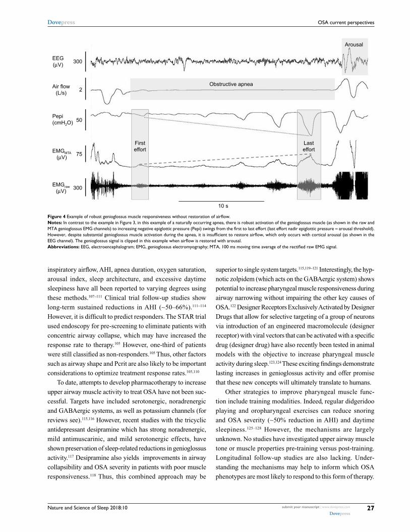
ficient to re-open the airway (Figure 4). Thus, the contribution
of the other upper airway muscles may also play a contributing
role.104 How the various components of upper airway muscle
function change over time is unknown. However, increased
weight gain over time and fat accumulation in the tongue are
predicted to worsen upper airway motion.79
Treatments to target the upper airway musclesOne approach to activate the upper airway muscles during
sleep is to deliver current to the muscles via direct stimulation

Nature and Science of Sleep 2018:10submit your manuscript | www.dovepress.com
Dovepress
Dovepress
26
Osman et al
or via stimulation of the hypoglossal nerve. Clinically, this
is achieved via surgical implantation of a stimulation device
connected to a cuff placed around the nerve.105 Mechanistic
studies have also used fine-wire electrodes or non-invasive
methods such as transcutaneous electrodes (see recent
review to compare different methods106). Improvements in
Figure 2 example of minimal genioglossus muscle responsiveness.Notes: in this example of a naturally occurring apnea, despite clear phasic activation of the genioglossus muscle (as shown in the raw and MTA genioglossus eMG channels), there is minimal activation of genioglossus during the respiratory event. This is despite substantial increasing negative epiglottic pressure (Pepi) swings from the first to last effort (last effort nadir epiglottic pressure = arousal threshold). it is only when cortical arousal occurs (as shown in the eeG channel) that major genioglossus activation occurs (signal clipped in this example) and airflow is restored.Abbreviations: EEG, electroencephalography; EMG, genioglossus electromyography; MTA, 100 ms moving time average of the rectified raw EMG signal.
EEG(µV)
EMGMTA(µV)
EMGraw(µV)
Air flow(L/s)
400
1
50
50
500
Firsteffort
Lasteffort
Obstructive apnea
Arousal
10 s
Pepi(cmH2O)
Figure 3 Example of robust genioglossus muscle responsiveness and restoration of airflow without cortical arousal.Notes: in contrast to the example in Figure 2, in this example of a naturally occurring hypopnea, there is robust activation of the genioglossus muscle (as shown in the raw and MTA genioglossus EMG channels), to increasing negative epiglottic pressure (Pepi) swings from the first to last effort which ultimately results in recovery of airflow without cortical arousal.Abbreviations: EEG, electroencephalogram; EMG, genioglossus electromyography; MTA, 100 ms moving time average of the rectified raw EMG signal.
EEG(µV)
EMGMTA(µV)
EMGraw(µV)
Air flow(L/s)
200
0.25
20
50
500
Pepi(cmH2O)
Firsteffort
40 s
Hypopnea
Lasteffort
Nature and Science of Sleep 2018:10 submit your manuscript | www.dovepress.com
Dovepress
Dovepress
27
OSA current perspectives
inspiratory airflow, AHI, apnea duration, oxygen saturation,
arousal index, sleep architecture, and excessive daytime
sleepiness have all been reported to varying degrees using
these methods.107–111 Clinical trial follow-up studies show
long-term sustained reductions in AHI (~50–66%).111–114
However, it is difficult to predict responders. The STAR trial
used endoscopy for pre-screening to eliminate patients with
concentric airway collapse, which may have increased the
response rate to therapy.105 However, one-third of patients
were still classified as non-responders.105 Thus, other factors
such as airway shape and Pcrit are also likely to be important
considerations to optimize treatment response rates.105,110
To date, attempts to develop pharmacotherapy to increase
upper airway muscle activity to treat OSA have not been suc-
cessful. Targets have included serotonergic, noradrenergic
and GABAergic systems, as well as potassium channels (for
reviews see).115,116 However, recent studies with the tricyclic
antidepressant desipramine which has strong noradrenergic,
mild antimuscarinic, and mild serotonergic effects, have
shown preservation of sleep-related reductions in genioglossus
activity.117 Desipramine also yields improvements in airway
collapsibility and OSA severity in patients with poor muscle
responsiveness.118 Thus, this combined approach may be
superior to single system targets.115,119–121 Interestingly, the hyp-
notic zolpidem (which acts on the GABAergic system) shows
potential to increase pharyngeal muscle responsiveness during
airway narrowing without impairing the other key causes of
OSA.122 Designer Receptors Exclusively Activated by Designer
Drugs that allow for selective targeting of a group of neurons
via introduction of an engineered macromolecule (designer
receptor) with viral vectors that can be activated with a specific
drug (designer drug) have also recently been tested in animal
models with the objective to increase pharyngeal muscle
activity during sleep.123,124 These exciting findings demonstrate
lasting increases in genioglossus activity and offer promise
that these new concepts will ultimately translate to humans.
Other strategies to improve pharyngeal muscle func-
tion include training modalities. Indeed, regular didgeridoo
playing and oropharyngeal exercises can reduce snoring
and OSA severity (~50% reduction in AHI) and daytime
sleepiness.125–128 However, the mechanisms are largely
unknown. No studies have investigated upper airway muscle
tone or muscle properties pre-training versus post-training.
Longitudinal follow-up studies are also lacking. Under-
standing the mechanisms may help to inform which OSA
phenotypes are most likely to respond to this form of therapy.
Figure 4 Example of robust genioglossus muscle responsiveness without restoration of airflow.Notes: in contrast to the example in Figure 3, in this example of a naturally occurring apnea, there is robust activation of the genioglossus muscle (as shown in the raw and MTA genioglossus EMG channels) to increasing negative epiglottic pressure (Pepi) swings from the first to last effort (last effort nadir epiglottic pressure = arousal threshold). However, despite substantial genioglossus muscle activation during the apnea, it is insufficient to restore airflow, which only occurs with cortical arousal (as shown in the EEG channel). The genioglossus signal is clipped in this example when airflow is restored with arousal.Abbreviations: EEG, electroencephalogram; EMG, genioglossus electromyography; MTA, 100 ms moving time average of the rectified raw EMG signal.
EEG(µV) 300
2
50
75
Firsteffort
10 s
Obstructive apnea
Lasteffort
Arousal
300
EMGMTA(µV)
EMGraw(µV)
Air flow(L/s)
Pepi(cmH2O)

Nature and Science of Sleep 2018:10submit your manuscript | www.dovepress.com
Dovepress
Dovepress
28
Osman et al
Prediction tools and simplified methods to estimate pharyngeal muscle functionIdentification of patients who have poor pharyngeal muscle
function may facilitate development of targeted therapies
directed toward this trait118 and improve treatment success
rates with existing therapies (e.g., hypoglossal nerve stimu-
lation). However, gold standard methodology to quantify
pharyngeal muscle activity is complex, invasive (fine-wire
electrodes inserted directly into the muscle), requires spe-
cialized personnel and equipment, and is time-consuming.34
There are no simplified tools to estimate pharyngeal muscle
function to identify people with this clinical phenotype
accurately. However, while multiple variables impact inspi-
ratory flow during sleep, mean peak inspiratory airflow
during airflow limitation appears to be a good surrogate for
active Pcrit, a measure that incorporates both anatomical
and neuromuscular components.40 Thus, if this approach
proves useful to predict treatment outcomes, automated sig-
nal processing algorithms based on routine PSG data could
be implemented.40 Nonetheless, additional practical tools to
determine pharyngeal muscle function are urgently required
in order to advance personalized treatment approaches that
target the upper airway muscles.
Respiratory arousal thresholdThe role of the respiratory arousal threshold in OSA patho-
genesis has been described in detail.129 Accordingly, these
concepts are outlined only briefly here. Historically, given
that most respiratory events are associated with a cortical
arousal, arousals were considered crucial to reopen the upper
airway following a respiratory event in OSA.130,131 However,
approximately 20% of respiratory events cease without corti-
cal arousal and an additional 20% occur after the upper airway
has already reopened and airflow has been restored.129,132–134
Indeed, 75% of adults with OSA have respiratory events that
terminate without an arousal or the arousal occurs following
airway reopening at some stage of the night.132 Thus, airway
reopening can occur without arousal. Rather, continual
unnecessary arousals can worsen OSA and contribute to OSA
pathophysiolgy.129,132 Specifically, repetitive arousals can per-
petuate blood-gas disturbances and cause sleep fragmentation
to promote cyclical breathing and prevent establishment and
maintenance of more stable, deeper stages of sleep.135–138
Numerous respiratory stimuli can contribute to arousal
from sleep during a respiratory event.129,139–142 Increased
respiratory effort due to a narrowed pharyngeal airway
increases negative intrathoracic pressure. Although the
amount of negative intrathoracic pressure generated can
vary greatly between individuals and in different stages of
sleep,35,132,143–147 the magnitude of negative pressure required
to cause an arousal from sleep is relatively constant within
an individual.147–149 This is the case regardless of whether the
respiratory disturbance is caused by hypoxia, hypercapnia,
or respiratory loading.147
Accordingly, the gold standard method to quantify the
threshold for arousal to respiratory stimuli requires an epi-
glottic or esophageal pressure catheter combined with PSG
recording equipment. Specifically, the respiratory arousal
threshold is the nadir pressure immediately prior to corti-
cal arousal (Figures 1, 2, and 4). The respiratory arousal
threshold is quantified by averaging multiple pressure val-
ues throughout the night to an experimental intervention
designed to cause airway narrowing or activate respiratory
afferents35,147 or during naturally occurring respiratory
events.144,145
In people with OSA who require large intrathoracic
pressure swings to cause an arousal (i.e., patients with high
respiratory arousal thresholds ≤25cmH2O), respiratory
events are often prolonged, particularly if these patients also
have poor upper airway muscle responsiveness.129 Thus, in
the absence of neuromuscular compensation, arousal from
sleep and reintroduction of wakefulness drive can act as a
last line of defense to facilitate rapid reopening of the air-
way to re-establish airflow and normalize blood-gas levels
in these individuals.129,130,150 The consequences of OSA such
as sleep deprivation increase, while CPAP therapy decreases
the respiratory arousal threshold.151,152 Nonetheless, approxi-
mately 30–50% of OSA patients wake to relatively small
intrathoracic/epiglottic pressure swings (i.e., patients with
low respiratory arousal thresholds ≥−15 cmH2O).35,38,129,145 In
these patients, given that the stimuli for arousal are the same
as the stimuli required to recruit the pharyngeal dilator mus-
cles (i.e., blood gas changes and negative pressure swings),
premature arousal limits the opportunity for neuromuscular
compensation mechanisms to overcome airway narrowing
and stabilize breathing.132 Frequent arousals can also cause
sleep fragmentation and sleep instability, prevent deeper
stages of sleep, and perpetuate unstable breathing.132,142 Thus,
strategies to reduce arousals in these patients may allow for
more stable breathing during sleep.
Treatments to target the respiratory arousal thresholdGiven the abovementioned rationale, the potential thera-
peutic role of hypnotics to treat OSA in patients with a low
respiratory arousal threshold phenotype has been an area

Nature and Science of Sleep 2018:10 submit your manuscript | www.dovepress.com
Dovepress
Dovepress
29
OSA current perspectives
of recent research focus.150 This strategy requires a care-
ful targeted approach to optimize benefit in those with the
desired phenotype and avoid harm in those with high arousal
thresholds. In particular, the selected agent to increase the
threshold for arousal to respiratory stimuli must do so with-
out impairment in pharyngeal muscle activity. Apart from
an early wakefulness benzodiazepine study,22 subsequent
studies during sleep have not shown systematic reductions
in pharyngeal muscle activity or responsiveness to negative
pharyngeal pressure with common doses of zopiclone,96,144
trazodone,153 temazepam,122 or zolpidem.122 While a recent
study found more variable effects on genioglossus muscle
responsiveness with temazepam, paradoxically, on average,
zolpidem increased muscle responsiveness 3-fold in people
with and without OSA.122
The other concern with hypnotic use in OSA is prolonga-
tion of respiratory events and worsening hypoxemia due to
blunted arousal responses in those individuals with a high
threshold for respiratory arousal. Indeed, this can occur with
high doses or in obese patients with very severe disease.129
By contrast, the hypnotics eszopiclone,145 zopiclone,22 and
trazodone154 can reduce OSA severity as measured by the AHI
without worsening hypoxemia. In the eszopiclone study,145
reductions in AHI occurred invariably in those with a low
arousal threshold phenotype. The number of arousals per
hour of sleep also decreased.145 Given the contrasting effects
of hypnotics in OSA, screening tools to distinguish between
patients with low and high respiratory arousal thresholds and
to determine who will benefit versus be susceptible to harm
are important.
Prediction tools and simplified methods to estimate the respiratory arousal thresholdThe gold standard approach to quantify the respiratory
arousal threshold is impractical for routine clinical use as it
is time-consuming, costly, and somewhat invasive (requires
an airway pressure catheter). Preliminarily findings indicate
that respiratory sensation to inspiratory loading during
wakefulness is related to the respiratory arousal threshold
during sleep.155 Edwards et al have developed a simple tool to
estimate the respiratory arousal threshold with high sensitiv-
ity and specificity based on three measures from a standard
overnight PSG (AHI, nadir oxygen saturation, and the apnea
to hypopnea ratio).38 Thus, while prospective intervention
studies are required, this simple approach could easily be
implemented in the clinical setting to inform treatment
decisions. Given that over 40% of OSA patients may also
have insomnia,156–158 simple accurate tools to determine which
OSA patients will benefit versus those at risk of harm with
hypnotics would be invaluable.
Loop gainLoop gain is a term used to describe the stability of a feedback
control system. In the context of respiratory physiology, loop
gain is the ventilatory response to ventilatory disturbance
ratio. It comprises three principal components: 1) plant gain
(i.e., tissues, blood, and lungs where CO2 is stored) and 2)
delays in circulation (i.e., time it takes for a change in CO2
to mix with the existing blood to arrive and be detected by
the chemoreceptors) and 3) controller gain (i.e., chemosen-
sitivity). Any medical condition that modifies one or more of
these components (e.g., heart failure) will alter loop gain.34
Components of OSA such as intermittent hypoxia can also
alter respiratory control.159 People with high loop gain have
exaggerated ventilatory responses to minimal changes in
CO2. This is a marker of an unstable control system. This can
be reduced with CPAP therapy.160 On the other hand, those
with extremely low loop gain often experience hypoven-
tilation during sleep, as is the case in people with obesity
hypoventilation syndrome.34
In their landmark study using proportional assist ventila-
tion to induce breathing oscillations during sleep after the
airway was stabilized with CPAP, Younes et al showed that
severe OSA patients have high loop gain.138 Subsequent stud-
ies confirmed that many people with OSA have high loop
gain.35,161 When combined with even a modest impairment
in upper airway anatomy, high loop gain can drive OSA
pathogenesis.35,161 Similar to the other phenotypic traits, loop
gain can be quantified using transient reductions in CPAP
during sleep to create a disturbance to breathing.161,162 Rapid
reintroduction of CPAP is then applied so that the ventilatory
response (overshoot) can be quantified (using a breathing
mask and pneumotachograph) to calculate loop gain. This
procedure is repeated as many times as possible throughout
the night. Loop gain is then calculated as the ventilatory
response divided by the ventilatory disturbance ratio after
scaling for the different levels of ventilatory disturbances
presented.161,162 This technique results in a negative number
such that more negative numbers reflect higher loop gain.162
Approximately one-third of OSA patients have high loop
gain (<−5) which indicates a >5 L/min increase in minute
ventilation in response to 1 L/min reduction in minute
ventilation.35

Nature and Science of Sleep 2018:10submit your manuscript | www.dovepress.com
Dovepress
Dovepress
30
Osman et al
Treatments to target loop gain and simplified methods to estimate loop gainO
2 therapy reduces loop gain and OSA severity in people with
high loop gain.163 Carbonic anhydrase inhibitors such as acet-
azolamide also reduce loop gain by approximately 40% and
OSA severity.164 Similarly, zonisamide reduces OSA severity.24
In addition, stabilization of CO2 and hypercapnia can prevent
hypoventilation and unstable respiratory control during sleep
in OSA.165 Strategies that combine O2 therapy to reduce loop
gain with a hypnotic to increase the arousal threshold can
yield major reductions in OSA severity.146 In addition, recent
studies from the Mateika group indicate that several of the
key contributors to OSA such as airway collapsibility, arousal
threshold, and respiratory control are influenced by circadian
phase.166–168 Thus, novel strategies that target the circadian
system may have therapeutic potential in OSA.
The current approaches to quantify loop gain during sleep
require experienced personnel to perform CPAP manipula-
tions and analyze the data. However, Terrill et al have devel-
oped an analysis approach that uses the nasal pressure signal
from a standard PSG to estimate loop gain.41 This approach
has been used to explain and predict changes to a range of
interventions in OSA.30,47,146 Wakefulness tests of respira-
tory control may also be helpful in estimating responses to
pharmacotherapies.44,45
Potential links between phenotypic traits, treatment, and health consequencesIn addition to the role that the phenotypic traits play in con-
tributing to OSA pathogenesis, the traits may also provide
insight into disease consequences and physiological reasons
for treatment failure. For example, a low arousal threshold
trait may be a physiological contributor to poor CPAP adher-
ence and compliance.31 A low arousal threshold trait and
its consequences such as sleep fragmentation and frequent
hypopneas with minimal changes in oxygenation may be a
marker for cognitive impairment and daytime sleepiness.54,169
Similarly, patients who tend to have more “intense” arousals,
which appear to be an inherent trait,47,170 may experience more
daytime sleepiness than those who do not. People who have
OSA that is driven by high loop gain may be more vulnerable
to the cardiovascular consequences of OSA. Conversely, strat-
egies that target certain components of respiratory control
such as mild intermittent hypoxia to elicit respiratory plastic-
ity may help improve CPAP compliance and potentially also
directly target the autonomic, cardiovascular, neurocognitive,
and metabolic systems.171,172 While possible links between
the phenotypic traits and specific disease consequences have
not yet been investigated, emergence of simplified tools to
estimate the traits will enable these theoretical concepts to
be tested systematically in cohort studies.
ConclusionCharacterization of the different causes or phenotypes of OSA
in recent years has provided new pathways for targeted therapy.
New simplified approaches to estimate each of the key causes
of OSA have recently been developed. While more work is
required, particularly directed toward the impaired pharyngeal
muscle trait, these new tools offer promise for the translation
of detailed phenotyping concepts to the clinic. Identification
of the traits may also provide insight in to which patients are
more likely to develop specific disease consequences.
DisclosureDJE is supported by a National Health and Medical Research
Council of Australia Senior Research Fellowship (1116942),
has a Commonwealth Government of Australia Cooperative
Research Centre grant (industry partner: Oventus Medical),
and serves as a consultant for Bayer. The other authors report
no conflicts of interest in this work.
References 1. Punjabi NM. The epidemiology of adult obstructive sleep apnea. Proc
Am Thorac Soc. 2008;5(2):136–143. 2. Peppard PE, Young T, Barnet JH, Palta M, Hagen EW, Hla KM.
Increased prevalence of sleep-disordered breathing in adults. Am J Epidemiol. 2013;177(9):1006–1014.
3. Heinzer R, Vat S, Marques-Vidal P, et al. Prevalence of sleep-disordered breathing in the general population: the HypnoLaus study. Lancet Respir Med. 2015;3(4):310–318.
4. Kapur VK, Resnick HE, Gottlieb DJ; Sleep Heart Health Study Group. Sleep disordered breathing and hypertension: does self-reported sleepi-ness modify the association? Sleep. 2008;31(8):1127–1132.
5. Walia HK, Li H, Rueschman M, et al. Association of severe obstruc-tive sleep apnea and elevated blood pressure despite antihypertensive medication use. J Clin Sleep Med. 2014;10(8):835–843.
6. Drager LF, Togeiro SM, Polotsky VY, Lorenzi-Filho G. Obstructive sleep apnea: a cardiometabolic risk in obesity and the metabolic syndrome. J Am Coll Cardiol. 2013;62(7):569–576.
7. Olaithe M, Bucks RS, Hillman DR, Eastwood PR. Cognitive deficits in obstructive sleep apnea: insights from a meta-review and comparison with deficits observed in COPD, insomnia, and sleep deprivation. Sleep Med Rev. Epub 2017 Mar 30.
8. Wheaton AG, Perry GS, Chapman DP, Croft JB. Sleep disordered breathing and depression among U.S. adults: National Health and Nutri-tion Examination Survey, 2005–2008. Sleep. 2012;35(4):461–467.
9. Antic NA, Catcheside P, Buchan C, et al. The effect of CPAP in normal-izing daytime sleepiness, quality of life, and neurocognitive function in patients with moderate to severe OSA. Sleep. 2011;34(1):111–119.
10. Romero E, Krakow B, Haynes P, Ulibarri V. Nocturia and snoring: predictive symptoms for obstructive sleep apnea. Sleep Breath. 2010;14(4):337–343.
11. Mulgrew AT, Ryan CF, Fleetham JA, et al. The impact of obstructive sleep apnea and daytime sleepiness on work limitation. Sleep Med. 2007;9(1):42–53.

Nature and Science of Sleep 2018:10 submit your manuscript | www.dovepress.com
Dovepress
Dovepress
31
OSA current perspectives
12. Howard ME, Desai AV, Grunstein RR, et al. Sleepiness, sleep-disordered breathing, and accident risk factors in commercial vehicle drivers. Am J Respir Crit Care Med. 2004;170(9):1014–1021.
13. Stoohs RA, Guilleminault C, Itoi A, Dement WC. Traffic accidents in commercial long-haul truck drivers: the influence of sleep-disordered breathing and obesity. Sleep. 1994;17(7):619–623.
14. Hillman DR, Murphy AS, Pezzullo L. The economic cost of sleep disorders. Sleep. 2006;29(3):299–305.
15. Sleep Health Foundation. Asleep on the job: Costs of inadequate sleep in Australia; 2017:1–112.
16. Montserrat JM, Ferrer M, Hernandez L, et al. Effectiveness of CPAP treatment in daytime function in sleep apnea syndrome: a randomized controlled study with an optimized placebo. Am J Resp Crit Care Med. 2001;164(4):608–613.
17. Knudsen TB, Laulund AS, Ingerslev J, Homøe P, Pinholt EM. Improved apnea-hypopnea index and lowest oxygen saturation after maxillomandibular advancement with or without counterclockwise rotation in patients with obstructive sleep apnea: a meta-analysis. J Oral Maxillofac Surg 2015;73(4):719–726.
18. George CF. Reduction in motor vehicle collisions following treatment of sleep apnoea with nasal CPAP. Thorax. 2001;56(7):508–512.
19. Dong Y, Dai Y, Wei G, Cha L, Li X. Effect of continuous positive air-way pressure on blood pressure in hypertensive patients with coronary artery bypass grafting and obstructive sleep apnea. Int J Clin Exp Med. 2014;7(11):4308–4315. eCollection 2014.
20. Olsen S, Smith S, Oei T, Douglas J. Health belief model predicts adherence to CPAP before experience with CPAP. Eur Respir J. 2008; 32(3):710–717.
21. Libman E, Bailes S, Fichten CS, et al. CPAP treatment adher-ence in women with obstructive sleep apnea. Sleep Disord. 2017; 2017:2760650.
22. Ferguson KA, Ono T, Lowe AA, Keenan SP, Fleetham JA. A random-ized crossover study of an oral appliance vs nasal-continuous positive airway pressure in the treatment of mild-moderate obstructive sleep apnea. Chest. 1996;109(5):1269–1275.
23. Noller MW, Guilleminault C, Gouveia CJ, et al. Mandibular advance-ment for adult obstructive sleep apnea: a systematic review and meta-analysis. J Craniomaxillofac Surg. Epub 2017 Oct 13.
24. Choi JH, Lee JY, Cha J, Kim K, Hong SN, Lee SH. Predictive models of objective oropharyngeal OSA surgery outcomes: success rate and AHI reduction ratio. PLoS One. 2017;12(9):e0185201.
25. Phillips CL, Grunstein RR, Darendeliler MA, et al. Health outcomes of continuous positive airway pressure versus oral appliance treatment for obstructive sleep apnea: a randomized controlled trial. Am J Respir Crit Care Med. 2013;187(8):879–887.
26. Appleton SL, Vakulin A, McEvoy RD, et al. Undiagnosed obstructive sleep apnea is independently associated with reductions in quality of life in middle-aged, but not elderly men of a population cohort. Sleep Breath. 2015;19(4):1309–1316.
27. Simpson L, Hillman DR, Cooper MN, et al. High prevalence of undiagnosed obstructive sleep apnoea in the general population and methods for screening for representative controls. Sleep Breath. 2013;17(3):967–973.
28. Kapur V, Strohl KP, Redline S, Iber C, O’Connor G, Nieto J. Underdi-agnosis of sleep apnea syndrome in U.S. communities. Sleep Breath. 2002;6(2):49–54.
29. Jaiswal SJ, Owens RL, Malhotra A. Raising awareness about sleep disorders. Lung India. 2017;34(3):262–268.
30. Bixler EO, Vgontzas AN, Lin HM, Calhoun SL, Vela-Bueno A, Kales A. Excessive daytime sleepiness in a general population sample: the role of sleep apnea, age, obesity, diabetes, and depression. J Clin Endocrinol Metab. 2005;90(8):4510–4515.
31. Gray EL, McKenzie DK, Eckert DJ. Obstructive sleep apnea with-out obesity is common and difficult to treat: evidence for a distinct pathophysiological phenotype. J Clin Sleep Med. 2017;13(1): 81–88.
32. Ye L, Pien GW, Ratcliffe SJ, et al. The different clinical faces of obstructive sleep apnoea: a cluster analysis. Eur Respir J. 2014;44(6): 1600–1607.
33. Arnardottir ES, Bjornsdottir E, Olafsdottir KA, Benediktsdottir B, Gislason T. Obstructive sleep apnoea in the general population: highly prevalent but minimal symptoms. Eur Respir J. 2016;47(1):194–202.
34. Eckert DJ. Phenotypic approaches to obstructive sleep apnoea – new pathways for targeted therapy. Sleep Med Rev. 2018;37:45–59.
35. Eckert DJ, White DP, Jordan AS, Malhotra A, Wellman A. Defining phenotypic causes of obstructive sleep apnea. Identification of novel therapeutic targets. Am J Respir Crit Care Med. 2013;188(8):996–1004.
36. Carberry JC, Amatoury J, Eckert DJ. Personalized management approach for OSA. Chest. Epub 2017 June 16.
37. Landry SA, Joosten SA, Eckert DJ, et al. Therapeutic CPAP level predicts upper airway collapsibility in patients with obstructive sleep apnea. Sleep. 2017;40(6).
38. Edwards BA, Eckert DJ, McSharry DG, et al. Clinical predictors of the respiratory arousal threshold in patients with obstructive sleep apnea. Am J Respir Crit Care Med. 2014;190(11):1293–1300.
39. Genta PR, Sands SA, Butler JP, et al. Airflow shape is associated with the pharyngeal structure causing OSA. Chest. 2017;152(3):537–546.
40. Azarbarzin A, Sands SA, Taranto-Montemurro L, et al. Estimation of pharyngeal collapsibility during sleep by peak inspiratory airflow. Sleep. 2017;40(1).
41. Terrill PI, Edwards BA, Nemati S, et al. Quantifying the ventilatory control contribution to sleep apnoea using polysomnography. Eur Respir J. 2015;45(2):408–418.
42. Hirata RP, Schorr F, Kayamori F, et al. Upper airway collapsibility assessed by negative expiratory pressure while awake is associ-ated with upper airway anatomy. J Clin Sleep Med. 2016;12(10): 1339–1346.
43. Osman A, Nguyen C, Carberry JC, et al. Awake upper airway col-lapsibility is related to airway collapsibility during sleep (Pcrit) in obstructive sleep apnea. J Sleep Res. 2016;25(2):A57.
44. Wang D, Marshall NS, Duffin J, et al. Phenotyping interindividual variability in obstructive sleep apnoea response to temazepam using ventilatory chemoreflexes during wakefulness. J Sleep Res. 2011;20(4):526–532.
45. Wang D, Somogyi AA, Yee BJ, et al. The effects of a single mild dose of morphine on chemoreflexes and breathing in obstructive sleep apnea. Respir Physiol Neurobiol. 2013;185(3):526–532.
46. Edwards BA, Wellman A, Owens RL. PSGs: more than just the AHI. J Clin Sleep Med. 2013;9(6):527–528.
47. Cairns A, Poulos G, Bogan R. Who is getting tested for obstructive sleep apnea using a portable recording system? Test results from 193,221 patients. J Clin Sleep Med. 2014;10(11):1193–1198.
48. Donovan LM, Patel SR. Making the most of simplified sleep apnea testing. Ann Intern Med. 2017;166(5):366–367.
49. Collop NA, Anderson WM, Boehlecke B, et al; Portable Monitoring Task Force of the American Academy of Sleep Medicine. Clinical guidelines for the use of unattended portable monitors in the diagnosis of obstructive sleep apnea in adult patients. Portable Monitoring Task Force of the American Academy of Sleep Medicine. J Clin Sleep Med. 2007;3(7):737–747.
50. Muraja-Murro A, Nurkkala J, Tiihonen P, et al. Total duration of apnea and hypopnea events and average desaturation show significant variation in patients with a similar apnea-hypopnea index. J Med Eng Technol. 2012;36(8):393–398.
51. Alex R, Manchikatla S, Machiraju K, et al. Effect of apnea dura-tion on apnea induced variations in cerebral blood flow velocity and arterial blood pressure. Conf Proc IEEE Eng Med Biol Soc. 2014;2014:270–273.
52. Wu H, Zhan X, Zhao M, Wei Y. Mean apnea-hypopnea duration (but not apnea-hypopnea index) is associated with worse hyperten-sion in patients with obstructive sleep apnea. Medicine (Baltimore). 2016;95(48):e5493.

Nature and Science of Sleep 2018:10submit your manuscript | www.dovepress.com
Dovepress
Dovepress
32
Osman et al
53. Sankari A, Pranathiageswaran S, Maresh S, Hosni AM, Badr MS. Characteristics and consequences of non-apneic respiratory events during sleep. Sleep. 2017;40(1).
54. Koch H, Schneider LD, Finn LA, et al. Breathing disturbances without hypoxia are associated with objective sleepiness in sleep apnea. Sleep. 2017;40(11).
55. Weaver EM, Woodson BT, Steward DL. Polysomnography indexes are discordant with quality of life, symptoms, and reaction times in sleep apnea patients. Otolaryngol Head Neck Surg. 2005;132(2):255–262.
56. Alzoubaidi M, Mokhlesi B. Obstructive sleep apnea during rapid eye movement sleep: clinical relevance and therapeutic implications. Curr Opin Pulm Med. 2016;22(6):545–554.
57. Appleton SL, Vakulin A, Martin SA, et al. Hypertension is associated with undiagnosed OSA during rapid eye movement sleep. Chest. 2016;150(3):495–505.
58. Aurora RN, Crainiceanu C, Gottlieb DJ, Kim JS, Punjabi NM. Obstruc-tive sleep apnea during rapid eye movement sleep and cardiovascular disease. Am J Respir Crit Care Med. Epub 2017 Nov 7.
59. Chami HA, Gottlieb DJ, Redline S, Punjabi NM. Association between glucose metabolism and sleep-disordered breathing during REM sleep. Am J Respir Crit Care Med. 2015;192(9):1118–1126.
60. Neelapu BC, Kharbanda OP, Sardana HK, et al. Craniofacial and upper airway morphology in adult obstructive sleep apnea patients: a systematic review and meta-analysis of cephalometric studies. Sleep Med Rev. 2017;31:79–90.
61. Edwards BA, Eckert DJ, Jordan AS. Obstructive sleep apnoea patho-genesis from mild to severe: is it all the same? Respirology. 2017;22(1): 33–42.
62. Eckert DJ, Wellman A. Physiological phenotypes. In: Barbé F, Pépin JL, editors. European Respiratory Monograph: Obstructive Sleep Apnoea. Plymouth, UK: European Respiratory Society; 2015:9–23.
63. Dempsey JA, Xie A, Patz DS, Wang D. Physiology in medicine: obstructive sleep apnea pathogenesis and treatment--considerations beyond airway anatomy. J Appl Physiol (1985). 2014;116(1):3–12.
64. Carlisle T, Carthy ER, Glasser M, et al. Upper airway factors that protect against obstructive sleep apnoea in healthy older males. Eur Respir J. 2014;44(3):685–693.
65. Safiruddin F, Koutsourelakis I, de Vries N. Upper airway collapse during drug induced sleep endoscopy: head rotation in supine position compared with lateral head and trunk position. Eur Arch Otorhinolar-yngol. 2015;272(2):485–488.
66. Coxson HO, Eastwood PR, Williamson JP, Sin DD. Phenotyping airway disease with optical coherence tomography. Respirology. 2011;16(1):34–43.
67. Ito E, Tsuiki S, Maeda K, Okajima I, Inoue Y. Oropharyngeal crowding closely relates to aggravation of OSA. Chest. 2016;150(2):346–352.
68. Schwab RJ, Pasirstein M, Pierson R, et al. Identification of upper air-way anatomic risk factors for obstructive sleep apnea with volumetric magnetic resonance imaging. Am J Respir Crit Care Med. 2003;168(5): 522–530.
69. Segal Y, Malhotra A, Pillar G. Upper airway length may be associated with the severity of obstructive sleep apnea syndrome. Sleep Breath. 2008;12(4):311–316.
70. Morrison DL, Launois SH, Isono S, Feroah TR, Whitelaw WA, Rem-mers JE. Pharyngeal narrowing and closing pressures in patients with obstructive sleep apnea. Am Rev Respir Dis. 1993;148(3):606–611.
71. Young T, Peppard PE, Taheri S. Excess weight and sleep-disordered breathing. J Appl Physiol (1985). 2005;99(4):1592–1599.
72. Lai CC, Friedman M, Lin HC, et al. Clinical predictors of effective continuous positive airway pressure in patients with obstructive sleep apnea/hypopnea syndrome. Laryngoscope. 2015;125(8):1983–1987.
73. Pinto JA, Godoy LB, Marquis VW, Sonego TB, Leal Cde F, Artico MS. Anthropometric data as predictors of Obstructive Sleep Apnea Severity. Braz J Otorhinolaryngol. 2011;77(4):516–521.
74. Caron CJJM, Pluijmers BI, Maas BDPJ, et al. Obstructive sleep apnoea in craniofacial microsomia: analysis of 755 patients. Int J Oral Maxil-lofac Surg. 2017;46(10):1330–1337.
75. Schorr F, Kayamori F, Hirata RP, et al. Different craniofacial charac-teristics predict upper airway collapsibility in Japanese-Brazilian and white men. Chest. 2016;149(3):737–746.
76. Sforza E, Bacon W, Weiss T, Thibault A, Petiau C, Krieger J. Upper airway collapsibility and cephalometric variables in patients with obstructive sleep apnea. Am J Respir Crit Care Med. 2000;161(2 Pt 1): 347–352.
77. Verma M, Seto-Poon M, Wheatley JR, Amis TC, Kirkness JP. Influ-ence of breathing route on upper airway lining liquid surface tension in humans. J Physiol. 2006;574(Pt 3):859–866.
78. Weiss TM, Atanasov S, Calhoun KH. The association of tongue scalloping with obstructive sleep apnea and related sleep pathology. Otolaryngol Head Neck Surg. 2005;133(6):966–971.
79. Kim AM, Keenan BT, Jackson N, et al. Tongue fat and its relationship to obstructive sleep apnea. Sleep. 2014;37(10):1639–1648.
80. Gleadhill IC, Schwartz AR, Schubert N, Wise RA, Permutt S, Smith PL. Upper airway collapsibility in snorers and in patients with obstruc-tive hypopnea and apnea. Am Rev Respir Dis. 1991;143(6):1300–1303.
81. Eastwood PR, Szollosi I, Platt PR, Hillman DR. Collapsibility of the upper airway during anesthesia with isoflurane. Anesthesiology. 2002;97(4):786–793.
82. Schwartz AR, O’Donnell CP, Baron J, et al. The hypotonic upper air-way in obstructive sleep apnea: role of structures and neuromuscular activity. Am J Respir Crit Care Med. 1998;157(4 Pt 1):1051–1057.
83. Carberry JC, Jordan AS, White DP, Wellman A, Eckert DJ. Upper airway collapsibility (Pcrit) and pharyngeal dilator muscle activity are sleep stage dependent. Sleep. 2016;39(3):511–521.
84. Kirkness JP, Peterson LA, Squier SB, et al. Performance characteristics of upper airway critical collapsing pressure measurements during sleep. Sleep. 2011;34(4):459–467.
85. Genta PR, Schorr F, Eckert DJ, et al. Upper airway collapsibility is asso-ciated with obesity and hyoid position. Sleep. 2014;37(10):1673–1678.
86. Kirkness JP, Schwartz AR, Schneider H, et al. Contribution of male sex, age, and obesity to mechanical instability of the upper airway during sleep. J Appl Physiol (1985). 2008;104(6):1618–1624.
87. Bachar G, Feinmesser R, Shpitzer T, Yaniv E, Nageris B, Eidelman L. Laryngeal and hypopharyngeal obstruction in sleep disordered breath-ing patients, evaluated by sleep endoscopy. Eur Arch Otorhinolaryngol. 2008;265(11):1397–1402.
88. Horner RL, Hughes SW, Malhotra A. State-dependent and reflex drives to the upper airway: basic physiology with clinical implications. J Appl Physiol (1985). 2014;116(3):325–336.
89. Kubin L. Neural control of the upper airway: respiratory and state-dependent mechanisms. Compr Physiol. 2016;6(4):1801–1850.
90. Saboisky JP, Butler JE, Fogel RB, et al. Tonic and phasic respiratory drives to human genioglossus motoneurons during breathing. J Neu-rophysiol. 2006;95(4):2213–2221.
91. Wilkinson V, Malhotra A, Nicholas CL, et al. Discharge patterns of human genioglossus motor units during sleep onset. Sleep. 2008;31(4): 525–533.
92. Nicholas CL, Jordan AS, Heckel L, et al. Discharge patterns of human tensor palatini motor units during sleep onset. Sleep. 2012;35(5):699–707.
93. Eckert DJ, Saboisky JP, Jordan AS, White DP, Malhotra A. A second-ary reflex suppression phase is present in genioglossus but not tensor palatini in response to negative upper airway pressure. J Appl Physiol (1985). 2010;108(6):1619–1624.
94. Eckert DJ, Malhotra A, Lo YL, White DP, Jordan AS. The influence of obstructive sleep apnea and gender on genioglossus activity during rapid eye movement sleep. Chest. 2009;135(4):957–964.
95. Eckert DJ, McEvoy RD, George KE, Thomson KJ, Catcheside PG. Genioglossus reflex inhibition to upper-airway negative-pressure stimuli during wakefulness and sleep in healthy males. J Physiol. 2007;581(Pt 3):1193–1205.
96. Loewen AH, Ostrowski M, Laprairie J, Maturino F, Hanly PJ, Younes M. Response of genioglossus muscle to increasing chemical drive in sleeping obstructive apnea patients. Sleep. 2011;34(8):1061–1073.

Nature and Science of Sleep 2018:10 submit your manuscript | www.dovepress.com
Dovepress
Dovepress
33
OSA current perspectives
97. Jordan AS, Wellman A, Heinzer RC, et al. Mechanisms used to restore ventilation after partial upper airway collapse during sleep in humans. Thorax. 2007;62(10):861–867.
98. Sands SA, Eckert DJ, Jordan AS, et al. Enhanced upper-airway muscle responsiveness is a distinct feature of overweight/obese individuals without sleep apnea. Am J Respir Crit Care Med. 2014;190(8):930–937.
99. Dotan Y, Pillar G, Tov N, et al. Dissociation of electromyogram and mechanical response in sleep apnoea during propofol anaesthesia. Eur Respir J. 2013;41(1):74–84.
100. Cheng S, Butler JE, Gandevia SC, Bilston LE. Movement of the tongue during normal breathing in awake healthy humans. J Physiol. 2008;586(17):4283–4294.
101. Brown EC, Cheng S, McKenzie DK, Butler JE, Gandevia SC, Bilston LE. Respiratory movement of upper airway tissue in obstructive sleep apnea. Sleep. 2013;36(7):1069–1076.
102. Cheng S, Brown EC, Hatt A, Butler JE, Gandevia SC, Bilston LE. Healthy humans with a narrow upper airway maintain patency during quiet breathing by dilating the airway during inspiration. J Physiol. 2014;592(21):4763–4774.
103. Jordan AS, White DP, Owens RL, et al. The effect of increased genioglossus activity and end-expiratory lung volume on pharyngeal collapse. J Appl Physiol (1985). 2010;109(2):469–475.
104. Dotan Y, Pillar G, Schwartz AR, Oliven A. Asynchrony of lingual muscle recruitment during sleep in obstructive sleep apnea. J Appl Physiol (1985). 2015;118(12):1516–1524.
105. Strollo PJ Jr, Soose RJ, Maurer JT, et al; STAR Trial Group. Upper-airway stimulation for obstructive sleep apnea. N Eng J Med. 2014;370(2):139–149.
106. Bisogni V, Pengo MF, De Vito A, et al. Electrical stimulation for the treatment of obstructive sleep apnoea: a review of the evidence. Expert Rev Respir Med. 2017;11(9):711–720.
107. Hida W, Okabe S, Miki H, et al. Effects of submental stimulation for several consecutive nights in patients with obstructive sleep apnoea. Thorax. 1994;49(5):446–452.
108. Hofauer B, Philip P, Wirth M, Knopf A, Heiser C. Effects of upper-airway stimulation on sleep architecture in patients with obstructive sleep apnea. Sleep Breath. 2017;21(4):901–908.
109. Schwartz AR, Barnes M, Hillman D, et al. Acute upper airway responses to hypoglossal nerve stimulation during sleep in obstructive sleep apnea. Am J Respir Crit Care Med. 2012;185(4):420–426.
110. Schwartz AR, Smith PL, Oliven A. Electrical stimulation of the hypo-glossal nerve: a potential therapy. J Appl Physiol (1985). 2014;116(3): 337–344.
111. Eastwood PR, Barnes M, Walsh JH, et al. Treating obstructive sleep apnea with hypoglossal nerve stimulation. Sleep. 2011;34(11): 1479–1486.
112. Steffen A, Sommer JU, Hofauer B, Maurer JT, Hasselbacher K, Heiser C. Outcome after one year of upper airway stimulation for obstructive sleep apnea in a multicenter German post-market study. Laryngoscope. Epub 2017 May 31.
113. Gillespie MB, Soose RJ, Woodson BT, et al; STAR Trial Investigators. Upper airway stimulation for obstructive sleep apnea: patient-reported outcomes after 48 months of follow-up. Otolaryngol Head Neck Surg. 2017;156(4):765–771.
114. Woodson BT, Soose RJ, Gillespie MB, et al; STAR Trial Investigators. Three-year outcomes of cranial nerve stimulation for obstructive sleep apnea: the STAR Trial. Otolaryngol Head Neck Surg. 2016;154(1):181–188.
115. White DP. Pharmacologic approaches to the treatment of obstructive sleep apnea. Sleep Med Clin. 2016;11(2):203–212.
116. Horner RL, Grace KP, Wellman A. A resource of potential drug targets and strategic decision-making for obstructive sleep apnoea pharma-cotherapy. Respirology. 2017;22(5):861–873.
117. Taranto-Montemurro L, Edwards BA, Sands SA, et al. Desipramine increases genioglossus activity and reduces upper airway collapsibility during Non-REM sleep in healthy subjects. Am J Respir Crit Care Med. 2016;194(7):878–885.
118. Taranto-Montemurro L, Sands SA, Edwards BA, et al. Desipramine improves upper airway collapsibility and reduces OSA sever-ity in patients with minimal muscle compensation. Eur Respir J. 2016;48(5):1340–1350.
119. Marshall NS, Yee BJ, Desai AV, et al. Two randomized placebo-con-trolled trials to evaluate the efficacy and tolerability of mirtazapine for the treatment of obstructive sleep apnea. Sleep. 2008;31(6):824–831.
120. Kraiczi H, Hedner J, Dahlöf P, Ejnell H, Carlson J. Effect of serotonin uptake inhibition on breathing during sleep and daytime symptoms in obstructive sleep apnea. Sleep. 1999;22(1):61–67.
121. Taranto-Montemurro L, Sands SA, Azarbarzin A, et al. Effect of 4-aminopyridine on genioglossus muscle activity during sleep in healthy adults. Ann Am Thorac Soc. 2017;14(7):1177–1183.
122. Carberry JC, Fisher LP, Grunstein RR, et al. Role of common hypnotics on the phenotypic causes of OSA: paradoxical effects of zolpidem. Eur Respir J. 2017;50(6):pii:1701344.
123. Horton GA, Fraigne JJ, Torontali ZA, et al. Activation of the hypoglos-sal to tongue musculature motor pathway by remote control. Sci Rep. 2017;7:45860.
124. Fleury Curado T, Fishbein K, Pho H, et al. Chemogenetic stimulation of the hypoglossal neurons improves upper airway patency. Sci Rep. 2017;7:44392.
125. Ieto V, Kayamori F, Montes MI, et al. Effects of oropharyngeal exercises on snoring: a randomized trial. Chest. 2015;148(3):683–691.
126. Guimarães KC, Drager LF, Genta PR, Marcondes BF, Lorenzi-Filho G. Effects of oropharyngeal exercises on patients with moderate obstructive sleep apnea syndrome. Am J Respir Crit Care Med. 2009;179(10):962–966.
127. Puhan MA, Suarez A, Lo Cascio C, Zahn A, Heitz M, Braendli O. Didg-eridoo playing as alternative treatment for obstructive sleep apnoea syn-drome: randomised controlled trial. BMJ. 2006;332(7536):266–270.
128. Camacho M, Certal V, Abdullatif J, et al. Myofunctional therapy to treat obstructive sleep apnea: a systematic review and meta-analysis. Sleep. 2015;38(5):669–675.
129. Eckert DJ, Younes MK. Arousal from sleep: implications for obstruc-tive sleep apnea pathogenesis and treatment. J Appl Physiol (1985). 2014;116(3):302–313.
130. Phillipson EA, Sullivan CE. Arousal: the forgotten response to respira-tory stimuli. Am Rev Respir Dis. 1978;118(5):807–809.
131. Remmers JE, deGroot WJ, Sauerland EK, Anch AM. Pathogenesis of upper airway occlusion during sleep. J Appl Physiol Respir Environ Exerc Physiol. 1978;44(6):931–938.
132. Younes M. Role of arousals in the pathogenesis of obstructive sleep apnea. Am J Respir Crit Care Med. 2004;169(5):623–633.
133. Rees K, Spence DP, Earis JE, Calverley PM. Arousal responses from apneic events during non-rapid-eye-movement sleep. Am J Respir Crit Care Med. 1995;152(3):1016–1021.
134. Younes M, Ostrowski M, Atkar R, Laprairie J, Siemens A, Hanly P. Mechanisms of breathing instability in patients with obstructive sleep apnea. J Appl Physiol (1985). 2007;103(6):1929–1941.
135. Ratnavadivel R, Chau N, Stadler D, Yeo A, McEvoy RD, Catcheside PG. Marked reduction in obstructive sleep apnea severity in slow wave sleep. J Clin Sleep Med. 2009;5(6):519–524.
136. White DP, Younes MK. Obstructive sleep apnea. Compr Physiol. 2012;2(4):2541–2594.
137. Younes M. Role of respiratory control mechanisms in the patho-genesis of obstructive sleep disorders. J Appl Physiol (1985). 2008;105(5):1389–1405.
138. Younes M, Ostrowski M, Thompson W, Leslie C, Shewchuk W. Chemical control stability in patients with obstructive sleep apnea. Am J Respir Crit Care Med. 2001;163(5):1181–1190.
139. Puddy A, Giesbrecht G, Sanii R, Younes M. Mechanism of detec-tion of resistive loads in conscious humans. J Appl Physiol (1985). 1992;72(6):2267–2270.
140. Younes M, Jung D, Puddy A, Giesbrecht G, Sanii R. Role of the chest wall in detection of added elastic loads. J Appl Physiol (1985). 1990;68(5):2241–2245.

Nature and Science of Sleep 2018:10submit your manuscript | www.dovepress.com
Dovepress
Dovepress
Nature and Science of Sleep
Publish your work in this journal
Submit your manuscript here: https://www.dovepress.com/nature-and-science-of-sleep-journal
Nature and Science of Sleep is an international, peer-reviewed, open access journal covering all aspects of sleep science and sleep medicine, including the neurophysiology and functions of sleep, the genetics of sleep, sleep and society, biological rhythms, dreaming, sleep disorders and therapy, and strategies to optimize healthy sleep. The manuscript
management system is completely online and includes a very quick and fair peer-review system, which is all easy to use. Visit http://www. dovepress.com/testimonials.php to read real quotes from published authors.
Dovepress
34
Osman et al
141. Banzett RB, Lansing RW, Brown R, et al. ‘Air hunger’ from increased PCO2 persists after complete neuromuscular block in humans. Respir Physiol. 1990;81(1):1–17.
142. Chamberlin NL. Brain circuitry mediating arousal from obstructive sleep apnea. Curr Opin Neurobiol. 2013;23(5):774–779.
143. Berry RB, Gleeson K. Respiratory arousal from sleep: mechanisms and significance. Sleep. 1997;20(8):654–675.
144. Carter SG, Berger MS, Carberry JC, et al. Zopiclone increases the arousal threshold without impairing genioglossus activity in obstruc-tive sleep apnea. Sleep. 2016;39(4):757–766.
145. Eckert DJ, Owens RL, Kehlmann GB, et al. Eszopiclone increases the respiratory arousal threshold and lowers the apnoea/hypopnoea index in obstructive sleep apnoea patients with a low arousal threshold. Clin Sci (Lond). 2011;120(12):505–514.
146. Edwards BA, Sands SA, Owens RL, et al. The combination of supple-mental oxygen and a hypnotic markedly improves obstructive sleep apnea in patients with a mild to moderate upper airway collapsibility. Sleep. 2016;39(11):1973–1983.
147. Gleeson K, Zwillich CW, White DP. The influence of increas-ing ventilatory effort on arousal from sleep. Am Rev Respir Dis. 1990;142(2):295–300.
148. Sforza E, Krieger J, Petiau C. Arousal threshold to respiratory stimuli in OSA patients: evidence for a sleep-dependent temporal rhythm. Sleep. 1999;22(1):69–75.
149. Berry RB, Asyali MA, McNellis MI, Khoo MC. Within-night varia-tion in respiratory effort preceding apnea termination and EEG delta power in sleep apnea. J Appl Physiol (1985). 1998;85(4):1434–1441.
150. Jordan AS, O’Donoghue FJ, Cori JM, Trinder J. Physiology of arousal in obstructive sleep apnea and potential impacts for sedative treatment. Am J Respir Crit Care Med. 2017;196(7):814–821.
151. Berry RB, Kouchi KG, Der DE, Dickel MJ, Light RW. Sleep apnea impairs the arousal response to airway occlusion. Chest. 1996;109(6): 1490–1496.
152. Haba-Rubio J, Sforza E, Weiss T, Schröder C, Krieger J. Effect of CPAP treatment on inspiratory arousal threshold during NREM sleep in OSAS. Sleep Breath. 2005;9(1):12–19.
153. Eckert DJ, Malhotra A, Wellman A, White DP. Trazodone increases the respiratory arousal threshold in patients with obstructive sleep apnea and a low arousal threshold. Sleep. 2014;37(4):811–819.
154. Smales ET, Edwards BA, Deyoung PN, et al. Trazodone effects on obstructive sleep apnea and non-REM arousal threshold. Ann Am Thorac Soc. 2015;12(5):758–764.
155. Carberry JC, Fisher LP, Carter SG, et al. Respiratory sensation during wakefulness is related to the respiratory arousal threshold during sleep in obstructive sleep apnoea. Sleep Biol Rhythms. 2015;13:58.
156. Benetó A, Gomez-Siurana E, Rubio-Sanchez P. Comorbidity between sleep apnea and insomnia. Sleep Med Rev. 2009;13(4):287–293.
157. Krakow B, Romero E, Ulibarri VA, Kikta S. Prospective assessment of nocturnal awakenings in a case series of treatment-seeking chronic insomnia patients: a pilot study of subjective and objective causes. Sleep. 2012;35(12):1685–1692.
158. Krakow B, Melendrez D, Ferreira E, et al. Prevalence of insom-nia symptoms in patients with sleep-disordered breathing. Chest. 2001;120(6):1923–1929.
159. Mateika JH, Panza G, Alex R, El-Chami M. The impact of intermittent or sustained carbon dioxide on intermittent hypoxia initiated respira-tory plasticity. What is the effect of these combined stimuli on apnea severity? Respir Physiol Neurobiol. Epub 2017 Oct 31.
160. Loewen A, Ostrowski M, Laprairie J, et al. Determinants of ventilatory instability in obstructive sleep apnea: inherent or acquired? Sleep. 2009;32(10):1355–1365.
161. Wellman A, Edwards BA, Sands SA, et al. A simplified method for determining phenotypic traits in patients with obstructive sleep apnea. J Appl Physiol (1985). 2013;114(7):911–922.
162. Wellman A, Eckert DJ, Jordan AS, et al. A method for measuring and modeling the physiological traits causing obstructive sleep apnea. J Appl Physiol (1985). 2011;110(6):1627–1637.
163. Wellman A, Malhotra A, Jordan AS, Stevenson KE, Gautam S, White DP. Effect of oxygen in obstructive sleep apnea: role of loop gain. Respir Physiol Neurobiol. 2008;162(2):144–151.
164. Edwards BA, Sands SA, Eckert DJ, et al. Acetazolamide improves loop gain but not the other physiological traits causing obstructive sleep apnoea. J Physiol. 2012;590(5):1199–1211.
165. Xie A, Teodorescu M, Pegelow DF, et al. Effects of stabilizing or increasing respiratory motor outputs on obstructive sleep apnea. J Appl Physiol (1985). 2013;115(1):22–33.
166. El-Chami M, Shaheen D, Ivers B, et al. Time of day affects chemore-flex sensitivity and the carbon dioxide reserve during NREM sleep in participants with sleep apnea. J Appl Physiol (1985). 2014;117(10): 1149–1156.
167. El-Chami M, Shaheen D, Ivers B, et al. Time of day affects the frequency and duration of breathing events and the critical closing pressure during NREM sleep in participants with sleep apnea. J Appl Physiol (1985). 2015;119(6):617–626.
168. Wains SA, El-Chami M, Lin HS, Mateika JH. Impact of arousal thresh-old and respiratory effort on the duration of breathing events across sleep stage and time of night. Respir Physiol Neurobiol. 2017;237: 35–41.
169. Djonlagic I, Saboisky J, Carusona A, Stickgold R, Malhotra A. Increased sleep fragmentation leads to impaired off-line consolidation of motor memories in humans. PLoS One. 2012;7(3):e34106.
170. Amatoury J, Azarbarzin A, Younes M, Jordan AS, Wellman A, Eckert DJ. Arousal intensity is a distinct pathophysiological trait in obstructive sleep apnea. Sleep. 2016;39(12):2091–2100.
171. Mateika JH, El-Chami M, Shaheen D, Ivers B. Intermittent hypoxia: a low-risk research tool with therapeutic value in humans. J Appl Physiol (1985). 2015;118(5):520–532.
172. Mateika JH, Komnenov D. Intermittent hypoxia initiated plastic-ity in humans: a multipronged therapeutic approach to treat sleep apnea and overlapping co-morbidities. Exp Neurol. 2017;287(Pt 2): 113–129.