Cognitive profile and brain morphological changes in obstructive sleep apnea
7
Cognitive prole and brain morphological changes in obstructive sleep apnea Federico Torelli a,d , Nicola Moscufo a , Girolamo Garreffa c , Fabio Placidi d , Andrea Romigi d , Silvana Zannino c,d , Marco Bozzali c , Fabrizio Fasano c , Giovanni Giulietti c , Ina Djonlagic b , Atul Malhotra b , Maria Grazia Marciani c,d , Charles R.G. Guttmann a, a Center for Neurological Imaging, Brigham & Women's Hospital - Harvard Medical School, 221 Longwood Avenue, Boston, MA 02115, USA b Division of Sleep Medicine, Brigham & Women's Hospital - Harvard Medical School, 221 Longwood Avenue, Boston, MA 02115, USA c Neuroimaging Laboratory, IRCCS Santa Lucia Foundation, Via Ardeatina 306, 00179 Rome, Italy d Department of Neuroscience, University Tor Vergata, Viale Oxford 81, 00133 Rome, Italy abstract article info Article history: Received 26 May 2010 Revised 1 September 2010 Accepted 24 September 2010 Available online 1 October 2010 Keywords: Sleep apnea Neurocognitive Magnetic resonance imaging Voxel-based morphometry Segmentation Lung Obstructive sleep apnea (OSA) is accompanied by neurocognitive impairment, likely mediated by injury to various brain regions. We evaluated brain morphological changes in patients with OSA and their relationship to neuropsychological and oximetric data. Sixteen patients affected by moderate-severe OSA (age: 55.8 ± 6.7 years, 13 males) and fourteen control subjects (age: 57.6 ± 5.1 years, 9 males) underwent 3.0 Tesla brain magnetic resonance imaging (MRI) and neuropsychological testing evaluating short- and long-term memory, executive functions, language, attention, praxia and non-verbal learning. Volumetric segmentation of cortical and subcortical structures and voxel-based morphometry (VBM) were performed. Patients and controls differed signicantly in Rey Auditory-Verbal Learning test (immediate and delayed recall), Stroop test and Digit span backward scores. Volumes of cortical gray matter (GM), right hippocampus, right and left caudate were smaller in patients compared to controls, with also brain parenchymal fraction (a normalized measure of cerebral atrophy) approaching statistical signicance. Differences remained signicant after controlling for comorbidities (hypertension, diabetes, smoking, hypercholesterolemia). VBM analysis showed regions of decreased GM volume in right and left hippocampus and within more lateral temporal areas in patients with OSA. Our ndings indicate that the signicant cognitive impairment seen in patients with moderate- severe OSA is associated with brain tissue damage in regions involved in several cognitive tasks. We conclude that OSA can increase brain susceptibility to the effects of aging and other clinical and pathological occurrences. © 2010 Elsevier Inc. All rights reserved. Introduction Obstructive sleep apnea (OSA) is characterized by repetitive episodes of complete (apnea) or partial (hypopnea) obstruction of the upper airway during sleep. These episodes result in decreased arterial oxygen saturation and transient arousals with marked disruption of normal sleep architecture (American Academy of Sleep Medicine, 1999). Excessive daytime sleepiness (EDS), fatigue and neuropsychological impairments are common clinical features in individuals with OSA. OSA is accompanied by impairment in several cognitive domains, including attention and vigilance decrements, memory gaps, and abnormalities in executive functions (Kim et al., 1997; Ferini-Strambi et al., 2003; Saunamaki and Jehkonen, 2007), although the reported presence and degree of such impairment shows great variability between studies (Décary et al., 2000; Aloia et al., 2004). These functional alterations are likely related to structural tissue damage and metabolic stress occurring in different brain tissue compartments and neural structures. Previous structural neuroima- ging studies in patients with OSA have reported inconsistent ndings. Although routine magnetic resonance imaging (MRI) often fails to demonstrate obvious cerebral damage (Davies et al., 2001), a higher prevalence of silent cerebrovascular lesions has been recently reported in patients with moderate-severe OSA (Nishibayashi et al., 2008). More sensitive and quantitative MRI techniques can reveal structural alterations in specic brain regions implicated in cognitive functions. Such alterations were detected both in white matter (WM) using diffusion tensor imaging (Macey et al., 2008) and in several gray matter (GM) regions using voxel-based morphometry (VBM) (Macey et al., 2002; Morrell et al., 2003; Yaouhi et al., 2009; Joo et al., 2010). NeuroImage 54 (2011) 787–793 Corresponding author. Center for Neurological Imaging, 221 Longwood Avenue, Boston, MA 02115, USA. Fax: +1 6172645154. E-mail addresses: [email protected] (F. Torelli), [email protected] (N. Moscufo), [email protected] (G. Garreffa), [email protected] (F. Placidi), [email protected] (A. Romigi), [email protected] (S. Zannino), [email protected] (M. Bozzali), [email protected] (F. Fasano), [email protected] (G. Giulietti), [email protected] (I. Djonlagic), [email protected] (A. Malhotra), [email protected] (M.G. Marciani), [email protected] (C.R.G. Guttmann). 1053-8119/$ – see front matter © 2010 Elsevier Inc. All rights reserved. doi:10.1016/j.neuroimage.2010.09.065 Contents lists available at ScienceDirect NeuroImage journal homepage: www.elsevier.com/locate/ynimg
-
Upload
independent -
Category
Documents
-
view
0 -
download
0
Transcript of Cognitive profile and brain morphological changes in obstructive sleep apnea

Cognitive pro!le and brain morphological changes in obstructive sleep apnea
Federico Torelli a,d, Nicola Moscufo a, Girolamo Garreffa c, Fabio Placidi d, Andrea Romigi d,Silvana Zannino c,d, Marco Bozzali c, Fabrizio Fasano c, Giovanni Giulietti c, Ina Djonlagic b, Atul Malhotra b,Maria Grazia Marciani c,d, Charles R.G. Guttmann a,!a Center for Neurological Imaging, Brigham & Women's Hospital - Harvard Medical School, 221 Longwood Avenue, Boston, MA 02115, USAb Division of Sleep Medicine, Brigham & Women's Hospital - Harvard Medical School, 221 Longwood Avenue, Boston, MA 02115, USAc Neuroimaging Laboratory, IRCCS Santa Lucia Foundation, Via Ardeatina 306, 00179 Rome, Italyd Department of Neuroscience, University Tor Vergata, Viale Oxford 81, 00133 Rome, Italy
a b s t r a c ta r t i c l e i n f o
Article history:Received 26 May 2010Revised 1 September 2010Accepted 24 September 2010Available online 1 October 2010
Keywords:Sleep apneaNeurocognitiveMagnetic resonance imagingVoxel-based morphometrySegmentationLung
Obstructive sleep apnea (OSA) is accompanied by neurocognitive impairment, likely mediated by injury tovarious brain regions. We evaluated brain morphological changes in patients with OSA and their relationshipto neuropsychological and oximetric data. Sixteen patients affected by moderate-severe OSA (age: 55.8±6.7 years, 13 males) and fourteen control subjects (age: 57.6±5.1 years, 9 males) underwent 3.0 Tesla brainmagnetic resonance imaging (MRI) and neuropsychological testing evaluating short- and long-termmemory,executive functions, language, attention, praxia and non-verbal learning. Volumetric segmentation of corticaland subcortical structures and voxel-based morphometry (VBM) were performed. Patients and controlsdiffered signi!cantly in Rey Auditory-Verbal Learning test (immediate and delayed recall), Stroop test andDigit span backward scores. Volumes of cortical gray matter (GM), right hippocampus, right and left caudatewere smaller in patients compared to controls, with also brain parenchymal fraction (a normalizedmeasure ofcerebral atrophy) approaching statistical signi!cance. Differences remained signi!cant after controlling forcomorbidities (hypertension, diabetes, smoking, hypercholesterolemia). VBM analysis showed regionsof decreased GM volume in right and left hippocampus and within more lateral temporal areas in patientswith OSA. Our !ndings indicate that the signi!cant cognitive impairment seen in patients with moderate-severe OSA is associated with brain tissue damage in regions involved in several cognitive tasks. We concludethat OSA can increase brain susceptibility to the effects of aging and other clinical and pathologicaloccurrences.
© 2010 Elsevier Inc. All rights reserved.
Introduction
Obstructive sleep apnea (OSA) is characterized by repetitiveepisodes of complete (apnea) or partial (hypopnea) obstruction ofthe upper airway during sleep. These episodes result in decreasedarterial oxygen saturation and transient arousals with markeddisruption of normal sleep architecture (American Academy of SleepMedicine, 1999). Excessive daytime sleepiness (EDS), fatigue andneuropsychological impairments are common clinical features inindividuals with OSA. OSA is accompanied by impairment in several
cognitive domains, including attention and vigilance decrements,memory gaps, and abnormalities in executive functions (Kim et al.,1997; Ferini-Strambi et al., 2003; Saunamaki and Jehkonen, 2007),although the reported presence and degree of such impairment showsgreat variability between studies (Décary et al., 2000; Aloia et al.,2004). These functional alterations are likely related to structuraltissue damage and metabolic stress occurring in different brain tissuecompartments and neural structures. Previous structural neuroima-ging studies in patients with OSA have reported inconsistent !ndings.Although routine magnetic resonance imaging (MRI) often fails todemonstrate obvious cerebral damage (Davies et al., 2001), a higherprevalence of silent cerebrovascular lesions has been recently reportedin patients with moderate-severe OSA (Nishibayashi et al., 2008).More sensitive and quantitative MRI techniques can reveal structuralalterations in speci!c brain regions implicated in cognitive functions.Such alterations were detected both in white matter (WM) usingdiffusion tensor imaging (Macey et al., 2008) and in several graymatter (GM) regions using voxel-based morphometry (VBM) (Maceyet al., 2002; Morrell et al., 2003; Yaouhi et al., 2009; Joo et al., 2010).
NeuroImage 54 (2011) 787–793
! Corresponding author. Center for Neurological Imaging, 221 Longwood Avenue,Boston, MA 02115, USA. Fax: +1 6172645154.
E-mail addresses: [email protected] (F. Torelli), [email protected](N. Moscufo), [email protected] (G. Garreffa), [email protected] (F. Placidi),[email protected] (A. Romigi), [email protected] (S. Zannino),[email protected] (M. Bozzali), [email protected] (F. Fasano),[email protected] (G. Giulietti), [email protected] (I. Djonlagic),[email protected] (A. Malhotra), [email protected] (M.G. Marciani),[email protected] (C.R.G. Guttmann).
1053-8119/$ – see front matter © 2010 Elsevier Inc. All rights reserved.doi:10.1016/j.neuroimage.2010.09.065
Contents lists available at ScienceDirect
NeuroImage
j ourna l homepage: www.e lsev ie r.com/ locate /yn img

However, other researchers have not con!rmed the VBM !ndings inOSA patients (O'Donoghue et al., 2005), probably because morestringent and conservative statistical methods were applied. VBM isused in GM volume studies to detect regional group differences intissue volume, density or concentration, and to investigate correla-tions between regional GM measures and clinical or neuropsycholog-ical variables (Ashburner and Friston, 2000; Good et al., 2001). Wehave undertaken a similar approach and, in addition, we performed ahypothesis-driven segmentation of cortical and subcortical structureswith the following objectives: (i) to evaluate brain structural changesand neurocognitive pro!le in a group of moderate-to-severe OSAindividuals compared to a control population; (ii) to assess therelationship between MRI outcome variables and neuropsychologicaltests, as well as nocturnal respiratory data.
Materials and methods
Subjects
The study cohort comprised thirty subjects, sixteen affected by OSAand fourteen normal controls. Sixteen newly diagnosed right-handedpatients affected by moderate-to-severe OSA (mean apnea-hypopneaindex, AHI, [standard deviation, SD]: 52.5 [26] events/hour) wereenrolled (13 males and 3 females; mean age [SD]: 55.8 [6.7] years)(Table 1). All patients were untreated for OSA. Twelve of them (75%)had severe OSA (AHI of 31.6 to 106.3/h, mean [SD]: 63.3 [20.3]/h),while the remaining four (25%) hadmoderateOSA (AHI of 15.8 to 25.6/h, mean [SD]: 20.2 [4.1]/h). Fourteen right-handed control subjects (9males and 5 females; mean age [SD]: 57.6 [5.2] years) who did notsuffer from OSA based on thorough history and physical examination(including an interview with the bed partner, when available) werealso recruited. Patients and controls were assessed by a detailedclinical interview, physical examination and the administration ofquestionnaires for the evaluation of daytime sleepiness (Epworthsleepiness scale—ESS [Johns, 1991]). Exclusion criteria were history ofhead injury, cerebral ischemia, encephalitis, mental disorder, majorcardiovascular disorder, alcohol or illicit drug abuse, score below28/30on the Mini Mental State Examination (Folstein et al., 1975), bodymass index (BMI)N40 kg/m2, claustrophobia and body metallicimplants or devices. The study was approved by the local institutionalEthics Committee and all subjects gave their informed consent prior totheir participation.
Nocturnal cardiorespiratory monitoring
OSA patients underwent a complete cardiorespiratory monitoringrun using a handheld device (Embletta, Flaga-Iceland). Thoraco-abdominal respiratory movements were recorded by strain gauges.Nasal air"owwasmeasuredby anasal cannula, oral air"owbya thermalsensor, snoring by a microphone, one-lead electrocardiogram bythoracic electrodes, and oxygen saturation by a !nger pulse oximeter.In addition all patients underwent respiratory function tests (spirom-etry and blood gas analysis) to exclude chronic obstructive pulmonarydisease. In accordance with the American Academy of Sleep Medicineguidelines, obstructive apneawasde!ned as a reduction in air"ow N90%lasting at least 10 s and associated with continued or increasedinspiratory effort; hypopnea was de!ned as a reduction in air"ow!30% lasting at least 10 s and accompanied by a 4% or greater oxygendesaturation.
Neuropsychological evaluation
The Mini-Mental State Examination (Folstein et al., 1975) and anextended version of the Mental Deterioration Battery (Caltagirone etal., 1979; Carlesimo et al., 1996) were carried out in all participantswithin 48 h of MRI examination. Administration of the batteryrequired approximately 90 min and scores were corrected for ageand education level. Short- and long-term memory, executivefunctions, language, attention, non-verbal learning and praxia (i.e.the ability to perform complex motor sequences and exercises) wereevaluated:
Short- and long-term memory:– Rey Auditory-Verbal Learning (RAVL) test: a list of 15 words
is read to the subject !ve times. Measures include immediaterecall (the sum of the words recalled in the !ve trials) and a15-min delayed recall (the number of words recalled 15 minafter the last word presentation).
– Digit span forward and backward tests: subjects are asked tolisten to and repeat sequences of single digits; the number ofdigits in each sequence is gradually increased. In thebackward part of the test, subjects repeat the sequences inreverse order (Orsini et al., 1987).
– Visual memory test: subjects are required to view a simple!gure for 3 s and then to recognize it in a multiple-choicecondition. Score ranges from 0 to 22.
– Rey–Osterreith Complex Figure recall: the subject is asked toreproduce a bi-dimensional complex !gure from memorywithout forewarning, 15 min after copy. Score ranges from 0to 36 (Osterrieth, 1944).
Constructional Praxia:– Copying drawings with and without landmarks: this task
requires the reproduction of a geometrical !gure both byfreehand and by joining landmarks already traced on thesheet.
– Rey–Osterreith Complex Figure copy: the subject is asked tocopy a bi-dimensional complex !gure. Score ranges from 0 to36 (Osterrieth, 1944).
General intelligence:– Raven's Advanced Progressive Matrices (36 items): a set of 3
subtests (labeled A, Ab and B) to evaluate non-verbalintelligence, visual processing speed, cognitive speed and"exibility. It consists in choosing from a set of distractors theitem logically missing in a given visual/spatial set (Raven,1947).
Language:- Semantic verbal "uency task: subjects have to produce asmany words as they can that fall into 3 semantic categories,in a time limit of 1 min per sub-test.
Table 1Demographic and clinical characteristics of study participants. Means±standarddeviations are reported; p-Values refer to 2-tailed Student's t-test.
Characteristic Patients (n=16) Controls (n=14) p-Value
Age (years) 55.8±6.7 57.6±5.2 0.42Gender 13 M, 3 F 9 M, 5 F 0.31 a
Education (years) 12.3±4.1 14.1±4.6 0.26Body mass index (BMI) 31.7±4.4 25.5±2.4 b0.01Epworth sleepiness scale(ESS)
8.5±4.5 2.6±1.6 b0.01
Apnea-hypopnea index (AHI) 52.5±26.0 – –
Oxygen desaturation index(ODI)
51.0±23.3 – –
Oxygen saturation (mean) (%) 92.0±3.1 – –
Desaturation time (%) b90% 21.9±20.7 – -b80% 3.7±8.2 – –
b70% 0.5±1.8 – –
Hypertension 10 (62.5%) 5 (35.7%) 0.27 a
Diabetes (Type 2) 1 (6.2%) 1 (7.1%) 1 a
Hypercholesterolemia 4 (25%) 1 (7.1%) 0.34 a
History of smoking 6 (37.5%) 3 (21.4%) 0.44 a
Intake of cardiovascularmedications
9 (56.2%) 4 (28.6%) 0.16 a
a p-Value refers to Fisher's Exact Test.
788 F. Torelli et al. / NeuroImage 54 (2011) 787–793

Executive functions:- Stroop Color/Word test: participants are required to namethe color ink that a color-word (e.g. RED) is presented in,both in congruent (e.g. when the word RED is printed in redlink) and in incongruent conditions (e.g. when the wordBLUE is printed in red link). In the latter condition an increasein the number of errors and the time taken to respond isobserved (“Stroop interference effect”) (Stroop, 1935). Thetest is considered the “paradigmatic measure of selectiveattention” (Carter et al., 1995).
– Phonological verbal "uency task: subjects have to produce asmany words as they can beginning with a given letter (A, F,S), in a time limit of 1 min per sub-test.
– Digit span backward test (see above).
Brain MR Imaging
High-resolutionMRI scans of thewhole brain were performed usinga 3.0 Tesla system (3T Allegra, Siemens Medical Solutions, Erlangen,Germany).
The following sequences were acquired:
• Axial 3D T1-weighted magnetization-prepared rapid-acquisitiongradient echo (MPRAGE) (FOV 20.8!25.6 cm2, matrix 208!256, inplane resolution 1!1 mm2, 176 slices, 1 mm slice thickness).
• Sagittal 3D TSE T2-weighted (T2) (FOV 22.0!25.6 cm2, matrix220!256, in plane resolution 1!1 mm2, 176 slices, 1 mm slicethickness).
• Sagittal 3D TSE T2 Fluid-attenuated inversion recovery (FLAIR) (FOV20.8!25.6 cm2, matrix 208!256, in plane resolution 1!1 mm2,144 slices, 1.3 mm slice thickness).
Image analysis
Brain tissue class segmentationMR image pre-processing included correction ofmagnetic !eld and
radio-frequency-related signal inhomogeneities (Sled et al., 1998) andlinear af!ne registration of FLAIR and T2 series to the MPRAGE series(Jenkinson and Smith, 2001). Brain parenchyma classi!cation intoGM,normal appearing WM, abnormal T2-hyperintense WM (WMH) andcerebrospinal "uid (CSF) was performed using a semi-automatedsegmentation pipeline previously described (Moscufo et al., 2009).Two segmentation modules from 3D-Slicer (www.slicer.org) (Pohl etal., 2004) and FreeSurfer (www.surfer.nmr.mgh.harvard.edu) (Fischlet al., 2002) were used on the MPRAGE and FLAIR series. In order tomaximize exclusion of false WMH, outputs from above were mergedand only those WMH areas identi!ed in both modules were selected.The brain parenchyma segmentation maps were reviewed and theWMHsweremanually edited by an expertwhen appropriate. Volumes
in milliliters (mL) were calculated for each subject by multiplying thenumber of voxels in each tissue class by the nominal volume of a singlevoxel (i.e., 0.001 mL). To account for subjects' head size differences, allvolumeswere normalized and expressed as percent of the intracranialcavity volume (ICV). The ICVwas outlined on the T2 series using an in-house semi-automated method and included cortical CSF. The brainparenchymal fraction (BPF), an indicator of brain atrophy, wasdetermined as follows: [WM+GM]/ICV (Fig. 1).
Anatomical brain parcellationParcellation and volumetry of cortical and subcortical brain
structures was performed with the FreeSurfer image analysis suite.This image processing includes several steps that have beenpreviously described (Segonne et al., 2004; Fischl et al., 2002, 2004).FreeSurfer morphometric procedures have been demonstrated toshow good test–retest reliability across scanner manufacturers andacross !eld strengths (Han et al., 2006). The volume of cortical GMand subcortical structures (hippocampus, amygdala, cerebellum,caudate, putamen, thalamus) were obtained with this technique.Visual quality control of the !nal segmentation output was performedfor each subject.
VBM analysisAutomated comparison of the GM volume between the groups on
a voxel-by-voxel basis was performed using MRI images that werespatially normalized into stereotactic space (Ashburner and Friston,2000). All T1 images were normalized and segmented into GM, WMand CSF, using respectively NewSegment and Dartel modulesincluded in SPM8 (Wellcome Department of Cognitive Neurology,London, UK).
Statistical analysis
Statistical analysis was performed using the SPSS statistical softwarepackage (version 13.0, SPSS, Chicago, IL). Mean differences in clinical,neuropsychological and volumetricMRI variables between patients andcontrols were compared by means of the two-tailed t-test; non-continuous data comparisons between groups were analyzed using theFisher's Exact Test.
For VBM between-group comparisons two ANOVA models, includ-ing respectively GM andWMmodulated and smoothed (10 mmFWHMkernel)maps,were employed. In eachmodel controls and patientswithOSA were entered as independent groups. Additional multiple regres-sion analyses, including GMmaps from all subjects, were performed toinvestigate correlations between scores obtained on individual cogni-tive tests, regional MRI volumes, and nocturnal respiratory data. Onlythose tests where patients reported signi!cantly different scores thancontrols were considered for correlation analysis. As in cross-sectional
Fig. 1. Series from 3D-Slicer segmentation module. Figure shows a same-level slice from the MPRAGE (A), FLAIR (B) and T2 (C) series and the segmentation output of 3D-Slicermodule (D) which was obtained using MPRAGE and FLAIR as inputs. Colors in panel D: yellow (WM), pale blue (GM), pink (CSF), red (WMH) and white (putamen). The ICV isoutlined in green (panel C) and includes cortical CSF.
789F. Torelli et al. / NeuroImage 54 (2011) 787–793
group analyses, demographic characteristics, comorbidities and ICVwere entered as nuisance variables together with education level. Thethreshold for statistical signi!cance was pb0.05.
Results
Subjects characteristics
Subjects with OSA and controls had similar age and comparablerepresentation of the two genders. Presence of more men thanwomen in the study cohort re"ects the higher prevalence of OSAamong men in the general population (Young et al., 1993). Patientsand controls differed signi!cantly for BMI and ESS scores (pb0.01).Despite a higher prevalence of comorbidities (hypertension, diabetes,hypercholesterolemia, history of smoking and medication intake) inthe OSA group, the differences with the control group were notsigni!cant (pN0.05) (Table 1).
Neuropsychological results
Compared to the controls, subjects with OSA reported a lowerperformance at the RAVL test, both for immediate (p=0.026) anddelayed recall (p=0.011), at the Stroop test (p=0.021), and at theDigit span backward test (p=0.049). No other signi!cant differencesbetween the two groups were detected (Table 2).
MRI results
Global brain atrophyOSA subjects had reduced cortical GM volume compared to
controls (p=0.012). This difference remained signi!cant aftercontrolling for age, gender, ESS and comorbidities, but not aftercontrolling for BMI. BPF was lower in OSA patients compared tocontrols, with a difference approaching the statistical signi!cance(p=0.068). No signi!cant difference was found between patients andcontrols in global WM volumes and global burden of WMH (pN0.40)(Table 3).
VBM analysisPatients with OSA compared to control subjects showed a region of
decreased GM volume in the right hippocampus (PFWE cluster levelcorr.b0.05; MNI Coordinates [x, y, z]=30; "5; "48). At uncorrectedlevel, a reduction in GM volume was also present in the lefthippocampus, and in some lateral temporal areas of both hemi-spheres. Moreover, patients with OSA compared to healthy controlsshowed two regions of reducedWMvolumewithin the right temporallobe (p uncorr.b0.001). Interestingly, one of them was localized
nearby the hippocampal region where GM atrophy was also detected(see above). Multiple regression analysis revealed a direct associationbetween scores obtained at the RAVL test and GM volume in the leftorbitofrontal cortex (OFC) (MNI Coordinates [x, y, z]="6; 41;"24; puncorr.b0.001) (Fig. 2).
Regional atrophy through structural segmentationNo relevant errors were detected in any of the automated
FreeSurfer segmentation outputs upon visual quality control. Volumeswere calculated from these maps and used in the analysis. We foundthat the right hippocampus was smaller in OSA patients compared tocontrols (p=0.024), with the signi!cance remaining also aftercontrolling for age, BMI, gender, ESS and comorbidities (a minorexception was represented by hypertension, p=0.058). The volumesof caudate bilaterally in the OSA group were smaller than controls(right caudate, p=0.044; left caudate p=0.030). However, thedifferences were no longer signi!cant after controlling for BMI,gender and hypertension (both sides) as well as after controlling forage and smoking (right side). No signi!cant differences were foundbetween patients and controls in the volumes of cerebellum,amygdala, putamen and thalamus (Table 3).
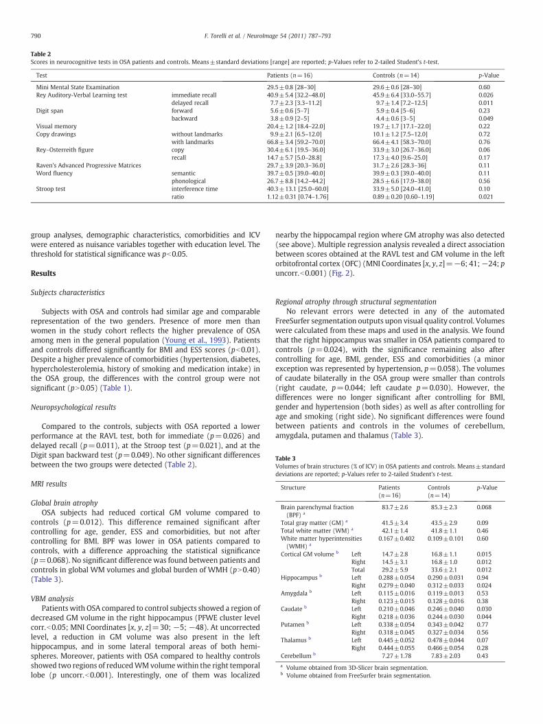
Table 2Scores in neurocognitive tests in OSA patients and controls. Means±standard deviations [range] are reported; p-Values refer to 2-tailed Student's t-test.
Test Patients (n=16) Controls (n=14) p-Value
Mini Mental State Examination 29.5±0.8 [28–30] 29.6±0.6 [28–30] 0.60Rey Auditory-Verbal Learning test immediate recall 40.9±5.4 [32.2–48.0] 45.9±6.4 [33.0–55.7] 0.026
delayed recall 7.7±2.3 [3.3–11.2] 9.7±1.4 [7.2–12.5] 0.011Digit span forward 5.6±0.6 [5–7] 5.9±0.4 [5–6] 0.23
backward 3.8±0.9 [2–5] 4.4±0.6 [3–5] 0.049Visual memory 20.4±1.2 [18.4–22.0] 19.7±1.7 [17.1–22.0] 0.22Copy drawings without landmarks 9.9±2.1 [6.5–12.0] 10.1±1.2 [7.5–12.0] 0.72
with landmarks 66.8±3.4 [59.2–70.0] 66.4±4.1 [58.3–70.0] 0.76Rey–Osterreith !gure copy 30.4±6.1 [19.5–36.0] 33.9±3.0 [26.7–36.0] 0.06
recall 14.7±5.7 [5.0–28.8] 17.3±4.0 [9.6–25.0] 0.17Raven's Advanced Progressive Matrices 29.7±3.9 [20.3–36.0] 31.7±2.6 [28.3–36] 0.11Word "uency semantic 39.7±0.5 [39.0–40.0] 39.9±0.3 [39.0–40.0] 0.11
phonological 26.7±8.8 [14.2–44.2] 28.5±6.6 [17.9–38.0] 0.56Stroop test interference time 40.3±13.1 [25.0–60.0] 33.9±5.0 [24.0–41.0] 0.10
ratio 1.12±0.31 [0.74–1.76] 0.89±0.20 [0.60–1.19] 0.021
Table 3Volumes of brain structures (% of ICV) in OSA patients and controls. Means±standarddeviations are reported; p-Values refer to 2-tailed Student's t-test.
Structure Patients(n=16)
Controls(n=14)
p-Value
Brain parenchymal fraction(BPF) a
83.7±2.6 85.3±2.3 0.068
Total gray matter (GM) a 41.5±3.4 43.5±2.9 0.09Total white matter (WM) a 42.1±1.4 41.8±1.1 0.46White matter hyperintensities(WMH) a
0.167±0.402 0.109±0.101 0.60
Cortical GM volume b Left 14.7±2.8 16.8±1.1 0.015Right 14.5±3.1 16.8±1.0 0.012Total 29.2±5.9 33.6±2.1 0.012
Hippocampus b Left 0.288±0.054 0.290±0.031 0.94Right 0.279±0.040 0.312±0.033 0.024
Amygdala b Left 0.115±0.016 0.119±0.013 0.53Right 0.123±0.015 0.128±0.016 0.38
Caudate b Left 0.210±0.046 0.246±0.040 0.030Right 0.218±0.036 0.244±0.030 0.044
Putamen b Left 0.338±0.054 0.343±0.042 0.77Right 0.318±0.045 0.327±0.034 0.56
Thalamus b Left 0.445±0.052 0.478±0.044 0.07Right 0.444±0.055 0.466±0.054 0.28
Cerebellum b 7.27±1.78 7.83±2.03 0.43a Volume obtained from 3D-Slicer brain segmentation.b Volume obtained from FreeSurfer brain segmentation.
790 F. Torelli et al. / NeuroImage 54 (2011) 787–793

Correlations between neuropsychological, clinical and MRI variables
For thewhole cohort, total hippocampal volumewas correlatedwiththe score in RAVL test (delayed recall) (r=0.388, p=0.034) andinversely correlated with the interference time recorded in the StroopColor/Word test (r="0.372, p=0.043). BPF was associated withscores in RAVL test (delayed recall) (r=0.387, p=0.034), in Word"uency semantic (r=0.465, p=0.010), in Rey–Osterreith ComplexFigure copy (r=0.423, p=0.020) and delayed recall (r=0.592,p=0.001). ESS was inversely related to scores in RAVL test (immediaterecall) (r="0.41, p=0.024) and in Digit span forward (r="0.37,p=0.046). In patients group, but not in controls, we found markednegative correlations between age and MRI !ndings, in particular totalhippocampal volume (r="0.589, p=0.016), left hippocampus vol-ume (r="0.620, p=0.010), total amygdala volume (r="0.511,p=0.043) and BPF (r="0.539, p=0.031). No signi!cant correlationsbetween nocturnal respiratory data, neuropsychological and MRI!ndings were found.
Discussion
In the present study, we foundmultiple structural changes in OSApatients using different quantitative approaches of MRI analysis.VBM showed a signi!cant GM reduction in right and left hippocam-pal volumes in OSA patients compared to controls. This !nding wascon!rmed for the right hippocampus also by the volumetric analysis.It can be argued that VBM is a more sensitive technique to showmildparenchymal damage not yet resulting in a loss of volume detectableby automated parcellation and volumetry of subcortical structures.The signi!cant decrease in cortical gray matter volume, affectingequally the right and the left hemispheres, re"ects on the BPF, anormalized measure of cerebral atrophy; no difference was found inwhite matter volume. Finally, a signi!cantly smaller caudate volumewas found bilaterally in OSA patients. Our neurocognitive resultsreveal a signi!cant impairment in OSA subjects, which is indepen-dent from cardiovascular comorbidities. In particular the mostimpaired cognitive domains were executive functions and verbalmemory.
A strength of the present study is represented by the comparisonof neuropsychological, neuroimaging and polygraphic !ndings. Inour sample, a verbal memory test score (RAVL test delayed recall)was signi!cantly correlated with total hippocampal volume. Hippo-campal volume was also inversely associated to the interferencetime recorded in the Stroop Color/Word test. On the contrary, we didnot !nd signi!cant relationships between neuropsychologicalde!cits and polygraphic variables. Also no correlation betweenseverity of OSA and reduction in cortical volume and BPF wasobserved. Finally, we found a signi!cant negative correlationbetween EDS (evaluated by means of the ESS score) and scoresobtained at verbal memory tests.
The presence and the extent of neurobehavioural changes inindividuals affected by sleep apnea is still a matter of debate. Someresearchers argue that EDS is the main cause of the neuropsycho-logical de!cits in patients with OSA and that the comorbiditiesusually observed in these patients (cardiovascular diseases, obesity,physical inactivity) are more important than sleep apnea per se inaffecting neurocognitive functions (Lim and Veasey, 2010). More-over there is a large heterogeneity in neuropsychological tests acrossdifferent studies in OSA, making dif!cult a direct comparison ofresults. To address this latter issue Décary et al. (2000) proposed astandardized neuropsychological test battery for the evaluation ofOSA patients. Our cohort showed more extensive neurocognitiveimpairment compared to previous studies (Redline et al., 1997;Salorio et al., 2002; Yaouhi et al., 2009). Yaouhi et al. (2009), whodemonstrated only a minor cognitive impairment in their cohort,studied less severe OSA patients than ours (mean AHI [SD]: 38.3[14.3] vs. 52.5 [26.0]/h). Other researchers who did not reportappreciable cognitive de!cits included only patients with mild tomoderate OSA (Redline et al., 1997). Similarly, Salorio et al. (2002),who identi!ed cognitive de!cits only on tasks requiring greaterintegration of executive control and long-term memory abilities,included a signi!cant number of patients with mild OSA in theirsample. Other neuropsychological studies on severe OSA subjectswith respiratory index similar to those of our patients reported morediffuse de!cits, particularly in terms of executive functioning,attention, learning and memory (Ferini-Strambi et al., 2003; Limet al., 2007). The !nding of impairment in memory and executivefunctions is essentially in agreement with the conclusions ofprevious reviews (Beebe and Gozal, 2002; Aloia et al., 2004;Saunamaki and Jehkonen, 2007). It is also in agreement withevidence from animal studies suggesting that both intermittenthypoxia (Kalaria et al., 2004) and sleep fragmentation (Tung et al.,2005) (the two essential features of OSA syndrome) can indepen-dently lead to neuronal loss in the hippocampus and pre-frontalcortex, areas that are closely associated with memory processes andexecutive functions.
The severity of OSA syndrome in our sample could also explain the!nding of global brain tissue damage, which has been rarely reportedin previous cross-sectional studies in the form of reduction of the ratioof total gray-to-white matter volumes (Macey et al., 2002). Thisoutcome is likely a consequence of apnea events and of the subsequentchronic intermittent hypoxemia. Cortical atrophymay also be inducedby factors other than hypoxic events, for example cardiovascularcomorbidities (mainly arterial hypertension), which are known toaffect brain tissue both globally (Enzinger et al., 2005; Ropele et al.,2010) and focally (den Heijer et al., 2005). This explanation howeverappears less probable in our cohort because patients and controlswerematched for cardiovascular disease and there was no signi!cantdifference in white matter lesion burden between the two groups. Alikely role of apnea events in affecting cortical volume seems to besupported also by the observation that the signi!cant difference ingray matter volume between OSA and controls is independent ofcardiovascular comorbidities. It is important to note that white matteris also susceptible to hypoxia and while we have not detected
Fig. 2. VBM analysis showing regions of GM and WM reduction in patients with OSAcompared to controls. Figure shows signi!cant changes of GM volume in righthippocampus (MNI: 30; "5; "48) and changes of WM volume in an anatomicallycontiguous region, across groups (p Family-wise Error corrected b0.05).
791F. Torelli et al. / NeuroImage 54 (2011) 787–793

signi!cant change we cannot rule out the presence of damage in thiscompartment. The presence of a degree of hippocampal atrophy in ourpatients appears to be consistent with the neuropsychological resultsand also with previous reports showing a decreased hippocampalvolume as the most consistent !nding provided by structuralneuroimaging studies in OSA (Zimmerman and Aloia, 2006). Thesigni!cantly smaller caudate volume that we found bilaterally in OSApatients may partially contribute to explain the impairment inexecutive functions, since this structure (especially the head of thecaudate) is thought to be involved in the so-called pre-frontal circuit(Saint-Cyr et al., 1988; Eslinger and Grattan, 1993). Furthermore, thisMRI !nding seems to con!rm a recent report of VBM analysis inpatients with severe OSA (Joo et al., 2010), showing several corticaland subcortical regions of reduced GM concentration (includingbilateral caudate nuclei) in severe OSA patients compared to healthycontrols. Functional neuroimaging studies in OSA have supported thehypothesis of an involvement of the pre-frontal circuit in theimpairment in executive functions. Absence of dorsolateral prefrontalactivation (Thomas et al., 2005) or, alternatively, increased bilateralactivation of prefrontal regions (Archbold et al., 2009) have beenreported during working memory tasks in patients with untreatedOSA.
Two previous studies (Gale and Hopkins, 2004; Yaouhi et al., 2009)correlated results in different neurobehavioural tests and MRI!ndings in OSA patients. Yaouhi and colleagues did not revealsigni!cant correlations in their sample (however they found correla-tions between neuropsychological/clinical data and brain metabolismevaluated by resting-state positron emission tomography). Gale andcolleagues, who detected hippocampal atrophy in 36% of their OSApatients, reported positive correlations between non-verbal memoryand information processing tests and both right and left hippocampus.Interestingly, in our sample, hippocampal volume was also associatedwith the score in an executive functions test. This outcome providesan additional evidence for an association between alterations in theorbitofrontal cortex (that we could detect by VBM) and in limbic areasand executive functioning impairment (Wagner et al., 2008; Keller etal., 2009). In the present study we could not !nd signi!cantcorrelations between neuropsychological and respiratory data. Thisresult is consistent with those from previous studies (Sauter et al.,2000; Adams et al., 2001) showing that the link between neuropsy-chological and sleep data (including nocturnal respiratory indexes) israrely strong and consistent. Moreover, it is well known that thecharacterization of disease severity should not be based only on theAHI, but also on clinical features (mainly the disease duration) whichare often not easily measurable. Previous studies showed thatimpairment in attention, vigilance and memory function is mostlyrelated to EDS, while hypoxemia correlates more with de!cits inexecutive functions (Bédard et al., 1991; Décary et al., 2000; Englemanet al., 2000; Brown, 2005). In our sample we found a negativecorrelation between EDS and verbal memory tests, while nosigni!cant correlation between oximetric data and executive func-tions tests was reported. This lack of correlation could be due to thefact that our sample was moderately hypoxemic (the meanpercentage of time spent under 90% and under 80% of desaturationwas 21.9% and 3.7%, respectively). However, this degree of desatura-tion could be suf!cient to cause the reduction in hippocampal volumeand in cortical gray matter that we observed, since these areas areknown to be particularly vulnerable to hypoxia (Gale and Hopkins,2004; Konaka et al., 2007).
The chronic intermittent hypoxemia observed in OSA patientsmight be seen as a factor which expedites the effects of otherconditions, particularly aging, that are known to cause brain atrophyor damage. Consistent with that, our patients showed negativecorrelations between age and several brain structure volumes (totaland left hippocampus, amygdala and BPF) while none of them weresigni!cant in the control group.
Conclusions
Our !ndings con!rm that moderate-severe OSA can be accompa-nied by signi!cant cognitive impairment. The brain image analysisperformed using different techniques highlighted the presence oftissue damage in regions involved in several cognitive domains. Suchdamage appeared to be independent of differences in age, gender, useof tobacco and cardiovascular comorbidities. Therefore the presenceof OSA could be seen as a factor which expedites the process of brainaging by increasing the susceptibility of speci!c cerebral structures toclinical and pathological occurrences.
Con!ict of interest statement
Dr. Torelli has no con"ict of interest.Dr. Moscufo has no con"ict of interest.Dr. Garreffa has no con"ict of interest.Dr. Placidi has no con"ict of interest.Dr. Romigi has no con"ict of interest.Dr. Zannino has no con"ict of interest.Dr. Bozzali has no con"ict of interest.Dr. Fasano has no con"ict of interest.Dr. Giulietti has no con"ict of interest.Dr. Djonlagic has no con"ict of interest.Dr. Malhotra has received consulting and/or research income from
Philips, P!zer, Merck, Cephalon, Itamar, Sleep Group Solutions, SleepHealthCenters, Apnex, Sepracor, Ethicon, Medtronic.
Prof. Marciani has no con"ict of interest.Dr. Guttmann receives research support from Teva Neuroscience,
the Maurice M. Pechet Foundation, the National Multiple SclerosisSociety (NMSS) (RG3574A1 and Pediatric MS Center of ExcellenceAward), the National Institutes of Health (NIH) (R01 NS036524-05,R01 NS055083-01A1, 1R01NS05270,* *UL1 RR025758-01,2P41RR013218 Supplement, 2U01AI063623-06), and has receivedsupport from the NMSS (RG 3798-A-2) and NIH (P41 RR013218). Dr.Guttmann also has received a honorarium for participation in anadvisory board meeting from Johnson & Johnson PharmaceuticalServices, LLC and is an inventor on U.S. patents number 6,080,164;6,684,098 B2; and 10/966,588.
Acknowledgments
This study was supported by NIH grants R01 HL 090897, R01HL085188, K24 HL 093218, 1 P01 HL 095491, R01 AG022092-01, PO1AG004390, and AHA 0840159N and the American Sleep MedicineFoundation.
References
Adams, N., Strauss, M., Schluchter, M., Redline, S., 2001. Relation of measures of sleep-disordered breathing to neuropsychological functioning. Am. J. Respir. Crit. CareMed.163 (7), 1626–1631.
Aloia, M.S., Arnedt, J.T., Davis, J.D., Riggs, R.L., Byrd, D., 2004. Neuropsychologicalsequelae of obstructive sleep apnea–hypopnea syndrome: a critical review. J. Int.Neuropsychol. Soc. 10 (5), 772–785.
American Academy of Sleep Medicine Task Force, 1999. Sleep-related breathingdisorders in adults: recommendations for syndrome de!nition and measurementtechniques in clinical research. Sleep 22 (5), 667–689.
Archbold, K.H., Borghesani, P.R., Mahurin, R.K., Kapur, V.K., Landis, C.A., 2009. Neuralactivation patterns working memory tasks and OSA disease severity: preliminary!ndings. J. Clin. Sleep Med. 5 (1), 21–27.
Ashburner, J., Friston, K.J., 2000. Voxel-based morphometry—the methods. Neuroimage11 (6 Pt 1), 805–821.
Bédard, M.A., Montplaisir, J., Richer, F., Rouleau, I., Malo, J., 1991. Obstructive sleep apneasyndrome: pathogenesis of neuropsychological de!cits. J. Clin. Exp. Neuropsychol. 13(6), 950–964.
Beebe, D.W., Gozal, D., 2002. Obstructive sleep apnea and the prefrontal cortex:towards a comprehensive model linking nocturnal upper airway obstruction todaytime cognitive and behavioral de!cits. J. Sleep Res. 11 (1), 1–16.
Brown, W.D., 2005. The psychosocial aspects of obstructive sleep apnea. Semin. Respir.Crit. Care Med. 26 (1), 33–43.
792 F. Torelli et al. / NeuroImage 54 (2011) 787–793

Caltagirone, C., Gainotti, G., Masullo, C., Miceli, G., 1979. Validity of some neuropsy-chological tests in the assessment of mental deterioration. Acta Psychiatr. Scand.60, 50–56.
Carlesimo, G.A., Caltagirone, C., Gainotti, C., 1996. The mental deterioration battery:normative data, diagnostic reliability and qualitative analysis of cognitiveimpairment. The Group of the standardization of the Mental Deterioration Battery.Eur. Neurol. 36 (6), 378–384.
Carter, C.S., Mintun, M., Cohen, J.D., 1995. Interference and facilitation effects duringselective attention: an H2
15O PET study of Stroop task performance. Neuroimage 2,264–272.
Davies, C.W., Crosby, J.H., Mullins, R.L., Traill, Z.C., Anslow, P., Davies, R.J., et al., 2001.Case control study of cerebrovascular damage de!ned by magnetic resonanceimaging in patients with OSA and normal matched control subjects. Sleep 24 (6),715–720.
Décary, A., Rouleau, I., Montplaisir, J., 2000. Cognitive de!cits associated with sleep apneasyndrome: a proposed neuropsychological test battery. Sleep 23 (3), 369–381.
den Heijer, T., Launer, L.J., Prins, N.D., van Dijk, E.J., Vermeer, S.E., Hofman, A., et al., 2005.Association between blood pressure, white matter lesions, and atrophy of themedial temporal lobe. Neurology 64 (2), 263–267.
Engleman, H.M., Kingshott, R.N., Martin, S.E., Douglas, N.J., 2000. Cognitive function inthe sleep apnea/hypopnea syndrome (SAHS). Sleep 23 (Suppl 4), S102–S108.
Enzinger, C., Fazekas, F., Matthews, P.M., Ropele, S., Schmidt, H., Smith, S., et al., 2005.Risk factors for progression of brain atrophy in aging: six-year follow-up of normalsubjects. Neurology 64 (10), 1704–1711.
Eslinger, P.J., Grattan, L.M., 1993. Frontal lobe and frontal–striatal substrates fordifferent forms of human cognitive "exibility. Neuropsychologia 31 (1), 17–28.
Ferini-Strambi, L., Baietto, C., Di Gioia, M.R., Castaldi, P., Castronovo, C., Zucconi, M., etal., 2003. Cognitive dysfunction in patients with obstructive sleep apnea (OSA):partial reversibility after continuous positive airway pressure (CPAP). Brain Res.Bull. 61 (1), 87–92.
Fischl, B., Salat, D.H., Busa, E., Albert, M., Dieterich, M., Haselgrove, C., et al., 2002. Wholebrain segmentation: automated labeling of neuroanatomical structures in thehuman brain. Neuron 33 (3), 341–355.
Fischl, B., Salat, D.H., van der Kouwe, A.J., Makris, N., Ségonne, F., Quinn, B.T., et al., 2004.Sequence-independent segmentation of magnetic resonance images. Neuroimage23 (Suppl 1), S69–S84.
Folstein, M.F., Folstein, S.E., McHugh, P.R., 1975. “Mini-mental state.” A practical methodfor grading the cognitive state of patients for the clinician. J. Psychiatr. Res. 12 (3),189–198.
Gale, S.D., Hopkins, R.O., 2004. Effects of hypoxia on the brain: neuroimaging andneuropsychological !ndings following carbon monoxide poisoning and obstructivesleep apnea. J. Int. Neuropsychol. Soc. 10 (1), 60–71.
Good, C.D., Johnsrude, I.S., Ashburner, J., Henson, R.N., Friston, K.J., Frackowiak, R.S.,2001. A voxel-based morphometric study of ageing in 465 normal adult humanbrains. Neuroimage 14 (1 Pt 1), 21–36.
Han, X., Jovicich, J., Salat, D., van der Kouwe, A., Quinn, B., Czanner, S., et al., 2006.Reliability of MRI-derived measurements of human cerebral cortical thickness: theeffects of !eld strength, scanner upgrade and manufacturer. Neuroimage 32 (1),180–194.
Jenkinson, M., Smith, S., 2001. A global optimisation method for robust af!neregistration of brain images. Med. Image Anal. 5, 143–156.
Johns, M.W., 1991. A new method for measuring daytime sleepiness: the EpworthSleepiness Scale. Sleep 14, 540–545.
Joo, E.Y., Tae, W.S., Lee, M.J., Kang, J.W., Park, H.S., Lee, J.Y., et al., 2010. Reduced braingray matter concentration in patients with obstructive sleep apnea syndrome.Sleep 33 (2), 235–241.
Kalaria, R.N., Spoors, L., Laude, E.A., Emery, C.J., Thwaites-Bee, D., Fairlie, J., et al., 2004.Hypoxia of sleep apnoea: cardiopulmonary and cerebral changes after intermittenthypoxia in rats. Respir. Physiol. Neurobiol. 140 (1), 53–62.
Keller, S.S., Baker, G., Downes, J.J., Roberts, N., 2009. Quantitative MRI of the prefrontalcortex and executive function in patients with temporal lobe epilepsy. EpilepsyBehav. 15 (2), 186–195.
Kim, H.C., Young, T., Matthews, C.G., Weber, S.M., Woodward, A.R., Palta, M., 1997.Sleep-disordered breathing and neuropsychological de!cits. A population-basedstudy. Am. J. Respir. Crit. Care Med. 156 (6), 1813–1819.
Konaka, K., Miyashita, K., Naritomi, H., 2007. Changes in diffusion-weighted magneticresonance imaging!ndings in the acute and subacute phases of anoxic encephalopathy.J. Stroke Cerebrovasc. Dis. 16 (2), 82–83.
Lim, D.C., Veasey, S.C., 2010. Neural injury in sleep apnea. Curr. Neurol. Neurosci. Rep.10 (1), 47–52.
Lim, W., Bardwell, W.A., Loredo, J.S., Kim, E.J., Ancoli-Israel, S., Morgan, E.E., et al., 2007.Neuropsychological effects of 2-week continuous positive airway pressure
treatment and supplemental oxygen in patients with obstructive sleep apnea: arandomized placebo-controlled study. J. Clin. Sleep Med. 3 (4), 380–386.
Macey, P.M., Henderson, L.A., Macey, K.E., Alger, J.R., Frysinger, R.C., Woo, M.A., et al.,2002. Brain morphology associated with obstructive sleep apnea. Am. J. Respir. Crit.Care Med. 166 (10), 1382–1387.
Macey, P.M., Kumar, R., Woo, M.A., Valladares, E.M., Yan-Go, F.L., Harper, R.M., 2008.Brain structural changes in obstructive sleep apnea. Sleep 31 (7), 967–977.
Morrell, M.J., McRobbie, D.W., Quest, R.A., Cummin, A.R., Ghiassi, R., Cor!eld, D.R., 2003.Changes in brain morphology associated with obstructive sleep apnea. SleepMed. 4(5), 451–454.
Moscufo, N., Guttmann, C.R., Meier, D., Csapo, I., Hildenbrand, P.G., Healy, B.C., et al.,2009. Brain regional lesion burden and impaired mobility in the elderly. Neurobiol.Aging. doi:10.1016/j.neurobiolaging.2009.04.010 [Epub ahead of print].
Nishibayashi, M., Miyamoto, M., Miyamoto, T., Suzuki, K., Hirata, K., 2008. Correlationbetween severity of obstructive sleep apnea and prevalence of silent cerebrovascularlesions. J. Clin. Sleep Med. 4 (3), 242–247.
O'Donoghue, F.J., Briellmann, R.S., Rochford, P.D., Abbott, D.F., Pell, G.S., Chan, C.H., et al.,2005. Cerebral structural changes in severe obstructive sleep apnea. Am. J. Respir.Crit. Care Med. 171 (10), 1185–1190.
Orsini, A., Grossi, D., Capitani, E., Laiacona, M., Pagagno, C., Vallar, G., 1987. Verbal andspatial immediate memory span: normative data from 1355 adults and 1112children. Ital. J. Neurol. Sci. 8, 539–548.
Osterrieth, P.A., 1944. Le test de copie d'une !gure complexe. Arch. Psychol. 30,206–356.
Pohl, K.M., Bouix, S., Kikinis, R., Grimson, W.E.L., 2004. Anatomical guided segmentationwith non-stationary tissue class distributions in an expectation-maximizationframework. IEEE International Symposium on Biomedical Imaging: From Nano toMacro, Arlington, pp. 81–84.
Raven, J.C., 1947. Guide to using coloured progressive matrices: Set A, Ab, B. Board andBook Forms, London.
Redline, S., Strauss, M.E., Adams, N., Winters, M., Roebuck, T., Spry, K., et al., 1997.Neuropsychological function in mild sleep-disordered breathing. Sleep 20 (2),160–167.
Ropele, S., Enzinger, C., Söllinger, M., Langkammer, C., Wallner-Blazek, M., Schmidt, R.,et al., 2010. The impact of sex and vascular risk factors on brain tissue changes withaging: magnetization transfer imaging results of the Austrian Stroke PreventionStudy. Am. J. Neuroradiol. 31 (7), 1297–1301.
Saint-Cyr, J.A., Taylor, A.E., Lang, A.E., 1988. Procedural learning and neostriataldysfunction in man. Brain 111 (Pt 4), 941–959.
Salorio, C.F., White, D.A., Piccirillo, J., Duntley, S.P., Uhles, M.L., 2002. Learning, memory,and executive control in individuals with obstructive sleep apnea syndrome. J. Clin.Exp. Neuropsychol. 24 (1), 93–100.
Saunamaki, T., Jehkonen, M., 2007. A review of executive functions in obstructive sleepapnea syndrome. Acta Neurol. Scand. 115, 1–11.
Sauter, C., Asenbaum, S., Popovic, R., Bauer, H., Lamm, C., Klösch, G., et al., 2000.Excessive daytime sleepiness in patients suffering from different levels ofobstructive sleep apnoea syndrome. J. Sleep Res. 9 (3), 293–301.
Segonne, F., Dale, A.M., Busa, E., Glessner, M., Salat, D., Hahn, H.K., et al., 2004. A hybridapproach to the skull stripping problem in MRI. Neuroimage 22 (3), 1060–1075.
Sled, J.G., Zijdenbos, A.P., Evans, A.C., 1998. A nonparametric method for automaticcorrection of intensity nonuniformity in MRI data. IEEE Trans. Med. Imaging 17,87–97.
Stroop, J.R., 1935. Studies of interference in serial verbal reactions. J. Exp. Psychol. 18,643–662.
Thomas, R.J., Rosen, B.R., Stern, C.E., Weiss, J.W., Kwong, K.K., 2005. Functional imagingof working memory in obstructive sleep-disordered breathing. J. Appl. Physiol. 98(6), 2226–2234.
Tung, A., Takase, L., Fornal, C., Jacobs, B., 2005. Effects of sleep deprivation and recoverysleep upon cell proliferation in adult rat dentate gyrus. Neuroscience 134 (3),721–723.
Wagner, G., Koch, K., Schachtzabel, C., Reichenbach, J.R., Sauer, H., Schlösser Md, R.G.,2008. Enhanced rostral anterior cingulate cortex activation during cognitivecontrol is related to orbitofrontal volume reduction in unipolar depression.J. Psychiatry Neurosci. 33 (3), 199–208.
Yaouhi, K., Bertran, F., Clochon, P., Mézenge, F., Denise, P., Foret, J., et al., 2009. Acombined neuropsychological and brain imaging study of obstructive sleep apnea.J. Sleep Res. 18 (1), 36–48.
Young, T., Palta, M., Dempsey, J., Skatrud, J., Weber, S., Badr, S., 1993. The occurrence ofsleep-disordered breathing among middle-aged adults. N Engl J. Med. 328 (17),1230–1235.
Zimmerman, M.E., Aloia, M.S., 2006. A review of neuroimaging in obstructive sleepapnea. J. Clin. Sleep Med. 2 (4), 461–471.
793F. Torelli et al. / NeuroImage 54 (2011) 787–793