Drug screen in iPSC-Neurons identifies nucleoside analogs ...
Bioorganic & Medicinal Chemistry 19 (2011) 2359–2367
Contents lists available at ScienceDirect
Bioorganic & Medicinal Chemistry
journal homepage: www.elsevier .com/locate /bmc
Acetyl analogs of combretastatin A-4: Synthesis and biological studies
Balaji Babu a, Megan Lee a, Lauren Lee a, Raymond Strobel a, Olivia Brockway a, Alexis Nickols a,Robert Sjoholm a, Samuel Tzou a, Sameer Chavda a, Dereje Desta b, Gregory Fraley b, Adam Siegfried c,William Pennington c, Rachel M. Hartley d, Cara Westbrook d, Susan L. Mooberry d,e, Konstantinos Kiakos f,John A. Hartley f, Moses Lee a,⇑a Department of Chemistry and the Division of Natural and Applied Sciences, Hope College 49423, USAb Department of Biology, Hope College 49423, USAc Department of Chemistry, Clemson University, SC 29634, USAd Department of Pharmacology, University of Texas Health Science Center at San Antonio, San Antonio, TX 78229, USAe Department of Medicine, University of Texas Health Science Center at San Antonio, San Antonio, TX 78229, USAf UCL Cancer Research Institute, University College London, Paul O’ Gorman Building, 72 Huntley Street, London WC1F 6BT, UK
a r t i c l e i n f o a b s t r a c t
Article history:Received 21 December 2010Revised 9 February 2011Accepted 11 February 2011Available online 17 February 2011
Keywords:Combretastatin A-4TrimethoxyphenylacetoneTubulinMicrotubuleCytotoxicityCancer
0968-0896/$ - see front matter � 2011 Elsevier Ltd. Adoi:10.1016/j.bmc.2011.02.018
⇑ Corresponding author. Tel.: +1 616 395 7190.E-mail address: [email protected] (M. Lee).
The combretastatins have received significant attention because of their simple chemical structures,excellent antitumor efficacy and novel antivascular mechanisms of action. Herein, we report the synthe-sis of 20 novel acetyl analogs of CA-4 (1), synthesized from 3,4,5-trimethoxyphenylacetone that com-prises the A ring of CA-4 with different aromatic aldehydes as the B ring. Molecular modeling studiesindicate that these new compounds possess a ‘twisted’ conformation similar to CA-4. The new analogseffectively inhibit the growth of human and murine cancer cells. The most potent compounds 6k, 6sand 6t, have IC50 values in the sub-lM range. Analog 6t has an IC50 of 182 nM in MDA-MB-435 cellsand has advantages over earlier analogs due to its enhanced water solubility (456 lM). This compoundinitiates microtubule depolymerization with an EC50 value of 1.8 lM in A-10 cells. In a murine L1210 syn-geneic tumor model 6t had antitumor activity and no apparent toxicity.
� 2011 Elsevier Ltd. All rights reserved.
1. Introduction
The ongoing efforts of US National Cancer Institute to identifyanticancer agents of natural origin led to the identification of com-bretastatins, which were initially isolated from the African willowtree Combretum caffrum (Combretaceae). The most potent of theseinitial combretastatins was combretastatin A-4 (CA-4 or 1, Fig. 1).1
CA-4 is an antimitotic agent that binds to the colchicine site ontubulin causing inhibition of tubulin polymerization and in cellsmicrotubule depolymerization and mitotic accumulation.2 CA-4(1) exhibits potent cytotoxicity against a broad spectrum of humancancer lines, including those that are multidrug resistance (MDR)positive.3 This compound also elicits irreversible vascular shut-down in solid tumors, leaving normal vasculature intact, therebyacting as a vascular disrupting agent.4 Major limitations of 1 areits poor aqueous solubility and bioavailability, which significantlyimpairs optimal in vivo antitumor activity.5 The relative simplicityof the 1,2-diarylethene scaffold of the CA-4 series, along with theirbiological properties, resulted in extensive structure–activity
ll rights reserved.
relationship (SAR) studies of CA-4 analogs. Consequently, it wasfound that a 3,4,5-trimethoxyphenyl as the A ring substituentsand a cis disposition of the double bond between two aryl groups(A and B) is optimal for biological activities. Many analogs of 1 havebeen developed over time that can retain the biological actions ofthe parent molecule but that have improved water solubility andbetter pharmacokinetic properties. These analogs include awater-soluble phosphate prodrug derivative of CA-4, CA-4P (2),as well as AVE 8062 (3) and the amino derivative AC-7739 (4)(Fig. 1). CA-4P (Zybrestat), CA-1 diphosphate (Oxi4503), AVE8062 and NPI-2358 are currently being evaluated in clinical trials.Another class of compound where the two aryl rings are bridgedthrough three atoms is represented by A-105972 (5a, Fig. 1), anoxadiazoline analog, which exhibits potency towards multidrugresistant cancer cells.6 Another cytotoxic oxadiazoline compound5b (Fig. 1) was developed in the authors’ laboratory, and it wasfound to cause microtubule depolymerization.7e
Our work has recently focused on the synthesis of novel CA-4analogs that retain the cytotoxic and microtubule disrupting activ-ities of the parent molecule (1a). However, we have not yet identi-fied an analog that preserves the cytotoxic potency of CA-4.8 Werecently reported the synthesis and cytotoxicity of a,b-unsaturated
H3COOCH3
H3CO
OCH3
R
1, R = OH, combretastatin A42, R = phosphate3, R = NH2, AC-77394, R = NHCOCHNH2CH2OH, AVE-8062
O
H3CO
H3CO
H3CO
NH2
OCH3
NN
Ac
5a, A-105972
H3COOCH3
H3CO
R3
R2
CH3
O
6a-t
O
H3CO
H3CO
H3CO
OCH3
NN
5b
Ac R1
R4
R5
6k, R1=R2=R4=R5=H, R3=Me6l, R1=R2=R3=R4=R5=H6m, R1=R4=R5=H; R2=R3=Cl6n, R1=R4=R5=H; R2=NO2; R3=OMe6o, R1=R4=OMe; R2=R3=R5=H6p, R1=R4=R5=H; R2=R3=OMe6q, R1=R3=R5=OMe; R2=R4=H6r, ; R1=R5=H;R2=R3=R4=OMe6s, R1=R4=R5=H; R2=OTBS; R3=OMe6t, R1=R4=R5=H; R2=OH; R3=OMe
6a, R1=R2=R4=R5=H; R3=NO26b, R1=R3=R4=R5=H; R2=NO26c, R1=R2=R4=R5=H; R3=Cl6d, R1=R3=R4=R5=H; R2=Br6e, R1=R2=R4=R5=H; R3=Br6f, R1=R2=R4=R5=H; R3=CN6g, R1=R2=R4=R5=H; R3=F6h, R1=R2=R4=R5=H; R3=OMe6i, R1=OMe;R2=R3=R4=R5=H6j, R1=R3=R4=R5=H; R2=OMe
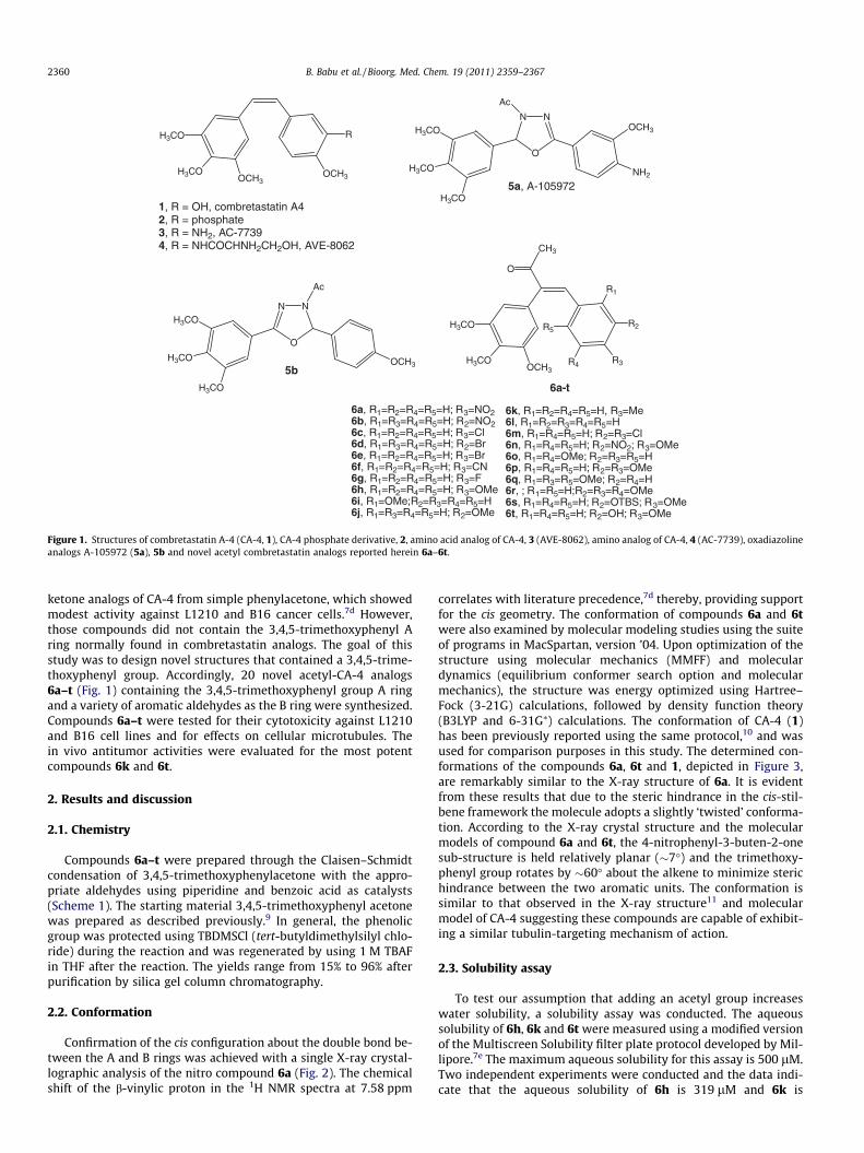
Figure 1. Structures of combretastatin A-4 (CA-4, 1), CA-4 phosphate derivative, 2, amino acid analog of CA-4, 3 (AVE-8062), amino analog of CA-4, 4 (AC-7739), oxadiazolineanalogs A-105972 (5a), 5b and novel acetyl combretastatin analogs reported herein 6a–6t.
2360 B. Babu et al. / Bioorg. Med. Chem. 19 (2011) 2359–2367
ketone analogs of CA-4 from simple phenylacetone, which showedmodest activity against L1210 and B16 cancer cells.7d However,those compounds did not contain the 3,4,5-trimethoxyphenyl Aring normally found in combretastatin analogs. The goal of thisstudy was to design novel structures that contained a 3,4,5-trime-thoxyphenyl group. Accordingly, 20 novel acetyl-CA-4 analogs6a–t (Fig. 1) containing the 3,4,5-trimethoxyphenyl group A ringand a variety of aromatic aldehydes as the B ring were synthesized.Compounds 6a–t were tested for their cytotoxicity against L1210and B16 cell lines and for effects on cellular microtubules. Thein vivo antitumor activities were evaluated for the most potentcompounds 6k and 6t.
2. Results and discussion
2.1. Chemistry
Compounds 6a–t were prepared through the Claisen–Schmidtcondensation of 3,4,5-trimethoxyphenylacetone with the appro-priate aldehydes using piperidine and benzoic acid as catalysts(Scheme 1). The starting material 3,4,5-trimethoxyphenyl acetonewas prepared as described previously.9 In general, the phenolicgroup was protected using TBDMSCl (tert-butyldimethylsilyl chlo-ride) during the reaction and was regenerated by using 1 M TBAFin THF after the reaction. The yields range from 15% to 96% afterpurification by silica gel column chromatography.
2.2. Conformation
Confirmation of the cis configuration about the double bond be-tween the A and B rings was achieved with a single X-ray crystal-lographic analysis of the nitro compound 6a (Fig. 2). The chemicalshift of the b-vinylic proton in the 1H NMR spectra at 7.58 ppm
correlates with literature precedence,7d thereby, providing supportfor the cis geometry. The conformation of compounds 6a and 6twere also examined by molecular modeling studies using the suiteof programs in MacSpartan, version ’04. Upon optimization of thestructure using molecular mechanics (MMFF) and moleculardynamics (equilibrium conformer search option and molecularmechanics), the structure was energy optimized using Hartree–Fock (3-21G) calculations, followed by density function theory(B3LYP and 6-31G⁄) calculations. The conformation of CA-4 (1)has been previously reported using the same protocol,10 and wasused for comparison purposes in this study. The determined con-formations of the compounds 6a, 6t and 1, depicted in Figure 3,are remarkably similar to the X-ray structure of 6a. It is evidentfrom these results that due to the steric hindrance in the cis-stil-bene framework the molecule adopts a slightly ‘twisted’ conforma-tion. According to the X-ray crystal structure and the molecularmodels of compound 6a and 6t, the 4-nitrophenyl-3-buten-2-onesub-structure is held relatively planar (�7�) and the trimethoxy-phenyl group rotates by �60� about the alkene to minimize sterichindrance between the two aromatic units. The conformation issimilar to that observed in the X-ray structure11 and molecularmodel of CA-4 suggesting these compounds are capable of exhibit-ing a similar tubulin-targeting mechanism of action.
2.3. Solubility assay
To test our assumption that adding an acetyl group increaseswater solubility, a solubility assay was conducted. The aqueoussolubility of 6h, 6k and 6t were measured using a modified versionof the Multiscreen Solubility filter plate protocol developed by Mil-lipore.7e The maximum aqueous solubility for this assay is 500 lM.Two independent experiments were conducted and the data indi-cate that the aqueous solubility of 6h is 319 lM and 6k is

OH3C
MeO
MeO
OMe
O
HR3
R4
R1 R2
piperidine, benzoic acidtoluene, reflux, 4h
6k, R1=R2=R4=R5=H, R3=Me6l, R1=R2=R3=R4=R5=H6m, R1=R4=R5=H; R2=R3=Cl6n, R1=R4=R5=H; R2=NO2; R3=OMe6o, R1=R4=OMe; R2=R3=R5=H6p, R1=R4=R5=H; R2=R3=OMe6q, R1=R3=R5=OMe; R2=R4=H6r, ; R1=R5=H;R2=R3=R4=OMe6s, R1=R4=R5=H; R2=OTBS; R3=OMe6t, R1=R4=R5=H; R2=OH; R3=OMe
MeOOMe
MeO
H3C
R1
R3R4
R5 R2
O
R5
6
6a, R1=R2=R4=R5=H; R3=NO26b, R1=R3=R4=R5=H; R2=NO26c, R1=R2=R4=R5=H; R3=Cl6d, R1=R3=R4=R5=H; R2=Br6e, R1=R2=R4=R5=H; R3=Br6f, R1=R2=R4=R5=H; R3=CN6g, R1=R2=R4=R5=H; R3=F6h, R1=R2=R4=R5=H; R3=OMe6i, R1=OMe;R2=R3=R4=R5=H6j, R1=R3=R4=R5=H; R2=OMe
Scheme 1. Synthesis of acetyl-combretastatin analogs 6a–t.
Figure 2. X-ray structure of compound 6a. Complete X-ray structural data for 6awere deposited with the Cambridge Crystallographic Data Centre (CCDC depositionnumber: 811797).
B. Babu et al. / Bioorg. Med. Chem. 19 (2011) 2359–2367 2361
443 lM. The analog 6t has an aqueous solubility of 456 lM, indi-cating that the addition an acetyl group on the ethenyl moiety in-creases the aqueous solubility of the compound when compared toCA-4 itself and acetyl oxadiazoline 5b.7e
2.4. Cytotoxicity
The compounds were evaluated for cytotoxic activity againstmurine B16 melanoma and L1210 leukemia cells. The results,
Figure 3. Molecular models of co
Table 1, show several interesting trends based upon the substitu-tion pattern of the aromatic ring B. The compound 6l (derived frombenzaldehyde), yielded IC50 values of 25 and 5.4 lM against B16and L1210 cells, respectively. For the B ring monosubstituted com-pounds (Table 1), a nitro group in the meta-position (6b) yieldedgood potency against L1210 and B16 cells (IC50 3.8 and 3.3 lM,respectively). Moving this nitro group to the para-position (6a)lowered the potency 4 to 5-fold against both cell lines in compar-ison to 6b, suggesting that the electronic influence (due to induc-tive effects) of the meta-nitro group enhances the interaction of6b with it is cellular targets. In contrast, the compounds with hal-ogens in the meta- and para-position (6c–6e and 6g) showed mod-est to poor activity (IC50 values in the range of 7.4–65 lM), with 6cand 6e (containing para-chloro and para-bromo substituents,respectively) exhibiting the highest potency against one or bothcell lines. Interestingly, incorporation of the slightly less polarand larger bromo substituent at the meta-position in 6d (in com-parison to 6c) resulted in additional loss in cytotoxic potency inboth cell lines (IC50 values of 39 and 35 lM in B16 and L1210 cells,respectively). Similar trends (based upon substitution pattern)seem apparent for compounds 6h–6k (Table 1). Electron-donatingmethoxy and methyl substituents in the para-position (6h and 6k)increased potency against cell lines and gave IC50 values for L1210cells in the sub-lM range; 0.38 and 0.36 lM, respectively. Onlymodest levels of cytotoxicity were observed for 6i and 6j. This sug-gests that substituent size is a factor contributing to the potency ofthese compounds, as could substituent polarizability in the para-position. Based upon the information obtained from the study ofthe monosubstituted compounds, we were curious to ascertainwhether some of the trends observed for monosubstituted com-pounds could be enhanced via incorporation of two substituentsinto the aromatic ring. In comparison to 6b, 6c and 6h, similar
mpounds 6a, 6t, and CA-4, 1.

Table 1Cytotoxicity of the compounds 6a–6t on L1210 and B16 cancer cell lines
Compound IC50 (lM)
ArMeOOMe
MeO
H3C O
B16 L1210 B16 L1210
Ar =
NO2
6a
18 ± 7.8 18 ± 9.2 CH3
6k2.9 0.36
NO2
6b
3.8 ± 0.3 3.3 ± 0.2
6l25 ± 4.2 5.4 ± 0.2
Cl
6c7.4 ± 2.5 5.0 ± 0.1
Cl
Cl 6m
6.4 ± 1.2 4.0
Br
6d
39 ± 1.4 35 ± 6.4
NO2
OMe
6n
3.8 ± 0.2 3.1
Br 6e 19 ± 9.2 4.0 ± 1.5
OMe
OMe
6o
4.0 ± 0.1 3.7 ± 0.01
CN 6f
29 ± 12 8.0 ± 4OMe
OMe
6p
42 ± 4.9 37 ± 11.3
F
6g65 ± 7.1 43 ± 4.2
OMe
OMe
MeO 6q
>100 3.7 ± 0.9
OMe 6h
7.3 ± 1.7 0.38 OMe
OMe
OMe
6r
23 ± 2.8 7.2 ± 2
OMe
6i
41 ± 13.4 40 OMe
OTBS
6s5.1 ± 2.9 0.18
OMe
6j31 ± 0.7 39 ± 0.7 OMe
OH
6t
3.5 ± 1.4 0.45 ± 0.1
IC50 values are expressed in lM.
2362 B. Babu et al. / Bioorg. Med. Chem. 19 (2011) 2359–2367
cytotoxic potency for the disubstituted counterparts 6m (contain-ing chlorine atoms in both the meta- and para-positions) and 6n(meta-nitro and para-methoxy) were observed, confirming thetrends seen with the mono substituted compounds. The molecules6q and 6r which contain trisubstituted methoxy groups in the 2,4,6and 3,4,5 positions (Table 1) showed no significant activity againsteither cell line, suggesting that the steric congestion resulting frommultiple methoxy groups prevents optimal orientation within thebinding site as compared to the analogs 6h and 6k. The synthe-sized exact acetyl analogs of CA-4 1 (6s and 6t) showed significant
activity (IC50 in 0.18–3.5 lM range, Table 1). Finally, the enhancedpotency of some of these molecules towards leukemia cells(L1210) over solid tumor cells (B16) is, in general, consistent withstudies on other combretastatin analogs from our laboratory.7,12
2.5. NCI data
Compound 6t was evaluated at the National Cancer Instituteagainst a panel of 60 human cancer cell lines.13 Inhibition of cellproliferation after 48 h continuous exposure was determined using
B. Babu et al. / Bioorg. Med. Chem. 19 (2011) 2359–2367 2363
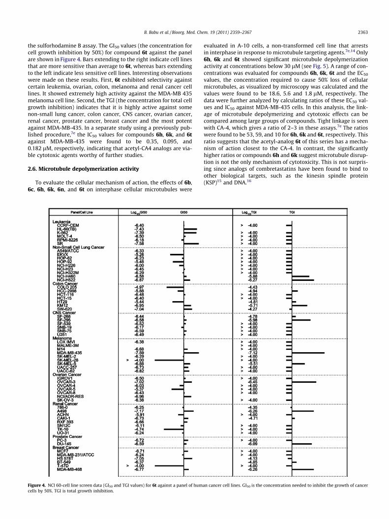
the sulforhodamine B assay. The GI50 values (the concentration forcell growth inhibition by 50%) for compound 6t against the panelare shown in Figure 4. Bars extending to the right indicate cell linesthat are more sensitive than average to 6t, whereas bars extendingto the left indicate less sensitive cell lines. Interesting observationswere made on these results. First, 6t exhibited selectivity againstcertain leukemia, ovarian, colon, melanoma and renal cancer celllines. It showed extremely high activity against the MDA-MB 435melanoma cell line. Second, the TGI (the concentration for total cellgrowth inhibition) indicates that it is highly active against somenon-small lung cancer, colon cancer, CNS cancer, ovarian cancer,renal cancer, prostate cancer, breast cancer and the most potentagainst MDA-MB-435. In a separate study using a previously pub-lished procedure,7e the IC50 values for compounds 6h, 6k, and 6tagainst MDA-MB-435 were found to be 0.35, 0.095, and0.182 lM, respectively, indicating that acetyl-CA4 analogs are via-ble cytotoxic agents worthy of further studies.
2.6. Microtubule depolymerization activity
To evaluate the cellular mechanism of action, the effects of 6b,6c, 6h, 6k, 6n, and 6t on interphase cellular microtubules were
Figure 4. NCI 60-cell line screen data (GI50 and TGI values) for 6t against a panel of humcells by 50%. TGI is total growth inhibition.
evaluated in A-10 cells, a non-transformed cell line that arrestsin interphase in response to microtubule targeting agents.7e,14 Only6h, 6k and 6t showed significant microtubule depolymerizationactivity at concentrations below 30 lM (see Fig. 5). A range of con-centrations was evaluated for compounds 6h, 6k, 6t and the EC50
values, the concentration required to cause 50% loss of cellularmicrotubules, as visualized by microscopy was calculated and thevalues were found to be 18.6, 5.6 and 1.8 lM, respectively. Thedata were further analyzed by calculating ratios of these EC50 val-ues and IC50 against MDA-MB-435 cells. In this analysis, the link-age of microtubule depolymerizing and cytotoxic effects can becompared among large groups of compounds. Tight linkage is seenwith CA-4, which gives a ratio of 2–3 in these assays.7e The ratioswere found to be 53, 59, and 10 for 6h, 6k and 6t, respectively. Thisratio suggests that the acetyl-analog 6t of this series has a mecha-nism of action closest to the CA-4. In contrast, the significantlyhigher ratios or compounds 6h and 6k suggest microtubule disrup-tion is not the only mechanism of cytotoxicity. This is not surpris-ing since analogs of combretastatins have been found to bind toother biological targets, such as the kinesin spindle protein(KSP)15 and DNA.16
an cancer cell lines. GI50 is the concentration needed to inhibit the growth of cancer
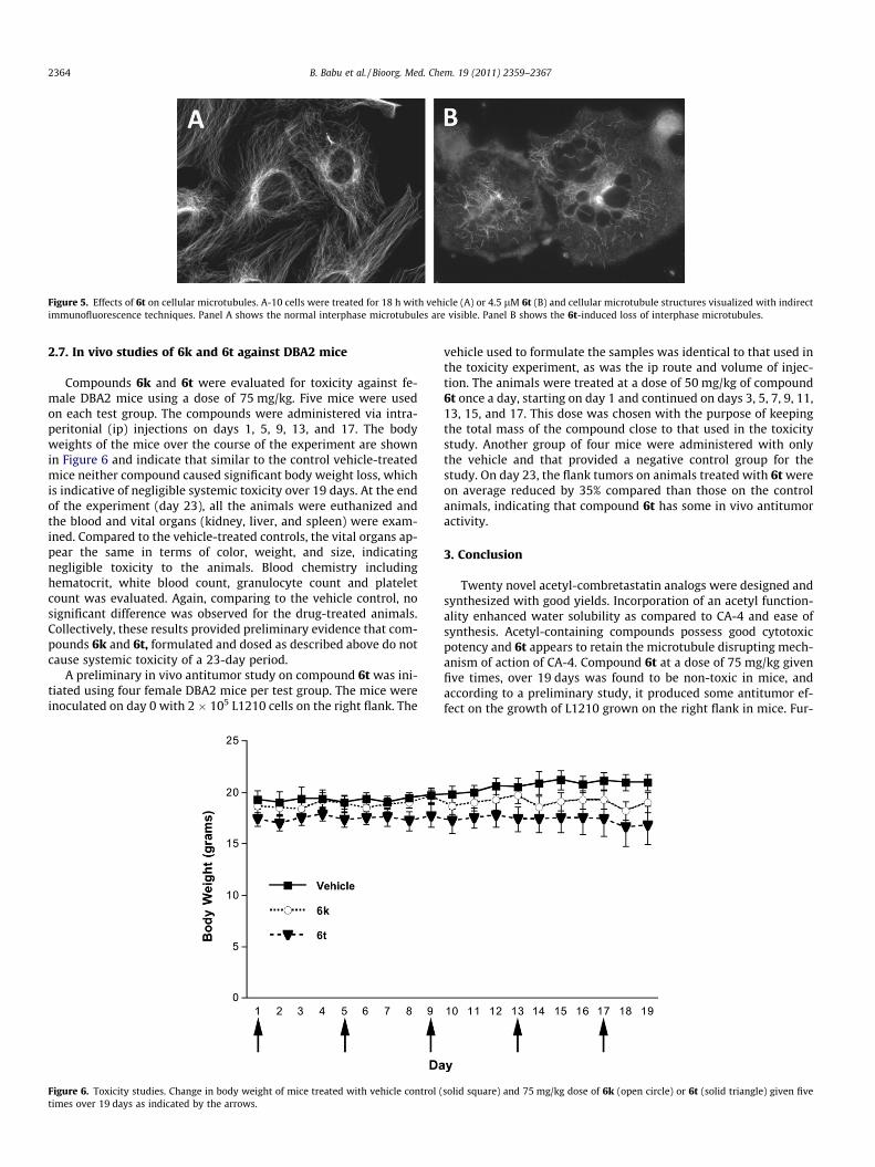
Figure 5. Effects of 6t on cellular microtubules. A-10 cells were treated for 18 h with vehicle (A) or 4.5 lM 6t (B) and cellular microtubule structures visualized with indirectimmunofluorescence techniques. Panel A shows the normal interphase microtubules are visible. Panel B shows the 6t-induced loss of interphase microtubules.
2364 B. Babu et al. / Bioorg. Med. Chem. 19 (2011) 2359–2367
2.7. In vivo studies of 6k and 6t against DBA2 mice
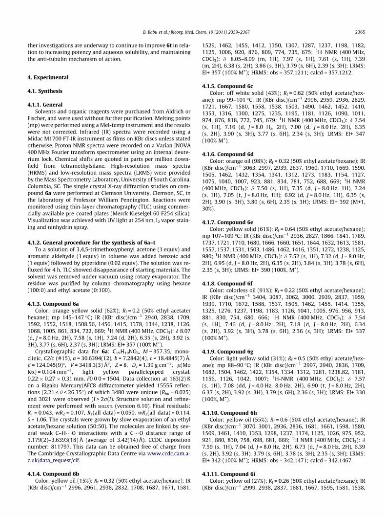
Compounds 6k and 6t were evaluated for toxicity against fe-male DBA2 mice using a dose of 75 mg/kg. Five mice were usedon each test group. The compounds were administered via intra-peritonial (ip) injections on days 1, 5, 9, 13, and 17. The bodyweights of the mice over the course of the experiment are shownin Figure 6 and indicate that similar to the control vehicle-treatedmice neither compound caused significant body weight loss, whichis indicative of negligible systemic toxicity over 19 days. At the endof the experiment (day 23), all the animals were euthanized andthe blood and vital organs (kidney, liver, and spleen) were exam-ined. Compared to the vehicle-treated controls, the vital organs ap-pear the same in terms of color, weight, and size, indicatingnegligible toxicity to the animals. Blood chemistry includinghematocrit, white blood count, granulocyte count and plateletcount was evaluated. Again, comparing to the vehicle control, nosignificant difference was observed for the drug-treated animals.Collectively, these results provided preliminary evidence that com-pounds 6k and 6t, formulated and dosed as described above do notcause systemic toxicity of a 23-day period.
A preliminary in vivo antitumor study on compound 6t was ini-tiated using four female DBA2 mice per test group. The mice wereinoculated on day 0 with 2 � 105 L1210 cells on the right flank. The
Figure 6. Toxicity studies. Change in body weight of mice treated with vehicle control (times over 19 days as indicated by the arrows.
vehicle used to formulate the samples was identical to that used inthe toxicity experiment, as was the ip route and volume of injec-tion. The animals were treated at a dose of 50 mg/kg of compound6t once a day, starting on day 1 and continued on days 3, 5, 7, 9, 11,13, 15, and 17. This dose was chosen with the purpose of keepingthe total mass of the compound close to that used in the toxicitystudy. Another group of four mice were administered with onlythe vehicle and that provided a negative control group for thestudy. On day 23, the flank tumors on animals treated with 6t wereon average reduced by 35% compared than those on the controlanimals, indicating that compound 6t has some in vivo antitumoractivity.
3. Conclusion
Twenty novel acetyl-combretastatin analogs were designed andsynthesized with good yields. Incorporation of an acetyl function-ality enhanced water solubility as compared to CA-4 and ease ofsynthesis. Acetyl-containing compounds possess good cytotoxicpotency and 6t appears to retain the microtubule disrupting mech-anism of action of CA-4. Compound 6t at a dose of 75 mg/kg givenfive times, over 19 days was found to be non-toxic in mice, andaccording to a preliminary study, it produced some antitumor ef-fect on the growth of L1210 grown on the right flank in mice. Fur-
solid square) and 75 mg/kg dose of 6k (open circle) or 6t (solid triangle) given five
B. Babu et al. / Bioorg. Med. Chem. 19 (2011) 2359–2367 2365
ther investigations are underway to continue to improve 6t in rela-tion to increasing potency and aqueous solubility, and maintainingthe anti-tubulin mechanism of action.
4. Experimental
4.1. Synthesis
4.1.1. GeneralSolvents and organic reagents were purchased from Aldrich or
Fischer, and were used without further purification. Melting points(mp) were performed using a Mel-temp instrument and the resultswere not corrected. Infrared (IR) spectra were recorded using aMidac M1700 FT-IR instrument as films on KBr discs unless statedotherwise. Proton NMR spectra were recorded on a Varian INOVA400 MHz Fourier transform spectrometer using an internal deute-rium lock. Chemical shifts are quoted in parts per million down-field from tetramethylsilane. High-resolution mass spectra(HRMS) and low-resolution mass spectra (LRMS) were providedby the Mass Spectrometry Laboratory, University of South Carolina,Columbia, SC. The single crystal X-ray diffraction studies on com-pound 6a were performed at Clemson University, Clemson, SC, inthe laboratory of Professor William Pennington. Reactions weremonitored using thin-layer chromatography (TLC) using commer-cially available pre-coated plates (Merck Kieselgel 60 F254 silica).Visualization was achieved with UV light at 254 nm, I2 vapor stain-ing and ninhydrin spray.
4.1.2. General procedure for the synthesis of 6a–tTo a solution of 3,4,5-trimethoxyphenyl acetone (1 equiv) and
aromatic aldehyde (1 equiv) in toluene was added benzoic acid(1 equiv) followed by piperidine (0.02 equiv). The solution was re-fluxed for 4 h. TLC showed disappearance of starting materials. Thesolvent was removed under vacuum using rotary evaporator. Theresidue was purified by column chromatography using hexane(100:0) and ethyl acetate (0:100).
4.1.3. Compound 6aColor: orange yellow solid (62%); Rf = 0.2 (50% ethyl acetate/
hexane); mp 145–147 �C; IR (KBr disc)/cm�1 2940, 2838, 1709,1592, 1552, 1518, 1508.56, 1456, 1415, 1378, 1344, 1238, 1126,1068, 1005, 861, 834, 722, 669; 1H NMR (400 MHz, CDCl3): d 8.07(d, J = 8.0 Hz, 2H), 7.58 (s, 1H), 7.24 (d, 2H), 6.35 (s, 2H), 3.92 (s,3H), 3.77 (s, 6H), 2.37 (s, 3H); LRMS: EI+ 357 (100% M+).
Crystallographic data for 6a: C19H19NO6, M = 357.35, mono-clinic, C2/c (#15), a = 30.6394(12), b = 7.2842(4), c = 18.4845(7) Å,b = 124.045(9)�, V = 3418.3(3) Å3, Z = 8, Dc = 1.39 g cm�3, l(MoKa) = 0.104 mm�1, light yellow parallelepiped crystal,0.22 � 0.27 � 0.31 mm, F0 0 0 = 1504. Data collection at 163(2) Kon a Rigaku Mercury/AFC8 diffractometer yielded 15555 reflec-tions (2.21 < h < 26.35�) of which 3480 were unique (Rint = 0.025)and 3021 were observed (I > 2r(I). Structure solution and refine-ment were performed with SHELXTL (version 6.10). Final residuals:R1 = 0.043, wR2 = 0.107, R1(all data) = 0.050, wR2(all data) = 0.114,S = 1.06. The crystals were grown by slow evaporation of an ethylacetate/hexane solution (50:50). The molecules are linked by sev-eral weak C–H� � �O interactions with a C� � �O distance range of3.179(2)–3.6393(18) Å (average of 3.42(14) Å). CCDC depositionnumber: 811797. This data can be obtained free of charge fromThe Cambridge Crystallographic Data Centre via www.ccdc.cam.a-c.uk/data_request/cif.
4.1.4. Compound 6bColor: yellow oil (15%); Rf = 0.32 (50% ethyl acetate/hexane); IR
(KBr disc)/cm�1 2996, 2961, 2938, 2832, 1708, 1687, 1671, 1581,
1529, 1462, 1455, 1412, 1350, 1307, 1287, 1237, 1198, 1182,1125, 1006, 920, 876, 809, 774, 735, 675; 1H NMR (400 MHz,CDCl3): d 8.05–8.09 (m, 1H), 7.97 (s, 1H), 7.61 (s, 1H), 7.39(m, 2H), 6.38 (s, 2H), 3.86 (s, 3H), 3.79 (s, 6H), 2.39 (s, 3H); LRMS:EI+ 357 (100% M+); HRMS: obs = 357.1211; calcd = 357.1212.
4.1.5. Compound 6cColor: off white solid (43%); Rf = 0.62 (50% ethyl acetate/hex-
ane); mp 99–101 �C; IR (KBr disc)/cm�1 2996, 2959, 2936, 2829,1721, 1667, 1580, 1558, 1538, 1503, 1490, 1462, 1452, 1410,1353, 1316, 1300, 1275, 1235, 1195, 1181, 1126, 1090, 1011,974, 876, 818, 772, 745, 679; 1H NMR (400 MHz, CDCl3): d 7.54(s, 1H), 7.16 (d, J = 8.0 Hz, 2H), 7.00 (d, J = 8.0 Hz, 2H), 6.35(s, 2H), 3.90 (s, 3H), 3.77 (s, 6H), 2.34 (s, 3H); LRMS: EI+ 347(100% M+).
4.1.6. Compound 6dColor: orange oil (98%); Rf = 0.32 (50% ethyl acetate/hexane); IR
(KBr disc)/cm�1 3063, 2997, 2939, 2837, 1960, 1710, 1669, 1590,1505, 1462, 1432, 1354, 1341, 1312, 1273, 1183, 1154, 1127,1075, 1040, 1007, 923, 881, 834, 781, 752, 688, 669; 1H NMR(400 MHz, CDCl3): d 7.50 (s, 1H), 7.35 (d, J = 8.0 Hz, 1H), 7.24(s, 1H), 7.05 (t, J = 8.0 Hz, 1H); 6.92 (d, J = 8.0 Hz, 1H), 6.35 (s,2H), 3.90 (s, 3H), 3.80 (s, 6H), 2.35 (s, 3H); LRMS: EI+ 392 (M+1,30%).
4.1.7. Compound 6eColor: yellow solid (61%); Rf = 0.64 (50% ethyl acetate/hexane);
mp 107–109 �C; IR (KBr disc)/cm�1 2936, 2827, 1866, 1841, 1789,1737, 1721, 1710, 1680, 1666, 1660, 1651, 1644, 1632, 1613, 1581,1557, 1537, 1531, 1503, 1486, 1462, 1416, 1351, 1272, 1238, 1125,980; 1H NMR (400 MHz, CDCl3): d 7.52 (s, 1H), 7.32 (d, J = 8.0 Hz,2H), 6.95 (d, J = 8.0 Hz, 2H), 6.35 (s, 2H), 3.84 (s, 3H), 3.78 (s, 6H),2.35 (s, 3H); LRMS: EI+ 390 (100%, M+).
4.1.8. Compound 6fColor: colorless oil (91%); Rf = 0.22 (50% ethyl acetate/hexane);
IR (KBr disc)/cm�1 3404, 3087, 3062, 3000, 2939, 2837, 1959,1939, 1710, 1672, 1588, 1537, 1505, 1462, 1455, 1414, 1355,1325, 1276, 1237, 1198, 1183, 1126, 1041, 1005, 976, 956, 913,881, 830, 754, 680, 666; 1H NMR (400 MHz, CDCl3): d 7.54(s, 1H), 7.46 (d, J = 8.0 Hz, 2H), 7.18 (d, J = 8.0 Hz, 2H), 6.34(s, 2H), 3.92 (s, 3H), 3.78 (s, 6H), 2.36 (s, 3H); LRMS: EI+ 337(100% M+).
4.1.9. Compound 6gColor: light yellow solid (31%); Rf = 0.5 (50% ethyl acetate/hex-
ane); mp 88–90 �C; IR (KBr disc)/cm�1 2997, 2940, 2836, 1709,1682, 1504, 1462, 1422, 1354, 1334, 1312, 1281, 1238.82, 1181,1156, 1126, 1042, 1007; 1H-NMR (400 MHz, CDCl3): d 7.57(s, 1H), 7.08 (dd, J = 4.0 Hz, 8.0 Hz, 2H), 6.90 (t, J = 8.0 Hz, 2H),6.37 (s, 2H), 3.92 (s, 3H), 3.79 (s, 6H), 2.36 (s, 3H); LRMS: EI+ 330(100%, M+).
4.1.10. Compound 6hColor: yellow oil (55%); Rf = 0.6 (50% ethyl acetate/hexane); IR
(KBr disc)/cm�1 3070, 3001, 2936, 2836, 1681, 1661, 1598, 1580,1509, 1461, 1410, 1353, 1298, 1237, 1174, 1125, 1026, 975, 952,921, 880, 830, 758, 698, 681, 666; 1H NMR (400 MHz, CDCl3): d7.59 (s, 1H), 7.04 (d, J = 8.0 Hz, 2H), 6.73 (d, J = 8.0 Hz, 2H), 6.39(s, 2H), 3.92 (s, 3H), 3.79 (s, 6H), 3.78 (s, 3H), 2.35 (s, 3H); LRMS:EI+ 342 (100% M+); HRMS: obs = 342.1471; calcd = 342.1467.
4.1.11. Compound 6iColor: yellow oil (27%); Rf = 0.26 (50% ethyl acetate/hexane); IR
(KBr disc)/cm�1 2999, 2938, 2837, 1681, 1667, 1595, 1581, 1538,
2366 B. Babu et al. / Bioorg. Med. Chem. 19 (2011) 2359–2367
1504, 1485, 1463, 1438, 1411, 1353, 1315, 1297, 1238, 1196, 1179,1163, 1125, 1048, 1024, 1007, 880, 824, 776, 755, 696; 1H NMR(400 MHz, CDCl3): d 7.98 (s, 1H), 7.20 (t, J = 8.0 Hz, 1H), 6.86 (d,J = 8.0 Hz, 1H), 6.76 (d, J = 8.0 Hz, 1H), 6.62 (t, J = 8.0 Hz, 1H), 3.88(s, 3H), 3.87 (s, 3H), 3.72 (s, 6H), 2.43 (s, 3H); LRMS: EI+ 342(100% M+).
4.1.12. Compound 6jColor: yellow oil (41%); Rf = 0.28 (50% ethyl acetate/hexane); IR
(KBr disc)/cm�1 3051, 2998, 2936, 2833, 1666, 1596, 1581, 1558,1537, 1531, 1503, 1488, 1462, 1432, 1410, 1352, 1313, 1260,1237, 1158, 1125, 1042, 1006, 911, 876, 783, 751, 694, 666; 1HNMR (400 MHz, CDCl3): d 7.59 (s, 1H), 7.14 (t, J = 8.0 Hz, 1H),6.60–6.80 (m, 1H), 6.59 (s, 1H), 6.40 (s, 2H), 3.90 (s, 3H), 3.79(s, 6H), 3.54 (s, 3H), 2.38 (s, 3H); LRMS: EI+ 342 (100% M+).
4.1.13. Compound 6kColor: yellow solid (48%); Rf = 0.6 (50% ethyl acetate/hexane);
mp 102–104 �C; IR (KBr disc)/cm�1 2997, 2928, 2835, 1667,1581, 1505, 1456, 1411, 1348, 1317, 1236, 1183, 1123, 1008,813; 1H NMR (400 MHz, CDCl3): d 7.60 (s, 1H), 7.02 (d, J = 8.0 Hz,2H), 6.99 (d, J = 8.0 Hz, 2H), 6.38 (s, 3H), 3.92 (s, 3H); 3.78(s, 6H); 2.37 (s, 3H); 2.30 (s, 3H); LRMS: EI+ 326 (100% M+); HRMS:obs = 326.1515; calcd = 326.1518.
4.1.14. Compound 6lColor: light yellow oil (29%); Rf = 0.3 (50% ethyl acetate/hex-
ane); IR (KBr disc)/cm�1 3059, 3022, 2998, 2937, 2831, 2592,2469, 2426, 2360, 2247, 2147, 1964, 1841, 1789, 1737, 1681,1666, 1580, 1537, 1503, 1462, 1448, 1410, 1386, 1354, 1327,1303, 1286, 1235, 1197, 1182, 1125, 1077, 1020, 1005, 953, 773,757, 679; 1H NMR (400 MHz, CDCl3): d 7.60 (s, 1H), 7.25–7.16(m, 3H), 7.09 (d, J = 4.0 Hz, 2H), 6.38 (s, 2H), 3.90 (s, 3H), 3.75(s, 6H), 2.37 (s, 3H); LRMS: EI+ 312 (100% M+).
4.1.15. Compound 6mColor: yellow oil (90%); Rf = 0.44 (50% ethyl acetate/hexane); IR
(KBr disc)/cm�1 2359, 1658, 1652, 1581, 1557, 1538, 1504, 1462,1455, 1410, 1378, 1353, 1316, 1277, 1236, 1126, 1026, 1003,916, 883, 819, 698, 695; 1H NMR (400 MHz, CDCl3): d 7.47(s, 1H), 7.24 (d, J = 8.0 Hz, 2H), 7.19 (d, J = 4 Hz 1H), 6.87–90 (dd,1H), 6.38 (s, 2H), 3.91 (s, 3 H), 3.79 (s, 6H), 2.35 (s, 3H); LRMS:EI+ 380 (100% M+).
4.1.16. Compound 6nColor: orange solid (85%); Rf = 0.25 (50% ethyl acetate/hexane);
mp 88–90 �C; IR (KBr disc)/cm�1 2998, 2940, 2840, 1709, 1666,1613, 1581, 1530, 1504.0, 1461, 1412, 1353, 1281.49, 1236,1198, 1184, 1159, 1123, 1088, 1010, 820, 755, 733, 619; 1H NMR(400 MHz, CDCl3): d 7.62 (s, 1H), 7.51 (s, 1H), 7.22–7.20 (dd, 1H),6.90 (d, J = 8.0 Hz, 1H), 6.38 (s, 2H), 3.94 (s, 3H), 3.93 (s, 3H), 3.81(s, 3H), 2.35 (s, 3H); LRMS: EI+ 387 (100% M+); HRMS:obs = 387.1311, calcd = 387.1318.
4.1.17. Compound 6oColor: yellow oil (81%); Rf = 0.33 (50% ethyl acetate/hexane); IR
(KBr disc)/cm�1 3000, 2938, 2831, 1684, 1666, 1598, 1582, 1506,1462, 1454, 1413, 1385, 1354, 1318, 1302, 1287, 1235, 1195; 1HNMR (400 MHz, CDCl3): d 8.01 (s, 1H), 6.80 (d, J = 4.0 Hz, 2H),6.40 (s, 2H), 6.36 (d, J = 4.0 Hz, 1H), 3.86 (s, 6H), 3.77 (s, 6H), 3.34(s, 3H), 2.45 (s, 3H); LRMS: EI+ 372 (75%, M+).
4.1.18. Compound 6pColor: yellow oil (91%); Rf = 0.66 (50% ethyl acetate/hexane); IR
(KBr disc)/cm�1 3001, 2935, 2835, 1712, 1659, 1581, 1512, 1462,1415, 1353, 1334, 1306, 1268, 1239, 1162, 1125, 1023, 879, 858,
809, 752, 667, 680; 1H NMR (400 MHz, CDCl3): d 7.59 (s, 1H),6.91–6.88 (dd, 1H), 6.76 (d, J = 8.0 Hz, 1H), 6.52 (s, 1H), 6.45(s, 2H), 3.88 (s, 3H), 3.85 (s, 3H), 3.81 (s, 6H), 3.49 (s, 3H), 2.37(s, 3H); LRMS: EI+ 372 (100% M+).
4.1.19. Compound 6qColor: yellow oil (72%); Rf = 0.61 (50% ethyl acetate/hexane); IR
(KBr disc)/cm�1 2998, 2938, 2838, 1955, 1709, 1667, 1601, 1504,1461, 1414, 1351, 1335, 1298, 1234, 1207, 1183, 1156, 1127,1058, 1033, 1006, 951, 815, 751, 733, 685; 1H NMR (400 MHz,CDCl3): d 7.59 (s, 1H), 6.28 (s, 2H), 5.98 (s, 2H), 3.80 (s, 3 H), 3.77(s, 3H), 3.65 (s, 6H), 3.51 (s, 6H), 2.50 (s, 3H); LRMS: EI+ 402(100% M+).
4.1.20. Compound 6rColor: yellow oil (96%); Rf = 0.4 (50% ethyl acetate/hexane); IR
(KBr disc)/cm�1 2997, 2937, 2835, 1707, 1681, 1663, 1578, 1502,1459, 1419, 1330, 1237, 1184, 1157, 1125, 1039, 1003, 953, 924,880, 833, 753, 695, 670; 1H NMR (400 MHz, CDCl3): d 7.54 (s,1H), 6.45 (s, 2H), 6.40 (s, 2H), 3.87 (s, 3H), 3.83 (s, 3H), 3.82 (s,6H), 3.61 (s, 6H), 2.37 (s, 3H); LRMS: EI+ 402 (M+, 100%).
4.1.21. Compound 6sColor: brown oil (42%); Rf = 0.5 (50% ethyl acetate/hexane); IR
(KBr disc)/cm�1 2996, 2938, 2848, 1710, 1662, 1584, 1507, 1456,1418, 1238, 1127, 1005, 844, 782, 762, 717; 1H NMR (400 MHz,CDCl3): d 7.53 (s, 1H), 6.83 (d, J = 8 Hz, 1H), 6.72 (d, J = 8 Hz, 1H),6.56 (s, 1H), 3.90 (s, 3H), 3.78 (s, 6H), 3.77 (s, 3H) 2.30 (s, 3H),0.89 (s, 9H), 0.07 (s, 6H); LRMS: EI+ 472 (M+, 30%).
4.1.22. Compound 6tColor: light yellow solid (48%); Rf = 0.25; (50% ethyl acetate/
hexane); mp 113–115 �C; IR (KBr disc)/cm�1 3014, 2937, 2843,1661, 1583, 1508, 1457, 1411, 1350, 1277, 1237, 1126, 1018,758, 668; 1H NMR (400 MHz, CDCl3): d 7.51 (s, 1H), 6.62–6.67(m, 3H), 6.37 (s, 2H), 5.46 (s, 1H), 3.91 (s, 3H), 3.86 (s, 3H), 3.79(s, 6H), 2.32 (s, 3H); LRMS: EI+ 358 (M+, 100%); HRMS:obs = 358.1423; calcd = 358.1416.
4.2. Biological studies
4.2.1. Cytotoxicity studies on L1210 murine lymphoma andB16-F0 melanoma cell lines
The L1210 and B16-F0 cell lines were obtained from AmericanType Tissue Culture Collection (ATCC) and the MMT based cytotox-icity studies were conducted exactly according to a recently pub-lished procedure.7,17
4.2.2. NCI in vitro cytotoxicity screenCompound 6t was also tested against a panel of 60 human can-
cer cell lines at the National Cancer Institute, Bethesda, MD.13a Thecytotoxicity studies were conducted using a 48 h exposure proto-col using the sulforhodamine B assay.13b Dose–response curveswere used to generate the GI50 (concentration of drug needed to in-hibit the growth by 50%), TGI (total growth inhibition), and LC50
values (concentration needed to inhibit the growth of cells by 50%).
4.2.3. Toxicity studies of compounds 6k and 6t on healthyfemale DBA2 mice
Compound 6k and 6t was formulated by mixing the appropriateamount of the compound with 1.5 mL of PET (polyethylene glycol400, absolute ethanol, and Tween 80 in 6:3:1 portions), 1.2 mL of5% glucose in water, and 0.3 mL of DMSO for 6t and 0.6 mL ofDMSO for 6k. The final concentration of ethanol was 15% andDMSO 10%. In each case clear, colorless solutions were obtained.Using an average weight of a mouse of 20 g, a 100 lL (for 6t) and
B. Babu et al. / Bioorg. Med. Chem. 19 (2011) 2359–2367 2367
110 lL (for 6k) injection of this solution provided a dose of 75 mg/kg. The solution was stored at 20 �C and the compound’s stabilitywas monitored by TLC analysis.
Week (5–7) old female DBA2 mice obtained from the JacksonLaboratory were treated via an intraperitoneal (IP) route usingthe abovementioned formulation. Five mice were included in eachgroup. Injections were made on days 1, 5, 9, 13 and 17. The controlanimals received only the vehicle (100 lL). Body weights weremeasured every other day and at the end of the experiment (day23) the animals were sacrificed. The blood was analyzed by Kent-wood Veterinary Hospital, Grand Rapids, MI and the appearanceand weights of the internal organs (liver, kidney, spleen) were alsoexamined.18
4.2.4. In vivo antitumor activity of 6k and 6t against L1210murine lymphocytic leukemia18
Week (5–7) old female DBA2 mice were used in this study.Groups of four mice were inoculated with L1210 cells that havebeen passaged through a mouse. The cells were washed threetimes with PBS and resuspended in PBS at a concentration of4.0 � 106 cells/mL. The cell suspensions (100 lL) were deliveredsubcutaneously (sc) to the left flank at day 0. At this concentration,each mouse received 200,000 tumor cells. The length and width ofthe tumors were measured almost every other day using a digitalcalipers and the tumor volume was calculated as: tumor volume(mm3) = [length (mm) � (width (mm)]2/2.
The drug solution was formulated as described. Each mouse re-ceived 100 lL (for 6t) and 110 lL (for 6k) of the drug solution viaan intraperitoneal (ip) administration route on days 1, 3, 5, 7, 9, 11,13, 15 and 17. Each injection delivered 50 mg/kg of compound 6kand 6t. Animal weights were taken every other day. The vehiclecontrol group of mice was treated on a similar schedule with100 lL of the vehicle.
Acknowledgments
The authors thank Conjura Pharmaceuticals, LLC for supportingthis project (M.L.). Support from the President’s Council Excellenceaward is acknowledged (S.L.M.).
Supplementary data
Supplementary data associated with this article can be found, inthe online version, at doi:10.1016/j.bmc.2011.02.018.
References and notes
1. (a) Pettit, G. R.; Singh, S. B.; Niven, M. L.; Hamel, E.; Schmidt, J. M. J. Nat. Prod.1987, 50, 119; (b) Dorr, R. T.; Dvorakova, K.; Snead, K.; Alberts, D. S.; Salmon, S.E.; Pettit, G. R. Invest. New Drugs 1996, 14, 131.
2. (a) Hsieh, H. P.; Liou, J. P.; Lin, Y. T.; Mahindroo, N.; Chang, J. Y.; Yang, Y. N.;Chern, S. S.; Tan, U. K.; Chang, C. W.; Chen, T. W.; Lin, C. H.; Chang, Y. Y.; Wang,C. C. Bioorg. Med. Chem. Lett. 2003, 13, 101; (b) Pettit, G. R.; Singh, S. B.; Boyd, M.R.; Hamel, E.; Pettit, R. K.; Schimdt, J. M.; Hogan, F. J. Med. Chem. 1995, 38, 1666.
3. McGown, A. T.; Fox, B. W. Cancer Chemother. Pharmacol. 1990, 26, 79.4. (a) Tozer, G. M.; Prise, V. E.; Wilson, J.; Locke, R. J.; Vojnovic, B.; Stratford, M. R.
L.; Dennis, M. F.; Chaplin, D. J. Cancer Res. 1999, 59, 1626; (b) Pero, R. W.;Sherris, D. Chem. Abstr. 2000, 133, 198575; (c) Tozer, G. M.; Kanthrou, C.;Parkins, C. S.; Hill, S. A. Int. J. Exp. Pathol. 2002, 83, 21; (d) Siemann, D. W.;Mercer, E.; Lepler, S.; Rojiani, A. M. Int. J. Cancer 2002, 99, 1.
5. Galbraith, S. M.; Maxwel, R. J.; Lodge, M. A.; Tozer, G. M.; Wilson, J.; Taylor, N. J.;Stirling, J. J.; Sena, L.; Padhani, A. R.; Rustin, G. S. J. Clin. Oncol. 2003, 15, 2831.
6. Wu-Wong, J. R.; Alder, J. D.; Alder, L.; Burns, D. J.; Han, E. K.-H.; Credo, B.; Tahir,S. K.; Dayton, B. D.; Ewing, P. J.; Chiou, W. J. Cancer Res. 2001, 61, 1486.
7. (a) LeBlanc, R.; Dickson, J.; Brown, T.; Stewart, M.; Pati, H. N.; Van Derveer, D.;Arman, H.; Harris, J.; Pennington, W.; Holt, H.; Lee, M. Bioorg. Med. Chem. 2005,13, 6025; (b) Johnson, M.; Younglove, B.; Lee, L.; LeBlanc, R.; Holt, H.; Patrice,H.; Mackay, H.; Brown, T.; Mooberry, S. L.; Lee, M. Bioorg. Med. Chem. Lett. 2007,17, 5897; (c) Lee, L.; Davis, R.; Vanderham, J.; Hills, P.; Mackay, H.; Brown, T.;Mooberry, S. L.; Lee, M. Eur. J. Med. Chem. 2008, 43, 2011; (d) Chavda, S.; Davis,R.; Ferguson, A.; Riddering, C.; Dittenhafer, K.; Mackay, H.; Babu, B.; Lee, M.;Siegfried, A.; Pennington, W.; Shadfan, M.; Mooberry, S. L.; Mishra, B. K.; Pati, H.N. Lett. Drug Des. Disc. 2009, 7, 531; (e) Lee, L.; Lyda, M. R.; Lee, M.; Davis, R.;Mackay, H.; Chavda, S.; Babu, B.; O’Brien, E. L.; Risinger, A. L.; Mooberry, S. L.;Lee, M. J. Med. Chem. 2010, 53, 325.
8. Gaukoger, K.; Hadfield, J. A.; Hepworth, L. A.; Lawrence, N. J.; McGown, A. T. J.Org. Chem. 2001, 66, 8135.
9. Takeya, T.; Okubo, T.; Nishida, S.; Tobinaga, S. Chem. Pharm. Bull. 1985, 33, 3599.10. Ruprich, J.; Prout, A.; Dickson, J.; Younglove, B.; Nolan, L.; Baxi, K.; LeBlanc, R.;
Forrest, L.; Hills, P.; Holt, H.; Mackay, H.; Brown, T.; Mooberry, S.; Lee, M. Lett.Drug Des. Disc. 2007, 4, 144.
11. (a) Lara-Ochoa, F.; Espinosa-Perez, G. Tetrahedron Lett. 2007, 48, 7007; (b)Pettit, G. R.; Rhodes, M. R.; Herald, D. L.; Hamel, E.; Schmidt, J. M.; Pettit, R. K. J.Med. Chem. 2005, 4, 4087.
12. Kupchinsky, S.; Centioni, S.; Howard, T.; Trzupek, J.; Roller, S.; Carnahan, V.;Townes, H.; Purnell, B.; Price, C.; Handl, H.; Summerville, K.; Johnson, K.; Toth,J.; Hudson, S.; Kiakos, K.; Hartley, J. A.; Lee, M. Bioorg. Med. Chem. 2004, 12,6221.
13. (a) Boyd, R. B. The NCI In Vitro Anticancer Drug Discovery Screen. In AnticancerDrug Development Guide: Preclinical Screening, Clinical Trials, and Approval;Teicher, B., Ed.; Humana Press Inc.: Totowa, NJ, 1997; pp 23–42; (b) Kehan, P.;Storeng, R.; Scudiero, D.; Monks, A.; McMahon, J.; Vistica, D., et al J. Natl. CancerInst. 1990, 82, 1107.
14. Blagosklonny, M. V.; Darzynkiewicz, Z.; Halicka, H. D.; Pozarowski, P.;Demidenko, Z. N.; Barry, J. J.; Kamath, K. R.; Herrmann, R. A. Cell Cycle 2004,3, 1048.
15. (a) Cox, C.; Breslin, M.; Mariano, B.; Coleman, P.; Buser, C.; Walsh, E.; Hamilton,K.; Huber, H.; Kohl, N.; Torrent, M.; Yan, Y.; Kuo, L.; Hartman, G. Bioorg. Med.Chem. Lett. 2005, 15, 2041; (b) Fraley, M.; Garbaccio, R. M.; Arrington, K. L.;Hoffman, W. F.; Tasber, E. S.; Coleman, P. J.; Buser, C. A.; Walsh, E. S.; Hamilton,K.; Fernandes, C.; Schaber, M. D.; Lobell, R. B.; Tao, W.; South, V. J.; Yan, Y.; Kuo,L.; Prueksaritanont, T.; Shu, C.; Torrent, M.; Heimbrook, D. C.; Kohl, N. E.;Huber, H. E.; Hartman, G. D. Bioorg. Med. Chem. Lett. 2006, 16, 1775; (c)Garbaccio, R. M.; Fraley, M. E.; Tasber, E. S.; Olson, C. M.; Hoffman, W. F.;Arrington, K. L.; Torrent, M.; Buser, C. A.; Walsh, E. S.; Hamilton, K.; Schaber, M.D.; Fernandes, C.; Lobell, R. B.; Tao, W.; South, V. J.; Yan, Y.; Kuo, L. C.;Prueksaritanont, T.; Slaughter, D. E.; Shu, C.; Heimbrook, D. C.; Kohl, N. E.;Huber, H. E.; Hartman, G. D. Bioorg. Med. Chem. Lett. 2006, 16, 1780.
16. Gupton, J. T.; Burham, B. S.; Krumpe, K.; Du, K.; Sikorski, J. A.; Warren, A. E.;Barnes, C. R.; Hall, I. H. Arch. Pharm. (Weinheim) 2000, 333, 3.
17. Carmichael, J.; Degraff, W. G.; Gazdar, A. F.; Minna, J. D.; Mitchell, J. B. CancerRes. 1987, 47, 936.
18. Sato, A.; McNulty, L.; Cox, K.; Kim, S.; Scott, A.; Daniell, K.; Summerville, K.;Price, C.; Hudson, S.; Kiakos, K.; Hartley, J. A.; Asao, T.; Lee, M. J. Med. Chem.2005, 48, 3903.
Copyright © 2022 FDOKUMEN