Crystal Structure of the N-Acetyltransferase Domain of Human N-Acetyl-L-Glutamate Synthase in...
10
Crystal Structure of the N-Acetyltransferase Domain of Human N-Acetyl-L-Glutamate Synthase in Complex with N-Acetyl-L-Glutamate Provides Insights into Its Catalytic and Regulatory Mechanisms Gengxiang Zhao 1 , Zhongmin Jin 2 , Norma M. Allewell 3 , Mendel Tuchman 1 , Dashuang Shi 1 * 1 Center for Genetic Medicine Research and Department of Integrative Systems Biology, Children’s National Medical Center, The George Washington University, Washington, D. C., United States of America, 2 Southeast Regional Collaborative Access Team, Advanced Photon Source, Argonne National Laboratory, Argonne, Illinois, United States of America, 3 Department of Cell Biology and Molecular Genetics and Department of Chemistry and Biochemistry, College of Computer, Mathematical, and Natural Sciences, University of Maryland, College Park, Maryland, United States of America Abstract N-acetylglutamate synthase (NAGS) catalyzes the conversion of AcCoA and L-glutamate to CoA and N-acetyl-L-glutamate (NAG), an obligate cofactor for carbamyl phosphate synthetase I (CPSI) in the urea cycle. NAGS deficiency results in elevated levels of plasma ammonia which is neurotoxic. We report herein the first crystal structure of human NAGS, that of the catalytic N-acetyltransferase (hNAT) domain with N-acetyl-L-glutamate bound at 2.1 A ˚ resolution. Functional studies indicate that the hNAT domain retains catalytic activity in the absence of the amino acid kinase (AAK) domain. Instead, the major functions of the AAK domain appear to be providing a binding site for the allosteric activator, L-arginine, and an N- terminal proline-rich motif that is likely to function in signal transduction to CPS1. Crystalline hNAT forms a dimer similar to the NAT-NAT dimers that form in crystals of bifunctional N-acetylglutamate synthase/kinase (NAGS/K) from Maricaulis maris and also exists as a dimer in solution. The structure of the NAG binding site, in combination with mutagenesis studies, provide insights into the catalytic mechanism. We also show that native NAGS from human and mouse exists in tetrameric form, similar to those of bifunctional NAGS/K. Citation: Zhao G, Jin Z, Allewell NM, Tuchman M, Shi D (2013) Crystal Structure of the N-Acetyltransferase Domain of Human N-Acetyl-L-Glutamate Synthase in Complex with N-Acetyl-L-Glutamate Provides Insights into Its Catalytic and Regulatory Mechanisms. PLoS ONE 8(7): e70369. doi:10.1371/journal.pone.0070369 Editor: Michael D.W. Griffin, University of Melbourne, Australia Received April 12, 2013; Accepted June 17, 2013; Published July 24, 2013 This is an open-access article, free of all copyright, and may be freely reproduced, distributed, transmitted, modified, built upon, or otherwise used by anyone for any lawful purpose. The work is made available under the Creative Commons CC0 public domain dedication. Funding: This work was supported by Public Health Service grants DK-DK064913 (MT) from the National Institute of Diabetes, Digestive and Kidney Diseases. Use of the Advanced Photon Source was supported by the U. S. Department of Energy, Office of Science and Office of Basic Energy Sciences, under Contract No. W-31- 109-Eng-38. The funders had no role in study design, data collection and analysis, decision to publish, or preparation of the manuscript. Competing Interests: The authors have declared that no competing interests exist. * E-mail: [email protected] Introduction N-acetylglutamate synthase (NAGS, EC 2.3.1.1) catalyzes the conversion of AcCoA and glutamate to CoA and N-acetylgluta- mate (NAG). In microorganisms and plants, NAG is further converted to NAG phosphate by NAG kinase (NAGK, EC 2.7.2.8) to continue the L-arginine biosynthetic pathway [1,2]. However, in mammals, NAG has an entirely different role as the essential cofactor for carbamyl phosphate synthetase I (CPSI) in the urea cycle [3]. Perhaps because NAG plays different roles in lower organisms and mammals, L-arginine has opposing regulatory effects on their NAGS enzymes. In bacteria, particularly those that use the linear pathway for L-arginine biosynthesis, NAGS is feedback inhibited by the end product, L-arginine. Conversely, in mammals, L-arginine enhances the NAGS activity [3]. Phylogenetic analysis of NAGS protein sequences classifies them into two distinct types: bacteria-like, classic NAGS and vertebrate- like NAGS [4]. Most bacterial and plant NAGS belong to the former, with high sequence similarity to Escherichia coli NAGS. The second type includes not only vertebrate NAGS, but also fungal NAGS and NAGK, and bacterial bifunctional NAGS/K. Nevertheless, in spite of structural similarities of the second type NAGS of various species, it is still inhibited by L-arginine in microorganisms that utilize it. Previously, we determined the structure of NAGS from N. gonorrhoeae (ngNAGS) and showed that this type of NAGS has a hexameric quaternary structure and that each subunit has two distinct domains: an N-terminal amino acid kinase (AAK) domain and a C-terminal N-acetyltransferase (NAT) domain [5]. The AAK domain has a structure similar to those of various N-acetylgluta- mate kinases (NAGK), but it is devoid of NAGK activity. It also has an L-arginine binding site similar to those in L-arginine sensitive NAGK structures [6]. The NAT domain has a typical GCN5-related NAT fold and a site that catalyzes NAG synthesis which is located .25 A ˚ away from the L-arginine binding site [7]. We have also previously determined the structures of bifunctional NAGS/K from Maricaulis maris (mmNAGS/K) and Xanthomonas campestris (xcNAGS/K) [8]. Surprisingly, bifunctional NAGS/K oligomerizes to form a novel tetramer. Although the subunits of NAGS/K have similar structures to ngNAGS subunits with two distinct domains, their domain-domain linkers and relative domain orientations are different from those of ngNAGS. Inhibition by L-arginine of NAGS/K was proposed to result from PLOS ONE | www.plosone.org 1 July 2013 | Volume 8 | Issue 7 | e70369
-
Upload
independent -
Category
Documents
-
view
3 -
download
0
Transcript of Crystal Structure of the N-Acetyltransferase Domain of Human N-Acetyl-L-Glutamate Synthase in...

Crystal Structure of the N-Acetyltransferase Domain ofHuman N-Acetyl-L-Glutamate Synthase in Complex withN-Acetyl-L-Glutamate Provides Insights into Its Catalyticand Regulatory MechanismsGengxiang Zhao1, Zhongmin Jin2, Norma M. Allewell3, Mendel Tuchman1, Dashuang Shi1*
1Center for Genetic Medicine Research and Department of Integrative Systems Biology, Children’s National Medical Center, The George Washington University,
Washington, D. C., United States of America, 2 Southeast Regional Collaborative Access Team, Advanced Photon Source, Argonne National Laboratory, Argonne, Illinois,
United States of America, 3Department of Cell Biology and Molecular Genetics and Department of Chemistry and Biochemistry, College of Computer, Mathematical, and
Natural Sciences, University of Maryland, College Park, Maryland, United States of America
Abstract
N-acetylglutamate synthase (NAGS) catalyzes the conversion of AcCoA and L-glutamate to CoA and N-acetyl-L-glutamate(NAG), an obligate cofactor for carbamyl phosphate synthetase I (CPSI) in the urea cycle. NAGS deficiency results in elevatedlevels of plasma ammonia which is neurotoxic. We report herein the first crystal structure of human NAGS, that of thecatalytic N-acetyltransferase (hNAT) domain with N-acetyl-L-glutamate bound at 2.1 A resolution. Functional studiesindicate that the hNAT domain retains catalytic activity in the absence of the amino acid kinase (AAK) domain. Instead, themajor functions of the AAK domain appear to be providing a binding site for the allosteric activator, L-arginine, and an N-terminal proline-rich motif that is likely to function in signal transduction to CPS1. Crystalline hNAT forms a dimer similar tothe NAT-NAT dimers that form in crystals of bifunctional N-acetylglutamate synthase/kinase (NAGS/K) from Maricaulis marisand also exists as a dimer in solution. The structure of the NAG binding site, in combination with mutagenesis studies,provide insights into the catalytic mechanism. We also show that native NAGS from human and mouse exists in tetramericform, similar to those of bifunctional NAGS/K.
Citation: Zhao G, Jin Z, Allewell NM, Tuchman M, Shi D (2013) Crystal Structure of the N-Acetyltransferase Domain of Human N-Acetyl-L-Glutamate Synthase inComplex with N-Acetyl-L-Glutamate Provides Insights into Its Catalytic and Regulatory Mechanisms. PLoS ONE 8(7): e70369. doi:10.1371/journal.pone.0070369
Editor: Michael D.W. Griffin, University of Melbourne, Australia
Received April 12, 2013; Accepted June 17, 2013; Published July 24, 2013
This is an open-access article, free of all copyright, and may be freely reproduced, distributed, transmitted, modified, built upon, or otherwise used by anyone forany lawful purpose. The work is made available under the Creative Commons CC0 public domain dedication.
Funding: This work was supported by Public Health Service grants DK-DK064913 (MT) from the National Institute of Diabetes, Digestive and Kidney Diseases. Useof the Advanced Photon Source was supported by the U. S. Department of Energy, Office of Science and Office of Basic Energy Sciences, under Contract No. W-31-109-Eng-38. The funders had no role in study design, data collection and analysis, decision to publish, or preparation of the manuscript.
Competing Interests: The authors have declared that no competing interests exist.
* E-mail: [email protected]
Introduction
N-acetylglutamate synthase (NAGS, EC 2.3.1.1) catalyzes the
conversion of AcCoA and glutamate to CoA and N-acetylgluta-
mate (NAG). In microorganisms and plants, NAG is further
converted to NAG phosphate by NAG kinase (NAGK, EC 2.7.2.8)
to continue the L-arginine biosynthetic pathway [1,2]. However,
in mammals, NAG has an entirely different role as the essential
cofactor for carbamyl phosphate synthetase I (CPSI) in the urea
cycle [3]. Perhaps because NAG plays different roles in lower
organisms and mammals, L-arginine has opposing regulatory
effects on their NAGS enzymes. In bacteria, particularly those that
use the linear pathway for L-arginine biosynthesis, NAGS is
feedback inhibited by the end product, L-arginine. Conversely, in
mammals, L-arginine enhances the NAGS activity [3].
Phylogenetic analysis of NAGS protein sequences classifies them
into two distinct types: bacteria-like, classic NAGS and vertebrate-
like NAGS [4]. Most bacterial and plant NAGS belong to the
former, with high sequence similarity to Escherichia coli NAGS. The
second type includes not only vertebrate NAGS, but also fungal
NAGS and NAGK, and bacterial bifunctional NAGS/K.
Nevertheless, in spite of structural similarities of the second type
NAGS of various species, it is still inhibited by L-arginine in
microorganisms that utilize it.
Previously, we determined the structure of NAGS from N.
gonorrhoeae (ngNAGS) and showed that this type of NAGS has a
hexameric quaternary structure and that each subunit has two
distinct domains: an N-terminal amino acid kinase (AAK) domain
and a C-terminal N-acetyltransferase (NAT) domain [5]. The AAK
domain has a structure similar to those of various N-acetylgluta-
mate kinases (NAGK), but it is devoid of NAGK activity. It also
has an L-arginine binding site similar to those in L-arginine
sensitive NAGK structures [6]. The NAT domain has a typical
GCN5-related NAT fold and a site that catalyzes NAG synthesis
which is located .25 A away from the L-arginine binding site [7].
We have also previously determined the structures of bifunctional
NAGS/K from Maricaulis maris (mmNAGS/K) and Xanthomonas
campestris (xcNAGS/K) [8]. Surprisingly, bifunctional NAGS/K
oligomerizes to form a novel tetramer. Although the subunits of
NAGS/K have similar structures to ngNAGS subunits with two
distinct domains, their domain-domain linkers and relative
domain orientations are different from those of ngNAGS.
Inhibition by L-arginine of NAGS/K was proposed to result from
PLOS ONE | www.plosone.org 1 July 2013 | Volume 8 | Issue 7 | e70369

changes in the relative orientations of AAK and NAT domains
that close the AcCoA binding site.
Even though extensive efforts have been made to determine the
mammalian NAGS structure, it has proven challenging because
the complete protein is unstable in solution. We succeeded in
obtaining stable and functional human NAGS NAT domain
(hNAT) (residues 377–534) with NAG bound at 2.1 A resolution.
This structure and related mutagenesis experiments allowed us to
define the catalytic mechanism. We have also confirmed by cross-
linking and gel-filtration experiments that both human and mouse
NAGS have tetrameric oligomeric structures similar to bifunc-
tional NAGS/K. Therefore, the mechanisms that L-arginine uses
to activate mammalian NAGS and inhibit bifunctional NAGS/K
may be similar despite its disparate effects on the catalytic
function.
Results and Discussion
Enzymatic Activity of the NAT DomainhNAT has detectable NAGS activity with a Vmax value of
1.1960.08 mmol/min/mg, but this value is approximately 6.6 fold
lower than the specific activity of the full-length wild type hNAGS
in the absence of L-arginine and 12.6 fold lower than the same in
the presence of L-arginine (1 mM) under similar buffer conditions
[9]. AcCoA and L-glutamate titration experiments (Figure 1)
indicate that the absence of the AAK domain affects AcCoA
binding affinity so that hNAT has a slightly higher apparent Kmvalue of 1.2360.05 mM than the complete protein
(0.9460.04 mM). Glutamate binding appears to be stronger, with
a Km value of 1.1860.03 mM lower than that of the complete
protein (2.5060.15 mM) in the absence of L-arginine, but close to
that of 1.4960.04 mM in the presence of L-arginine. AcCoA
binding for hNAT shows significantly cooperativity with a Hill
coefficient of 1.960.2, in contrast to the complete hNAGS which
shows no cooperativity [9].
Oligomerization State in SolutionTo determine the states of oligomerization of both complete
NAGS and the NAT domain in solution, cross-linking and
analytic gel filtration experiments were performed. Cross-linking
experiments using dimethyl suberimidate or suberic acid bis(3-
sulfo-N-hydroxysuccinimide ester) sodium salt showed at least four
bands on SDS-PAGE gels for both human and mouse complete
NAGS, with molecular weights corresponding to oligomers of 1, 2,
3 and 4 subunits (Figure 2). Gel filtration experiments also
demonstrated that complete hNAGS and mNAGS exist primarily
as tetramers in solution. The molecular weights of mNAGS and
hNAGS calculated from the standard curve are 199.2 and 220.1
KDa, respectively, consistent with tetramer molecular weights of
195.8 and 202.4 KDa for mNAGS and hNAGS, respectively.
Molecular weights of mNAT and hNAT calculated from the
standard curve are 36.2 and 36.1 kDa, respectively, implying they
exist as dimers in solution since molecular weights of mNAT and
hNAT dimers calculated based on the expected amino acid
sequenced are 36.1 kDa matching the observed weight. The
results are consistent with those for bifunctional mmNAGS/K and
xcNAGS/K and imply that the hNAGS and mNAGS have similar
tetrameric architectures to mmNAGS/K and xcNAGS/K in
solution.
Structure of hNAT with NAG BoundThe structure of hNAT (residue 377 to 534) was determined at
2.1 A resolution and refined to Rwork and Rfree values of 18.4%
and 24.4%, respectively (Table 1). The model has good geometry
with 92.5% of the residues located inside the most favored area of
a Ramachantran plot. Four copies of each subunit were identified
in the asymmetric unit. The structures of the four subunits were
not defined equally well with subunit A best defined, followed by
subunit X, subunit B and subunit Y, with average temperature B
factors of 35.0 A2, 44.9 A2, 54.2 A2 and 78.1 A2, respectively.
Superimpositions of the four subunits result in RMS deviations of
0.4–0.8 A (Table 2) with subunits A and B most similar, and
subunit A and X most different. As shown in Figure 3B, the core
secondary structures are very similar for all subunits, with the
major differences in loop regions and terminal residues, which are
usually highly flexible and easily affected by the different packing
environments in the crystal. Since the structure of subunit A has
the best quality, the structure description and discussion will be
mainly based on this subunit.
Figure 1. Biochemical properties of hNAT. A and B, dependence of enzyme activity on the concentration of AcCoA or L-glutamate. AcCoA or L-glutamate was varied in the range of 0.25–5.0 and 0.50–20.0, respectively, with L-glutamate and AcCoA fixed at 10 or 2.5 mM, respectively.doi:10.1371/journal.pone.0070369.g001
Structure of Human N-Acetyl-L-Glutamate Synthase
PLOS ONE | www.plosone.org 2 July 2013 | Volume 8 | Issue 7 | e70369

Each subunit has a central seven-strand b-sheet arranged as a V-
shaped structure with three anti-parallel b-strands in the C-
terminal arm and four anti-parallel b-strands in the N-terminal
arm (Figure 3A). The central b-sheet is flanked by five helices with
three helices on one side and four helices on the other. The
structure has a typical fold of GCN5-related N-acetyltransferase
and is similar to the NAT domain structure of the bifunctional
NAGS/K from M. maris (Table 2).
DimerizationEven though four subunits were identified in an asymmetric
unit, the PISA server [10] indicated that the stable molecule is
dimer. Subunit A and subunit B form a molecular dimer. The
molecular dimers for subunit X and subunit Y were generated via
crystallographic two-fold symmetries, respectively. At each dimer
interface (A-B, X-X or Y-Y), the C-terminal arm from one subunit
interacts with the C-terminal arm from the other subunit to form a
continuous 6-strand antiparallel b-sheet, similar to the NAT-NAT
domain interaction observed in the mmNAGS/K structure
(Figure 3C). This extensive interface has a buried interface of
1477 A2. The interactions in this interface involve extensive main-
chain (Asp490, Ser492 and Ser494) and side-chain (Ser494)
hydrogen bonding interactions, p-p interactions (Phe496–
Figure 2. Oligomeric structure of mNAGS and hNAGS in solution. A: Analytic gel chromatography of mNAGS and hNAGS. Elution profiles ofmNAGS and hNAGS are shown in dashed and solid lines, respectively. B: Analytic gel chromatography of mNAT and hNAT. Elution profiles of mNATand hNAT are shown in dashed and solid lines, respectively. C: Cross-linking of mNAGS. Lanes 1; protein size markers; 2, mNAGS (2.5 mg) withoutcross-linking reagent; 3, mNAGS (2.5 mg) with cross-linking reagent, suberic acid bis(3-sulfo-N-hydroxysuccinimide ester) sodium salt; 4, mNAGS withcross-linking reagent, dimethyl suberimidate dihydrochloride. D: Cross-linking of mNAGS. Lanes 1; protein size markers; 2, mNAGS without cross-linking reagent; 3, mNAGS (1.5 mg) with cross-linking reagent, dimethyl suberimidate dihydrochloride; 4, mNAGS (4.5 mg) with cross-linking reagent,dimethyl suberimidate dihydrochloride.doi:10.1371/journal.pone.0070369.g002
Structure of Human N-Acetyl-L-Glutamate Synthase
PLOS ONE | www.plosone.org 3 July 2013 | Volume 8 | Issue 7 | e70369
Phe4969), p-cation interactions (Tyr512–Arg5099) and other
hydrophobic interactions (Figure 3D).
NAG Binding SiteThe electron density map was readily interpretable with NAG
visible at the enzyme active site. NAG binds in a cavity surrounded
by the central b-sheet (strands b16 and b17), the loop connecting
helices a11 and a12, and the C-terminal segment (Figure 3A). The
side-chains of five residues, Lys444 from the strand b16, Arg474and Arg476 from strand b17, Asn479 from the loop connecting
b17 and a14 and Lys401 from the loop connecting helices a11and a12 are involved in hydrogen bonding to NAG (Figure 4A,
Table 3). The main-chain O of Asp443 and Arg473 and the main-
chain N of Phe445 and Arg476 are also involved in positioning
NAG by anchoring different functional groups of NAG. The side-
chains of Phe399, Leu442, Trp498 and Phe525 form hydrophobic
interactions with the side-chain of NAG holding the side-chain in
place. These extensive hydrogen bonding and hydrophobic
interactions place NAG or L-glutamate in the right position and
orientation to facilitate the catalytic reaction and define the
specificity of hNAGS. All these residues are either invariant
(Phe399, Leu442, Asp443, Lys444, Phe445, Arg474, Arg476,
Asn479, Trp498 and) or conservatively substituted (Lys401 and
Phe525) in vertebrate-like NAGS. However, in contrast to
bacterial-like NAGS such as Neisseria gonorrhoeae NAGS [5],
hNAGS uses different residues to bind NAG, supporting the
hypothesis that the NAT domains of vertebrate-like and bacterial-
like NAGS evolved from different ancestors.
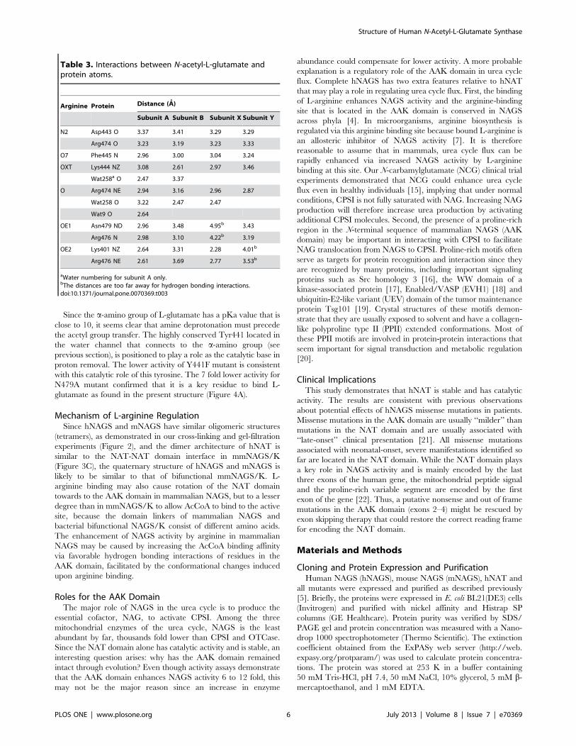
CoA Binding SiteEven though 10 mM CoA was present in the crystallization
solution, no continuous electron density corresponding to CoA
was observed in the ‘‘V-shaped’’ groove where the pantetheine
moiety of CoA usually binds, probably due to suboptimal
conditions for CoA binding. However, the unambiguous identi-
fication of NAG in the expected site suggests that CoA is likely to
bind in a site similar to those found in other GCN5-related
acetyltransferases. Structural comparison of hNAT with the NAT
domain of ngNAGS, which has both substrates bound, allows
identification of the CoA binding site and development of a model
of the catalytic mechanism. Superimposition of the hNAT with the
NAT domain of ngNAGS clearly indicates that the pantetheine
moiety of CoA interacts with the protein through hydrogen bonds
with the main-chain nitrogen of Phe445 and the main-chain
oxygen of Val447 from b16, in a way similar to an anti-parallel b-sheet (Figure 5). The thiol sulfur is oriented in a position within
hydrogen bonding range of the side-chains of Tyr495 and Ser475.
One water molecule (w37) was identified occupying the thiol sulfur
position in the present structure, 3.4 A away from the acetyl
carbon and perpendicular to the acetyl group plane of NAG. The
structure is consistent with hNAGS using a one-step direct attack
catalytic mechanism to transfer the acetyl group from AcCoA to
the amino group of L-glutamate, as is the case for most members
of the GCN5-related NAT family. The pyrophosphate moiety
appears to be in proximity to the sequence Gln452–Gly453–
Gln454–Gly455–Ser456–Gly457–Gln458, which conforms to
(Arg/Gln)-Xaa-Xaa-Gly-Xaa-(Gly/Ala) motif for AcCoA recog-
nition and binding in known GCN5-related N-acetyltransferases
[11]. Because of the absence of AcCoA or CoA binding, this part
of structure varied significantly among different subunits
(Figure 3B). Upon AcCoA or CoA binding, the structure needs
to adjust in order to bind the pyrophosphate moiety. The side-
chains of Gln454 and Gln458 of this motif, as well as Trp484,
seem to be involved in positioning CoA. The adenosine moiety of
CoA is located on the surface of the protein, as seen in other
GCN5-related NAT structures [12].
Table 1. Data collection and refinement statistics.
Data collection
Bound ligands NAG
Space group P43212
Wavelength (A) 1.0
Resolution (A) 50–2.10 (2.14–2.10)a
Unit-cell parameters (A) a= b = 116.1
c = 109.7
Measurements 600,087
Unique reflections 83,202 (4,107)
Redundancy 7.2 (5.1)
Completeness (%) 99.9 (99.1)
,I/s(I). 21.7 (1.7)
Rmerg(%)b 8.1 (65.6)
Refinement
Resolution range (A) 40–2.10 (2.15–2.10)
No. of protein atoms 4,963
No. of water atoms 266
No. of hetero atoms 52
Rmsd of bond lengths (A) 0.008
Rmsd of bond angle (u) 1.1
Rwork (%)c 18.5 (25.2)
Rfree (%)d 24.4 (31.5)
Ramachandran plot (%)
Favored 92.5
Allowed 7.3
Generous 0.2
Disallowed 0.0
aFigures in brackets apply to the highest-resolution shell.bRmerg =ShSi|I(h,i)-,I(h).|/ShSiI(h,i), where I(h,i) is the intensity of the ithobservation of reflection h, and,I(h). is the average intensity of redundantmeasurements of reflection h.cRwork =Sh||Fobs| – |Fcalc||/Sh |Fobs|.dRfree =Sh||Fobs| – |Fcalc||/Sh |Fobs| for 5% of the reserved reflections.doi:10.1371/journal.pone.0070369.t001
Table 2. RMSD values (A) among different subunit withinNAG bound structure and with the bifunctional mmNAGS/Knative structure.
Subunit A B X Y
A 1.08a 0.42 0.76 0.60
B 1.19 0.69 0.61
X 1.05 0.48
Y 1.04
aThe values in bold are RMSD of the subunit A in hNAT structure with subunitsin mmNAGS/K NAT domain structure (PDB 3S6H).doi:10.1371/journal.pone.0070369.t002
Structure of Human N-Acetyl-L-Glutamate Synthase
PLOS ONE | www.plosone.org 4 July 2013 | Volume 8 | Issue 7 | e70369

Comparison of the NAT Domain Structures of HumanNAGS and mmNAGS/KThe overall hNAT structure is similar to that of mmNAGS/K
(Figure 6, Table 2) and can be aligned with an RMS deviation of
,1.0 A, even though different subunits in mmNAGS/K have
different relative orientations of the AAK and NAT domains [8].
The major structural differences occur in the loop regions (a12–b14, b16–a13, b15–b16 and b19–a15 loops). The significant
conformational changes in the pyrophosphate moiety binding
motif in the loop connecting b16 and a13 demonstrate the high
flexibility in this region in the absence of AcCoA binding, as shown
in the variation among different subunits. The conformational
changes of the side-chain position of Arg476 may be functionally
significant. In all mmNAGS/K subunits, the side-chain of Arg388
(the equivalent residue of Arg476) points outwards (Figure 6)
whereas in the NAG bound hNAT structure, this side-chain moves
towards the substrate binding site to anchor the c-carboxyl groupof NAG. Another interesting difference is in the a12–b14 loop in
which two more residues are present in hNAT compared to
mmNAGS/K. The side-chain of Arg414 of this loop swings
towards the NAG binding site to form a hydrogen bond with the
side-chains of Asp433 and Asp443. At least 8 nearby water
molecules link the amino nitrogen of NAG to the side-chains of
Tyr441, Asp443, Lys444, Ser524, Arg414 and Ser410 in a string
that extends to the protein surface (Figure 4B). As proposed for
serotonin N-acetyltransferase [13], this chain of water molecules
may be a ‘‘proton wire’’ to ferry away a proton from the substrate
and facilitate a nucleophilic attack on AcCoA.
Implications for CatalysisTo confirm the catalytic mechanism, several residues in this site
were selected for biochemical studies. Tyr485, the equivalent
residue of Tyr397 in mmNAGS/K and Tyr405 in xcNAGS/K,
appears to act as a catalytic acid that donates a proton to the thiol
group of CoA, playing an important role in the catalytic reaction
(Figure 4A). This equivalent tyrosine could be identified in most
GCN5-related acetyltransferases [14]. Indeed, the Y485F mutant
showed 10 fold lower catalytic activity than wild-type protein
(Table 4).
Figure 3. Structure of hNAT. A: Ribbon diagram of hNAT subunit structure. Bound NAG is shown as sky-blue sticks. The electron density map(2Fo–Fc) around bound NAG (contoured at 1.0 s) is shown as blue cage. B: Superimposition of four hNAT subunits in asymmetric unit. The boundNAG is shown as sky-blue sticks. The proposed bound CoA is shown as green sticks. Subunits A, B, X and Y are shown in pink, yellow, green and blueribbons, respectively. C: The hNAT molecular dimer. Subunits A and B are shown in green and red ribbons, respectively. D: Details of the interactionsbetween subunits A and B. Side-chains of the residues in the interface are shown in sticks. Potential hydrogen bonding interactions are shown in reddashed lines.doi:10.1371/journal.pone.0070369.g003
Structure of Human N-Acetyl-L-Glutamate Synthase
PLOS ONE | www.plosone.org 5 July 2013 | Volume 8 | Issue 7 | e70369
Since the a-amino group of L-glutamate has a pKa value that is
close to 10, it seems clear that amine deprotonation must precede
the acetyl group transfer. The highly conserved Tyr441 located in
the water channel that connects to the a-amino group (see
previous section), is positioned to play a role as the catalytic base in
proton removal. The lower activity of Y441F mutant is consistent
with this catalytic role of this tyrosine. The 7 fold lower activity for
N479A mutant confirmed that it is a key residue to bind L-
glutamate as found in the present structure (Figure 4A).
Mechanism of L-arginine RegulationSince hNAGS and mNAGS have similar oligomeric structures
(tetramers), as demonstrated in our cross-linking and gel-filtration
experiments (Figure 2), and the dimer architecture of hNAT is
similar to the NAT-NAT domain interface in mmNAGS/K
(Figure 3C), the quaternary structure of hNAGS and mNAGS is
likely to be similar to that of bifunctional mmNAGS/K. L-
arginine binding may also cause rotation of the NAT domain
towards to the AAK domain in mammalian NAGS, but to a lesser
degree than in mmNAGS/K to allow AcCoA to bind to the active
site, because the domain linkers of mammalian NAGS and
bacterial bifunctional NAGS/K consist of different amino acids.
The enhancement of NAGS activity by arginine in mammalian
NAGS may be caused by increasing the AcCoA binding affinity
via favorable hydrogen bonding interactions of residues in the
AAK domain, facilitated by the conformational changes induced
upon arginine binding.
Roles for the AAK DomainThe major role of NAGS in the urea cycle is to produce the
essential cofactor, NAG, to activate CPSI. Among the three
mitochondrial enzymes of the urea cycle, NAGS is the least
abundant by far, thousands fold lower than CPSI and OTCase.
Since the NAT domain alone has catalytic activity and is stable, an
interesting question arises: why has the AAK domain remained
intact through evolution? Even though activity assays demonstrate
that the AAK domain enhances NAGS activity 6 to 12 fold, this
may not be the major reason since an increase in enzyme
abundance could compensate for lower activity. A more probable
explanation is a regulatory role of the AAK domain in urea cycle
flux. Complete hNAGS has two extra features relative to hNAT
that may play a role in regulating urea cycle flux. First, the binding
of L-arginine enhances NAGS activity and the arginine-binding
site that is located in the AAK domain is conserved in NAGS
across phyla [4]. In microorganisms, arginine biosynthesis is
regulated via this arginine binding site because bound L-arginine is
an allosteric inhibitor of NAGS activity [7]. It is therefore
reasonable to assume that in mammals, urea cycle flux can be
rapidly enhanced via increased NAGS activity by L-arginine
binding at this site. Our N-carbamylglutamate (NCG) clinical trial
experiments demonstrated that NCG could enhance urea cycle
flux even in healthy individuals [15], implying that under normal
conditions, CPSI is not fully saturated with NAG. Increasing NAG
production will therefore increase urea production by activating
additional CPSI molecules. Second, the presence of a proline-rich
region in the N-terminal sequence of mammalian NAGS (AAK
domain) may be important in interacting with CPSI to facilitate
NAG translocation from NAGS to CPSI. Proline-rich motifs often
serve as targets for protein recognition and interaction since they
are recognized by many proteins, including important signaling
proteins such as Src homology 3 [16], the WW domain of a
kinase-associated protein [17], Enabled/VASP (EVH1) [18] and
ubiquitin-E2-like variant (UEV) domain of the tumor maintenance
protein Tsg101 [19]. Crystal structures of these motifs demon-
strate that they are usually exposed to solvent and have a collagen-
like polyproline type II (PPII) extended conformations. Most of
these PPII motifs are involved in protein-protein interactions that
seem important for signal transduction and metabolic regulation
[20].
Clinical ImplicationsThis study demonstrates that hNAT is stable and has catalytic
activity. The results are consistent with previous observations
about potential effects of hNAGS missense mutations in patients.
Missense mutations in the AAK domain are usually ‘‘milder’’ than
mutations in the NAT domain and are usually associated with
‘‘late-onset’’ clinical presentation [21]. All missense mutations
associated with neonatal-onset, severe manifestations identified so
far are located in the NAT domain. While the NAT domain plays
a key role in NAGS activity and is mainly encoded by the last
three exons of the human gene, the mitochondrial peptide signal
and the proline-rich variable segment are encoded by the first
exon of the gene [22]. Thus, a putative nonsense and out of frame
mutations in the AAK domain (exons 2–4) might be rescued by
exon skipping therapy that could restore the correct reading frame
for encoding the NAT domain.
Materials and Methods
Cloning and Protein Expression and PurificationHuman NAGS (hNAGS), mouse NAGS (mNAGS), hNAT and
all mutants were expressed and purified as described previously
[5]. Briefly, the proteins were expressed in E. coli BL21(DE3) cells
(Invitrogen) and purified with nickel affinity and Histrap SP
columns (GE Healthcare). Protein purity was verified by SDS/
PAGE gel and protein concentration was measured with a Nano-
drop 1000 spectrophotometer (Thermo Scientific). The extinction
coefficient obtained from the ExPASy web server (http://web.
expasy.org/protparam/) was used to calculate protein concentra-
tions. The protein was stored at 253 K in a buffer containing
50 mM Tris-HCl, pH 7.4, 50 mM NaCl, 10% glycerol, 5 mM b-mercaptoethanol, and 1 mM EDTA.
Table 3. Interactions between N-acetyl-L-glutamate andprotein atoms.
Arginine Protein Distance (A)
Subunit A Subunit B Subunit X Subunit Y
N2 Asp443 O 3.37 3.41 3.29 3.29
Arg474 O 3.23 3.19 3.23 3.33
O7 Phe445 N 2.96 3.00 3.04 3.24
OXT Lys444 NZ 3.08 2.61 2.97 3.46
Wat258a O 2.47 3.37
O Arg474 NE 2.94 3.16 2.96 2.87
Wat258 O 3.22 2.47 2.47
Wat9 O 2.64
OE1 Asn479 ND 2.96 3.48 4.95b 3.43
Arg476 N 2.98 3.10 4.22b 3.19
OE2 Lys401 NZ 2.64 3.31 2.28 4.01b
Arg476 NE 2.61 3.69 2.77 3.53b
aWater numbering for subunit A only.bThe distances are too far away for hydrogen bonding interactions.doi:10.1371/journal.pone.0070369.t003
Structure of Human N-Acetyl-L-Glutamate Synthase
PLOS ONE | www.plosone.org 6 July 2013 | Volume 8 | Issue 7 | e70369

Site-directed MutagenesisSite-directed mutant DNA sequences encoding hNAT were
created using primers containing the desired mutations and the
QuikChange Mutagenesis Kit according to the manufacturer’s
protocol (Strategene). The sequences of mutant DNA sequences
were verified by DNA sequencing.
Activity AssayEnzymatic activity was assayed using the method described
previously [23]. A stable isotope dilution method using liquid
chromatography mass spectrometry (LC–MS) to measure NAG
production was adapted. Each assay was performed in a 100 mlsolution containing 50 mM Tris, pH 8.5, 10 mM glutamate and
2.5 mM AcCoA. The reaction was initiated by the addition of
purified recombinant enzyme (20 mg), and the mixture was
incubated at 303 K for 5 min and quenched with 100 ml of
30% trichloroacetic acid containing 50 mg of N-acetyl-[13C5]glu-
tamate (13C-NAG) as an internal standard. Precipitated protein
was removed by micro-centrifugation. The supernatant (10 ml) wassubmitted to LC-MS (Agilent) analysis. The mobile phase
consisted of 92% solvent A (1 ml trifluoroacetic acid in 1 L water)
and 8% solvent B (1 ml trifluoroacetic acid in 1 L of 1:9 water/
acetonitrile) and the flow rate was 0.6 ml/min. Glutamate, NAG,
Figure 4. NAG binding site. A: Stereo diagram of NAG binding site. The bound NAG is shown in sky-blue sticks. The side-chains involved inhydrogen bonding interactions with NAG are shown in green sticks. The side-chains of other surrounding residues are shown in yellow sticks. Thewater molecule (w37) is shown in red ball. The electron density map (2Fo–Fc) around bound NAG (contoured at 1.0 s) is shown as blue cage. Potentialhydrogen bonding interactions are shown in red dashed lines. B: Stereo diagram of ‘‘water wire’’ channel. The bound NAG is shown in sky-blue sticks.Water molecules are shown in yellow balls. Residues involved in hydrogen bonding interactions are shown in brown sticks. Potential hydrogenbonding interactions are shown in red dashed lines.doi:10.1371/journal.pone.0070369.g004
Structure of Human N-Acetyl-L-Glutamate Synthase
PLOS ONE | www.plosone.org 7 July 2013 | Volume 8 | Issue 7 | e70369
and 13C-NAG were detected and quantified by selected ion
monitoring mass spectrometry.
AcCoA and glutamate titration experiments were carried out
with AcCoA or L-glutamate concentration varied in the range of
0.25–5.0 and 0.5–20 mM, respectively, and L-glutamate or
AcCoA concentration fixed at 10 and 2.5 mM, respectively. The
L-glutamate titration data were fit to Michaelis-Menten kinetics,
while AcCoA titration data were fit to sigmoidal kinetics (V=Vmax
[AcCoA]n/([AcCoA]n+Kmn), where Vmax is maximum activity, Km
is half-maximum activity and n is the Hill coefficient, using the
program GNUPLOT.
Cross-linking ExperimentCross-linking experiments were performed using the protocol
described by Davies and Stark [24]. mNAGS (2.5 mg ) and
hNAGS (1.5 and 4.5 mg) were incubated with the cross-linking
reagent dimethyl suberimidate (4.5 mg) or suberic acid bis(3-sulfo-
N-hydroxysuccinimide ester) sodium salt (9.0 mg) in 10 ml solution
Figure 5. Stereo diagram of the proposed CoA binding site. The proposed bound CoA is shown in green sticks. The bound NAG is shown insky-blue sticks. Side-chains of residues that potentially hydrogen bond to CoA are shown in yellow sticks. The water molecule (w37) that occupies thesimilar position of thiol S of CoA is shown in a red ball.doi:10.1371/journal.pone.0070369.g005
Figure 6. Superimposition of hNAT with the NAT domain of subunit X of mmNAGS/K. The structure of hNAT is shown in pink ribbons. Thestructure of the NAT domain of subunit X of mmNAGS/K is shown in yellow ribbons. The bound NAG is shown in sky-blue sticks. The proposed boundCoA is shown in green sticks. Residues that are mentioned in text are shown in sticks.doi:10.1371/journal.pone.0070369.g006
Structure of Human N-Acetyl-L-Glutamate Synthase
PLOS ONE | www.plosone.org 8 July 2013 | Volume 8 | Issue 7 | e70369
containing 200 mM triethanolamine, pH 8.25 for three hours at
298 K. Samples of mNAGS with and without cross-linking
reagent were subjected to sodium dodecyl sulfate polyacrylamide
get electrophoresis (NuPAGE 4–12% Bis-Tris gel) in MES SDS
buffer (50 mMMES, 50 mM Tris base, 0.1% SDS, 1 mM EDTA,
pH 7.3) and stained with Coomassie blue. Samples of hNAGS
with and without cross-linking reagent were subjected to sodium
dodecyl sulfate polyacrylamide get electrophoresis (NuPAGE 4–
12% Bis-Tris gel) in MES SDS buffer (50 mM MES, 50 mM Tris
base, 0.1% SDS, 1 mM EDTA, pH 7.3) and stained with silver.
Size marker controls consisted of proteins with defined molecular
weights of protein standards purchased from Invitrogen.
Gel-filtration ChromatographyMolecular weight of mNAGS and hNAGS were determined
with a Superdex 200 HR 10/30 column (Amersham Biosciences)
as previously described [5]. The running buffer contains 100 mM
NaH2PO4 pH 7.4, 150 mM NaCl, 10% glycerol, 1 mM b-mercaptoethanol. Thyroglobulin (669 kDa), ferritin (440 kDa),
ngNAGS (296.7 kDa), mmNAGS (200.5 kDa) and aldolase (158
kDa) were used as protein standards.
Molecular weights of hNAT and the NAT domain of mouse
NAGS (mNAT) were determined similarly, but with different
protein standards of ovalbumin (43 kDa), albumin (67 kDa),
chymotrypsinogen A (25 kDa) and ribonucleause (13.7 kDa).
CrystallizationCrystals were grown by the sitting-drop, vapor-diffusion
method. Before crystallization, the purified protein (,20 mg/ml)
was treated with thrombin (50 units) overnight at 277 K to remove
the his-tag, then incubated with 10 mM CoA, and 20 mM NAG
for 30 min. Screening of crystallization conditions was performed
using sitting-drop vapor diffusion in 96-well plates (Hampton
Research) at 291 K by mixing 2 ml of the protein solution with
2 ml of the reagent solution from the sparse matrix Crystal Screens
1 and 2, and Index Screen (Hampton Research). The best crystals
were grown from a reservoir solution containing 100 mM Bis-tris,
pH 6.5, 35% PEG3350. Crystals were stick-shaped and took 2–3
days to reach a maximal length of 0.6 mm.
Data Collection and Structure DeterminationCrystals were transferred from the crystallization plate to a well
solution supplemented with 25% glycerol and then frozen directly
by liquid nitrogen. Diffraction data were collected at beamline 22-
ID equipped with MAR300 CCD at the Advanced Photon source
(APS), Argonne National Laboratory, USA. All data were
processed using the HKL2000 package [25]; statistics are
summarized in Table 1. The structure was solved by molecular
replacement using Phaser [26,27] based on the NAT domain of
mmNAGS/K structure of subunit X as a search model. After
several cycles of refinements with Phenix [28] and model
adjustments with Coot [29], NAG was visible in the electron
density map and was built into the model. In the last run of the
refinement, the translation/liberation/screw parameters were
included and refined [30]. Two groups per subunit were selected
according to the N-terminal arm (residues 375–469) and the C-
terminal arm (470–527). Final R and Rfree values were 18.4% and
24.4%, respectively. Refinement statistics for the final refined
model are given in Table 1. The final refined coordinates for NAG
bound hNAT and its structure factors have been deposited in
RCSB Protein Data Bank with accession code 4K30 and provided
as Supplemental Materials.
Supporting Information
File S1 Coordinate file for the described structure.
(PDB)
File S2 Structure factors for the described structure.
(CIF)
Acknowledgments
We thank Dr. David Davies for facilitating the use of the diffraction
equipment in the Molecular Structure Section of the National Institutes of
Health and Dr. Fred Dyda for help in data collection.
Author Contributions
Conceived and designed the experiments: DS MT GZ. Performed the
experiments: DS GZ ZJ. Analyzed the data: DS GZ ZJ. Contributed
reagents/materials/analysis tools: DS GZ. Wrote the paper: DS MT NMA
GZ.
References
1. Slocum RD (2005) Genes, enzymes and regulation of arginine biosynthesis in
plants. Plant Physiol Biochem 43: 729–745.
2. Cunin R, Glansdorff N, Pierard A, Stalon V (1986) Biosynthesis and metabolism
of arginine in bacteria. Microbiol Rev 50: 314–352.
3. Haskins N, Panglao M, Qu Q, Majumdar H, Cabrera-Luque J, et al. (2008)
Inversion of allosteric effect of arginine on N-acetylglutamate synthase, a
molecular marker for evolution of tetrapods. BMC Biochem 9: 24.
4. Qu Q, Morizono H, Shi D, Tuchman M, Caldovic L (2007) A novel
bifunctional N-acetylglutamate synthase-kinase from Xanthomonas campestris
that is closely related to mammalian N-acetylglutamate synthase. BMC Biochem
8: 4.
5. Shi D, Sagar V, Jin Z, Yu X, Caldovic L, et al. (2008) The crystal structure of N-
acetyl-L-glutamate synthase from Neisseria gonorrhoeae provides insights into
mechanisms of catalysis and regulation. J Biol Chem 283: 7176–7184.
6. Ramon-Maiques S, Fernandez-Murga ML, Gil-Ortiz F, Vagin A, Fita I, et al.
(2006) Structural bases of feed-back control of arginine biosynthesis, revealed by
the structures of two hexameric N-acetylglutamate kinases, from Thermotoga
maritima and Pseudomonas aeruginosa. J Mol Biol 356: 695–713.
7. Min L, Jin Z, Caldovic L, Morizono H, Allewell NM, et al. (2009) Mechanism of
allosteric inhibition of N-acetyl-L-glutamate synthase by L-arginine. J Biol Chem
284: 4873–4880.
8. Shi D, Li Y, Cabrera-Luque J, Jin Z, Yu X, et al. (2011) A Novel N-
acetylglutamate synthase architecture revealed by the crystal structure of the
bifunctional enzyme from Maricaulis maris. PLoS One 6: e28825.
9. Caldovic L, Lopez GY, Haskins N, Panglao M, Shi D, et al. (2006) Biochemical
properties of recombinant human and mouse N-acetylglutamate synthase. Mol
Genet Metab 87: 226–232.
10. Krissinel E, Henrick K (2007) Inference of macromolecular assemblies from
crystalline state. J Mol Biol 372: 774–797.
11. Neuwald AF, Landsman D (1997) GCN5-related histone N-acetyltransferases
belong to a diverse superfamily that includes the yeast SPT10 protein. Trends
Biochem Sci 22: 154–155.
Table 4. Enzyme activities of hNAT and active site mutants.
Sample Activity (mmoles/min/mg)a
WT 1.0560.01
WT+L-arginine (1 mM) 1.0460.01
Y485F 0.07860.003
Y441F 0.85760.004
N479A 0.15460.002
aMeans 6 standard errors of means (n= 3) are shown.doi:10.1371/journal.pone.0070369.t004
Structure of Human N-Acetyl-L-Glutamate Synthase
PLOS ONE | www.plosone.org 9 July 2013 | Volume 8 | Issue 7 | e70369

12. Dyda F, Klein DC, Hickman AB (2000) GCN5-related N-acetyltransferases: a
structural overview. Annu Rev Biophys Biomol Struct 29: 81–103.13. Hickman AB, Namboodiri MA, Klein DC, Dyda F (1999) The structural basis of
ordered substrate binding by serotonin N-acetyltransferase: enzyme complex at
1.8 A resolution with a bisubstrate analog. Cell 97: 361–369.14. He H, Ding Y, Bartlam M, Sun F, Le Y, et al. (2003) Crystal structure of
tabtoxin resistance protein complexed with acetyl coenzyme A reveals themechanism for beta-lactam acetylation. J Mol Biol 325: 1019–1030.
15. Ah Mew N, Payan I, Daikhin Y, Nissim I, Nissim I, et al. (2009) Effects of a
single dose of N-carbamylglutamate on the rate of ureagenesis. Mol GenetMetab 98: 325–330.
16. Kaieda S, Matsui C, Mimori-Kiyosue Y, Ikegami T (2010) Structural basis ofthe recognition of the SAMP motif of adenomatous polyposis coli by the Src-
homology 3 domain. Biochemistry 49: 5143–5153.17. Macias MJ, Hyvonen M, Baraldi E, Schultz J, Sudol M, et al. (1996) Structure of
the WW domain of a kinase-associated protein complexed with a proline-rich
peptide. Nature 382: 646–649.18. Prehoda KE, Lee DJ, Lim WA (1999) Structure of the enabled/VASP homology
1 domain-peptide complex: a key component in the spatial control of actinassembly. Cell 97: 471–480.
19. Schlundt A, Sticht J, Piotukh K, Kosslick D, Jahnke N, et al. (2009) Proline-rich
sequence recognition: II. Proteomics analysis of Tsg101 ubiquitin-E2-like variant(UEV) interactions. Mol Cell Proteomics 8: 2474–2486.
20. Adzhubei AA, Sternberg MJ, Makarov AA (2013) Polyproline-II Helix inProteins: Structure and Function. J Mol Biol 425: 2100–2132.
21. Caldovic L, Morizono H, Tuchman M (2007) Mutations and polymorphisms in
the human N-acetylglutamate synthase (NAGS) gene. Hum Mutat 28: 754–759.22. Caldovic L, Morizono H, Gracia Panglao M, Gallegos R, Yu X, et al. (2002)
Cloning and expression of the human N-acetylglutamate synthase gene.
Biochem Biophys Res Commun 299: 581–586.23. Caldovic L, Morizono H, Yu X, Thompson M, Shi D, et al. (2002)
Identification, cloning and expression of the mouse N-acetylglutamate synthasegene. Biochem J 364: 825–831.
24. Davies GE, Stark GR (1970) Use of dimethyl suberimidate, a cross-linking
reagent, in studying the subunit structure of oligomeric proteins. Proc Natl AcadSci U S A 66: 651–656.
25. Otwinowski Z, Minor W (1997) Processing of X-ray diffraction data collected inoscillation mode. Methods Enzymol 276: 307–326.
26. Read RJ (2001) Pushing the boundaries of molecular replacement withmaximum likelihood. Acta Crystallogr D Biol Crystallogr 57: 1373–1382.
27. Storoni LC, McCoy AJ, Read RJ (2004) Likelihood-enhanced fast rotation
functions. Acta Crystallogr D Biol Crystallogr 60: 432–438.28. Adams PD, Afonine PV, Bunkoczi G, Chen VB, Davis IW, et al. (2010)
PHENIX: a comprehensive Python-based system for macromolecular structuresolution. Acta Crystallogr D Biol Crystallogr 66: 213–221.
29. Emsley P, Cowtan K (2004) Coot: model-building tools for molecular graphics.
Acta Crystallogr D Biol Crystallogr 60: 2126–2132.30. Winn MD, Isupov MN, Murshudov GN (2001) Use of TLS parameters to model
anisotropic displacements in macromolecular refinement. Acta Crystallogr D BiolCrystallogr 57: 122–133.
Structure of Human N-Acetyl-L-Glutamate Synthase
PLOS ONE | www.plosone.org 10 July 2013 | Volume 8 | Issue 7 | e70369


![2-[5-Methyl-2-(propan-2-yl)phenoxy]- N ′-{2-[5-methyl-2-(propan-2-yl)phenoxy]acetyl}acetohydrazide](https://static.fdokumen.com/doc/165x107/6344862303a48733920aed56/2-5-methyl-2-propan-2-ylphenoxy-n-2-5-methyl-2-propan-2-ylphenoxyacetylacetohydrazide.jpg)