N -methyl-d-aspartate receptor- and metabotropic glutamate receptor-dependent long-term depression...
14
NMDAR- and mGluR-dependent long term depression are differentially regulated by the ubiquitin-proteasome system Ami Citri * , Gilberto Soler-Llavina * , Samarjit Bhattacharyya, and Robert C. Malenka Nancy Pritzker Laboratory, Dept. of Psychiatry and Behavioral Sciences, Stanford University School of Medicine, Palo Alto CA 94304 Abstract Long-term depression (LTD) in CA1 pyramidal neurons can be induced by activation of either NMDA receptors (NMDARs) or metabotropic glutamate receptors (mGluRs), both of which elicit changes in synaptic efficacy through AMPA receptor (AMPAR) endocytosis. To address the role of the ubiquitin-proteasome system in regulating AMPAR endocytosis during these forms of LTD, we examined the effects of pharmacological inhibitors of proteasomal degradation and protein ubiquitination on endocytosis of GluR1-containing AMPARs in dissociated rat hippocampal cultures as well as LTD of excitatory synaptic responses in acute rat hippocampal slices. Our findings suggest that the contribution of the ubiquitin-proteasome system to NMDAR-induced versus mGluR-induced AMPAR endocytosis and the consequent LTD differs significantly. NMDAR-induced AMPAR endocytosis and LTD occur independently of proteasome function, but appear to depend, at least in part, on ubiquitination. In contrast, mGluR-induced AMPAR endocytosis and LTD are enhanced by inhibition of proteasomal degradation, as well as by the inhibitor of protein ubiquitination. Furthermore, the decay of mGluR-induced membrane depolarization and Erk activation is delayed following inhibition of either ubiquitination or proteasomal degradation. These results suggest that while NMDAR-dependent LTD may utilize ubiquitin as a signal for AMPAR endocytosis, mGluR-induced signaling and LTD is limited by a feedback mechanism that involves the ubiquitin-proteasome system. Keywords rat; synapse; long-term-depression; endocytosis; glutamate receptor Introduction Two forms of long-term depression (LTD) of synaptic transmission have been identified at excitatory synapses onto CA1 pyramidal neurons in the hippocampus and are distinguished on the basis of whether they are triggered via activation of postsynaptic N-methyl-D- aspartate receptors (NMDARs) (Dudek & Bear, 1992; Mulkey & Malenka, 1992) or group I metabotropic glutamate receptors (mGluRs) (Bashir et al., 1993; Bolshakov & Siegelbaum, 1994). Both forms of LTD can coexist at the same set of synapses (Oliet et al., 1997) and both are thought to be due primarily to the clathrin- and dynamin-dependent endocytosis of AMPARs (Carroll et al., 2001; Snyder et al., 2001; Xiao et al., 2001; Shepherd & Huganir, Corresponding Author: Robert Malenka Stanford University School of Medicine 1050 Arastradero Rd. Rm B249 Palo Alto, CA. 94304-5552 650-724-2730 650-498-4585 (fax) [email protected]. * These authors contributed equally to this work. Supporting Information Additional supporting information may be found in the online version of this article: NIH Public Access Author Manuscript Eur J Neurosci. Author manuscript; available in PMC 2010 October 1. Published in final edited form as: Eur J Neurosci. 2009 October ; 30(8): 1443–1450. doi:10.1111/j.1460-9568.2009.06950.x. NIH-PA Author Manuscript NIH-PA Author Manuscript NIH-PA Author Manuscript
Transcript of N -methyl-d-aspartate receptor- and metabotropic glutamate receptor-dependent long-term depression...

NMDAR- and mGluR-dependent long term depression aredifferentially regulated by the ubiquitin-proteasome system
Ami Citri*, Gilberto Soler-Llavina*, Samarjit Bhattacharyya, and Robert C. MalenkaNancy Pritzker Laboratory, Dept. of Psychiatry and Behavioral Sciences, Stanford UniversitySchool of Medicine, Palo Alto CA 94304
AbstractLong-term depression (LTD) in CA1 pyramidal neurons can be induced by activation of eitherNMDA receptors (NMDARs) or metabotropic glutamate receptors (mGluRs), both of which elicitchanges in synaptic efficacy through AMPA receptor (AMPAR) endocytosis. To address the roleof the ubiquitin-proteasome system in regulating AMPAR endocytosis during these forms of LTD,we examined the effects of pharmacological inhibitors of proteasomal degradation and proteinubiquitination on endocytosis of GluR1-containing AMPARs in dissociated rat hippocampalcultures as well as LTD of excitatory synaptic responses in acute rat hippocampal slices. Ourfindings suggest that the contribution of the ubiquitin-proteasome system to NMDAR-inducedversus mGluR-induced AMPAR endocytosis and the consequent LTD differs significantly.NMDAR-induced AMPAR endocytosis and LTD occur independently of proteasome function, butappear to depend, at least in part, on ubiquitination. In contrast, mGluR-induced AMPARendocytosis and LTD are enhanced by inhibition of proteasomal degradation, as well as by theinhibitor of protein ubiquitination. Furthermore, the decay of mGluR-induced membranedepolarization and Erk activation is delayed following inhibition of either ubiquitination orproteasomal degradation. These results suggest that while NMDAR-dependent LTD may utilizeubiquitin as a signal for AMPAR endocytosis, mGluR-induced signaling and LTD is limited by afeedback mechanism that involves the ubiquitin-proteasome system.
Keywordsrat; synapse; long-term-depression; endocytosis; glutamate receptor
IntroductionTwo forms of long-term depression (LTD) of synaptic transmission have been identified atexcitatory synapses onto CA1 pyramidal neurons in the hippocampus and are distinguishedon the basis of whether they are triggered via activation of postsynaptic N-methyl-D-aspartate receptors (NMDARs) (Dudek & Bear, 1992; Mulkey & Malenka, 1992) or group Imetabotropic glutamate receptors (mGluRs) (Bashir et al., 1993; Bolshakov & Siegelbaum,1994). Both forms of LTD can coexist at the same set of synapses (Oliet et al., 1997) andboth are thought to be due primarily to the clathrin- and dynamin-dependent endocytosis ofAMPARs (Carroll et al., 2001; Snyder et al., 2001; Xiao et al., 2001; Shepherd & Huganir,
Corresponding Author: Robert Malenka Stanford University School of Medicine 1050 Arastradero Rd. Rm B249 Palo Alto, CA.94304-5552 650-724-2730 650-498-4585 (fax) [email protected].*These authors contributed equally to this work.Supporting Information Additional supporting information may be found in the online version of this article:
NIH Public AccessAuthor ManuscriptEur J Neurosci. Author manuscript; available in PMC 2010 October 1.
Published in final edited form as:Eur J Neurosci. 2009 October ; 30(8): 1443–1450. doi:10.1111/j.1460-9568.2009.06950.x.
NIH
-PA Author Manuscript
NIH
-PA Author Manuscript
NIH
-PA Author Manuscript

2007). Thus, it has been assumed that they employ a common route of AMPAR endocytosisthat is activated by divergent signaling pathways.
Group I mGluRs, via coupling to Gαq, normally activate phospholipase C, resulting inphosphoinositide hydrolysis, calcium release from internal stores, protein kinase Cactivation and subsequent activation of the Erk/MAPK pathway (Conn, 2003). Theactivation of Erk (Gallagher et al., 2004) as well as PKC (Oliet et al., 1997) havespecifically been reported to be necessary for mGluR-dependent LTD in the hippocampus(Gallagher et al., 2004; Bellone et al., 2008). Interest in this form of LTD was greatlyincreased by the finding that it is enhanced in a mouse model of Fragile X syndrome (Huberet al., 2002), an observation that led to the hypothesis that excessive mGluR signalingunderlies this most common inherited form of human mental retardation (Bear et al., 2004).In contrast, NMDAR-dependent LTD is thought to be triggered, at least in part, byactivation of a cascade of protein phosphatases (Carroll et al., 2001; Citri & Malenka, 2008)leading to the dephosphorylation of critical synaptic substrates such as AMPAR subunits(Lee et al., 2000).
The elucidation of the role of ubiquitin conjugation in targeting substrate proteins forproteasomal degradation in a wide array of cell types and species (Hershko & Ciechanover,1998) including mammalian neurons (Ehlers, 2003), raised the possibility that this processmay play a role in LTD. Indeed, ubiquitination of the synaptic scaffold protein PSD-95 wassuggested to be critical for NMDAR-triggered AMPAR endocytosis and LTD (Colledge etal., 2003). However, several subsequent findings were not consistent with this model(Bingol & Schuman, 2004; Xu et al., 2008; Bhattacharyya et al., 2009). More recently,nonproteolytic roles for ubiquitin have been uncovered including the regulation ofendocytosis of membrane receptors such as G-protein coupled receptors and receptortyrosine kinases by mono-ubiquitination (Urbe, 2005; Chen & Sun, 2009). Here we usewell-established inhibitors of proteasomal function, as well as a recently developedcompound that inhibits the E1 activating enzyme of the ubiquitin cascade (Yang et al.,2007) to address the roles of ubiquitination and proteasomal degradation in NMDAR- versusmGluR-triggered AMPAR endocytosis and LTD. Surprisingly, our results suggest that thereare dramatic differences in the regulation of these forms of synaptic plasticity by theubiquitin-proteasome system.
Materials and MethodsAnimals
All experiments were carried out in Sprague-Dawley rats (Charles River, Wilmington, MA)in accordance with Stanford University guidelines regarding the care and use of animals forexperimental procedures. Either P0 (dissociated neuronal cultures) or P14–P28(electrophysiology) rats were used for experiments. Animals were housed in a temperatureand humidity-controlled room with a light/dark cycle of 12 hours. Food and water werealways available. All efforts were made to reduce the number of animals utilized in thisstudy. Newborn rats were anesthetized on ice, while juvenile rats were anesthetized withisoflurane prior to decapitation.
DrugsAll drugs were made as concentrated stock solutions and diluted 100–10,000 fold beforeuse. Drugs that could not be dissolved in H2O were dissolved in DMSO (except LY-341495which was prepared in 1.2 N NaOH). Drugs used in the study were D-AP5, LY341495,TTX, NBQX, DNQX, R,S-DHPG and NMDA (all from Tocris, Ellisville, MO); picrotoxin(Sigma, St. Louis, MO), UBEI-41 (50 mM in DMSO; Biogenova, Rockville, MD),
Citri et al. Page 2
Eur J Neurosci. Author manuscript; available in PMC 2010 October 1.
NIH
-PA Author Manuscript
NIH
-PA Author Manuscript
NIH
-PA Author Manuscript

lactacystin (Calbiochem, San Diego, CA and Boston Biochemicals, Cambridge, MA) andMG-132 (Biomol, Plymouth Meeting, PA and Tocris).
Dissociated hippocampal culturesPrimary hippocampal neuronal cultures were prepared from P0 rat pups as previouslydescribed with minor modifications (Beattie et al., 2000). Briefly, hippocampi weredissected and tissue was dissociated by papain treatment followed by trituration with glasspipettes. Cells were plated on poly D-lysine-coated coverslips at a density of approximately75,000 cells per 12 mm well for immunocytochemistry or 5*105 cells per 35 mm plate forimmunoblotting. Cells were grown in minimum essential media (MEM) (Invitrogen,Carlsbad, CA) with 0.5 mM glutamine and N2-supplement. Glial growth was inhibited byFUDR after 6 days in culture. Cells were assayed at 13–15 DIV.
ImmunocytochemistryTo study the effect of lactacystin, MG-132 and UBEI-41 on the surface expression ofAMPARs, cells were incubated in 50 μM UBEI-41, 5 μM lactacystin or 5, 50 μM MG-132for 30 min at 37°C. Subsequently, surface AMPARs were labeled in live neurons by 15 minincubation at 37°C with a rabbit polyclonal antibody (Calbiochem, San Diego, CA) directedagainst the N-terminus of the GluR1 subunit (1:20 in conditioned media, i.e., MEM + 0.5mM Glutamine). After washout of the antibody, cells were chilled on ice and fixed in 4%PFA on ice for 15 min without permeabilization. Surface receptors were then stained withgoat anti rabbit Alexa-568 (Molecular probes, Invitrogen, Carlsbad, CA) secondary antibodyfor visualization.
To assay the endocytosis of AMPARs, cells were first incubated with 5 μM lactacystin, 5,50 μM MG-132 or 50 μM UBEI-41 for 30 min at 37°C. Surface receptors were then labeledwith the rabbit polyclonal antibody directed against the N-terminus of the GluR1 subunit inlive cells as described above. After wash out of the antibody, cells were treated with theappropriate mixtures of antagonists (1 μM TTX + 100 μM LY341495 + 20 μM DNQX forNMDA application; 1 μM TTX, 20 μM DNQX and 50 μM APV for DHPG application) for5 min. Subsequently, 100 μM NMDA or 100 μM DHPG were applied for 3 or 5 minutes,respectively, in the presence of the appropriate antagonists as just described. Cells werewashed and returned to the incubator for a total of 15 min. Cells were then fixed in 4% PFAfor 15 min on ice without permeabilization. After that, surface receptors were labeled byusing a saturating concentration of goat-anti rabbit Alexa 568 conjugated secondaryantibody (Molecular Probes, Invitrogen, Carlsbad, CA), followed by permeabilization ofcells with 0.1% Triton X-100 for 30 min at room temperature and staining of internalizedreceptors with donkey-anti rabbit Cy5-conjugated secondary antibody (JacksonImmunoresearch, West Grove, PA).
To ensure that the Cy5 conjugated secondary antibody used to label internalized AMPARsin our assay did not label any surface AMPARs, we performed the following controlexperiment. In a non-permeabilized cell, application of a saturating concentration of thefirst, Alexa-568 conjugated secondary antibody that was used to stain surface GluR1prevented any further detectable staining of surface GluR1 upon subsequent application ofthe second, Cy5 conjugated, secondary antibody. Whereas, when cells were permeabilizedwith 0.1% Triton X-100 for 30 min at room temperature, application of the second, Cy5conjugated secondary antibody now labeled internalized GluR1-containing AMPARs. Theseexperiments showed that the second, Cy5 conjugated secondary antibody that was used forlabeling internalized AMPARs did not label surface AMPARs in our assay, indicating thatthe saturating concentration of the first, Alexa-568 conjugated secondary antibody occupiedall surface AMPARs (Bhattacharyya et al., 2009). We used a Cy5 conjugated secondary
Citri et al. Page 3
Eur J Neurosci. Author manuscript; available in PMC 2010 October 1.
NIH
-PA Author Manuscript
NIH
-PA Author Manuscript
NIH
-PA Author Manuscript

antibody to label the internalized receptors since Cy5 is not detectable in the visible range,enabling an unbiased sampling of cells in the different conditions.
Image acquisition and analysisCoverslips were mounted in Fluoromount (Electron Microscopy Sciences, Hatfield, PA) andcells were imaged with a 63X oil-immersion objective mounted on a Zeiss LSM 510 laserscanning confocal microscope. Images for all conditions in a particular experiment wereobtained using identical acquisition parameters (gain, offset, laser power, pinhole size, scanspeed etc.) and were analyzed with Metamorph software using identical parameters (Metaimaging series 6.1, Universal imaging). Untreated and treated cells from the same culturepreparation were always compared with one another. Images from each experiment werethresholded using identical values between different conditions and the total thresholdedarea of fluorescently labeled surface and internalized receptors was measured usingMetamorph software. Since UBEI-41 itself was found to have some autofluorescence(especially in the cell body area), experiments involving UBEI-41 were thresholded suchthat contribution from the autofluorescence due to UBEI-41 was minimized. The determinedthresholding value was identical between different conditions. To measure the surfacereceptors in our assay, surface fluorescence was divided by total cell area, which wasdetermined by measuring background fluorescence using a low threshold level. These valueswere then normalized to the average surface fluorescence of untreated control cells. Forcalculation of the proportion of surface AMPARs that were endocytosed in the dendrite (10μm away from the soma), dendritic intracellular fluorescence was divided by total dendriticfluorescence (intracellular plus surface) for each cell. These values were then normalized tothose of untreated control cells from the same experiment. Each experimental treatment andanalysis was performed on a minimum of two coverslips with most experiments includingon average six to eight coverslips. For presentation, images were processed using AdobePhotoshop software (Adobe Systems) by adjusting brightness and contrast levels to the samedegree for all conditions illustrated in each experiment.
The n value given for each experiment refers to the number of cells analyzed. Data arepresented as mean ± SEM. Group results were compared by using Student's t-test. P > 0.05was considered not significant (n.s).
ImmunoblottingDissociated hippocampal neurons at 13–15 DIV were pretreated for 40 minutes with DMSO,50 μM UBEI-41 or 5 μM lactacystin, in the presence of 1 μM TTX, 20 μM DNQX and 50μM APV. Cells were then treated for 5 minutes with 100 μM RS-DHPG in the presence of 1μM TTX, 20 μM DNQX and 50 μM APV, and followed for a total time as indicated. Cellswere harvested in RIPA buffer (50 mM Tris-Hcl pH=7.4, 150 mM NaCl, 1% NP-40, 0.1%SDS, 0.5% deoxycholic acid, 50 mM NaF, 200 μM Na3VO4 and a protease inhibitorcocktail [Roche]). SDS-PAGE sample buffer was added to cleared lysates, which wereseparated on 4%–12% gradient Bis-Tris gels (Invitrogen) and transferred to PVDFmembranes. Immunoblotting was performed using primary antibodies to activated Erk(pErk; V8031; 1:5000 Promega, Madison, WI) and Erk2 (Santa Cruz Biotechnology, SantaCruz, CA, sc-154; 1:1000), detected with horseradish peroxidase-conjugated secondaryantibodies and developed with the ECL Western blotting detection system (AmershamBiosciences, GE Healthcare). The levels of pErk and Erk2 were quantified from sub-saturated films using ImageJ software (NIH). Levels of pErk were then normalized to theintensity of the corresponding Erk2 band, and defined as percent (%) of the value at t=0.Averages and standard errors of the mean (SEM) are presented. Results from neuronstreated with lactacystin or UBEI-41 are compared to control experiments that wereperformed in parallel and resolved on the same western blot. Statistical significance was
Citri et al. Page 4
Eur J Neurosci. Author manuscript; available in PMC 2010 October 1.
NIH
-PA Author Manuscript
NIH
-PA Author Manuscript
NIH
-PA Author Manuscript

assessed by comparing each point in the time course to the appropriate control using a non-directional (two-tailed) Student's t-test.
Slice preparation and electrophysiological recordingsTransverse hippocampal slices (field recordings, 400 μm; whole-cell, 300 μm) wereprepared from 21–28 day old Sprague-Dawley rats (field recordings and whole cell currentclamp) or 14–21 day old rats (whole-cell voltage clamp experiments) in ice-cold sucrosesolution containing (in mM): sucrose 204, NaHCO3 26.2, glucose 11, KCl 2.5, MgSO4 2,NaH2PO4 1, CaCl2 0.5 saturated with 95% O2 and 5% CO2. Slices were incubated at roomtemperature (20–22°) in artificial cerebrospinal fluid (ACSF) containing (in mM): NaCl 119,NaHCO3 26.2, glucose 11, KCl 2.5, CaCl2 2.5, MgSO4 1.3, and NaH2PO4 for at least 1.5hours before use. Slices were transferred to a submerged recording chamber continuouslyperfused (1.5–2 ml/min) with warm (28–30°) ACSF containing 50 μM picrotoxin (to blockinhibitory transmission). Dendritic, field excitatory postsynaptic potentials (fEPSPs) wererecorded by placing a patch electrode filled with ACSF or 1M NaCl (adjusted to pH 7.4 withNaOH) in the stratum radiatum, 100–200 μm from the CA1 pyramidal cell layer. fEPSPswere evoked by small, brief (50–200 μA, 0.1 ms) current injections delivered to theSchaeffer collaterals using a bipolar glass electrodes filled with ACSF. Whole-cell currentclamp recordings were made with 3–5 MΩ electrodes filled with a solution containing (inmM): KMeSO4 130, HEPES 10, NaCl 10, Mg-ATP 2.5, EGTA 0.4, Na-GTP 0.25, QX-3141 (pH, 7.3 with KOH). Whole-cell voltage clamp recordings were made with 3–5 MΩelectrodes filled with a solution containing (in mM): CsMeSO4 117.5, HEPES 10, TEA-Cl10, CsCl, 15.5, NaCl 8, EGTA 0.25, Mg-ATP 4, Na-GTP 0.3, QX-314 5 (pH adjusted to 7.3with CsOH, Osmolarity 290–300 mOsm). MG-132 was included in patch pipettes at a finalconcentration of 1 μM and allowed to perfuse the cell for a minimum of 15 minutes prior toinduction of LTD (as in (Colledge et al., 2003).
Incubations in UBEI-41 (50 μM) or lactacystin (1 or 5 μM) were performed, following therecovery period in a holding chamber with oxygenated ACSF for 30–60 minutes prior toplacement in the recording chamber. For LTD experiments measured by field potentials,single stimuli were delivered at 0.1 Hz to monitor basal fEPSPs. After a 10 minute baseline,LTD was induced in one of two ways: NMDAR-dependent LTD was elicited by delivering900 stimuli at 5 Hz, while mGluR-dependent LTD was induced by a 10 minute applicationof RS-DHPG (100 μM). Responses were monitored at 0.1 Hz for at least 45 minutes post-induction. For whole-cell voltage clamp LTD experiments, baseline EPSCs were recorded at−60 mV while stimulating afferents at 0.1 Hz. LTD was induced by delivering 200 stimuliat 1 Hz paired with depolarization to −40 mV (as in (Colledge et al., 2003). In the whole-cell current clamp experiments, the resting membrane potential (Vm) was monitored at afrequency of 0.1 Hz. After a 5 minute stable baseline had been achieved, (within 15 minutesafter breaking into the cell) RS-DHPG (100 μM) was perfused for 3 minutes followed by a30 minute washout. Each experiment was normalized to the maximum change in Vm. Theaverage change in Vm relative to baseline (ΔVm) was then calculated and compared acrossconditions.
Field and whole-cell recordings were performed using either an Axopatch-1D amplifier(Axon Instruments, Molecular Devices, Sunnyvale, CA) or a multiclamp 700B amplifier(Molecular Devices, Sunnyvale, CA) and acquired with custom software written in Igor Pro(Wavemetrics, Lake Oswego, Oregon). All data were filtered at 2 kHz and digitized at 10kHz. For display purposes and averaging across experiments, fEPSP initial slopes (1 mswindow following the end of the fiber volley) and EPSC amplitudes were calculated, binnedin 2-minute intervals and normalized to the mean fEPSP slope or EPSC amplitude duringthe baseline period. In field experiments, the % LTD within each condition is defined as theaverage fEPSP slope during minutes 45–60 after induction compared to the average fEPSP
Citri et al. Page 5
Eur J Neurosci. Author manuscript; available in PMC 2010 October 1.
NIH
-PA Author Manuscript
NIH
-PA Author Manuscript
NIH
-PA Author Manuscript

slope during the baseline period. The same time window was used to compare the magnitudeof LTD across conditions (control vs preincubated slices). For the whole-cell LTDexperiments, the average EPSC amplitude during minutes 30–40 after induction was used todetermine % LTD. We used two different concentrations of lactacystin in experiments thatinvolved both NMDAR- and mGluR-dependent forms of LTD (1 and 5 μM). Themagnitudes of LTD in slices preincubated in either concentration of lactacystin were notdifferent and therefore pooled (NMDA LTD: 1μM, n = 5; 5 μM, n = 8; mGluR LTD: 1 μM,n = 14;5 μM, n = 8). To rule out a possible contribution of Group I mGluRs to NMDAR-dependent LTD we included 100 μM LY-341495 in a subset of experiments (Control n = 6;UBEI-41 n = 3; lactacystin n = 4). The results were identical to those carried out in theabsence of LY-341495, hence pooled. UBEI-41, Lactacystin and control experiments werecontinuously interleaved allowing us to compare both sets to the same control group.Because the MG-132 experiments (Fig. 3E,F) were performed separately, they werecompared to their own set of interleaved controls. The majority of the electrophysiologyexperiments were carried out by two independent experimenters, and the results were pooledfor analysis and presentation.
All data is presented as mean ± standard error of the mean (SEM) unless otherwise stated.For statistical comparisons, non-directional (two-tailed) Student's t-test was used.Differences were considered significant if P < 0.05.
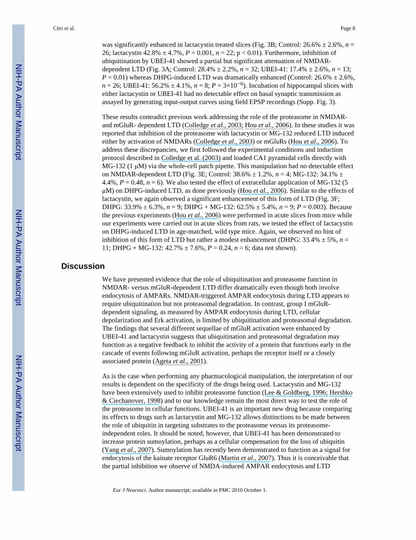
ResultsTo examine whether AMPAR endocytosis triggered by NMDAR or group I mGluRactivation requires proteasomal degradation, cultured neurons were pre-treated withlactacystin (5 μM) and surface GluR1-containing AMPARs were followed by live cellstaining (Bhattacharyya et al., 2009). Inhibition of the proteasome had no effect on NMDA-triggered endocytosis of dendritic AMPARs (Fig. 1A,B; control: 1.0 ± 0.08, n = 34; NMDA:1.7 ± 0.13, n = 17; P= 0.00008 [NMDA to control]; NMDA + lactacystin: 1.5 ± 0.13, n =21; P= 0.38 [NMDA + lactacystin to NMDA]). In contrast, AMPAR endocytosis triggeredby DHPG (3,5-Dihydroxyphenylglycine) was consistently enhanced in sister cultures treatedwith lactacystin (Fig. 1A,B; Control: 1.0 ± 0.08, n = 34; DHPG: 1.5 ± 0.11, n = 24; P=0.00078 [DHPG to control]; DHPG + lactacystin: 1.9 ± 0.14, n = 25); P= 0.035 [DHPG +lactacystin to DHPG]. Because the lack of effect of lactacystin on NMDA-triggeredAMPAR endocytosis contradicts previous work (Colledge et al., 2003), we repeated theseexperiments using another widely used, albeit less specific, (Lee & Goldberg, 1996)inhibitor of the proteasome, MG-132. Like lactacystin, pretreatment of cultures withMG-132 (5 μM) did not affect NMDA-induced AMPAR endocytosis (Fig. 1C,D; Control:1.0 ± 0.06, n = 52; NMDA: 1.9 ± 0.2, n = 28; P= 0.000002 [NMDA to control] NMDA +MG-132: 2.0 ± 0.21, n = 26), P= 0.9 [NMDA + MG-132 to NMDA] but consistentlyenhanced DHPG-induced AMPAR endocytosis (Fig. 1C,D; Control: 1.0 ± 0.06, n = 52;DHPG: 1.7 ± 0.18, n = 30; P= 0.00014 [DHPG to control]; DHPG + MG-132: 2.3 ± 0.18, n= 28; P= 0.029 [DHPG + MG-132 to DHPG]). We also repeated these experiments usingthe higher concentration of MG-132 (50 μM) that was used in previous work (Colledge etal., 2003) and again obtained the same results; no effect on NMDA-induced AMPARendocytosis and an enhancement of DHPG-induced AMPAR endocytosis (Supp. Fig. 1). Itis conceivable that differences in culture conditions could account for the discrepancybetween our results and those previously reported although our previous work on the role ofPSD-95 in LTD in slices (Xu et al., 2008) makes this possibility unlikely.
To address a potential role for protein ubiquitination in the different forms of AMPARendocytosis, we utilized UBEI-41/PYR-41, a novel cell-permeable compound thatirreversibly inhibits the E1 activating enzyme of the ubiquitin cascade by preventing E1-
Citri et al. Page 6
Eur J Neurosci. Author manuscript; available in PMC 2010 October 1.
NIH
-PA Author Manuscript
NIH
-PA Author Manuscript
NIH
-PA Author Manuscript

ubiquitin thioester bond formation (Yang et al., 2007). This is the first compound that hasthe potential of elucidating proteasome-dependent and independent roles of ubiquitin. At theconcentration used (50 μM), UBEI-41 has been demonstrated to inhibit E1-ubiquitinthioester bonds by 95% and robustly inhibit several ubiquitin-dependent intracellularprocesses (Yang et al., 2007; Dey et al., 2008; Gao et al., 2008; Zaarur et al., 2008;Satheshkumar et al., 2009). NMDA-induced endocytosis of dendritic AMPARs wassignificantly reduced, albeit not completely abolished, in the UBEI-41-treated cells (Fig.1E,F; Control: 1.0 ± 0.07, n = 31; NMDA: 2.1 ± 0.18, n = 28; P= 0.000001 [NMDA toControl], NMDA + UBEI-41: 1.3 ± 0.19, n = 25; P= 0.0078 [NMDA + UBEI-41 toNMDA]). In marked contrast, UBEI-41 treatment of the same set of cultures caused anenhancement of the dendritic AMPAR endocytosis triggered by application of the group ImGluR agonist DHPG (100 μM R,S) (Fig. 1E,F; Control: 1.0 ± 0.07, n = 31; DHPG: 1.9 ±0.17, n = 21; P= 0.0000047 [DHPG to Control]; DHPG + UBEI-41: 2.7 ± 0.27, n = 24; P=0.019 [DHPG + UBEI-41 to DHPG]). This marked difference in the effects of UBEI-41, aswell as the correlated enhancement of DHPG-induced AMPAR endocytosis by bothUBEI-41 and the proteasomal inhibitors suggest that independent mechanisms likelyunderlie the endocytosis of AMPARs following activation of NMDARs versus mGluRs.Surface expression of GluR1-containing AMPARs was not affected by any of the drugtreatments used (lactacystin, MG-132 or UBEI-41; Supp. Fig. 2).
The effects of UBEI-41 suggest that like the endocytosis of other membrane proteins (Hicke& Dunn, 2003; Urbe, 2005; Hanyaloglu & Von Zastrow, 2008) NMDAR-triggeredendocytosis of AMPARs may require mono-ubiquitination of either the receptorsthemselves or endocytic adaptors as a signal for the subsequent clathrin/dynamin-mediatedendocytosis. In contrast, the enhancement of DHPG-induced AMPAR endocytosis followinginhibition of ubiquitin activation or proteasomal function suggests not only the existence ofa distinct, ubiquitin-independent mechanism for AMPAR endocytosis but also that theubiquitin/proteasome system functions to limit a step in the cascade of events leading frommGluR activation to AMPAR endocytosis.
To address whether the ubiquitin/proteasome system functions as a feedback mechanism togenerally limit the consequences of group I mGluR activation, we examined the effects oflactacystin and UBEI-41 on two additional effects caused by DHPG; neuronal membranedepolarization (Gereau & Conn, 1995) and Erk/MAPK activation (Gallagher et al., 2004).The relative magnitude of the maintained depolarization induced by DHPG in CA1pyramidal cells (measured at 25–30 following DHPG application) was significantlyincreased by incubation of slices in either lactacystin or UBEI-41 when compared to controlcells (Fig. 2A,B; control cells, 0.31 ± 0.09, n = 10; lactacystin, 0.65 ± .12, P = 0.01, n = 7;UBEI-41, 0.62 ± 0.1, P = 0.02, n = 12). Similarly, the duration of DHPG-induced Erkphosphorylation in cultured neurons was clearly enhanced by these drugs (Fig. 2C–F).Statistical significance (p≤0.05) was observed for the 5,15,30 and 60 minute time points ofthe lactacystin treatment (p=0.01, 0.05, 0.04, and 0.03 respectively), as well as the 30 and 60minute time points of the UBEI-41 treated cells (p=0.01, 0.05 respectively). Thus, threedifferent effects of group I mGluR activation are all enhanced by inhibition of ubiquitinationand proteasome function, suggesting an important role for the ubiquitin/proteasome systemas a feedback mechanism that limits mGluR-triggered signaling.
The different effects of inhibiting proteasomal degradation and ubiquitination on NMDA-versus DHPG-induced endocytosis of AMPARs suggest that the drugs we have used willalso have different effects on NMDAR-versus mGluR-dependent LTD. Consistent with thisprediction, NMDAR-dependent LTD was not affected by inhibition of proteasomaldegradation by lactacystin (Fig. 3A; Control: 28.4% ± 2.2%, n = 32; lactacystin: 26.4% ±2.7%, P = 0.62, n = 17). In contrast, mGluR-dependent LTD induced by DHPG application
Citri et al. Page 7
Eur J Neurosci. Author manuscript; available in PMC 2010 October 1.
NIH
-PA Author Manuscript
NIH
-PA Author Manuscript
NIH
-PA Author Manuscript
was significantly enhanced in lactacystin treated slices (Fig. 3B; Control: 26.6% ± 2.6%, n =26; lactacystin 42.8% ± 4.7%, P = 0.001, n = 22; p < 0.01). Furthermore, inhibition ofubiquitination by UBEI-41 showed a partial but significant attenuation of NMDAR-dependent LTD (Fig. 3A; Control: 28.4% ± 2.2%, n = 32; UBEI-41: 17.4% ± 2.6%, n = 13;P = 0.01) whereas DHPG-induced LTD was dramatically enhanced (Control: 26.6% ± 2.6%,n = 26; UBEI-41: 56.2% ± 4.1%, n = 8; P = 3×10⁻⁶). Incubation of hippocampal slices witheither lactacystin or UBEI-41 had no detectable effect on basal synaptic transmission asassayed by generating input-output curves using field EPSP recordings (Supp. Fig. 3).
These results contradict previous work addressing the role of the proteasome in NMDAR-and mGluR- dependent LTD (Colledge et al., 2003; Hou et al., 2006). In these studies it wasreported that inhibition of the proteasome with lactacystin or MG-132 reduced LTD inducedeither by activation of NMDARs (Colledge et al., 2003) or mGluRs (Hou et al., 2006). Toaddress these discrepancies, we first followed the experimental conditions and inductionprotocol described in Colledge et al. (2003) and loaded CA1 pyramidal cells directly withMG-132 (1 μM) via the whole-cell patch pipette. This manipulation had no detectable effecton NMDAR-dependent LTD (Fig. 3E; Control: 38.6% ± 1.2%, n = 4; MG-132: 34.1% ±4.4%, P = 0.48, n = 6). We also tested the effect of extracellular application of MG-132 (5μM) on DHPG-induced LTD, as done previously (Hou et al., 2006). Similar to the effects oflactacystin, we again observed a significant enhancement of this form of LTD (Fig. 3F;DHPG: 33.9% ± 6.3%, n = 9; DHPG + MG-132: 62.5% ± 5.4%, n = 9; P = 0.003). Becausethe previous experiments (Hou et al., 2006) were performed in acute slices from mice whileour experiments were carried out in acute slices from rats, we tested the effect of lactacystinon DHPG-induced LTD in age-matched, wild type mice. Again, we observed no hint ofinhibition of this form of LTD but rather a modest enhancement (DHPG: 33.4% ± 5%, n =11; DHPG + MG-132: 42.7% ± 7.6%, P = 0.24, n = 6; data not shown).
DiscussionWe have presented evidence that the role of ubiquitination and proteasome function inNMDAR- versus mGluR-dependent LTD differ dramatically even though both involveendocytosis of AMPARs. NMDAR-triggered AMPAR endocytosis during LTD appears torequire ubiquitination but not proteasomal degradation. In contrast, group I mGluR-dependent signaling, as measured by AMPAR endocytosis during LTD, cellulardepolarization and Erk activation, is limited by ubiquitination and proteasomal degradation.The findings that several different sequellae of mGluR activation were enhanced byUBEI-41 and lactacystin suggests that ubiquitination and proteasomal degradation mayfunction as a negative feedback to inhibit the activity of a protein that functions early in thecascade of events following mGluR activation, perhaps the receptor itself or a closelyassociated protein (Ageta et al., 2001).
As is the case when performing any pharmacological manipulation, the interpretation of ourresults is dependent on the specificity of the drugs being used. Lactacystin and MG-132have been extensively used to inhibit proteasome function (Lee & Goldberg, 1996; Hershko& Ciechanover, 1998) and to our knowledge remain the most direct way to test the role ofthe proteasome in cellular functions. UBEI-41 is an important new drug because comparingits effects to drugs such as lactacystin and MG-132 allows distinctions to be made betweenthe role of ubiquitin in targeting substrates to the proteasome versus its proteasome-independent roles. It should be noted, however, that UBEI-41 has been demonstrated toincrease protein sumoylation, perhaps as a cellular compensation for the loss of ubiquitin(Yang et al., 2007). Sumoylation has recently been demonstrated to function as a signal forendocytosis of the kainate receptor GluR6 (Martin et al., 2007). Thus it is conceivable thatthe partial inhibition we observe of NMDA-induced AMPAR endocytosis and LTD
Citri et al. Page 8
Eur J Neurosci. Author manuscript; available in PMC 2010 October 1.
NIH
-PA Author Manuscript
NIH
-PA Author Manuscript
NIH
-PA Author Manuscript

following treatment with UBEI-41 is due to compensation by sumoylation. Nevertheless,under identical experimental conditions, we observed consistent yet opposing effects of thedrugs on NMDAR- versus mGluR-triggered AMPAR endocytosis and LTD. These resultsprovide strong evidence for significant differences in the role of the ubiquitin/proteasomesystem in the two forms of LTD investigated.
Our results also suggest that both ubiquitin-dependent and independent mechanisms can beused during the endocytosis of AMPARs and which route is used depends on the initialtriggering signal. This conclusion is similar to that drawn from prior results reporting thatthe endocytosis of AMPARs and their subsequent intracellular trafficking also differsdepending on whether NMDARs versus AMPARs are activated (Beattie et al., 2000; Ehlers,2000; Lee et al., 2004; Bhattacharyya et al., 2009). Furthermore, similar dependence on theinitial triggering event has been described for the ligand-induced endocytosis andsubsequent trafficking of many membrane proteins including G-protein coupled receptorsand growth factor receptors (Mukhopadhyay & Riezman, 2007).
Inappropriate activation of group I mGluR signaling has been suggested to be important inthe pathophysiology of cognitive disorders such as Alzheimer's disease and Fragile Xsyndrome, animal models of which demonstrate abnormal mGluR-dependent LTD (Hsieh etal., 2006; Dolen & Bear, 2008). Our results raise the possibility that impairment of theubiquitin/proteasome system, as observed in several other brain disorders (Ciechanover andBrundin, 2003; Bennett et al., 2007), could contribute to these disorders by reducingnegative feedback regulation of mGluR-signaling.
Supplementary MaterialRefer to Web version on PubMed Central for supplementary material.
AcknowledgmentsA.C. gratefully acknowledges support of The International Human Frontier Science Program Organization. Thiswork was supported by NIH grant MH63394 to R.C.M. The authors thank Malenka lab members for criticalreading of the manuscript.
Abbreviations
NMDA N-methyl- D-aspartate
mGluR metabotropic gluatamae receptor
LTD long term depression
AMPA α-amino-3-hydroxyl-5-methyl-4-isoxazole-propionate
DHPG 3,5-Dihydroxyphenylglycine
AP5 (2R)-amino-5-phosphonopentanoate
TTX tetrodotoxin
NBQX 2,3-dihydroxy-6-nitro-7-sulfamoyl-benzo[f]quinoxaline-2,3-dione
DNQX 6,7-Dinitroquinoxaline- 2,3-dione
Citri et al. Page 9
Eur J Neurosci. Author manuscript; available in PMC 2010 October 1.
NIH
-PA Author Manuscript
NIH
-PA Author Manuscript
NIH
-PA Author Manuscript

ReferencesAgeta H, Kato A, Hatakeyama S, Nakayama K, Isojima Y, Sugiyama H. Regulation of the level of
Vesl-1S/Homer-1a proteins by ubiquitin-proteasome proteolytic systems. J Biol Chem. 2001;276:15893–15897. [PubMed: 11278836]
Bashir ZI, Jane DE, Sunter DC, Watkins JC, Collingridge GL. Metabotropic glutamate receptorscontribute to the induction of long-term depression in the CA1 region of the hippocampus. Eur JPharmacol. 1993; 239:265–266. [PubMed: 8223907]
Bear M, Huber K, Warren S. The mGluR theory of fragile X mental retardation. Trends inNeurosciences. 2004; 27:370–377. [PubMed: 15219735]
Beattie EC, Carroll RC, Yu X, Morishita W, Yasuda H, von Zastrow M, Malenka RC. Regulation ofAMPA receptor endocytosis by a signaling mechanism shared with LTD. Nat Neurosci. 2000;3:1291–1300. [PubMed: 11100150]
Bellone C, Luscher C, Mameli M. Mechanisms of synaptic depression triggered by metabotropicglutamate receptors. Cell Mol Life Sci. 2008; 65:2913–2923. [PubMed: 18712277]
Bhattacharyya S, Biou V, Xu W, Schluter O, Malenka RC. A critical role for PSD-95/AKAPinteractions in endocytosis of synaptic AMPA receptors. Nat Neurosci. 2009; 12:172–181.[PubMed: 19169250]
Bingol B, Schuman EM. A proteasome-sensitive connection between PSD-95 and GluR1 endocytosis.Neuropharmacology. 2004; 47:755–763. [PubMed: 15458847]
Bolshakov VY, Siegelbaum SA. Postsynaptic induction and presynaptic expression of hippocampallong-term depression. Science. 1994; 264:1148–1152. [PubMed: 7909958]
Carroll RC, Beattie EC, von Zastrow M, Malenka RC. Role of AMPA receptor endocytosis in synapticplasticity. Nat Rev Neurosci. 2001; 2:315–324. [PubMed: 11331915]
Chen ZJ, Sun LJ. Nonproteolytic functions of ubiquitin in cell signaling. Molecular Cell. 2009;33:275–286. [PubMed: 19217402]
Citri A, Malenka RC. Synaptic plasticity: multiple forms, functions, and mechanisms.Neuropsychopharmacology. 2008; 33:18–41. [PubMed: 17728696]
Colledge M, Snyder E, Crozier RA, Soderling JA, Jin Y, Langeberg LK, Lu H, Bear M, Scott JD.Ubiquitination regulates PSD-95 degradation and AMPA receptor surface expression. Neuron.2003; 40:595–607. [PubMed: 14642282]
Conn PJ. Physiological roles and therapeutic potential of metabotropic glutamate receptors. Ann N YAcad Sci. 2003; 1003:12–21. [PubMed: 14684432]
Dey A, Tergaonkar V, Lane DP. Double-edged swords as cancer therapeutics: simultaneouslytargeting p53 and NF-kappaB pathways. Nat Rev Drug Discov. 2008; 7:1031–1040. [PubMed:19043452]
Dolen G, Bear MF. Role for metabotropic glutamate receptor 5 (mGluR5) in the pathogenesis offragile X syndrome. J Physiol. 2008; 586:1503–1508. [PubMed: 18202092]
Dudek SM, Bear MF. Homosynaptic long-term depression in area CA1 of hippocampus and effects ofN-methyl-D-aspartate receptor blockade. Proc Natl Acad Sci USA. 1992; 89:4363–4367.[PubMed: 1350090]
Ehlers MD. Reinsertion or degradation of AMPA receptors determined by activity-dependentendocytic sorting. Neuron. 2000; 28:511–525. [PubMed: 11144360]
Ehlers MD. Activity level controls postsynaptic composition and signaling via the ubiquitin-proteasome system. Nat Neurosci. 2003; 6:231–242. [PubMed: 12577062]
Gallagher SM, Daly CA, Bear MF, Huber KM. Extracellular signal-regulated protein kinase activationis required for metabotropic glutamate receptor-dependent long-term depression in hippocampalarea CA1. J Neurosci. 2004; 24:4859–4864. [PubMed: 15152046]
Gao Y, Chotoo CK, Balut CM, Sun F, Bailey MA, Devor DC. Role of S3 and S4 transmembranedomain charged amino acids in channel biogenesis and gating of KCa2.3 and KCa3.1. J BiolChem. 2008; 283:9049–9059. [PubMed: 18227067]
Gereau, R.W.t.; Conn, PJ. Roles of specific metabotropic glutamate receptor subtypes in regulation ofhippocampal CA1 pyramidal cell excitability. J Neurophysiol. 1995; 74:122–129. [PubMed:7472317]
Citri et al. Page 10
Eur J Neurosci. Author manuscript; available in PMC 2010 October 1.
NIH
-PA Author Manuscript
NIH
-PA Author Manuscript
NIH
-PA Author Manuscript

Hanyaloglu AC, von Zastrow M. Regulation of GPCRs by endocytic membrane trafficking and itspotential implications. Annu Rev Pharmacol Toxicol. 2008; 48:537–568. [PubMed: 18184106]
Hershko A, Ciechanover A. The ubiquitin system. Annu Rev Biochem. 1998; 67:425–479. [PubMed:9759494]
Hicke L, Dunn R. Regulation of membrane protein transport by ubiquitin and ubiquitin-bindingproteins. Annu Rev Cell Dev Biol. 2003; 19:141–172. [PubMed: 14570567]
Hou L, Antion MD, Hu D, Spencer CM, Paylor R, Klann E. Dynamic translational and proteasomalregulation of fragile X mental retardation protein controls mGluR-dependent long-termdepression. Neuron. 2006; 51:441–454. [PubMed: 16908410]
Hsieh H, Boehm J, Sato C, Iwatsubo T, Tomita T, Sisodia S, Malinow R. AMPAR removal underliesAbeta-induced synaptic depression and dendritic spine loss. Neuron. 2006; 52:831–843. [PubMed:17145504]
Huber KM, Gallagher SM, Warren ST, Bear MF. Altered synaptic plasticity in a mouse model offragile X mental retardation. Proc Natl Acad Sci USA. 2002; 99:7746–7750. [PubMed: 12032354]
Lee DH, Goldberg AL. Selective inhibitors of the proteasome-dependent and vacuolar pathways ofprotein degradation in Saccharomyces cerevisiae. J Biol Chem. 1996; 271:27280–27284.[PubMed: 8910302]
Lee HK, Barbarosie M, Kameyama K, Bear MF, Huganir RL. Regulation of distinct AMPA receptorphosphorylation sites during bidirectional synaptic plasticity. Nature. 2000; 405:955–959.[PubMed: 10879537]
Lee SH, Simonetta A, Sheng M. Subunit rules governing the sorting of internalized AMPA receptorsin hippocampal neurons. Neuron. 2004; 43:221–236. [PubMed: 15260958]
Martin S, Nishimune A, Mellor JR, Henley JM. SUMOylation regulates kainate-receptor-mediatedsynaptic transmission. Nature. 2007; 447:321–325. [PubMed: 17486098]
Mukhopadhyay D, Riezman H. Proteasome-independent functions of ubiquitin in endocytosis andsignaling. Science. 2007; 315:201–205. [PubMed: 17218518]
Mulkey RM, Malenka RC. Mechanisms underlying induction of homosynaptic long-term depressionin area CA1 of the hippocampus. Neuron. 1992; 9:967–975. [PubMed: 1419003]
Oliet SH, Malenka RC, Nicoll RA. Two distinct forms of long-term depression coexist in CA1hippocampal pyramidal cells. Neuron. 1997; 18:969–982. [PubMed: 9208864]
Satheshkumar PS, Anton LC, Sanz P, Moss B. Inhibition of the ubiquitin-proteasome system preventsvaccinia virus DNA replication and expression of intermediate and late genes. J Virol. 2009;83:2469–2479. [PubMed: 19129442]
Shepherd JD, Huganir RL. The cell biology of synaptic plasticity: AMPA receptor trafficking. AnnuRev Cell Dev Biol. 2007; 23:613–643. [PubMed: 17506699]
Snyder EM, Philpot BD, Huber KM, Dong X, Fallon JR, Bear MF. Internalization of ionotropicglutamate receptors in response to mGluR activation. Nat Neurosci. 2001; 4:1079–1085. [PubMed:11687813]
Urbe S. Ubiquitin and endocytic protein sorting. Essays Biochem. 2005; 41:81–98. [PubMed:16250899]
Xiao MY, Zhou Q, Nicoll RA. Metabotropic glutamate receptor activation causes a rapid redistributionof AMPA receptors. Neuropharmacology. 2001; 41:664–671. [PubMed: 11640920]
Xu W, Schluter OM, Steiner P, Czervionke BL, Sabatini B, Malenka RC. Molecular dissociation ofthe role of PSD-95 in regulating synaptic strength and LTD. Neuron. 2008; 57:248–262. [PubMed:18215622]
Yang Y, Kitagaki J, Dai RM, Tsai YC, Lorick KL, Ludwig RL, Pierre SA, Jensen JP, Davydov IV,Oberoi P, Li CC, Kenten JH, Beutler JA, Vousden KH, Weissman AM. Inhibitors of ubiquitin-activating enzyme (E1), a new class of potential cancer therapeutics. Cancer Res. 2007; 67:9472–9481. [PubMed: 17909057]
Zaarur N, Meriin AB, Gabai VL, Sherman MY. Triggering aggresome formation. Dissectingaggresome-targeting and aggregation signals in synphilin 1. J Biol Chem. 2008; 283:27575–27584. [PubMed: 18635553]
Citri et al. Page 11
Eur J Neurosci. Author manuscript; available in PMC 2010 October 1.
NIH
-PA Author Manuscript
NIH
-PA Author Manuscript
NIH
-PA Author Manuscript
FIG. 1.The ubiquitin-proteasome system differentially regulates NMDA- and DHPG- inducedAMPAR endocytosis. Representative examples (A, C, E) and quantification (B, D, F)demonstrating effects of lactacystin (5 μM, A, B), UBEI-41 (50 μM, C, D) and MG-132 (5μM, E, F) on NMDA- versus DHPG-induced AMPAR endocytosis. Scale bar: 20 μm. ***indicates p < 0.001, * indicates p < 0.05 and n.s indicates p > 0.05.
Citri et al. Page 12
Eur J Neurosci. Author manuscript; available in PMC 2010 October 1.
NIH
-PA Author Manuscript
NIH
-PA Author Manuscript
NIH
-PA Author Manuscript

FIG. 2.Inhibition of ubiquitination and proteasomal degradation prolongs DHPG-inducedmembrane depolarization and Erk activation. Representative experiments (A) and summaryof the effects of lactacystin (5 μM) and UBEI-41 (50 μM) on the depolarization caused byapplication of DHPG (3 min, 100 μM) to CA1 pyramidal neurons. (C–D) Representativewestern blots showing effects of lactacystin and UBEI-41 on the increase in thephosphorylated form of Erk (pErk) in cultured neurons treated with DHPG (5 min, 100 μM)at time=0. (E–F) Summary graphs of the effect of lactacystin (n=5) and UBEI-41 (n=3) onErk activation induced by DHPG. pErk levels were normalized to the intensity of thecorresponding Erk2 band, and presented as % of the value at t=0 min (* indicates p < 0.05).
Citri et al. Page 13
Eur J Neurosci. Author manuscript; available in PMC 2010 October 1.
NIH
-PA Author Manuscript
NIH
-PA Author Manuscript
NIH
-PA Author Manuscript

FIG. 3.The ubiquitin-proteasome system differentially regulates NMDAR- and mGluR-dependentLTD. (A–D) Summary graphs of field EPSP experiments in which NMDAR-dependentLTD (induced by 5 Hz, 3 min stimulation) and mGluR-dependent LTD (induced by 100 μMDHPG applied for 10 minutes) were induced in control slices or slices preincubated inlactacystin (1–5 μM) or UBEI-41 (50 μM). (E) Summary graph of whole-cell voltage clampexperiments in which NMDAR-dependent LTD was induced in control cells or cells loadedwith MG-132 (1 μM). (F) Summary graph of experiments of mGluR-dependent LTDinduced in control slices or slices perfused with MG-132 (5 μM). Numbers in parenthesesindicate number of slices (A–D, F) or cells (E). Representative traces are shown to the rightof each experimental summary graph (scale bars: vertical = 100 μV or 20 pA, horizontal = 5ms).
Citri et al. Page 14
Eur J Neurosci. Author manuscript; available in PMC 2010 October 1.
NIH
-PA Author Manuscript
NIH
-PA Author Manuscript
NIH
-PA Author Manuscript
![In vivo positron emission tomography imaging with [ 11 C]ABP688: binding variability and specificity for the metabotropic glutamate receptor subtype 5 in baboons](https://static.fdokumen.com/doc/165x107/6316b26ad18b031ae106426d/in-vivo-positron-emission-tomography-imaging-with-11-cabp688-binding-variability.jpg)