Adenosine A2A receptors and metabotropic glutamate 5 receptors are co-localized and functionally...
13
Adenosine A 2A receptors and metabotropic glutamate 5 receptors are co-localized and functionally interact in the hippocampus: a possible key mechanism in the modulation of N-methyl-D-aspartate effects M. T. Tebano,* A. Martire,* N. Rebola, R. Pepponi,* M. R. Domenici,* M. C. Gro `,* M. A. Schwarzschild,à J. F. Chen,§ R. A. Cunha and P. Popoli* *Department of Drug Research and Evaluation, Istituto Superiore di Sanita `, Rome, Italy Center for Neuroscience of Coimbra, Institute of Biochemistry, Faculty of Medicine, University of Coimbra, Coimbra, Portugal àMassGeneral Institute for Neurodegenerative Disease, Boston, Massachusetts, USA §Molecular Neuropharmacology Laboratory, Department of Neurology, Boston University School of Medicine, Boston, Massachusetts, USA Abstract Hippocampal metabotropic glutamate 5 receptors (mGlu5Rs) regulate both physiological and pathological responses to glutamate. Because mGlu5R activation enhances NMDA- mediated effects, and given the role played by NMDA recep- tors in synaptic plasticity and excitotoxicity, modulating mGlu5R may influence both the physiological and the patho- logical effects elicited by NMDA receptor stimulation. We evaluated whether adenosine A 2A receptors (A 2A Rs) modu- lated mGlu5R-dependent effects in the hippocampus, as they do in the striatum. Co-application of the A 2A R agonist CGS 21680 with the mGlu5R agonist (RS)-2-chloro-s-hydr- oxyphenylglycine(CHPG) synergistically reduced field excita- tory postsynaptic potentials in the CA1 area of rat hippocampal slices. Endogenous tone at A 2A Rs seemed to be required to enable mGlu5R-mediated effects, as the ability of CHPG to potentiate NMDA effects was antagonized by the selective A 2A R antagonist ZM 241385 in rat hippocampal sli- ces and cultured hippocampal neurons, and abolished in the hippocampus of A 2A R knockout mice. Evidence for the inter- action between A 2A Rs and mGlu5Rs was further strengthened by demonstrating their co-localization in hippocampal synap- ses. This is the first evidence showing that hippocampal A 2A Rs and mGlu5Rs are co-located and act synergistically, and that A 2A Rs play a permissive role in mGlu5R receptor- mediated potentiation of NMDA effects in the hippocampus. Keywords: adenosine A 2A receptors, hippocampus, metabo- tropic glutamate 5 receptors, N-methyl-D-aspartate. J. Neurochem. (2005) 95, 1188–1200. In the hippocampus, group I metabotropic glutamate receptors (mGluRs, which include the mGlu1R and mGlu5R subtypes) regulate synaptic plasticity and spatial learning (reviewed in Anwyl 1999; Balschun et al. 1999; Bortolotto et al. 1999; Balschun and Wetzel 2002), contribute to the generation of epileptiform activity (Sacaan and Schoepp 1992), and modu- late excitotoxic processes (Attucci et al. 2002). Because mGlu5R is the most abundant group I mGluR in the hippocampus (Romano et al. 1995; Lujan et al. 1996, 1997; Shigemoto et al. 1997), this subtype is thought to play a predominant role. One of the most noteworthy effects of mGlu5R activation is an enhancement of NMDA-mediated effects (reviewed in Anwyl 1999; Bortolotto et al. 1999) and, Received March 23, 2005; revised manuscript received July 15, 2005; accepted July 18, 2005. Address correspondence and reprint requests to P. Popoli, Department of Drug Research and Evaluation, Istituto Superiore di Sanita `, Viale Regina Elena 299, 00161 Rome, Italy. E-mail: [email protected] Abbreviations used: ACSF, artificial cerebrospinal fluid; A 2A R, adenosine A 2A receptor; cAMP, cyclic AMP; CHPG, (RS)-2-chloro-s- hydroxyphenylglycine; DARPP-32, dopamine- and cAMP-regulated phosphoproten of mr 32,000; DHPG, RS3,5-dihydroxyphenylglycine; fEPSP, field excitatory postsynaptic potential; KO, knockout; LDH, lactate dehydrogenase; mGluR, metabotrophic glutamate receptor; MPEP, 2- methyl-6-(phenylethyny) pyridine hydrochloride; PAGE, polyacrylamide gel electrophoresis; PBS, phosphate-buffered saline; PKA, protein kinase A; PMSF, phenylmethylsulfonyl fluoride; PPS, paired-pulse stimulation; PSD, postsynaptic density; SDS, sodium dodecyl sulfate; TBST, Tris- buffered saline with Tween 20; vGluT, vesicular glutamate transporter. Journal of Neurochemistry , 2005, 95, 1188–1200 doi:10.1111/j.1471-4159.2005.03455.x 1188 Ó 2005 International Society for Neurochemistry, J. Neurochem. (2005) 95, 1188–1200
Transcript of Adenosine A2A receptors and metabotropic glutamate 5 receptors are co-localized and functionally...

Adenosine A2A receptors and metabotropic glutamate 5 receptorsare co-localized and functionally interact in the hippocampus: apossible key mechanism in the modulation of N-methyl-D-aspartateeffects
M. T. Tebano,* A. Martire,* N. Rebola,� R. Pepponi,* M. R. Domenici,* M. C. Gro,*M. A. Schwarzschild,� J. F. Chen,§ R. A. Cunha� and P. Popoli*
*Department of Drug Research and Evaluation, Istituto Superiore di Sanita, Rome, Italy
�Center for Neuroscience of Coimbra, Institute of Biochemistry, Faculty of Medicine, University of Coimbra, Coimbra, Portugal
�MassGeneral Institute for Neurodegenerative Disease, Boston, Massachusetts, USA
§Molecular Neuropharmacology Laboratory, Department of Neurology, Boston University School of Medicine, Boston,
Massachusetts, USA
Abstract
Hippocampal metabotropic glutamate 5 receptors (mGlu5Rs)
regulate both physiological and pathological responses to
glutamate. Because mGlu5R activation enhances NMDA-
mediated effects, and given the role played by NMDA recep-
tors in synaptic plasticity and excitotoxicity, modulating
mGlu5R may influence both the physiological and the patho-
logical effects elicited by NMDA receptor stimulation. We
evaluated whether adenosine A2A receptors (A2ARs) modu-
lated mGlu5R-dependent effects in the hippocampus, as they
do in the striatum. Co-application of the A2AR agonist
CGS 21680 with the mGlu5R agonist (RS)-2-chloro-s-hydr-
oxyphenylglycine(CHPG) synergistically reduced field excita-
tory postsynaptic potentials in the CA1 area of rat
hippocampal slices. Endogenous tone at A2ARs seemed to be
required to enable mGlu5R-mediated effects, as the ability of
CHPG to potentiate NMDA effects was antagonized by the
selective A2AR antagonist ZM 241385 in rat hippocampal sli-
ces and cultured hippocampal neurons, and abolished in the
hippocampus of A2AR knockout mice. Evidence for the inter-
action between A2ARs and mGlu5Rs was further strengthened
by demonstrating their co-localization in hippocampal synap-
ses. This is the first evidence showing that hippocampal
A2ARs and mGlu5Rs are co-located and act synergistically,
and that A2ARs play a permissive role in mGlu5R receptor-
mediated potentiation of NMDA effects in the hippocampus.
Keywords: adenosine A2A receptors, hippocampus, metabo-
tropic glutamate 5 receptors, N-methyl-D-aspartate.
J. Neurochem. (2005) 95, 1188–1200.
In the hippocampus, group I metabotropic glutamate receptors(mGluRs, which include the mGlu1R and mGlu5R subtypes)regulate synaptic plasticity and spatial learning (reviewed inAnwyl 1999; Balschun et al. 1999; Bortolotto et al. 1999;Balschun and Wetzel 2002), contribute to the generation ofepileptiform activity (Sacaan and Schoepp 1992), and modu-late excitotoxic processes (Attucci et al. 2002). BecausemGlu5R is the most abundant group I mGluR in thehippocampus (Romano et al. 1995; Lujan et al. 1996, 1997;Shigemoto et al. 1997), this subtype is thought to play apredominant role. One of the most noteworthy effects ofmGlu5R activation is an enhancement of NMDA-mediatedeffects (reviewed in Anwyl 1999; Bortolotto et al. 1999) and,
Received March 23, 2005; revised manuscript received July 15, 2005;accepted July 18, 2005.Address correspondence and reprint requests to P. Popoli, Department
of Drug Research and Evaluation, Istituto Superiore di Sanita, VialeRegina Elena 299, 00161 Rome, Italy. E-mail: [email protected] used: ACSF, artificial cerebrospinal fluid; A2AR,
adenosine A2A receptor; cAMP, cyclic AMP; CHPG, (RS)-2-chloro-s-hydroxyphenylglycine; DARPP-32, dopamine- and cAMP-regulatedphosphoproten of mr 32,000; DHPG, RS3,5-dihydroxyphenylglycine;fEPSP, field excitatory postsynaptic potential; KO, knockout; LDH, lactatedehydrogenase; mGluR, metabotrophic glutamate receptor; MPEP, 2-methyl-6-(phenylethyny) pyridine hydrochloride; PAGE, polyacrylamidegel electrophoresis; PBS, phosphate-buffered saline; PKA, protein kinaseA; PMSF, phenylmethylsulfonyl fluoride; PPS, paired-pulse stimulation;PSD, postsynaptic density; SDS, sodium dodecyl sulfate; TBST, Tris-buffered saline with Tween 20; vGluT, vesicular glutamate transporter.
Journal of Neurochemistry, 2005, 95, 1188–1200 doi:10.1111/j.1471-4159.2005.03455.x
1188 � 2005 International Society for Neurochemistry, J. Neurochem. (2005) 95, 1188–1200

in general, it is well accepted that mGlu5Rs ‘set the tone’ ofNMDA receptor-mediated neurotransmission (Alagarsamyet al. 1999). Given the key role of NMDA receptors in bothsynaptic plasticity (Collingridge and Bliss 1995) and excito-toxicity (Rothman andOlney 1995), modulatingmGlu5Rmayhelp regulate both the physiological and the pathologicaleffects elicited by NMDA receptor stimulation in the hippo-campus. Indeed, hippocampal long-term potentiation isblocked in rats by the selective mGlu5R antagonist2-methyl-6-(phenylethyny) pyridine hydrochloride (MPEP)(e.g. Balschun and Wetzel 2002), and reduced in mice lackingmGlu5Rs (Lu et al. 1997). In another brain region, thestriatum, mGlu5Rs are under the tight control of adenosineA2A receptors (A2ARs). Indeed, A2ARs and mGlu5Rs havebeen reported to interact in models of Parkinson disease(Popoli et al. 2001; Coccurello et al. 2004), and to synergis-tically modulate GABA (Diaz-Cabiale et al. 2002) andglutamate release (Rodrigues et al. 2005). Furthermore, theexistence of A2AR–mGlu5R heteromeric complexes in striatalmembranes and a synergistic interaction between the tworeceptors in inducing c-fos expression has been reported (Ferreet al. 2002; Fuxe et al. 2003). Very recently, it has been shownthat the state of activation of striatal A2ARs influences somemGlu5R-dependent effects, such as dopamine-and cAMP-regulated phosphoproten of Mr 32,000 (DARPP-32) phos-phorylation (Nishi et al. 2003) and NMDA potentiation(Domenici et al. 2004).
Although A2ARs are most abundant in the striatum, theyare also present in the hippocampus, where they modulatesynaptic transmission and excitotoxicity (reviewed in Cunha2005). However, unlike the striatum, nothing is known aboutthe possible co-localization of A2ARs and mGlu5Rs in thehippocampus, nor whether A2ARs can modulate hippocam-pal mGlu5Rs, thus influencing NMDA-mediated responses.
The primary aim of the present study was to explore thehypothesis that A2ARs can regulate mGlu5R-mediatedeffects in the hippocampus. Having found a clear functionalA2AR–mGlu5R interaction in electrophysiological and cellculture experiments, we then confirmed the hypothesis thathippocampal A2ARs exert a permissive role on mGlu5R-mediated effects (namely the potentiation of NMDAresponses) by using mice lacking A2ARs. The interactionbetween A2ARs and mGlu5Rs was further strengthened bythe finding of their co-localization in hippocampal synapses.
Materials and Methods
Animals
Male and pregnant female Wistar rats (2–3 months old) obtained
either from Harlan-Nossan (Udine, Italy) or from Charles River
(Barcelona, Spain), and A2AR knockout (KO) and wild-type (WT)
mice (2–4 months old, see below) were used. The animals were kept
under standardized temperature, humidity and lighting conditions,
and had free access to water and food. All animal procedures were
carried out according to the European Community Guidelines for
Animal Care, DL 116/92, application of the European Communities
Council Directive (86/609/EEC).
Preparation and maintenance of slices
The animals were decapitated under ether anesthesia, and brains were
quickly removed from the skull. Transverse hippocampal slices
(400 lm thick) were cut with a McIlwain tissue chopper. Slices were
maintained at room temperature (22–25�C) in artificial cerebrospinalfluid (ACSF) containing 126 mM NaCl, 3.5 mM KCl, 1.2 mM
NaH2PO4, 1.2 mM MgCl2, 2 mM CaCl2, 25 mM NaHCO3 and
11 mM glucose (pH 7.3) saturated with 95% O2 and 5% CO2. After
incubation in ACSF for at least 1 h, a single slice was transferred to a
submerged recording chamber and continuously superfused at 32–
33�C with ACSF at rate of 2.7–3 mL/min. The drugs were added to
this superfusion solution. Extracellular field excitatory postsynaptic
potentials (fEPSPs) were recorded through a glass microelectrode
filled with 2 M NaCl (pipette resistance 2–5 MW) placed in the
stratum radiatum of the CA1 area. A bipolar twisted NiCr-insulated
electrode (50 lm outer diameter), placed in the stratum radiatum,
was used to stimulate the Schaffer collaterals. Stimulation was
delivered every 20 s (square pulses of 100-ls duration at a frequencyof 0.05 Hz) and every three consecutive responses were averaged.
Signals were acquired with a DAM-80 AC differential amplifier
(WPI Instruments, Woltham, MA, USA) and analyzed with the LTP
program (Anderson and Collingridge 2001). At least 10 min of stable
baseline recording preceded drug application. To allow comparisons
between experiments, in each experiment the slope values were
normalized, taking the average of the values obtained over the 10-
min period immediately before applying the test compound as 100%.
The washout period lasted at least 30 min.
In order to establish a possible involvement of presynaptic
mechanisms in the effects of A2AR and mGlu5R agonists, a series of
experiments was performed under a protocol of paired-pulse
stimulation (PPS), in which the Schaffer fibers were stimulated
twice with an interpulse interval of 50 ms. Under control conditions,
such a protocol normally elicits a condition of paired-pulse
facilitation, in which the response elicited by the second stimulus
(R2) is greater than that elicited by the first stimulus (R1). The
degree of paired-pulse facilitation is quantified by the R2/R1 ratio
and a modification of this ratio is an indication of a presynaptic
action on neurotransmitter release (Schulz et al. 1994). Data were
expressed as mean ± SEM from N slices.
Lactate dehydrogenase (LDH) release from cultured
hippocampal neurons
Neurons were isolated from hippocampi dissected from E17 Wistar
rat embryos. Pregnant rats were anesthetized with ether, decapitated
and the fetuses were collected and rapidly decapitated. After
removal of the meninges, the hippocampi were collected in Hank’s
balanced salt solution (HBSS) and dissociated. Hippocampal cells
were then washed in HBSS and resuspended in Neurobasal medium
supplemented with 0.5 mM L-glutamine, 2% B-27 supplement and
gentamicin (50 lg/mL) (referred as complete medium). Aliquots of
2–3 · 105 cells were placed in 24-well culture plates coated with
poly-L-lysine (5 lg/mL) and maintained at 37�C in humidified air
with 5% CO2.
A2AR–mGlu5R interaction in the hippocampus 1189
� 2005 International Society for Neurochemistry, J. Neurochem. (2005) 95, 1188–1200

Every 4 days, 0.5 mL medium was removed and replaced by
the same volume of fresh complete medium. Assays were done on
12–14-day-old cultures. At the time of the experiment, culture
medium was removed and substituted with an appropriate
volume of Neurobasal medium supplemented with gentamicin
(50 lg/mL). Cultured cells were then exposed to NMDA (300 lM)and/or CHPG (500–1000 lM) for 60 min. ZM 241385 (30 nM) or
MPEP (30 lM) were applied 15 min before and then co-applied
with NMDA and/or CHPG. Following exposure to the drugs, the
culture medium was removed and replaced with fresh complete
medium. Cultures were then returned to the incubator and cellular
damage was evaluated 24 h later by measuring the amount of LDH
released into the medium using a cytotoxicity detection kit (Roche
Diagnostic, Indianapolis, IN, USA). Results are expressed as a
percentage of control (100%), and represent mean ± SEM values
of 3–4 independent experiments, assayed in triplicate.
Experiments in A2AR KO mice
A2AR KO mice were generated as described previously (Chen
et al. 1999) to produce the near congenic (N6) line used here.
A2AR KO mice (A2AR–/–) and their WT littermates (A2AR+/+)were generated by cross-breeding heterozygous A2AR mice
(A2AR+/–). They were genotyped on the basis of PCR analysis
performed on DNA isolated from tail samples. PCR amplification
products were routinely fractionated through 2% agarose gels and
stained with ethidium bromide. Photographs of the gels were
made by using incident ultraviolet light to record the results of
electrophoresis. In each experiment, age- and sex-matched A2ARKO mice and WT littermates were used.
The experimental procedure described for rats was used for
electrophysiological recordings in mice hippocampal slices.
To verify whether changes in mGlu5R density occurred in the
hippocampus of A2ARKO versus WTmice, western blot experiments
were performed. Proteins for mGlu5R analysis were extracted from
previously frozen hippocampal tissues by homogenization in cold
buffer containing 0.32 M sucrose, 10 mM HEPES, 0.1 mM EGTA,
0.1 mM EDTA, 0.1 mM phenylmethylsulfonyl fluoride (PMSF),
10 lg/mL leupeptin and 10 lg/mL aprotinin (pH 7.4). Homogenates
were centrifuged at 500 g for 20 min and the resulting supernatant
was centrifuged at 20 000 g. Pellets were resuspended in ice-cold
25 mM Tris-HCl buffer (pH 7.4) containing 1 mM PMSF, and an
aliquot was used for protein determinations (Bio-Rad DC protein
assay; Bio-Rad, Hercules, CA, USA). Samples were resuspended in
Laemmli sample buffer, boiled for 5 min at 90�C and resolved by
sodium dodecyl sulfate (SDS)–polyacrylamide gel electrophoresis
(PAGE) using 10% gels (Laemmli 1970). For western blot analysis,
proteins (5, 10 or 20 lg) were separated by SDS–PAGE (10% gels)
and transferred overnight to nitrocellulose paper (Shleicher & Schuell
BioScience, Dassel, Germany) at 35 V. Blots werewashedwith TBST
buffer (20 mM Tris, pH 7.5, 0.05% Tween 20, 150 mM NaCl) and
blocked with TBST containing 1% bovine serum albumin for 1 h.
Blots were then incubated at room temperature with the primary
antibody, a rabbit anti-mGlu5R antibody (1 : 1000 dilution; Upstate
Biotechnology, Lake Placid, NY, USA) for 1 h. After extensive
washes in TBST, the immunoreactive bands were detected by
incubation with alkaline phosphatase-conjugated secondary antibod-
ies (Promega Corporation, Madison, WI, USA) and revealed by
Western blue substrate for alkaline phosphatase (Promega).
Subcellular distribution of A2AR and mGlu5R immunoreactivity
in cultured hippocampal neurons
Immunocytochemistry in the coverslip-mounted neurons was carried
out as described previously (Rebola et al. 2005). Briefly, the cultureswere washed twice with 1 mL phosphate-buffered saline (PBS;
140 mM NaCl, 3 mM KCl, 20 mM Na2HPO4, 1.5 mM KH2PO4) kept
at 37�C during 10 min, and fixed with 4% paraformaldehyde with 4%
sucrose for 30 min at 37�C. Coverslip-mounted cells were then
permeabilized with 0.2% Triton X-100 for 2 min at room temperature
(22–25�C) and non-specific binding subsequently blocked with 3%
bovine serum albumin for 30 min at room temperature. Cells then
incubated for 1 h at room temperature with mouse anti-A2AR (1 : 500
dilution; Upstate Biotechnology), rabbit anti-mGlu5R receptor
antibody (1 : 1000 dilution; Upstate Biotechnology), rabbit or mouse
anti-synaptophysin antibody (1 : 200 dilution; Sigma, Sintra, Portu-
gal) and guinea pig anti-vesicular glutamate transporter (vGluT) type
1 (1 : 5000; Chemicon, Temecula, CA, USA) and guinea pig vGluT2
(1 : 5000; Chemicon), and then washed three times with 200 lL PBS
for 5 min. Incubation with secondary antibody, an AlexaFluor-598
(green)-labeled goat anti-guinea pig IgG antibody (1 : 200 dilution;
Amersham, Little Chalfont, UK), AlexaFluor-598 (red)-labeled goat
anti-mouse IgG antibody (1 : 200 dilution; Amersham), AlexaFluor-
488 (green)-labeled goat anti-rabbit IgG antibody (1 : 200 dilution;
Amersham) or AlexaFluor-488 (green)-labeled goat anti-mouse IgG
antibody (1 : 200 dilution; Amersham), was conducted for 1 h at
room temperature. We confirmed that none of the secondary
antibodies produced any signal in preparations from which the
corresponding primary antibody was omitted. After three washing
periods of 5 min with 200 lL PBS, the cells were mounted using a
ProlongAntifade kit (Amersham) and, after drying, were visualized in
a Zeiss Axiovert 200 fluorescence microscope (Obertcochen,
Germany) equipped with a cooled camera (Coolsnap Photometrics,
Tucson, AZ, USA) or with a Bio-Rad 600 confocal microscope
(Hercules, CA, USA) and analyzed with MetaFluor 4.0 software
(Molecular Devices, Downingtown, PA, USA).
Subsynaptic distribution of A2ARs and mGlu5Rs in rat
hippocampus
Separation of the presynaptic active zone, postsynaptic density (PSD)
and non-synaptic fractions from hippocampal nerve terminals was
carried out as initially described by Phillips et al. (2001) with minor
modifications (see Rebola et al. 2003). Briefly, the hippocampi from
eight male Wistar rats were homogenized at 4�C with a Teflon–glass
homogenizer in 15 mL isolation solution (0.32 M sucrose, 0.1 mM
CaCl2, 1 mM MgCl2, 0.1 mM PMSF). The concentration of sucrose
was raised to 1.25 M by addition of 75 mL 2 M sucrose and 30 mL
0.1 mM CaCl2, and the suspension was divided into 10 ultracentri-
fuge tubes. The homogenate was overlaid with 8 mL of a solution
containing 1.0 M sucrose and 0.1 mM CaCl2, and with 5 mL
homogenization solution and centrifuged at 100 000 g for 3 h at
4�C. Synaptosomes were collected at the 1.25/1.0 M sucrose
interface, diluted 1 : 10 in cold 0.32 M sucrose containing 0.1 mM
CaCl2 and pelleted by centrifugation at 15 000 g for 30 min at 4�C.The pellets were resuspended in 1 mL 0.32 M sucrose with 0.1 mM
CaCl2 and a small sample was stored for western blot analysis.
Synaptosomes were then diluted 1 : 10 in cold 0.1 mM CaCl2 and an
equal volume of 2 · solubilization buffer (2% Triton X-100, 40 mM
Tris, pH 6.0) was added to the suspension. The membranes were
1190 M. T. Tebano et al.
� 2005 International Society for Neurochemistry, J. Neurochem. (2005) 95, 1188–1200

incubated for 30 min on ice with mild agitation and the insoluble
material (synaptic junctions) was pelleted by centrifugation at
40 000 g for 30 min at 4�C. The supernatant (extra-synaptic fraction)was decanted, and proteins were precipitated with six volumes of
acetone at )20�C and recovered by centrifugation at 18 000 g for
30 min at ) 15�C. The synaptic junction pellet was washed in
solubilization buffer (pH 6.0) and resuspended in 10 volumes of a
second solubilization buffer (1% Triton X-100, 20 mM Tris but at
pH 8.0). This increase in pH allows the dissociation of the
extracellular matrix that maintains the presynaptic active zone tightly
bound to the PSD (Phillips et al. 2001). Hence, the active zone is
solubilized whereas the PSD is essentially preserved because the
amount of detergent is not enough for its solubilization (Phillips et al.2001). After incubation for 30 min on ice with mild agitation, the
mixture was centrifuged (18 000 g for 30 min at )15�C) and the
supernatant (presynaptic fraction corresponding to the active zone)
processed as described for the extra-synaptic fraction. The protease
inhibitor PMSF (1 mM) was added to the suspension in all extraction
steps. The pellets from the supernatants and the final insoluble pellet
(postsynaptic fraction) were solubilized in 5% SDS and the protein
concentration determined by the bicinchoninic acid protein assay.
The samples were added to an equal volume of 2 · SDS–PAGE
sample buffer before freezing at )20�C. As reported previously
(Rebola et al. 2003), this fractionation procedure allows an effective
separation (over 90% efficiency) of markers of the presynaptic
(containing syntaxin or SNAP25), postsynaptic (containing PSD-95
or NMDA receptor subunits) and non-active zone (containing
synaptophysin) fractions, and can be used to determine the
subsynaptic distribution of mGluRs by western blot analysis (e.g.
Rebola et al. 2003; Rodrigues et al. 2005).Western blot analysis was carried out using 20–140 lg of each
protein fraction, obtained as described above. Samples were
loaded on to SDS–polyacrylamide gels (7.5%) and transferred on
to polyvinylidene difluoride membranes. The membranes were
then blocked for 1 h at room temperature in 5% low-fat milk in
Tris-buffered saline medium with 0.1% Tween 20 (Sigma) before
being probed with primary antibodies raised against the A2AR
(1 : 500) and mGlu5R (1 : 1000), applied overnight at 4�C.Detection was performed using alkaline phosphatase-conjugated
secondary antibodies goat anti-rabbit IgG (1 : 20 000 dilution;
Amersham) or rabbit anti-goat IgG (1 : 5000 dilution; Santa Cruz
Biotechnology, Santa Cruz, CA, USA). Immunoblots were
visualized using ECF detection reagent (Amersham) and a
VersaDoc 3000 (Bio-Rad).
Drugs
CHPG, MPEP, CGS 21680, ZM 241385, forskolin, cyclopenthy-
ladenosine (CPA) and KT 5720 were obtained from Tocris Cookson
(Northpoint, UK); NMDA was from RBI (Natik, MA, USA) and
bicuculline was from Sigma-Aldrich (Milan, Italy).
Results
A2AR activation facilitates CHPG-induced effects
in rat hippocampal slices
As shown in Fig. 1(a and c), the selective mGlu5R agonistCHPG (500 lM over 10 min) did not significantly affect the
fEPSP recorded in the CA1 area (p > 0.05). Only atconcentration of 1 mM did CHPG induce a reduction in thefEPSP slope () 21.8 ± 4% of basal, N ¼ 13; p < 0.005versus baseline, Wilcoxon signed rank test) that completelyrecovered after washout. This effect was significantlyreduced by the selective mGlu5R antagonist MPEP(30 lM) () 7.1 ± 1.5% of basal, N ¼ 4; p < 0.01 vs. 1 mM
CHPG, Mann–Whitney U-test) (Fig. 1c). MPEP (30 lM) byitself did not affect synaptic transmission.
We then investigated whether the co-activation of A2ARsand mGlu5Rs had a synergistic effect on synaptic transmis-sion, as reported to occur in the striatum (Popoli et al. 2001;Domenici et al. 2004; Rodrigues et al. 2005). Co-applicationof the selective A2AR agonist CGS 21680 (50 nM) andCHPG (500 lM) elicited a significant reduction in the fEPSPslope () 25.0 ± 5.4%, N ¼ 6; p < 0.03 vs. CHPG orCGS 21680 alone, Mann–Whitney U-test; p < 0.005 vs.baseline, Wilcoxon signed rank test) (Figs 1b and c),whereas neither CGS 21680 (50 nM) nor CHPG (500 lM)alone affected synaptic transmission (Fig. 1c). The selectiveA2AR antagonist ZM 241385 (100 nM) abolished the syner-gistic effect resulting from the co-activation of A2ARs andmGlu5Rs (data not shown), whereas ZM 241385 had noeffect on its own, as reported previously (Cunha et al. 1997).MPEP (30 lM) also prevented the fEPSP slope reductioninduced by CGS 21680 + CHPG (500 lM) (data not shown).These data indicate that activation of A2ARs facilitatesmGlu5R receptor-mediated effects.
In order to explore the mechanisms responsible for theA2AR–mGlu5R interaction, we investigated the possibleinvolvement of the cyclic AMP (cAMP)–protein kinase A(PKA) pathway, the canonical transduction mechanism ofA2ARs (Fredholm et al. 2003). To this end, we testedwhether the adenylyl cyclase activator forskolin also poten-tiated the effect of CHPG. As shown in Fig. 2(a), applicationof forskolin (30 lM) plus CHPG failed to significantlymodify the fEPSP slope () 5.2 ± 2.4%, N ¼ 5, p > 0.05 vs.baseline). Moreover, application of the PKA inhibitorKT 5720 (1 lM, added 10 min before and then along withCGS and CHPG) did not influence the fEPSP slope reductionwith respect to that observed with CGS 21680 + CHPG(mean fEPSP slope 84.2 ± 1.8%, N ¼ 4) (Fig. 2a). Forskolin(30 lM) and KT 5720 (1 lM) did not influence the fEPSPslope on their own (data not shown).
We next investigated whether the interaction betweenA2ARs and mGlu5Rs involved GABAergic transmission,which has been reported to be controlled by both A2ARs(Cunha and Ribeiro 2000a) and mGlu5Rs (Mori and Gerber2002). We observed that CGS 21680 (50 nM) and CHPG(500 lM) still inhibited the fEPSP slope in the presence ofbicuculline (10 lM) () 32.2 ± 8.5%, N ¼ 5; p > 0.05 vs.CGS + CHPG in the absence of bicuculline) (Fig. 2a). Thissuggests that GABAergic transmission is not involved in the
A2AR–mGlu5R interaction in the hippocampus 1191
� 2005 International Society for Neurochemistry, J. Neurochem. (2005) 95, 1188–1200
synergistic reduction of the fEPSP slope induced by theco-administration of CGS 21680 and CHPG.
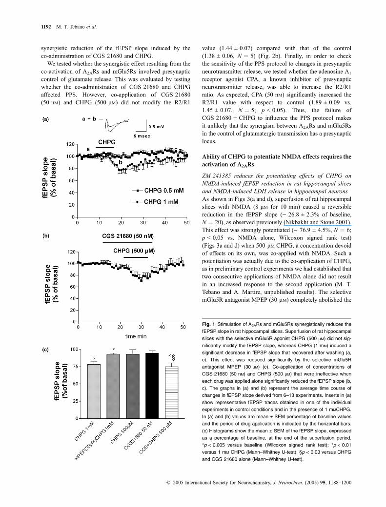
We tested whether the synergistic effect resulting from theco-activation of A2ARs and mGlu5Rs involved presynapticcontrol of glutamate release. This was evaluated by testingwhether the co-administration of CGS 21680 and CHPGaffected PPS. However, co-application of CGS 21680(50 nM) and CHPG (500 lM) did not modify the R2/R1
value (1.44 ± 0.07) compared with that of the control(1.38 ± 0.06, N ¼ 5) (Fig. 2b). Finally, in order to checkthe sensitivity of the PPS protocol to changes in presynapticneurotransmitter release, we tested whether the adenosine A1
receptor agonist CPA, a known inhibitor of presynapticneurotransmitter release, was able to increase the R2/R1ratio. As expected, CPA (50 nM) significantly increased theR2/R1 value with respect to control (1.89 ± 0.09 vs.1.45 ± 0.07, N ¼ 5; p < 0.05). Thus, the failure ofCGS 21680 + CHPG to influence the PPS protocol makesit unlikely that the synergism between A2ARs and mGlu5Rsin the control of glutamatergic transmission has a presynapticlocus.
Ability of CHPG to potentiate NMDA effects requires the
activation of A2ARs
ZM 241385 reduces the potentiating effects of CHPG onNMDA-induced fEPSP reduction in rat hippocampal slicesand NMDA-induced LDH release in hippocampal neuronsAs shown in Figs 3(a and d), superfusion of rat hippocampalslices with NMDA (8 lM for 10 min) caused a reversiblereduction in the fEPSP slope () 26.8 ± 2.3% of baseline,N ¼ 20), as observed previously (Nikbakht and Stone 2001).This effect was strongly potentiated () 76.9 ± 4.5%, N ¼ 6;p < 0.05 vs. NMDA alone, Wilcoxon signed rank test)(Figs 3a and d) when 500 lM CHPG, a concentration devoidof effects on its own, was co-applied with NMDA. Such apotentiation was actually due to the co-application of CHPG,as in preliminary control experiments we had established thattwo consecutive applications of NMDA alone did not resultin an increased response to the second application (M. T.Tebano and A. Martire, unpublished results). The selectivemGlu5R antagonist MPEP (30 lM) completely abolished the
Fig. 1 Stimulation of A2ARs and mGlu5Rs synergistically reduces the
fEPSP slope in rat hippocampal slices. Superfusion of rat hippocampal
slices with the selective mGlu5R agonist CHPG (500 lM) did not sig-
nificantly modify the fEPSP slope, whereas CHPG (1 mM) induced a
significant decrease in fEPSP slope that recovered after washing (a,
c). This effect was reduced significantly by the selective mGlu5R
antagonist MPEP (30 lM) (c). Co-application of concentrations of
CGS 21680 (50 nM) and CHPG (500 lM) that were ineffective when
each drug was applied alone significantly reduced the fEPSP slope (b,
c). The graphs in (a) and (b) represent the average time course of
changes in fEPSP slope derived from 6–13 experiments. Inserts in (a)
show representative fEPSP traces obtained in one of the individual
experiments in control conditions and in the presence of 1 mMCHPG.
In (a) and (b) values are mean ± SEM percentage of baseline values
and the period of drug application is indicated by the horizontal bars.
(c) Histograms show the mean ± SEM of the fEPSP slope, expressed
as a percentage of baseline, at the end of the superfusion period.
�p < 0.005 versus baseline (Wilcoxon signed rank test); *p < 0.01
versus 1 mM CHPG (Mann–Whitney U-test); §p < 0.03 versus CHPG
and CGS 21680 alone (Mann–Whitney U-test).
1192 M. T. Tebano et al.
� 2005 International Society for Neurochemistry, J. Neurochem. (2005) 95, 1188–1200

CHPG-induced potentiation of NMDA effects () 19.1 ±6.9%; N ¼ 3; p < 0.03 vs. NMDA + CHPG, Mann–Whit-ney U-test) (Figs 3b and d). Interestingly, the selective A2ARantagonist ZM 241385 (30 nM) also significantly reduced theability of CHPG to potentiate the effect of NMDA() 50.1 ± 8.3%, N ¼ 8; p < 0.05 vs. NMDA + CHPG,Mann–Whitney U-test) (Figs 3c and d). When given alone,
neither MPEP nor ZM 241385 modified basal synaptictransmission or the NMDA-induced inhibition of the fEPSPslope (data not shown).
To evaluate whether the A2AR–mGlu5R interaction alsoplayed a role in the modulation of NMDA-induced toxicity,primary cultures of rat hippocampal neurons were studied.Incubation of hippocampal neuronal cultures for 1 h with300 lM NMDA induced a significant increase in LDHrelease with respect to basal levels (190.2 ± 32.4%, N ¼ 4;p < 0.01, Mann–Whitney U-test) (Fig. 4). CHPG alone didnot modify LDH release up to a concentration of 1 mM
(100.9 ± 3.2%, N ¼ 3). However, when co-applied withNMDA, CHPG (1 mM) significantly potentiated the NMDA-induced LDH release (312.4 ± 60.1%, N ¼ 4; p < 0.05 vs.NMDA alone, Mann–Whitney U-test) (Fig. 4). The poten-tiating effect of CHPG on the NMDA-induced LDH releasewas prevented by MPEP (30 lM). Pretreatment with MPEPfor 15 min before administration of CHPG + NMDA wasable to restore the same degree of LDH release as inducedby NMDA alone (174.2 ± 5.5%, N ¼ 3; p < 0.05 vs.NMDA + CHPG, Mann–Whitney U-test) (Fig. 4). Similarly,ZM 241385 (30 nM) also abolished the ability of CHPG topotentiate NMDA-induced LDH release (160.8 ± 20.9%,N ¼ 4; p < 0.02 vs. NMDA + CHPG, Mann–WhitneyU-test) (Fig. 4). When applied alone, neither MPEP norZM 241385 influenced basal or NMDA-induced LDHrelease (data not shown).
Ability of CHPG to potentiate NMDA effects is lost in thehippocampus of A2AR KO miceTo further test the hypothesis that hippocampal A2ARs mightplay a permissive role in mGlu5R-dependent effects, theability of CHPG to potentiate NMDA-induced fEPSP slopereduction in hippocampal slices was compared in A2AR KOand WT mice.
As observed in rat hippocampal slices, application ofNMDA (8 lM during 10 min) in WT mice significantlydepressed the fEPSP slope () 33.3 ± 8.5%, N ¼ 8; p < 0.05vs. baseline, Wilcoxon signed rank test), an effect that wasfully reversed after 30 min of washout (Figs 5a and c). Anidentical effect of NMDA was observed in A2AR KO mice() 29.3 ± 6.6%, N ¼ 8; p < 0.05 vs. baseline, Wilcoxonsigned rank test) (Figs 5b and c). As CHPG (500 lM)depressed fEPSP on its own in mouse hippocampal slices(M. R. Domenici and A. Martire, unpublished results), aconcentration of 300 lM was used. Ten minutes of subse-quent co-administration of CHPG (300 lM) and NMDA(8 lM) produced a marked and significant potentiation of theNMDA-induced reduction in fEPSP slope in WT mice() 46.4 ± 8.0%, N ¼ 8; p < 0.05 vs. NMDA alone,Wilcoxon signed rank test) (Figs 5a and c), again in amanner similar to that observed in rat hippocampal slices.Likewise, the potentiating effect of CHPG was blocked eitherby MPEP (30 lM) () 24.0 ± 2.3%, N ¼ 7; p < 0.02 vs.
Fig. 2 Synergism between A2ARs and mGlu5Rs in the control of
fEPSPs in rat hippocampal slices does not appear to involve PKA
activation, GABAergic neurotransmission or presynaptic mechanisms.
(a) Histograms show that inhibition of the fEPSP slope caused by the
simultaneous application of CGS 21680 (50 nM) and CHPG (500 lM)
was not mimicked by the co-application of CHPG (500 lM) together
with the adenylyl cyclase activator forskolin (30 lM) instead of
CGS 21680. Moreover, the PKA inhibitor KT 5720 (1 lM) did
not prevent inhibition of the fEPSP slope induced by
CGS 21680 + CHPG. Application of bicuculline (10 lM), which pre-
vents GABAergic transmission, failed to modify the synergistic
reduction in the fEPSP slope induced by CGS 21680 + CHPG. Values
are mean ± SEM of five experiments. *p < 0.05 versus baseline
(Wilcoxon signed rank test). (b) Application of CGS 21680 and CHPG,
alone or in combination, did not modify the paired pulse facilitation
ratio (R2/R1) in comparison to that in control (i.e. no added drugs).
Values are mean ± SEM of five experiments.
A2AR–mGlu5R interaction in the hippocampus 1193
� 2005 International Society for Neurochemistry, J. Neurochem. (2005) 95, 1188–1200

CHPG + NMDA, Mann–Whitney U-test) (data not shown)or by ZM 241385 (30 nM) () 25.1 ± 5.4%, N ¼ 5; p < 0.05vs. CHPG, Mann–Whitney U-test) (data not shown). Incontrast, in slices from A2AR KO mice, CHPG (300 lM)failed to potentiate the fEPSP slope reduction induced byNMDA () 38.4 ± 7.4%, N ¼ 8; p > 0.05 vs. NMDA alone)(Figs 5b and c). When given alone, CHPG (300 lM) did notmodify basal synaptic transmission. The impairment ofCHPG-mediated potentiation of NMDA responses did notappear to depend on a reduced density of mGlu5Rs in thehippocampus of A2AR KO mice. Indeed, the density ofmGlu5Rs was not modified in the hippocampus of A2AR KOcompared with WT mice, as evaluated by western blotanalysis Fig. 5c.
Subcellular distribution of A2AR and mGlu5R
immunoreactivity in cultured hippocampal neurons
The synergistic effect achieved after stimulating A2ARs andmGlu5Rs raises the possibility that these two receptors are
co-localized in the hippocampus. We decided to evaluate byimmunocytochemistry the distribution of both receptors incultured hippocampal neurons using antibodies against theA2AR and mGlu5R. As observed previously (Rebola et al.2005), the A2AR was highly localized in synapses inhippocampal neurons (Fig. 6a). The mGlu5R had a muchbroader distribution, being present all over the neurons,namely in the cell body, axons, dendrites and synapses(Fig. 6b).
Co-localization studies with synaptophysin (located insynaptic vescicles and considered to be a marker of nerveterminals or synaptic contacts) and vGluT1 and vGluT2(markers of glutamatergic synapses) indicated that themGlu5R was present in synaptic contacts (although alsoelsewhere in neurons) and, in particular, in glutamatergicsynapses (Figs 6d–f). The A2AR was found to be highlyconcentrated in synaptic contacts (co-located with synapto-physin; see Rebola et al. 2005) and was present inglutamatergic synapses (co-located with vGluT1 and
Fig. 3 Both the mGlu5R antagonist MPEP and the adenosine A2A
receptor antagonist ZM 241385 prevent CHPG-induced potentiation of
NMDA effects in rat hippocampal slices (a–c). Time-course of changes
in fEPSP slope recorded in rat hippocampal slices. A 10 min super-
fusion period with NMDA (8 lM) reduced the fEPSP slope, an effect
that was potentiated by the co-application of 500 lM CHPG (a, d).
Each point/bar represents the mean ± S.E.M. of 6–20 experiments,
except for the group MPEP + CHPG + NMDA (N = 3). Representative
fEPSP traces recorded in control conditions and in the presence of
tested drugs are recorded in panel A. The CHPG-induced potentiation
of NMDA effects was abolished by 30 lMMPEP (b,d) and significantly
attenuated by 30 nM ZM 241385 (c, d). For clarity, the lack of effects
of 30 lM MPEP or 30 nM ZM 241385 alone on fEPSP slope are not
presented. *p < 0.05 versus NMDA (Wilcoxon signed rank test);
�p < 0.05 versus CHPG + NMDA (Mann–Whitney U-test).
1194 M. T. Tebano et al.
� 2005 International Society for Neurochemistry, J. Neurochem. (2005) 95, 1188–1200

VGluT2) (Figs 6g–i). Double immunocytochemical labelingwith anti-A2AR and anti-mGlu5R antibodies revealed co-localization of these two receptors mostly in synapticcontacts, where the A2AR was concentrated (Fig. 6c).
Subsynaptic distribution of A2ARs and mGlu5Rs in rat
hippocampus
The resolution of the above immunocytochemical studies didnot allow discrimination between presynaptic and postsy-naptic localization of either receptor. For example, under ourexperimental conditions, we observed co-localization of anti-synaptophysin (a presynaptic marker) and anti-PSD-95 (amarker of PSDs) immunoreactivities (data not shown). Thus,we decided to investigate the relative distribution of A2ARsand mGlu5Rs in the presynaptic active zone and in the PSDof hippocampal synapses. For this purpose, we used apH-based fractionation of synaptic contacts into their mainconstituents (Phillips et al. 2001), namely the PSD, theactive zone (a presynaptic specialization lining the PSD) anda presynaptic non-active zone fraction (that mostly includesthe majority of presynaptic constituents apart from the activezone). As observed in Fig. 7, the mGlu5R was mostlylocated in the PSD but there was also some immunoreactivitypresent at the presynaptic active zone and extra-active zonefractions. Quantification of the relative density of mGlu5Rimmunoreactivity in the three fractions in three differentseparations from different groups of rats revealed that
mGlu5R immunoreactivity was most abundant in the PSDfraction (61.3 ± 4.3% of the total immunoreactivity, N ¼ 3)but was also present in the presynaptic active zone(25.0 ± 2.3% of total immunoreactivity, N ¼ 3) and had alower relative abundance in the extra-active zone fraction of
(a)
(b)
(c)
Fig. 5 Ability of CHPG to potentiate the effects of NMDA is abolished
in the hippocampus of A2AR KO mice. (a, b) Representative traces
showing time course of changes in fEPSP slope recorded in mice
hippocampal slices from (a) WT and (b) A2AR KO mice. Values are
mean ± SEM. (c) Histogram shows mean ± SEM values from eight
experiments. In hippocampal slices from WT (a, c) and KO (b, c) mice
application of 8 lM NMDA induced a very similar reduction in fEPSP
slope. CHPG (300 lM) significantly potentiated this effect in WT mice
(a, c) but not in KO mice (b, c). No changes in mGlu5R expression
were revealed by western blotting of hippocampus from KO and WT
mice (c). *p < 0.05 versus NMDA alone (Wilcoxon signed rank test).
500
400
300
200
100
0
NMDA 300 µM
CHPG 1 mM
MPEP 30 µM
ZM 241385 30 µM
+
-
-
-
-
+
-
-
+
+
-
-
+
+
+
-
+
-
+
-
+
+
-
+
+
-
-
+
LD
H r
elea
se (
% o
f b
asel
) *
Fig. 4 Blockade of either A2ARs or mGlu5Rs prevents CHPG from
potentiating NMDA-induced LDH release from cultured hippocampal
neurons. Application of NMDA (300 lM for 60 min) to rat hippocampal
neurons induced a significant increase in LDH release. The mGlu5R
agonist CHPG (1 mM) had no effect on its own, but it significantly
potentiated NMDA-induced LDH release. The mGlu5R antagonist
MPEP (30 lM) and the A2AR antagonist ZM 241385 (30 nM) had no
effect by themselves but prevented CHPG from potentiating the
release of LDH. Values are mean ± SEM of 3–4 independent experi-
ments, assayed in triplicate. *p < 0.05 versus NMDA alone, �p < 0.05
versus CHPG + NMDA (Mann–Whitney U-test).
A2AR–mGlu5R interaction in the hippocampus 1195
� 2005 International Society for Neurochemistry, J. Neurochem. (2005) 95, 1188–1200

hippocampal nerve terminals (13.7 ± 3.6% of total immu-noreactivity, N ¼ 3). The A2AR was enriched in the presy-naptic active zone fraction (�60%) but was also present atthe PSD. Quantification of the relative density of A2ARimmunoreactivity in the three fractions in three differentseparations from different groups of rats revealed that A2ARimmunoreactivity was most abundant in the presynapticactive zone fraction (56.2 ± 3.3% of total immunoreactivity,N ¼ 3) but was also present in the PSD (35.2 ± 2.7% of totalimmunoreactivity, N ¼ 3) and had a lower relative abun-dance in the extra-synaptic fraction of hippocampal nerveterminals (8.6 ± 1.8% of total immunoreactivity, N ¼ 3).
As also shown in Fig. 7, we validated this subsynapticfractionation by confirming over 90% separation in the threesubsynaptic fractions of the PSD marker PSD-95, presynap-tic active zone marker syntaxin or SNAP-25, and extra-synaptic marker synaptophysin (present in synaptic vesicles;hence this protein is a presynaptic marker but is not locatedin the active zone, which is a restricted zone of thepresynaptic boutton). In particular, we confirmed previouslyobtained data showing that NMDA receptor subunits werehighly enriched in the PSD fraction and nearly absent in thepresynaptic or extra-synaptic fractions (see Rebola et al.2003) (Fig. 7). Thus, although A2ARs and mGlu5Rs werealso co-localized in the presynaptic active zone, their abilityto control NMDA-mediated effects may result from apostsynaptic interaction, as the PSD was the only subsynap-
tic compartment that contained all three receptors, i.e.A2ARs, mGlu5Rs and NMDA receptors.
Discussion
The present study demonstrated, for the first time, that A2ARsand mGlu5Rs are co-localized and functionally interact in therodent hippocampus.
A synergism between A2ARs and mGlu5Rs was demon-strated by the finding that, in rat hippocampal slices,co-application of ineffective concentrations of CGS 21680and CHPG (50 nM and 500 lM respectively) significantlyreduced the fEPSP slope. Furthermore, selective antagonistsof either mGlu5Rs (MPEP) or A2ARs (ZM 241385) preven-ted this synergistic effect, providing a pharmacologicaldemonstration for the involvement of both receptors. Thisfinding strictly reproduces our previous results in the striatum(Domenici et al. 2004; Rodrigues et al. 2005), a brain area inwhich the occurrence of functional interactions betweenA2ARs and mGlu5Rs has been reported in several models(see Introduction).
In order to investigate the mechanisms underlying theA2AR–mGlu5R interaction, we tested the possible involve-ment of GABAergic transmission. The inability of bicucul-line to modify the synergism between A2ARs and mGlu5Rssuggests that this interaction does not involve GABAergicneurotransmission. This is in agreement with previous
Fig. 6 Co-localization of A2ARs and
mGlu5Rs in rat hippocampal neurons in
culture. (a–c) Immunocytochemical identifi-
cation of A2A Rs (a) and mGlu5Rs (b) and
merged image (c) illustrate the partial
co-localization of the two receptors (yellow
color); the insert in (c) corresponds to an
amplification (6-8 times) of the region indi-
cated by the arrow, clearly illustrating the co-
localization of both receptors in neuronal
branches. (d–e) Immunocytochemical iden-
tification of vGluT1 and vGluT2 (d; markers
of glutamatergic nerve terminals) and
mGlu5Rs (e) and the merged image (f)
illustrate the partial co-localization of
mGlu5Rs in glutamatergic nerve terminals
(yellow color). (g–i) Immunocytochemical
identification of vGluT1 and vGluT2 (g) and
A2ARs (h) and the merged image (i) illustrate
the partial co-localization of A2ARs in gluta-
matergic nerve terminals (yellow color).
These images are representative of three
different fields per coverslip, in experiments
carried out 3–4 times using different hippo-
campal neurons prepared from different
groups of rat embryos. Scale bars are 50 lm
in (a)-(c) and 10 lm in (d)–(i).
1196 M. T. Tebano et al.
� 2005 International Society for Neurochemistry, J. Neurochem. (2005) 95, 1188–1200

reports showing that GABA receptor blockade did notinfluence CGS 21680-mediated effects in hippocampal slices(Lopes et al. 2002) and that, unlike the mGlu1R subtype ofgroup I mGluRs, the mGlu5R subtype predominantly affectsglutamatergic rather than GABAergic transmission (Battagliaet al. 2001). It can therefore be concluded that the interactionbetween A2ARs and mGlu5Rs might occur at glutamatergicsynapses. Accordingly, the studies carried out in culturedhippocampal neurons allowed us to conclude that A2ARs andmGlu5Rs are co-localized in synapses, and in particular inglutamatergic synapses, although they did not allow dis-crimination between a presynaptic or postsynaptic localiza-tion. In electrophysiological experiments, we found that theapplication of CGS 21680 and CHPG did not influence theratio of fEPSP responses under a protocol of PPS, which is
an index of presynaptic neurotransmitter release. This findingsuggests that, although both A2ARs (Lopes et al. 2002;Marchi et al. 2002; but see Nikbakht and Stone 2001) andmGlu5Rs (Fazal et al. 2003; Wang and Sihra 2004; Rodri-gues et al. 2005) might control the evoked release ofglutamate presynaptically, the interaction between thesetwo receptors observed in the present study is unlikely tooccur presynaptically. The most likely hypothesis is that theinteraction between A2ARs and mGlu5Rs has a postsynapticlocus, in accordance with the known postsynaptic effects ofboth A2ARs (Li and Henry 1998; O’Kane and Stone 1998)and mGlu5Rs (Mannaioni et al. 2001). This possibility isreinforced by the predominant postsynaptic localization ofmGlu5Rs, in particular in the hippocampus (Lujan et al.1996, 1997; Shigemoto et al. 1997; present results), and bythe present finding that one of the main consequences of theinteraction between A2ARs and mGlu5Rs is the control ofNMDA receptor-mediated effects, which is likely to occurpostsynaptically, because the PSD was the only neuronalcompartment shown to contain all three receptors.
The possible molecular mechanisms involved in thesynergistic interaction between A2ARs and mGlu5Rs remainto be elucidated. Although the cAMP/PKA transductionpathway is considered the canonical transduction systemoperated by A2ARs (Fredholm et al. 2003), the inability offorskolin to reproduce the effects of CGS 21680, and thefinding that KT 5720 did not inhibit the synergistic effect ofCGS 21680 and CHPG, excludes the involvement of thecAMP/PKA pathway in the synergism between A2ARs andmGlu5Rs, in contrast to findings in striatal slices (Domeniciet al. 2004). On the other hand, hippocampal A2ARs, besidescoupling to the classical cAMP/PKA pathway (Okada et al.2001; Rebola et al. 2002), also use a protein kinase C-dependent transduction pathway, in particular in the controlof glutamatergic transmission (Cunha and Ribeiro 2000b;Lopes et al. 2002). However, we did not attempt to testwhether protein kinase C might be involved in this poten-tiation of mGlu5R responses by A2ARs, as the manipulationof protein kinase C activity is also expected to directly affectthe responses mediated by mGlu5Rs in the hippocampus(Benquet et al. 2002; Kotecha et al. 2003). Thus, at thisstage, it can only be excluded that cAMP/PKA is involvedand hypothesized that A2AR stimulation facilitates mGlu5R-dependent effects through activation of the protein kinase Cpathway.
We found that application of CHPG enhanced NMDA-mediated responses in hippocampal slices from rats and WTmice. This finding is in full agreement with some reportsshowing that CHPG and RS3,5-dihydroxyphenylglycine(DHPG) potentiated NMDA-induced depolarization in therat hippocampus (Fitzjohn et al. 1996; Doherty et al. 1997),and is in line with several studies showing the ability ofmGlu5Rs to increase NMDA responses in several otherbrain areas (Ugolini et al. 1999; Attucci et al. 2001; Pisani
Syntaxin(a)
(b) (c)
PSD-95
Synaptophysin
NR2A
pre post extra
pre
mGluR5 (150 kDa) A2A (45 kDa)
post extra
PRE
706050403020100
60
50
40
30
20
10
0
% O
F T
OTA
L I
MM
UN
OR
EA
CT
IVIT
Y
% O
F T
OTA
L I
MM
UN
OR
EA
CT
IVIT
Y
POST EXTRA PRE POST EXTRA
synap pre post extra synap
Fig. 7 Subsynaptic distribution of mGlu5Rs (b) and A2ARs (c) in the
rat hippocampus. Hippocampal nerve terminals were prepared
(synap) and further fractionated to obtain fractions enriched in the
presynaptic active zone (pre), the PSD (post), in nerve terminals
outside the active zone (extra). These fractions were over 90% pure,
as illustrated by the ability to recover the immunoreactivity for syntaxin
in the presynaptic active zone fraction, PSD95 in the PSD fraction and
synaptophysin (a protein located in synaptic vesicles) in the extra-
active zone fraction (a). One NMDA receptor subunit (NR2A) was
nearly confined to the PSD (a). (b) Representative western blot
showing mGlu5R immunoreactivity, corresponding to a 150-kDa band,
in the four fractions and histogram showing average mGlu5R immu-
noreactivity in each fraction. Values are mean ± SEM of three
experiments using fractions prepared from different groups of rats. (c)
Representative western blot showing A2AR immunoreactivity, corres-
ponding to a 45-kDa band, in the four fractions and histogram showing
average A2AR immunoreactivity in each of the fractions. Values are
mean ± SEM of three experiments using fractions prepared from dif-
ferent groups of rats.
A2AR–mGlu5R interaction in the hippocampus 1197
� 2005 International Society for Neurochemistry, J. Neurochem. (2005) 95, 1188–1200

et al. 2001; Domenici et al. 2004). In the present study, theability of CHPG to potentiate NMDA-induced fEPSPreduction was prevented not only by MPEP (whichconfirms the selective involvement of mGlu5Rs in thiseffect of CHPG), but also by the A2AR antagonistZM 241385, indicating that A2ARs enable mGlu5R-medi-ated effects (namely, the potentiation of NMDA responses)in the hippocampus. This view was confirmed by thefinding that CHPG was no longer able to potentiate theeffects of NMDA effects in hippocampal slices from A2ARKO mice. According to our western blot experiments, thereduced functional ability of CHPG in A2AR KO mice doesnot appear to depend on changes in mGlu5R density. Thisfinding is not surprising, because in rat hippocampal slicesa single application of ZM 241385 (i.e. an ‘acute’ A2ARblockade, not implying changes in mGlu5R density) wasenough to block the ability of CHPG to potentiate NMDAeffects. Therefore, the present findings demonstrate thatboth the pharmacological blockade (use of ZM 241385 inrats and in WT mice) and the genetic inactivation (KOmice) of A2ARs seriously impair the ability of hippocampalmGlu5Rs to potentiate NMDA responses. These results arein line with our recent results showing that the state ofactivation of A2ARs influences mGlu5R-dependent NMDApotentiation in the striatum (Domenici et al. 2004). Thus,the ability of A2ARs to control mGlu5R-dependent effectsmight be a general feature of A2ARs in different brainregions, irrespective of their density (which is considerablygreater in the striatum than in the hippocampus; Fredholmet al. 2003). This further emphasizes the role of A2ARs as afine-tuning modulatory system (i.e. modulator of othermodulators) in the hippocampus (Sebastiao and Ribeiro2000).
The permissive role played by A2ARs on mGlu5R-mediated effects also appears to be relevant in the modula-tion of NMDA-mediated neurotoxicity. We observed herethat CHPG significantly potentiated NMDA-induced LDHrelease from cultured hippocampal neurons in a mannerabolished by MPEP, whereas the mGlu5R ligands alone hadno effect. Most importantly, we found that the NMDApotentiating effects of CHPG were fully prevented not onlyby MPEP but also by ZM 241385. This provides animportant clue to our understanding of the surprisingneuroprotective effect afforded by blockade of A2ARs(pharmacological or genetic) in an extra-striatal region wherethis receptor is scarcely located (reviewed in Cunha 2005). Itis important to note that ZM 241385 alone did not influencethe NMDA effects, and that WT and A2AR KO mice showedvery similar responses to NMDA application, indicating thatA2AR inactivation does not directly impair NMDA receptor-mediated effects.
In conclusion, the present results show that A2ARs andmGlu5Rs are co-localized and interact functionally in thehippocampus. The stimulation of A2ARs facilitates CHPG-
induced effects (namely fEPSP reduction) and, even moreinterestingly, hippocampal A2ARs need to be activated inorder to elicit the NMDA potentiating effects of mGlu5Rs.These data suggest that A2ARs might represent an interestingtarget for the development of new therapeutic strategies fordisorders involving changes in NMDA receptor signaling,such as Alzheimer’s disease, epilepsy and schizophrenia,where they might exert a neuroprotective effect similar tothat recognized for striatal diseases (Chen et al. 2001; Popoliet al. 2002).
Besides its possible role in modulating excitotoxicity, theA2AR–mGlu5R interaction might also be important as far asthe physiological effects mediated by hippocampal NMDAreceptors are concerned. Indeed, because hippocampalmGlu5Rs are highly involved in the modulation ofNMDA-dependent plasticity (see Introduction), this interac-tion might represent a major mechanism in the regulation oflearning and memory processes, an issue that requires furtherstudy.
Acknowledgements
This work was supported by grants from the Ministry of Health
(strategic projects ALZ4 and RF03.182) to PP and Fundacao para a
Ciencia e para a Tecnologia (POCTI/44740/2002) to RAC.
References
Alagarsamy S., Marino M. J., Rouse S. T., Gereau R. W. IV, HeinemannS. F. and Conn P. J. (1999) Activation of NMDA receptors reversesdesensitization of mGlu5R in native and recombinant systems. Nat.Neurosci. 2, 234–240.
Anderson W. W. and Collingridge G. L. (2001) The LTP Program: a dataacquisition program for on-line analysis of long-term potentiationand other synaptic events. J. Neurosci. Methods 108, 71–83.
Anwyl R. (1999) Metabotropic glutamate receptors: electrophysiologicalproperties and role in plasticity. Brain Res. Rev. 29, 83–120.
Attucci S., Carla V., Mannaioni G. and Moroni F. (2001) Activation oftype 5 metabotropic receptors enhances NMDA responses in cor-tical wedges. Br. J. Pharmacol. 132, 799–806.
Attucci S., Clodfelter G. V., Thibault O., Staton J., Moroni F., Landfield P.W. and Porter N.M. (2002) Group Imetabotropic glutamate receptorinhibition selectively blocks a prolonged Ca2+ elevation associatedwith age-dependent excitotoxicity. Neuroscience 112, 183–194.
Balschun D. and Wetzel W. (2002) Inhibition of mGlu5R blocks hip-pocampal LTP in vivo and spatial learning in rats. Pharmacol.Biochem. Behav. 73, 375–380.
Balschun D., Manahan-Vaughan D., Wagner T., Behnisch T., ReymannK. G. and Wetzel W. (1999) A specific role for group I mGluRs inhippocampal LTP and hippocampus-dependent spatial learning.Learn. Mem. 6, 138–152.
Battaglia G., Bruno V., Pisani A., Centonze D., Catania M. V., CalabresiP. and Nicoletti F. (2001) Selective blockade of type-1 metabo-tropic glutamate receptors induces neuroprotection by enhancinggabaergic transmission. Mol. Cell. Neurosci. 17, 1071–1083.
Benquet P., Gee C. E. and Gerber U. (2002) Two distinct signalingpathways upregulate NMDA receptor responses via two distinctmetabotropic glutamate receptor subtypes. J. Neurosci. 22, 9679–9686.
1198 M. T. Tebano et al.
� 2005 International Society for Neurochemistry, J. Neurochem. (2005) 95, 1188–1200

Bortolotto Z. A., Fitzjohn S. M. and Collingridge G. L. (1999) Roles ofmetabotropic glutamate receptors in LTP and LTD in the hippo-campus. Curr. Opin. Neurobiol. 9, 299–304.
Chen J. F., Huang Z., Ma J., Zhu J., Moratalla R., Standaert D.,Moskowitz M. A., Fink J. S. and Schwarzschild M. A. (1999) A2A
adenosine receptor deficiency attenuates brain injury induced bytransient focal ischemia in mice. J. Neurosci. 19, 9192–9200.
Chen J. F., Xu K., Petzer J. P., Staal R., Xu Y. H., Beilstein M., SonsallaP. K., Castagnoli K., Castagnoli N. and jr and Schwarzschild M. A.(2001) Neuroprotection by caffeine and A2A adenosine receptorinactivation in a model of Parkinson’s disease. J Neurosci. 21, RC143.
Coccurello R., Breysse N. and Amalric M. (2004) Simultaneousblockade of adenosine A2A and metaboptropic glutamate mGlu5receptors increase their efficacy in reversing Parkinsonian deficitsin rats. Neuropsychopharmacology 29, 1451–1461.
Collingridge G. L. and Bliss T. V. (1995) Memories of NMDA receptorsand LTP. Trends Neurosci. 18, 54–56.
Cunha R. A. (2005) Neuroprotection by adenosine in the brain: from A1
receptor activation to A2A receptor blockade. Purinergic Signalling1, 111–134.
Cunha R. A., Constantino M. D. and Ribeiro J. A. (1997) ZM241385 isan antagonist of the facilitatory responses produced by the A2A
adenosine receptor agonists CGS21680 and HENECA in the rathippocampus. Br. J. Pharmacol. 122, 1279–1284.
Cunha R. A. and Ribeiro J. A. (2000a) Purinergic modulation of[3H]GABA release from rat hippocampal nerve terminals. Neuro-pharmacology 39, 1156–1167.
Cunha R. A. and Ribeiro J. A. (2000b) Adenosine A2A facilitation ofsynaptic transmission in the CA1 area of the rat hippocampusrequires protein kinase C but not protein kinase A activation.Neurosci. Lett. 289, 127–130.
Diaz-Cabiale Z., Vivo M., Del Arco A., O’Connor W. T., Harte M. K.,Muller C. E., Martinez E., Popoli P., Fuxe K. and Ferre S. (2002)Metabotropic glutamate mGlu5 receptor-mediated modulation ofthe ventral striopallidal GABA pathway in rats: interactions withadenosine A2A and dopamine D2 receptors. Neurosci. Lett. 324,154–158.
Doherty A. J., Palmer M. J., Henley J. M., Collingridge G. L. and JaneD. E. (1997) (RS)-2-Chloro-5-hydroxyphenylglycine (CHPG)activates mGlu5, but not mGlu1, receptors expressed in CHO cellsand potentiates NMDA responses in hippocampus. Neurophar-macology 36, 265–267.
Domenici M. R., Pepponi R., Martire A., Tebano M. T., Potenza R. L.and Popoli P. (2004) Permissive role of adenosine A2A receptors onmetabotropic glutamate receptor 5 (mGluR5)-mediated effects inthe striatum. J. Neurochem. 90, 1276–1279.
Fazal A., Parker F., Palmer A. M. and Croucher M. J. (2003) Charac-terisation of the actions of group I metabotropic glutamate receptorsubtype selective ligands on excitatory amino acid release andsodium-dependent re-uptake in rat cerebrocortical minislices.J. Neurochem. 86, 1346–1358.
Ferre S., Karcz-Kubicha M., Hope B. et al. (2002) Synergistic interac-tion between adenosine A2A and glutamate mGlu5 receptors:implications for striatal neuronal function. Proc. Natl Acad. Sci.USA 99, 11 940–11 945.
Fitzjohn S. M., Irving A. J., Palmer M. J., Harvey J., Lodge D. andCollingridge G. L. (1996) Activation of group I mGluRs potenti-ates NMDA responses in rat hippocampal slices. Neurosci. Lett.203, 211–213.
Fredholm B. B., Cunha R. A. and Svenningsson P. (2003) Pharmacologyof adenosine A2A receptors and therapeutic applications. Curr. Top.Med. Chem. 3, 413–426.
Fuxe K., Agnati L. F., Jacobsen J. et al. (2003) Receptor heteromeri-zation in adenosine A2A receptor signaling: relevance for striatalfunction and Parkinson’s disease. Neurology 61, S19–S23.
Kotecha S. A., Jackson M. F., Al-Mahrouki A., Roder J. C., Orser B. A.and MacDonald J. F. (2003) Co-stimulation of mGluR5 andN-methyl-D-aspartate receptors is required for potentiation of ex-citatory synaptic transmission in hippocampal neurons. J. Biol.Chem. 278, 27 742–27 749.
Laemmli U. K. (1970) Cleavage of structural proteins during theassembly of the head of bacteriophage T4. Nature 227, 680–685.
Li H. and Henry J. L. (1998) Adenosine A2 receptor mediation of pre-and postsynaptic excitatory effects of adenosine in rat hippocam-pus in vitro. Eur. J. Pharmacol. 347, 173–182.
Lopes L. V., Cunha R. A., Kull B., Fredholm B. B. and Ribeiro J. A.(2002) Adenosine A2A receptor facilitation of hippocampal syn-aptic transmission is dependent on tonic A1 receptor inhibition.Neuroscience 112, 319–329.
Lu Y. M., Jia Z., Janus C., Henderson J. T., Gerlai R., Wojtowicz M. J.and Roder J. C. (1997) Mice lacking metabotropic glutamatereceptor 5 show impaired learning and reduced CA1 long-termpotentiation (LTP) but normal CA3 LTP. J. Neurosci. 17, 5196–5205.
Lujan R., Nusser Z., Roberts J. D., Shigemoto R. and Somogyi P. (1996)Perisynaptic location of metabotropic glutamate receptors mGlu1Rand mGluR5 on dendrites and dendritic spines in the rat hippo-campus. Eur. J. Neurosci. 8, 1488–1500.
Lujan R., Roberts J. D., Shigemoto R., Ohishi H. and Somogyi P. (1997)Differential plasma membrane distribution of metabotropic glu-tamate receptors mGluR1 alpha, mGluR2 and mGluR5, relative toneurotransmitter release sites. J. Chem. Neuroanat. 13, 219–241.
Mannaioni G., Marino M. J., Valemnti O., Traynelis S. F. and Conn J. P.(2001) Metabotropic glutamate receptors 1 and 5 differentiallyregulate CA1 pyramidal cell function. J. Neurosci. 21, 5925–5934.
Marchi M., Raiteri L., Risso F., Vallarino A., Bonfanti A., Monopoli A.,Ongini E. and Raiteri M. (2002) Effects of adenosine A1 and A2A
receptor activation on the evoked release of glutamate from ratcerebrocortical synaptosomes. Br. J. Pharmacol. 136, 434–440.
Mori M. and Gerber U. (2002) Slow feedback inhibition in the CA3 areaof the rat hippocampus by synergistic synaptic activation ofmGluR1 and mGluR5. J. Physiol. 544, 793–799.
Nikbakht M. R. and Stone T. W. (2001) Suppression of presynapticresponses to adenosine by activation of NMDA receptors. Eur. J.Pharmacol. 427, 13–25.
Nishi A., Liu F., Matsuyama S., Hamada M., Higashi H., Nairn A. C.and Greengard P. (2003) Metabotropic mGlu5 receptors regulateadenosine A2A receptor signalling. Proc. Natl Acad. Sci. USA 100,1322–1327.
O’Kane E. M. and Stone T. W. (1998) Interaction between adenosineA1 and A2 receptor-mediated responses in the rat hippocampusin vitro. Eur. J. Pharmacol. 362, 17–25.
Okada M., Nutt D. J., Murakami T., Zhu G., Kamata A., Kawata Y. andKaneko S. (2001) Adenosine receptor subtypes modulate twomajor functional pathways for hippocampal serotonin release.J. Neurosci. 21, 628–640.
Phillips G. R., Huang J. K., Wang Y., Tanaka H., Shapiro L., Zhang M.,Gordon R. E., Gawinowicz M. A., Zhao Y. and Colman D. R.(2001) The presynaptic particle web: ultrastructure, composition,dissolution, and reconstitution. Neuron 32, 1–20.
Pisani A., Gubellini P., Bonsi P., Conquet F., Picconi B., Centonze D.,Bernardi G. and Calabresi P. (2001) Metabotropic glutamatereceptor 5 mediates the potentiation of N-methyl-D-aspartateresponses in medium spiny striatal neurons. Neuroscience 106,579–587.
A2AR–mGlu5R interaction in the hippocampus 1199
� 2005 International Society for Neurochemistry, J. Neurochem. (2005) 95, 1188–1200

Popoli P., Pezzola A., Torvinen M., Reggio R., Pintor A., Scarchilli L.,Fuxe K. and Ferre S. (2001) The selective mGlu5 receptor agonistCHPG inhibits quinpirole-induced turning in 6-hydroxydopamine-lesioned rats and modulates the binding characteristics of dopam-ine D2 receptors in the striatum: interaction with adenosine A2A
receptors. Neuropsychopharmacology 25, 505–513.Popoli P., Pintor A., Domenici M. R. et al. (2002) Blockade of sriatal
adenosine A2A receptor reduces, through a presynaptic mechanism,quinolinic acid-induced excitotoxicity: possibile relevance to neu-roprotective interventions in neurodegenerative diseases of thestriatum. J. Neurosci. 22, 1967–1975.
Popoli P., Pintor A., Tebano M. T. et al. (2004) Neuroprotective effectsof the mGlu5R antagonist MPEP towards quinolinic acid-inducedstriatal toxicity: involvment of pre and post-synaptic mechanismsand lack of direct NMDA blocking activity. J. Neurochem. 89,1479–1489.
Rebola N., Oliveira C. R. and Cunha R. A. (2002) Transducing systemoperated by adenosine A2A receptors to facilitate acetylcholinerelease in the rat hippocampus. Eur. J. Pharmacol. 454, 31–38.
Rebola N., Pinheiro P. C., Oliveira C. R., Malva J. O. and Cunha R. A.(2003) Subcellular localization of adenosine A1 receptors in nerveterminals and synapses of the rat hippocampus. Brain Res. 987,49–58.
Rebola N., Canas P., Oliveira C. R. and Cunha R. A. (2005) Differentsynaptic and subsynaptic localization of adenosine A2A receptorsin the hippocampus and striatum of the rat. Neuroscience 132,893–903.
Rodrigues R. J., Alfaro T. M., Rebola N., Oliveira C. R. and CunhaR. A. (2005) Co-localization and functional interaction between
adenosine A2A and metabotropic group 5 receptors in gluta-matergic nerve terminals of the rat striatum. J. Neurochem. 92,433–441.
Romano C., Sesma M. A., McDonald C. T., O’Malley K., van den PolA. N. and Olney J. W. (1995) Distribution of metabotropic glu-tamate receptor mGluR5 immunoreactivity in rat brain. J. Comp.Neurol. 355, 455–469.
Rothman S. M. and Olney J. W. (1995) Excitotoxicity and the NMDAreceptor – still lethal after eight years. Trends Neurosci. 18, 57–58.
Sacaan A. I. and Schoepp D. D. (1992) Activation of hippocampalmetabotropic excitatory amino acid receptors leads to seizures andneuronal damage. Neurosci. Lett. 139, 77–82.
Schulz P. E., Cook E. P. and Johnston D. (1994) Changes in paired-pulsefacilitation suggest presynaptic involvement in long-term potenti-ation. J. Neurosci. 14, 5325–5337.
Sebastiao A. M. and Ribeiro J. A. (2000) Fine-tuning neuromodulationby adenosine. Trends Pharmacol. Sci. 21, 341–346.
Shigemoto R., Kinoshita A., Wada E. et al. (1997) Differential presy-naptic localization of metabotropic glutamate receptor subtypes inthe rat hippocampus. J. Neurosci. 17, 7503–7522.
Ugolini A., Corsi M. and Bordi F. (1999) Potentiation of NMDA andAMPA responses by the specific mGluR5 agonist CHPG in spinalcord motoneurons. Neuropharmacology 38, 1569–1576.
Wang S. J. and Sihra T. S. (2004) Noncompetitive metabotropic glu-tamate5 receptor antagonist (E)-2-methyl-6-styryl-pyridine(SIB1893) depresses glutamate release through inhibition of volt-age-dependent Ca2+ entry in rat cerebrocortical nerve terminals(synaptosomes). J. Pharmacol. Exp. Ther. 309, 951–958.
1200 M. T. Tebano et al.
� 2005 International Society for Neurochemistry, J. Neurochem. (2005) 95, 1188–1200