Reduced expression of thyroid hormone receptors and beta-adrenergic receptors in human failing...
7
Reduced expression of thyroid hormone receptors and beta- adrenergic receptors in human failing cardiomyocytes Pietro Amedeo Modesti a, *, Matilde Marchetta a , Tania Gamberi a , Gianluca Lucchese b , Massimo Maccherini b , Mario Chiavarelli b , Alessandra Modesti a a Department of Critical Care Medicine, University of Florence, Viale Morgagni 85, 50134 Florence, Italy b Department of Cardiothoracic Surgery, University of Siena, Siena, Italy 1. Introduction Many of the clinical manifestations of thyroid diseases are mediated by changes in cardiovascular hemodynamics [1]. These changes, in particular heart rate, are known to improve in response to treatment with beta-adrenergic receptor antagonists [2]. 3,5,3 0 -Triiodo-L-thyronine (T3) was indeed reported to affect cardiac function by enhancing the expres- sion of genes involved in the regulation of beta-adrenergic signalling, such as sarcoplasmic reticulum Ca 2+ ATPase [3], and of the beta-adrenergic receptor itself [4], in addition to its effects on the expression of other cardiac specific genes [3–7]. This aspect is particularly important in the treatment of thyroid storm disease where the use of beta-adrenergic antagonism is a priority. On the other hand, human studies have shown that the progression to failure is characterized not only by a significant reduction of adrenergic receptors [8], but also by cardiac changes in phenotype and gene expression similar to those described for hypothyroidism [9–11]. A low triiodiothyronine (T3) syndrome, characterized by low circu- lating levels of the biologically active form of T3 in the presence of normal thyrotropin (TSH) and of thyroxine was indeed reported to occur in approximately 30% of patients with advanced heart failure [12]. However, a reduced expres- sion of cardiac genes stimulated by thyroid hormone was observed also in explanted hearts removed from patients who were clinically and chemically euthyroid [9,13,14]. Therefore a potential alteration of cardiac T3 signal transduc- tion in failing heart was hypothesized so that several studies investigated the expression of receptors for thyroid hormone biochemical pharmacology 75 (2008) 900–906 article info Article history: Received 10 August 2007 Accepted 9 October 2007 Keywords: Heart failure Cardiomyopathy 3,5,3 0 -Triiodo-L-thyronine Myocytes abstract An altered thyroid hormone profile has been reported in patients with congestive heart failure. However, information regarding the status of thyroid hormone receptors in human failing cardiomyocytes is lacking. Therefore the expression of thyroid hormone and beta- adrenergic receptors was investigated in human ventricular cardiomyocytes isolated from patients with end-stage heart failure (FM, n = 12), or from tentative donors (C, n = 4). The expression of thyroid (TRalpha1, and TRbeta1) and beta-adrenergic receptors (ARB1 and ARB2) was measured at both the gene, and at the protein level. In FM the reduced mRNA expression of ARB1 ( p < 0.05, 37%) and ARB2 ( p < 0.05, 42%) was associated with a reduction of the messenger for TRalpha1 ( p < 0.05, 85%) and TRalpha2 ( p < 0.05, 73%). These findings were confirmed at the protein level for ARB1, ARB2 and TRalpha1. These data reveal that in human heart failure the reduction of beta-adrenergic receptors is associated with reduced expression of both TRalpha1 and TRalpha2 isoforms of thyroid hormone receptors. # 2007 Elsevier Inc. All rights reserved. * Corresponding author. Tel.: +39 055 7949376; fax: +39 055 7949376. E-mail address: pamodesti@unifi.it (P.A. Modesti). available at www.sciencedirect.com journal homepage: www.elsevier.com/locate/biochempharm 0006-2952/$ – see front matter # 2007 Elsevier Inc. All rights reserved. doi:10.1016/j.bcp.2007.10.011
-
Upload
independent -
Category
Documents
-
view
0 -
download
0
Transcript of Reduced expression of thyroid hormone receptors and beta-adrenergic receptors in human failing...
Reduced expression of thyroid hormone receptors and beta-adrenergic receptors in human failing cardiomyocytes
Pietro Amedeo Modesti a,*, Matilde Marchetta a, Tania Gamberi a, Gianluca Lucchese b,Massimo Maccherini b, Mario Chiavarelli b, Alessandra Modesti a
aDepartment of Critical Care Medicine, University of Florence, Viale Morgagni 85, 50134 Florence, ItalybDepartment of Cardiothoracic Surgery, University of Siena, Siena, Italy
b i o c h e m i c a l p h a r m a c o l o g y 7 5 ( 2 0 0 8 ) 9 0 0 – 9 0 6
a r t i c l e i n f o
Article history:
Received 10 August 2007
Accepted 9 October 2007
Keywords:
Heart failure
Cardiomyopathy
3,5,30-Triiodo-L-thyronine
Myocytes
a b s t r a c t
An altered thyroid hormone profile has been reported in patients with congestive heart
failure. However, information regarding the status of thyroid hormone receptors in human
failing cardiomyocytes is lacking. Therefore the expression of thyroid hormone and beta-
adrenergic receptors was investigated in human ventricular cardiomyocytes isolated from
patients with end-stage heart failure (FM, n = 12), or from tentative donors (C, n = 4). The
expression of thyroid (TRalpha1, and TRbeta1) and beta-adrenergic receptors (ARB1 and
ARB2) was measured at both the gene, and at the protein level.
In FM the reduced mRNA expression of ARB1 ( p < 0.05, �37%) and ARB2 ( p < 0.05, �42%)
was associated with a reduction of the messenger for TRalpha1 (p < 0.05, �85%) and
TRalpha2 ( p < 0.05, �73%). These findings were confirmed at the protein level for ARB1,
ARB2 and TRalpha1.
These data reveal that in human heart failure the reduction of beta-adrenergic receptors
is associated with reduced expression of both TRalpha1 and TRalpha2 isoforms of thyroid
hormone receptors.
# 2007 Elsevier Inc. All rights reserved.
avai lable at www.sc iencedi rec t .com
journal homepage: www.e lsev ier .com/ locate /b iochempharm
1. Introduction
Many of the clinical manifestations of thyroid diseases are
mediated by changes in cardiovascular hemodynamics [1].
These changes, in particular heart rate, are known to improve
in response to treatment with beta-adrenergic receptor
antagonists [2]. 3,5,30-Triiodo-L-thyronine (T3) was indeed
reported to affect cardiac function by enhancing the expres-
sion of genes involved in the regulation of beta-adrenergic
signalling, such as sarcoplasmic reticulum Ca2+ ATPase [3],
and of the beta-adrenergic receptor itself [4], in addition to its
effects on the expression of other cardiac specific genes [3–7].
This aspect is particularly important in the treatment of
thyroid storm disease where the use of beta-adrenergic
antagonism is a priority. On the other hand, human studies
* Corresponding author. Tel.: +39 055 7949376; fax: +39 055 7949376.E-mail address: [email protected] (P.A. Modesti).
0006-2952/$ – see front matter # 2007 Elsevier Inc. All rights reserveddoi:10.1016/j.bcp.2007.10.011
have shown that the progression to failure is characterized not
only by a significant reduction of adrenergic receptors [8], but
also by cardiac changes in phenotype and gene expression
similar to those described for hypothyroidism [9–11]. A low
triiodiothyronine (T3) syndrome, characterized by low circu-
lating levels of the biologically active form of T3 in the
presence of normal thyrotropin (TSH) and of thyroxine was
indeed reported to occur in approximately 30% of patients
with advanced heart failure [12]. However, a reduced expres-
sion of cardiac genes stimulated by thyroid hormone was
observed also in explanted hearts removed from patients
who were clinically and chemically euthyroid [9,13,14].
Therefore a potential alteration of cardiac T3 signal transduc-
tion in failing heart was hypothesized so that several studies
investigated the expression of receptors for thyroid hormone
.
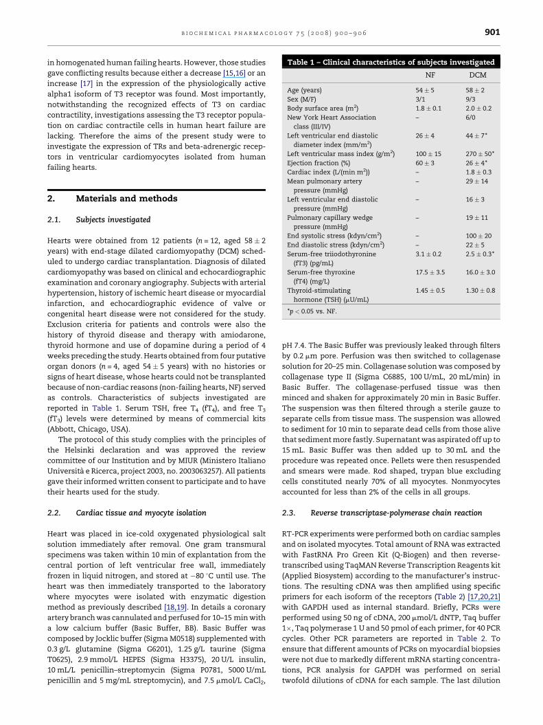
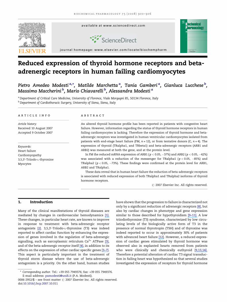
Table 1 – Clinical characteristics of subjects investigated
NF DCM
Age (years) 54 � 5 58 � 2
Sex (M/F) 3/1 9/3
Body surface area (m2) 1.8 � 0.1 2.0 � 0.2
New York Heart Association
class (III/IV)
– 6/0
Left ventricular end diastolic
diameter index (mm/m2)
26 � 4 44 � 7*
Left ventricular mass index (g/m2) 100 � 15 270 � 50*
Ejection fraction (%) 60 � 3 26 � 4*2
b i o c h e m i c a l p h a r m a c o l o g y 7 5 ( 2 0 0 8 ) 9 0 0 – 9 0 6 901
in homogenated human failing hearts. However, those studies
gave conflicting results because either a decrease [15,16] or an
increase [17] in the expression of the physiologically active
alpha1 isoform of T3 receptor was found. Most importantly,
notwithstanding the recognized effects of T3 on cardiac
contractility, investigations assessing the T3 receptor popula-
tion on cardiac contractile cells in human heart failure are
lacking. Therefore the aims of the present study were to
investigate the expression of TRs and beta-adrenergic recep-
tors in ventricular cardiomyocytes isolated from human
failing hearts.
Cardiac index (L/(min m )) – 1.8 � 0.3Mean pulmonary artery
pressure (mmHg)
– 29 � 14
Left ventricular end diastolic
pressure (mmHg)
– 16 � 3
Pulmonary capillary wedge
pressure (mmHg)
– 19 � 11
End systolic stress (kdyn/cm2) – 100 � 20
End diastolic stress (kdyn/cm2) – 22 � 5
Serum-free triiodothyronine
(fT3) (pg/mL)
3.1 � 0.2 2.5 � 0.3*
Serum-free thyroxine
(fT4) (mg/L)
17.5 � 3.5 16.0 � 3.0
Thyroid-stimulating
hormone (TSH) (mU/mL)
1.45 � 0.5 1.30 � 0.8
*p < 0.05 vs. NF.
2. Materials and methods
2.1. Subjects investigated
Hearts were obtained from 12 patients (n = 12, aged 58 � 2
years) with end-stage dilated cardiomyopathy (DCM) sched-
uled to undergo cardiac transplantation. Diagnosis of dilated
cardiomyopathy was based on clinical and echocardiographic
examination and coronary angiography. Subjects with arterial
hypertension, history of ischemic heart disease or myocardial
infarction, and echocardiographic evidence of valve or
congenital heart disease were not considered for the study.
Exclusion criteria for patients and controls were also the
history of thyroid disease and therapy with amiodarone,
thyroid hormone and use of dopamine during a period of 4
weeks preceding the study. Hearts obtained from four putative
organ donors (n = 4, aged 54 � 5 years) with no histories or
signs of heart disease, whose hearts could not be transplanted
because of non-cardiac reasons (non-failing hearts, NF) served
as controls. Characteristics of subjects investigated are
reported in Table 1. Serum TSH, free T4 (fT4), and free T3
(fT3) levels were determined by means of commercial kits
(Abbott, Chicago, USA).
The protocol of this study complies with the principles of
the Helsinki declaration and was approved the review
committee of our Institution and by MIUR (Ministero Italiano
Universita e Ricerca, project 2003, no. 2003063257). All patients
gave their informed written consent to participate and to have
their hearts used for the study.
2.2. Cardiac tissue and myocyte isolation
Heart was placed in ice-cold oxygenated physiological salt
solution immediately after removal. One gram transmural
specimens was taken within 10 min of explantation from the
central portion of left ventricular free wall, immediately
frozen in liquid nitrogen, and stored at �80 8C until use. The
heart was then immediately transported to the laboratory
where myocytes were isolated with enzymatic digestion
method as previously described [18,19]. In details a coronary
artery branch was cannulated and perfused for 10–15 min with
a low calcium buffer (Basic Buffer, BB). Basic Buffer was
composed by Jocklic buffer (Sigma M0518) supplemented with
0.3 g/L glutamine (Sigma G6201), 1.25 g/L taurine (Sigma
T0625), 2.9 mmol/L HEPES (Sigma H3375), 20 U/L insulin,
10 mL/L penicillin–streptomycin (Sigma P0781, 5000 U/mL
penicillin and 5 mg/mL streptomycin), and 7.5 mmol/L CaCl2,
pH 7.4. The Basic Buffer was previously leaked through filters
by 0.2 mm pore. Perfusion was then switched to collagenase
solution for 20–25 min. Collagenase solution was composed by
collagenase type II (Sigma C6885, 100 U/mL, 20 mL/min) in
Basic Buffer. The collagenase-perfused tissue was then
minced and shaken for approximately 20 min in Basic Buffer.
The suspension was then filtered through a sterile gauze to
separate cells from tissue mass. The suspension was allowed
to sediment for 10 min to separate dead cells from those alive
that sediment more fastly. Supernatant was aspirated off up to
15 mL. Basic Buffer was then added up to 30 mL and the
procedure was repeated once. Pellets were then resuspended
and smears were made. Rod shaped, trypan blue excluding
cells constituted nearly 70% of all myocytes. Nonmyocytes
accounted for less than 2% of the cells in all groups.
2.3. Reverse transcriptase-polymerase chain reaction
RT-PCR experiments were performed both on cardiac samples
and on isolated myocytes. Total amount of RNA was extracted
with FastRNA Pro Green Kit (Q-Biogen) and then reverse-
transcribed using TaqMAN Reverse Transcription Reagents kit
(Applied Biosystem) according to the manufacturer’s instruc-
tions. The resulting cDNA was then amplified using specific
primers for each isoform of the receptors (Table 2) [17,20,21]
with GAPDH used as internal standard. Briefly, PCRs were
performed using 50 ng of cDNA, 200 mmol/L dNTP, Taq buffer
1�, Taq polymerase 1 U and 50 pmol of each primer, for 40 PCR
cycles. Other PCR parameters are reported in Table 2. To
ensure that different amounts of PCRs on myocardial biopsies
were not due to markedly different mRNA starting concentra-
tions, PCR analysis for GAPDH was performed on serial
twofold dilutions of cDNA for each sample. The last dilution
Table 2 – Sequences and complementary DNA (cDNA) sizes of primers used for investigation of thyroid hormone receptoralpha1 (THRA1), alpha2 (THRA2) and beta1 (THRB1), of beta-adrenergic receptors beta1 (ARB1) and beta2 (ARB2) and ofGAPDH gene expression and their thermal profiles
cDNA Tm (8C) cDNA (bp) Size Sequence Ref.
THRA1 55 325 Sense GGT GCT GCA TGG AGA TCA TG [16]
Antisense GGA ATG TTG TGT TTG CGG TG
THRA2 55 259 Sense GGT GCT GCA TGG AGA TCA TG [16]
Antisense TCG ATC TTG TCC ACA CAC AG
THRB1 55 421 Sense CGG AGG AGA AGA AAT GTA AAG G [16]
Antisense GCT TCG GTG ACA GTT TTG ATG
ARB1 55 522 Sense CTC ACC AAC CTC TTC ATC ATG [19]
Antisense GAA ACG GCG CTC GCA GCT
ARB1 47 371 Sense CCT CCT AAA TTG GAT AGG [19]
Antisense AGT CTG TTT AGT GTT CTG
GAPDH 57 983 Sense TGA AGG TCG GAG TCA ACG GA [20]
Antisense CAT GTG GGC CAT GAG GTC CA
b i o c h e m i c a l p h a r m a c o l o g y 7 5 ( 2 0 0 8 ) 9 0 0 – 9 0 6902
giving a positive reaction for GAPDH was used to equalize
the amount of cDNA used in each PCR. The PCR products
were then separated using a 1% agarose gel in TAE and the
bands were quantified by a densitometer software (Quantity
One, BioRad) to calculate the ratio for each isoform to
GAPDH [21].
2.4. Western blot analysis
Isolated myocytes or myocardial samples were resuspended
in ice-cold lyses buffer with protease inhibitors mix for
eukaryotic cells [19]. Lyses was performed using FastPrep120
instrument (Bio101 ThermoSavant), keeping samples on ice
between two consecutive lyses cycles to avoid heating. Lysates
were centrifuged for 10 min at 13,000 rpm and supernatants
were collected. Proteins concentration was assayed with BCA
Protein Assay kit (Pierce).
Samples (50 mg) were separated by 12% SDS-PAGE and
transferred to a PVDF membrane (Amersham Bioscience). The
membranes were incubated over night with non-fat milk
(non-fat dry milk Biorad) and rabbit anti-human TRalpha1
(ab5621, Abcam) or rabbit anti-human TRbeta1 antibody
(ab5622, Abcam). Then the PVDF membrane was washed
twice in PBS and tween 0.01%, and incubated 1 h at room
temperature with anti-rabbit horseradish peroxidase conju-
gated secondary antibody. After washing, antibody-specific
proteins were visualized by chemioluminescent detection
system (ECL detection reagents, Amersham Bioscience). The
amount of each band was quantified by a densitometer
software (Quantity One, BioRad) and normalized using the
total protein amount detected by ponceau red solution
(Ponceau S solution, Sigma).
2.5. Receptor binding studies
Equilibrium binding studies were performed by incubating
freshly isolated myocytes (1 � 106 cell/mL) or cell membranes
obtained from homogenated hearts (300 mg/mL) with
100 pmol/L of [125I]iodocyanopindolol (ICYP) (2000 Ci/mmol,
Amersham Biosciences) and increasing concentrations of
isoproterenol (0–100 mmol/L) at 22 8C for 90 min. Receptors
subtypes were characterized in competition studies using
selective antagonists for adrenergic receptors beta1 (ARB1)
(metoprolol) or beta2 (ARB2) (ICI118551). Binding data were
analyzed by a non-linear fitting computer program (LIGAND)
as previously described [21].
2.6. Statistics
Data shown are mean � S.D. Individual study groups were
compared using the Mann–Whitney rank sum test and
multivariate analysis of variance.
3. Results
The characteristics of subjects investigated are reported in
Table 1. Plasma levels of fT3 were significantly lower in DCM
patients than in controls, although no subject had fT3 values
lower than reference limits.
3.1. Expression of 3,5,30-triiodo-L-thyronine and beta-adrenergic receptors in human failing cardiomyocytes
3.1.1. Thyroid hormone receptorsIn myocytes isolated from DCM heart, both TRalpha1
(0.20 � 0.05) and TRalpha2/GAPDH mRNA ratio (0.30 � 0.07)
were significantly reduced when compared to NF myocytes
(1.30 � 0.35, �85%; 1.10 � 0.40, �73%, respectively, p < 0.05 for
both), while the expression of TRbeta1 was unchanged
(0.30 � 0.15 and 0.30 � 0.10, respectively) (Fig. 1). Likewise
the expression of TRalpha1 and TRalpha2 was also reduced in
homogenated DCM hearts (0.30 � 0.05 vs. 0.90 � 0.15, �67%
and 0.35 � 0.07 vs. 1.10 � 0.20, �68%, respectively, p < 0.05 for
both), with no changes of TRbeta1 mRNA (1.00 � 0.30 vs.
0.80 � 0.20 in NF hearts) (Fig. 1).
Western blots for TRalpha1 and TRbeta1 confirmed those
results at the protein level both in isolated myocytes, where
the protein amount of TRalpha1 was reduced by 22% when
compared to control myocytes (Fig. 2), and in total cardiac
tissue where TRalpha1 was reduced by 30% in comparison to
NF hearts (Fig. 2). No changes in TRbeta1 receptor were found
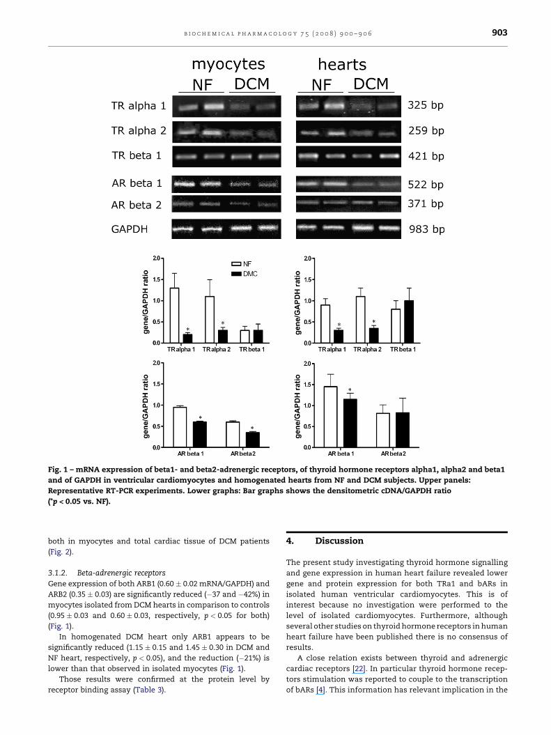
Fig. 1 – mRNA expression of beta1- and beta2-adrenergic receptors, of thyroid hormone receptors alpha1, alpha2 and beta1
and of GAPDH in ventricular cardiomyocytes and homogenated hearts from NF and DCM subjects. Upper panels:
Representative RT-PCR experiments. Lower graphs: Bar graphs shows the densitometric cDNA/GAPDH ratio
(*p < 0.05 vs. NF).
b i o c h e m i c a l p h a r m a c o l o g y 7 5 ( 2 0 0 8 ) 9 0 0 – 9 0 6 903
both in myocytes and total cardiac tissue of DCM patients
(Fig. 2).
3.1.2. Beta-adrenergic receptors
Gene expression of both ARB1 (0.60 � 0.02 mRNA/GAPDH) and
ARB2 (0.35 � 0.03) are significantly reduced (�37 and �42%) in
myocytes isolated from DCM hearts in comparison to controls
(0.95 � 0.03 and 0.60 � 0.03, respectively, p < 0.05 for both)
(Fig. 1).
In homogenated DCM heart only ARB1 appears to be
significantly reduced (1.15 � 0.15 and 1.45 � 0.30 in DCM and
NF heart, respectively, p < 0.05), and the reduction (�21%) is
lower than that observed in isolated myocytes (Fig. 1).
Those results were confirmed at the protein level by
receptor binding assay (Table 3).
4. Discussion
The present study investigating thyroid hormone signalling
and gene expression in human heart failure revealed lower
gene and protein expression for both TRa1 and bARs in
isolated human ventricular cardiomyocytes. This is of
interest because no investigation were performed to the
level of isolated cardiomyocytes. Furthermore, although
several other studies on thyroid hormone receptors in human
heart failure have been published there is no consensus of
results.
A close relation exists between thyroid and adrenergic
cardiac receptors [22]. In particular thyroid hormone recep-
tors stimulation was reported to couple to the transcription
of bARs [4]. This information has relevant implication in the
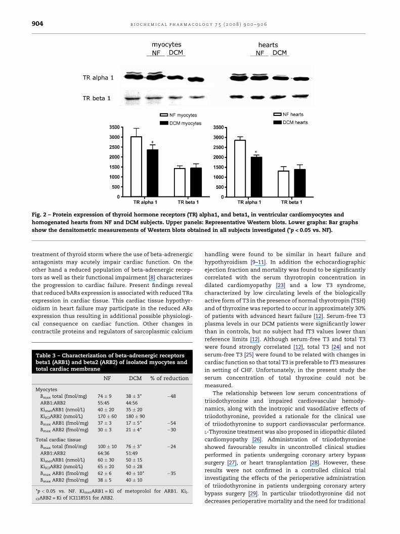
Fig. 2 – Protein expression of thyroid hormone receptors (TR) alpha1, and beta1, in ventricular cardiomyocytes and
homogenated hearts from NF and DCM subjects. Upper panels: Representative Western blots. Lower graphs: Bar graphs
show the densitometric measurements of Western blots obtained in all subjects investigated (*p < 0.05 vs. NF).
b i o c h e m i c a l p h a r m a c o l o g y 7 5 ( 2 0 0 8 ) 9 0 0 – 9 0 6904
treatment of thyroid storm where the use of beta-adrenergic
antagonists may acutely impair cardiac function. On the
other hand a reduced population of beta-adrenergic recep-
tors as well as their functional impairment [8] characterizes
the progression to cardiac failure. Present findings reveal
that reduced bARs expression is associated with reduced TRa
expression in cardiac tissue. This cardiac tissue hypothyr-
oidism in heart failure may participate in the reduced ARs
expression thus resulting in additional possible physiologi-
cal consequence on cardiac function. Other changes in
contractile proteins and regulators of sarcoplasmic calcium
Table 3 – Characterization of beta-adrenergic receptorsbeta1 (ARB1) and beta2 (ARB2) of isolated myocytes andtotal cardiac membrane
NF DCM % of reduction
Myocytes
Bmax total (fmol/mg) 74 � 9 38 � 3* �48
ARB1:ARB2 55:45 44:56
KimetARB1 (nmol/L) 40 � 20 35 � 20
KiICIARB2 (nmol/L) 170 � 60 180 � 90
Bmax ARB1 (fmol/mg) 37 � 3 17 � 5* �54
Bmax ARB2 (fmol/mg) 30 � 3 21 � 4* �30
Total cardiac tissue
Bmax total (fmol/mg) 100 � 10 76 � 3* �24
ARB1:ARB2 64:36 51:49
KimetARB1 (nmol/L) 60 � 30 50 � 15
KiICIARB2 (nmol/L) 65 � 20 50 � 28
Bmax ARB1 (fmol/mg) 62 � 6 40 � 10* �35
Bmax ARB2 (fmol/mg) 38 � 5 40 � 10
*p < 0.05 vs. NF. KimetARB1 = Ki of metoprolol for ARB1. KiI-
CIARB2 = Ki of ICI118551 for ARB2.
handling were found to be similar in heart failure and
hypothyroidism [9–11]. In addition the echocardiographic
ejection fraction and mortality was found to be significantly
correlated with the serum thyrotropin concentration in
dilated cardiomyopathy [23] and a low T3 syndrome,
characterized by low circulating levels of the biologically
active form of T3 in the presence of normal thyrotropin (TSH)
and of thyroxine was reported to occur in approximately 30%
of patients with advanced heart failure [12]. Serum-free T3
plasma levels in our DCM patients were significantly lower
than in controls, but no subject had fT3 values lower than
reference limits [12]. Although serum-free T3 and total T3
were found strongly correlated [12], total T3 [24] and not
serum-free T3 [25] were found to be related with changes in
cardiac function so that total T3 is preferable to fT3 measures
in setting of CHF. Unfortunately, in the present study the
serum concentration of total thyroxine could not be
measured.
The relationship between low serum concentrations of
triiodothyronine and impaired cardiovascular hemody-
namics, along with the inotropic and vasodilative effects of
triiodothyronine, provided a rationale for the clinical use
of triiodothyronine to support cardiovascular performance.
L-Thyroxine treatment was also proposed in idiopathic dilated
cardiomyopathy [26]. Administration of triiodothyronine
showed favourable results in uncontrolled clinical studies
performed in patients undergoing coronary artery bypass
surgery [27], or heart transplantation [28]. However, these
results were not confirmed in a controlled clinical trial
investigating the effects of the perioperative administration
of triiodothyronine in patients undergoing coronary artery
bypass surgery [29]. In particular triiodothyronine did not
decreases perioperative mortality and the need for traditional
b i o c h e m i c a l p h a r m a c o l o g y 7 5 ( 2 0 0 8 ) 9 0 0 – 9 0 6 905
inotropic agents in patients with preexisting impairment of
ventricular function [29].
In a therapeutic context to combat reactivation of fetal
gene expression thyroid hormone is also hampered by
controversial results of investigations of TR mRNA expres-
sion in failing human hearts. Kinugawa et al. [15] reported
that the failing human heart possesses higher levels of TRa2,
with diminished levels of TR a1, and unchanged TRb1 [15].
Other investigations of TR mRNA expression in the left
ventricles of failing human hearts found an increased
expression of TRb1 with lowered levels of TRa1 when
compared to donor hearts [16] or even an increased expres-
sion of all TR [17]. Adding to this complexity, Kinugawa et al.
[30] reported that TR expression is linked to the kind of
hypertrophic stimulation, namely physiological versus
pathological. Physiological stimulation was associated with
increased SERCA2a and a-MHC gene expression, and b-MHC
repression. Further experiments identified TRb to mediate
the physiological response, while TRa1 and -2 mediate the
pathological response [30]. Interestingly, TRb expression was
found to be up-regulated when cultured cardiomyocytes were
incubated with thyroid hormone, which may explain the
clinical observation that short-term thyroid hormone treat-
ment is beneficial in heart failure [31]. Therefore, studies
performed on tissue homogenates of human failing hearts
yielded conflicting results. However the investigation of
homogenated heart does not allow to selectively assess the
participation of different cell types. Therefore the use of
isolated human ventricular myocytes certainly constitutes an
advance because the present findings demonstrates reduced
gene expression of adrenoreceptor (AR) b1 and TRa1 not only
in human failing myocardium but specially in isolated
cardiomyocytes when compared with non-failing myocar-
dium and cardiomyocytes. The total expression of mRNA for
TRalpha receptor was reduced when compared to control
cells although we did not observe an increased mRNA
expression of the TRalpha2 isoform, probably due to the
low sensitivity of our methods.
The reduction in thyroid hormone receptors was closely
associated with a reduced myocyte expression of both beta1-
and beta2-adrenergic receptors. Although the reduction of
cardiac beta-adrenergic receptors is known to characterize the
progression to failure [20], at our knowledge the present is the
first study investigating adrenergic receptors in isolated failing
human ventricular myocytes. This aspect might be important
because the administration of thyroid hormone in patients
with heart failure improved cardiac output, left ventricular
ejection fraction, and decreased isovolumetric relaxation time
with a parallel induction of a beta-adrenergic receptor up-
regulation on peripheral lymphocyte surfaces [32]. Therefore it
cannot be excluded that the reduced expression of the active
form of thyroid receptor hormone might play a role in beta1-
adrenergic receptor reduction in human heart failure. On the
other hand the TH receptor alteration may also represent a
defensive mechanism for the myocyte to reduce oxygen
consumption.
In conclusion, in addition to the impaired population of
adrenergic receptors, a reduced expression of thyroid hor-
mone receptors characterizes cardiac myocytes changes in
advanced heart failure.
Acknowledgement
This work was supported by the Ministero dell’Universita e
della Ricerca Scientifica e Tecnologica, Rome, Italy (grant no.
2003063257-006, to P.A. Modesti).
r e f e r e n c e s
[1] Klein I, Ojamaa K. Thyroid hormone and the cardiovascularsystem. N Engl J Med 2001;344:501–9.
[2] Biondi B, Fazio S, Carella C, Sabatini D, Amato G, CittadiniA, et al. Control of adrenergic overactivity by beta-blockadeimproves the quality of life in patients receiving long termsuppressive therapy with levothyroxine. J Clin EndocrinolMetab 1994;78:1028–33.
[3] Rohrer D, Dillmann WH. Thyroid hormone markedlyincreased the mRNA coding for sarcoplasmic reticulumCa++ ATPase in the rat heart. J Biol Chem 1988;263:6941–4.
[4] Bahouth SW, Cui X, Beauchamp MJ, Park EA. Thyroidhormone induces beta1-adrenergic receptor genetranscription through a direct repeat separated by fivenucleotides. J Mol Cell Cardiol 1997;29:3223–37.
[5] Izumo S, Nadal-Ginard B, Mahdavi V. All members ofthe MHC multigene family respond to thyroid hormonein a highly tissue-specific manner. Science 1986;231:597–600.
[6] Ojamaa K, Klemperer JD, MacGilvray SS, Klein, Samarel A.Thyroid hormone and hemodynamic regulation of beta-myosin heavy chain promoter in the heart. Endocrinology1996;137:802–8.
[7] Nishiyama A, Kambe F, Kamiya K, Seo H, Toyama J. Effectsof thyroid status on expression of voltage-gated potassiumchannels in rat left ventricle. Cardiovasc Res 1998;40:343–51.
[8] Bristow MR, Minobe WA, Raynolds MV, Port JD, RasmussenR, Ray PE, et al. Reduced beta 1 receptor messenger RNAabundance in the failing human heart. J Clin Invest1993;92:2737–45.
[9] Lowes BD, Minobe W, Abraham WT, Rizeq MN, BohlmeyerTJ, Quaife RA, et al. Changes in gene expression in theintact human heart. Downregulation of alpha-myosinheavy chain in hypertrophied, failing ventricularmyocardium. J Clin Invest 1997;100:2315–24.
[10] Klein I. Thyroid and the heart. Thyroid 2002;12:439.[11] Danzi S, Klein I. Thyroid hormone-regulated cardiac gene
expression and cardiovascular disease. Thyroid2002;12:467–72.
[12] Iervasi G, Pingitore A, Landi P, Raciti M, Ripoli A, ScarlattiniM, et al. Low-T3 syndrome: a strong prognostic predictor ofdeath in patients with heart disease. Circulation2003;107:708–13.
[13] Nakao K, Minobe W, Roden R, Bristow MR, Leinwand LA.Myosin heavy chain gene expression in human heartfailure. J Clin Invest 1997;100:2362–70.
[14] Miyata S, Minobe W, Bristow MR, Leinwand LA. Myosinheavy chain isoform expression in the failing andnonfailing human heart. Circ Res 2000;86:386–90.
[15] Kinugawa K, Minobe WA, Wood WM, Ridgway EC, BaxterJD, Ribeiro RC, et al. Signaling pathways responsible forfetal gene induction in the failing human heart: evidencefor altered thyroid hormone receptor gene expression.Circulation 2001;103:1089–94.
[16] Sylven C, Jansson E, Sotonyi P, Waagstein F, Barkhem T,Bronnegard M. Cardiac nuclear hormone receptor mRNA inheart failure in man. Life Sci 1996;59:1917–22.
b i o c h e m i c a l p h a r m a c o l o g y 7 5 ( 2 0 0 8 ) 9 0 0 – 9 0 6906
[17] d’Amati G, di Gioia CR, Mentuccia D, Pistilli D, Proietti-Pannunzi L, Miraldi F, et al. Increased expression of thyroidhormone receptor isoforms in end-stage human congestiveheart failure. J Clin Endocrinol Metab 2001;86:2080–4.
[18] Modesti PA, Vanni S, Paniccia R, Bandinelli B, Bertolozzi I,Polidori G, et al. Characterization of endothelin-1 receptorsubtypes in isolated human cardiomyocytes. J CardiovascPharmacol 1999;34:333–9.
[19] Modesti A, Bertolozzi I, Gamberi T, Marchetta M, LumachiC, Coppo M, et al. Hyperglycemia activates JAK2 signalingpathway in human failing myocytes via angiotensin II-mediated oxidative stress. Diabetes 2005;54:394–401.
[20] Engelhardt S, Bohm M, Erdmann E, Lohse MJ. Analysis ofbeta-adrenergic receptor mRNA levels in humanventricular biopsy specimens by quantitative polymerasechain reactions: progressive reduction of beta 1-adrenergicreceptor mRNA in heart failure. J Am Coll Cardiol1996;27:146–54.
[21] Neri Serneri GG, Cecioni I, Vanni S, Paniccia R, Bandinelli B,Vetere A, et al. Selective upregulation of cardiac endothelinsystem in patients with ischemic but not idiopathic dilatedcardiomyopathy: endothelin-1 system in the human failingheart. Circ Res 2000;86:377–85.
[22] Walker JD, Crawford Jr FA, Mukherjee R, Spinale FG. Thedirect effects of 3,5,30-triiodo-L-thyronine (T3) on myocytecontractile processes. Insights into mechanisms of action. JThorac Cardiovasc Surg 1995;110:1369–79.
[23] Kozdag G, Ural D, Vural A, Agacdiken A, Kahraman G, SahinT, et al. Relation between free triiodothyronine/freethyroxine ratio, echocardiographic parameters andmortality in dilated cardiomyopathy. Eur J Heart Fail2005;7:113–8.
[24] Pingitore A, Iervasi G, Barison A, Prontera C, Pratali L,Emdin M, et al. Early activation of an altered thyroidhormone profile in asymptomatic or mildly symptomatic
idiopathic left ventricular dysfunction. J Card Fail2006;12:520–6.
[25] Calvo-Romero JM, Rodriguez EM. Serum-free thyroxine andthyrotropin concentrations in euthyroid patients withdecompensated congestive heart failure. Int J Cardiol2005;102:367–8.
[26] Moruzzi P, Doria E, Agostoni PG, Capacchione V, SganzerlaP. Usefulness of L-thyroxine to improve cardiac andexercise performance in idiopathic dilatedcardiomyopathy. Am J Cardiol 1994;73:374–8.
[27] Novitzky D, Cooper DKC, Swanepoel A. Inotropic effect oftriiodothyronine (T3) in low cardiac output followingcardioplegic arrest and cardiopulmonary bypass: an initialexperience in patients undergoing open heart surgery. Eur JCardiothorac Surg 1989;3:140–5.
[28] Jeevanandam V, Todd B, Regillo T, Hellman S, Eldridge C,McClurken J. Reversal of donor myocardial dysfunction bytriiodothyronine replacement therapy. J Heart LungTransplant 1994;13:681–7.
[29] Klemperer JD, Klein I, Gomez M, Helm RE, Ojamaa K,Thomas SJ, et al. Thyroid hormone treatment aftercoronary-artery bypass surgery. N Engl J Med1995;333:1522–7.
[30] Kinugawa K, Yonekura K, Ribeiro RC, Eto Y, Aoyagi T,Baxter JD, et al. Regulation of thyroid hormone receptorisoforms in physiological and pathological cardiachypertrophy. Circ Res 2001;89:591–8.
[31] Hamilton MA, Stevenson LW, Fonarow GC, Steimle A,Goldhaber JI, Child JS, et al. Safety and hemodynamiceffects of intravenous triiodothyronine in advancedcongestive heart failure. Am J Cardiol 1998;81:443–7.
[32] Lu X, Huang J, Zhang X, Li X, Wang C, Zhang P, et al. Effectsof thyroxine on cardiac function and lymphocyte beta-adrenoreceptors in patients with chronic congestive heartfailure. Chin Med J (Engl) 2003;116:1697–700.