Myelin localization of peptidylarginine deiminases 2 and 4: comparison of PAD2 and PAD4 activities
11
Myelin localization of peptidylarginine deiminases 2 and 4: comparison of PAD2 and PAD4 activities Dorothy D Wood 1 , Cameron A Ackerley 2 , Ben van den Brand 1 , Li Zhang 1 , Reinout Raijmakers 3 , Fabrizio G Mastronardi 1 and Mario A Moscarello 1 An understanding of the structure and composition of the myelin sheath is essential to understand the pathogenesis of demyelinating diseases such as multiple sclerosis (MS). The presence of citrulline in myelin proteins in particular myelin basic protein (MBP) causes an important change in myelin structure, which destabilizes myelin. The peptidylarginine deiminases (PADs) are responsible for converting arginine in proteins to citrulline. Two of these, PAD2 and PAD4, were localized to the myelin sheath by immunogold electron microscopy. Deimination of MBP by the recombinant forms of these enzymes showed that it was extensive, that is, PAD2 deiminated 18 of 19 arginyl residues in MBP, whereas PAD4 deiminated 14 of 19 residues. In the absence of PAD2 (the PAD2-knockout mouse) PAD4 remained active with limited deimination of arginyl residues. In myelin isolated from patients with MS, the amounts of both PAD2 and PAD4 enzymes were increased compared with that in normals, and the citrullinated proteins were also increased. These data support the view that an increase in citrullinated proteins resulting from increased PAD2 and 4 is an important change in the pathogenesis of MS. Laboratory Investigation (2008) 88, 354–364; doi:10.1038/labinvest.3700748; published online 28 January 2008 KEYWORDS: peptidylarginine deiminase; citrullinated myelin basic protein; myelin; multiple sclerosis; neurodegenerative disease Peptidylarginine deiminases (PADs) are enzymes which dei- minate arginyl residues in proteins. Five isozymes, termed PADs 1–4 and PAD6, are known, which have some tissue specificity, although PAD2 is widely distributed. 1 It is the principal PAD enzyme in brain, where it has been shown to be responsible for the deimination of arginyl residues in myelin basic protein (MBP), a principal protein in myelin 2 and a possible autoantigen in multiple sclerosis (MS). 3,4 The presence of other PAD enzymes in myelin has not been re- ported. For all PADs, the deimination reaction involves the conversion of arginyl residues in proteins to citrulline and the formation of ammonia. 5 The conversion of arginine to ci- trulline involves the loss of one positive charge from the protein for each arginine deiminated. In earlier studies, we reported that MBP isolated from white matter from normal human brain could be fractio- nated into several components by column chromatography. One of these components, which accounted for 20% of all the MBP, contained six citrullinyl residues, the sites of which we determined by protein sequencing. 6 More recently, the deimination has been shown to be more extensive by mass spectrometry with partial deimination of many arginines. 7 The loss of six positive charges accounted for the chroma- tographic behavior on the cation-exchange column. This same fraction isolated from patients with MS accounted for 45% of the total MBP, and in a case of fulminating MS, it accounted for 80–90% of all the MBP. 8 Instead of six arginyl residues in normal MBP, 6 18 of 19 arginyl residues in the Marburg’s MBP were deiminated. In a number of in vitro studies in protein–lipid interaction systems, we showed that this deiminated protein was unable to compact lipid bilayers, causing destabilization, which might be the mechanism of demyelination. 9 Our recent studies have revealed that the deiminated MBP was more susceptible to proteolysis by myelin-associated proteases, 10,11 and the citrullinated MBP was digested autocatalytically in vitro, releasing the im- munodominant epitope in the absence of proteases. 12 These studies have been reviewed recently. 4 Although the con- tribution of PAD enzymes in the pathogenesis of MS is unclear at this time, the presence of large amounts of Received 17 October 2007; revised 03 December 2007; accepted 10 December 2007 1 Molecular Structure and Function, The Hospital for Sick Children, Toronto, ON, Canada; 2 Department of Pathology, The Hospital for Sick Children, Toronto, ON, Canada and 3 Department of Biomolecular Chemistry, Nijmegen Center for Molecular Life Sciences, Radboud University Nijmegen, Nijmegen, The Netherlands Correspondence: Dr MA Moscarello, MD, PhD, Molecular Structure and Function, The Hospital for Sick Children, 555 University Avenue, Toronto, Ontario, Canada M5G 1X8. E-mail: [email protected] Laboratory Investigation (2008) 88, 354–364 & 2008 USCAP, Inc All rights reserved 0023-6837/08 $30.00 354 Laboratory Investigation | Volume 88 April 2008 | www.laboratoryinvestigation.org
-
Upload
independent -
Category
Documents
-
view
3 -
download
0
Transcript of Myelin localization of peptidylarginine deiminases 2 and 4: comparison of PAD2 and PAD4 activities

Myelin localization of peptidylarginine deiminases2 and 4: comparison of PAD2 and PAD4 activitiesDorothy D Wood1, Cameron A Ackerley2, Ben van den Brand1, Li Zhang1, Reinout Raijmakers3,Fabrizio G Mastronardi1 and Mario A Moscarello1
An understanding of the structure and composition of the myelin sheath is essential to understand the pathogenesis ofdemyelinating diseases such as multiple sclerosis (MS). The presence of citrulline in myelin proteins in particular myelinbasic protein (MBP) causes an important change in myelin structure, which destabilizes myelin. The peptidylargininedeiminases (PADs) are responsible for converting arginine in proteins to citrulline. Two of these, PAD2 and PAD4, werelocalized to the myelin sheath by immunogold electron microscopy. Deimination of MBP by the recombinant forms ofthese enzymes showed that it was extensive, that is, PAD2 deiminated 18 of 19 arginyl residues in MBP, whereas PAD4deiminated 14 of 19 residues. In the absence of PAD2 (the PAD2-knockout mouse) PAD4 remained active with limiteddeimination of arginyl residues. In myelin isolated from patients with MS, the amounts of both PAD2 and PAD4 enzymeswere increased compared with that in normals, and the citrullinated proteins were also increased. These data support theview that an increase in citrullinated proteins resulting from increased PAD2 and 4 is an important change in thepathogenesis of MS.Laboratory Investigation (2008) 88, 354–364; doi:10.1038/labinvest.3700748; published online 28 January 2008
KEYWORDS: peptidylarginine deiminase; citrullinated myelin basic protein; myelin; multiple sclerosis; neurodegenerative disease
Peptidylarginine deiminases (PADs) are enzymes which dei-minate arginyl residues in proteins. Five isozymes, termedPADs 1–4 and PAD6, are known, which have some tissuespecificity, although PAD2 is widely distributed.1 It is theprincipal PAD enzyme in brain, where it has been shown tobe responsible for the deimination of arginyl residues inmyelin basic protein (MBP), a principal protein in myelin2
and a possible autoantigen in multiple sclerosis (MS).3,4 Thepresence of other PAD enzymes in myelin has not been re-ported. For all PADs, the deimination reaction involves theconversion of arginyl residues in proteins to citrulline and theformation of ammonia.5 The conversion of arginine to ci-trulline involves the loss of one positive charge from theprotein for each arginine deiminated.
In earlier studies, we reported that MBP isolated fromwhite matter from normal human brain could be fractio-nated into several components by column chromatography.One of these components, which accounted for 20% of all theMBP, contained six citrullinyl residues, the sites of whichwe determined by protein sequencing.6 More recently, the
deimination has been shown to be more extensive by massspectrometry with partial deimination of many arginines.7
The loss of six positive charges accounted for the chroma-tographic behavior on the cation-exchange column. Thissame fraction isolated from patients with MS accounted for45% of the total MBP, and in a case of fulminating MS, itaccounted for 80–90% of all the MBP.8 Instead of six arginylresidues in normal MBP,6 18 of 19 arginyl residues in theMarburg’s MBP were deiminated. In a number of in vitrostudies in protein–lipid interaction systems, we showed thatthis deiminated protein was unable to compact lipid bilayers,causing destabilization, which might be the mechanism ofdemyelination.9 Our recent studies have revealed that thedeiminated MBP was more susceptible to proteolysis bymyelin-associated proteases,10,11 and the citrullinated MBPwas digested autocatalytically in vitro, releasing the im-munodominant epitope in the absence of proteases.12 Thesestudies have been reviewed recently.4 Although the con-tribution of PAD enzymes in the pathogenesis of MS isunclear at this time, the presence of large amounts of
Received 17 October 2007; revised 03 December 2007; accepted 10 December 2007
1Molecular Structure and Function, The Hospital for Sick Children, Toronto, ON,Canada; 2Department of Pathology, The Hospital for Sick Children, Toronto, ON, Canada and 3Department of Biomolecular Chemistry, Nijmegen Center for MolecularLife Sciences, Radboud University Nijmegen, Nijmegen, The NetherlandsCorrespondence: Dr MA Moscarello, MD, PhD, Molecular Structure and Function, The Hospital for Sick Children, 555 University Avenue, Toronto, Ontario, CanadaM5G 1X8. E-mail: [email protected]
Laboratory Investigation (2008) 88, 354–364
& 2008 USCAP, Inc All rights reserved 0023-6837/08 $30.00
354 Laboratory Investigation | Volume 88 April 2008 | www.laboratoryinvestigation.org

citrullinated MBP suggests a strong link between citrullinatedMBP and peptidylarginine deiminases.
Since MBP is a major myelin protein and citrullinatedMBP was localized in myelin by immunogold electronmicroscopy,13 the localization of PAD2 in myelin was un-dertaken. Immunogold studies were carried out with an anti-PAD2 antibody. The data demonstrate clearly that PAD2 islocalized in myelin, in the vicinity of MBP its natural sub-strate. The number of gold particles in myelin from MS brainwas increased threefold over normal, in agreement with aprevious study in which we reported a threefold increase inthe amount of PAD 2 by immunoblots.14 In a study of thedeiminated sites, no restrictions were observed, that is, allarginyl residues except one were targets for deimination inaqueous media. Since citrullinated MBP is generated by PAD,these studies suggest that decreased PAD enzyme activity maybe a therapeutic target with PAD-specific inhibitors, as sug-gested for rheumatoid arthritis.15,16
We extended our studies on PAD2 in myelin by studying aPAD2-knockout mouse to determine if citrullinated MBPcould be formed in the absence of PAD2. We demonstratedthat citrullinated MBP was present and was formed by thePAD4 enzyme. Immunogold localization of PAD4 showed itto differ from that of PAD2. PAD2 was found in clusters,whereas PAD4 was distributed as single particles in myelinand in axons. Nuclear labeling of PAD4 was a prominentfeature. The presence of PAD4 in myelin was also confirmedby Western blot of myelin proteins isolated from PAD2 nullmice.
MATERIALS AND METHODSMaterialsa-N-benzoyl-L-arginine ethyl ester (BAEE), L-citrulline andcalf thymus histone type IIA were purchased from SigmaChemical Company (St Louis, MO, USA). MBP was preparedby the method of Cheifetz and Moscarello.17 The chargeisomers were isolated on CM52 columns as described by us.6
Normal human brain was obtained from the Canadian BrainTissue Bank. Normal white matter samples from brains ofMS patients and white matter from non-neurologicalcontrols were obtained from the MS tissue bank at UCLA.Human recombinant PAD2 and PAD4 (EC 3.5–3.15) wereprepared as described by Raijmakers et al18 and supplied tous for this study. The normal mice used for this study wereon the CD-1 background, and were housed in the Hospitalfor Sick Children Animal Care facility. These studies wereapproved by the Animal Care Committee at The Hospitalfor Sick Children. The brains from PAD2-null mice19 andnormal controls were provided in a frozen condition fromthe Netherlands, and were obtained in accordance withapproved animal protocols.
Preparation of MyelinMyelin was prepared from normal-appearing white matterfrom brains of MS tissue and normal human white matter
obtained from the brain tissue bank at UCLA, by the methodof Cruz and Moscarello.20
AntibodiesPrimary antibodies included anti-MBP monoclonal antibodyfrom Chemicon (MAb348). The epitope for this antibodyhas been characterized by Groome et al.21 The polyclonalantibody to PAD isozymes1–4 was a gift from Dr H Takahara(Department of Applied Biological Resource Sciences, Schoolof Agriculture, Ibaraki University, Ami-machi, Inashiki-gun,Ibaraki, Japan). It reacts with all the PAD enzymes because ofthe high degree of sequence homology at the C-terminus ofthese enzymes.22 The isotype-specific anti-PAD2 antibodywas generated by immunization with two peptides com-prising amino acids 3–18 and 516–531 of human PAD2.18,19
The PAD4 isotype-specific antibody was raised againstpeptides 180–194 of human PAD4.18 The monoclonal anti-citrulline antibody, MAbF9523 was obtained from Dr ANicholas, Birmingham, Alabama.
Immunogold Electron MicroscopyPellets of myelin isolated from white matter from normalindividuals and normal-appearing white matter from pa-tients with MS and optic nerves from normal CD-1 micewere fixed in 4% paraformaldehyde containing 0.1% glutar-aldehyde in 0.1 M phosphate buffer, pH 7.4, for a minimumof 4 h. Following a thorough rinse in phosphate buffer,samples were infused with 2–3 M sucrose for several hoursbefore freezing in liquid nitrogen. The sucrose-infused ma-terial was then mounted on aluminum ultramicrotomy pins,plunged frozen in liquid nitrogen and ultrathin cryosectionswere prepared with a cryo-ultramicrotome at �951C with adiamond knife. Sections were transferred to formvar nickelgrids with a loop of molten sucrose and labeled usingimmunogold. Following blocking in PBS containing 0.15%glycine and 0.5% BSA, the grids were rinsed with PBS/BSAbefore incubation with a polyclonal antibody against PAD for1 h. The grids were then washed several times with PBS/BSAbefore incubation with either goat anti-rabbit IgG (10 nm)for isolated myelin or 5 nm gold particles for murine opticnerve (Amersham Life Sciences, IL, USA), for an additionalhour. In double-labeling experiments, samples were rinsedthoroughly with PBS/BSA and then incubated with MAb26,21
a monoclonal antibody that recognizes MBP. Again, thesamples were rinsed thoroughly in PBS/BSA and incubatedwith 10 nm gold goat anti-murine complexes (AmershamLife Sciences). After a thorough rinsing in PBS and distilledwater, sections were stabilized in a thin film of methylcellu-lose containing 0.2% uranyl acetate. Controls included theomission of either primary antibody or antibody gold com-plexes. All of the grids were then examined in JEOL JEM 1230transmission electron microscope (JEOL USA; Peabody, MA,USA) and images were acquired with a digital camera (AMTCorp, Danvers, MA, USA). Gold particle densities were cal-culated in particles/mm2 of myelin. Images acquired with the
Peptidylarginine deiminase 2 and 4 in myelin
DD Wood et al
www.laboratoryinvestigation.org | Laboratory Investigation | Volume 88 April 2008 355

camera were exported into an image analysis program(SCION Image, Frederick, MD, USA) and 28 images fromnormal and MS were analyzed. Data were expressed as meanand standard error. Significance was determined usingStudent’s t-test.
Immunoslot Blot for PAD2 and PAD4 in Myelin Fractionof CD-1 MiceCD-1 mouse brains were homogenized in 0.25 M sucrose andseparated on sucrose gradient of 0.25, 0.88, 1 and 1.2 Msucrose by centrifuging for 1 h at 28 000 r.p.m. The interphasebetween 0.25 and 0.88 M contained the myelin fraction,which was collected and washed three times. Samples werediluted to 0.1 mg/ml in 3 M urea/PBS and 10 mg protein wasapplied onto nitrocellulose membrane using Bio-Dot SFMicrofiltration Apparatus. After blocking for 1 h in 5% milkin Tris-buffered saline and 0.05% Tween 20 (TBS/T), mem-branes were incubated for 1.5 h at room temperature withpolyclonal anti-PAD2 (1:1000), polyclonal anti-PAD4(1:1000) or polyclonal anti-actin antibodies (1:200) (Sigma)diluted in 5% milk in TBS/T. Subsequently, membranes wereincubated with HRP-conjugated anti-rabbit (1:5000) (Bio-Rad) antibody diluted in 5% milk in TBS/T for 1 h at roomtemperature, and then visualized with ECL (GE Healthcare).
Numbers of pixels on film were measured with the ImageSXM image analysis software, using a Macintosh (Apple Inc.,Cupertino, CA, USA) computer with OSX version 10.4.10.The average of two measurements per sample was used tocalculate the PAD-protein/actin ratio. Student’s t-test wasused to determine significance between the PAD2 and PAD4measurements.
PAD Enzyme AssaysPAD activity was measured with recombinant PAD2 andPAD4, using benzoylarginine ethyl ester (BAEE) as substrate,as previously described.2 MBP was deiminated by PAD2 andPAD4 in separate experiments using similar conditions. Forthe mass spectrometry experiments, MBP was deiminated for2 h to ensure complete deimination.
When BAEE was used as substrate, varying concentrationsof BAEE were incubated in 0.1 M CaCl2, 1.0 M HEPES bufferpH 7.6, 0.08 M DTT and 1 ml of enzyme solution containing2.6 mg PAD2 or 2.2 mg PAD4. The reaction was stopped with100 ml of 5 N perchloric acid. Each sample was centrifugedat 41C for 3 min at 1500 r.p.m. in a Hettich (Tuttingen,Germany) tabletop centrifuge. Aliquots of 400 ml were used inthe citrulline assays. The aliquots were mixed with 1 ml ofreagent B (10% antipyrineþ 2.5% ferric ammonium sulfate,3.5 N sulfuric acid and 3.5 N orthophosphoric acid. ReagentA (0.5 ml), containing 0.5% diacetylmonoxime and 15%sodium chloride, was added and the mixture was vortexedbefore boiling the sample for 15 min in a water bath. Thesamples were cooled on ice and absorbance was read at464 nm. When MBP was used as substrate, component 1
(C-1 the least modified of the MBP components) was addedin amounts varying from 0 to 250 mg of protein.
Preparation of MBP from Mouse MyelinMBP was extracted from myelin with 0.2 N sulfuric acid andcentrifuged for 1 h at 41C. The supernatant was removed anddialyzed against water overnight. Protein assays were carriedout by the method of Peterson.24
ImmunodetectionMyelin proteins were resolved through a 12.5%. SDS-poly-acrylamide gel by electrophoresis and western blotted ontonitrocellulose membrane. Non-protein sites on the nitro-cellulose membranes were blocked with 5% milk in TBS/0.05% Tween 20 and subsequently incubated with thefollowing primary antibodies diluted in blocking buffer:anti-PAD4 (1:250), F95 (1:50) and anti-MBP (1:10 000) for1 h at room temperature. The following secondary antibodieswere diluted in blocking buffer: anti-rabbit IgG HRP-con-jugated (1:5000) for antibody anti-PAD4, anti-mouse IgMHRP-conjugated antibody for F95 (1:50) and anti-mouseIgG HRP-conjugated antibody for anti-MBP (1:10 000), andincubation was performed for 1 h at room temperature.Antibodies were visualized with ECL (GE Healthcare).
For immunoslot blot analysis, the number of pixels on filmwere measured with Image SXM. The average from threemeasurements per sample was used to calculate citrulline/MBP ratios. A Student’s t-test was used to determinesignificance.
Preparation of Lipid VesiclesEgg phosphatidylcholine (PC) and bovine brain phosphati-dylserine (PS) were purchased from Avanti (Birmingham,AL, USA) as dry powders. For preparation of the vesicles, thelipids were dissolved in chloroform at a concentration of20 mg/ml (PC) and 10 mg/ml (PS), and stored under nitro-gen at �201C.6 Aliquots of the lipids generating a mixture of92.2% PC and 7.8% PS were transferred to autoclavable tubesand the chloroform was evaporated under a stream ofnitrogen and further dried by lyophilizing for 10 min. Thelipids were suspended in 1 ml of HEPES buffer, pH 7.6,containing 0.1 M CaCl2 and 0.08 M DTT, and then sonicatedin a water bath at room temperature until clarified(approximately 30 min). The vesicles were centrifuged at15 000 r.p.m. for 15 min in Hettich centrifuge. The lipidvesicles were suspended in 490 ml of HEPES buffer and theMBP was added followed by 10 ml of PAD2 or PAD4, and themixture was incubated at 521C for 2 h. After incubation,ether was added to remove the lipid and the insoluble ma-terial was taken up in water and dialyzed for 20 min usingthree changes of water. The retentates were lyophilized.
Mass SpectrometryMolecular masses of intact human MBP (component C-1)and its citrullinated forms were measured with a QSTAR XL
Peptidylarginine deiminase 2 and 4 in myelin
DD Wood et al
356 Laboratory Investigation | Volume 88 April 2008 | www.laboratoryinvestigation.org

electrospray ionization QTOF mass spectrometer (AppliedBiosystems/MDS Sciex, Concord, ON, Canada) in positiveion mode. Samples of 5 mg each were dissolved in a solutionof acetonitrile and deionized water (1:1, v/v) containing 0.2%of formic acid at a concentration of 10 pmol/ml. Samplesconsisting 2 ml of each were injected using an HPLC system(Waters, Milford, MA, USA) and carried over to the nanos-pray source by a mobile phase of 50% acetonitrile in deio-nized water containing 0.2% of formic acid. The flow ratewas kept at 6 ml/min and the capillary voltage was maintainedat 3000 eV. The mass spectra obtained were deconvolutedusing the Bayesian Protein reconstruct script, which comeswith the ABI BioAnalyst 1.1 software.
Protein Sequence DeterminationMBP samples (5 mg) were digested in solution using 0.05 mgof trypsin in 25 mM ammonium bicarbonate solution in atotal volume of 50 ml. After overnight incubation at 371C, thepeptide solution was lyophilized by SpeedVac centrifugationand resuspended in 20 ml 0.1% trifluoroacetic acid (TFA).Each sample consisting of 1ml solution was mixed with 1 mlof 2,5-dihydroxybenzoic acid (120 mg/ml in acetonitrile/H2O, 1:1, v/v) and spotted on the target plate for MALDI-QTOF MS analysis. Peptide mass measurements were per-formed with a QSTAR XL MALDI-QTOF mass spectrometer(Applied Biosystems/MDS Sciex). The instrument wasequipped with a UV nitrogen laser (337 nm) and the accele-ration voltage was 4 kV. Nitrogen and argon were used ascurtain and collision gases, respectively.
LC/MS/MS experiments were performed with a QSTAR XLelectrospray ionization QTOF mass spectrometer (AppliedBiosystems/MDS Sciex) coupled with an Agilent 1100NanoLC system (Santa Clara, CA, USA). Samples (5ml) werefirst loaded onto a pre-column (100 mm i.d.� 5 cm) and theneluted to an analytical column (75 mm i.d.� 10 cm) for fur-ther separation. Both columns were home made and packedwith Pursuit C18 (5 mm particle size, 200 A pore size; VarianInc., Palo Alto, CA, USA). The mobile phase was composedof solutions A and B, where A was 2% acetonitrile in waterand B was 90% acetonitrile in water, each containing 0.1%formic acid and 0.02% TFA. The gradient started at 0% Band ramped to 100% B in 65 min. Flow rate was set at 0.2 ml/min. MS and MS/MS experiments were performed usinginformation-dependent acquisition with each cycle time of10 s (1-s MS scan followed by three 3 s MS/MS scans). Thedata set was converted into a peak list file called MascotGeneric File (MGF) using the Mascot script, which comeswith the Analyst QS 1.1 software. The MGF files were thenused to search against the human MBP FASTA sequencedatabase, using our in-house MASCOT search engine. Pro-tein N-terminal acetylation, arginine methylation, dimethy-lation, arginine citrullination and methionine oxidation wereset as variable modifications in the search. Peptide masstolerance and MS/MS tolerance was set to 0.3 Da.
RESULTSLocalization of PAD in MyelinTo demonstrate the presence of PAD in myelin, cryosectionsof myelin isolated from normal and MS white matter werelabeled with a polyclonal PAD antibody (which recognizesall PAD isoforms) as described in Materials and Methods.Representative sections are shown in Figure 1a and b. Goldparticles were found in the myelin sheath isolated fromnormal human white matter (Figure 1a). An increasednumber of gold particles were observed in myelin isolatedfrom MS normal-appearing white matter (Figure 1b).The increase in gold particles was quantitated by obtainingparticle densities (particles/mm2 of myelin). These areshown in Table 1. The number of particles/mm2 was increasedapproximately threefold in the MS samples, showing anapproximately threefold increase in PAD enzyme inwhite matter from MS tissue. This is consistent with ourimmunoslot blot measurements showing a threefold increasein the amount of PAD2 enzyme in normal-appearing whitematter from MS tissue.14
Localization of PAD in Murine White MatterSince the myelin shown in Figure 1 was prepared fromnormal-appearing white matter obtained at autopsy, we ex-amined optic nerve from normal CD-1 mice to examine goldlabeling of myelin in situ. These are shown in Figure 2a–c forPAD2 and Figure 3a–c for PAD4. PAD2 labeling was found inthe oligodendrocyte (Figure 2a) and in the myelin (upperright-hand corner). The large particles in this figure representMBP labeling, whereas the small particles represent PAD2labeling. Although PAD2 labeling was distributed throughoutthe cell, much of the labeling was found in clusters, suggestiveof a vesicular localization. PAD2 labeling was found in theaxon (Figure 2b) largely in clusters (arrows), and in theperiaxonal region (arrowheads). Clusters of PAD2-labeledgold particles were also found in myelin (Figure 2c, arrow-heads).
Immunogold labeling for PAD4 is shown in Figure 3a–c.PAD4 labeling was found in the oligodendrocyte cytoplasm(Figure 3a) and in the nucleus associated with chromatin. Inthe axon, particles were associated with intermediate fila-ments (Figure 3b). Gold labeling of intermediate filaments, aswell as myelin labeling, is also shown in Figure 3c. Much ofthe myelin labeling was in the periaxonal space. The goldlabeling for PAD2 was in clusters, whereas that for PAD4was generally observed as single particles. Control sectionswere incubated with either secondary gold-conjugatedantibody alone (Supplementary Figure 1A), or with primaryanti-PAD2 antibody preadsorbed with PAD2 protein(Supplementary Figure 1B).
Quantitation of PAD2 and PAD4 in Isolated MyelinTo determine the amount of PAD2 and PAD4 in myelinisolated from normal and MS white matter, we performedimmunoslot blots with specific PAD2 and PAD4 antibodies
Peptidylarginine deiminase 2 and 4 in myelin
DD Wood et al
www.laboratoryinvestigation.org | Laboratory Investigation | Volume 88 April 2008 357
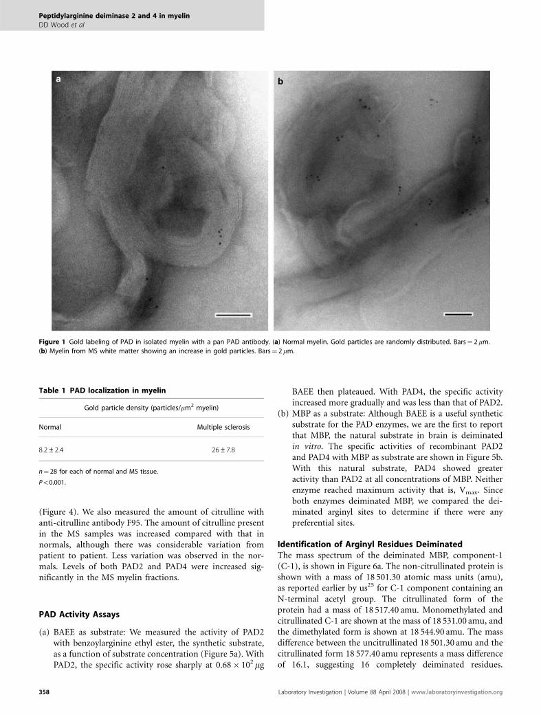
(Figure 4). We also measured the amount of citrulline withanti-citrulline antibody F95. The amount of citrulline presentin the MS samples was increased compared with that innormals, although there was considerable variation frompatient to patient. Less variation was observed in the nor-mals. Levels of both PAD2 and PAD4 were increased sig-nificantly in the MS myelin fractions.
PAD Activity Assays
(a) BAEE as substrate: We measured the activity of PAD2with benzoylarginine ethyl ester, the synthetic substrate,as a function of substrate concentration (Figure 5a). WithPAD2, the specific activity rose sharply at 0.68� 102mg
BAEE then plateaued. With PAD4, the specific activityincreased more gradually and was less than that of PAD2.
(b) MBP as a substrate: Although BAEE is a useful syntheticsubstrate for the PAD enzymes, we are the first to reportthat MBP, the natural substrate in brain is deiminatedin vitro. The specific activities of recombinant PAD2and PAD4 with MBP as substrate are shown in Figure 5b.With this natural substrate, PAD4 showed greateractivity than PAD2 at all concentrations of MBP. Neitherenzyme reached maximum activity that is, Vmax. Sinceboth enzymes deiminated MBP, we compared the dei-minated arginyl sites to determine if there were anypreferential sites.
Identification of Arginyl Residues DeiminatedThe mass spectrum of the deiminated MBP, component-1(C-1), is shown in Figure 6a. The non-citrullinated protein isshown with a mass of 18 501.30 atomic mass units (amu),as reported earlier by us25 for C-1 component containing anN-terminal acetyl group. The citrullinated form of theprotein had a mass of 18 517.40 amu. Monomethylated andcitrullinated C-1 are shown at the mass of 18 531.00 amu, andthe dimethylated form is shown at 18 544.90 amu. The massdifference between the uncitrullinated 18 501.30 amu and thecitrullinated form 18 577.40 amu represents a mass differenceof 16.1, suggesting 16 completely deiminated residues.
Table 1 PAD localization in myelin
Gold particle density (particles/mm2 myelin)
Normal Multiple sclerosis
8.2±2.4 26±7.8
n¼ 28 for each of normal and MS tissue.
Po0.001.
Figure 1 Gold labeling of PAD in isolated myelin with a pan PAD antibody. (a) Normal myelin. Gold particles are randomly distributed. Bars¼ 2 mm.
(b) Myelin from MS white matter showing an increase in gold particles. Bars¼ 2 mm.
Peptidylarginine deiminase 2 and 4 in myelin
DD Wood et al
358 Laboratory Investigation | Volume 88 April 2008 | www.laboratoryinvestigation.org

Since we found 18 of 19 arginyl residues to be deiminated(see below), a small amount of non-deiminated arginylresidue accounted for the overall mass change of 16 amu.In order to determine the arginyl residues deiminatedin MBP, the protein was isolated from the reaction mixture,digested with trypsin and the peptides were analyzed bymass spectrometry as described in Materials and Methods.
The arginyl residues converted to citrulline were determinedby fragmentation analysis. The entire sequence of MBPshowing the deiminated sites is shown in Figure 6b forPAD2 and PAD4 reactions. The underlined arginines(R; Figure 6b) were deiminated by the PAD2 reaction, whilethe asterisks represent arginines deiminated in the PAD4reaction.
Figure 2 Immunogold electron microscopy of normal mouse optic nerve, with anti PAD2 antibody. (a) Oligodendrocyte showing PAD2 labeling (small
particles) and MBP labeling (large particles). Ol, oligodendrocyte; My, myelin. Bars¼ 2 mm. (b) PAD2 labeling in the axon (arrows) and periaxonal distribution
(arrowhead). Ax, axon. Bars¼ 2 mm. (c) PAD2 labeling in myelin showing distribution in clusters (arrowheads). My, myelin. Bars¼ 2 mm.
Peptidylarginine deiminase 2 and 4 in myelin
DD Wood et al
www.laboratoryinvestigation.org | Laboratory Investigation | Volume 88 April 2008 359

Figure 3 Immunogold electron microscopy of mouse optic nerve using anti-PAD4 antibody. (a) Oligodendrocyte showing PAD4 labeling (arrows and
arrowheads) in the cytoplasm (asterisk) and nuclear labeling associated with chromatin. N, nucleus. (b) Myelin labeling distributed as single particles
(arrowheads). Ax, axon; IF, intermediate filaments. (c) Axonal labeling, axolema labeling (arrows) and intermediate filaments within the axon and myelin
labeling. My, myelin; IF, intermediate filaments. Bars¼ 2 mm.
Peptidylarginine deiminase 2 and 4 in myelin
DD Wood et al
360 Laboratory Investigation | Volume 88 April 2008 | www.laboratoryinvestigation.org

Several interesting conclusions arise from the data. It canbe seen that most of the arginyl residues were deiminated byboth PAD2 and PAD4, although PAD2 deiminated more sitesthan PAD4, for example, PAD2 deiminated 18 of 19 arginylresidues, whereas PAD4 deiminated 14 of 19 residues. Threeof the sites not deiminated by PAD4 were in the N-terminalportion of the molecule (R5, R9 and R43), suggesting thatstructural factors either primary or secondary may be in-volved. In an earlier report, deimination of arginyl residues intrichohyalin adjacent to glutamic acid were only slightlymodified, showing that amino-acid sequence influences PADactivity.26 Although many sites were deiminated to someextent, none was completely deiminated. This can be seenfrom the intensity of the peaks for the different peptides,which varied considerably (Supplementary data). The GRGsite, which contains R107, the major methylated R, is mono-or dimethylated, but is also deiminated in those MBPmolecules that are not methylated at this site. Since massspectrometry of the protein after 2 h in the presence of eitherPAD2 or PAD4 revealed that both mono- and dimethylatedarginines remained (Figure 6a), it suggested that deimina-tion5,27 of the methylated arginine did not occur.28–30
Consistent with our data, Raijmakers et al31 showed that thehuman, mouse and rabbit PAD enzymes were not able to‘reverse’ the methylation of arginine residues by convertingmonomethylated arginine into citrulline in synthetic pep-tides. These authors showed that the human and mousePAD enzymes can only convert non-methylated peptidy-larginine into peptidylcitrulline in vitro. Our data supportthis conclusion.
PAD4 and Citrullinated Proteins from Myelin ofPAD2-Knockout MiceBy western blot we were unable to detect PAD2 in solubilizedmyelin prepared from PAD2-null mouse brain. However,western blots with antibody to PAD4 showed good im-munoreactivity (Figure 7), in agreement with the im-munogold labeling (Figure 2), and the amount was similar tothat found in the wild-type mice. To establish that MBPisolated from myelin of knockout mice was citrullinated, weisolated the acid-soluble MBP fraction from myelin, digestedit with trypsin and subjected the digest to mass spectrometricanalysis. In MBP from normal mouse myelin, we identifiedseven peptides in which arginine was deiminated. In thePAD2-knockout, we identified three citrullinated peptides,two of which represented the mono- and dimethylatedarginyl residues in the GRG sequence. The third peptide
Figure 4 Immunoslot blot of solubilized myelin represented as a bar
diagram showing reactivity to PAD2 and PAD4. Slot blots were probed with
anti-citrulline antibody, F95, anti-PAD2 antibody and anti-PAD4 antibody.
MS, multiple sclerosis; n¼ 6 normal and 7 MS individuals. Representative
slot blots are shown in Supplementary Figure 2. ***¼ significant difference.
Figure 5 Velocity–substrate relationship of recombinant PAD2 and PAD4.
(a) BAEE substrate (mg cit/min/mg enzyme� 10�3). (b) MBP substrate
(mg cit/min/mg enzyme� 10�3).
Peptidylarginine deiminase 2 and 4 in myelin
DD Wood et al
www.laboratoryinvestigation.org | Laboratory Investigation | Volume 88 April 2008 361
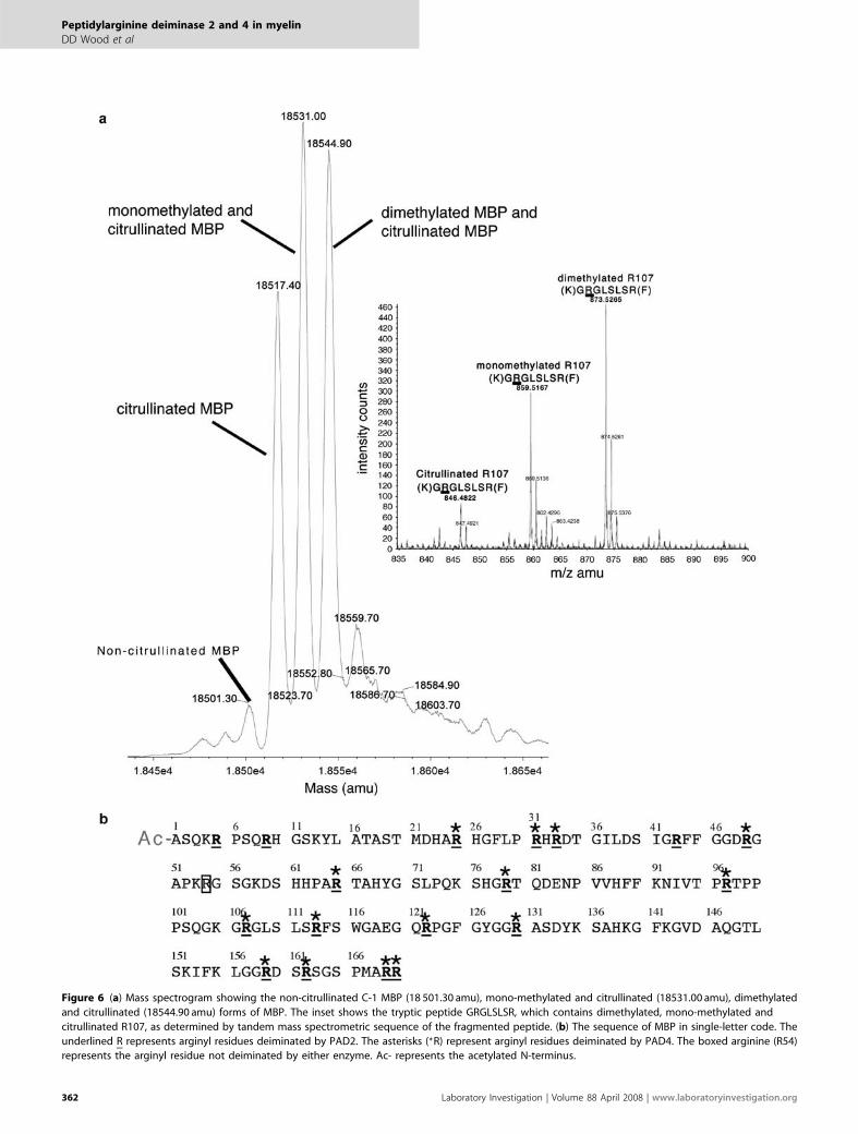
Figure 6 (a) Mass spectrogram showing the non-citrullinated C-1 MBP (18 501.30 amu), mono-methylated and citrullinated (18531.00 amu), dimethylated
and citrullinated (18544.90 amu) forms of MBP. The inset shows the tryptic peptide GRGLSLSR, which contains dimethylated, mono-methylated and
citrullinated R107, as determined by tandem mass spectrometric sequence of the fragmented peptide. (b) The sequence of MBP in single-letter code. The
underlined R represents arginyl residues deiminated by PAD2. The asterisks (*R) represent arginyl residues deiminated by PAD4. The boxed arginine (R54)
represents the arginyl residue not deiminated by either enzyme. Ac- represents the acetylated N-terminus.
Peptidylarginine deiminase 2 and 4 in myelin
DD Wood et al
362 Laboratory Investigation | Volume 88 April 2008 | www.laboratoryinvestigation.org
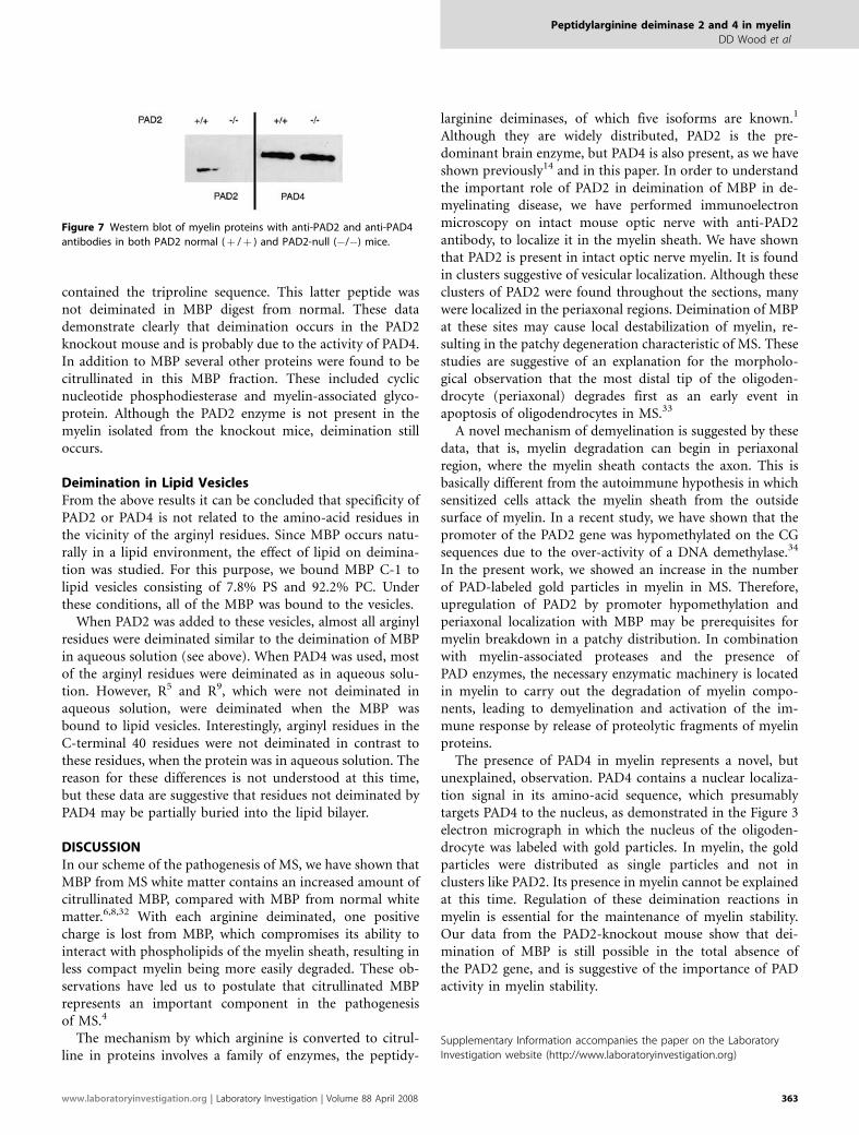
contained the triproline sequence. This latter peptide wasnot deiminated in MBP digest from normal. These datademonstrate clearly that deimination occurs in the PAD2knockout mouse and is probably due to the activity of PAD4.In addition to MBP several other proteins were found to becitrullinated in this MBP fraction. These included cyclicnucleotide phosphodiesterase and myelin-associated glyco-protein. Although the PAD2 enzyme is not present in themyelin isolated from the knockout mice, deimination stilloccurs.
Deimination in Lipid VesiclesFrom the above results it can be concluded that specificity ofPAD2 or PAD4 is not related to the amino-acid residues inthe vicinity of the arginyl residues. Since MBP occurs natu-rally in a lipid environment, the effect of lipid on deimina-tion was studied. For this purpose, we bound MBP C-1 tolipid vesicles consisting of 7.8% PS and 92.2% PC. Underthese conditions, all of the MBP was bound to the vesicles.
When PAD2 was added to these vesicles, almost all arginylresidues were deiminated similar to the deimination of MBPin aqueous solution (see above). When PAD4 was used, mostof the arginyl residues were deiminated as in aqueous solu-tion. However, R5 and R9, which were not deiminated inaqueous solution, were deiminated when the MBP wasbound to lipid vesicles. Interestingly, arginyl residues in theC-terminal 40 residues were not deiminated in contrast tothese residues, when the protein was in aqueous solution. Thereason for these differences is not understood at this time,but these data are suggestive that residues not deiminated byPAD4 may be partially buried into the lipid bilayer.
DISCUSSIONIn our scheme of the pathogenesis of MS, we have shown thatMBP from MS white matter contains an increased amount ofcitrullinated MBP, compared with MBP from normal whitematter.6,8,32 With each arginine deiminated, one positivecharge is lost from MBP, which compromises its ability tointeract with phospholipids of the myelin sheath, resulting inless compact myelin being more easily degraded. These ob-servations have led us to postulate that citrullinated MBPrepresents an important component in the pathogenesisof MS.4
The mechanism by which arginine is converted to citrul-line in proteins involves a family of enzymes, the peptidy-
larginine deiminases, of which five isoforms are known.1
Although they are widely distributed, PAD2 is the pre-dominant brain enzyme, but PAD4 is also present, as we haveshown previously14 and in this paper. In order to understandthe important role of PAD2 in deimination of MBP in de-myelinating disease, we have performed immunoelectronmicroscopy on intact mouse optic nerve with anti-PAD2antibody, to localize it in the myelin sheath. We have shownthat PAD2 is present in intact optic nerve myelin. It is foundin clusters suggestive of vesicular localization. Although theseclusters of PAD2 were found throughout the sections, manywere localized in the periaxonal regions. Deimination of MBPat these sites may cause local destabilization of myelin, re-sulting in the patchy degeneration characteristic of MS. Thesestudies are suggestive of an explanation for the morpholo-gical observation that the most distal tip of the oligoden-drocyte (periaxonal) degrades first as an early event inapoptosis of oligodendrocytes in MS.33
A novel mechanism of demyelination is suggested by thesedata, that is, myelin degradation can begin in periaxonalregion, where the myelin sheath contacts the axon. This isbasically different from the autoimmune hypothesis in whichsensitized cells attack the myelin sheath from the outsidesurface of myelin. In a recent study, we have shown that thepromoter of the PAD2 gene was hypomethylated on the CGsequences due to the over-activity of a DNA demethylase.34
In the present work, we showed an increase in the numberof PAD-labeled gold particles in myelin in MS. Therefore,upregulation of PAD2 by promoter hypomethylation andperiaxonal localization with MBP may be prerequisites formyelin breakdown in a patchy distribution. In combinationwith myelin-associated proteases and the presence ofPAD enzymes, the necessary enzymatic machinery is locatedin myelin to carry out the degradation of myelin compo-nents, leading to demyelination and activation of the im-mune response by release of proteolytic fragments of myelinproteins.
The presence of PAD4 in myelin represents a novel, butunexplained, observation. PAD4 contains a nuclear localiza-tion signal in its amino-acid sequence, which presumablytargets PAD4 to the nucleus, as demonstrated in the Figure 3electron micrograph in which the nucleus of the oligoden-drocyte was labeled with gold particles. In myelin, the goldparticles were distributed as single particles and not inclusters like PAD2. Its presence in myelin cannot be explainedat this time. Regulation of these deimination reactions inmyelin is essential for the maintenance of myelin stability.Our data from the PAD2-knockout mouse show that dei-mination of MBP is still possible in the total absence ofthe PAD2 gene, and is suggestive of the importance of PADactivity in myelin stability.
Supplementary Information accompanies the paper on the Laboratory
Investigation website (http://www.laboratoryinvestigation.org)
Figure 7 Western blot of myelin proteins with anti-PAD2 and anti-PAD4
antibodies in both PAD2 normal (þ /þ ) and PAD2-null (�/�) mice.
Peptidylarginine deiminase 2 and 4 in myelin
DD Wood et al
www.laboratoryinvestigation.org | Laboratory Investigation | Volume 88 April 2008 363

ACKNOWLEDGEMENTS
We gratefully acknowledge the support by Prinses Beatrix Fonds to BvB. RR
was funded by the Dutch Technology Foundation through grant
790.35.898. We thank Dr Brian Werneburg (Boehringer Ingelheim,
Ridgefield, USA) for supplying PAD2-knockout mouse tissue samples.
We are also grateful for the technical assistance provided by Mrs Teresa
Miani, Ms Aina Tilups and Mr Howard Rosenberg in the Division of
Pathology at The Hospital for Sick Children, Toronto. We thank both the
Canadian Brain bank and Drs RM Nagra and WW Tourtellotte for providing
normal and MS brain samples from the Human Brain and Spinal Fluid
Resource Center’s, VA West Los Angeles Healthcare Center, Los Angeles,
CA 90073, which is sponsored by NINDS/NIMH, National Multiple Sclerosis
Society (USA), and the Department of Veterans Affairs. This work was
supported by Canadian Institute of Health Research (CIHR) grant to MAM
and Multiple Sclerosis Society of Canada (MSSC) grant to MAM and FGM.
1. Vossenaar ER, Zendman AJ, van Venrooij WJ, et al. PAD, a growingfamily of citrullinating enzymes: genes, features and involvement indisease. Bioessays 2003;25:1106–1118.
2. Lamensa JW, Moscarello MA. Deimination of human myelinbasic protein by a peptidylarginine deiminase from bovine brain.J Neurochem 1993;61:987–996.
3. Mastronardi FG, Moscarello MA. Molecules affecting myelin stability:a novel hypothesis regarding the pathogenesis of multiple sclerosis.J Neurosci Res 2005;80:301–308.
4. Moscarello MA, Mastronardi FG, Wood DD. The role of citrullinatedproteins suggests a novel mechanism in the pathogenesis of multiplesclerosis. Neurochem Res 2007;32:251–256.
5. Kearney PL, Bhatia M, Jones NG, et al. Kinetic characterization ofprotein arginine deiminase 4: a transcriptional corepressor implicatedin the onset and progression of rheumatoid arthritis. Biochemistry2005;44:10570–10582.
6. Wood DD, Moscarello MA. The isolation, characterization, andlipid-aggregating properties of a citrulline containing myelin basicprotein. J Biol Chem 1989;264:5121–5127.
7. Kim JK, Mastronardi FG, Wood DD, et al. Multiple sclerosis: animportant role for post-translational modifications of myelin basicprotein in pathogenesis. Mol Cell Proteomics 2003;2:453–462.
8. Wood DD, Bilbao JM, O’Connors P, et al. Acute multiple sclerosis(Marburg type) is associated with developmentally immature myelinbasic protein. Ann Neurol 1996;40:18–24.
9. Brady GW, Fein DB, Wood DD, et al. The interaction of basic proteinsfrom normal and multiple sclerosis myelin with phosphatidylglycerolvesicles. FEBS Lett 1981;125:159–160.
10. D’Souza CA, Moscarello MA. Differences in susceptibility of MBP chargeisomers to digestion by stromelysin-1 (MMP-3) and release of animmunodominant epitope. Neurochem Res 2006;31:1045–1054.
11. Pritzker LB, Joshi S, Gowan JJ, et al. Deimination of myelin basicprotein. 1. Effect of deimination of arginyl residues of myelin basicprotein on its structure and susceptibility to digestion by cathepsin D.Biochemistry 2000;39:5374–5381.
12. D’Souza CA, Wood DD, She YM, et al. Autocatalytic cleavage of myelinbasic protein: an alternative to molecular mimicry. Biochemistry2005;44:12905–12913.
13. McLaurin J, Ackerley CA, Moscarello MA. Localization of basic proteinsin human myelin. J Neurosci Res 1993;35:618–628.
14. Mastronardi FG, Wood DD, Mei J, et al. Increased citrullination ofhistone H3 in multiple sclerosis brain and animal models ofdemyelination: a role for tumor necrosis factor-inducedpeptidylarginine deiminase 4 translocation. J Neurosci 2006;26:11387–11396.
15. Luo Y, Arita K, Bhatia M, et al. Inhibitors and inactivators of proteinarginine deiminase 4: functional and structural characterization.Biochemistry 2006;45:11727–11736.
16. Stone EM, Schaller TH, Bianchi H, et al. Inactivation of two diverseenzymes in the amidinotransferase superfamily by 2-chloroacetamidine: dimethylargininase and peptidylargininedeiminase. Biochemistry 2005;44:13744–13752.
17. Cheifetz S, Moscarello MA. Effect of bovine basic protein chargemicroheterogeneity on protein-induced aggregation of unilamellarvesicles containing a mixture of acidic and neutral phospholipids.Biochemistry 1985;24:1909–1914.
18. Raijmakers R, Vogelzangs J, Croxford JL, et al. Citrullination ofcentral nervous system proteins during the development ofexperimental autoimmune encephalomyelitis. J Comp Neurol2005;486:243–253.
19. Raijmakers R, Vogelzangs J, Raats J, et al. Experimental autoimmuneencephalomyelitis induction in peptidylarginine deiminase 2 knockoutmice. J Comp Neurol 2006;498:217–226.
20. Cruz TF, Moscarello MA. Characterization of myelin fractions fromhuman brain white matter. J Neurochem 1985;44:1411–1418.
21. Groome N, Dawkes A, Barry R, et al. New monoclonal antibodiesreactive with defined sequential epitopes in human myelin basicprotein. J Neuroimmunol 1988;19:305–315.
22. Rus’d AA, Ikejiri Y, Ono H, et al. Molecular cloning of cDNAs of mousepeptidylarginine deiminase type I, type III and type IV, and theexpression pattern of type I in mouse. Eur J Biochem 1999;259:660–669.
23. Nicholas AP, Sambandam T, Echols JD, et al. Expression of citrullinatedproteins in murine experimental autoimmune encephalomyelitis.J Comp Neurol 2005;486:254–266.
24. Peterson GL. A simplification of the protein assay method ofLowry et al which is more generally applicable. Anal Biochem1977;83:346–356.
25. Pritzker LB, Joshi S, Harauz G, et al. Deimination of myelin basicprotein. 2. Effect of methylation of MBP on its deimination bypeptidylarginine deiminase. Biochemistry 2000;39:5382–5388.
26. Tarcsa E, Marekov LN, Mei G, et al. Protein unfolding bypeptidylarginine deiminase. Substrate specificity and structuralrelationships of the natural substrates trichohyalin and filaggrin. J BiolChem 1996;271:30709–30716.
27. Hidaka Y, Hagiwara T, Yamada M. Methylation of the guanidino groupof arginine residues prevents citrullination by peptidylargininedeiminase IV. FEBS Lett 2005;579:4088–4092.
28. Klose RJ, Zhang Y. Regulation of histone methylation bydemethylimination and demethylation. Nat Rev Mol Cell Biol2007;8:307–318.
29. Lee YH, Coonrod SA, Kraus WL, et al. Regulation of coactivator complexassembly and function by protein arginine methylation anddemethylimination. Proc Natl Acad Sci USA 2005;102:3611–3616.
30. Wang Y, Wysocka J, Sayegh J, et al. Human PAD4 regulates histonearginine methylation levels via demethylimination. Science2004;306:279–283.
31. Raijmakers R, Zendman AJ, Egberts WV, et al. Methylation of arginineresidues interferes with citrullination by peptidylarginine deiminasesin vitro. J Mol Biol 2007;367:1118–1129.
32. Moscarello MA, Wood DD, Ackerley C, et al. Myelin in multiple sclerosisis developmentally immature. J Clin Invest 1994;94:146–154.
33. Ludwin SK. The pathogenesis of multiple sclerosis: relating humanpathology to experimental studies. J Neuropathol Exp Neurol2006;65:305–318.
34. Mastronardi FG, Noor A, Wood DD, et al. Peptidyl arginine deiminase 2CpG island in multiple sclerosis white matter is hypomethylated.J Neurosci Res 2007;85:2006–2016.
Peptidylarginine deiminase 2 and 4 in myelin
DD Wood et al
364 Laboratory Investigation | Volume 88 April 2008 | www.laboratoryinvestigation.org