Cathepsin S and an asparagine-specific endoprotease dominate the proteolytic processing of human...
11
0014-2980/01/1212-3726$17.50 + .50/0 © WILEY-VCH Verlag GmbH, D-69451 Weinheim, 2001 Cathepsin S and an asparagine-specific endoprotease dominate the proteolytic processing of human myelin basic protein in vitro Hermann Beck 1 , Gerold Schwarz 1 , Christian J. Schröter 1 , Martin Deeg 1 , Daniel Baier 1 , Stefan Stevanovic 2 , Ekkehard Weber 3 , Christoph Driessen 1,4 and Hubert Kalbacher 1 1 Medical and Natural Sciences Research Center, University of Tübingen, Tübingen, Germany 2 Department of Immunology, Institute of Cell Biology, University of Tübingen, Tübingen, Germany 3 Institute of Physiological Chemistry, University of Halle, Halle, Germany 4 Department of Medicine II, University of Tübingen, Tübingen, Germany The biochemical characterization of antigen degradation is an important basis for a better understanding of both the immune response and autoimmune diseases mediated by MHC class II molecules. In this study we used high-performance liquid chromatography and mass spectrometry to analyze the processing of myelin basic protein (MBP), a potential autoanti- gen implicated in the pathogenesis of multiple sclerosis. We resolved the kinetics of MBP processing by lysosomal extracts or purified endocytic proteases, identified the major cleav- age sites during this process and assigned them to the activity of proteolytic enzymes. Pro- teolytic processing of MBP is mostly guided along the hydrophobic regions of the protein. It is initiated by two proteolytic steps (after N 92 and S 110 ) that are performed by an asparagine- specific endopeptidase (AEP) and by cathepsin (Cat) S, respectively. The resulting process- ing intermediates are converted into more than 60 different species of 20–40-mers due to the activity of endopeptidases including CatS, D and L. The fragments thus generated are sub- sequently degraded by C- or N-terminal trimming. Strikingly, the initial cleavages during MBP processing affect two immunodominant regions of the potential autoantigen [MBP(85–99) and MBP(111-129)] in an inverse manner. CatS directly generates the N termi- nus of the epitope MBP(111–129) in large quantities during the initial phase of processing, which might explain the immunogenicity of this region in spite of its relatively poor binding to HLA-DR4. In contrast, the dominant cleavage by AEP mediates the destruction of MBP(85–99) unless the epitope is protected, e.g. by binding to HLA-DR. Our results thus characterize the proteolytic events during processing of MBP on a molecular level and sug- gest a biochemical basis for the immunogenicity of the immunodominant epitopes, which could serve as a guideline for future therapeutic strategies. Key words: Antigen processing / Myelin basic protein / Cathepsin / MHC class II / B lymphocyte Received 15/12/01 Revised 12/9/01 Accepted 9/10/01 [I 20267] Abbreviations: AEP: Asparagine-specific endopeptidase Cat: Cathepsin TTCF: Tetanus toxoid C fragment AMCA: 7-amino-4-methyl-coumarin-3-acetic acid B-LC-line: Lym- phoblastoid B cell line ESI-MS: Electrospray ionization source mass spectrometry E-64: Trans-epoxysuccinyl-l- leucylamido-(4-guanidino)butane HPSEC: High- performance size exclusion chromatography MALDI-MS: Matrix-assisted laser desorption ionization mass spectrom- etry MBP: Myelin basic protein TFA: Trifluoroacetic acid Z: Benzyloxycarbonyl 1 Introduction While many details of the assembly, transport, and pro- cessing of MHC class II molecules have been resolved in recent years, the generation of antigenic peptide from complex protein and its binding to MHC class II are still poorly understood [1–3]. Cathepsins (Cat) represent the most abundant family of endocytic proteases and new members of this group continue to be discovered [4, 5]. CatB, D, E, L and S have been implicated in antigen pro- cessing and the generation of immunogenic epitopes [6]. However, cathepsins represent by no means the entire spectrum of endocytic proteases and unrelated proteases might be equally important. Recently, the asparagine-specific endopeptidase (AEP) has been identified as the key enzyme that mediates the rate- limiting step in the intracellular processing of tetanus toxoid C fragment (TTCF) [7]. For TTCF, the generation of dominant processing intermediates by AEP precedes the 3726 H. Beck et al. Eur. J. Immunol. 2001. 31: 3726–3736
-
Upload
independent -
Category
Documents
-
view
3 -
download
0
Transcript of Cathepsin S and an asparagine-specific endoprotease dominate the proteolytic processing of human...
0014-2980/01/1212-3726$17.50+.50/0 © WILEY-VCH Verlag GmbH, D-69451 Weinheim, 2001
Cathepsin S and an asparagine-specificendoprotease dominate the proteolytic processingof human myelin basic protein in vitro
Hermann Beck1, Gerold Schwarz1, Christian J. Schröter1, Martin Deeg1, Daniel Baier1,Stefan Stevanovic2, Ekkehard Weber3, Christoph Driessen1,4 and Hubert Kalbacher1
1 Medical and Natural Sciences Research Center, University of Tübingen, Tübingen, Germany2 Department of Immunology, Institute of Cell Biology, University of Tübingen, Tübingen,
Germany3 Institute of Physiological Chemistry, University of Halle, Halle, Germany4 Department of Medicine II, University of Tübingen, Tübingen, Germany
The biochemical characterization of antigen degradation is an important basis for a betterunderstanding of both the immune response and autoimmune diseases mediated by MHCclass II molecules. In this study we used high-performance liquid chromatography and massspectrometry to analyze the processing of myelin basic protein (MBP), a potential autoanti-gen implicated in the pathogenesis of multiple sclerosis. We resolved the kinetics of MBPprocessing by lysosomal extracts or purified endocytic proteases, identified the major cleav-age sites during this process and assigned them to the activity of proteolytic enzymes. Pro-teolytic processing of MBP is mostly guided along the hydrophobic regions of the protein. Itis initiated by two proteolytic steps (after N92 and S110) that are performed by an asparagine-specific endopeptidase (AEP) and by cathepsin (Cat) S, respectively. The resulting process-ing intermediates are converted into more than 60 different species of 20–40-mers due to theactivity of endopeptidases including CatS, D and L. The fragments thus generated are sub-sequently degraded by C- or N-terminal trimming. Strikingly, the initial cleavages duringMBP processing affect two immunodominant regions of the potential autoantigen[MBP(85–99) and MBP(111-129)] in an inverse manner. CatS directly generates the N termi-nus of the epitope MBP(111–129) in large quantities during the initial phase of processing,which might explain the immunogenicity of this region in spite of its relatively poor bindingto HLA-DR4. In contrast, the dominant cleavage by AEP mediates the destruction ofMBP(85–99) unless the epitope is protected, e.g. by binding to HLA-DR. Our results thuscharacterize the proteolytic events during processing of MBP on a molecular level and sug-gest a biochemical basis for the immunogenicity of the immunodominant epitopes, whichcould serve as a guideline for future therapeutic strategies.
Key words: Antigen processing / Myelin basic protein / Cathepsin / MHC class II / B lymphocyte
Received 15/12/01Revised 12/9/01Accepted 9/10/01
[I 20267]
Abbreviations: AEP: Asparagine-specific endopeptidaseCat: Cathepsin TTCF: Tetanus toxoid C fragment AMCA:7-amino-4-methyl-coumarin-3-acetic acid B-LC-line: Lym-phoblastoid B cell line ESI-MS: Electrospray ionizationsource mass spectrometry E-64: Trans-epoxysuccinyl-l-leucylamido-(4-guanidino)butane HPSEC: High-performance size exclusion chromatography MALDI-MS:Matrix-assisted laser desorption ionization mass spectrom-etry MBP: Myelin basic protein TFA: Trifluoroacetic acid Z:Benzyloxycarbonyl
1 Introduction
While many details of the assembly, transport, and pro-cessing of MHC class II molecules have been resolved in
recent years, the generation of antigenic peptide fromcomplex protein and its binding to MHC class II are stillpoorly understood [1–3]. Cathepsins (Cat) represent themost abundant family of endocytic proteases and newmembers of this group continue to be discovered [4, 5].CatB, D, E, L and S have been implicated in antigen pro-cessing and the generation of immunogenic epitopes[6]. However, cathepsins represent by no means theentire spectrum of endocytic proteases and unrelatedproteases might be equally important. Recently, theasparagine-specific endopeptidase (AEP) has beenidentified as the key enzyme that mediates the rate-limiting step in the intracellular processing of tetanustoxoid C fragment (TTCF) [7]. For TTCF, the generation ofdominant processing intermediates by AEP precedes the
3726 H. Beck et al. Eur. J. Immunol. 2001. 31: 3726–3736
Fig. 1. Reversed-phase HPLC profiles of human MBP afterdifferent incubation times with disrupted lysosomes at pH5.4. Substrate (0.1 mM) was incubated with disrupted lyso-somes equivalent to 5×10⁵ cells/ ? mol substrate. (A) Recom-binant MBP, (B) MBP purified from human CNS. The peaksindicated consist of: I, MBP(111–170); II, MBP(93–170); III,MBP(1–92); IV, MBP(1–110), as inferred from microsequenc-ing.
formation of the immunogenic epitopes [8]. Whethersuch initial ”unlocking” of intact antigenic protein is amore general characteristic of the processing of exoge-nous antigen, whether it is performed by a singleenzyme, a class of enzymes or by unrelated proteases,whether the sites of such dominant processing steps aredetermined by the specificity of certain proteases ormerely by the structure of the antigenic protein itself,remain largely open questions.
Several lines of evidence suggest an important role ofmyelin basic protein (MBP) as an autoantigen in thepathogenesis of the MHC class II-associated autoim-mune disease multiple sclerosis (MS). A dominant auto-reactive T cell response against certain regions of humanMBP can be found in a large portion of patients with thedisease [9, 10]. The immunodominant epitope in the con-text of HLA-DR2, the haplotype associated with thestrongest susceptibility for MS, has been localized toMBP residues 82–100, with MBP(85–99) constituting theminimum T cell epitope [11–13]. Similarly, DRB1*0401,the most common DR4 allele in the Caucasian popu-lation, is associated with susceptibility for MS.DRB1*0401restricted autoreactive T cells were found torecognize MBP(111–129), with the major T cell epitopecorresponding to residues MBP(116–123) [10]. Process-ing of MBP and the generation of the antigenic determi-nants remain poorly characterized to date. A betterunderstanding of the processing of MBP should not onlyprovide new insights in the function of the antigen-processing machinery in general, but might also openways to interfere with the presentation of epitopes impli-cated in the pathogenesis of MS.
We here show that proteolytic processing of MBP bylysosomal proteases in vitro is dominated by two initialcleavages performed by CatS and AEP. Further proteoly-sis of the resulting intermediates is largely guided alongthe hydrophobic regions of the protein. Surprisingly, oneof the major processing sites lies within the immunodo-minant region MBP(85–99). Binding to MHC class II ade-quately protects this epitope from destruction, highlight-ing the importance of determinant capture by class IImolecules for the preservation of the major immunogenicepitope of MBP.
2 Results
2.1 The initial stage of proteolytic processing ofMBP is controlled by AEP and CatS
The endocytic compartment of APC contains a complexmixture of individual proteases each of which is poten-tially involved in antigen processing. To characterize the
processing of MBP in vitro, lysosomes were isolatedfrom a human B cell line by differential centrifugation andrecombinant human MBP was digested with lysosomalextracts. At different time points the resulting pattern ofdegradation products was resolved by RP-HPLC and UVabsorption was detected at 214 nm (Fig. 1a). During theinitial phase ( X 80% of substrate turnover) of proteolyticin vitro processing, the bulk of protein eluted in fourdistinct peaks (marked I-IV) shortly before intact MBP.After prolonged incubation intact MBP was no longerdetected, and peaks I-IV slowly converted into poorlyresolved material that eluted after shorter retentiontimes. Human MBP, obtained from human white matterpreparation by semipreparative HPLC as described [14],represents a mixture of the unmodified protein along withits monomethylated (6%) and dimethylated (60%) spe-cies. This results in more complex HPLC profiles afterprocessing with lysosomal extracts (Fig. 1b), where thekinetics and the ratio of the MBP degradation productsII, III and IV were altered compared to recombinant MBP.However, dominant molecular masses corresponding toidentical degradation fragments were identified frompeaks I-IV by matrix-assisted laser desorption ionizationmass spectrometry (MALDI-MS) when either recombi-nant MBP or MBP purified from human CNS was sub-jected to in vitro proteolysis in the same manner (see alsobelow), confirming that the initial phase of proteolyticprocessing of purified MBP vs. recombinant MBP in vitrofollowed a similar cleavage pattern.
Eur. J. Immunol. 2001. 31: 3726–3736 Processing of human MBP in vitro 3727
Fig. 2. Processing of human MBP under various conditions. Left panel: top: disrupted lysosomes preincubated with 10 ? M E-64;middle: disrupted lysosomes preincubated at pH 7.5 for 1 h, 37°C; MBP incubated with Cathepsin S at pH 5.4. Right panel: top:co-incubation with 5 mg/ml Z-AENK-NH2; middle: co-incubation with 5 mg/ml Z-AEQK-NH2, 10 ? M E-64; bottom: co-incubationwith 5 mg/ml Z-AENK-NH2, 5 nM LVHS. The experiments were run in parallel and processing was stopped at the same timepoint. Fragments of human MBP in the indicated peaks: I, MBP(111–170); II, MBP(93–170); III, MBP(1–92); IV, MBP(1–110). Sub-strate (0.1 mM) was incubated with disrupted lysosomes equivalent to 5×10⁵ cells/ ? mol substrate. In all cases, identification wasachieved by sequencing.
The degradation pattern observed suggested that pro-cessing of MBP in vitro occurred in a stepwise fashion:during an initial phase MBP was converted into a limitednumber of dominant processing intermediates that weredegraded into multiple smaller fragments as processingproceeds. Using protease inhibitors, we tried to identifythe enzymes responsible for the generation of the domi-nant degradation products at the first stage of process-ing (50–80% of substrate turnover). While inhibition ofserine-, metallo- and aspartate-proteases had no de-tectable influence on the in vitro-degradation patternresolved by HPLC (data not shown), the addition oftrans-epoxysuccinyl-l-leucylamido-(4-guanidino) butane(E-64), a generic inhibitor of cysteine proteases, inter-fered with formation of HPLC peaks I and IV, while peaksII and III were still present (Fig. 2). This strongly sug-gested that peaks I and IV were the result of MBP pro-cessing mediated by a cysteine protease. CatS, L and Brepresent the most abundant cysteine proteases in theendocytic compartment of antigen-presenting cells.While the former has a rather broad pH optimum rangingfrom pH 4.5–pH 7, the latter two are inactivated by expo-sure to neutral pH [15–17]. To narrow down the prote-
ases possibly involved in the generation of peaks I andIV, we performed a similar set of in vitro processingexperiments at neutral pH. Under these conditions,peaks I and IV remained unchanged, consistent withtheir possible generation by CatS, while peaks II and IIIdisappeared. Finally, when MBP was digested with puri-fied CatS, only protein peaks eluting at the retentiontimes of peaks I and IV were observed, which stronglysupported that CatS is responsible for one of the initialcleavages of MBP during processing in vitro.
Based on the results described above, the endocyticprotease(s) responsible for the generation of the domi-nant peaks II and III was/were characterized by (i) inac-tivity at neutral pH and (ii) resistance to common genericprotease inhibitors. The legumain-type protease AEPthat was recently discovered as the key proteaseinvolved in the unlocking of TTCF [7] matched these cri-teria. We therefore synthesized a competitive inhibitor ofAEP activity, benzyloxycarbonyl (Z)-AENK-NH2, alongwith its control analogue Z-AEQK-NH2, to test whetherspecific inhibition of AEP during in vitro proteolytic pro-cessing of MBP would affect the formation of HPLC-
3728 H. Beck et al. Eur. J. Immunol. 2001. 31: 3726–3736
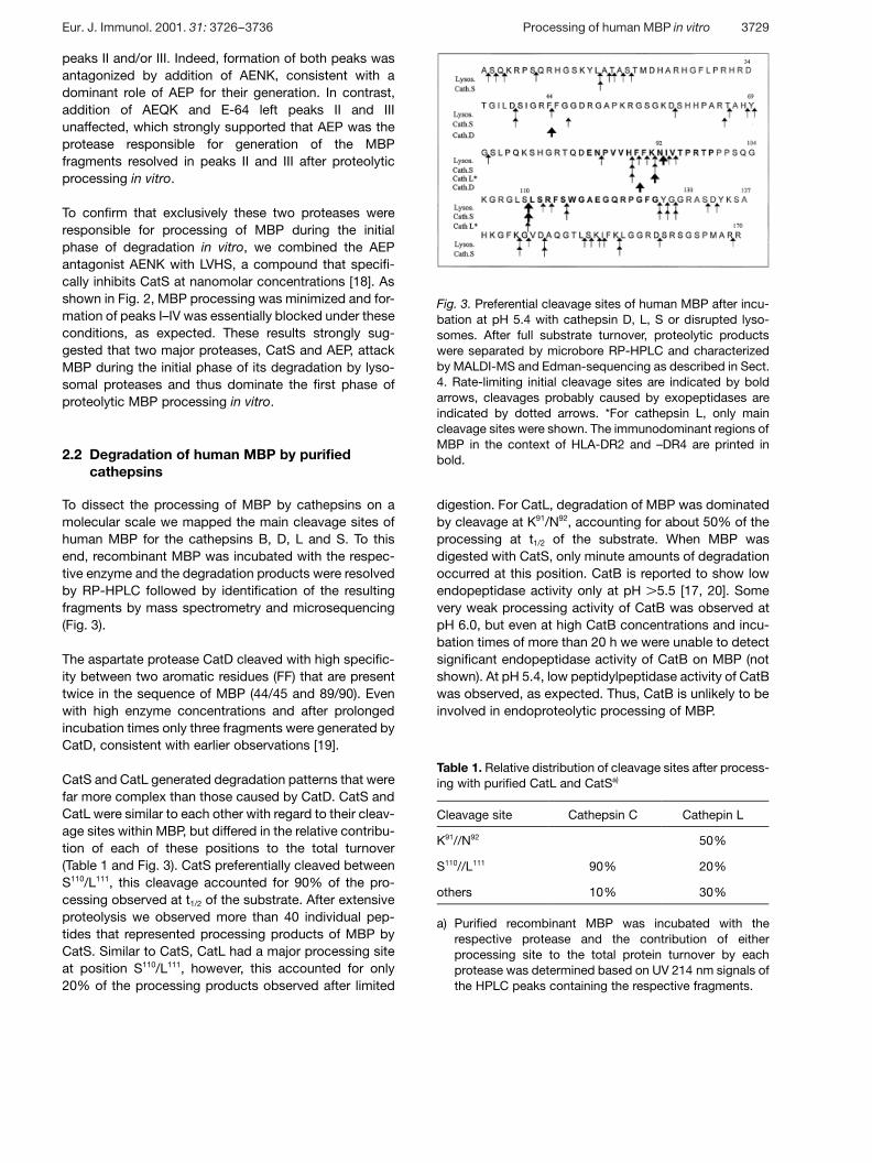
Fig. 3. Preferential cleavage sites of human MBP after incu-bation at pH 5.4 with cathepsin D, L, S or disrupted lyso-somes. After full substrate turnover, proteolytic productswere separated by microbore RP-HPLC and characterizedby MALDI-MS and Edman-sequencing as described in Sect.4. Rate-limiting initial cleavage sites are indicated by boldarrows, cleavages probably caused by exopeptidases areindicated by dotted arrows. *For cathepsin L, only maincleavage sites were shown. The immunodominant regions ofMBP in the context of HLA-DR2 and –DR4 are printed inbold.
Table 1. Relative distribution of cleavage sites after process-ing with purified CatL and CatSa)
Cleavage site Cathepsin C Cathepin L
K91//N92 50%
S110//L111 90% 20%
others 10% 30%
a) Purified recombinant MBP was incubated with therespective protease and the contribution of eitherprocessing site to the total protein turnover by eachprotease was determined based on UV 214 nm signals ofthe HPLC peaks containing the respective fragments.
peaks II and/or III. Indeed, formation of both peaks wasantagonized by addition of AENK, consistent with adominant role of AEP for their generation. In contrast,addition of AEQK and E-64 left peaks II and IIIunaffected, which strongly supported that AEP was theprotease responsible for generation of the MBPfragments resolved in peaks II and III after proteolyticprocessing in vitro.
To confirm that exclusively these two proteases wereresponsible for processing of MBP during the initialphase of degradation in vitro, we combined the AEPantagonist AENK with LVHS, a compound that specifi-cally inhibits CatS at nanomolar concentrations [18]. Asshown in Fig. 2, MBP processing was minimized and for-mation of peaks I–IV was essentially blocked under theseconditions, as expected. These results strongly sug-gested that two major proteases, CatS and AEP, attackMBP during the initial phase of its degradation by lyso-somal proteases and thus dominate the first phase ofproteolytic MBP processing in vitro.
2.2 Degradation of human MBP by purifiedcathepsins
To dissect the processing of MBP by cathepsins on amolecular scale we mapped the main cleavage sites ofhuman MBP for the cathepsins B, D, L and S. To thisend, recombinant MBP was incubated with the respec-tive enzyme and the degradation products were resolvedby RP-HPLC followed by identification of the resultingfragments by mass spectrometry and microsequencing(Fig. 3).
The aspartate protease CatD cleaved with high specific-ity between two aromatic residues (FF) that are presenttwice in the sequence of MBP (44/45 and 89/90). Evenwith high enzyme concentrations and after prolongedincubation times only three fragments were generated byCatD, consistent with earlier observations [19].
CatS and CatL generated degradation patterns that werefar more complex than those caused by CatD. CatS andCatL were similar to each other with regard to their cleav-age sites within MBP, but differed in the relative contribu-tion of each of these positions to the total turnover(Table 1 and Fig. 3). CatS preferentially cleaved betweenS110/L111, this cleavage accounted for 90% of the pro-cessing observed at t1/2 of the substrate. After extensiveproteolysis we observed more than 40 individual pep-tides that represented processing products of MBP byCatS. Similar to CatS, CatL had a major processing siteat position S110/L111, however, this accounted for only20% of the processing products observed after limited
digestion. For CatL, degradation of MBP was dominatedby cleavage at K91/N92, accounting for about 50% of theprocessing at t1/2 of the substrate. When MBP wasdigested with CatS, only minute amounts of degradationoccurred at this position. CatB is reported to show lowendopeptidase activity only at pH G 5.5 [17, 20]. Somevery weak processing activity of CatB was observed atpH 6.0, but even at high CatB concentrations and incu-bation times of more than 20 h we were unable to detectsignificant endopeptidase activity of CatB on MBP (notshown). At pH 5.4, low peptidylpeptidase activity of CatBwas observed, as expected. Thus, CatB is unlikely to beinvolved in endoproteolytic processing of MBP.
Eur. J. Immunol. 2001. 31: 3726–3736 Processing of human MBP in vitro 3729
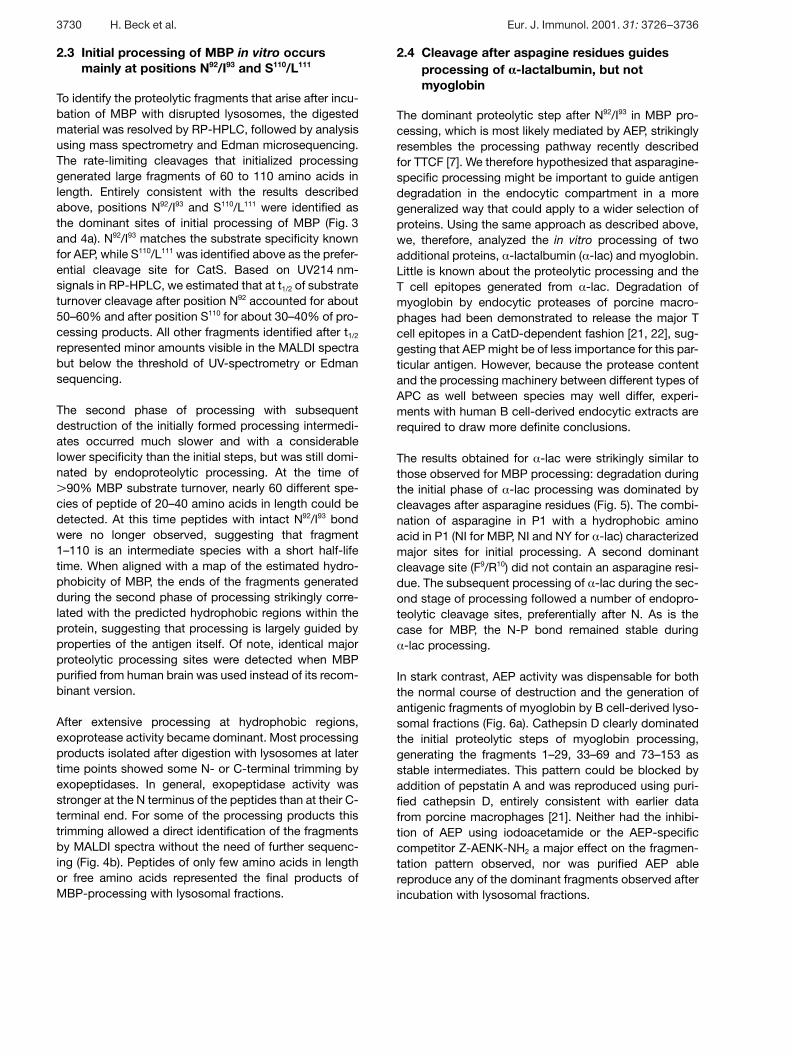
2.3 Initial processing of MBP in vitro occursmainly at positions N92/I93 and S110/L111
To identify the proteolytic fragments that arise after incu-bation of MBP with disrupted lysosomes, the digestedmaterial was resolved by RP-HPLC, followed by analysisusing mass spectrometry and Edman microsequencing.The rate-limiting cleavages that initialized processinggenerated large fragments of 60 to 110 amino acids inlength. Entirely consistent with the results describedabove, positions N92/I93 and S110/L111 were identified asthe dominant sites of initial processing of MBP (Fig. 3and 4a). N92/I93 matches the substrate specificity knownfor AEP, while S110/L111 was identified above as the prefer-ential cleavage site for CatS. Based on UV214 nm-signals in RP-HPLC, we estimated that at t1/2 of substrateturnover cleavage after position N92 accounted for about50–60% and after position S110 for about 30–40% of pro-cessing products. All other fragments identified after t1/2
represented minor amounts visible in the MALDI spectrabut below the threshold of UV-spectrometry or Edmansequencing.
The second phase of processing with subsequentdestruction of the initially formed processing intermedi-ates occurred much slower and with a considerablelower specificity than the initial steps, but was still domi-nated by endoproteolytic processing. At the time ofG 90% MBP substrate turnover, nearly 60 different spe-
cies of peptide of 20–40 amino acids in length could bedetected. At this time peptides with intact N92/I93 bondwere no longer observed, suggesting that fragment1–110 is an intermediate species with a short half-lifetime. When aligned with a map of the estimated hydro-phobicity of MBP, the ends of the fragments generatedduring the second phase of processing strikingly corre-lated with the predicted hydrophobic regions within theprotein, suggesting that processing is largely guided byproperties of the antigen itself. Of note, identical majorproteolytic processing sites were detected when MBPpurified from human brain was used instead of its recom-binant version.
After extensive processing at hydrophobic regions,exoprotease activity became dominant. Most processingproducts isolated after digestion with lysosomes at latertime points showed some N- or C-terminal trimming byexopeptidases. In general, exopeptidase activity wasstronger at the N terminus of the peptides than at their C-terminal end. For some of the processing products thistrimming allowed a direct identification of the fragmentsby MALDI spectra without the need of further sequenc-ing (Fig. 4b). Peptides of only few amino acids in lengthor free amino acids represented the final products ofMBP-processing with lysosomal fractions.
2.4 Cleavage after aspagine residues guidesprocessing of > -lactalbumin, but notmyoglobin
The dominant proteolytic step after N92/I93 in MBP pro-cessing, which is most likely mediated by AEP, strikinglyresembles the processing pathway recently describedfor TTCF [7]. We therefore hypothesized that asparagine-specific processing might be important to guide antigendegradation in the endocytic compartment in a moregeneralized way that could apply to a wider selection ofproteins. Using the same approach as described above,we, therefore, analyzed the in vitro processing of twoadditional proteins, § -lactalbumin ( § -lac) and myoglobin.Little is known about the proteolytic processing and theT cell epitopes generated from § -lac. Degradation ofmyoglobin by endocytic proteases of porcine macro-phages had been demonstrated to release the major Tcell epitopes in a CatD-dependent fashion [21, 22], sug-gesting that AEP might be of less importance for this par-ticular antigen. However, because the protease contentand the processing machinery between different types ofAPC as well between species may well differ, experi-ments with human B cell-derived endocytic extracts arerequired to draw more definite conclusions.
The results obtained for § -lac were strikingly similar tothose observed for MBP processing: degradation duringthe initial phase of § -lac processing was dominated bycleavages after asparagine residues (Fig. 5). The combi-nation of asparagine in P1 with a hydrophobic aminoacid in P1 (NI for MBP, NI and NY for § -lac) characterizedmajor sites for initial processing. A second dominantcleavage site (F9/R10) did not contain an asparagine resi-due. The subsequent processing of § -lac during the sec-ond stage of processing followed a number of endopro-teolytic cleavage sites, preferentially after N. As is thecase for MBP, the N-P bond remained stable during§ -lac processing.
In stark contrast, AEP activity was dispensable for boththe normal course of destruction and the generation ofantigenic fragments of myoglobin by B cell-derived lyso-somal fractions (Fig. 6a). Cathepsin D clearly dominatedthe initial proteolytic steps of myoglobin processing,generating the fragments 1–29, 33–69 and 73–153 asstable intermediates. This pattern could be blocked byaddition of pepstatin A and was reproduced using puri-fied cathepsin D, entirely consistent with earlier datafrom porcine macrophages [21]. Neither had the inhibi-tion of AEP using iodoacetamide or the AEP-specificcompetitor Z-AENK-NH2 a major effect on the fragmen-tation pattern observed, nor was purified AEP ablereproduce any of the dominant fragments observed afterincubation with lysosomal fractions.
3730 H. Beck et al. Eur. J. Immunol. 2001. 31: 3726–3736
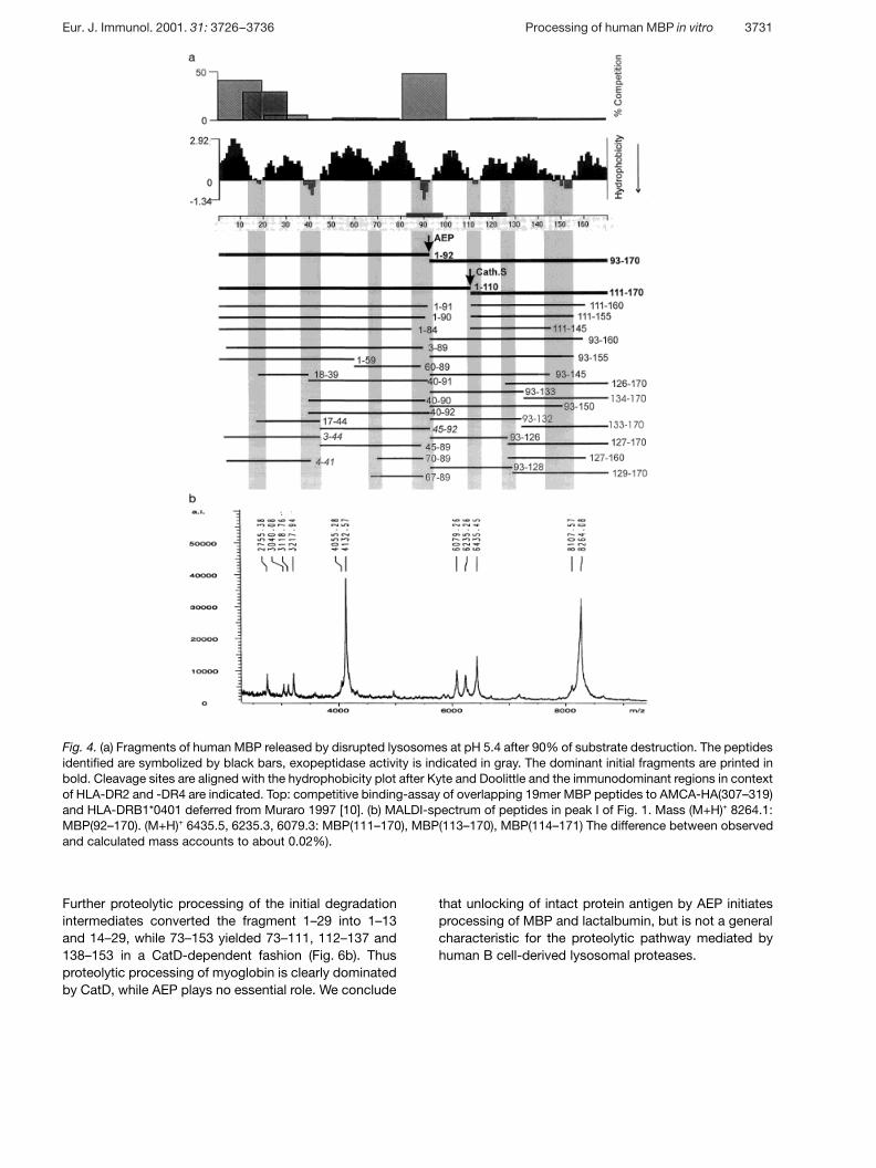
Fig. 4. (a) Fragments of human MBP released by disrupted lysosomes at pH 5.4 after 90% of substrate destruction. The peptidesidentified are symbolized by black bars, exopeptidase activity is indicated in gray. The dominant initial fragments are printed inbold. Cleavage sites are aligned with the hydrophobicity plot after Kyte and Doolittle and the immunodominant regions in contextof HLA-DR2 and -DR4 are indicated. Top: competitive binding-assay of overlapping 19mer MBP peptides to AMCA-HA(307–319)and HLA-DRB1*0401 deferred from Muraro 1997 [10]. (b) MALDI-spectrum of peptides in peak I of Fig. 1. Mass (M+H)+ 8264.1:MBP(92–170). (M+H)+ 6435.5, 6235.3, 6079.3: MBP(111–170), MBP(113–170), MBP(114–171) The difference between observedand calculated mass accounts to about 0.02%).
Further proteolytic processing of the initial degradationintermediates converted the fragment 1–29 into 1–13and 14–29, while 73–153 yielded 73–111, 112–137 and138–153 in a CatD-dependent fashion (Fig. 6b). Thusproteolytic processing of myoglobin is clearly dominatedby CatD, while AEP plays no essential role. We conclude
that unlocking of intact protein antigen by AEP initiatesprocessing of MBP and lactalbumin, but is not a generalcharacteristic for the proteolytic pathway mediated byhuman B cell-derived lysosomal proteases.
Eur. J. Immunol. 2001. 31: 3726–3736 Processing of human MBP in vitro 3731
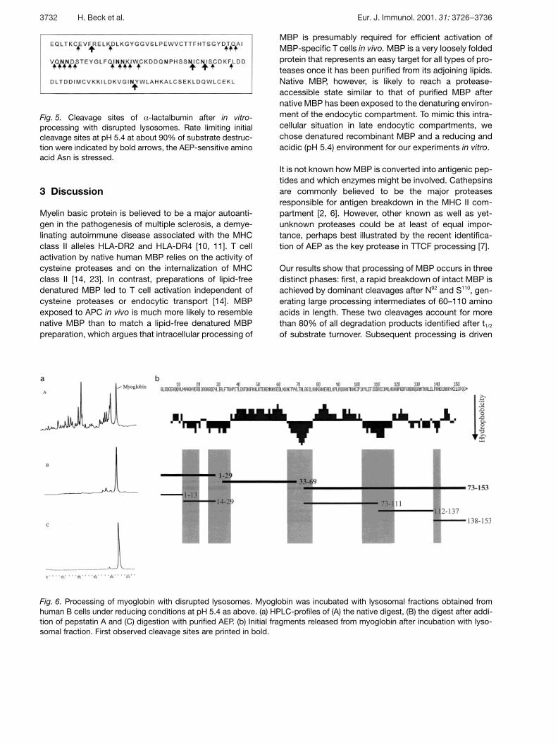
Fig. 5. Cleavage sites of § -lactalbumin after in vitro-processing with disrupted lysosomes. Rate limiting initialcleavage sites at pH 5.4 at about 90% of substrate destruc-tion were indicated by bold arrows, the AEP-sensitive aminoacid Asn is stressed.
Fig. 6. Processing of myoglobin with disrupted lysosomes. Myoglobin was incubated with lysosomal fractions obtained fromhuman B cells under reducing conditions at pH 5.4 as above. (a) HPLC-profiles of (A) the native digest, (B) the digest after addi-tion of pepstatin A and (C) digestion with purified AEP. (b) Initial fragments released from myoglobin after incubation with lyso-somal fraction. First observed cleavage sites are printed in bold.
3 Discussion
Myelin basic protein is believed to be a major autoanti-gen in the pathogenesis of multiple sclerosis, a demye-linating autoimmune disease associated with the MHCclass II alleles HLA-DR2 and HLA-DR4 [10, 11]. T cellactivation by native human MBP relies on the activity ofcysteine proteases and on the internalization of MHCclass II [14, 23]. In contrast, preparations of lipid-freedenatured MBP led to T cell activation independent ofcysteine proteases or endocytic transport [14]. MBPexposed to APC in vivo is much more likely to resemblenative MBP than to match a lipid-free denatured MBPpreparation, which argues that intracellular processing of
MBP is presumably required for efficient activation ofMBP-specific T cells in vivo. MBP is a very loosely foldedprotein that represents an easy target for all types of pro-teases once it has been purified from its adjoining lipids.Native MBP, however, is likely to reach a protease-accessible state similar to that of purified MBP afternative MBP has been exposed to the denaturing environ-ment of the endocytic compartment. To mimic this intra-cellular situation in late endocytic compartments, wechose denatured recombinant MBP and a reducing andacidic (pH 5.4) environment for our experiments in vitro.
It is not known how MBP is converted into antigenic pep-tides and which enzymes might be involved. Cathepsinsare commonly believed to be the major proteasesresponsible for antigen breakdown in the MHC II com-partment [2, 6]. However, other known as well as yet-unknown proteases could be at least of equal impor-tance, perhaps best illustrated by the recent identifica-tion of AEP as the key protease in TTCF processing [7].
Our results show that processing of MBP occurs in threedistinct phases: first, a rapid breakdown of intact MBP isachieved by dominant cleavages after N92 and S110, gen-erating large processing intermediates of 60–110 aminoacids in length. These two cleavages account for morethan 80% of all degradation products identified after t1/2
of substrate turnover. Subsequent processing is driven
3732 H. Beck et al. Eur. J. Immunol. 2001. 31: 3726–3736
by less specific endopeptidase activity at a much slowerrate, which results in peptides that comprise 20–40amino acids. Strikingly, endopeptidase activity is largelyguided along the hydrophobic regions of MBP. This is notan artifact due to the use of recombinant MBP, since thesame major processing sites were identified for purifiedhuman MBP. Similar patterns of processing along hydro-phobic regions were found for additional antigens test-ed (e.g. § -lactalbumin, myoglobin, hen-egg lysozyme;Fig. 5, 6 and H. Beck, unpublished observations). Thissuggests that antigen processing is largely guided bystructural properties of the protein itself, e.g. by the dis-tribution of hydrophobic regions over the sequence. Thisis perhaps not surprising given that most lysosomal pro-teases prefer to cleave next to hydrophobic amino acids[24]. In the third phase of processing exopeptidase activ-ity becomes dominant, with a significantly stronger activ-ity at the N terminus compared to the C-terminal end ofthe fragments. This finally leads to a stage of almostcomplete degradation, where only short peptides or freeamino acids are present.
According to our data the first stage of MBP processingis dominated by the cysteine protease CatS (cleavageafter position S110) and by the legumain-like AEP thatcleaves after N92. This is supported by (i) blockade of thegeneration of the respective dominant HPLC peaks byaddition of specific inhibitors against AEP- and CatS-activity; (ii) generation of corresponding processing inter-mediates and HPLC fractions by the purified enzymes;and (iii) identification of major processing products thatmatch the substrate preferences of either enzyme afterincubation of intact MBP with lysosomal extracts. ThusCatS not only plays a unique role for Ii degradation in Bcells and dendritic cells, but also represents one of thekey enzymes that initialize MBP processing. Given thatCatS is a major endopeptidase that has been shown tobe active in early endocytic compartments of dendriticcells [25] we speculate that CatS may be involved inMBP processing in intact cells in a similar manner. AEP isnot only the rate-limiting protease for TTCF processing[7], it also initiates degradation of MBP in vitro (thisreport). In contrast to TTCF processing, however, com-petitive inhibition of AEP does not halt processing ofMBP, because proteolysis of MBP is still initiated byCatS. Thus the initial unlocking of antigenic protein forfurther processing can be performed by different prote-ases of distinct specificity at different sites. Our data fur-ther suggest that AEP activity could be of more generalrelevance during the initial events of antigen processing:in vitro processing experiments of a similar type, using § -lactalbumin as a model substrate, revealed a similar pat-tern of dominant initial cleavage sites that were entirelyconsistent with their generation by AEP. However, a thirdprotein substrate tested (myoglobin) did not follow this
pattern but was almost exclusively degraded by CatD,entirely consistent with earlier observations in othertypes of APC [21]. “Unlocking” by AEP might thereforebe important for the initiation of processing in a variety ofantigens, however, we do not consider it a general rulefor antigen processing in the MHC class II compartment.
CatS and CatL showed a similar profile with respectto their specificity in MBP processing, although weobserved clear-cut differences in the quantitative contri-bution of different processing sites between bothenzymes. The shared specificity between CatS and CatLcorresponds well with the similar role of both enzymes inIi processing in B cells/dendritic cells, and thymic epithe-lial cells, respectively [26, 27]. CatD generates a relativelysmall set of degradation products, in agreement with itsnarrow substrate specificity [2]. Accordingly, fragmentsthat match the major MBP-processing sites for CatDwere only found in a low percentage in lysosomalextracts. This suggests that CatD is dispensable for pro-cessing of MBP, consistent with earlier results obtainedin CatD -/- mice in vivo [28].
Strikingly, the dominant proteolytic events during the ini-tial phase of MBP-processing affect the two immunodo-minant regions of MBP in an exactly inverse manner:while the initial cleavage by CatS (after S110) directlycontributes to the generation of MBP(111–129), thesecond dominant proteolytic event (cleavage after N92
by AEP) destroys the central immunodominant regionMBP(85–99) eliminating the first two anchor positions ofthe epitope. In fact, MBP(85–99) represented the regionwith the fastest turnover, and peptides containingMBP(85–99) were exclusively found at early time points.How can these differences be reconciled?
The guidance of antigen processing by protective matri-ces like MHC molecules or specific receptors has beenfrequently suggested [29–31]. Intact MBP is able to bindto HLA-DR molecules already on the cell surface [14, 23].Since MBP(85–99) binds to class II with a high affinity,binding of this region to class II might protect the centralepitope from degradation by AEP. We confirmed thatclass II binding is indeed sufficient for this type of protec-tion in vitro, in agreement with earlier observations (datanot shown and [31]). Thus, we suggest that the highaffinity of MBP(85–99) for class II rescues the epitopefrom degradation by AEP which in part could explain itsimmunogenicity. In this scenario, the generation of theimmunogenic peptide is therefore largely guided by theMHC molecule itself. AEP here acts as an epitope-destructive protease, in contrast to the situation encoun-tered for TTCF processing, where AEP is required for thegeneration of the immunodominant epitope.
Eur. J. Immunol. 2001. 31: 3726–3736 Processing of human MBP in vitro 3733
The inverse situation is encountered in case ofMBP(111–129): this epitope shows only a low affinity toHLA-DRB1*0401, but still represents an immunodomin-ant region in vivo [10]. Our data demonstrate that the Nterminus of this epitope is directly generated by one ofthe dominant proteolytic steps that initiate MBP pro-cessing, the cleavage by CatS after S110. In addition, weobserved a remarkable stability of the residues 111–126against proteolytic attack. We suggest that generation ofMBP(111–129) is not guided by MHC binding, consistentwith its low affinity for class II, but by the substrate pref-erence of CatS. The high cleavage rate of CatS after resi-due S110 guides the generation of relatively high amountsof MBP(111–129). This way MBP(111–129) can becomean immunodominant region irrespective of its relativelypoor HLA-DR4 binding.
Based on these data, what might represent a promisingtherapeutic strategy to possibly influence the generationof the immunodominant epitopes of MBP? The initialsteps of processing largely predetermine the peptidesgenerated and therefore represent attractive targetsfor intervention. Different strategies might be needed,depending on the way immunogenicity of a given epi-tope is achieved. In the case of MBP(85–99), peptidomi-metics, that bind to HLA-DR with high affinity but are atthe same time relatively protease resistant might be ableto displace the immunodominant region from its protec-tive binding to HLA-DR and thus might initiate itsdestruction by AEP. For MBP(111–129), pharmacologicalinhibition of CatS might be a promising strategy, too.Both types of agents, protease-resistant HLA-DR-binding peptidomimetics and a specific pharmacologicalinhibitor of CatS are available [25, 32].
In summary, we find that hydrophobic regions guide theprocessing of human MBP by lysosomal enzymes. Pro-teolytic processing of MBP is not a stochastic event, butdominated by rate-limiting initial cleavages by AEP andCatS, while the bulk of subsequent endoproteolytic pro-cessing is performed by cysteine proteases. While theproteolytic breakdown of § -lactalbumine appears to fol-low similar rules, the proteolytic processing of myoglobinis clearly distinct and largely dominated by cathepsin D.
The immunodominant epitope MBP(85–99) is likely to beonly generated when efficient protection from destruc-tion by AEP is provided, possibly via MHC class II bind-ing. In contrast, MBP(111–129) is generated during theinitial phase of MBP processing and may thereforebecome immunodominant even as a poor binder toHLA-DR4.
4 Materials and methods
4.1 Protein expression and purification
Human recombinant MBP (18.5-kDa form, Swiss-Prot:P02686) was produced by recombinant expression of a vec-tor (pET 21, Novagene, Madison) containing the cDNA cod-ing for human MBP in E. coli host BL21 (DE) (Novagene)according to the manufacturers advice. The crude productwas purified by RP-HPLC using a Grom-Sil 120 Cyano-1column (Grom, Herrenberg, Germany) with a gradient of0.05% TFA/acetonitrile and monitoring at 214 nm. Peakswere collected and characterized by mass spectrometry.Product purity was assessed by SDS-PAGE and HPLC to bemore than 98%. Purified human MBP was a gift from M. Ver-gelli and purified by HPLC as published [14]. AEP was puri-fied as described by Chen et al. [33].
4.2 In vitro processing
Lysosomal fractions were obtained from the human EBV-transformed homozygous lymphoblastoid B cell line (B-LCL)BSM (DRB1*0401, DRw53) by differential centrifugation andhypotonic lysis as recently described [34].
CatL was prepared from human procathepsin L [35] by pro-cessing the latent proenzyme to the mature single-chainenzyme on a negatively charged surface. CatS was purifiedfrom human spleen according to the method described byKirschke et al. [15] with minor modifications.
Substrate solution containing 2 mg/ml MBP (human § -lactalbumin, equine heart myoglobin) in 150 mM citrate/phosphate, containing 4 mM DTT, adjusted to the desiredpH, was incubated at 37°C with different endo- and exopep-tidases and subcellular fractions for 10 min to 20 h. Pro-cessing studies were performed with 20 ? g/ml human CatB(Sigma, St. Louis, MO), 10 ? g/ml bovine spleen CatD(Sigma), 1–3 ? g/ml CatL, 1–5 ? g/ml CatS, 1–3 ? g/ml AEPand with lysosomal fractions equivalent to 0.1×10⁶–1×10⁶
cells/ ? mol substrate. In some experiments, specific prote-ase inhibitors (0.2 mM PMSF, 1 ? M pepstatin A, or 10 ? ME-64 (Sigma)) were added to the substrate solution. In addi-tion, an inhibitor cocktail also containing 10 ? M chymosta-tin, 5 mM EDTA, and 5 ? M leupeptin was used if maximalinhibition was intended. For competitive inhibition of AEP,processing studies were performed in the presence of 5 mg/ml Z-AENK-NH2 (prepared by standard solid-phase synthe-sis and HPLC purification) or Z-AEQK-NH2 as control.
For pH-induced enzyme inactivation, disrupted lysosomeswere preincubated in 20 mM citrate pH 5.4, containing8 mM DTT and 1 ? M pepstatin A at 37° C. After 1 h, aliquotsof the solution were diluted with 0.2 M phosphate pH 7.5 at37°C for 1 h. For processing studies at pH 5.4, 0.2 M citratepH 5.2 was added until the solution reached the desired pH.
3734 H. Beck et al. Eur. J. Immunol. 2001. 31: 3726–3736
For specific inhibition of CatS, an aliquot of lysosomal frac-tion was 1:4 diluted with 0.2 M citrate pH 5.3, containing20 nM LVHS and incubated at 37°C for 30 min before use inprocessing experiments.
4.3 Analysis of in vitro processing products
In experiments with whole human MBP as substrate, pro-teolytic products were separated by microbore reversed-phase HPLC using a C8 or C4 column (protein and peptide,2.1×150 mm, Vydac). The column was equilibrated with100% system A. Elution was carried out at a flow rate of0.2 ml/min using an acetonitrile gradient: 0–5 min 0–15%system B, 5–50 min 15–55% System B (System A = 0.05%TFA in water, system B = 80% acetonitrile, 0.05 % TFA inwater). The column effluent was monitored with a diodearray detector (1000S, Applied Biosystems, Weiterstadt,Germany). Dominant eluting peak fractions were collectedand dried in a speed vac. Of each sample 1 ? l was subjectedto MALDI-MS (Bruker Reflex III, Bruker, Bremen, Germany).Some of the fractions were also subjected to Edman proteinmicrosequencing (494A “procise”, Applied Biosystems). Therelative quantity of MBP processing products was estimatedby UV signals in RP-HPLC and Edman sequencing.
Acknowledgements: We gratefully acknowledge Prof. Dr.H. Meyer (University of Bochum, Germany) for performinginitial MALDI-ionization mass spectrometry studies and H.L.Ploegh (Harvard Medical School) for providing the Cathep-sin S-inhibitor LHVS. This work was supported by grantsfrom the Deutsche Forschungsgemeinschaft (SFB 510, Ka767/4–2, DR 378/2–1). C.J.S. is a fellow of the BoehringerIngelheim Fonds.
References
1 Watts, C., Capture and processing of exogenous antigens forpresentation on MHC molecules. Annu. Rev. Immunol. 1997. 15:821–850.
2 Chapman, H. A., Endosomal proteolysis and MHC class II func-tion. Curr. Opin. Immunol. 1998. 10: 93–102.
3 Nakagawa, T. Y. and Rudensky, A. Y., The role of lysosomal pro-teinases in MHC class II-mediated antigen processing and pre-sentation. Immunol. Rev. 1999. 172: 121–129.
4 Shi, G. P., Bryant, R. A., Riese, R., Verhelst, S., Driessen, C., Li,Z., Bromme, D., Ploegh, H. L. and Chapman, H. A., Role forcathepsin F in invariant chain processing and major histocompat-ibility complex class II peptide loading by macrophages. J. Exp.Med. 2000. 191: 1177–1186.
5 Bogyo, M. and Ploegh, H. L., Antigen presentation. A proteasedraws first blood. Nature 1998. 396: 626–627.
6 Villadangos, J. A. and Ploegh, H. L., Proteolysis in MHC class IIantigen presentation: who’s in charge? Immunity 2000. 12:233–239.
7 Manoury, B., Hewitt, E. W., Morrice, N., Dando, P. M., Barrett,A. J. and Watts, C., An asparaginyl endopeptidase processes amicrobial antigen for class II MHC presentation. Nature 1998.396: 695–699.
8 Antoniou, A. N., Blackwood, S. L., Mazzeo, D. and Watts, C.,Control of antigen presentation by a single protease cleavagesite. Immunity 2000. 12: 391–398.
9 Martin, R., Howell, M. D., Jaraquemada, D., Flerlage, M.,Richert, J., Brostoff, S., Long, E. O., McFarlin, D. E. andMcFarland, H. F., A myelin basic protein peptide is recognizedby cytotoxic T cells in the context of four HLA-DR types associ-ated with multiple sclerosis. J. Exp. Med. 1991. 173: 19–24.
10 Muraro, P. A., Vergelli, M., Kalbus, M., Banks, D. E., Nagle, J.W., Tranquill, L. R., Nepom, G. T., Biddison, W. E., McFarland,H. F. and Martin, R., Immunodominance of a low-affinity majorhistocompatibility complex-binding myelin basic protein epitope(residues 111–129) in HLA-DR4 (B1*0401) subjects is associatedwith a restricted T cell receptor repertoire. J. Clin. Invest. 1997.100: 339–349.
11 Martin, R., Jaraquemada, D., Flerlage, M., Richert, J., Whita-ker, J., Long, E. O., McFarlin, D. E. and McFarland, H. F., Finespecificity and HLA restriction of myelin basic protein-specificcytotoxic T cell lines from multiple sclerosis patients and healthyindividuals. J. Immunol. 1990. 145: 540–548.
12 Ota, K., Matsui, M., Milford, E. L., Mackin, G. A., Weiner, H. L.and Hafler, D. A., T cell recognition of an immunodominant mye-lin basic protein epitope in multiple sclerosis. Nature 1990. 346:183–187.
13 Wucherpfennig, K. W., Sette, A., Southwood, S., Oseroff, C.,Matsui, M., Strominger, J. L. and Hafler, D. A., Structuralrequirements for binding of an immunodominant myelin basicprotein peptide to DR2 isotypes and for its recognition by humanT cell clones. J. Exp. Med. 1994. 179: 279–290.
14 Vergelli, M., Pinet, V., Vogt, A. B., Kalbus, M., Malnati, M. S.,Riccio, P., Long, E. O. and Martin, R., HLA DR-restricted pre-sentation of purified myelin basic protein is independent of intra-cellular processing. Eur. J. Immunol. 1997. 27: 941–951.
15 Kirschke, H., Wiederanders, B., Brömme, D. and Rinne, A.,Cathepsin S from bovine spleen. Purification, distribution, intra-cellular localization and action on proteins. Biochem. J. 1989.264: 467–473.
16 Turk, B., Dolenc, I., Turk, V. and Bieth, J. G., Kinetics of the pH-induced inactivation of human cathepsin L. Biochemistry 1993.32: 375–380.
17 Chapman, H. A., Riese, R. J. and Shi, G. P., Emerging roles forcysteine proteases in human biology. Annu. Rev. Physiol 1997.59: 63–88.
18 Palmer, J. T., Rasnick, D., Klaus, J. L. and Bromme, D., Vinylsulfones as mechanism-based cysteine protease inhibitors.J. Med. Chem. 1995. 38: 3193–3196.
19 Whitaker, J. N. and Seyer, J. M., The sequential limited degra-dation of bovine myelin basic protein by bovine brain cathepsinD. J. Biol. Chem. 1979. 254: 6956–6963.
20 Brömme, D., Steinert, A., Friebe, S., Fittkau, S., Wiederan-ders, B. and Kirschke, H., The specificity of bovine spleencathepsin S. A comparison with rat liver cathepsins L and B.Biochem. J. 1989. 264: 475–481.
21 Van Noort, J. M. and Jacobs, M. J., Cathepsin D, but notcathepsin B, releases T cell stimulatory fragments from lysozymethat are functional in the context of multiple murine class II MHCmolecules. Eur. J. Immunol. 1994. 24: 2175–2180.
Eur. J. Immunol. 2001. 31: 3726–3736 Processing of human MBP in vitro 3735
22 Van Noort, J. M., Boon, J., Van der Drift, A. C. M., Wagenaar,J. P. A., Boots, A. M. H. and Boog, J. P.G., Antigen processingby endosomal proteases determines which sites of sperm-whalemyoglobin are eventually recognized by T cells. Eur. J. Immunol.1991. 21: 1989–1996.
23 Pinet, V., Vergelli, M., Martin, R., Bakke, O. and Long, E. O.,Antigen presentation mediated by recycling of surface HLA-DRmolecules. Nature 1995. 375: 603–606.
24 Bohley, P., Surface hydrophobicity and intracellular degradationof proteins. Biol. Chem. 1996. 377: 425–435.
25 Driessen, C., Bryant, R. A., Lennon Dumenil, A. M., Villadan-gos, J. A., Bryant, P. W., Shi, G. P., Chapman, H. A. andPloegh, H. L., Cathepsin S controls the trafficking and matura-tion of MHC class II molecules in dendritic cells. J. Cell Biol.1999. 147: 775–790.
26 Nakagawa, T., Roth, W., Wong, P., Nelson, A., Farr, A., Deus-sing, J., Villadangos, J. A., Ploegh, H. L., Peters, C. andRudensky, A. Y., Cathepsin L: critical role in Ii degradation andCD4 T cell selection in the thymus. Science 1998. 280: 450–453.
27 Shi, G. P., Villadangos, J. A., Dranoff, G., Small, C., Gu, L. J.,Haley, K. J., Riese, R., Ploegh, H. L. and Chapman, H. A.,Cathepsin S required for normal MHC class II peptide loadingand germinal center development. Immunity 1999. 10: 197–206.
28 Deussing, J., Roth, W., Saftig, P., Peters, C., Ploegh, H. L. andVilladangos, J. A., Cathepsins B and D are dispensable for majorhistocompatibility complex class II-mediated antigen presenta-tion. Proc. Natl. Acad. Sci. USA 1998. 95: 4516–4521.
29 Deng, H., Apple, R., Clare Salzler, M., Trembleau, S., Mathis,D., Adorini, L. and Sercarz, E., Determinant capture as a possi-ble mechanism of protection afforded by major histocompatibilitycomplex class II molecules in autoimmune disease. J. Exp. Med.1993. 178: 1675–1680.
30 Simitsek, P. D., Campbell, D. G., Lanzavecchia, A., Fair-weather, N. and Watts, C., Modulation of antigen processing bybound antibodies can boost or suppress class II major histocom-patibility complex presentation of different T cell determinants.J. Exp. Med. 1995. 181: 1957–1963.
31 Mouritsen, S., Meldal, M., Werdelin, O., Hansen, A.S. andBuus, S., MHC molecules protect T cell epitopes against proteo-lytic destruction. J. Immunol. 1992. 149: 1987–1993.
32 Bolin, D. R., Swain, A. L., Sarabu, R., Berthel, S. J., Gillespie,P., Huby, N. J., Makofske, R., Orzechowski, L., Perrotta, A.,Toth, K., Cooper, J. P., Jiang, N., Falcioni, F., Campbell, R.,Cox, D., Gaizband, D., Belunis, C. J., Vidovic, D., Ito, K., Crow-ther, R., Kammlott, U., Zhang, X., Palermo, R., Weber, D., Gue-not, J., Nagy, Z. and Olson, G. L., Peptide and peptide mimeticinhibitors of antigen presentation by HLA-DR class II MHC mole-cules. Design, structure-activity relationships, and X-ray crystalstructures. J. Med. Chem. 2000. 43: 2135–2148.
33 Chen, J. M., Dando, P. M., Rawlings, N. D., Brown, M. A.,Young, N. E., Stevens, R. A., Hewitt, E., Watts, C., and Barrett,A. J., Cloning, isolation, and characterisation of mammalian legu-main, an asparaginyl endopeptidase. J. Biol. Chem. 1997. 272:8090–8098.
34 Schröter, C. J., Braun, M., Englert, J., Beck, H., Schmid, H.and Kalbacher, H., A rapid method to separate endosomes fromlysosomal contents using differential centrifugation and hypo-tonic lysis of lysosomes. J. Immunol. Methods 1999. 227:161–168.
35 Weber, E., Bahn, H. and Günther, D., Monoclonal antibodiesagainst cathepsin L and procathepsin L of different species.Hybridoma 1997. 16: 159–166.
Correspondence: Hubert Kalbacher, Medical and NaturalSciences Research Center, University of Tübingen, Ob demHimmelreich 7, D-72074 Tübingen, GermanyFax: +49–707187815e-mail: kalbacher — uni-tuebingen.de
3736 H. Beck et al. Eur. J. Immunol. 2001. 31: 3726–3736